
Filters
Clonality
Type
Reactivity
Gene Name
Isotype
Host
Application
Clone
10000 results for "Recombinant Proteins" - showing 7550-7600
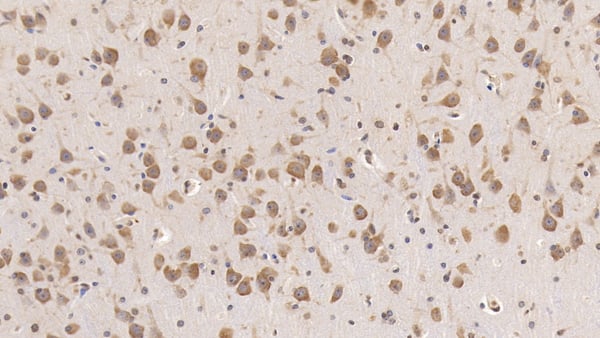
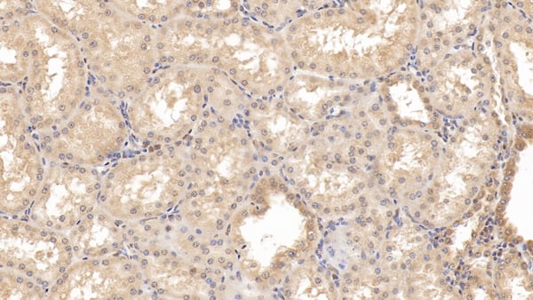 IHC (Immunohistochemisry)
(DAB staining on IHCP;Samples: Human Kidney Tissue;Primary Ab: 30ug/ml Mouse AntiHuman RAB5A AntibodySecond Ab: 2ug/mL HRPLinked Caprine AntiMouse IgG Polyclonal Antibody(Catalog: SAA544Mu19))
IHC (Immunohistochemisry)
(DAB staining on IHCP;Samples: Human Kidney Tissue;Primary Ab: 30ug/ml Mouse AntiHuman RAB5A AntibodySecond Ab: 2ug/mL HRPLinked Caprine AntiMouse IgG Polyclonal Antibody(Catalog: SAA544Mu19))
RAB5A, Member RAS Oncogene Family (RAB5A), Monoclonal Antibody (Cat# AAA151899)
Full Name
Monoclonal Antibody to RAB5A, Member RAS Oncogene Family (RAB5A)
Gene Names
RAB5A; RAB5
Reactivity
Human, Rat, Pig
Applications
Immunoprecipitation, Immunocytochemistry, Immunohistochemistry, Western Blot
Purity
Protein A + Protein G affinity chromatography
Pricing
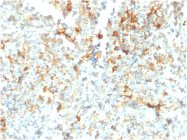
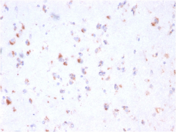
 Application Data
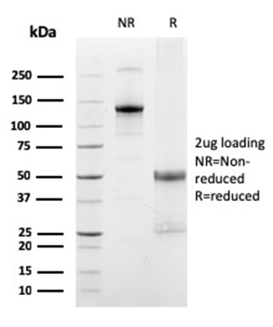
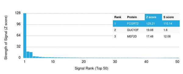
(Analysis of Protein Array containing >19,000 full-length human proteins using FCGRT Mouse Monoclonal Antibody (FCGRT/2932) Z- and S- Score: The Z-score represents the strength of a signal that a monoclonal antibody (MAb) (in combination with a fluorescently-tagged anti-IgG secondary antibody) produces when binding to a particular protein on the HuProtTM array. Z-scores are described in units of standard deviations (SD's) above the mean value of all signals generated on that array. If targets on HuProtTM are arranged in descending order of the Z-score, the S-score is the difference (also in units of SD's) between the Z-score. S-score therefore represents the relative target specificity of a MAb to its intended target. A MAb is considered to specific to its intended target, if the MAb has an S-score of at least 2.5. For example, if a MAb binds to protein X with a Z-score of 43 and to protein Y with a Z-score of 14, then the S-score for the binding of that MAb to protein X is equal to 29.)
Application Data
(Analysis of Protein Array containing >19,000 full-length human proteins using FCGRT Mouse Monoclonal Antibody (FCGRT/2932) Z- and S- Score: The Z-score represents the strength of a signal that a monoclonal antibody (MAb) (in combination with a fluorescently-tagged anti-IgG secondary antibody) produces when binding to a particular protein on the HuProtTM array. Z-scores are described in units of standard deviations (SD's) above the mean value of all signals generated on that array. If targets on HuProtTM are arranged in descending order of the Z-score, the S-score is the difference (also in units of SD's) between the Z-score. S-score therefore represents the relative target specificity of a MAb to its intended target. A MAb is considered to specific to its intended target, if the MAb has an S-score of at least 2.5. For example, if a MAb binds to protein X with a Z-score of 43 and to protein Y with a Z-score of 14, then the S-score for the binding of that MAb to protein X is equal to 29.)
FCGRT/FcRn, Monoclonal Antibody (Cat# AAA214969)
Full Name
FCGRT/FcRn (IgG Transporter)
Gene Names
FCGRT; FCRN; alpha-chain
Reactivity
Human
Applications
Immunohistochemistry
Purity
Purified Ab with BSA and Azide at 200ug/ml OR Purified Ab WITHOUT BSA and Azide at 1.0mg/ml
Pricing
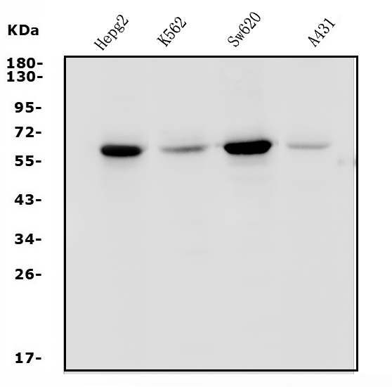
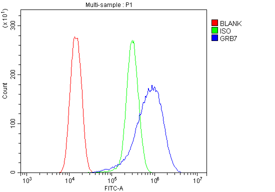 FCM/FACS (Flow Cytometry)
(Figure 2. Flow Cytometry analysis of U20S cells using anti-GRB7 antibody (AAA125663).Overlay histogram showing U20S cells stained with AAA125663 (Blue line). The cells were blocked with 10% normal goat serum. And then incubated with rabbit anti-GRB7 Antibody (AAA125663,1μg/1x106 cells) for 30 min at 20 degree C. DyLight®488 conjugated goat anti-rabbit IgG (5-10μg/1x106 cells) was used as secondary antibody for 30 minutes at 20 degree C. Isotype control antibody (Green line) was rabbit IgG (1μg/1x106) used under the same conditions. Unlabelled sample (Red line) was also used as a control.)
FCM/FACS (Flow Cytometry)
(Figure 2. Flow Cytometry analysis of U20S cells using anti-GRB7 antibody (AAA125663).Overlay histogram showing U20S cells stained with AAA125663 (Blue line). The cells were blocked with 10% normal goat serum. And then incubated with rabbit anti-GRB7 Antibody (AAA125663,1μg/1x106 cells) for 30 min at 20 degree C. DyLight®488 conjugated goat anti-rabbit IgG (5-10μg/1x106 cells) was used as secondary antibody for 30 minutes at 20 degree C. Isotype control antibody (Green line) was rabbit IgG (1μg/1x106) used under the same conditions. Unlabelled sample (Red line) was also used as a control.)
GRB7, Polyclonal Antibody (Cat# AAA125663)
Full Name
Anti-GRB7 Antibody
Reactivity
Human
Applications
Direct ELISA, Flow Cytometry, Western Blot
Purity
Immunogen affinity purified.
Pricing
 WB (Western Blot)
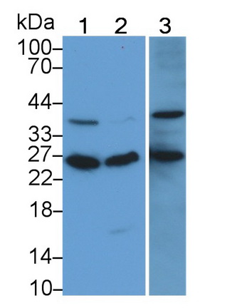
(Western Blot; Sample: Lane1: Mouse Cerebrum lysate; Lane2: Rat Cerebrum lysate Primary Ab: 2ug/mL Mouse AntiHuman NRGN Antibody Second Ab: 0.2ug/mL HRPLinked Caprine AntiMouse IgG Polyclonal Antibody (Catalog: SAA544Mu19))
WB (Western Blot)
(Western Blot; Sample: Lane1: Mouse Cerebrum lysate; Lane2: Rat Cerebrum lysate Primary Ab: 2ug/mL Mouse AntiHuman NRGN Antibody Second Ab: 0.2ug/mL HRPLinked Caprine AntiMouse IgG Polyclonal Antibody (Catalog: SAA544Mu19))
Neurogranin (NRGN), Monoclonal Antibody (Cat# AAA151532)
Full Name
Monoclonal Antibody to Neurogranin (NRGN)
Gene Names
NRGN; RC3; hng
Reactivity
Human, Mouse, Rat, Pig
Applications
Immunoprecipitation, Immunocytochemistry, Immunohistochemistry, Western Blot
Purity
Protein A + Protein G affinity chromatography
Pricing
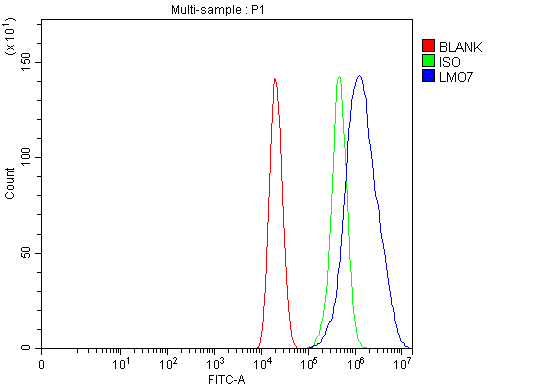 FCM/FACS (Flow Cytometry)
(Figure 2. Flow Cytometry analysis of Caco-2 cells using anti-LMO7 antibody (AAA127321).Overlay histogram showing Caco-2 cells stained with AAA127321 (Blue line). To facilitate intracellular staining, cells were fixed with 4% paraformaldehyde and permeabilized with permeabilization buffer. The cells were blocked with 10% normal goat serum. And then incubated with rabbit anti-LMO7 Antibody (AAA127321, 1ug/1x106 cells) for 30 min at 20 degree C. DyLight488 conjugated goat anti-rabbit IgG was used as secondary antibody for 30 minutes at 20 degree C. Isotype control antibody (Green line) was rabbit IgG (1ug/1x106) used under the same conditions. Unlabelled sample (Red line) was also used as a control.)
FCM/FACS (Flow Cytometry)
(Figure 2. Flow Cytometry analysis of Caco-2 cells using anti-LMO7 antibody (AAA127321).Overlay histogram showing Caco-2 cells stained with AAA127321 (Blue line). To facilitate intracellular staining, cells were fixed with 4% paraformaldehyde and permeabilized with permeabilization buffer. The cells were blocked with 10% normal goat serum. And then incubated with rabbit anti-LMO7 Antibody (AAA127321, 1ug/1x106 cells) for 30 min at 20 degree C. DyLight488 conjugated goat anti-rabbit IgG was used as secondary antibody for 30 minutes at 20 degree C. Isotype control antibody (Green line) was rabbit IgG (1ug/1x106) used under the same conditions. Unlabelled sample (Red line) was also used as a control.)
LMO7, Polyclonal Antibody (Cat# AAA127321)
Full Name
Anti-LMO7 Antibody Picoband
Gene Names
LMO7; LOMP; FBX20; FBXO20
Reactivity
Human
Applications
Flow Cytometry, Western Blot
Purity
Immunogen affinity purified.
Pricing
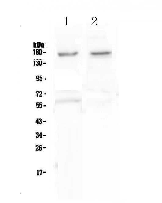
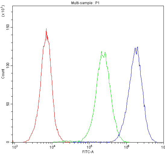 FCM/FACS (Flow Cytometry)
(Figure 2. Flow Cytometry analysis of U251 cells using anti-NRXN1 antibody (AAA124650).Overlay histogram showing U251 cells stained with AAA124650 (Blue line).The cells were blocked with 10% normal goat serum. And then incubated with rabbit anti-NRXN1 Antibody (AAA124650,1ug/1x10^6 cells) for 30 min at 20 degree C. DyLight®488 conjugated goat anti-rabbit IgG (5-10ug/1x10^6 cells) was used as secondary antibody for 30 minutes at 20 degree C. Isotype control antibody (Green line) was rabbit IgG (1ug/1x106) used under the same conditions. Unlabelled sample (Red line) was also used as a control.)
FCM/FACS (Flow Cytometry)
(Figure 2. Flow Cytometry analysis of U251 cells using anti-NRXN1 antibody (AAA124650).Overlay histogram showing U251 cells stained with AAA124650 (Blue line).The cells were blocked with 10% normal goat serum. And then incubated with rabbit anti-NRXN1 Antibody (AAA124650,1ug/1x10^6 cells) for 30 min at 20 degree C. DyLight®488 conjugated goat anti-rabbit IgG (5-10ug/1x10^6 cells) was used as secondary antibody for 30 minutes at 20 degree C. Isotype control antibody (Green line) was rabbit IgG (1ug/1x106) used under the same conditions. Unlabelled sample (Red line) was also used as a control.)
Neurexin 1, Polyclonal Antibody (Cat# AAA124650)
Full Name
Anti-Neurexin 1 antibody
Gene Names
NRXN1; PTHSL2; SCZD17; Hs.22998
Reactivity
Human
Applications
Western Blot, Flow Cytometry
Purity
Immunogen affinity purified
Pricing


 Application Data
(Analysis of Protein Array containing more than 19,000 full-length human proteins using Desmoglein-3 Mouse Monoclonal Antibody (DSG3/2839) Z- and S- Score: The Z-score represents the strength of a signal that a monoclonal antibody (MAb) (in combination with a fluorescently-tagged anti-IgG secondary antibody) produces when binding to a particular protein on the HuProtTM array. Z-scores are described in units of standard deviations (SD's) above the mean value of all signals generated on that array. If targets on HuProtTM are arranged in descending order of the Z-score, the S-score is the difference (also in units of SD's) between the Z-score. S-score therefore represents the relative target specificity of a MAb to its intended target. A MAb is considered to specific to its intended target, if the MAb has an S-score of at least 2.5. For example, if a MAb binds to protein X with a Z-score of 43 and to protein Y with a Z-score of 14, then the S-score for the binding of that MAb to protein X is equal to 29.)
Application Data
(Analysis of Protein Array containing more than 19,000 full-length human proteins using Desmoglein-3 Mouse Monoclonal Antibody (DSG3/2839) Z- and S- Score: The Z-score represents the strength of a signal that a monoclonal antibody (MAb) (in combination with a fluorescently-tagged anti-IgG secondary antibody) produces when binding to a particular protein on the HuProtTM array. Z-scores are described in units of standard deviations (SD's) above the mean value of all signals generated on that array. If targets on HuProtTM are arranged in descending order of the Z-score, the S-score is the difference (also in units of SD's) between the Z-score. S-score therefore represents the relative target specificity of a MAb to its intended target. A MAb is considered to specific to its intended target, if the MAb has an S-score of at least 2.5. For example, if a MAb binds to protein X with a Z-score of 43 and to protein Y with a Z-score of 14, then the S-score for the binding of that MAb to protein X is equal to 29.)
Desmoglein-3, Monoclonal Antibody (Cat# AAA214935)
Full Name
Desmoglein-3 (Squamous Cell Marker)
Gene Names
DSG3; PVA; CDHF6
Reactivity
Human
Applications
Immunohistochemistry
Purity
Purified Ab with BSA and Azide at 200ug/ml OR Purified Ab WITHOUT BSA and Azide at 1.0mg/ml
Pricing
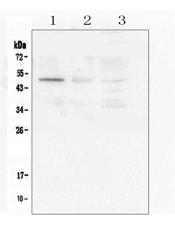
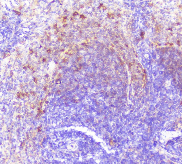 IHC (Immunohiostchemistry)
(Figure 3. IHC analysis of Caspase-2 using anti-Caspase-2 antibody.Caspase-2 was detected in paraffin-embedded section of human lung cancer tissue. Heat mediated antigen retrieval was performed in citrate buffer (pH6, epitope retrieval solution) for 20 mins. The tissue section was blocked with 10% goat serum. The tissue section was then incubated with 1ug/ml rabbit anti-Caspase-2 antibody.Caspase-2 was detected in paraffin-embedded section of human lung cancer tissue. Heat mediated antigen retrieval was performed in citrate buffer (pH6, epitope retrieval solution) for 20 mins. The tissue section was blocked with 10% goat serum. The tissue section was then incubated with 1ug/ml rabbit anti-Caspase-2 antibody.Caspase-2 was detected in paraffin-embedded section of mouse intestine tissue. Heat mediated antigen retrieval was performed in citrate buffer (pH6, epitope retrieval solution) for 20 mins. The tissue section was blocked with 10% goat serum. The tissue section was then incubated with 1ug/ml rabbit anti-Caspase-2 antibody.Caspase-2 was detected in paraffin-embedded section of rat spleen tissue. Heat mediated antigen retrieval was performed in citrate buffer (pH6, epitope retrieval solution) for 20 mins. The tissue section was blocked with 10% goat serum. The tissue section was then incubated with 1ug/ml rabbit anti-Caspase-2 Antibody overnight at 4 degree C. Biotinylated goat anti-rabbit IgG was used as secondary antibody and incubated for 30 minutes at 37 degree C. The tissue section was developed using Strepavidin-Biotin-Complex (SABC) with DAB as the chromogen.)
IHC (Immunohiostchemistry)
(Figure 3. IHC analysis of Caspase-2 using anti-Caspase-2 antibody.Caspase-2 was detected in paraffin-embedded section of human lung cancer tissue. Heat mediated antigen retrieval was performed in citrate buffer (pH6, epitope retrieval solution) for 20 mins. The tissue section was blocked with 10% goat serum. The tissue section was then incubated with 1ug/ml rabbit anti-Caspase-2 antibody.Caspase-2 was detected in paraffin-embedded section of human lung cancer tissue. Heat mediated antigen retrieval was performed in citrate buffer (pH6, epitope retrieval solution) for 20 mins. The tissue section was blocked with 10% goat serum. The tissue section was then incubated with 1ug/ml rabbit anti-Caspase-2 antibody.Caspase-2 was detected in paraffin-embedded section of mouse intestine tissue. Heat mediated antigen retrieval was performed in citrate buffer (pH6, epitope retrieval solution) for 20 mins. The tissue section was blocked with 10% goat serum. The tissue section was then incubated with 1ug/ml rabbit anti-Caspase-2 antibody.Caspase-2 was detected in paraffin-embedded section of rat spleen tissue. Heat mediated antigen retrieval was performed in citrate buffer (pH6, epitope retrieval solution) for 20 mins. The tissue section was blocked with 10% goat serum. The tissue section was then incubated with 1ug/ml rabbit anti-Caspase-2 Antibody overnight at 4 degree C. Biotinylated goat anti-rabbit IgG was used as secondary antibody and incubated for 30 minutes at 37 degree C. The tissue section was developed using Strepavidin-Biotin-Complex (SABC) with DAB as the chromogen.)
Caspase-2/CASP2, Polyclonal Antibody (Cat# AAA124940)
Full Name
Anti-Caspase-2/CASP2 Antibody
Gene Names
CASP2; ICH1; NEDD2; CASP-2; NEDD-2; PPP1R57
Reactivity
Human, Mouse, Rat
Applications
Direct ELISA, Immunohistochemistry, Western Blot
Purity
Immunogen Affinity Purified
Pricing

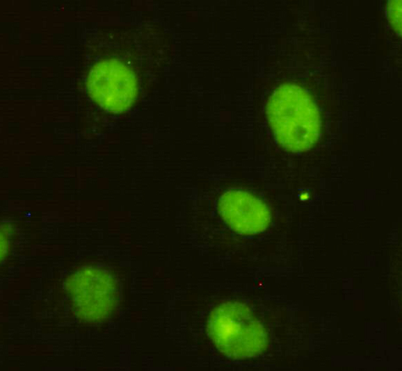
 ICC (Immunocytochemistry)
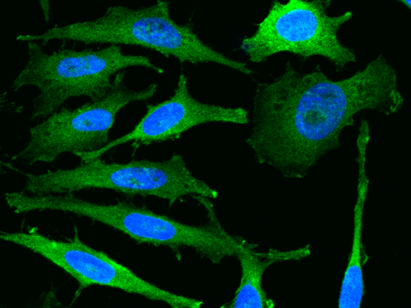
(Immunocytochemistry of HeLa cells using anti-DBC1 mouse mAb diluted 1:200.)
ICC (Immunocytochemistry)
(Immunocytochemistry of HeLa cells using anti-DBC1 mouse mAb diluted 1:200.)
DBC1, Monoclonal Antibody (Cat# AAA320295)
Full Name
DBC1 Mouse mAb
Gene Names
KIAA1967; DBC1; DBC-1; NET35; p30DBC; p30 DBC
Reactivity
Human, Mouse, Rat, Monkey
Applications
Immunoprecipitation, Immunocytochemistry, Western Blot
Purity
The antibody was affinity-purified from mouse ascites by affinity-chromatography using epitope-specific immunogen.
Pricing
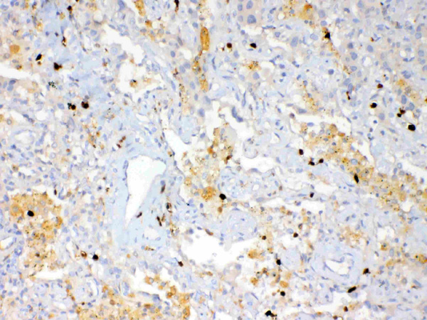
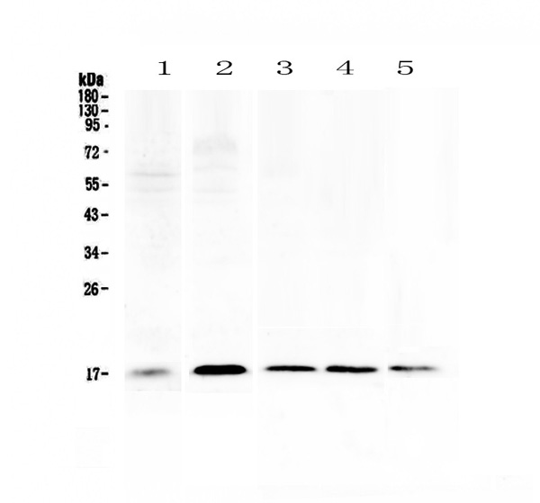
 IHC (Immunohistochemisry)
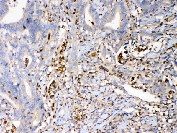
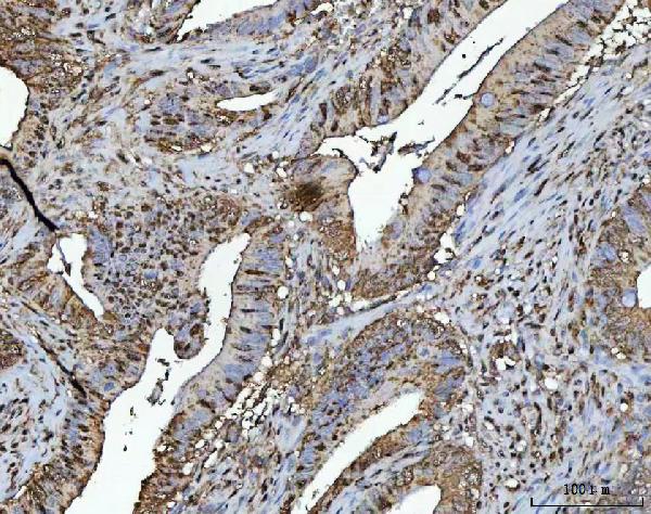
(Figure 3. IHC analysis of Psoriasin using anti-Psoriasin antibody (AAA124685).Psoriasin was detected in paraffin-embedded section of human rectal cancer tissue. Heat mediated antigen retrieval was performed in citrate buffer (pH6, epitope retrieval solution) for 20 mins. The tissue section was blocked with 10% goat serum. The tissue section was then incubated with 1ug/ml rabbit anti-Psoriasin Antibody (AAA124685) overnight at 4 degree C. Biotinylated goat anti-rabbit IgG was used as secondary antibody and incubated for 30 minutes at 37 degree C. The tissue section was developed using Strepavidin-Biotin-Complex (SABC) with DAB as the chromogen.)
IHC (Immunohistochemisry)
(Figure 3. IHC analysis of Psoriasin using anti-Psoriasin antibody (AAA124685).Psoriasin was detected in paraffin-embedded section of human rectal cancer tissue. Heat mediated antigen retrieval was performed in citrate buffer (pH6, epitope retrieval solution) for 20 mins. The tissue section was blocked with 10% goat serum. The tissue section was then incubated with 1ug/ml rabbit anti-Psoriasin Antibody (AAA124685) overnight at 4 degree C. Biotinylated goat anti-rabbit IgG was used as secondary antibody and incubated for 30 minutes at 37 degree C. The tissue section was developed using Strepavidin-Biotin-Complex (SABC) with DAB as the chromogen.)
Psoriasin, Polyclonal Antibody (Cat# AAA124685)
Full Name
Anti-Psoriasin Picoband antibody
Gene Names
S100A7; PSOR1; S100A7c
Reactivity
Human, Mouse, Rat
No cross reactivity with other proteins.
No cross reactivity with other proteins.
Applications
Western Blot, Immunohistochemistry
Pricing
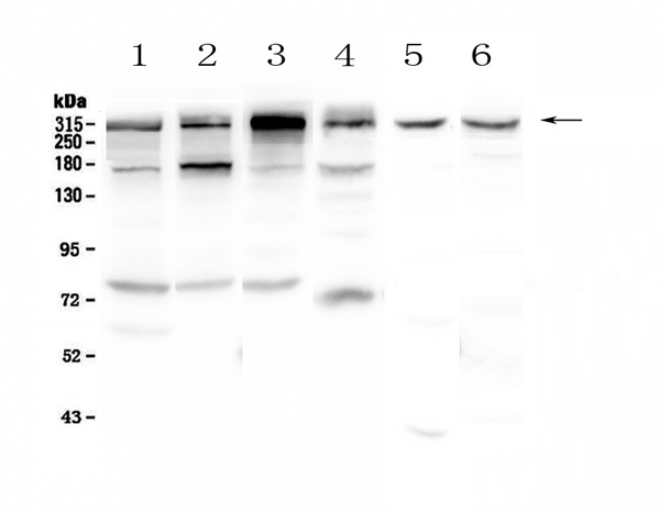
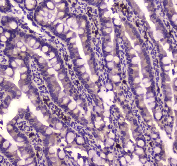 IHC (Immunohistochemistry)
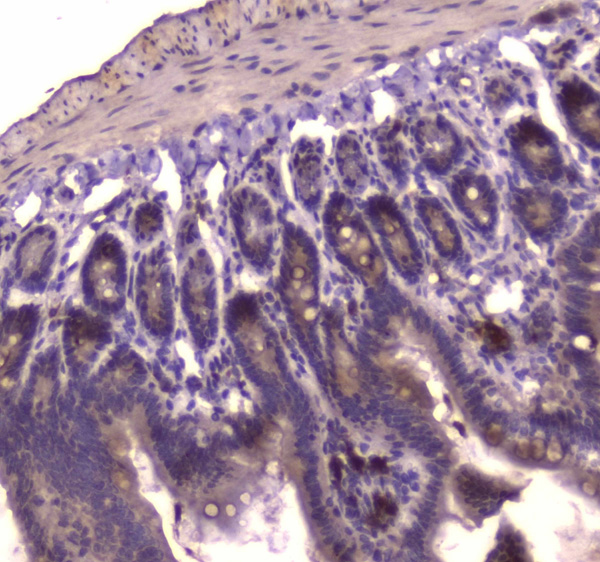
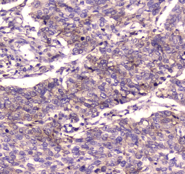
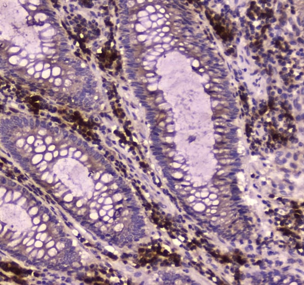
(Figure 5. IHC analysis of IGF2R using anti-IGF2R antibody (AAA124606).IGF2R was detected in paraffin-embedded section of rat small intestine tissue. Heat mediated antigen retrieval was performed in citrate buffer (pH6, epitope retrieval solution) for 20 mins. The tissue section was blocked with 10% goat serum. The tissue section was then incubated with 2ug/ml rabbit anti-IGF2R Antibody (AAA124606) overnight at 4 degree C. Biotinylated goat anti-rabbit IgG was used as secondary antibody and incubated for 30 minutes at 37 degree C. The tissue section was developed using Strepavidin-Biotin-Complex (SABC) with DAB as the chromogen.)
IHC (Immunohistochemistry)
(Figure 5. IHC analysis of IGF2R using anti-IGF2R antibody (AAA124606).IGF2R was detected in paraffin-embedded section of rat small intestine tissue. Heat mediated antigen retrieval was performed in citrate buffer (pH6, epitope retrieval solution) for 20 mins. The tissue section was blocked with 10% goat serum. The tissue section was then incubated with 2ug/ml rabbit anti-IGF2R Antibody (AAA124606) overnight at 4 degree C. Biotinylated goat anti-rabbit IgG was used as secondary antibody and incubated for 30 minutes at 37 degree C. The tissue section was developed using Strepavidin-Biotin-Complex (SABC) with DAB as the chromogen.)
IGF2R, Polyclonal Antibody (Cat# AAA124606)
Full Name
Anti-IGF2R Picoband antibody
Gene Names
IGF2R; MPR1; MPRI; CD222; CIMPR; M6P-R; MPR300; CI-M6PR; MPR 300; M6P/IGF2R
Reactivity
Human, Mouse, Rat
No cross reactivity with other proteins.
No cross reactivity with other proteins.
Applications
Western Blot, Immunohistochemistry
Pricing
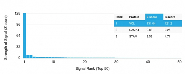 Application Data
(Analysis of Protein Array containing more than 19, 000 full-length human proteins using Vinculin Mouse Monoclonal Antibody (VCL/2572).Z- and S- Score: The Z-score represents the strength of a signal that a monoclonal antibody (MAb) (in combination with a fluorescently-tagged anti-IgG secondary antibody) produces when binding to a particular protein on the HuProtTM array. Z-scores are described in units of standard deviations (SD's) above the mean value of all signals generated on that array. If targets on HuProtTM are arranged in descending order of the Z-score, the S-score is the difference (also in units of SD's) between the Z-score. S-score therefore represents the relative target specificity of a MAb to its intended target. A MAb is considered to specific to its intended target, if the MAb has an S-score of at least 2.5. For example, if a MAb binds to protein X with a Z-score of 43 and to protein Y with a Z-score of 14, then the S-score for the binding of that MAb to protein X is equal to 29.)
Application Data
(Analysis of Protein Array containing more than 19, 000 full-length human proteins using Vinculin Mouse Monoclonal Antibody (VCL/2572).Z- and S- Score: The Z-score represents the strength of a signal that a monoclonal antibody (MAb) (in combination with a fluorescently-tagged anti-IgG secondary antibody) produces when binding to a particular protein on the HuProtTM array. Z-scores are described in units of standard deviations (SD's) above the mean value of all signals generated on that array. If targets on HuProtTM are arranged in descending order of the Z-score, the S-score is the difference (also in units of SD's) between the Z-score. S-score therefore represents the relative target specificity of a MAb to its intended target. A MAb is considered to specific to its intended target, if the MAb has an S-score of at least 2.5. For example, if a MAb binds to protein X with a Z-score of 43 and to protein Y with a Z-score of 14, then the S-score for the binding of that MAb to protein X is equal to 29.)
Vinculin, Monoclonal Antibody (Cat# AAA214707)
Full Name
Vinculin (Marker of Age-related Macular Degeneration)
Gene Names
VCL; MV; MVCL; CMD1W; CMH15; HEL114
Reactivity
Human. Others not known.
Applications
Immunohistochemistry
Pricing
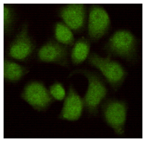
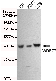 ICC (Immunocytochemistry)
(Immunocytochemistry staining of HeLa cells fixed in 1% Paraformaldehyde and then permeabilized in 0.1% Triton X-100.next using WDR77 mouse mAb (dilution 1:100).)
ICC (Immunocytochemistry)
(Immunocytochemistry staining of HeLa cells fixed in 1% Paraformaldehyde and then permeabilized in 0.1% Triton X-100.next using WDR77 mouse mAb (dilution 1:100).)
WDR77, Monoclonal Antibody (Cat# AAA320373)
Full Name
WDR77 Mouse mAb
Gene Names
WDR77; p44; MEP50; MEP-50; HKMT1069; Nbla10071; p44/Mep50
Reactivity
Human, Mouse, Rat
Applications
Immunocytochemistry, Western Blot
Purity
The antibody was affinity-purified from mouse ascites by affinity-chromatography using epitope-specific immunogen.
Pricing
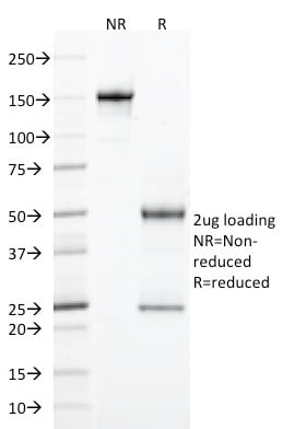
 Application Data
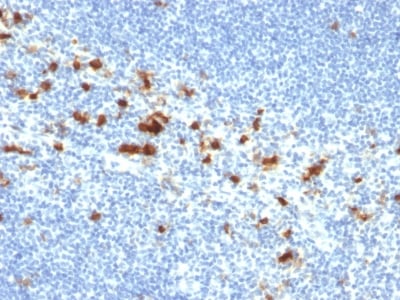
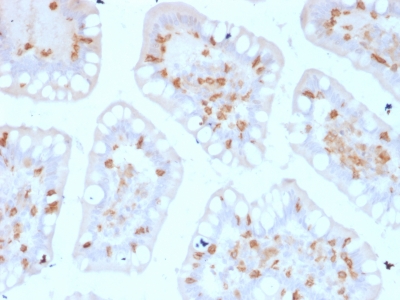
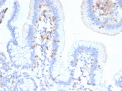
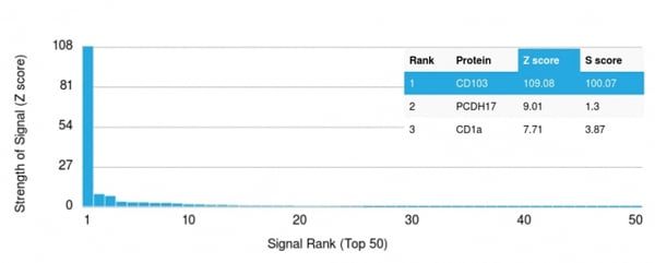
(Analysis of Protein Array containing more than 19, 000 full-length human proteins using CD103 Mouse Monoclonal Antibody (ITGAE/2474). Z- and S- Score: The Z-score represents the strength of a signal that a monoclonal antibody (MAb) (in combination with a fluorescently-tagged anti-IgG secondary antibody) produces when binding to a particular protein on the HuProtTM array. Z-scores are described in units of standard deviations (SD's) above the mean value of all signals generated on that array. If targets on HuProtTM are arranged in descending order of the Z-score, the S-score is the difference (also in units of SD's) between the Z-score. S-score therefore represents the relative target specificity of a MAb to its intended target. A MAb is considered to specific to its intended target, if the MAb has an S-score of at least 2.5. For example, if a MAb binds to protein X with a Z-score of 43 and to protein Y with a Z-score of 14, then the S-score for the binding of that MAb to protein X is equal to 29.)
Application Data
(Analysis of Protein Array containing more than 19, 000 full-length human proteins using CD103 Mouse Monoclonal Antibody (ITGAE/2474). Z- and S- Score: The Z-score represents the strength of a signal that a monoclonal antibody (MAb) (in combination with a fluorescently-tagged anti-IgG secondary antibody) produces when binding to a particular protein on the HuProtTM array. Z-scores are described in units of standard deviations (SD's) above the mean value of all signals generated on that array. If targets on HuProtTM are arranged in descending order of the Z-score, the S-score is the difference (also in units of SD's) between the Z-score. S-score therefore represents the relative target specificity of a MAb to its intended target. A MAb is considered to specific to its intended target, if the MAb has an S-score of at least 2.5. For example, if a MAb binds to protein X with a Z-score of 43 and to protein Y with a Z-score of 14, then the S-score for the binding of that MAb to protein X is equal to 29.)
CD103/Integrin alpha E, Monoclonal Antibody (Cat# AAA214659)
Full Name
CD103/Integrin alpha E (T-Cell Lymphoma & Hairy Cell Leukemia Marker)
Gene Names
ITGAE; CD103; HUMINAE
Reactivity
Human. Others not known.
Applications
Immunohistochemistry
Pricing
 Application Data
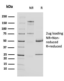
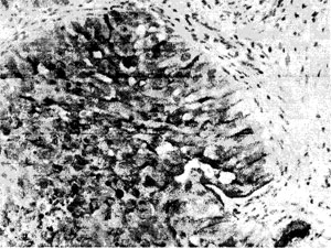
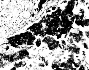
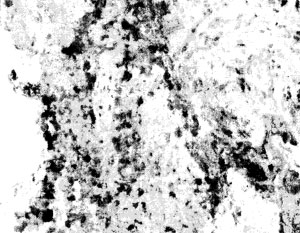
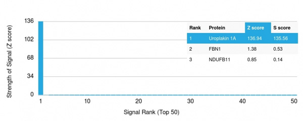
(Analysis of Protein Array containing more than 19,000 full-length human proteins using Uroplakin 1A Mouse Monoclonal Antibody (UPK1A/2924) Z- and S- Score: The Z-score represents the strength of a signal that a monoclonal antibody (MAb) (in combination with a fluorescently-tagged anti-IgG secondary antibody) produces when binding to a particular protein on the HuProtTM array. Z-scores are described in units of standard deviations (SD’s) above the mean value of all signals generated on that array. If targets on HuProtTM are arranged in descending order of the Z-score, the S-score is the difference (also in units of SD’s) between the Z-score. S-score therefore represents the relative target specificity of a MAb to its intended target. A MAb is considered to specific to its intended target, if the MAb has an S-score of at least 2.5. For example, if a MAb binds to protein X with a Z-score of 43 and to protein Y with a Z-score of 14, then the S-score for the binding of that MAb to protein X is equal to 29.)
Application Data
(Analysis of Protein Array containing more than 19,000 full-length human proteins using Uroplakin 1A Mouse Monoclonal Antibody (UPK1A/2924) Z- and S- Score: The Z-score represents the strength of a signal that a monoclonal antibody (MAb) (in combination with a fluorescently-tagged anti-IgG secondary antibody) produces when binding to a particular protein on the HuProtTM array. Z-scores are described in units of standard deviations (SD’s) above the mean value of all signals generated on that array. If targets on HuProtTM are arranged in descending order of the Z-score, the S-score is the difference (also in units of SD’s) between the Z-score. S-score therefore represents the relative target specificity of a MAb to its intended target. A MAb is considered to specific to its intended target, if the MAb has an S-score of at least 2.5. For example, if a MAb binds to protein X with a Z-score of 43 and to protein Y with a Z-score of 14, then the S-score for the binding of that MAb to protein X is equal to 29.)
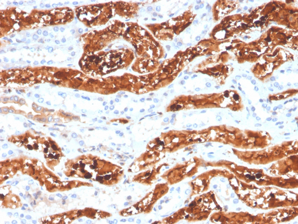
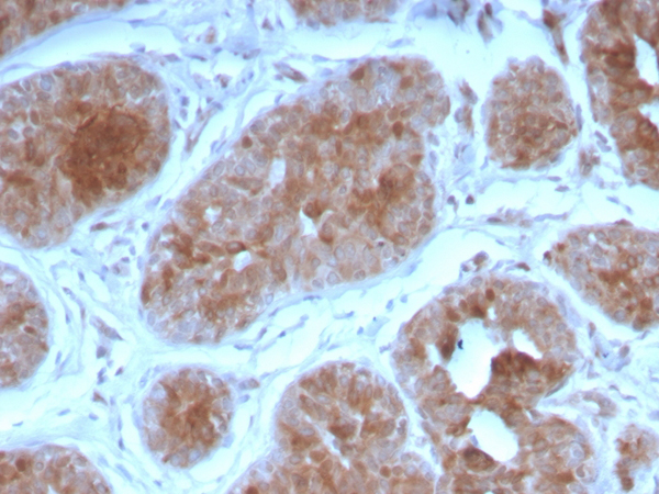
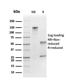
Uroplakin 1A, Monoclonal Antibody (Cat# AAA214890)
Full Name
Uroplakin 1A (Urothelial Differentiation Marker)
Gene Names
UPK1A; UP1A; UPIA; UPKA; TSPAN21
Reactivity
Human
Applications
Immunohistochemistry
Purity
Purified Ab with BSA and Azide at 200ug/ml OR Purified Ab WITHOUT BSA and Azide at 1.0mg/ml
Pricing
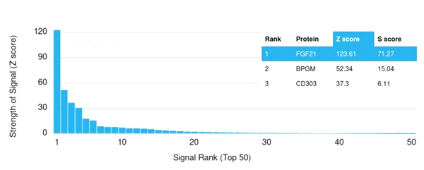 Application Data
(Analysis of Protein Array containing more than 19,000 full-length human proteins using FGF21 Mouse Monoclonal antibody (FGF21/3691). Z- and S- Score: The Z-score represents the strength of a signal that a monoclonal antibody (MAb) (in combination with a fluorescently-tagged anti-IgG secondary antibody) produces when binding to a particular protein on the HuProtTM array. Z-scores are described in units of standard deviations (SD's) above the mean value of all signals generated on that array. If targets on HuProtTM are arranged in descending order of the Z-score, the S-score is the difference (also in units of SD's) between the Z-score. S-score therefore represents the relative target specificity of a MAb to its intended target. A MAb is considered to specific to its intended target, if the MAb has an S-score of at least 2.5. For example, if a MAb binds to protein X with a Z-score of 43 and to protein Y with a Z-score of 14, then the S-score for the binding of that MAb to protein X is equal to 29.)
Application Data
(Analysis of Protein Array containing more than 19,000 full-length human proteins using FGF21 Mouse Monoclonal antibody (FGF21/3691). Z- and S- Score: The Z-score represents the strength of a signal that a monoclonal antibody (MAb) (in combination with a fluorescently-tagged anti-IgG secondary antibody) produces when binding to a particular protein on the HuProtTM array. Z-scores are described in units of standard deviations (SD's) above the mean value of all signals generated on that array. If targets on HuProtTM are arranged in descending order of the Z-score, the S-score is the difference (also in units of SD's) between the Z-score. S-score therefore represents the relative target specificity of a MAb to its intended target. A MAb is considered to specific to its intended target, if the MAb has an S-score of at least 2.5. For example, if a MAb binds to protein X with a Z-score of 43 and to protein Y with a Z-score of 14, then the S-score for the binding of that MAb to protein X is equal to 29.)
FGF21, Monoclonal Antibody (Cat# AAA215399)
Full Name
FGF21 (Fibroblast growth factor 21)
Reactivity
Human
Applications
Immunohistochemistry
Purity
Purified Ab with BSA and Azide at 200ug/ml or Purified Ab with BSA and Azide at 200ug/ml or Purified Ab WITHOUT BSA and Azide at 1.0mg/ml
Pricing


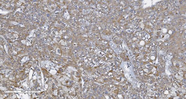 IHC (Immunohistochemisry)
(Figure 3. IHC analysis of CARD4/NOD1 using anti-CARD4/NOD1 antibody (AAA125553).CARD4/NOD1 was detected in paraffin-embedded section of human liver cancer tissue. Heat mediated antigen retrieval was performed in EDTA buffer (pH8. 0, epitope retrieval solution). The tissue section was blocked with 10% goat serum. The tissue section was then incubated with 2μg/ml rabbit anti-CARD4/NOD1 Antibody (AAA125553) overnight at 4 degree C. Biotinylated goat anti-rabbit IgG was used as secondary antibody and incubated for 30 minutes at 37 degree C. The tissue section was developed using Strepavidin-Biotin-Complex (SABC) with DAB as the chromogen.)
IHC (Immunohistochemisry)
(Figure 3. IHC analysis of CARD4/NOD1 using anti-CARD4/NOD1 antibody (AAA125553).CARD4/NOD1 was detected in paraffin-embedded section of human liver cancer tissue. Heat mediated antigen retrieval was performed in EDTA buffer (pH8. 0, epitope retrieval solution). The tissue section was blocked with 10% goat serum. The tissue section was then incubated with 2μg/ml rabbit anti-CARD4/NOD1 Antibody (AAA125553) overnight at 4 degree C. Biotinylated goat anti-rabbit IgG was used as secondary antibody and incubated for 30 minutes at 37 degree C. The tissue section was developed using Strepavidin-Biotin-Complex (SABC) with DAB as the chromogen.)
CARD4/NOD1, Polyclonal Antibody (Cat# AAA125553)
Full Name
Anti-CARD4/NOD1 Antibody
Gene Names
NOD1; CARD4; NLRC1; CLR7.1
Reactivity
Human
Applications
Direct ELISA, Flow Cytometry, Immunohistochemistry, Western Blot
Purity
Immunogen affinity purified.
Pricing
 WB (Western Blot)
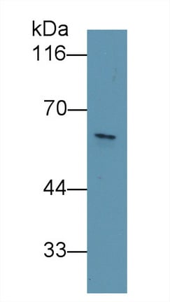
(Western Blot; Sample: 293T cell lysate Primary Ab: 2ug/ml Mouse AntiHuman KPNa2 Antibody Second Ab: 0.2ug/mL HRPLinked Caprine AntiMouse IgG Polyclonal Antibody (Catalog: SAA544Mu19))
WB (Western Blot)
(Western Blot; Sample: 293T cell lysate Primary Ab: 2ug/ml Mouse AntiHuman KPNa2 Antibody Second Ab: 0.2ug/mL HRPLinked Caprine AntiMouse IgG Polyclonal Antibody (Catalog: SAA544Mu19))
Karyopherin Alpha 2 (KPNa2), Monoclonal Antibody (Cat# AAA151860)
Full Name
Monoclonal Antibody to Karyopherin Alpha 2 (KPNa2)
Gene Names
KPNA2; QIP2; RCH1; IPOA1; SRP1alpha
Reactivity
Human, Pig
Applications
Immunoprecipitation, Immunocytochemistry, Immunohistochemistry, Western Blot
Purity
Protein A + Protein G affinity chromatography
Pricing
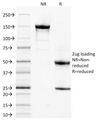
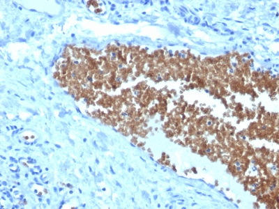
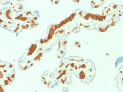
 Application Data
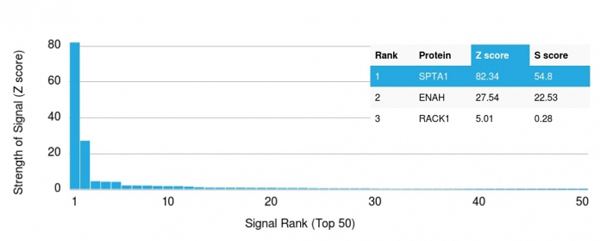
(Analysis of Protein Array containing more than 19, 000 full-length human proteins using Spectrin, alpha 1 Mouse Monoclonal Antibody (SPTA1/1832)Z- and S- Score: The Z-score represents the strength of a signal that a monoclonal antibody (MAb) (in combination with a fluorescently-tagged anti-IgG secondary antibody) produces when binding to a particular protein on the HuProtTM array. Z-scores are described in units of standard deviations (SD's) above the mean value of all signals generated on that array. If targets on HuProtTM are arranged in descending order of the Z-score, the S-score is the difference (also in units of SD's) between the Z-score. S-score therefore represents the relative target specificity of a MAb to its intended target. A MAb is considered to specific to its intended target, if the MAb has an S-score of at least 2.5. For example, if a MAb binds to protein X with a Z-score of 43 and to protein Y with a Z-score of 14, then the S-score for the binding of that MAb to protein X is equal to 29.)
Application Data
(Analysis of Protein Array containing more than 19, 000 full-length human proteins using Spectrin, alpha 1 Mouse Monoclonal Antibody (SPTA1/1832)Z- and S- Score: The Z-score represents the strength of a signal that a monoclonal antibody (MAb) (in combination with a fluorescently-tagged anti-IgG secondary antibody) produces when binding to a particular protein on the HuProtTM array. Z-scores are described in units of standard deviations (SD's) above the mean value of all signals generated on that array. If targets on HuProtTM are arranged in descending order of the Z-score, the S-score is the difference (also in units of SD's) between the Z-score. S-score therefore represents the relative target specificity of a MAb to its intended target. A MAb is considered to specific to its intended target, if the MAb has an S-score of at least 2.5. For example, if a MAb binds to protein X with a Z-score of 43 and to protein Y with a Z-score of 14, then the S-score for the binding of that MAb to protein X is equal to 29.)
Spectrin Alpha 1, Monoclonal Antibody (Cat# AAA214690)
Full Name
Spectrin Alpha 1 (Erythrocyte Marker)
Gene Names
SPTA1; EL2; HPP; HS3; SPH3; SPTA
Reactivity
Human. Others not known.
Applications
Immunohistochemistry
Pricing
 FCM/FACS (Flow Cytometry)
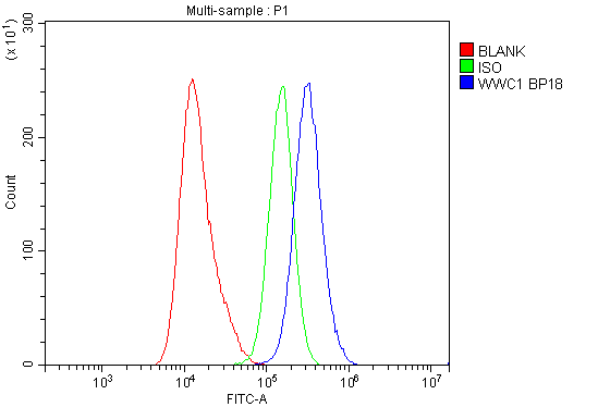
(Figure 3. Flow Cytometry analysis of A431 cells using anti-KIBRA/WWC1 antibody (AAA125673).Overlay histogram showing A431 cells stained with AAA125673 (Blue line). The cells were blocked with 10% normal goat serum. And then incubated with rabbit anti-KIBRA/WWC1 Antibody (AAA125673, 1μg/1x106 cells) for 30 min at 20 degree C. DyLight®488 conjugated goat anti-rabbit IgG (5-10μg/1x106 cells) was used as secondary antibody for 30 minutes at 20 degree C. Isotype control antibody (Green line) was rabbit IgG (1μg/1x106) used under the same conditions. Unlabelled sample (Red line) was also used as a control.)
FCM/FACS (Flow Cytometry)
(Figure 3. Flow Cytometry analysis of A431 cells using anti-KIBRA/WWC1 antibody (AAA125673).Overlay histogram showing A431 cells stained with AAA125673 (Blue line). The cells were blocked with 10% normal goat serum. And then incubated with rabbit anti-KIBRA/WWC1 Antibody (AAA125673, 1μg/1x106 cells) for 30 min at 20 degree C. DyLight®488 conjugated goat anti-rabbit IgG (5-10μg/1x106 cells) was used as secondary antibody for 30 minutes at 20 degree C. Isotype control antibody (Green line) was rabbit IgG (1μg/1x106) used under the same conditions. Unlabelled sample (Red line) was also used as a control.)
KIBRA/WWC1, Polyclonal Antibody (Cat# AAA125673)
Full Name
Anti-KIBRA/WWC1 Antibody
Gene Names
WWC1; KIBRA; HBEBP3; HBEBP36; MEMRYQTL; PPP1R168
Reactivity
Human, Mouse, Rat
Applications
Direct ELISA, Flow Cytometry, Immunofluorescence, Immunocytochemistry, Western Blot
Purity
Immunogen affinity purified.
Pricing
 IF (Immunofluorescence)
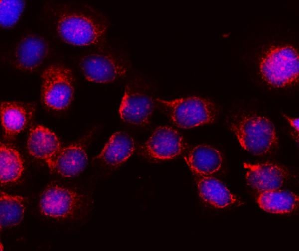
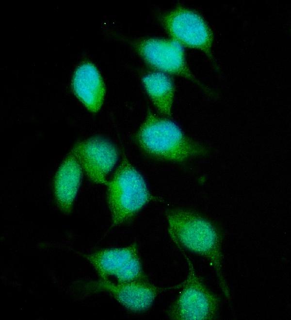
(Figure 3. IF analysis of XPC using anti- XPC antibody (AAA125551).XPC was detected in immunocytochemical section of HELA cells. Enzyme antigen retrieval was performed using IHC enzyme antigen retrieval reagent for 15 mins. The cells were blocked with 10% goat serum. And then incubated with 5μg/mL rabbit anti-XPC Antibody (AAA125551) overnight at 4 degree C. DyLight®488 Conjugated Goat Anti-Rabbit IgG was used as secondary antibody at 1:100 dilution and incubated for 30 minutes at 37 degree C. The section was counterstained with DAPI. Visualize using a fluorescence microscope and filter sets appropriate for the label used.)
IF (Immunofluorescence)
(Figure 3. IF analysis of XPC using anti- XPC antibody (AAA125551).XPC was detected in immunocytochemical section of HELA cells. Enzyme antigen retrieval was performed using IHC enzyme antigen retrieval reagent for 15 mins. The cells were blocked with 10% goat serum. And then incubated with 5μg/mL rabbit anti-XPC Antibody (AAA125551) overnight at 4 degree C. DyLight®488 Conjugated Goat Anti-Rabbit IgG was used as secondary antibody at 1:100 dilution and incubated for 30 minutes at 37 degree C. The section was counterstained with DAPI. Visualize using a fluorescence microscope and filter sets appropriate for the label used.)
XPC, Polyclonal Antibody (Cat# AAA125551)
Full Name
Anti-XPC Antibody
Gene Names
XPC; XP3; RAD4; XPCC; p125
Reactivity
Human, Mouse, Rat
Applications
Direct ELISA, Flow Cytometry, Immunofluorescence, Immunocytochemistry, Western Blot
Purity
Immunogen affinity purified.
Pricing
 WB (Western Blot)
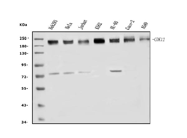
(Figure 1. Western blot analysis of CDK12 using anti-CDK12 antibody (AAA125695).Electrophoresis was performed on a 5-20% SDS-PAGE gel at 70V (Stacking gel) / 90V (Resolving gel) for 2-3 hours. The sample well of each lane was loaded with 50ug of sample under reducing conditions.Lane 1: human HEK293 whole cell lysatesLane 2: human HELA whole cell lysatesLane 3: human Jurkat whole cell lysatesLane 4: human K562 whole cell lysatesLane 5: human HL-60 whole cell lysatesLane 6: human CACO-2 whole cell lysatesLane 7: human A549 whole cell lysates.After Electrophoresis, proteins were transferred to a Nitrocellulose membrane at 150mA for 50-90 minutes. Blocked the membrane with 5% Non-fat Milk/ TBS for 1. 5 hour at RT. The membrane was incubated with rabbit anti-CDK12 antigen affinity purified polyclonal antibody (Catalog # AAA125695) at 0. 5 μg/mL overnight at 4 degree C, then washed with TBS-0. 1%Tween 3 times with 5 minutes each and probed with a goat anti-rabbit IgG-HRP secondary antibody at a dilution of 1:5000 for 1. 5 hour at RT. The signal is developed using an Enhanced Chemiluminescent detection (ECL) kit with Tanon 5200 system. A specific band was detected for CDK12 at approximately 205KD. The expected band size for CDK12 is at 205KD.)
WB (Western Blot)
(Figure 1. Western blot analysis of CDK12 using anti-CDK12 antibody (AAA125695).Electrophoresis was performed on a 5-20% SDS-PAGE gel at 70V (Stacking gel) / 90V (Resolving gel) for 2-3 hours. The sample well of each lane was loaded with 50ug of sample under reducing conditions.Lane 1: human HEK293 whole cell lysatesLane 2: human HELA whole cell lysatesLane 3: human Jurkat whole cell lysatesLane 4: human K562 whole cell lysatesLane 5: human HL-60 whole cell lysatesLane 6: human CACO-2 whole cell lysatesLane 7: human A549 whole cell lysates.After Electrophoresis, proteins were transferred to a Nitrocellulose membrane at 150mA for 50-90 minutes. Blocked the membrane with 5% Non-fat Milk/ TBS for 1. 5 hour at RT. The membrane was incubated with rabbit anti-CDK12 antigen affinity purified polyclonal antibody (Catalog # AAA125695) at 0. 5 μg/mL overnight at 4 degree C, then washed with TBS-0. 1%Tween 3 times with 5 minutes each and probed with a goat anti-rabbit IgG-HRP secondary antibody at a dilution of 1:5000 for 1. 5 hour at RT. The signal is developed using an Enhanced Chemiluminescent detection (ECL) kit with Tanon 5200 system. A specific band was detected for CDK12 at approximately 205KD. The expected band size for CDK12 is at 205KD.)
CDK12, Polyclonal Antibody (Cat# AAA125695)
Full Name
Anti-CDK12 Antibody
Gene Names
CDK12; CRK7; CRKR; CRKRS
Reactivity
Human
Applications
Direct ELISA, Flow Cytometry, Immunofluorescence, Immunocytochemistry, Western Blot
Purity
Immunogen affinity purified.
Pricing
 Application Data
Crossveinless-2/CV-2/BMPER, ELISA Kit (Cat# AAA125839)
Full Name
Human Crossveinless-2/CV-2/BMPER ELISA Kit
Gene Names
BMPER; CV2; CV-2; CRIM3
Reactivity
Human
Pricing
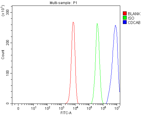 FCM/FACS (Flow Cytometry)
(Figure 3. Flow Cytometry analysis of K562 cells using anti-CDCA8 antibody (AAA127388).Overlay histogram showing K562 cells stained with AAA127388 (Blue line). To facilitate intracellular staining, cells were fixed with 4% paraformaldehyde and permeabilized with permeabilization buffer. The cells were blocked with 10% normal goat serum. And then incubated with rabbit anti-CDCA8 Antibody (AAA127388, 1ug/1x106 cells) for 30 min at 20 degree C. DyLight488 conjugated goat anti-rabbit IgG was used as secondary antibody for 30 minutes at 20 degree C. Isotype control antibody (Green line) was rabbit IgG (1ug/1x106) used under the same conditions. Unlabelled sample (Red line) was also used as a control.)
FCM/FACS (Flow Cytometry)
(Figure 3. Flow Cytometry analysis of K562 cells using anti-CDCA8 antibody (AAA127388).Overlay histogram showing K562 cells stained with AAA127388 (Blue line). To facilitate intracellular staining, cells were fixed with 4% paraformaldehyde and permeabilized with permeabilization buffer. The cells were blocked with 10% normal goat serum. And then incubated with rabbit anti-CDCA8 Antibody (AAA127388, 1ug/1x106 cells) for 30 min at 20 degree C. DyLight488 conjugated goat anti-rabbit IgG was used as secondary antibody for 30 minutes at 20 degree C. Isotype control antibody (Green line) was rabbit IgG (1ug/1x106) used under the same conditions. Unlabelled sample (Red line) was also used as a control.)
CDCA8, Polyclonal Antibody (Cat# AAA127388)
Full Name
Anti-CDCA8 Antibody Picoband
Gene Names
CDCA8; BOR; DasraB; MESRGP; BOREALIN
Reactivity
Human
Applications
Flow Cytometry, Immunofluorescence, Immunocytochemistry, Western Blot
Purity
Immunogen affinity purified.
Pricing
 WB (Western Blot)
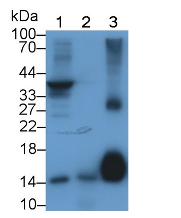
(Western Blot; Sample: Lane1: Porcine Heart lysate; Lane2: Rat Testis lysate; Lane3: Gallus Adipose lysate Primary Ab: 2ug/ml Mouse AntiHuman FABP4 Antibody Second Ab: 0.2ug/mL HRPLinked Caprine AntiMouse IgG Polyclonal Antibody (Catalog: SAA544Mu19))
WB (Western Blot)
(Western Blot; Sample: Lane1: Porcine Heart lysate; Lane2: Rat Testis lysate; Lane3: Gallus Adipose lysate Primary Ab: 2ug/ml Mouse AntiHuman FABP4 Antibody Second Ab: 0.2ug/mL HRPLinked Caprine AntiMouse IgG Polyclonal Antibody (Catalog: SAA544Mu19))
Fatty Acid Binding Protein 4 (FABP4), Monoclonal Antibody (Cat# AAA151731)
Full Name
Monoclonal Antibody to Fatty Acid Binding Protein 4 (FABP4)
Gene Names
FABP4; aP2; ALBP; AFABP; A-FABP; HEL-S-104
Reactivity
Human, Rat, Pig, Chicken
Applications
Immunoprecipitation, Immunocytochemistry, Immunohistochemistry, Western Blot
Purity
Protein A + Protein G affinity chromatography
Pricing
 Application Data
 FCM/FACS (Flow Cytometry)
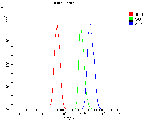
(Figure 3. Flow Cytometry analysis of SH-SY5Y cells using anti-MPST antibody (AAA127241).Overlay histogram showing SH-SY5Y cells stained with AAA127241 (Blue line). To facilitate intracellular staining, cells were fixed with 4% paraformaldehyde and permeabilized with permeabilization buffer. The cells were blocked with 10% normal goat serum. And then incubated with rabbit anti-MPST Antibody (AAA127241, 1ug/1x106 cells) for 30 min at 20 degree C. DyLight488 conjugated goat anti-rabbit IgG was used as secondary antibody for 30 minutes at 20 degree C. Isotype control antibody (Green line) was rabbit IgG (1ug/1x106) used under the same conditions. Unlabelled sample (Red line) was also used as a control.)
FCM/FACS (Flow Cytometry)
(Figure 3. Flow Cytometry analysis of SH-SY5Y cells using anti-MPST antibody (AAA127241).Overlay histogram showing SH-SY5Y cells stained with AAA127241 (Blue line). To facilitate intracellular staining, cells were fixed with 4% paraformaldehyde and permeabilized with permeabilization buffer. The cells were blocked with 10% normal goat serum. And then incubated with rabbit anti-MPST Antibody (AAA127241, 1ug/1x106 cells) for 30 min at 20 degree C. DyLight488 conjugated goat anti-rabbit IgG was used as secondary antibody for 30 minutes at 20 degree C. Isotype control antibody (Green line) was rabbit IgG (1ug/1x106) used under the same conditions. Unlabelled sample (Red line) was also used as a control.)
MPST, Polyclonal Antibody (Cat# AAA127241)
Full Name
Anti-MPST Antibody Picoband
Gene Names
MPST; MST; TST2; TUM1
Reactivity
Human
Applications
Flow Cytometry, Immunofluorescence, Immunocytochemistry, Western Blot
Purity
Immunogen affinity purified.
Pricing
 IP (Immunoprecipitation)
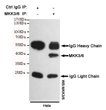
(Immunoprecipitation analysis of Hela cell lysates using MKK3/6 mouse mAb.)
IP (Immunoprecipitation)
(Immunoprecipitation analysis of Hela cell lysates using MKK3/6 mouse mAb.)
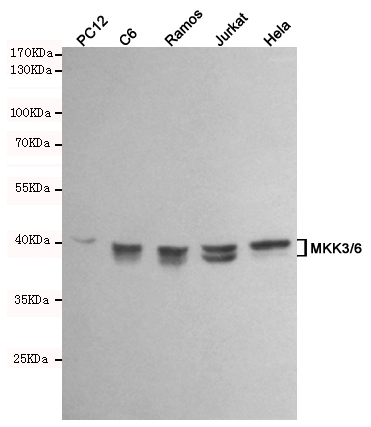
MKK3/6, Monoclonal Antibody (Cat# AAA320402)
Full Name
MKK3/6 Mouse mAb
Gene Names
MAP2K6; MEK6; MKK6; MAPKK6; PRKMK6; SAPKK3; SAPKK-3
Reactivity
Human, Rat
Applications
Immunoprecipitation, Western Blot
Purity
The antibody was affinity-purified from mouse ascites by affinity-chromatography using epitope-specific immunogen.
Pricing
 IF (Immunofluorescence)
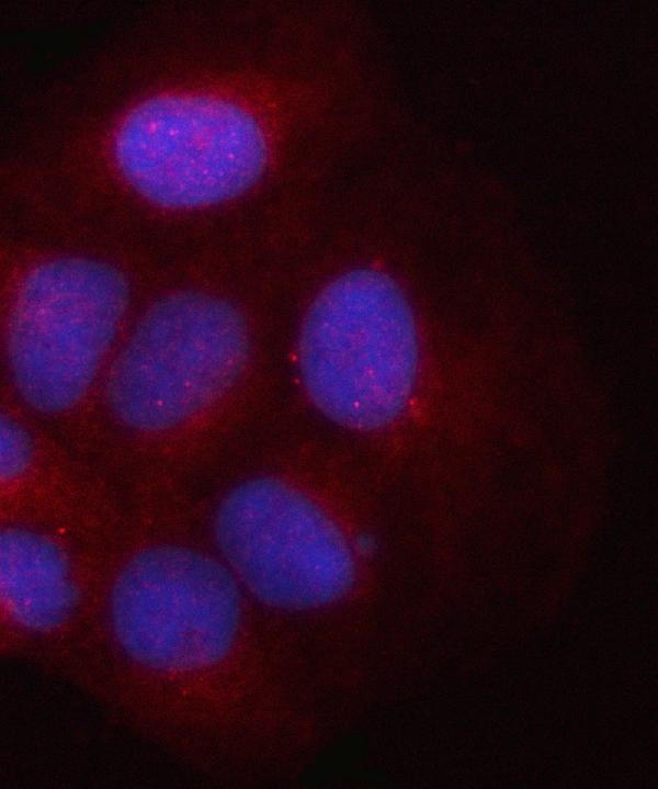
(Figure 2. IF analysis of PSMD2 using anti-PSMD2 antibody (AAA127391).PSMD2 was detected in an immunocytochemical section of U2OS cells. Enzyme antigen retrieval was performed using IHC enzyme antigen retrieval reagent for 15 mins. The cells were blocked with 10% goat serum. And then incubated with 5ug/mL rabbit anti-PSMD2 Antibody (AAA127391) overnight at 4 degree C. Cy3 Conjugated Goat Anti-Rabbit IgG (BA1032) was used as secondary antibody at 1:500 dilution and incubated for 30 minutes at 37 degree C. The section was counterstained with DAPI. Visualize using a fluorescence microscope and filter sets appropriate for the label used.)
IF (Immunofluorescence)
(Figure 2. IF analysis of PSMD2 using anti-PSMD2 antibody (AAA127391).PSMD2 was detected in an immunocytochemical section of U2OS cells. Enzyme antigen retrieval was performed using IHC enzyme antigen retrieval reagent for 15 mins. The cells were blocked with 10% goat serum. And then incubated with 5ug/mL rabbit anti-PSMD2 Antibody (AAA127391) overnight at 4 degree C. Cy3 Conjugated Goat Anti-Rabbit IgG (BA1032) was used as secondary antibody at 1:500 dilution and incubated for 30 minutes at 37 degree C. The section was counterstained with DAPI. Visualize using a fluorescence microscope and filter sets appropriate for the label used.)
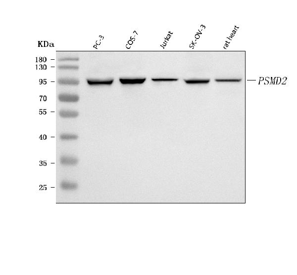
PSMD2, Polyclonal Antibody (Cat# AAA127391)
Full Name
Anti-PSMD2 Antibody Picoband
Gene Names
PSMD2; S2; P97; Rpn1; TRAP2
Reactivity
Human, Monkey, Rat
Applications
Immunofluorescence, Immunocytochemistry, Western Blot
Purity
Immunogen affinity purified.
Pricing
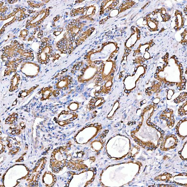
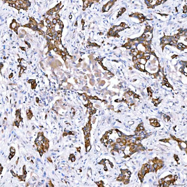
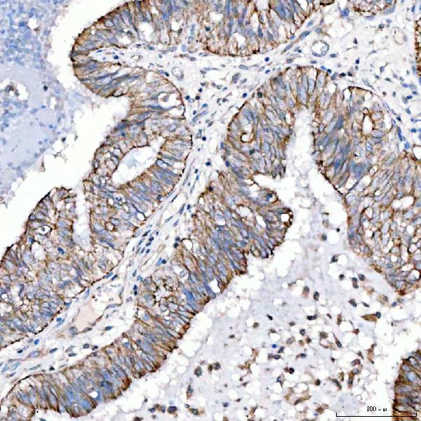
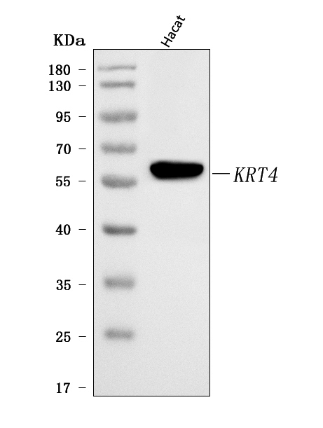
 IF (Immunofluorescence)
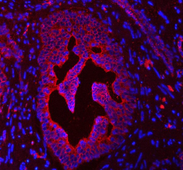
(Figure 5. IF analysis of Cytokeratin 4/KRT4 using anti-Cytokeratin 4/KRT4 antibody (AAA127443).Cytokeratin 4/KRT4 was detected in a paraffin-embedded section of human prostatic cancer tissue. Heat mediated antigen retrieval was performed in EDTA buffer (pH 8.0, epitope retrieval solution). The tissue section was blocked with 10% goat serum. The tissue section was then incubated with 5ug/mL rabbit anti-Cytokeratin 4/KRT4 Antibody (AAA127443) overnight at 4 degree C. Cy3 Conjugated Goat Anti-Rabbit IgG (BA1032) was used as secondary antibody at 1:500 dilution and incubated for 30 minutes at 37 degree C. The section was counterstained with DAPI. Visualize using a fluorescence microscope and filter sets appropriate for the label used.)
IF (Immunofluorescence)
(Figure 5. IF analysis of Cytokeratin 4/KRT4 using anti-Cytokeratin 4/KRT4 antibody (AAA127443).Cytokeratin 4/KRT4 was detected in a paraffin-embedded section of human prostatic cancer tissue. Heat mediated antigen retrieval was performed in EDTA buffer (pH 8.0, epitope retrieval solution). The tissue section was blocked with 10% goat serum. The tissue section was then incubated with 5ug/mL rabbit anti-Cytokeratin 4/KRT4 Antibody (AAA127443) overnight at 4 degree C. Cy3 Conjugated Goat Anti-Rabbit IgG (BA1032) was used as secondary antibody at 1:500 dilution and incubated for 30 minutes at 37 degree C. The section was counterstained with DAPI. Visualize using a fluorescence microscope and filter sets appropriate for the label used.)
Cytokeratin 4/KRT4, Polyclonal Antibody (Cat# AAA127443)
Full Name
Anti-Cytokeratin 4/KRT4 Antibody Picoband
Gene Names
KRT4; K4; CK4; CK-4; CYK4; WSN1
Reactivity
Human
Applications
Western Blot, Immunohistochemistry, Immunofluorescence
Purity
Immunogen affinity purified.
Pricing
 IP (Immunoprecipitation)
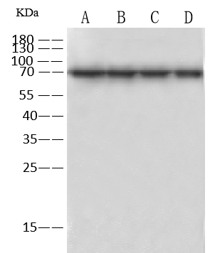
(HSPA1A was immunoprecipitated using: Lane A:0.5 mg A431 Whole Cell Lysate Lane B:0.5 mg Hela Whole Cell Lysate Lane C:0.5 mg HepG2 Whole Cell Lysate Lane D:0.5 mg Jurkat Whole Cell Lysate 4 uL anti- HSPA1A rabbit polyclonal antibody and 60 ug of Immunomagnetic beads Protein A/G. Primary antibody: Anti- HSPA1A rabbit polyclonal antibody,at 1:100 dilution Secondary antibody: Goat Anti-Rabbit IgG (H+L)/HRP at 1/10000 dilution Developed using the ECL technique. Performed under reducing conditions. Predicted band size: 70kDa Observed band size :70 kDa)
IP (Immunoprecipitation)
(HSPA1A was immunoprecipitated using: Lane A:0.5 mg A431 Whole Cell Lysate Lane B:0.5 mg Hela Whole Cell Lysate Lane C:0.5 mg HepG2 Whole Cell Lysate Lane D:0.5 mg Jurkat Whole Cell Lysate 4 uL anti- HSPA1A rabbit polyclonal antibody and 60 ug of Immunomagnetic beads Protein A/G. Primary antibody: Anti- HSPA1A rabbit polyclonal antibody,at 1:100 dilution Secondary antibody: Goat Anti-Rabbit IgG (H+L)/HRP at 1/10000 dilution Developed using the ECL technique. Performed under reducing conditions. Predicted band size: 70kDa Observed band size :70 kDa)
HSP70, Polyclonal Antibody (Cat# AAA258524)
Full Name
Anti-HSP70 Antibody, Rabbit Polyclonal
Gene Names
HSPA1B; HSP72; HSPA1; HSX70; HSP70-1; HSP70-2; HSP70.1; HSP70.2; HSP70-1B
Reactivity
Human
Applications
Immunoprecipitation, Immunofluorescence, Immunocytochemistry, Western Blot
Purity
Protein A & Antigen Affinity
Pricing

 FCM/FACS (Flow Cytometry)
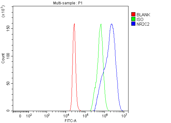
(Figure 4. Flow Cytometry analysis of 293T cells using anti-NR2C2 antibody (AAA127140).Overlay histogram showing 293T cells stained with AAA127140 (Blue line). To facilitate intracellular staining, cells were fixed with 4% paraformaldehyde and permeabilized with permeabilization buffer. The cells were blocked with 10% normal goat serum. And then incubated with rabbit anti-NR2C2 Antibody (AAA127140, 1ug/1x106 cells) for 30 min at 20 degree C. DyLight488 conjugated goat anti-rabbit IgG was used as secondary antibody for 30 minutes at 20 degree C. Isotype control antibody (Green line) was rabbit IgG (1ug/1x106) used under the same conditions. Unlabelled sample (Red line) was also used as a control.)
FCM/FACS (Flow Cytometry)
(Figure 4. Flow Cytometry analysis of 293T cells using anti-NR2C2 antibody (AAA127140).Overlay histogram showing 293T cells stained with AAA127140 (Blue line). To facilitate intracellular staining, cells were fixed with 4% paraformaldehyde and permeabilized with permeabilization buffer. The cells were blocked with 10% normal goat serum. And then incubated with rabbit anti-NR2C2 Antibody (AAA127140, 1ug/1x106 cells) for 30 min at 20 degree C. DyLight488 conjugated goat anti-rabbit IgG was used as secondary antibody for 30 minutes at 20 degree C. Isotype control antibody (Green line) was rabbit IgG (1ug/1x106) used under the same conditions. Unlabelled sample (Red line) was also used as a control.)
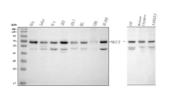
NR2C2, Polyclonal Antibody (Cat# AAA127140)
Full Name
Anti-NR2C2 Antibody Picoband
Gene Names
NR2C2; TR4; TAK1
Reactivity
Human, Monkey, Mouse, Rat
Applications
Flow Cytometry, Western Blot, Immunohistochemistry
Purity
Immunogen affinity purified.
Pricing
 FCM/FACS (Flow Cytometry)
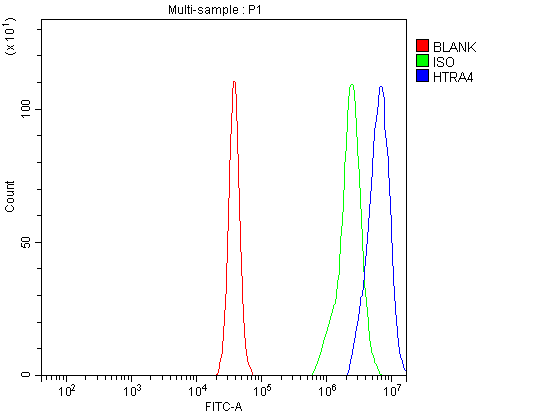
(Figure 2. Flow Cytometry analysis of Daudi cells using anti-HTRA4 antibody (AAA127779).Overlay histogram showing Daudi cells stained with AAA127779 (Blue line). The cells were fixed with 4% paraformaldehyde and blocked with 10% normal goat serum. And then incubated with rabbit anti-HTRA4 Antibody (AAA127779, 1ug/1x106 cells) for 30 min at 20 degree C. DyLight488 conjugated goat anti-rabbit IgG was used as secondary antibody for 30 minutes at 20 degree C. Isotype control antibody (Green line) was rabbit IgG (1ug/1x106) used under the same conditions. Unlabelled sample (Red line) was also used as a control.)
FCM/FACS (Flow Cytometry)
(Figure 2. Flow Cytometry analysis of Daudi cells using anti-HTRA4 antibody (AAA127779).Overlay histogram showing Daudi cells stained with AAA127779 (Blue line). The cells were fixed with 4% paraformaldehyde and blocked with 10% normal goat serum. And then incubated with rabbit anti-HTRA4 Antibody (AAA127779, 1ug/1x106 cells) for 30 min at 20 degree C. DyLight488 conjugated goat anti-rabbit IgG was used as secondary antibody for 30 minutes at 20 degree C. Isotype control antibody (Green line) was rabbit IgG (1ug/1x106) used under the same conditions. Unlabelled sample (Red line) was also used as a control.)
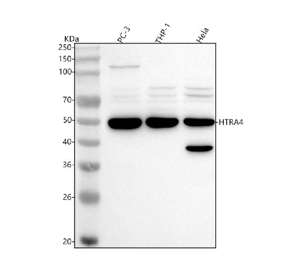
HTRA4, Polyclonal Antibody (Cat# AAA127779)
Full Name
Anti-HTRA4 Antibody Picoband
Reactivity
Human
Applications
Flow Cytometry, Western Blot
Purity
Immunogen affinity purified.
Pricing
 FCM/FACS (Flow Cytometry)
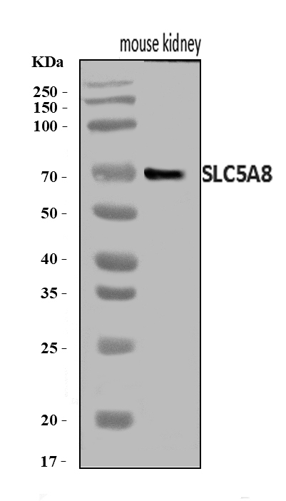
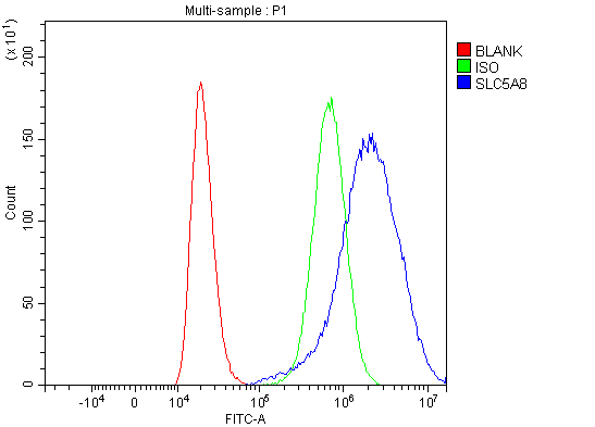
(Figure 2. Flow Cytometry analysis of 293T cells using anti-SLC5A8 antibody (AAA126340).Overlay histogram showing 293T cells stained with AAA126340 (Blue line). The cells were blocked with 10% normal goat serum. And then incubated with rabbit anti-SLC5A8 Antibody (AAA126340, 1 ug/1x10^6 cells) for 30 min at 20 degree C. DyLight488 conjugated goat anti-rabbit IgG was used as secondary antibody for 30 minutes at 20 degree C. Isotype control antibody (Green line) was rabbit IgG (1 ug/1x10^6) used under the same conditions. Unlabelled sample (Red line) was also used as a control.)
FCM/FACS (Flow Cytometry)
(Figure 2. Flow Cytometry analysis of 293T cells using anti-SLC5A8 antibody (AAA126340).Overlay histogram showing 293T cells stained with AAA126340 (Blue line). The cells were blocked with 10% normal goat serum. And then incubated with rabbit anti-SLC5A8 Antibody (AAA126340, 1 ug/1x10^6 cells) for 30 min at 20 degree C. DyLight488 conjugated goat anti-rabbit IgG was used as secondary antibody for 30 minutes at 20 degree C. Isotype control antibody (Green line) was rabbit IgG (1 ug/1x10^6) used under the same conditions. Unlabelled sample (Red line) was also used as a control.)
SLC5A8, Polyclonal Antibody (Cat# AAA126340)
Full Name
Anti-SLC5A8 Antibody Picoband
Gene Names
SLC5A8; AIT; SMCT; SMCT1
Reactivity
Human, Mouse
Applications
Western Blot, Flow Cytometry
Purity
Immunogen affinity purified.
Pricing
 FCM/FACS (Flow Cytometry)
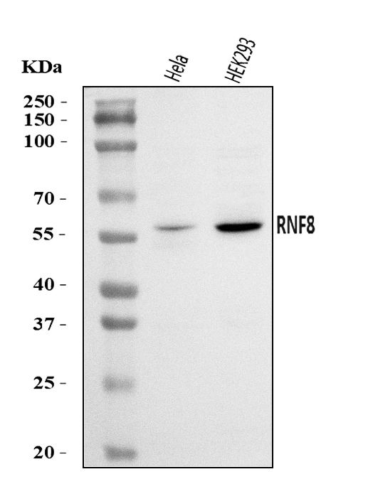
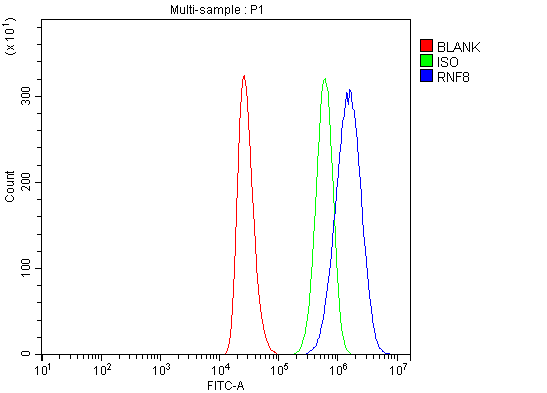
(Figure 2. Flow Cytometry analysis of THP-1 cells using anti-RNF8 antibody (AAA126060).Overlay histogram showing THP-1 cells stained with AAA126060 (Blue line). The cells were blocked with 10% normal goat serum. And then incubated with rabbit anti-RNF8 Antibody (AAA126060, 1 ug/1x10^6 cells) for 30 min at 20 degree C. DyLight488 conjugated goat anti-rabbit IgG was used as secondary antibody for 30 minutes at 20 degree C. Isotype control antibody (Green line) was rabbit IgG (1 ug/1x10^6) used under the same conditions. Unlabelled sample (Red line) was also used as a control.)
FCM/FACS (Flow Cytometry)
(Figure 2. Flow Cytometry analysis of THP-1 cells using anti-RNF8 antibody (AAA126060).Overlay histogram showing THP-1 cells stained with AAA126060 (Blue line). The cells were blocked with 10% normal goat serum. And then incubated with rabbit anti-RNF8 Antibody (AAA126060, 1 ug/1x10^6 cells) for 30 min at 20 degree C. DyLight488 conjugated goat anti-rabbit IgG was used as secondary antibody for 30 minutes at 20 degree C. Isotype control antibody (Green line) was rabbit IgG (1 ug/1x10^6) used under the same conditions. Unlabelled sample (Red line) was also used as a control.)
RNF8, Polyclonal Antibody (Cat# AAA126060)
Full Name
Anti-RNF8 Antibody Picoband
Gene Names
RNF8; hRNF8
Reactivity
Human
Applications
Flow Cytometry, Western Blot
Purity
Immunogen affinity purified.
Pricing
 IF (Immunofluorescence)
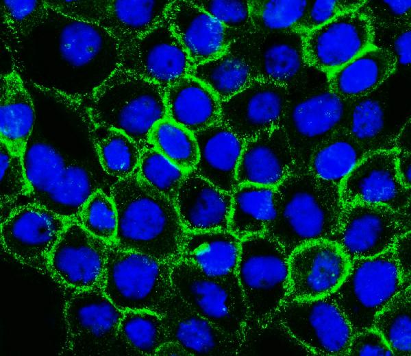
(Figure 2. IF analysis of P Cadherin/CDH3 using anti-P Cadherin/CDH3 antibody (AAA126302).P Cadherin/CDH3 was detected in an immunocytochemical section of A431 cells. Enzyme antigen retrieval was performed using IHC enzyme antigen retrieval reagent (AR0022) for 15 mins. The cells were blocked with 10% goat serum. And then incubated with 5 ug/mL rabbit anti-P Cadherin/CDH3 Antibody (AAA126302) overnight at 4 degree C. DyLight488 Conjugated Goat Anti-Rabbit IgG was used as secondary antibody at 1:100 dilution and incubated for 30 minutes at 37 degree C. The section was counterstained with DAPI. Visualize using a fluorescence microscope and filter sets appropriate for the label used.)
IF (Immunofluorescence)
(Figure 2. IF analysis of P Cadherin/CDH3 using anti-P Cadherin/CDH3 antibody (AAA126302).P Cadherin/CDH3 was detected in an immunocytochemical section of A431 cells. Enzyme antigen retrieval was performed using IHC enzyme antigen retrieval reagent (AR0022) for 15 mins. The cells were blocked with 10% goat serum. And then incubated with 5 ug/mL rabbit anti-P Cadherin/CDH3 Antibody (AAA126302) overnight at 4 degree C. DyLight488 Conjugated Goat Anti-Rabbit IgG was used as secondary antibody at 1:100 dilution and incubated for 30 minutes at 37 degree C. The section was counterstained with DAPI. Visualize using a fluorescence microscope and filter sets appropriate for the label used.)
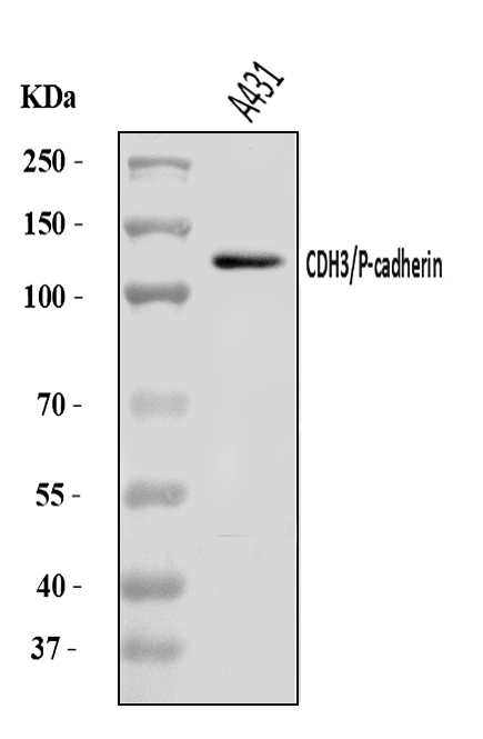
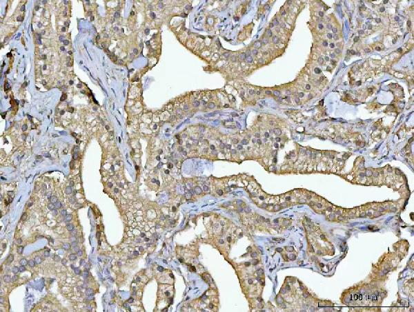
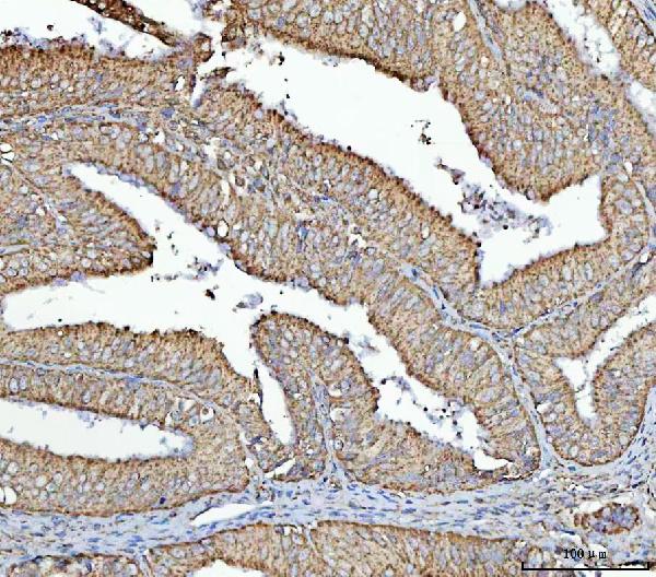
P cadherin/CDH3, Polyclonal Antibody (Cat# AAA126302)
Full Name
Anti-P cadherin/CDH3 Antibody Picoband
Gene Names
CDH3; CDHP; HJMD; PCAD
Reactivity
Human
Applications
Immunofluorescence, Immunocytochemistry, Western Blot
Purity
Immunogen affinity purified.
Pricing

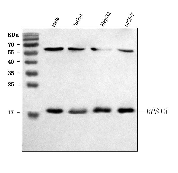
 FCM/FACS (Flow Cytometry)
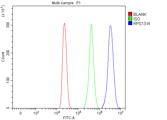
(Figure 5. Flow Cytometry analysis of HL-60 cells using anti-RPS13 antibody (AAA127351).Overlay histogram showing HL-60 cells stained with AAA127351 (Blue line). To facilitate intracellular staining, cells were fixed with 4% paraformaldehyde and permeabilized with permeabilization buffer. The cells were blocked with 10% normal goat serum. And then incubated with rabbit anti-RPS13 Antibody (AAA127351, 1ug/1x106 cells) for 30 min at 20 degree C. DyLight488 conjugated goat anti-rabbit IgG was used as secondary antibody for 30 minutes at 20 degree C. Isotype control antibody (Green line) was rabbit IgG (1ug/1x106) used under the same conditions. Unlabelled sample (Red line) was also used as a control.)
FCM/FACS (Flow Cytometry)
(Figure 5. Flow Cytometry analysis of HL-60 cells using anti-RPS13 antibody (AAA127351).Overlay histogram showing HL-60 cells stained with AAA127351 (Blue line). To facilitate intracellular staining, cells were fixed with 4% paraformaldehyde and permeabilized with permeabilization buffer. The cells were blocked with 10% normal goat serum. And then incubated with rabbit anti-RPS13 Antibody (AAA127351, 1ug/1x106 cells) for 30 min at 20 degree C. DyLight488 conjugated goat anti-rabbit IgG was used as secondary antibody for 30 minutes at 20 degree C. Isotype control antibody (Green line) was rabbit IgG (1ug/1x106) used under the same conditions. Unlabelled sample (Red line) was also used as a control.)
RPS13, Polyclonal Antibody (Cat# AAA127351)
Full Name
Anti-RPS13 Antibody Picoband
Gene Names
RPS13; S13
Reactivity
Human
Applications
Flow Cytometry, Immunohistochemistry, Western Blot
Purity
Immunogen affinity purified.
Pricing
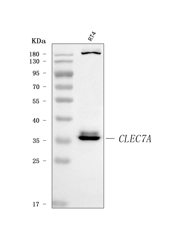
 FCM/FACS (Flow Cytometry)
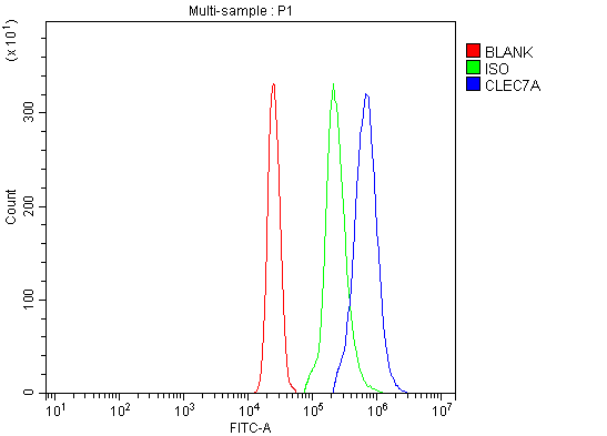
(Figure 2. Flow Cytometry analysis of U937 cells using anti-Dectin-1/CLEC7A antibody (AAA126247).Overlay histogram showing U937 cells stained with AAA126247 (Blue line). The cells were blocked with 10% normal goat serum. And then incubated with rabbit anti-Dectin-1/CLEC7A Antibody (AAA126247, 1 ug/1x10^6 cells) for 30 min at 20 degree C. DyLight488 conjugated goat anti-rabbit IgG was used as secondary antibody for 30 minutes at 20 degree C. Isotype control antibody (Green line) was rabbit IgG (1 ug/1x10^6) used under the same conditions. Unlabelled sample (Red line) was also used as a control.)
FCM/FACS (Flow Cytometry)
(Figure 2. Flow Cytometry analysis of U937 cells using anti-Dectin-1/CLEC7A antibody (AAA126247).Overlay histogram showing U937 cells stained with AAA126247 (Blue line). The cells were blocked with 10% normal goat serum. And then incubated with rabbit anti-Dectin-1/CLEC7A Antibody (AAA126247, 1 ug/1x10^6 cells) for 30 min at 20 degree C. DyLight488 conjugated goat anti-rabbit IgG was used as secondary antibody for 30 minutes at 20 degree C. Isotype control antibody (Green line) was rabbit IgG (1 ug/1x10^6) used under the same conditions. Unlabelled sample (Red line) was also used as a control.)
Dectin-1/CLEC7A, Polyclonal Antibody (Cat# AAA126247)
Full Name
Anti-Dectin-1/CLEC7A Antibody Picoband
Gene Names
CLEC7A; BGR; CANDF4; DECTIN1; CLECSF12
Reactivity
Human
Applications
Flow Cytometry, Western Blot
Purity
Immunogen affinity purified.
Pricing
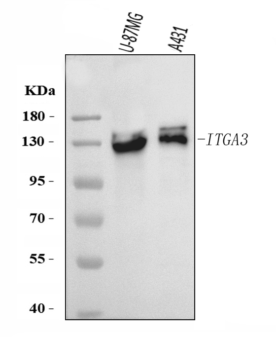
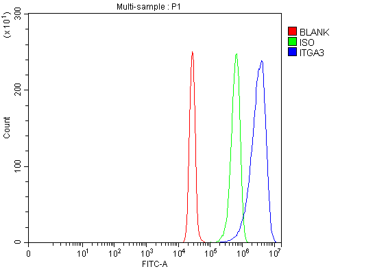 FCM/FACS (Flow Cytometry)
(Figure 2. Flow Cytometry analysis of SiHa cells using anti-Integrin Alpha 3/ITGA3 antibody (AAA126269).Overlay histogram showing SiHa cells stained with AAA126269 (Blue line). The cells were blocked with 10% normal goat serum. And then incubated with rabbit anti-Integrin Alpha 3/ITGA3 Antibody (AAA126269, 1 ug/1x10^6 cells) for 30 min at 20 degree C. DyLight488 conjugated goat anti-rabbit IgG was used as secondary antibody for 30 minutes at 20 degree C. Isotype control antibody (Green line) was rabbit IgG (1 ug/1x10^6) used under the same conditions. Unlabelled sample (Red line) was also used as a control.)
FCM/FACS (Flow Cytometry)
(Figure 2. Flow Cytometry analysis of SiHa cells using anti-Integrin Alpha 3/ITGA3 antibody (AAA126269).Overlay histogram showing SiHa cells stained with AAA126269 (Blue line). The cells were blocked with 10% normal goat serum. And then incubated with rabbit anti-Integrin Alpha 3/ITGA3 Antibody (AAA126269, 1 ug/1x10^6 cells) for 30 min at 20 degree C. DyLight488 conjugated goat anti-rabbit IgG was used as secondary antibody for 30 minutes at 20 degree C. Isotype control antibody (Green line) was rabbit IgG (1 ug/1x10^6) used under the same conditions. Unlabelled sample (Red line) was also used as a control.)
Integrin alpha 3/ITGA3, Polyclonal Antibody (Cat# AAA126269)
Full Name
Anti-Integrin alpha 3/ITGA3 Antibody Picoband
Gene Names
ITGA3; VL3A; CD49C; GAPB3; ILNEB; MSK18; VCA-2; VLA3a; GAP-B3
Reactivity
Human
Applications
Flow Cytometry, Western Blot
Purity
Immunogen affinity purified.
Pricing
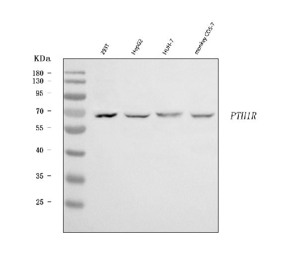
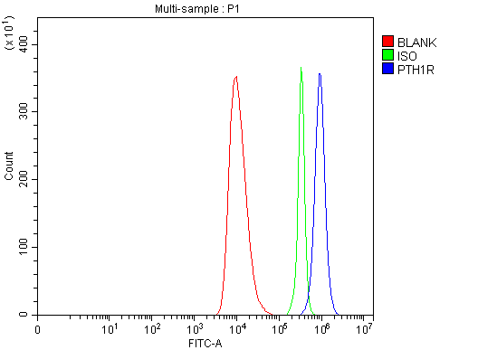 FCM/FACS (Flow Cytometry)
(Figure 2. Flow Cytometry analysis of HEL cells using anti-Parathyroid Hormone Receptor 1/PTH1R antibody (AAA126153).Overlay histogram showing HEL cells stained with AAA126153 (Blue line). The cells were blocked with 10% normal goat serum. And then incubated with rabbit anti-Parathyroid Hormone Receptor 1/PTH1R Antibody (AAA126153, 1 ug/1x10^6 cells) for 30 min at 20 degree C. DyLight488 conjugated goat anti-rabbit IgG was used as secondary antibody for 30 minutes at 20 degree C. Isotype control antibody (Green line) was rabbit IgG (1 ug/1x10^6) used under the same conditions. Unlabelled sample (Red line) was also used as a control.)
FCM/FACS (Flow Cytometry)
(Figure 2. Flow Cytometry analysis of HEL cells using anti-Parathyroid Hormone Receptor 1/PTH1R antibody (AAA126153).Overlay histogram showing HEL cells stained with AAA126153 (Blue line). The cells were blocked with 10% normal goat serum. And then incubated with rabbit anti-Parathyroid Hormone Receptor 1/PTH1R Antibody (AAA126153, 1 ug/1x10^6 cells) for 30 min at 20 degree C. DyLight488 conjugated goat anti-rabbit IgG was used as secondary antibody for 30 minutes at 20 degree C. Isotype control antibody (Green line) was rabbit IgG (1 ug/1x10^6) used under the same conditions. Unlabelled sample (Red line) was also used as a control.)
Parathyroid Hormone Receptor 1/PTH1R, Polyclonal Antibody (Cat# AAA126153)
Full Name
Anti-Parathyroid Hormone Receptor 1/PTH1R Antibody Picoband
Gene Names
PTH1R; PFE; PTHR; PTHR1
Reactivity
Human, Monkey
Applications
Flow Cytometry, Western Blot
Purity
Immunogen affinity purified.
Pricing
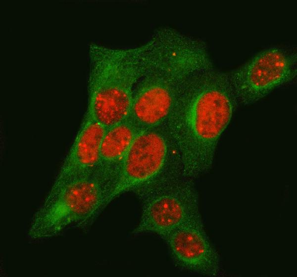
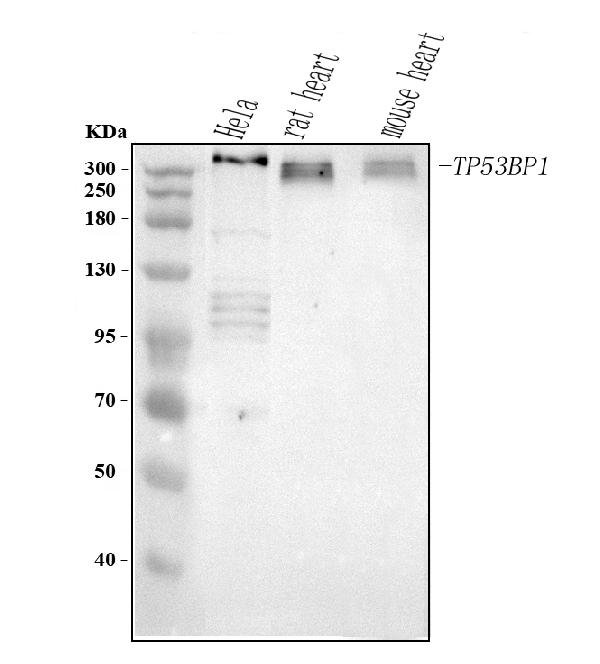
 FCM/FACS (Flow Cytometry)
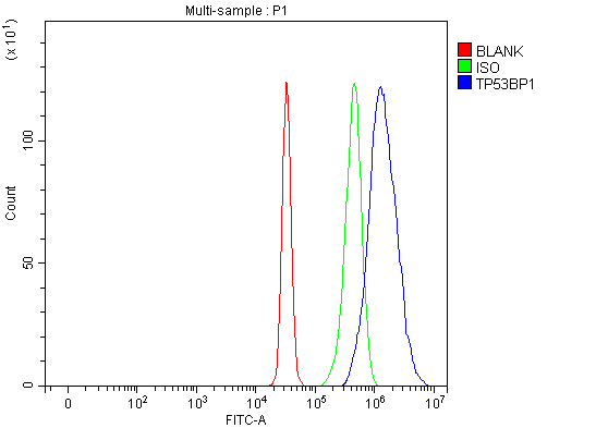
(Figure 3. Flow Cytometry analysis of HepG2 cells using anti-TP53BP1 antibody (AAA126987).Overlay histogram showing HepG2 cells stained with AAA126987 (Blue line). To facilitate intracellular staining, cells were fixed with 4% paraformaldehyde and permeabilized with permeabilization buffer. The cells were blocked with 10% normal goat serum. And then incubated with rabbit anti-TP53BP1 Antibody (AAA126987, 1ug/1x106 cells) for 30 min at 20 degree C. DyLight488 conjugated goat anti-rabbit IgG was used as secondary antibody for 30 minutes at 20 degree C. Isotype control antibody (Green line) was rabbit IgG (1ug/1x106) used under the same conditions. Unlabelled sample (Red line) was also used as a control.)
FCM/FACS (Flow Cytometry)
(Figure 3. Flow Cytometry analysis of HepG2 cells using anti-TP53BP1 antibody (AAA126987).Overlay histogram showing HepG2 cells stained with AAA126987 (Blue line). To facilitate intracellular staining, cells were fixed with 4% paraformaldehyde and permeabilized with permeabilization buffer. The cells were blocked with 10% normal goat serum. And then incubated with rabbit anti-TP53BP1 Antibody (AAA126987, 1ug/1x106 cells) for 30 min at 20 degree C. DyLight488 conjugated goat anti-rabbit IgG was used as secondary antibody for 30 minutes at 20 degree C. Isotype control antibody (Green line) was rabbit IgG (1ug/1x106) used under the same conditions. Unlabelled sample (Red line) was also used as a control.)
TP53BP1, Polyclonal Antibody (Cat# AAA126987)
Full Name
Anti-TP53BP1 Antibody Picoband
Gene Names
TP53BP1; p202; 53BP1
Reactivity
Human, Mouse, Rat
Applications
Flow Cytometry, Immunofluorescence, Immunocytochemistry, Western Blot
Purity
Immunogen affinity purified.
Pricing
 FCM/FACS (Flow Cytometry)
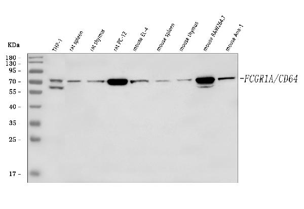
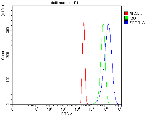
(Figure 2. Flow Cytometry analysis of SiHa cells using anti-CD64/FCGR1A antibody (AAA126227).Overlay histogram showing SiHa cells stained with AAA126227 (Blue line). The cells were blocked with 10% normal goat serum. And then incubated with rabbit anti-CD64/FCGR1A Antibody (AAA126227, 1 ug/1x10^6 cells) for 30 min at 20 degree C. DyLight488 conjugated goat anti-rabbit IgG was used as secondary antibody for 30 minutes at 20 degree C. Isotype control antibody (Green line) was rabbit IgG (1 ug/1x10^6) used under the same conditions. Unlabelled sample (Red line) was also used as a control.)
FCM/FACS (Flow Cytometry)
(Figure 2. Flow Cytometry analysis of SiHa cells using anti-CD64/FCGR1A antibody (AAA126227).Overlay histogram showing SiHa cells stained with AAA126227 (Blue line). The cells were blocked with 10% normal goat serum. And then incubated with rabbit anti-CD64/FCGR1A Antibody (AAA126227, 1 ug/1x10^6 cells) for 30 min at 20 degree C. DyLight488 conjugated goat anti-rabbit IgG was used as secondary antibody for 30 minutes at 20 degree C. Isotype control antibody (Green line) was rabbit IgG (1 ug/1x10^6) used under the same conditions. Unlabelled sample (Red line) was also used as a control.)
CD64/FCGR1A, Polyclonal Antibody (Cat# AAA126227)
Full Name
Anti-CD64/FCGR1A Antibody Picoband
Gene Names
FCGR1A; CD64; FCRI; CD64A; IGFR1
Reactivity
Human, Mouse, Rat
Applications
Western Blot, Flow Cytometry
Purity
Immunogen affinity purified.
Pricing

 FCM/FACS (Flow Cytometry)
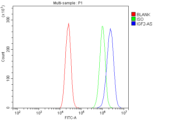
(Figure 3. Flow Cytometry analysis of CACO-2 cells using anti-IGF2-AS antibody (AAA126606).Overlay histogram showing CACO-2 cells stained with AAA126606 (Blue line).The cells were blocked with 10% normal goat serum. And then incubated with rabbit anti-IGF2-AS Antibody (AAA126606, 1ug/1x10^6 cells) for 30 min at 20 degree C. DyLight488 conjugated goat anti-rabbit IgG was used as secondary antibody for 30 minutes at 20 degree C. Isotype control antibody (Green line) was rabbit IgG (1ug/1x10^6) used under the same conditions. Unlabelled sample (Red line) was also used as a control.)
FCM/FACS (Flow Cytometry)
(Figure 3. Flow Cytometry analysis of CACO-2 cells using anti-IGF2-AS antibody (AAA126606).Overlay histogram showing CACO-2 cells stained with AAA126606 (Blue line).The cells were blocked with 10% normal goat serum. And then incubated with rabbit anti-IGF2-AS Antibody (AAA126606, 1ug/1x10^6 cells) for 30 min at 20 degree C. DyLight488 conjugated goat anti-rabbit IgG was used as secondary antibody for 30 minutes at 20 degree C. Isotype control antibody (Green line) was rabbit IgG (1ug/1x10^6) used under the same conditions. Unlabelled sample (Red line) was also used as a control.)
IGF2-AS, Polyclonal Antibody (Cat# AAA126606)
Full Name
Anti-IGF2-AS Antibody Picoband
Gene Names
IGF2-AS; PEG8; IGF2AS; IGF2-AS1
Reactivity
Human
Applications
Western Blot, Immunocytochemistry, Immunofluorescence, Flow Cytometry
Purity
Immunogen affinity purified.
Pricing
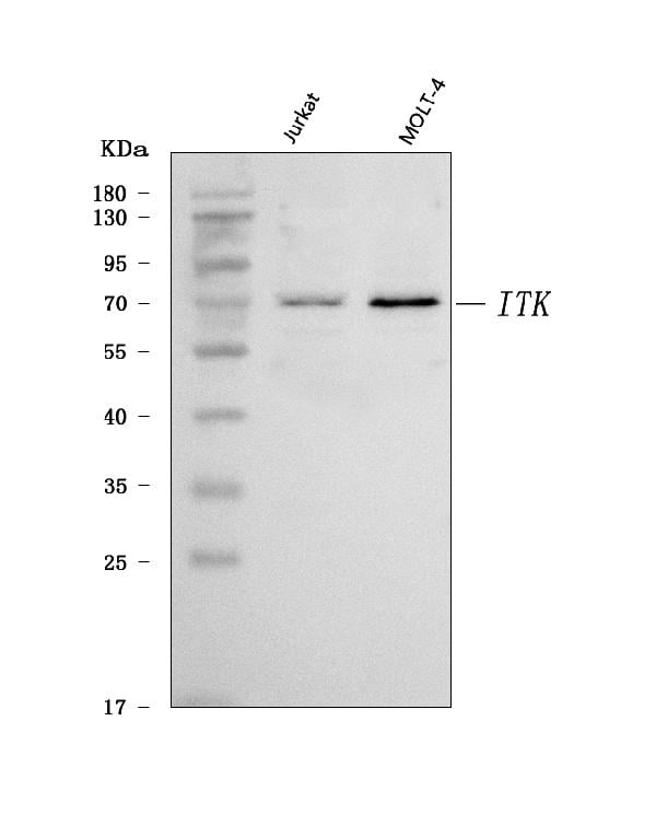
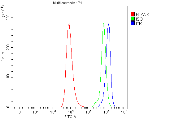 FCM/FACS (Flow Cytometry)
(Figure 3. Flow Cytometry analysis of HEL cells using anti-ITK antibody (AAA126137).Overlay histogram showing HEL cells stained with AAA126137 (Blue line). The cells were blocked with 10% normal goat serum. And then incubated with rabbit anti-ITK Antibody (AAA126137, 1 ug/1x10^6 cells) for 30 min at 20 degree C. DyLight488 conjugated goat anti-rabbit IgG was used as secondary antibody for 30 minutes at 20 degree C. Isotype control antibody (Green line) was rabbit IgG (1 ug/1x10^6) used under the same conditions. Unlabelled sample (Red line) was also used as a control.)
FCM/FACS (Flow Cytometry)
(Figure 3. Flow Cytometry analysis of HEL cells using anti-ITK antibody (AAA126137).Overlay histogram showing HEL cells stained with AAA126137 (Blue line). The cells were blocked with 10% normal goat serum. And then incubated with rabbit anti-ITK Antibody (AAA126137, 1 ug/1x10^6 cells) for 30 min at 20 degree C. DyLight488 conjugated goat anti-rabbit IgG was used as secondary antibody for 30 minutes at 20 degree C. Isotype control antibody (Green line) was rabbit IgG (1 ug/1x10^6) used under the same conditions. Unlabelled sample (Red line) was also used as a control.)
ITK, Polyclonal Antibody (Cat# AAA126137)
Full Name
Anti-ITK Antibody Picoband
Gene Names
ITK; EMT; LYK; LPFS1; PSCTK2
Reactivity
Human
Applications
Flow Cytometry, Immunohistochemistry, Western Blot
Purity
Immunogen affinity purified.
Pricing
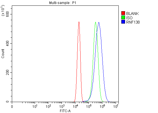
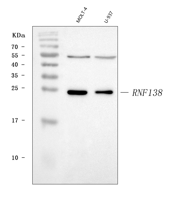
 FCM/FACS (Flow Cytometry)
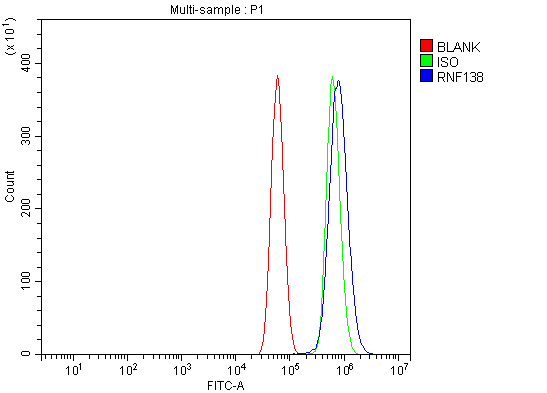
(Figure 3. Flow Cytometry analysis of U87 cells using anti-HSD-4/RNF138 antibody (AAA126542).Overlay histogram showing U87 cells stained with AAA126542 (Blue line). The cells were blocked with 10% normal goat serum. And then incubated with rabbit anti-HSD-4/RNF138 Antibody (AAA126542, 1 ug/1x10^6 cells) for 30 min at 20 degree C. DyLight488 conjugated goat anti-rabbit IgG was used as secondary antibody for 30 minutes at 20 degree C. Isotype control antibody (Green line) was rabbit IgG (1 ug/1x10^6) used under the same conditions. Unlabelled sample (Red line) was also used as a control.)
FCM/FACS (Flow Cytometry)
(Figure 3. Flow Cytometry analysis of U87 cells using anti-HSD-4/RNF138 antibody (AAA126542).Overlay histogram showing U87 cells stained with AAA126542 (Blue line). The cells were blocked with 10% normal goat serum. And then incubated with rabbit anti-HSD-4/RNF138 Antibody (AAA126542, 1 ug/1x10^6 cells) for 30 min at 20 degree C. DyLight488 conjugated goat anti-rabbit IgG was used as secondary antibody for 30 minutes at 20 degree C. Isotype control antibody (Green line) was rabbit IgG (1 ug/1x10^6) used under the same conditions. Unlabelled sample (Red line) was also used as a control.)
HSD-4/RNF138, Polyclonal Antibody (Cat# AAA126542)
Full Name
Anti-HSD-4/RNF138 Antibody Picoband
Gene Names
RNF138; NARF; HSD-4; STRIN; hNARF
Reactivity
Human
Applications
Flow Cytometry, Western Blot
Purity
Immunogen affinity purified.
Pricing
 IHC (Immunohistochemistry)
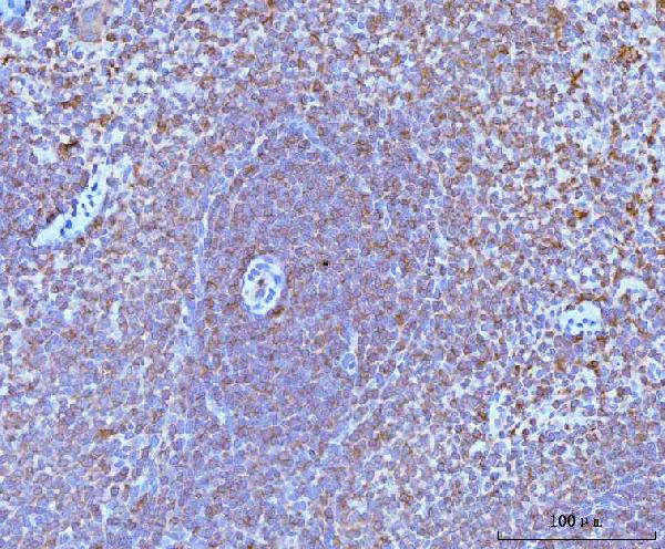
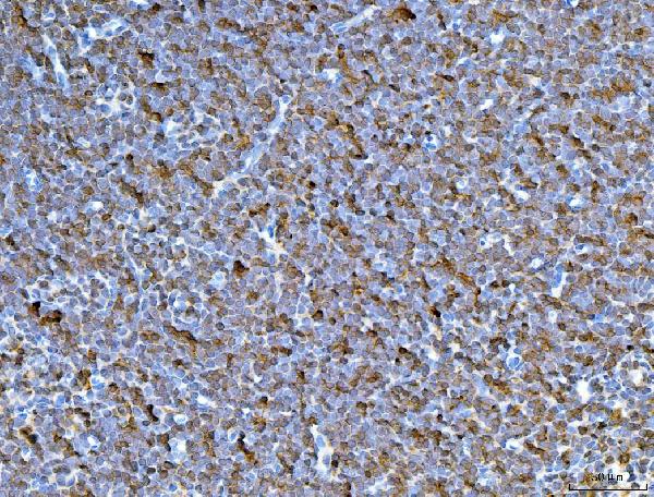
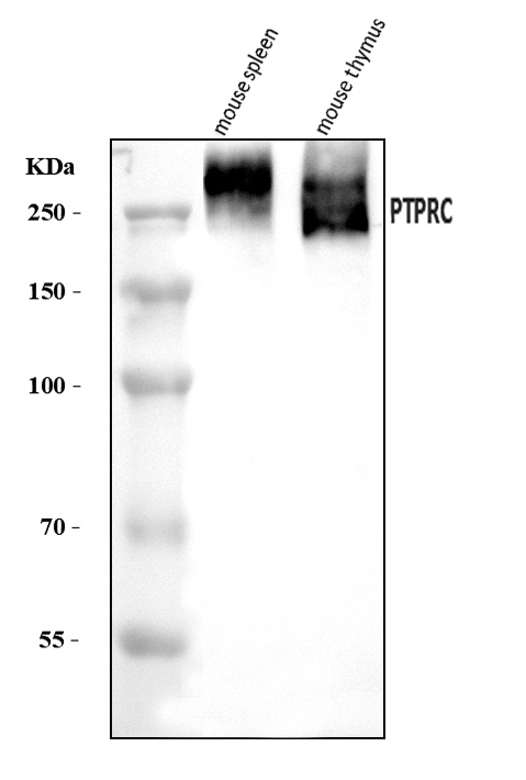
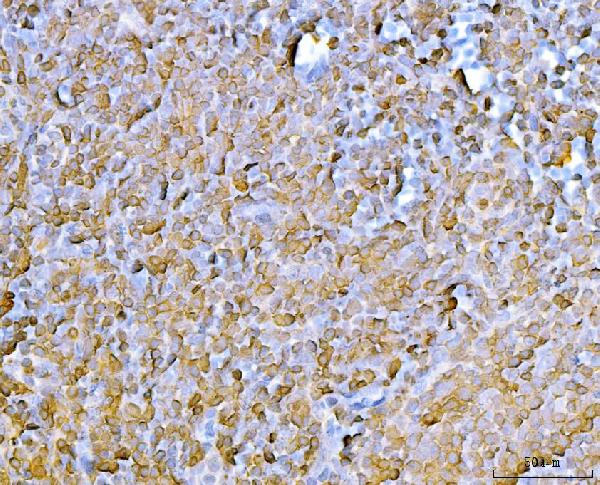
(Figure 4. IHC analysis of CD45/Ptprc using anti-CD45/Ptprc antibody (AAA126038).CD45/Ptprc was detected in a paraffin-embedded section of mouse lymph node tissue. Heat mediated antigen retrieval was performed in EDTA buffer (pH 8.0, epitope retrieval solution). The tissue section was blocked with 10% goat serum. The tissue section was then incubated with 2 ug/ml rabbit anti-CD45/Ptprc Antibody (AAA126038) overnight at 4 degree C. Biotinylated goat anti-rabbit IgG was used as secondary antibody and incubated for 30 minutes at 37 degree C. The tissue section was developed using Strepavidin-Biotin-Complex (SABC) with DAB as the chromogen.)
IHC (Immunohistochemistry)
(Figure 4. IHC analysis of CD45/Ptprc using anti-CD45/Ptprc antibody (AAA126038).CD45/Ptprc was detected in a paraffin-embedded section of mouse lymph node tissue. Heat mediated antigen retrieval was performed in EDTA buffer (pH 8.0, epitope retrieval solution). The tissue section was blocked with 10% goat serum. The tissue section was then incubated with 2 ug/ml rabbit anti-CD45/Ptprc Antibody (AAA126038) overnight at 4 degree C. Biotinylated goat anti-rabbit IgG was used as secondary antibody and incubated for 30 minutes at 37 degree C. The tissue section was developed using Strepavidin-Biotin-Complex (SABC) with DAB as the chromogen.)
CD45/Ptprc, Polyclonal Antibody (Cat# AAA126038)
Full Name
Anti-CD45/Ptprc Antibody Picoband
Reactivity
Mouse, Rat
Applications
Immunohistochemistry, Western Blot
Purity
Immunogen affinity purified.
Pricing
 FCM/FACS (Flow Cytometry)
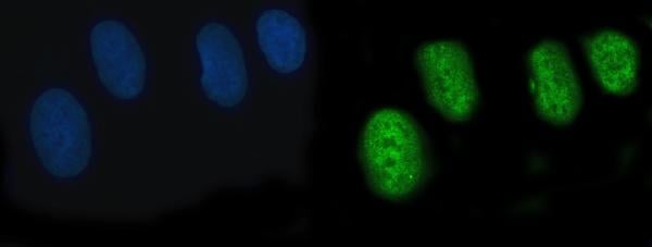
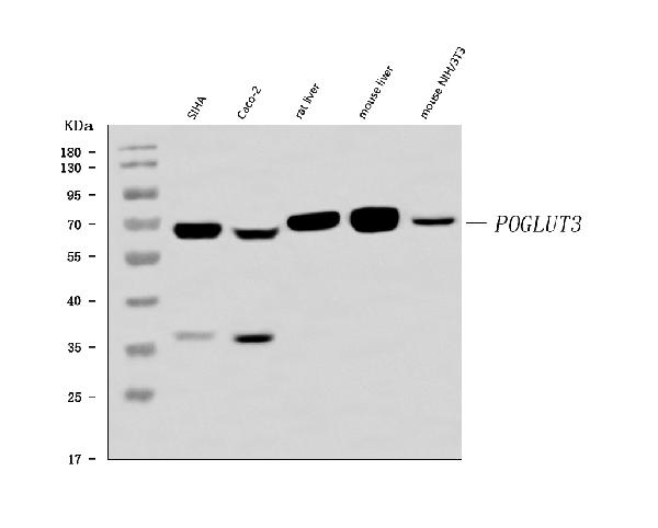
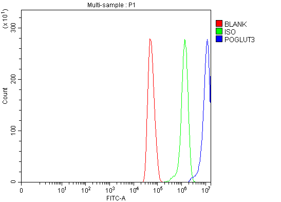
(Figure 3. Flow Cytometry analysis of U251 cells using anti-POGLUT3 antibody (A15507-1).Overlay histogram showing U251 cells stained with A15507-1 (Blue line). The cells were blocked with 10% normal goat serum. And then incubated with rabbit anti-POGLUT3 Antibody (A15507-1, 1 ug/1x10^6 cells) for 30 min at 20 degree C. DyLight488 conjugated goat anti-rabbit IgG was used as secondary antibody for 30 minutes at 20 degree C. Isotype control antibody (Green line) was rabbit IgG (1 ug/1x10^6) used under the same conditions. Unlabelled sample (Red line) was also used as a control.)
FCM/FACS (Flow Cytometry)
(Figure 3. Flow Cytometry analysis of U251 cells using anti-POGLUT3 antibody (A15507-1).Overlay histogram showing U251 cells stained with A15507-1 (Blue line). The cells were blocked with 10% normal goat serum. And then incubated with rabbit anti-POGLUT3 Antibody (A15507-1, 1 ug/1x10^6 cells) for 30 min at 20 degree C. DyLight488 conjugated goat anti-rabbit IgG was used as secondary antibody for 30 minutes at 20 degree C. Isotype control antibody (Green line) was rabbit IgG (1 ug/1x10^6) used under the same conditions. Unlabelled sample (Red line) was also used as a control.)
ERp72/PDIA4, Polyclonal Antibody (Cat# AAA126490)
Full Name
Anti-ERp72/PDIA4 Antibody Picoband
Gene Names
PDIA4; ERP70; ERP72; ERp-72
Reactivity
Human, Mouse, Rat
Applications
Flow Cytometry, Western Blot
Purity
Immunogen affinity purified.
Pricing
 FCM/FACS (Flow Cytometry)
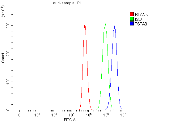
(Figure 3. Flow Cytometry analysis of U87 cells using anti-TSTA3/GFUS antibody (AAA126565).Overlay histogram showing U87 cells stained with AAA126565 (Blue line). The cells were blocked with 10% normal goat serum. And then incubated with rabbit anti-TSTA3/GFUS Antibody (AAA126565, 1 ug/1x10^6 cells) for 30 min at 20 degree C. DyLight488 conjugated goat anti-rabbit IgG was used as secondary antibody for 30 minutes at 20 degree C. Isotype control antibody (Green line) was rabbit IgG (1 ug/1x10^6) used under the same conditions. Unlabelled sample (Red line) was also used as a control.)
FCM/FACS (Flow Cytometry)
(Figure 3. Flow Cytometry analysis of U87 cells using anti-TSTA3/GFUS antibody (AAA126565).Overlay histogram showing U87 cells stained with AAA126565 (Blue line). The cells were blocked with 10% normal goat serum. And then incubated with rabbit anti-TSTA3/GFUS Antibody (AAA126565, 1 ug/1x10^6 cells) for 30 min at 20 degree C. DyLight488 conjugated goat anti-rabbit IgG was used as secondary antibody for 30 minutes at 20 degree C. Isotype control antibody (Green line) was rabbit IgG (1 ug/1x10^6) used under the same conditions. Unlabelled sample (Red line) was also used as a control.)
TSTA3/GFUS, Polyclonal Antibody (Cat# AAA126565)
Full Name
Anti-TSTA3/GFUS Antibody Picoband
Gene Names
TSTA3; FX; P35B; SDR4E1
Reactivity
Human, Mouse, Rat
Applications
Flow Cytometry, Immunofluorescence, Immunocytochemistry, Western Blot
Purity
Immunogen affinity purified.
Pricing
 IF (Immunofluorescence)
(Figure 5. IF analysis of MDH2 using anti-MDH2 antibody (AAA126941).MDH2 was detected in an immunocytochemical section of HeLa cells. Enzyme antigen retrieval was performed using IHC enzyme antigen retrieval reagent (AR0022) for 15 mins. The cells were blocked with 10% goat serum. And then incubated with 5 ug/mL mouse anti-MDH2 Antibody (AAA126941) overnight at 4 degree C. Cy3 Conjugated Goat Anti-Mouse IgG (BA1031) was used as secondary antibody at 1:100 dilution and incubated for 30 minutes at 37 degree C. The section was counterstained with DAPI. Visualize using a fluorescence microscope and filter sets appropriate for the label used.)
IF (Immunofluorescence)
(Figure 5. IF analysis of MDH2 using anti-MDH2 antibody (AAA126941).MDH2 was detected in an immunocytochemical section of HeLa cells. Enzyme antigen retrieval was performed using IHC enzyme antigen retrieval reagent (AR0022) for 15 mins. The cells were blocked with 10% goat serum. And then incubated with 5 ug/mL mouse anti-MDH2 Antibody (AAA126941) overnight at 4 degree C. Cy3 Conjugated Goat Anti-Mouse IgG (BA1031) was used as secondary antibody at 1:100 dilution and incubated for 30 minutes at 37 degree C. The section was counterstained with DAPI. Visualize using a fluorescence microscope and filter sets appropriate for the label used.)
MDH2, Monoclonal Antibody (Cat# AAA126941)
Full Name
Anti-MDH2 Antibody Picoband (monoclonal, 5D8C1)
Gene Names
MDH2; MDH; MOR1; M-MDH; MGC:3559
Reactivity
Human
Applications
Immunofluorescence, Immunocytochemistry, Immunohistochemistry, Western Blot
Purity
Immunogen affinity purified.
Pricing
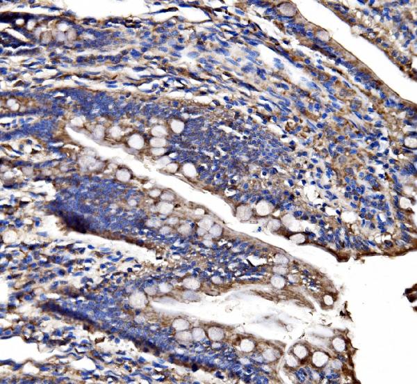
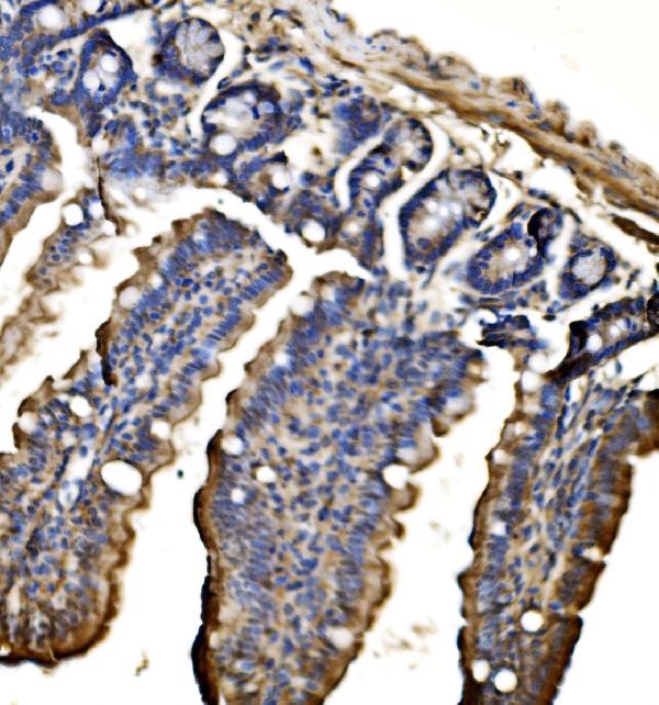
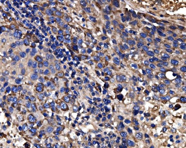
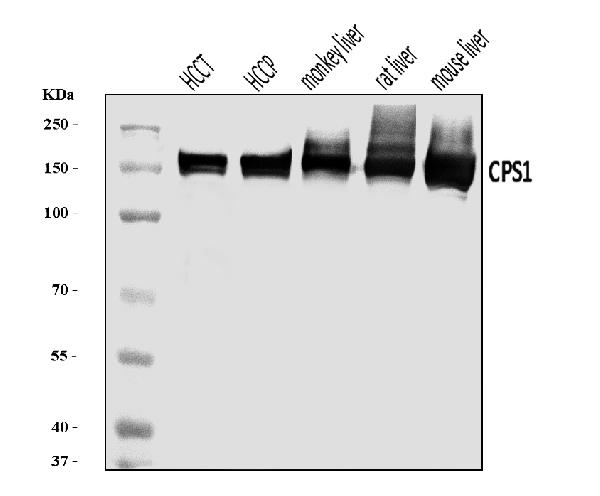
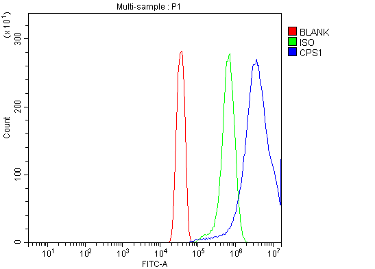 FCM/FACS (Flow Cytometry)
(Figure 5. Flow Cytometry analysis of HeLa cells using anti-CPS1 antibody (AAA126131).Overlay histogram showing HeLa cells stained with AAA126131 (Blue line). The cells were blocked with 10% normal goat serum. And then incubated with rabbit anti-CPS1 Antibody (AAA126131, 1 ug/1x10^6 cells) for 30 min at 20 degree C. DyLight488 conjugated goat anti-rabbit IgG was used as secondary antibody for 30 minutes at 20 degree C. Isotype control antibody (Green line) was rabbit IgG (1 ug/1x10^6) used under the same conditions. Unlabelled sample (Red line) was also used as a control.)
FCM/FACS (Flow Cytometry)
(Figure 5. Flow Cytometry analysis of HeLa cells using anti-CPS1 antibody (AAA126131).Overlay histogram showing HeLa cells stained with AAA126131 (Blue line). The cells were blocked with 10% normal goat serum. And then incubated with rabbit anti-CPS1 Antibody (AAA126131, 1 ug/1x10^6 cells) for 30 min at 20 degree C. DyLight488 conjugated goat anti-rabbit IgG was used as secondary antibody for 30 minutes at 20 degree C. Isotype control antibody (Green line) was rabbit IgG (1 ug/1x10^6) used under the same conditions. Unlabelled sample (Red line) was also used as a control.)
CPS1, Polyclonal Antibody (Cat# AAA126131)
Full Name
Anti-CPS1 Antibody Picoband
Gene Names
CPS1; PHN; CPSASE1
Reactivity
Human, Mouse, Rat, Monkey
Applications
Flow Cytometry, Immunohistochemistry, Western Blot
Purity
Immunogen affinity purified.
Pricing