
Filters
Clonality
Type
Reactivity
Gene Name
Isotype
Host
Application
Clone
10000 results for "Recombinant Proteins" - showing 7700-7750
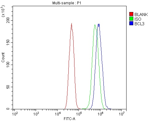 FCM/FACS (Flow Cytometry)
(Figure 2. Flow Cytometry analysis of U251 cells using anti-BCL3 antibody (AAA125669).Overlay histogram showing U251 cells stained with AAA125669 (Blue line). The cells were blocked with 10% normal goat serum. And then incubated with rabbit anti-BCL3 Antibody (AAA125669, 1μg/1x106 cells) for 30 min at 20 degree C. DyLight®488 conjugated goat anti-rabbit IgG (5-10μg/1x106 cells) was used as secondary antibody for 30 minutes at 20 degree C. Isotype control antibody (Green line) was rabbit IgG (1μg/1x106) used under the same conditions. Unlabelled sample (Red line) was also used as a control.)
FCM/FACS (Flow Cytometry)
(Figure 2. Flow Cytometry analysis of U251 cells using anti-BCL3 antibody (AAA125669).Overlay histogram showing U251 cells stained with AAA125669 (Blue line). The cells were blocked with 10% normal goat serum. And then incubated with rabbit anti-BCL3 Antibody (AAA125669, 1μg/1x106 cells) for 30 min at 20 degree C. DyLight®488 conjugated goat anti-rabbit IgG (5-10μg/1x106 cells) was used as secondary antibody for 30 minutes at 20 degree C. Isotype control antibody (Green line) was rabbit IgG (1μg/1x106) used under the same conditions. Unlabelled sample (Red line) was also used as a control.)
BCL3, Polyclonal Antibody (Cat# AAA125669)
Full Name
Anti-BCL3 Antibody
Gene Names
BCL3; BCL4; D19S37
Reactivity
Human, Mouse, Rat
Applications
Direct ELISA, Flow Cytometry, Western Blot
Purity
Immunogen affinity purified.
Pricing
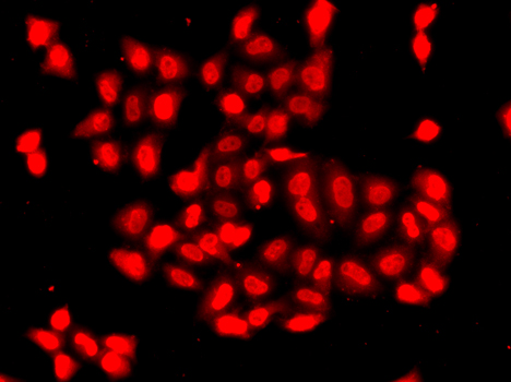 IF (Immunofluorescence)
(Immunofluorescence analysis of A549 cells using CAMK1D.)
IF (Immunofluorescence)
(Immunofluorescence analysis of A549 cells using CAMK1D.)
CAMK1D, Polyclonal Antibody (Cat# AAA314055)
Full Name
CAMK1D Rabbit Polyclonal Antibody
Gene Names
CAMK1D; CKLiK; CaM-K1; CaMKID
Reactivity
Human, Mouse, Rat
Applications
Immunofluorescence, Immunohistochemistry, Western Blot
Purity
Affinity purification
Pricing
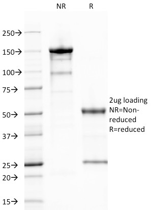
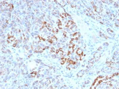
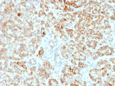
 Application Data
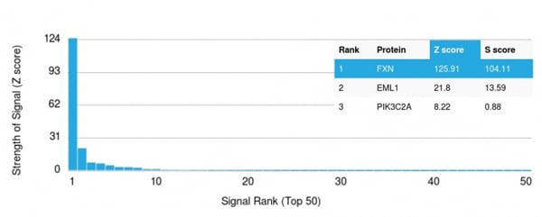
(Analysis of Protein Array containing more than 19,000 full-length human proteins using Frataxin Mouse Monoclonal Antibody (FXN/2124). Z- and S- Score: The Z-score represents the strength of a signal that a monoclonal antibody (MAb) (in combination with a fluorescently-tagged anti-IgG secondary antibody) produces when binding to a particular protein on the HuProtTM array. Z-scores are described in units of standard deviations (SD's) above the mean value of all signals generated on that array. If targets on HuProtTM are arranged in descending order of the Z-score, the S-score is the difference (also in units of SD's) between the Z-score. S-score therefore represents the relative target specificity of a MAb to its intended target. A MAb is considered to specific to its intended target, if the MAb has an S-score of at least 2.5. For example, if a MAb binds to protein X with a Z-score of 43 and to protein Y with a Z-score of 14, then the S-score for the binding of that MAb to protein X is equal to 29.)
Application Data
(Analysis of Protein Array containing more than 19,000 full-length human proteins using Frataxin Mouse Monoclonal Antibody (FXN/2124). Z- and S- Score: The Z-score represents the strength of a signal that a monoclonal antibody (MAb) (in combination with a fluorescently-tagged anti-IgG secondary antibody) produces when binding to a particular protein on the HuProtTM array. Z-scores are described in units of standard deviations (SD's) above the mean value of all signals generated on that array. If targets on HuProtTM are arranged in descending order of the Z-score, the S-score is the difference (also in units of SD's) between the Z-score. S-score therefore represents the relative target specificity of a MAb to its intended target. A MAb is considered to specific to its intended target, if the MAb has an S-score of at least 2.5. For example, if a MAb binds to protein X with a Z-score of 43 and to protein Y with a Z-score of 14, then the S-score for the binding of that MAb to protein X is equal to 29.)
Frataxin, Monoclonal Antibody (Cat# AAA214743)
Full Name
Frataxin (Marker of Friedreich Ataxia)
Gene Names
FXN; FA; X25; CyaY; FARR; FRDA
Reactivity
Human. Others not known.
Applications
Immunohistochemistry
Purity
Purified Ab with BSA and Azide at 200ug/ml OR Purified Ab WITHOUT BSA and Azide at 1.0mg/ml
Pricing
 Application Data
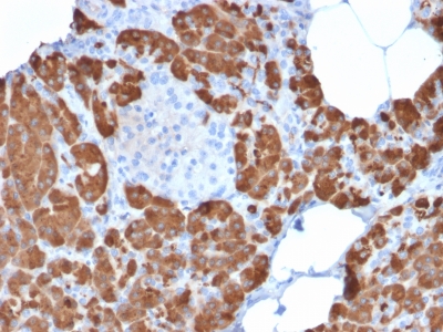
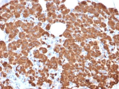
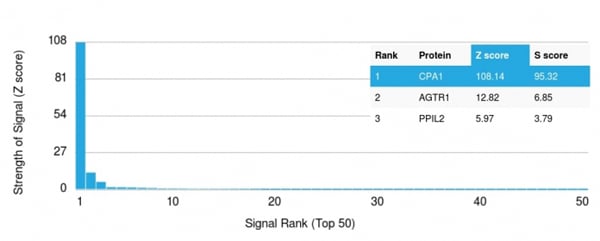
(Analysis of Protein Array containing more than 19,000 full-length human proteins using Carboxypeptidase A1/CPA1 Mouse Monoclonal Antibody (CPA1/2711). Z- and S- Score: The Z-score represents the strength of a signal that a monoclonal antibody (MAb) (in combination with a fluorescently-tagged anti-IgG secondary antibody) produces when binding to a particular protein on the HuProtTM array. Z-scores are described in units of standard deviations (SD's) above the mean value of all signals generated on that array. If targets on HuProtTM are arranged in descending order of the Z-score, the S-score is the difference (also in units of SD's) between the Z-score. S-score therefore represents the relative target specificity of a MAb to its intended target. A MAb is considered to specific to its intended target, if the MAb has an S-score of at least 2.5. For example, if a MAb binds to protein X with a Z-score of 43 and to protein Y with a Z-score of 14, then the S-score for the binding of that MAb to protein X is equal to 29.)
Application Data
(Analysis of Protein Array containing more than 19,000 full-length human proteins using Carboxypeptidase A1/CPA1 Mouse Monoclonal Antibody (CPA1/2711). Z- and S- Score: The Z-score represents the strength of a signal that a monoclonal antibody (MAb) (in combination with a fluorescently-tagged anti-IgG secondary antibody) produces when binding to a particular protein on the HuProtTM array. Z-scores are described in units of standard deviations (SD's) above the mean value of all signals generated on that array. If targets on HuProtTM are arranged in descending order of the Z-score, the S-score is the difference (also in units of SD's) between the Z-score. S-score therefore represents the relative target specificity of a MAb to its intended target. A MAb is considered to specific to its intended target, if the MAb has an S-score of at least 2.5. For example, if a MAb binds to protein X with a Z-score of 43 and to protein Y with a Z-score of 14, then the S-score for the binding of that MAb to protein X is equal to 29.)
Carboxypeptidase A1/CPA1, Monoclonal Antibody (Cat# AAA214723)
Full Name
Carboxypeptidase A1/CPA1 (Pancreatic Cancer Marker)
Gene Names
CPA1; CPA
Reactivity
Human. Others not known.
Applications
Immunohistochemistry
Purity
Purified Ab with BSA and Azide at 200ug/ml OR Purified Ab WITHOUT BSA and Azide at 1.0mg/ml
Pricing
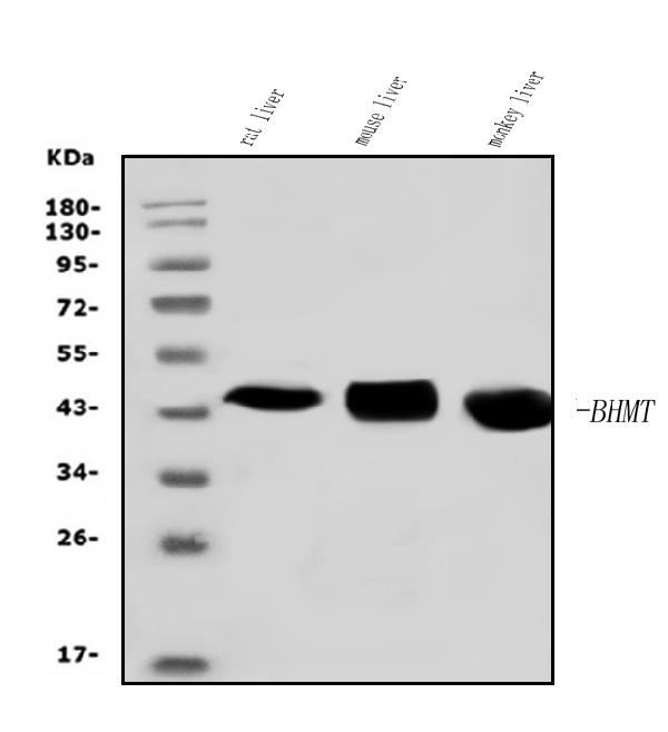
 FCM/FACS (Flow Cytometry)
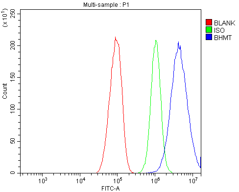
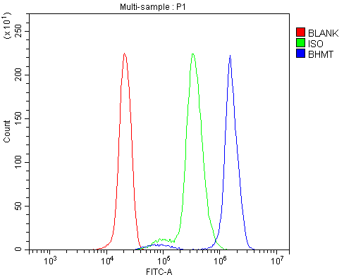
(Figure 3. Flow Cytometry analysis of U937 cells using anti-BHMT antibody (AAA125778).Overlay histogram showing U937 cells stained with AAA125778 (Blue line). The cells were blocked with 10% normal goat serum. And then incubated with rabbit anti-BHMT Antibody (AAA125778, 1μg/1x106 cells) for 30 min at 20 degree C. DyLight®488 conjugated goat anti-rabbit IgG (5-10μg/1x106 cells) was used as secondary antibody for 30 minutes at 20 degree C. Isotype control antibody (Green line) was rabbit IgG (1μg/1x106) used under the same conditions. Unlabelled sample (Red line) was also used as a control.)
FCM/FACS (Flow Cytometry)
(Figure 3. Flow Cytometry analysis of U937 cells using anti-BHMT antibody (AAA125778).Overlay histogram showing U937 cells stained with AAA125778 (Blue line). The cells were blocked with 10% normal goat serum. And then incubated with rabbit anti-BHMT Antibody (AAA125778, 1μg/1x106 cells) for 30 min at 20 degree C. DyLight®488 conjugated goat anti-rabbit IgG (5-10μg/1x106 cells) was used as secondary antibody for 30 minutes at 20 degree C. Isotype control antibody (Green line) was rabbit IgG (1μg/1x106) used under the same conditions. Unlabelled sample (Red line) was also used as a control.)
BHMT, Polyclonal Antibody (Cat# AAA125778)
Full Name
Anti-BHMT Antibody
Gene Names
BHMT; BHMT1; HEL-S-61p
Reactivity
Human, Mouse, Rat, Monkey
Applications
Direct ELISA, Flow Cytometry, Western Blot
Purity
Immunogen affinity purified.
Pricing
 Application Data
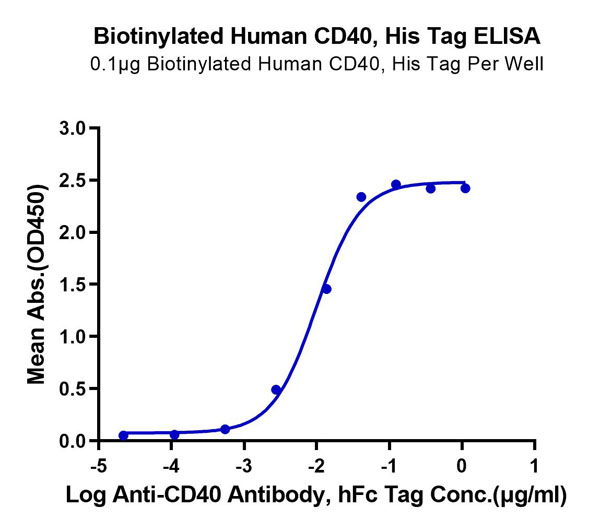
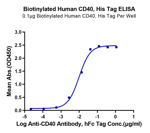
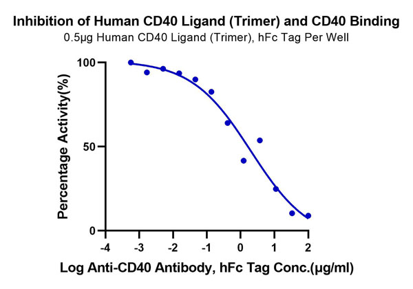
(Biotinylated Human CD40/TNFRSF5 Protein Blocking DataSerial dilutions of Anti-CD40 Antibody were added into Biotinylated Human CD40, His Tag : Human CD40 Ligand (Trimer) , hFc Tag binding reactioins. The half maximal inhibitiory concentration (IC50) is 2.0ug/ml.)
Application Data
(Biotinylated Human CD40/TNFRSF5 Protein Blocking DataSerial dilutions of Anti-CD40 Antibody were added into Biotinylated Human CD40, His Tag : Human CD40 Ligand (Trimer) , hFc Tag binding reactioins. The half maximal inhibitiory concentration (IC50) is 2.0ug/ml.)
CD40/TNFRSF5, Recombinant Protein (Cat# AAA76149)
Full Name
Biotinylated Human CD40/TNFRSF5 Protein Recombinant
Gene Names
CD40; p50; Bp50; CDW40; TNFRSF5
Purity
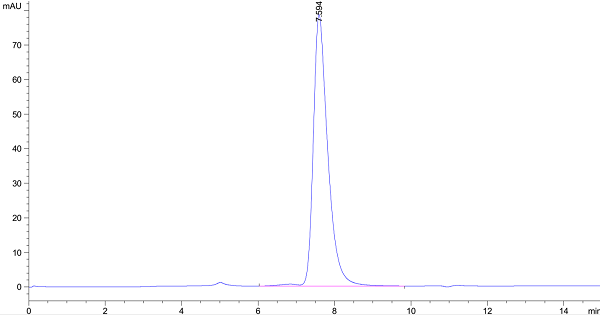
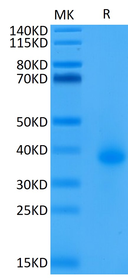
>95%, as determined by SDS-PAGE and HPLC
Pricing
 FCM/FACS (Flow Cytometry)
(Figure 2. Flow Cytometry analysis of C2C12 cells using anti-Bim/Bcl2l11 antibody (AAA127065).Overlay histogram showing C2C12 cells stained with AAA127065 (Blue line). To facilitate intracellular staining, cells were fixed with 4% paraformaldehyde and permeabilized with permeabilization buffer. The cells were blocked with 10% normal goat serum. And then incubated with rabbit anti-Bim/Bcl2l11 Antibody (AAA127065, 1ug/1x106 cells) for 30 min at 20 degree C. DyLight488 conjugated goat anti-rabbit IgG was used as secondary antibody for 30 minutes at 20 degree C. Isotype control antibody (Green line) was rabbit IgG (1ug/1x106) used under the same conditions. Unlabelled sample (Red line) was also used as a control.)
FCM/FACS (Flow Cytometry)
(Figure 2. Flow Cytometry analysis of C2C12 cells using anti-Bim/Bcl2l11 antibody (AAA127065).Overlay histogram showing C2C12 cells stained with AAA127065 (Blue line). To facilitate intracellular staining, cells were fixed with 4% paraformaldehyde and permeabilized with permeabilization buffer. The cells were blocked with 10% normal goat serum. And then incubated with rabbit anti-Bim/Bcl2l11 Antibody (AAA127065, 1ug/1x106 cells) for 30 min at 20 degree C. DyLight488 conjugated goat anti-rabbit IgG was used as secondary antibody for 30 minutes at 20 degree C. Isotype control antibody (Green line) was rabbit IgG (1ug/1x106) used under the same conditions. Unlabelled sample (Red line) was also used as a control.)
Bim/Bcl2l11, Polyclonal Antibody (Cat# AAA127065)
Full Name
Anti-Bim/Bcl2l11 Antibody Picoband
Gene Names
Bcl2l11; Bim; Bod; bcl2-L-11; 1500006F24Rik
Reactivity
Mouse
Applications
Flow Cytometry, Western Blot
Purity
Immunogen affinity purified.
Pricing
 Application Data
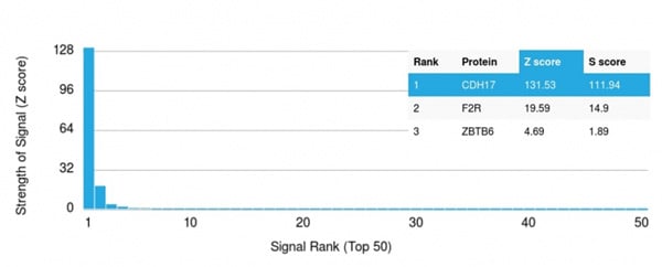
(Analysis of Protein Array containing more than 19,000 full-length human proteins using Cadherin 17 (CDH17) Mouse Monoclonal Antibody (CDH17/2617). Z- and S- Score: The Z-score represents the strength of a signal that a monoclonal antibody (Monoclonal Antibody) (in combination with a fluorescently-tagged anti-IgG secondary antibody) produces when binding to a particular protein on the HuProtTM array. Z-scores are described in units of standard deviations (SD’s) above the mean value of all signals generated on that array. If targets on HuProtTM are arranged in descending order of the Z-score, the S-score is the difference (also in units of SD’s) between the Z-score. S-score therefore represents the relative target specificity of a Monoclonal Antibody to its intended target. A Monoclonal Antibody is considered to specific to its intended target, if the Monoclonal Antibody has an S-score of at least 2.5. For example, if a Monoclonal Antibody binds to protein X with a Z-score of 43 and to protein Y with a Z-score of 14, then the S-score for the binding of that Monoclonal Antibody to protein X is equal to 29.)
Application Data
(Analysis of Protein Array containing more than 19,000 full-length human proteins using Cadherin 17 (CDH17) Mouse Monoclonal Antibody (CDH17/2617). Z- and S- Score: The Z-score represents the strength of a signal that a monoclonal antibody (Monoclonal Antibody) (in combination with a fluorescently-tagged anti-IgG secondary antibody) produces when binding to a particular protein on the HuProtTM array. Z-scores are described in units of standard deviations (SD’s) above the mean value of all signals generated on that array. If targets on HuProtTM are arranged in descending order of the Z-score, the S-score is the difference (also in units of SD’s) between the Z-score. S-score therefore represents the relative target specificity of a Monoclonal Antibody to its intended target. A Monoclonal Antibody is considered to specific to its intended target, if the Monoclonal Antibody has an S-score of at least 2.5. For example, if a Monoclonal Antibody binds to protein X with a Z-score of 43 and to protein Y with a Z-score of 14, then the S-score for the binding of that Monoclonal Antibody to protein X is equal to 29.)
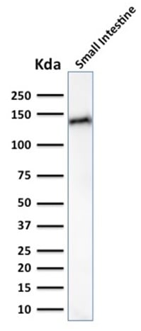
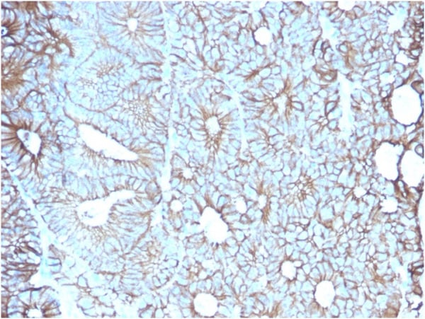
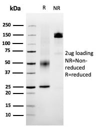
Cadherin 17/LI Cadherin, Monoclonal Antibody (Cat# AAA214874)
Full Name
Cadherin 17/LI Cadherin (Liver-Intestine Marker)
Gene Names
CDH17; HPT1; CDH16; HPT-1
Reactivity
Human
Applications
Immunohistochemistry, Western Blot
Purity
Purified Ab with BSA and Azide at 200ug/ml OR Purified Ab WITHOUT BSA at 1.0mg/ml
Pricing
 Application Data
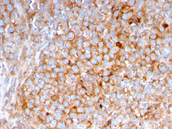
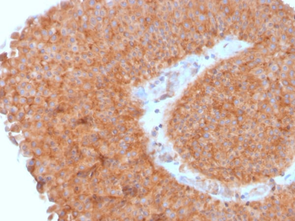
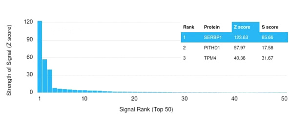
(Analysis of Protein Array containing more than 19,000 full-length human proteins using PAI-RBP1 Mouse Monoclonal Antibody (SERBP1/3495). Z- and S- Score: The Z-score represents the strength of a signal that a monoclonal antibody (MAb) (in combination with a fluorescently-tagged anti-IgG secondary antibody) produces when binding to a particular protein on the HuProtTM array. Z-scores are described in units of standard deviations (SD's) above the mean value of all signals generated on that array. If targets on HuProtTM are arranged in descending order of the Z-score, the S-score is the difference (also in units of SD's) between the Z-score. S-score therefore represents the relative target specificity of a MAb to its intended target. A MAb is considered to specific to its intended target, if the MAb has an S-score of at least 2.5. For example, if a MAb binds to protein X with a Z-score of 43 and to protein Y with a Z-score of 14, then the S-score for the binding of that MAb to protein X is equal to 29.)
Application Data
(Analysis of Protein Array containing more than 19,000 full-length human proteins using PAI-RBP1 Mouse Monoclonal Antibody (SERBP1/3495). Z- and S- Score: The Z-score represents the strength of a signal that a monoclonal antibody (MAb) (in combination with a fluorescently-tagged anti-IgG secondary antibody) produces when binding to a particular protein on the HuProtTM array. Z-scores are described in units of standard deviations (SD's) above the mean value of all signals generated on that array. If targets on HuProtTM are arranged in descending order of the Z-score, the S-score is the difference (also in units of SD's) between the Z-score. S-score therefore represents the relative target specificity of a MAb to its intended target. A MAb is considered to specific to its intended target, if the MAb has an S-score of at least 2.5. For example, if a MAb binds to protein X with a Z-score of 43 and to protein Y with a Z-score of 14, then the S-score for the binding of that MAb to protein X is equal to 29.)
PAI-RBP1/SERBP1/SERPINE1, Monoclonal Antibody (Cat# AAA214985)
Full Name
PAI-RBP1/SERBP1/SERPINE1
Gene Names
SERBP1; CGI-55; CHD3IP; HABP4L; PAIRBP1; PAI-RBP1
Reactivity
Human
Applications
Immunohistochemistry
Purity
Purified Ab with BSA and Azide at 200ug/ml OR Purified Ab WITHOUT BSA and Azide at 1.0mg/ml
Pricing

 FCM/FACS (Flow Cytometry)
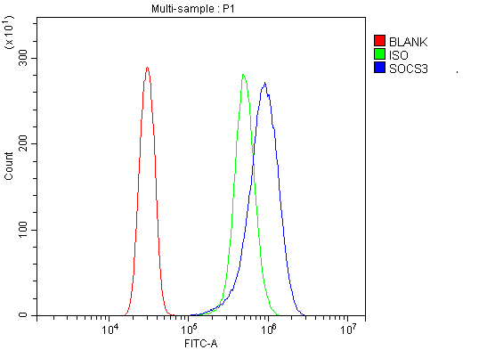
(Figure 2. Flow Cytometry analysis of SiHa cells using anti-SOCS3 antibody (AAA125532).Overlay histogram showing SiHa cells stained with AAA125532 (Blue line). The cells were blocked with 10% normal goat serum. And then incubated with rabbit anti-SOCS3 Antibody (AAA125532, 1μg/1x106 cells) for 30 min at 20 degree C. DyLight®488 conjugated goat anti-rabbit IgG (5-10μg/1x106 cells) was used as secondary antibody for 30 minutes at 20 degree C. Isotype control antibody (Green line) was rabbit IgG (1μg/1x106) used under the same conditions. Unlabelled sample (Red line) was also used as a control.)
FCM/FACS (Flow Cytometry)
(Figure 2. Flow Cytometry analysis of SiHa cells using anti-SOCS3 antibody (AAA125532).Overlay histogram showing SiHa cells stained with AAA125532 (Blue line). The cells were blocked with 10% normal goat serum. And then incubated with rabbit anti-SOCS3 Antibody (AAA125532, 1μg/1x106 cells) for 30 min at 20 degree C. DyLight®488 conjugated goat anti-rabbit IgG (5-10μg/1x106 cells) was used as secondary antibody for 30 minutes at 20 degree C. Isotype control antibody (Green line) was rabbit IgG (1μg/1x106) used under the same conditions. Unlabelled sample (Red line) was also used as a control.)
SOCS3, Polyclonal Antibody (Cat# AAA125532)
Full Name
Anti-SOCS3 Antibody
Gene Names
SOCS3; CIS3; SSI3; ATOD4; Cish3; SSI-3; SOCS-3
Reactivity
Human, Mouse, Rat
Applications
Direct ELISA, Flow Cytometry, Western Blot
Purity
Immunogen affinity purified.
Pricing
 IHC (Immunohiostchemistry)
(Figure 3. IHC analysis of Alpha 1 microglobulin using anti-Alpha 1 microglobulin antibody.Alpha 1 microglobulin was detected in paraffin-embedded section of human liver cancer tissue. Heat mediated antigen retrieval was performed in citrate buffer (pH6, epitope retrieval solution) for 20 mins. The tissue section was blocked with 10% goat serum. The tissue section was then incubated with 2ug/ml rabbit anti-Alpha 1 microglobulin antibody.Alpha 1 microglobulin was detected in paraffin-embedded section of human liver cancer tissue. Heat mediated antigen retrieval was performed in citrate buffer (pH6, epitope retrieval solution) for 20 mins. The tissue section was blocked with 10% goat serum. The tissue section was then incubated with 2ug/ml rabbit anti-Alpha 1 microglobulin Antibody overnight at 4 degree C. Biotinylated goat anti-rabbit IgG was used as secondary antibody and incubated for 30 minutes at 37 degree C. The tissue section was developed using Strepavidin-Biotin-Complex (SABC) with DAB as the chromogen.)
IHC (Immunohiostchemistry)
(Figure 3. IHC analysis of Alpha 1 microglobulin using anti-Alpha 1 microglobulin antibody.Alpha 1 microglobulin was detected in paraffin-embedded section of human liver cancer tissue. Heat mediated antigen retrieval was performed in citrate buffer (pH6, epitope retrieval solution) for 20 mins. The tissue section was blocked with 10% goat serum. The tissue section was then incubated with 2ug/ml rabbit anti-Alpha 1 microglobulin antibody.Alpha 1 microglobulin was detected in paraffin-embedded section of human liver cancer tissue. Heat mediated antigen retrieval was performed in citrate buffer (pH6, epitope retrieval solution) for 20 mins. The tissue section was blocked with 10% goat serum. The tissue section was then incubated with 2ug/ml rabbit anti-Alpha 1 microglobulin Antibody overnight at 4 degree C. Biotinylated goat anti-rabbit IgG was used as secondary antibody and incubated for 30 minutes at 37 degree C. The tissue section was developed using Strepavidin-Biotin-Complex (SABC) with DAB as the chromogen.)
Alpha 1 microglobulin/AMBP, Polyclonal Antibody (Cat# AAA124941)
Full Name
Anti-Alpha 1 microglobulin/AMBP Antibody
Gene Names
AMBP; A1M; HCP; ITI; UTI; EDC1; HI30; ITIL; IATIL; ITILC
Reactivity
Human
Applications
Direct ELISA, Immunohistochemistry, Western Blot
Purity
Immunogen Affinity Purified
Pricing
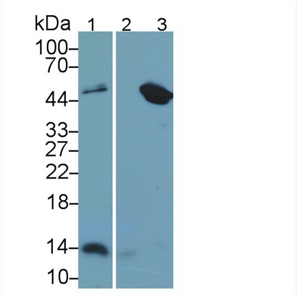 WB (Western Blot)
(Western Blot Sample: Lane1: Human Urine; Lane2: A431 cell lysate; Lane3: Raji cell lysate Primary Ab: 2ug/ml Mouse AntiHuman b2M Antibody Second Ab: 0.2ug/mL HRPLinked Caprine AntiMouse IgG Polyclonal Antibody (Catalog: SAA544Mu19))
WB (Western Blot)
(Western Blot Sample: Lane1: Human Urine; Lane2: A431 cell lysate; Lane3: Raji cell lysate Primary Ab: 2ug/ml Mouse AntiHuman b2M Antibody Second Ab: 0.2ug/mL HRPLinked Caprine AntiMouse IgG Polyclonal Antibody (Catalog: SAA544Mu19))
Beta2Microglobulin (b2M), Monoclonal Antibody (Cat# AAA151517)
Full Name
Monoclonal Antibody to Beta2Microglobulin (b2M)
Reactivity
Human, Pig
Applications
Immunoprecipitation, Immunocytochemistry, Immunohistochemistry, Western Blot
Purity
Protein A + Protein G affinity chromatography
Pricing
 FCM/FACS (Flow Cytometry)
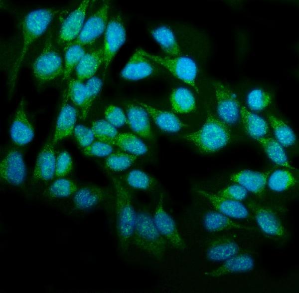
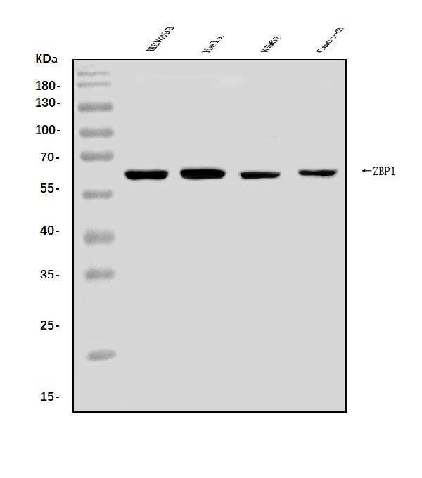
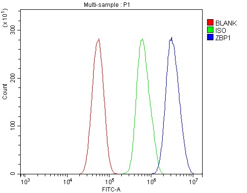
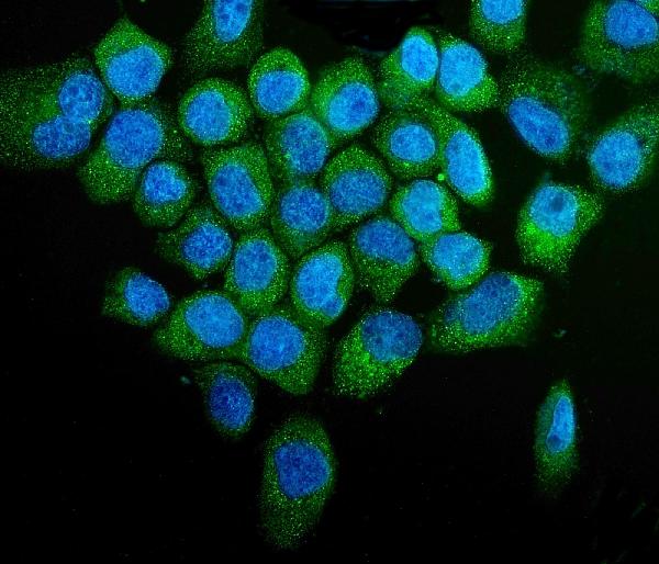
(Figure 3. Flow Cytometry analysis of U87 cells using anti-DAI/ZBP1 antibody (AAA125730).Overlay histogram showing U87 cells stained with AAA125730 (Blue line). The cells were blocked with 10% normal goat serum. And then incubated with rabbit anti-DAI/ZBP1 Antibody (AAA125730, 1μg/1x106 cells) for 30 min at 20 degree C. DyLight®488 conjugated goat anti-rabbit IgG (5-10μg/1x106 cells) was used as secondary antibody for 30 minutes at 20 degree C. Isotype control antibody (Green line) was rabbit IgG (1μg/1x106) used under the same conditions. Unlabelled sample (Red line) was also used as a control.)
FCM/FACS (Flow Cytometry)
(Figure 3. Flow Cytometry analysis of U87 cells using anti-DAI/ZBP1 antibody (AAA125730).Overlay histogram showing U87 cells stained with AAA125730 (Blue line). The cells were blocked with 10% normal goat serum. And then incubated with rabbit anti-DAI/ZBP1 Antibody (AAA125730, 1μg/1x106 cells) for 30 min at 20 degree C. DyLight®488 conjugated goat anti-rabbit IgG (5-10μg/1x106 cells) was used as secondary antibody for 30 minutes at 20 degree C. Isotype control antibody (Green line) was rabbit IgG (1μg/1x106) used under the same conditions. Unlabelled sample (Red line) was also used as a control.)
DAI/ZBP1, Polyclonal Antibody (Cat# AAA125730)
Full Name
Anti-DAI/ZBP1 Antibody
Gene Names
ZBP1; DAI; DLM1; DLM-1; C20orf183
Reactivity
Human
Applications
Direct ELISA, Flow Cytometry, Immunofluorescence, Immunocytochemistry, Western Blot
Purity
Immunogen affinity purified.
Pricing
 FCM/FACS (Flow Cytometry)
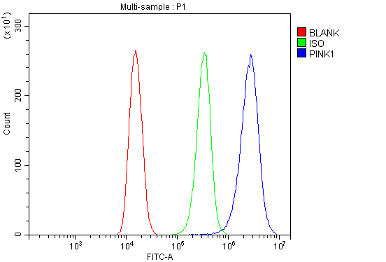
(Figure 3. Flow Cytometry analysis of 293T cells using anti-PINK1 antibody (AAA125528).Overlay histogram showing 293T cells stained with AAA125528 (Blue line). The cells were blocked with 10% normal goat serum. And then incubated with rabbit anti-PINK1 Antibody (AAA125528, 1μg/1x106 cells) for 30 min at 20 degree C. DyLight®488 conjugated goat anti-rabbit IgG (5-10μg/1x106 cells) was used as secondary antibody for 30 minutes at 20 degree C. Isotype control antibody (Green line) was rabbit IgG (1μg/1x106) used under the same conditions. Unlabelled sample (Red line) was also used as a control.)
FCM/FACS (Flow Cytometry)
(Figure 3. Flow Cytometry analysis of 293T cells using anti-PINK1 antibody (AAA125528).Overlay histogram showing 293T cells stained with AAA125528 (Blue line). The cells were blocked with 10% normal goat serum. And then incubated with rabbit anti-PINK1 Antibody (AAA125528, 1μg/1x106 cells) for 30 min at 20 degree C. DyLight®488 conjugated goat anti-rabbit IgG (5-10μg/1x106 cells) was used as secondary antibody for 30 minutes at 20 degree C. Isotype control antibody (Green line) was rabbit IgG (1μg/1x106) used under the same conditions. Unlabelled sample (Red line) was also used as a control.)
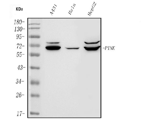
PINK1, Polyclonal Antibody (Cat# AAA125528)
Full Name
Anti-PINK1 Antibody
Gene Names
PINK1; BRPK; PARK6
Reactivity
Human
Applications
Direct ELISA, Flow Cytometry, Immunofluorescence, Immunocytochemistry, Western Blot
Purity
Immunogen affinity purified.
Pricing
 IHC (Immunohiostchemistry)
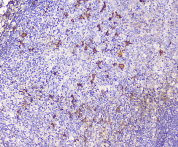
(Figure 3. IHC analysis of CXCL12 using anti-CXCL12 antibody.CXCL12 was detected in paraffin-embedded section of mouse spleen tissue. Heat mediated antigen retrieval was performed in citrate buffer (pH6, epitope retrieval solution) for 20 mins. The tissue section was blocked with 10% goat serum. The tissue section was then incubated with 1ug/ml rabbit anti-CXCL12 antibody.CXCL12 was detected in paraffin-embedded section of mouse spleen tissue. Heat mediated antigen retrieval was performed in citrate buffer (pH6, epitope retrieval solution) for 20 mins. The tissue section was blocked with 10% goat serum. The tissue section was then incubated with 1ug/ml rabbit anti-CXCL12 antibody.CXCL12 was detected in paraffin-embedded section of human tonsil tissue. Heat mediated antigen retrieval was performed in citrate buffer (pH6, epitope retrieval solution) for 20 mins. The tissue section was blocked with 10% goat serum. The tissue section was then incubated with 1ug/ml rabbit anti-CXCL12 Antibody overnight at 4 degree C. Biotinylated goat anti-rabbit IgG was used as secondary antibody and incubated for 30 minutes at 37 degree C. The tissue section was developed using Strepavidin-Biotin-Complex (SABC) with DAB as the chromogen.)
IHC (Immunohiostchemistry)
(Figure 3. IHC analysis of CXCL12 using anti-CXCL12 antibody.CXCL12 was detected in paraffin-embedded section of mouse spleen tissue. Heat mediated antigen retrieval was performed in citrate buffer (pH6, epitope retrieval solution) for 20 mins. The tissue section was blocked with 10% goat serum. The tissue section was then incubated with 1ug/ml rabbit anti-CXCL12 antibody.CXCL12 was detected in paraffin-embedded section of mouse spleen tissue. Heat mediated antigen retrieval was performed in citrate buffer (pH6, epitope retrieval solution) for 20 mins. The tissue section was blocked with 10% goat serum. The tissue section was then incubated with 1ug/ml rabbit anti-CXCL12 antibody.CXCL12 was detected in paraffin-embedded section of human tonsil tissue. Heat mediated antigen retrieval was performed in citrate buffer (pH6, epitope retrieval solution) for 20 mins. The tissue section was blocked with 10% goat serum. The tissue section was then incubated with 1ug/ml rabbit anti-CXCL12 Antibody overnight at 4 degree C. Biotinylated goat anti-rabbit IgG was used as secondary antibody and incubated for 30 minutes at 37 degree C. The tissue section was developed using Strepavidin-Biotin-Complex (SABC) with DAB as the chromogen.)
CXCL12, Polyclonal Antibody (Cat# AAA124886)
Full Name
Anti-CXCL12 Antibody
Gene Names
CXCL12; IRH; PBSF; SDF1; TLSF; TPAR1; SCYB12
Reactivity
Human, Mouse, Rat
Applications
Direct ELISA, Immunohistochemistry, Western Blot
Purity
Immunogen Affinity Purified
Pricing
 Application Data
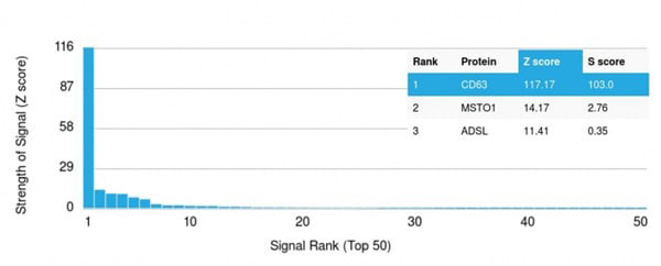
(Analysis of Protein Array containing >19,000 full-length human proteins using CD63 Mouse Monoclonal Antibody (LAMP3/2881) Z- and S- Score: The Z-score represents the strength of a signal that a monoclonal antibody (MAb) (in combination with a fluorescently-tagged anti-IgG secondary antibody) produces when binding to a particular protein on the HuProtTM array. Z-scores are described in units of standard deviations (SD's) above the mean value of all signals generated on that array. If targets on HuProtTM are arranged in descending order of the Z-score, the S-score is the difference (also in units of SD's) between the Z-score. S-score therefore represents the relative target specificity of a MAb to its intended target. A MAb is considered to specific to its intended target, if the MAb has an S-score of at least 2.5. For example, if a MAb binds to protein X with a Z-score of 43 and to protein Y with a Z-score of 14, then the S-score for the binding of that MAb to protein X is equal to 29.)
Application Data
(Analysis of Protein Array containing >19,000 full-length human proteins using CD63 Mouse Monoclonal Antibody (LAMP3/2881) Z- and S- Score: The Z-score represents the strength of a signal that a monoclonal antibody (MAb) (in combination with a fluorescently-tagged anti-IgG secondary antibody) produces when binding to a particular protein on the HuProtTM array. Z-scores are described in units of standard deviations (SD's) above the mean value of all signals generated on that array. If targets on HuProtTM are arranged in descending order of the Z-score, the S-score is the difference (also in units of SD's) between the Z-score. S-score therefore represents the relative target specificity of a MAb to its intended target. A MAb is considered to specific to its intended target, if the MAb has an S-score of at least 2.5. For example, if a MAb binds to protein X with a Z-score of 43 and to protein Y with a Z-score of 14, then the S-score for the binding of that MAb to protein X is equal to 29.)
CD63, Monoclonal Antibody (Cat# AAA215309)
Full Name
CD63 (Late Endosomes Marker)
Gene Names
CD63; MLA1; ME491; LAMP-3; OMA81H; TSPAN30
Reactivity
Human
Applications
Immunohistochemistry, Western Blot
Purity
Purified Ab with BSA and Azide at 200ug/ml OR Purified Ab WITHOUT BSA and Azide at 1.0mg/ml
Pricing
 FCM/FACS (Flow Cytometry)
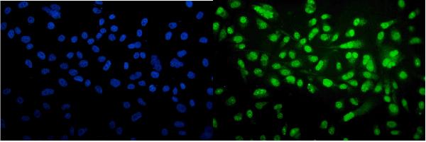
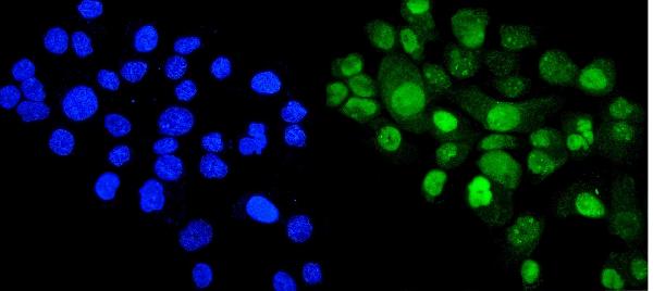
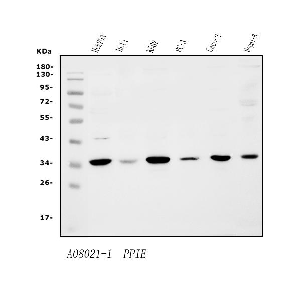
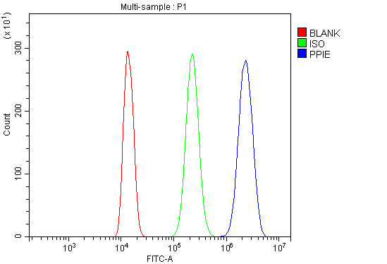
(Figure 4. Flow Cytometry analysis of THP-1 cells using anti-Cyclophilin E/PPIE antibody (AAA125797).Overlay histogram showing THP-1 cells stained with AAA125797 (Blue line). The cells were blocked with 10% normal goat serum. And then incubated with rabbit anti-Cyclophilin E/PPIE Antibody (AAA125797, 1μg/1x106 cells) for 30 min at 20 degree C. DyLight®488 conjugated goat anti-rabbit IgG (5-10μg/1x106 cells) was used as secondary antibody for 30 minutes at 20 degree C. Isotype control antibody (Green line) was rabbit IgG (1μg/1x106) used under the same conditions. Unlabelled sample (Red line) was also used as a control.)
FCM/FACS (Flow Cytometry)
(Figure 4. Flow Cytometry analysis of THP-1 cells using anti-Cyclophilin E/PPIE antibody (AAA125797).Overlay histogram showing THP-1 cells stained with AAA125797 (Blue line). The cells were blocked with 10% normal goat serum. And then incubated with rabbit anti-Cyclophilin E/PPIE Antibody (AAA125797, 1μg/1x106 cells) for 30 min at 20 degree C. DyLight®488 conjugated goat anti-rabbit IgG (5-10μg/1x106 cells) was used as secondary antibody for 30 minutes at 20 degree C. Isotype control antibody (Green line) was rabbit IgG (1μg/1x106) used under the same conditions. Unlabelled sample (Red line) was also used as a control.)
Cyclophilin E/PPIE, Polyclonal Antibody (Cat# AAA125797)
Full Name
Anti-Cyclophilin E/PPIE Antibody
Gene Names
PPIE; CYP33; CYP-33
Reactivity
Human, Mouse
Applications
Direct ELISA, Flow Cytometry, Immunofluorescence, Immunocytochemistry, Western Blot
Purity
Immunogen affinity purified.
Pricing
 FCM/FACS (Flow Cytometry)
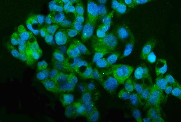
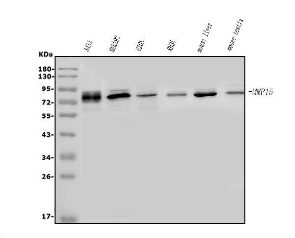
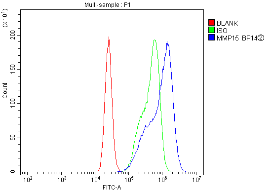
(Figure 3. Flow Cytometry analysis of CACO-2 cells using anti-MT2-MMP/MMP15 antibody (AAA125772).Overlay histogram showing CACO-2 cells stained with AAA125772 (Blue line). The cells were blocked with 10% normal goat serum. And then incubated with rabbit anti-MT2-MMP/MMP15 Antibody (AAA125772, 1μg/1x106 cells) for 30 min at 20 degree C. DyLight®488 conjugated goat anti-rabbit IgG (5-10μg/1x106 cells) was used as secondary antibody for 30 minutes at 20 degree C. Isotype control antibody (Green line) was rabbit IgG (1μg/1x106) used under the same conditions. Unlabelled sample (Red line) was also used as a control.)
FCM/FACS (Flow Cytometry)
(Figure 3. Flow Cytometry analysis of CACO-2 cells using anti-MT2-MMP/MMP15 antibody (AAA125772).Overlay histogram showing CACO-2 cells stained with AAA125772 (Blue line). The cells were blocked with 10% normal goat serum. And then incubated with rabbit anti-MT2-MMP/MMP15 Antibody (AAA125772, 1μg/1x106 cells) for 30 min at 20 degree C. DyLight®488 conjugated goat anti-rabbit IgG (5-10μg/1x106 cells) was used as secondary antibody for 30 minutes at 20 degree C. Isotype control antibody (Green line) was rabbit IgG (1μg/1x106) used under the same conditions. Unlabelled sample (Red line) was also used as a control.)
MT2-MMP/MMP15, Polyclonal Antibody (Cat# AAA125772)
Full Name
Anti-MT2-MMP/MMP15 Antibody
Gene Names
MMP15; MTMMP2; SMCP-2; MT2-MMP
Reactivity
Human, Mouse, Rat
Applications
Direct ELISA, Flow Cytometry, Immunofluorescence, Immunocytochemistry, Western Blot
Purity
Immunogen affinity purified.
Pricing
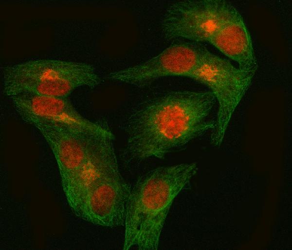
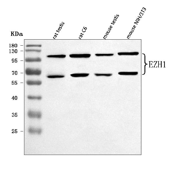
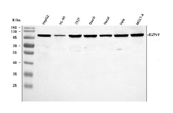
 FCM/FACS (Flow Cytometry)
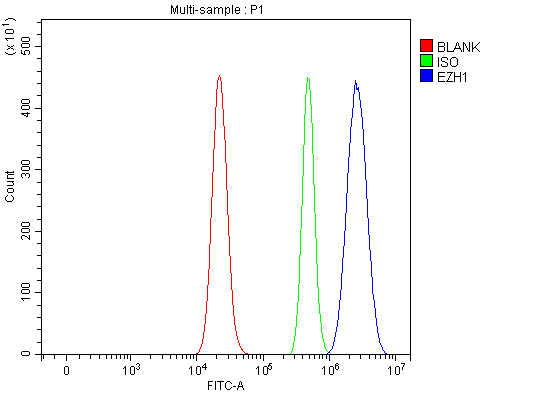
(Figure 4. Flow Cytometry analysis of RT4 cells using anti-EZH1 antibody (AAA127122).Overlay histogram showing RT4 cells stained with AAA127122 (Blue line). To facilitate intracellular staining, cells were fixed with 4% paraformaldehyde and permeabilized with permeabilization buffer. The cells were blocked with 10% normal goat serum. And then incubated with rabbit anti-EZH1 Antibody (AAA127122, 1ug/1x106 cells) for 30 min at 20 degree C. DyLight488 conjugated goat anti-rabbit IgG was used as secondary antibody for 30 minutes at 20 degree C. Isotype control antibody (Green line) was rabbit IgG (1ug/1x106) used under the same conditions. Unlabelled sample without incubation with primary antibody and secondary antibody (Red line) was used as a blank control.)
FCM/FACS (Flow Cytometry)
(Figure 4. Flow Cytometry analysis of RT4 cells using anti-EZH1 antibody (AAA127122).Overlay histogram showing RT4 cells stained with AAA127122 (Blue line). To facilitate intracellular staining, cells were fixed with 4% paraformaldehyde and permeabilized with permeabilization buffer. The cells were blocked with 10% normal goat serum. And then incubated with rabbit anti-EZH1 Antibody (AAA127122, 1ug/1x106 cells) for 30 min at 20 degree C. DyLight488 conjugated goat anti-rabbit IgG was used as secondary antibody for 30 minutes at 20 degree C. Isotype control antibody (Green line) was rabbit IgG (1ug/1x106) used under the same conditions. Unlabelled sample without incubation with primary antibody and secondary antibody (Red line) was used as a blank control.)
EZH1, Polyclonal Antibody (Cat# AAA127122)
Full Name
Anti-EZH1 Antibody Picoband
Gene Names
EZH1; KMT6B
Reactivity
Human, Mouse, Rat
Applications
Flow Cytometry, Immunofluorescence, Immunocytochemistry, Western Blot
Purity
Immunogen affinity purified.
Pricing
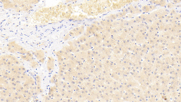
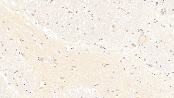 IHC (Immunohiostchemistry)
(DAB staining on IHCP;Sample: Human Cerebrum Tissue; Primary Ab: 30ug/ml Mouse AntiHuman bACE1 AntibodySecond Ab: 2ug/mL HRPLinked Caprine AntiMouse IgG Polyclonal Antibody(Catalog: SAA544Mu19))
IHC (Immunohiostchemistry)
(DAB staining on IHCP;Sample: Human Cerebrum Tissue; Primary Ab: 30ug/ml Mouse AntiHuman bACE1 AntibodySecond Ab: 2ug/mL HRPLinked Caprine AntiMouse IgG Polyclonal Antibody(Catalog: SAA544Mu19))
BetaSite APP Cleaving Enzyme 1 (bACE1), Monoclonal Antibody (Cat# AAA151617)
Full Name
Monoclonal Antibody to BetaSite APP Cleaving Enzyme 1 (bACE1)
Gene Names
BACE1; ASP2; BACE; HSPC104
Reactivity
Human, Pig
Applications
Immunoprecipitation, Immunocytochemistry, Immunohistochemistry, Western Blot
Purity
Protein A + Protein G affinity chromatography
Pricing
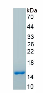
 Bioactivity
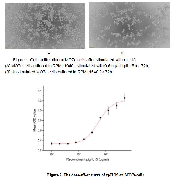
(Interleukin 15 (IL15) is a widely expressed cytokine that is structurally and functionally related to IL2, which plays an important role in many immunological diseases. IL15 also regulates T and natural killer (NK) cell activation and proliferation. The activity of recombinant pig IL15 was measured in a cell proliferation assay using MO7e human megakaryocytic leukemic cells. MO7e cells were seeded into triplicate wells of 96-well plates at a density of 30,000 cells/well in RPMI-1640 with the addition of various concentrations of rpIL15. After incubated for 72h, cells were observed by inverted microscope and cell proliferation was measured by Cell Counting Kit-8 (CCK-8). Briefly, 10 ul of CCK-8 solution was added to each well of the plate, then the absorbance at 450 nm was measured using a microplate reader after incubating the plate for 1-4 hours at 37 degree C. Cell proliferation of MO7e cells after incubation with rpIL15 for 72h observed by inverted microscope was shown in Figure 1. The dose-effect curve of rpIL15 was shown in Figure 2. It was obvious that rpIL15 significantly promoted cell proliferation of MO7e cells.The EC50 for this effect is typically 0.55 ug/ml.)
Bioactivity
(Interleukin 15 (IL15) is a widely expressed cytokine that is structurally and functionally related to IL2, which plays an important role in many immunological diseases. IL15 also regulates T and natural killer (NK) cell activation and proliferation. The activity of recombinant pig IL15 was measured in a cell proliferation assay using MO7e human megakaryocytic leukemic cells. MO7e cells were seeded into triplicate wells of 96-well plates at a density of 30,000 cells/well in RPMI-1640 with the addition of various concentrations of rpIL15. After incubated for 72h, cells were observed by inverted microscope and cell proliferation was measured by Cell Counting Kit-8 (CCK-8). Briefly, 10 ul of CCK-8 solution was added to each well of the plate, then the absorbance at 450 nm was measured using a microplate reader after incubating the plate for 1-4 hours at 37 degree C. Cell proliferation of MO7e cells after incubation with rpIL15 for 72h observed by inverted microscope was shown in Figure 1. The dose-effect curve of rpIL15 was shown in Figure 2. It was obvious that rpIL15 significantly promoted cell proliferation of MO7e cells.The EC50 for this effect is typically 0.55 ug/ml.)
Interleukin 15 (IL15), Active Protein (Cat# AAA161674)
Full Name
Active Interleukin 15 (IL15)
Gene Names
IL15; IL-15
Applications
Activity Assay, Cell Culture
Purity
Greater than 90% by SDS-PAGE
Pricing
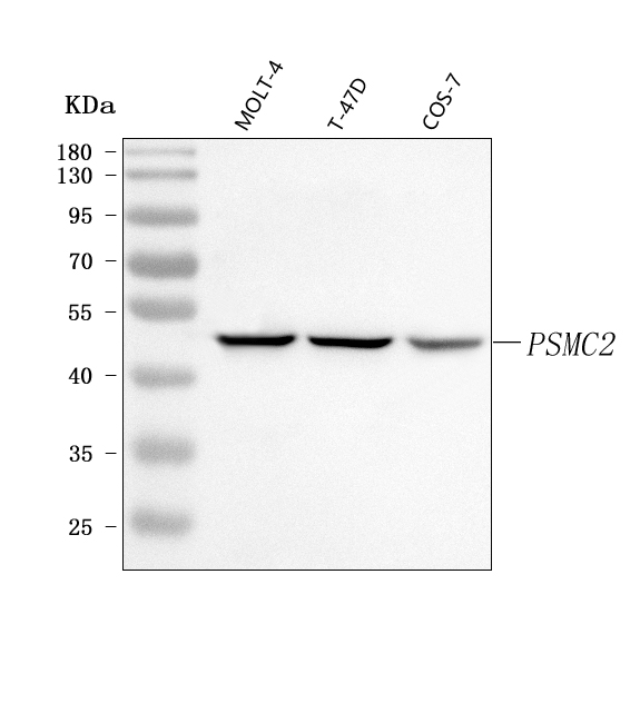
 IF (Immunofluorescence)
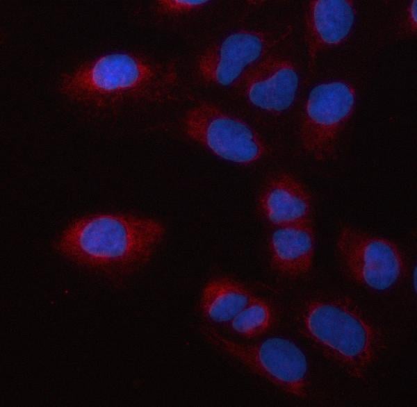
(Figure 2. IF analysis of PSMC2 using anti-PSMC2 antibody (AAA127577).PSMC2 was detected in an immunocytochemical section of U2OS cells. Enzyme antigen retrieval was performed using IHC enzyme antigen retrieval reagent for 15 mins. The cells were blocked with 10% goat serum. And then incubated with 5ug/mL rabbit anti-PSMC2 Antibody (AAA127577) overnight at 4 degree C. Cy3 Conjugated Goat Anti-Rabbit IgG (BA1032) was used as secondary antibody at 1:500 dilution and incubated for 30 minutes at 37 degree C. The section was counterstained with DAPI. Visualize using a fluorescence microscope and filter sets appropriate for the label used.)
IF (Immunofluorescence)
(Figure 2. IF analysis of PSMC2 using anti-PSMC2 antibody (AAA127577).PSMC2 was detected in an immunocytochemical section of U2OS cells. Enzyme antigen retrieval was performed using IHC enzyme antigen retrieval reagent for 15 mins. The cells were blocked with 10% goat serum. And then incubated with 5ug/mL rabbit anti-PSMC2 Antibody (AAA127577) overnight at 4 degree C. Cy3 Conjugated Goat Anti-Rabbit IgG (BA1032) was used as secondary antibody at 1:500 dilution and incubated for 30 minutes at 37 degree C. The section was counterstained with DAPI. Visualize using a fluorescence microscope and filter sets appropriate for the label used.)
PSMC2, Polyclonal Antibody (Cat# AAA127577)
Full Name
Anti-PSMC2 Antibody Picoband
Gene Names
PSMC2; S7; MSS1; Nbla10058
Reactivity
Human, Monkey
Applications
Immunofluorescence, Immunocytochemistry, Western Blot
Purity
Immunogen affinity purified.
Pricing
 FCM/FACS (Flow Cytometry)
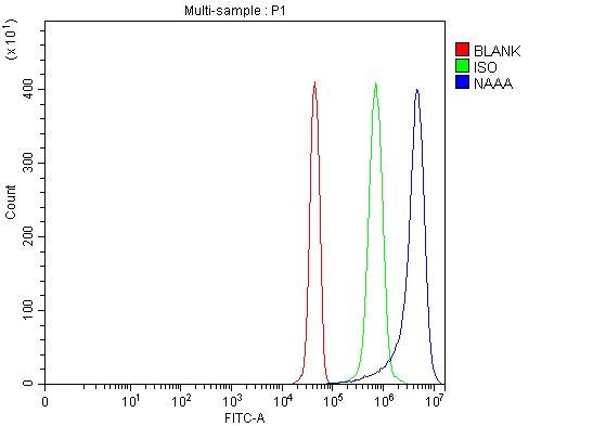
(Figure 2. Flow Cytometry analysis of JK cells using anti-NAAA antibody (AAA127349).Overlay histogram showing JK cells stained with AAA127349 (Blue line). To facilitate intracellular staining, cells were fixed with 4% paraformaldehyde and permeabilized with permeabilization buffer. The cells were blocked with 10% normal goat serum. And then incubated with rabbit anti-NAAA Antibody (AAA127349, 1ug/1x106 cells) for 30 min at 20 degree C. DyLight488 conjugated goat anti-rabbit IgG was used as secondary antibody for 30 minutes at 20 degree C. Isotype control antibody (Green line) was rabbit IgG (1ug/1x106) used under the same conditions. Unlabelled sample (Red line) was also used as a control.)
FCM/FACS (Flow Cytometry)
(Figure 2. Flow Cytometry analysis of JK cells using anti-NAAA antibody (AAA127349).Overlay histogram showing JK cells stained with AAA127349 (Blue line). To facilitate intracellular staining, cells were fixed with 4% paraformaldehyde and permeabilized with permeabilization buffer. The cells were blocked with 10% normal goat serum. And then incubated with rabbit anti-NAAA Antibody (AAA127349, 1ug/1x106 cells) for 30 min at 20 degree C. DyLight488 conjugated goat anti-rabbit IgG was used as secondary antibody for 30 minutes at 20 degree C. Isotype control antibody (Green line) was rabbit IgG (1ug/1x106) used under the same conditions. Unlabelled sample (Red line) was also used as a control.)
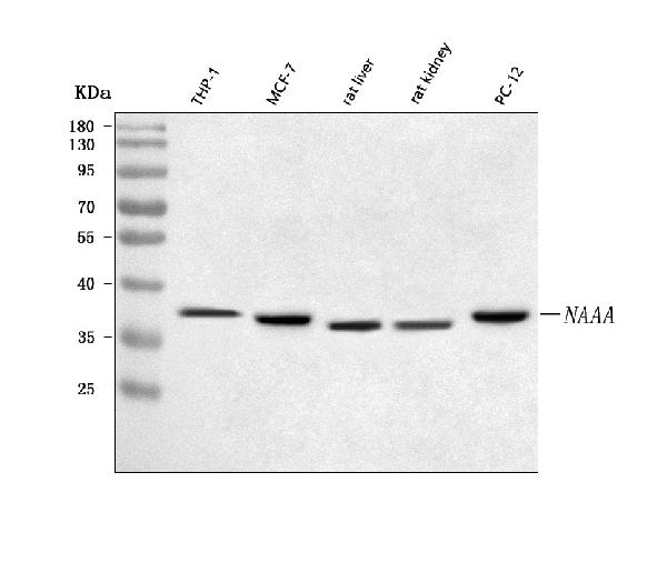
NAAA, Polyclonal Antibody (Cat# AAA127349)
Full Name
Anti-NAAA Antibody Picoband
Gene Names
NAAA; PLT; ASAHL
Reactivity
Human, Rat
Applications
Flow Cytometry, Western Blot
Purity
Immunogen affinity purified.
Pricing
 FCM/FACS (Flow Cytometry)
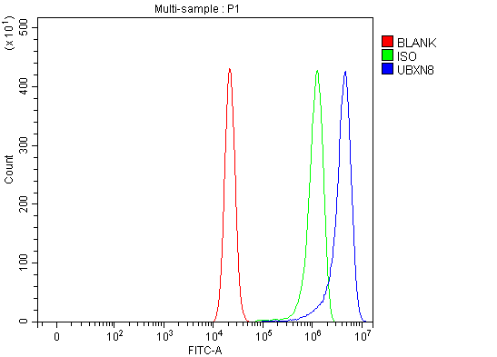
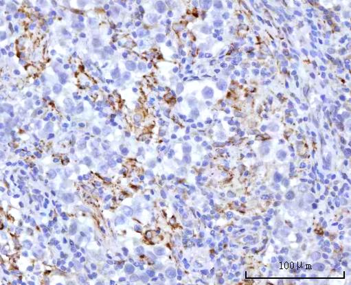
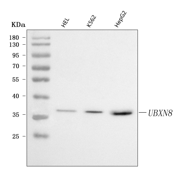
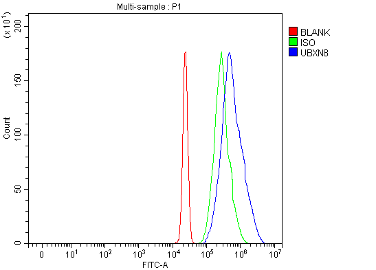
(Figure 4. Flow Cytometry analysis of U937 cells using anti-UBXN8 antibody (AAA127791).Overlay histogram showing U937 cells stained with AAA127791 (Blue line). To facilitate intracellular staining, cells were fixed with 4% paraformaldehyde and permeabilized with permeabilization buffer. The cells were blocked with 10% normal goat serum. And then incubated with rabbit anti-UBXN8 Antibody (AAA127791, 1ug/1x106 cells) for 30 min at 20 degree C. DyLight488 conjugated goat anti-rabbit IgG was used as secondary antibody for 30 minutes at 20 degree C. Isotype control antibody (Green line) was rabbit IgG (1ug/1x106) used under the same conditions. Unlabelled sample without incubation with primary antibody and secondary antibody (Red line) was used as a blank control.)
FCM/FACS (Flow Cytometry)
(Figure 4. Flow Cytometry analysis of U937 cells using anti-UBXN8 antibody (AAA127791).Overlay histogram showing U937 cells stained with AAA127791 (Blue line). To facilitate intracellular staining, cells were fixed with 4% paraformaldehyde and permeabilized with permeabilization buffer. The cells were blocked with 10% normal goat serum. And then incubated with rabbit anti-UBXN8 Antibody (AAA127791, 1ug/1x106 cells) for 30 min at 20 degree C. DyLight488 conjugated goat anti-rabbit IgG was used as secondary antibody for 30 minutes at 20 degree C. Isotype control antibody (Green line) was rabbit IgG (1ug/1x106) used under the same conditions. Unlabelled sample without incubation with primary antibody and secondary antibody (Red line) was used as a blank control.)
UBXN8, Polyclonal Antibody (Cat# AAA127791)
Full Name
Anti-UBXN8 Antibody Picoband
Gene Names
UBXN8; REP8; UBXD6; D8S2298E
Reactivity
Human
Applications
Flow Cytometry, Immunohistochemistry, Western Blot
Purity
Immunogen affinity purified.
Pricing
 IHC (Immunohistochemistry)
(DAB staining on IHCP;Sample: Porcine Cerebrum Tissue; Primary Ab: 10ug/ml Mouse AntiHuman IFNa/bR1 AntibodySecond Ab: 2ug/mL HRPLinked Caprine AntiMouse IgG Polyclonal Antibody(Catalog: SAA544Mu19))
IHC (Immunohistochemistry)
(DAB staining on IHCP;Sample: Porcine Cerebrum Tissue; Primary Ab: 10ug/ml Mouse AntiHuman IFNa/bR1 AntibodySecond Ab: 2ug/mL HRPLinked Caprine AntiMouse IgG Polyclonal Antibody(Catalog: SAA544Mu19))
Interferon Alpha/Beta Receptor 1 (IFNa/bR1), Monoclonal Antibody (Cat# AAA151711)
Full Name
Monoclonal Antibody to Interferon Alpha/Beta Receptor 1 (IFNa/bR1)
Gene Names
IFNAR1; AVP; IFRC; IFNAR; IFNBR; IFN-alpha-REC
Reactivity
Human, Pig
Applications
Immunoprecipitation, Immunocytochemistry, Immunohistochemistry, Western Blot
Purity
Protein A + Protein G affinity chromatography
Pricing
 FCM/FACS (Flow Cytometry)
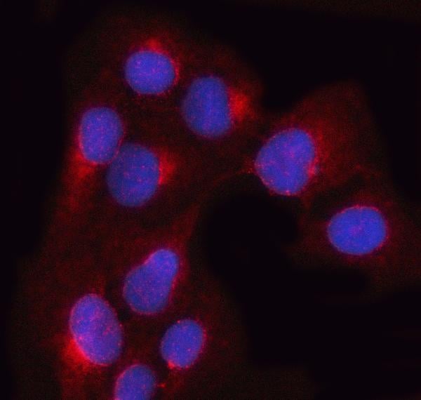
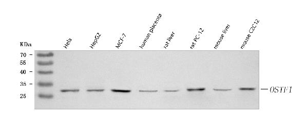
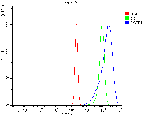
(Figure 3. Flow Cytometry analysis of U20S cells using anti-OSTF1 antibody (AAA127341).Overlay histogram showing U20S cells stained with AAA127341 (Blue line). To facilitate intracellular staining, cells were fixed with 4% paraformaldehyde and permeabilized with permeabilization buffer. The cells were blocked with 10% normal goat serum. And then incubated with rabbit anti-OSTF1 Antibody (AAA127341, 1ug/1x106 cells) for 30 min at 20 degree C. DyLight488 conjugated goat anti-rabbit IgG was used as secondary antibody for 30 minutes at 20 degree C. Isotype control antibody (Green line) was rabbit IgG (1ug/1x106) used under the same conditions. Unlabelled sample without incubation with primary antibody and secondary antibody (Red line) was used as a blank control.)
FCM/FACS (Flow Cytometry)
(Figure 3. Flow Cytometry analysis of U20S cells using anti-OSTF1 antibody (AAA127341).Overlay histogram showing U20S cells stained with AAA127341 (Blue line). To facilitate intracellular staining, cells were fixed with 4% paraformaldehyde and permeabilized with permeabilization buffer. The cells were blocked with 10% normal goat serum. And then incubated with rabbit anti-OSTF1 Antibody (AAA127341, 1ug/1x106 cells) for 30 min at 20 degree C. DyLight488 conjugated goat anti-rabbit IgG was used as secondary antibody for 30 minutes at 20 degree C. Isotype control antibody (Green line) was rabbit IgG (1ug/1x106) used under the same conditions. Unlabelled sample without incubation with primary antibody and secondary antibody (Red line) was used as a blank control.)
OSTF1, Polyclonal Antibody (Cat# AAA127341)
Full Name
Anti-OSTF1 Antibody Picoband
Gene Names
OSTF1; OSF; SH3P2; bA235O14.1
Reactivity
Human, Mouse, Rat
Applications
Flow Cytometry, Immunofluorescence, Immunocytochemistry, Western Blot
Purity
Immunogen affinity purified.
Pricing
 FCM/FACS (Flow Cytometry)
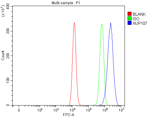
(Figure 3. Flow Cytometry analysis of HEL cells using anti-NUP107 antibody (AAA127196).Overlay histogram showing HEL cells stained with AAA127196 (Blue line). To facilitate intracellular staining, cells were fixed with 4% paraformaldehyde and permeabilized with permeabilization buffer. The cells were blocked with 10% normal goat serum. And then incubated with rabbit anti-NUP107 Antibody (AAA127196, 1ug/1x106 cells) for 30 min at 20 degree C. DyLight488 conjugated goat anti-rabbit IgG was used as secondary antibody for 30 minutes at 20 degree C. Isotype control antibody (Green line) was rabbit IgG (1ug/1x106) used under the same conditions. Unlabelled sample (Red line) was also used as a control.)
FCM/FACS (Flow Cytometry)
(Figure 3. Flow Cytometry analysis of HEL cells using anti-NUP107 antibody (AAA127196).Overlay histogram showing HEL cells stained with AAA127196 (Blue line). To facilitate intracellular staining, cells were fixed with 4% paraformaldehyde and permeabilized with permeabilization buffer. The cells were blocked with 10% normal goat serum. And then incubated with rabbit anti-NUP107 Antibody (AAA127196, 1ug/1x106 cells) for 30 min at 20 degree C. DyLight488 conjugated goat anti-rabbit IgG was used as secondary antibody for 30 minutes at 20 degree C. Isotype control antibody (Green line) was rabbit IgG (1ug/1x106) used under the same conditions. Unlabelled sample (Red line) was also used as a control.)
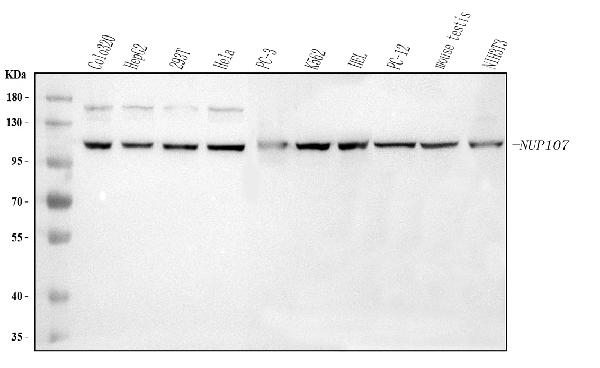
NUP107, Polyclonal Antibody (Cat# AAA127196)
Full Name
Anti-NUP107 Antibody Picoband
Gene Names
NUP107; NUP84
Reactivity
Human, Mouse, Rat
Applications
Flow Cytometry, Western Blot, Immunofluorescence, Immunocytochemistry
Purity
Immunogen affinity purified.
Pricing
 FCM/FACS (Flow Cytometry)
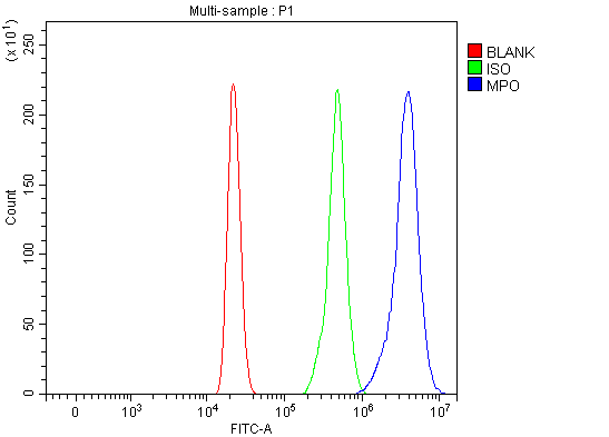
(Figure 2. Flow Cytometry analysis of HL-60 cells using anti-MPO antibody (AAA126985).Overlay histogram showing HL-60 cells stained with AAA126985 (Blue line). To facilitate intracellular staining, cells were fixed with 4% paraformaldehyde and permeabilized with permeabilization buffer. The cells were blocked with 10% normal goat serum. And then incubated with rabbit anti-MPO Antibody (AAA126985, 1ug/1x106 cells) for 30 min at 20 degree C. DyLight488 conjugated goat anti-rabbit IgG was used as secondary antibody for 30 minutes at 20 degree C. Isotype control antibody (Green line) was rabbit IgG (1ug/1x106) used under the same conditions. Unlabelled sample without incubation with primary antibody and secondary antibody (Red line) was used as a blank control.)
FCM/FACS (Flow Cytometry)
(Figure 2. Flow Cytometry analysis of HL-60 cells using anti-MPO antibody (AAA126985).Overlay histogram showing HL-60 cells stained with AAA126985 (Blue line). To facilitate intracellular staining, cells were fixed with 4% paraformaldehyde and permeabilized with permeabilization buffer. The cells were blocked with 10% normal goat serum. And then incubated with rabbit anti-MPO Antibody (AAA126985, 1ug/1x106 cells) for 30 min at 20 degree C. DyLight488 conjugated goat anti-rabbit IgG was used as secondary antibody for 30 minutes at 20 degree C. Isotype control antibody (Green line) was rabbit IgG (1ug/1x106) used under the same conditions. Unlabelled sample without incubation with primary antibody and secondary antibody (Red line) was used as a blank control.)
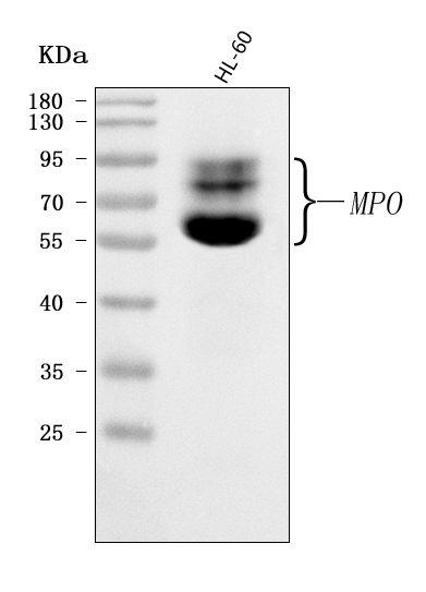
MPO, Polyclonal Antibody (Cat# AAA126985)
Full Name
Anti-MPO Antibody Picoband
Reactivity
Human
Applications
Flow Cytometry, Western Blot
Purity
Immunogen affinity purified.
Pricing
 FCM/FACS (Flow Cytometry)
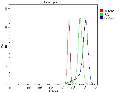
(Figure 2. Flow Cytometry analysis of MCF-7 cells using anti-TYK2 antibody (AAA126160).Overlay histogram showing MCF-7 cells stained with AAA126160 (Blue line). The cells were blocked with 10% normal goat serum. And then incubated with rabbit anti-TYK2 Antibody (AAA126160, 1 ug/1x10^6 cells) for 30 min at 20 degree C. DyLight488 conjugated goat anti-rabbit IgG was used as secondary antibody for 30 minutes at 20 degree C. Isotype control antibody (Green line) was rabbit IgG (1 ug/1x10^6) used under the same conditions. Unlabelled sample (Red line) was also used as a control.)
FCM/FACS (Flow Cytometry)
(Figure 2. Flow Cytometry analysis of MCF-7 cells using anti-TYK2 antibody (AAA126160).Overlay histogram showing MCF-7 cells stained with AAA126160 (Blue line). The cells were blocked with 10% normal goat serum. And then incubated with rabbit anti-TYK2 Antibody (AAA126160, 1 ug/1x10^6 cells) for 30 min at 20 degree C. DyLight488 conjugated goat anti-rabbit IgG was used as secondary antibody for 30 minutes at 20 degree C. Isotype control antibody (Green line) was rabbit IgG (1 ug/1x10^6) used under the same conditions. Unlabelled sample (Red line) was also used as a control.)
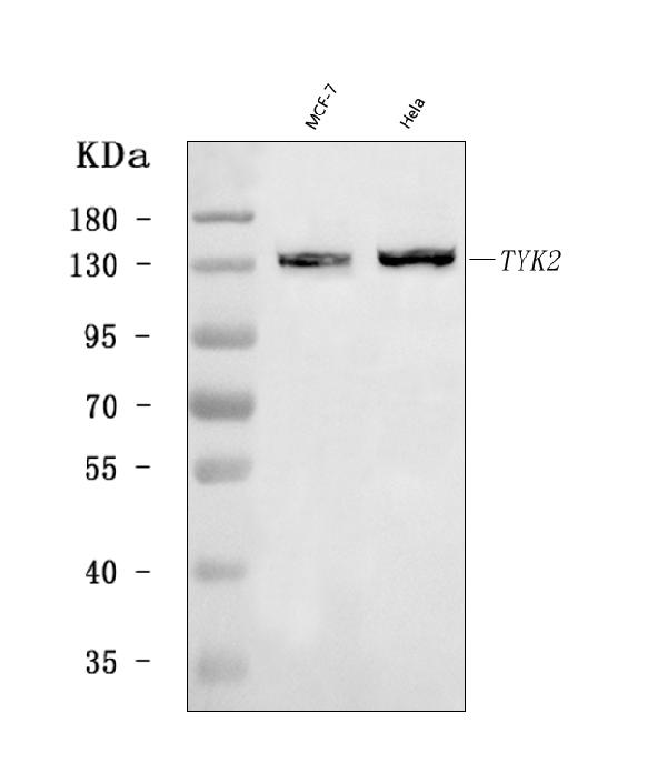
TYK2, Polyclonal Antibody (Cat# AAA126160)
Full Name
Anti-TYK2 Antibody Picoband
Gene Names
TYK2; JTK1
Reactivity
Human
Applications
Flow Cytometry, Western Blot
Purity
Immunogen affinity purified.
Pricing
 IF (Immunofluorescence)
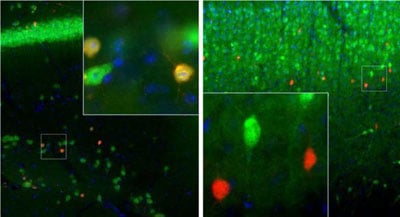
(Image: Left: Adult mouse brain hippocampal section (45 uM; fixed by transcardial perfusion with 4% paraformaldehyde) was co-stained with Calretinin AAA76584 (1:1000, red) and our rabbit polyclonal antibody to Fox3/NeuN (RA22113, green). Calretinin stains a small number of interneuron in the stratum radiatum of CA1 region, and Fox/NeuN is expressed in most neurons in the brain. As a result, cells are positive for calretinin appear to be yellow. Right: Adult rat cortical section (45 uM; fixed by transcardial perfusion with 4% paraformaldehyde) was co-stained with AAA76584 (red) and our chicken polyclonal antibody to calbinidin (CH22118, green). In cortex, calretinin is expressed in a small population of interneurons concentrated in Layer 4 area, while calbindin is expressed in cells concentrated in Layer 2/3. Because each antibody specifically labels a different population of cells exclusively, the cells are either stained with green, or red in the cortex. Insets are high-magnification images of the boxed area in each picture. Blue is Dapi staining that labels DNA.)
IF (Immunofluorescence)
(Image: Left: Adult mouse brain hippocampal section (45 uM; fixed by transcardial perfusion with 4% paraformaldehyde) was co-stained with Calretinin AAA76584 (1:1000, red) and our rabbit polyclonal antibody to Fox3/NeuN (RA22113, green). Calretinin stains a small number of interneuron in the stratum radiatum of CA1 region, and Fox/NeuN is expressed in most neurons in the brain. As a result, cells are positive for calretinin appear to be yellow. Right: Adult rat cortical section (45 uM; fixed by transcardial perfusion with 4% paraformaldehyde) was co-stained with AAA76584 (red) and our chicken polyclonal antibody to calbinidin (CH22118, green). In cortex, calretinin is expressed in a small population of interneurons concentrated in Layer 4 area, while calbindin is expressed in cells concentrated in Layer 2/3. Because each antibody specifically labels a different population of cells exclusively, the cells are either stained with green, or red in the cortex. Insets are high-magnification images of the boxed area in each picture. Blue is Dapi staining that labels DNA.)
Calretinin, Monoclonal Antibody (Cat# AAA76584)
Full Name
Calretinin (IgG1)
Reactivity
Human, Mouse, Rat, Cow
Applications
Immunofluorescence, Immunohistochemistry, Western Blot, Immunocytochemistry
Purity
Affinity Purified
Pricing
 IF (Immunofluorescence)
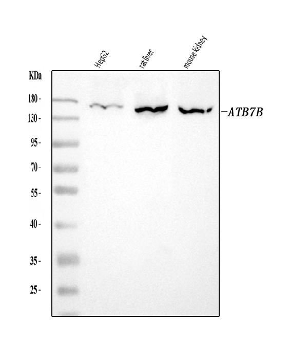
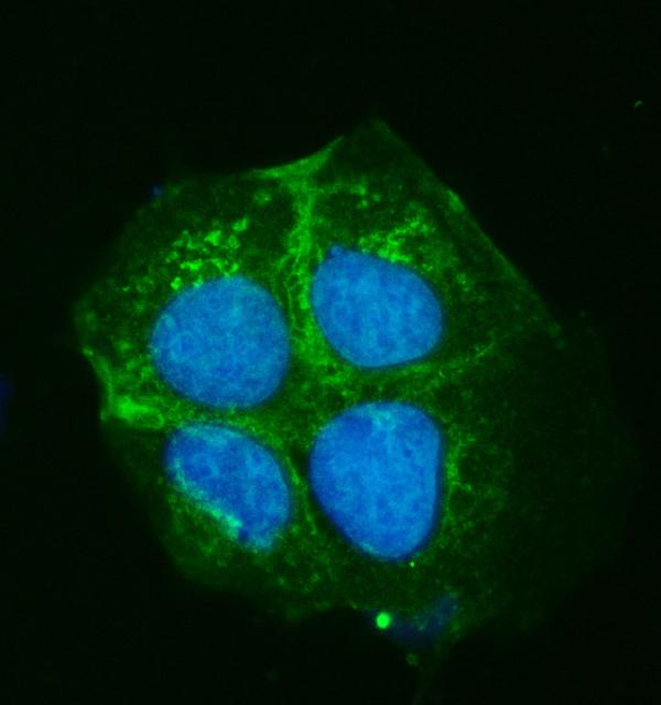
(Figure 2. IF analysis of ATP7B using anti-ATP7B antibody (AAA126056).ATP7B was detected in an immunocytochemical section of Caco-2 cells. Enzyme antigen retrieval was performed using IHC enzyme antigen retrieval reagent (AR0022) for 15 mins. The cells were blocked with 10% goat serum. And then incubated with 5 ug/mL rabbit anti-ATP7B Antibody (AAA126056) overnight at 4 degree C. DyLight488 Conjugated Goat Anti-Rabbit IgG was used as secondary antibody at 1:100 dilution and incubated for 30 minutes at 37 degree C. The section was counterstained with DAPI. Visualize using a fluorescence microscope and filter sets appropriate for the label used.)
IF (Immunofluorescence)
(Figure 2. IF analysis of ATP7B using anti-ATP7B antibody (AAA126056).ATP7B was detected in an immunocytochemical section of Caco-2 cells. Enzyme antigen retrieval was performed using IHC enzyme antigen retrieval reagent (AR0022) for 15 mins. The cells were blocked with 10% goat serum. And then incubated with 5 ug/mL rabbit anti-ATP7B Antibody (AAA126056) overnight at 4 degree C. DyLight488 Conjugated Goat Anti-Rabbit IgG was used as secondary antibody at 1:100 dilution and incubated for 30 minutes at 37 degree C. The section was counterstained with DAPI. Visualize using a fluorescence microscope and filter sets appropriate for the label used.)
ATP7B, Polyclonal Antibody (Cat# AAA126056)
Full Name
Anti-ATP7B Antibody Picoband
Gene Names
ATP7B; WD; PWD; WC1; WND
Reactivity
Human, Mouse, Rat
Applications
Immunofluorescence, Immunocytochemistry, Western Blot
Purity
Immunogen affinity purified.
Pricing
 FCM/FACS (Flow Cytometry)
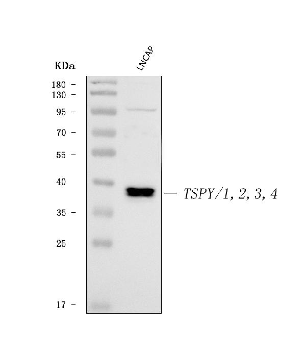
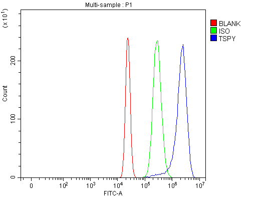
(Figure 2. Flow Cytometry analysis of U2OS cells using anti-TSPY1/2/3/4 antibody (AAA126368).Overlay histogram showing U2OS cells stained with AAA126368 (Blue line). The cells were blocked with 10% normal goat serum. And then incubated with rabbit anti-TSPY1/2/3/4 Antibody (AAA126368, 1 ug/1x10^6 cells) for 30 min at 20 degree C. DyLight488 conjugated goat anti-rabbit IgG was used as secondary antibody for 30 minutes at 20 degree C. Isotype control antibody (Green line) was rabbit IgG (1 ug/1x10^6) used under the same conditions. Unlabelled sample (Red line) was also used as a control.)
FCM/FACS (Flow Cytometry)
(Figure 2. Flow Cytometry analysis of U2OS cells using anti-TSPY1/2/3/4 antibody (AAA126368).Overlay histogram showing U2OS cells stained with AAA126368 (Blue line). The cells were blocked with 10% normal goat serum. And then incubated with rabbit anti-TSPY1/2/3/4 Antibody (AAA126368, 1 ug/1x10^6 cells) for 30 min at 20 degree C. DyLight488 conjugated goat anti-rabbit IgG was used as secondary antibody for 30 minutes at 20 degree C. Isotype control antibody (Green line) was rabbit IgG (1 ug/1x10^6) used under the same conditions. Unlabelled sample (Red line) was also used as a control.)
TSPY1/2/3/4, Polyclonal Antibody (Cat# AAA126368)
Full Name
Anti-TSPY1/2/3/4 Antibody Picoband
Gene Names
TSPY1; CT78; TSPY; DYS14; pJA923
Reactivity
Human
Applications
Flow Cytometry, Western Blot
Purity
Immunogen affinity purified.
Pricing
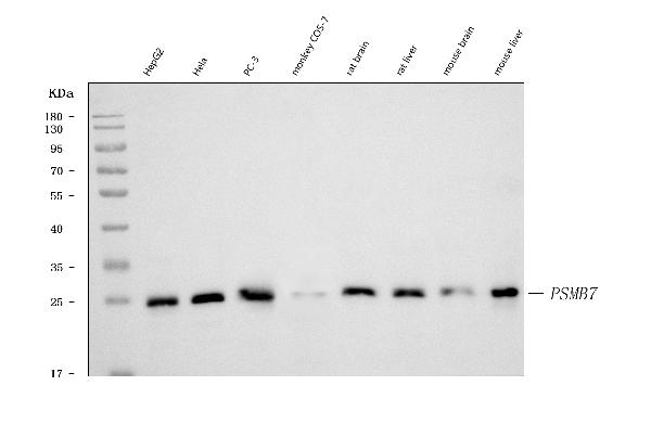
 FCM/FACS (Flow Cytometry)
(Figure 2. Flow Cytometry analysis of CACO-2 cells using anti-PSMB7 antibody (AAA126955).Overlay histogram showing CACO-2 cells stained with AAA126955 (Blue line). The cells were blocked with 10% normal goat serum. And then incubated with mouse anti-PSMB7 Antibody (AAA126955, 1 ug/1x10^6 cells) for 30 min at 20 degree C. DyLight488 conjugated goat anti-mouse IgG was used as secondary antibody for 30 minutes at 20 degree C. Isotype control antibody (Green line) was mouse IgG (1 ug/1x10^6) used under the same conditions. Unlabelled sample (Red line) was also used as a control.)
FCM/FACS (Flow Cytometry)
(Figure 2. Flow Cytometry analysis of CACO-2 cells using anti-PSMB7 antibody (AAA126955).Overlay histogram showing CACO-2 cells stained with AAA126955 (Blue line). The cells were blocked with 10% normal goat serum. And then incubated with mouse anti-PSMB7 Antibody (AAA126955, 1 ug/1x10^6 cells) for 30 min at 20 degree C. DyLight488 conjugated goat anti-mouse IgG was used as secondary antibody for 30 minutes at 20 degree C. Isotype control antibody (Green line) was mouse IgG (1 ug/1x10^6) used under the same conditions. Unlabelled sample (Red line) was also used as a control.)
Proteasome 20S beta 7/PSMB7, Monoclonal Antibody (Cat# AAA126955)
Full Name
Anti-Proteasome 20S beta 7/PSMB7 Antibody Picoband (monoclonal, 3H6C8)
Gene Names
PSMB7; Z
Reactivity
Human, Monkey, Mouse, Rat
Applications
Flow Cytometry, Western Blot
Purity
Immunogen affinity purified.
Pricing
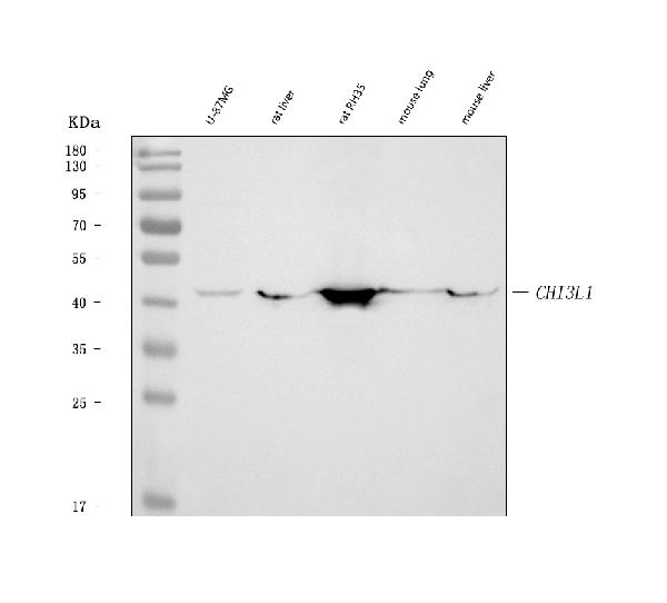
 IF (Immunofluorescence)
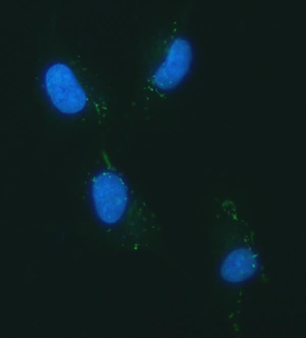
(Figure 2. IF analysis of YKL-40/CHI3L1 using anti-YKL-40/CHI3L1 antibody (AAA126129).YKL-40/CHI3L1 was detected in an immunocytochemical section of U87 cells. Enzyme antigen retrieval was performed using IHC enzyme antigen retrieval reagent (AR0022) for 15 mins. The cells were blocked with 10% goat serum. And then incubated with 5 ug/mL rabbit anti-YKL-40/CHI3L1 Antibody (AAA126129) overnight at 4 degree C. DyLight488 Conjugated Goat Anti-Rabbit IgG was used as secondary antibody at 1:100 dilution and incubated for 30 minutes at 37 degree C. The section was counterstained with DAPI. Visualize using a fluorescence microscope and filter sets appropriate for the label used.)
IF (Immunofluorescence)
(Figure 2. IF analysis of YKL-40/CHI3L1 using anti-YKL-40/CHI3L1 antibody (AAA126129).YKL-40/CHI3L1 was detected in an immunocytochemical section of U87 cells. Enzyme antigen retrieval was performed using IHC enzyme antigen retrieval reagent (AR0022) for 15 mins. The cells were blocked with 10% goat serum. And then incubated with 5 ug/mL rabbit anti-YKL-40/CHI3L1 Antibody (AAA126129) overnight at 4 degree C. DyLight488 Conjugated Goat Anti-Rabbit IgG was used as secondary antibody at 1:100 dilution and incubated for 30 minutes at 37 degree C. The section was counterstained with DAPI. Visualize using a fluorescence microscope and filter sets appropriate for the label used.)
YKL-40/CHI3L1, Polyclonal Antibody (Cat# AAA126129)
Full Name
Anti-YKL-40/CHI3L1 Antibody Picoband
Gene Names
CHI3L1; GP39; ASRT7; GP-39; YKL40; CGP-39; YKL-40; YYL-40; HC-gp39; HCGP-3P; hCGP-39
Reactivity
Human, Mouse, Rat
Applications
Immunofluorescence, Immunocytochemistry, Western Blot
Purity
Immunogen affinity purified.
Pricing
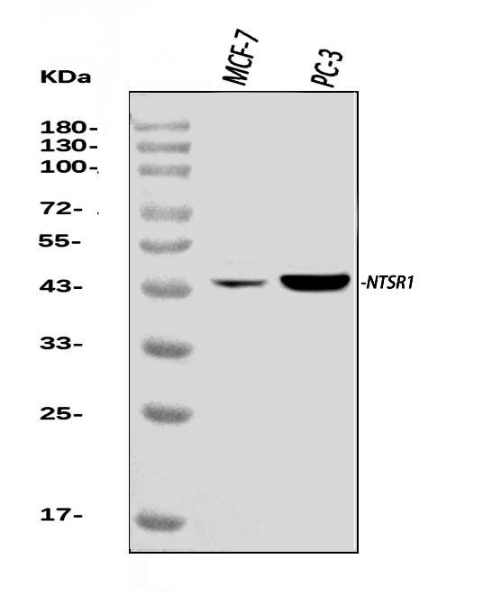
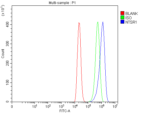 FCM/FACS (Flow Cytometry)
(Figure 2. Flow Cytometry analysis of THP-1 cells using anti-Neurotensin Receptor 1/NTSR1 antibody (AAA126393).Overlay histogram showing THP-1 cells stained with AAA126393 (Blue line). The cells were blocked with 10% normal goat serum. And then incubated with rabbit anti-Neurotensin Receptor 1/NTSR1 Antibody (AAA126393, 1 ug/1x10^6 cells) for 30 min at 20 degree C. DyLight488 conjugated goat anti-rabbit IgG was used as secondary antibody for 30 minutes at 20 degree C. Isotype control antibody (Green line) was rabbit IgG (1 ug/1x10^6) used under the same conditions. Unlabelled sample (Red line) was also used as a control.)
FCM/FACS (Flow Cytometry)
(Figure 2. Flow Cytometry analysis of THP-1 cells using anti-Neurotensin Receptor 1/NTSR1 antibody (AAA126393).Overlay histogram showing THP-1 cells stained with AAA126393 (Blue line). The cells were blocked with 10% normal goat serum. And then incubated with rabbit anti-Neurotensin Receptor 1/NTSR1 Antibody (AAA126393, 1 ug/1x10^6 cells) for 30 min at 20 degree C. DyLight488 conjugated goat anti-rabbit IgG was used as secondary antibody for 30 minutes at 20 degree C. Isotype control antibody (Green line) was rabbit IgG (1 ug/1x10^6) used under the same conditions. Unlabelled sample (Red line) was also used as a control.)
Neurotensin Receptor 1/NTSR1, Polyclonal Antibody (Cat# AAA126393)
Full Name
Anti-Neurotensin Receptor 1/NTSR1 Antibody Picoband
Gene Names
NTSR1; NTR
Reactivity
Human
Applications
Flow Cytometry, Western Blot
Purity
Immunogen affinity purified.
Pricing
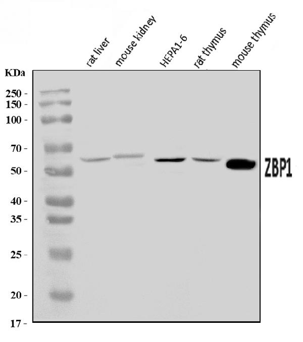
 IF (Immunofluorescence)
(Figure 2. IF analysis of DAI/Zbp1 using anti-DAI/Zbp1 antibody (AAA126400).DAI/Zbp1 was detected in an immunocytochemical section of RM-1 cells. Enzyme antigen retrieval was performed using IHC enzyme antigen retrieval reagent (AR0022) for 15 mins. The cells were blocked with 10% goat serum. And then incubated with 5 ug/mL rabbit anti-DAI/Zbp1 Antibody (AAA126400) overnight at 4 degree C. DyLight488 Conjugated Goat Anti-Rabbit IgG was used as secondary antibody at 1:100 dilution and incubated for 30 minutes at 37 degree C. The section was counterstained with DAPI. Visualize using a fluorescence microscope and filter sets appropriate for the label used.)
IF (Immunofluorescence)
(Figure 2. IF analysis of DAI/Zbp1 using anti-DAI/Zbp1 antibody (AAA126400).DAI/Zbp1 was detected in an immunocytochemical section of RM-1 cells. Enzyme antigen retrieval was performed using IHC enzyme antigen retrieval reagent (AR0022) for 15 mins. The cells were blocked with 10% goat serum. And then incubated with 5 ug/mL rabbit anti-DAI/Zbp1 Antibody (AAA126400) overnight at 4 degree C. DyLight488 Conjugated Goat Anti-Rabbit IgG was used as secondary antibody at 1:100 dilution and incubated for 30 minutes at 37 degree C. The section was counterstained with DAPI. Visualize using a fluorescence microscope and filter sets appropriate for the label used.)
DAI/Zbp1, Polyclonal Antibody (Cat# AAA126400)
Full Name
Anti-DAI/Zbp1 Antibody Picoband
Gene Names
Zbp1; Dai; Dlm1; mZaDLM; 2010010H03Rik
Reactivity
Mouse, Rat
Applications
Immunofluorescence, Immunocytochemistry, Western Blot
Purity
Immunogen affinity purified.
Pricing
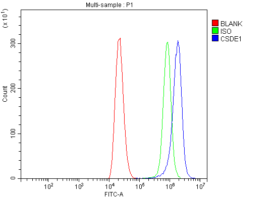 FCM/FACS (Flow Cytometry)
(Figure 2. Flow Cytometry analysis of THP-1 cells using anti-CSDE1/NRU antibody (AAA126411).Overlay histogram showing THP-1 cells stained with AAA126411 (Blue line). The cells were blocked with 10% normal goat serum. And then incubated with rabbit anti-CSDE1/NRU Antibody (AAA126411, 1 ug/1x10^6 cells) for 30 min at 20 degree C. DyLight488 conjugated goat anti-rabbit IgG was used as secondary antibody for 30 minutes at 20 degree C. Isotype control antibody (Green line) was rabbit IgG (1 ug/1x10^6) used under the same conditions. Unlabelled sample (Red line) was also used as a control.)
FCM/FACS (Flow Cytometry)
(Figure 2. Flow Cytometry analysis of THP-1 cells using anti-CSDE1/NRU antibody (AAA126411).Overlay histogram showing THP-1 cells stained with AAA126411 (Blue line). The cells were blocked with 10% normal goat serum. And then incubated with rabbit anti-CSDE1/NRU Antibody (AAA126411, 1 ug/1x10^6 cells) for 30 min at 20 degree C. DyLight488 conjugated goat anti-rabbit IgG was used as secondary antibody for 30 minutes at 20 degree C. Isotype control antibody (Green line) was rabbit IgG (1 ug/1x10^6) used under the same conditions. Unlabelled sample (Red line) was also used as a control.)
CSDE1/NRU, Polyclonal Antibody (Cat# AAA126411)
Full Name
Anti-CSDE1/NRU Antibody Picoband
Gene Names
CSDE1; UNR; D1S155E
Reactivity
Human
Applications
Western Blot, Immunocytochemistry, Immunohistochemistry, Immunofluorescence, Flow Cytometry
Purity
Immunogen affinity purified.
Pricing
 FCM/FACS (Flow Cytometry)
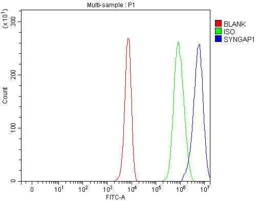
(Figure 2. Flow Cytometry analysis of K562 cells using anti-SYNGAP1 antibody (AAA127260).Overlay histogram showing K562 cells stained with AAA127260 (Blue line). To facilitate intracellular staining, cells were fixed with 4% paraformaldehyde and permeabilized with permeabilization buffer. The cells were blocked with 10% normal goat serum. And then incubated with rabbit anti-SYNGAP1 Antibody (AAA127260, 1ug/1x106 cells) for 30 min at 20 degree C. DyLight488 conjugated goat anti-rabbit IgG was used as secondary antibody for 30 minutes at 20 degree C. Isotype control antibody (Green line) was rabbit IgG (1ug/1x106) used under the same conditions. Unlabelled sample (Red line) was also used as a control.)
FCM/FACS (Flow Cytometry)
(Figure 2. Flow Cytometry analysis of K562 cells using anti-SYNGAP1 antibody (AAA127260).Overlay histogram showing K562 cells stained with AAA127260 (Blue line). To facilitate intracellular staining, cells were fixed with 4% paraformaldehyde and permeabilized with permeabilization buffer. The cells were blocked with 10% normal goat serum. And then incubated with rabbit anti-SYNGAP1 Antibody (AAA127260, 1ug/1x106 cells) for 30 min at 20 degree C. DyLight488 conjugated goat anti-rabbit IgG was used as secondary antibody for 30 minutes at 20 degree C. Isotype control antibody (Green line) was rabbit IgG (1ug/1x106) used under the same conditions. Unlabelled sample (Red line) was also used as a control.)
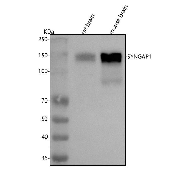
SYNGAP1, Polyclonal Antibody (Cat# AAA127260)
Full Name
Anti-SYNGAP1 Antibody Picoband
Gene Names
SYNGAP1; MRD5; RASA1; RASA5; SYNGAP
Reactivity
Human, Mouse, Rat
Applications
Flow Cytometry, Western Blot
Purity
Immunogen affinity purified.
Pricing
 FCM/FACS (Flow Cytometry)
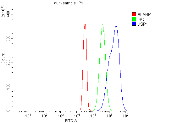
(Figure 2. Flow Cytometry analysis of SiHa cells using anti-USP1 antibody (AAA126339).Overlay histogram showing SiHa cells stained with AAA126339 (Blue line). The cells were blocked with 10% normal goat serum. And then incubated with rabbit anti-USP1 Antibody (AAA126339, 1 ug/1x10^6 cells) for 30 min at 20 degree C. DyLight488 conjugated goat anti-rabbit IgG was used as secondary antibody for 30 minutes at 20 degree C. Isotype control antibody (Green line) was rabbit IgG (1 ug/1x10^6) used under the same conditions. Unlabelled sample (Red line) was also used as a control.)
FCM/FACS (Flow Cytometry)
(Figure 2. Flow Cytometry analysis of SiHa cells using anti-USP1 antibody (AAA126339).Overlay histogram showing SiHa cells stained with AAA126339 (Blue line). The cells were blocked with 10% normal goat serum. And then incubated with rabbit anti-USP1 Antibody (AAA126339, 1 ug/1x10^6 cells) for 30 min at 20 degree C. DyLight488 conjugated goat anti-rabbit IgG was used as secondary antibody for 30 minutes at 20 degree C. Isotype control antibody (Green line) was rabbit IgG (1 ug/1x10^6) used under the same conditions. Unlabelled sample (Red line) was also used as a control.)
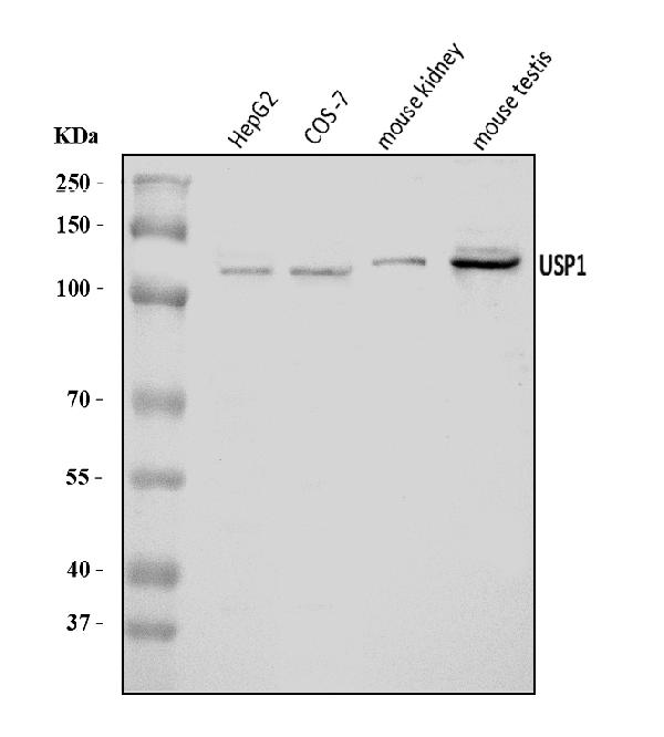
USP1, Polyclonal Antibody (Cat# AAA126339)
Full Name
Anti-USP1 Antibody Picoband
Gene Names
USP1; UBP
Reactivity
Human, Mouse, Monkey
Applications
Western Blot, Flow Cytometry
Purity
Immunogen affinity purified.
Pricing
 FCM/FACS (Flow Cytometry)
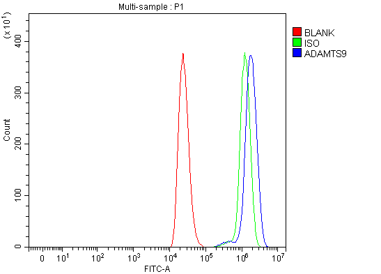
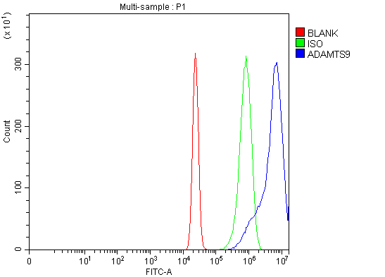
(Figure 3. Flow Cytometry analysis of U2OS cells using anti-ADAMTS9 antibody (AAA126395).Overlay histogram showing U2OS cells stained with AAA126395 (Blue line). The cells were blocked with 10% normal goat serum. And then incubated with rabbit anti-ADAMTS9 Antibody (AAA126395, 1 ug/1x10^6 cells) for 30 min at 20 degree C. DyLight488 conjugated goat anti-rabbit IgG was used as secondary antibody for 30 minutes at 20 degree C. Isotype control antibody (Green line) was rabbit IgG (1 ug/1x10^6) used under the same conditions. Unlabelled sample (Red line) was also used as a control.)
FCM/FACS (Flow Cytometry)
(Figure 3. Flow Cytometry analysis of U2OS cells using anti-ADAMTS9 antibody (AAA126395).Overlay histogram showing U2OS cells stained with AAA126395 (Blue line). The cells were blocked with 10% normal goat serum. And then incubated with rabbit anti-ADAMTS9 Antibody (AAA126395, 1 ug/1x10^6 cells) for 30 min at 20 degree C. DyLight488 conjugated goat anti-rabbit IgG was used as secondary antibody for 30 minutes at 20 degree C. Isotype control antibody (Green line) was rabbit IgG (1 ug/1x10^6) used under the same conditions. Unlabelled sample (Red line) was also used as a control.)
ADAMTS9, Polyclonal Antibody (Cat# AAA126395)
Full Name
Anti-ADAMTS9 Antibody Picoband
Reactivity
Human, Rat
Applications
Flow Cytometry, Western Blot
Purity
Immunogen affinity purified.
Pricing
 FCM/FACS (Flow Cytometry)
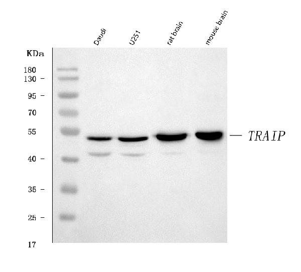
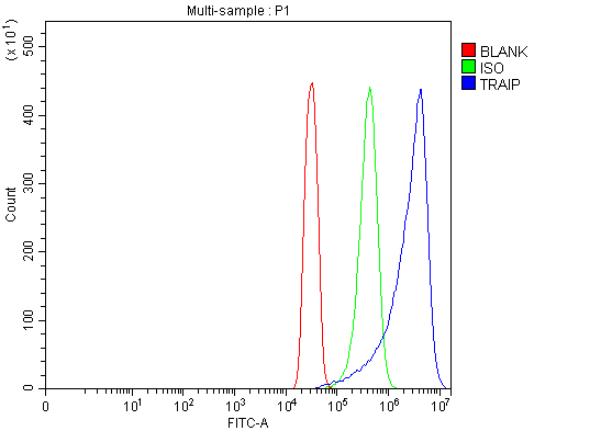
(Figure 3. Flow Cytometry analysis of SiHa cells using anti-TRIP/TRAIP antibody (AAA126507).Overlay histogram showing SiHa cells stained with AAA126507 (Blue line). The cells were blocked with 10% normal goat serum. And then incubated with rabbit anti-TRIP/TRAIP Antibody (AAA126507, 1 ug/1x10^6 cells) for 30 min at 20 degree C. DyLight488 conjugated goat anti-rabbit IgG was used as secondary antibody for 30 minutes at 20 degree C. Isotype control antibody (Green line) was rabbit IgG (1 ug/1x10^6) used under the same conditions. Unlabelled sample (Red line) was also used as a control.)
FCM/FACS (Flow Cytometry)
(Figure 3. Flow Cytometry analysis of SiHa cells using anti-TRIP/TRAIP antibody (AAA126507).Overlay histogram showing SiHa cells stained with AAA126507 (Blue line). The cells were blocked with 10% normal goat serum. And then incubated with rabbit anti-TRIP/TRAIP Antibody (AAA126507, 1 ug/1x10^6 cells) for 30 min at 20 degree C. DyLight488 conjugated goat anti-rabbit IgG was used as secondary antibody for 30 minutes at 20 degree C. Isotype control antibody (Green line) was rabbit IgG (1 ug/1x10^6) used under the same conditions. Unlabelled sample (Red line) was also used as a control.)
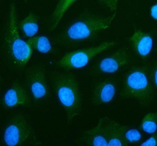
TRIP/TRAIP, Polyclonal Antibody (Cat# AAA126507)
Full Name
Anti-TRIP/TRAIP Antibody Picoband
Gene Names
TRAIP; TRIP; RNF206
Reactivity
Human, Mouse, Rat
Applications
Flow Cytometry, Immunofluorescence, Immunocytochemistry, Western Blot
Purity
Immunogen affinity purified.
Pricing
 FCM/FACS (Flow Cytometry)
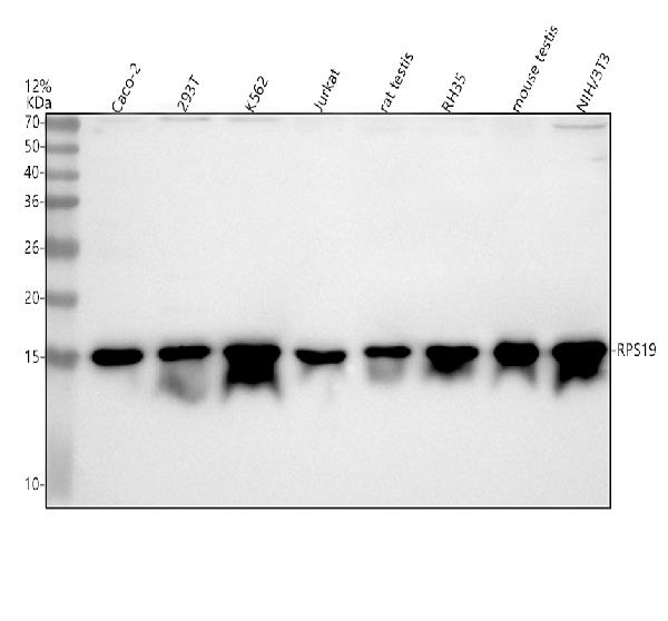
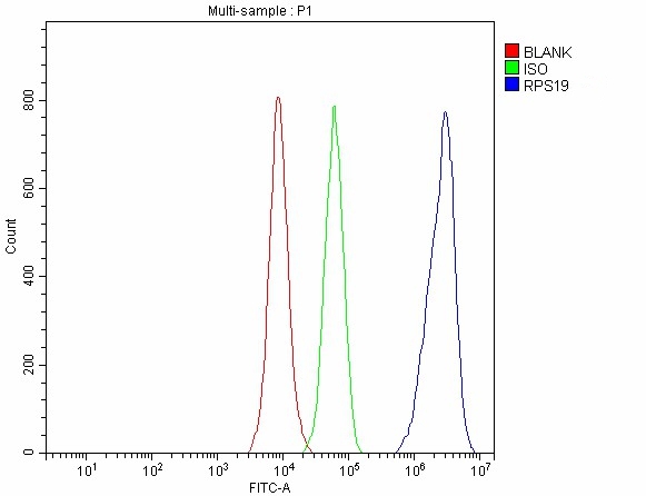
(Figure 3. Flow Cytometry analysis of 293T cells using anti-RPS19 antibody (AAA127107).Overlay histogram showing 293T cells stained with AAA127107 (Blue line). To facilitate intracellular staining, cells were fixed with 4% paraformaldehyde and permeabilized with permeabilization buffer. The cells were blocked with 10% normal goat serum. And then incubated with rabbit anti-RPS19 Antibody (AAA127107, 1ug/1x106 cells) for 30 min at 20 degree C. DyLight488 conjugated goat anti-rabbit IgG was used as secondary antibody for 30 minutes at 20 degree C. Isotype control antibody (Green line) was rabbit IgG (1ug/1x106) used under the same conditions. Unlabelled sample without incubation with primary antibody and secondary antibody (Red line) was used as a blank control.)
FCM/FACS (Flow Cytometry)
(Figure 3. Flow Cytometry analysis of 293T cells using anti-RPS19 antibody (AAA127107).Overlay histogram showing 293T cells stained with AAA127107 (Blue line). To facilitate intracellular staining, cells were fixed with 4% paraformaldehyde and permeabilized with permeabilization buffer. The cells were blocked with 10% normal goat serum. And then incubated with rabbit anti-RPS19 Antibody (AAA127107, 1ug/1x106 cells) for 30 min at 20 degree C. DyLight488 conjugated goat anti-rabbit IgG was used as secondary antibody for 30 minutes at 20 degree C. Isotype control antibody (Green line) was rabbit IgG (1ug/1x106) used under the same conditions. Unlabelled sample without incubation with primary antibody and secondary antibody (Red line) was used as a blank control.)
RPS19, Polyclonal Antibody (Cat# AAA127107)
Full Name
Anti-RPS19 Antibody Picoband
Gene Names
RPS19; DBA; S19; DBA1
Reactivity
Human, Mouse, Rat
Applications
Flow Cytometry, Immunofluorescence, Immunocytochemistry, Western Blot
Purity
Immunogen affinity purified.
Pricing
 FCM/FACS (Flow Cytometry)
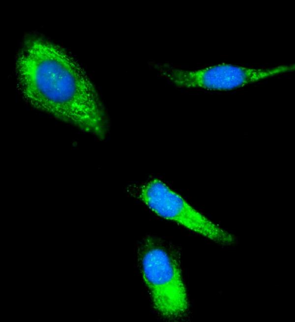
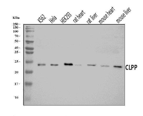
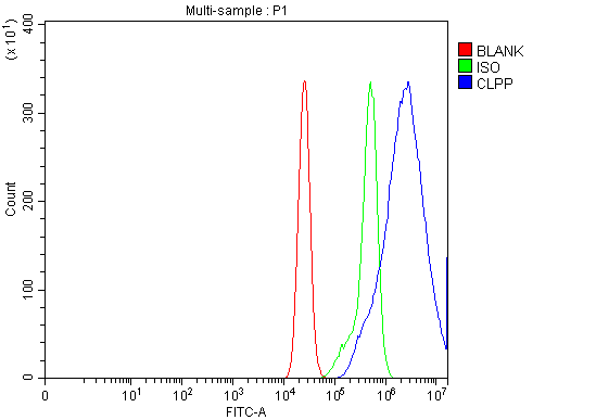
(Figure 3. Flow Cytometry analysis of CACO-2 cells using anti-CLPP antibody (AAA126111).Overlay histogram showing CACO-2 cells stained with AAA126111 (Blue line). The cells were blocked with 10% normal goat serum. And then incubated with rabbit anti-CLPP Antibody (AAA126111, 1 ug/1x10^6 cells) for 30 min at 20 degree C. DyLight488 conjugated goat anti-rabbit IgG was used as secondary antibody for 30 minutes at 20 degree C. Isotype control antibody (Green line) was rabbit IgG (1 ug/1x10^6) used under the same conditions. Unlabelled sample (Red line) was also used as a control.)
FCM/FACS (Flow Cytometry)
(Figure 3. Flow Cytometry analysis of CACO-2 cells using anti-CLPP antibody (AAA126111).Overlay histogram showing CACO-2 cells stained with AAA126111 (Blue line). The cells were blocked with 10% normal goat serum. And then incubated with rabbit anti-CLPP Antibody (AAA126111, 1 ug/1x10^6 cells) for 30 min at 20 degree C. DyLight488 conjugated goat anti-rabbit IgG was used as secondary antibody for 30 minutes at 20 degree C. Isotype control antibody (Green line) was rabbit IgG (1 ug/1x10^6) used under the same conditions. Unlabelled sample (Red line) was also used as a control.)
CLPP, Polyclonal Antibody (Cat# AAA126111)
Full Name
Anti-CLPP Antibody Picoband
Gene Names
CLPP; DFNB81; PRLTS3
Reactivity
Human, Mouse, Rat
Applications
Flow Cytometry, Immunofluorescence, Immunocytochemistry, Western Blot
Purity
Immunogen affinity purified.
Pricing
 IHC (Immunohistochemistry)
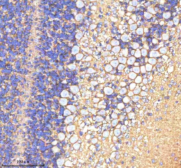
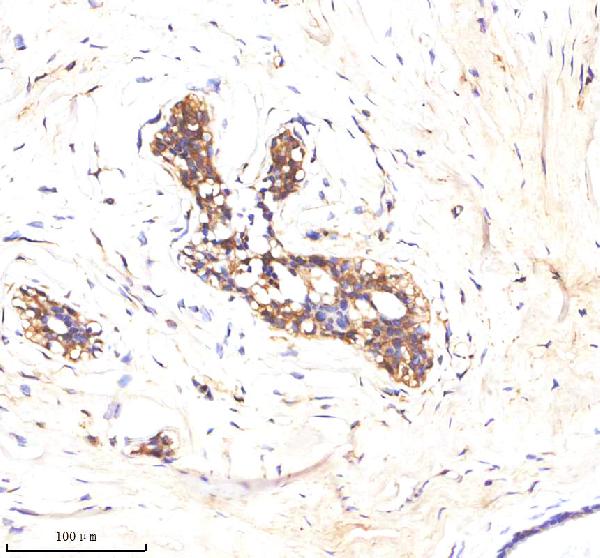
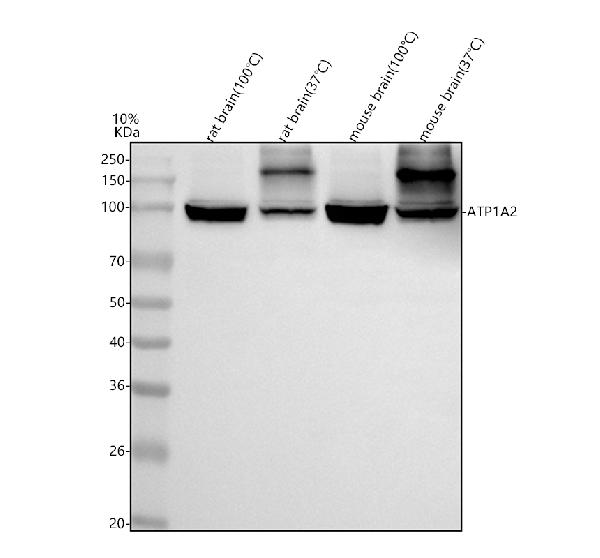
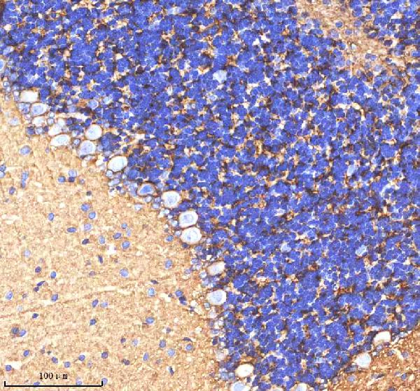
(Figure 4. IHC analysis of ATP1A2 using anti-ATP1A2 antibody (AAA127097).ATP1A2 was detected in a paraffin-embedded section of rat cerebellum tissue. Heat mediated antigen retrieval was performed in EDTA buffer (pH 8.0, epitope retrieval solution). The tissue section was blocked with 10% goat serum. The tissue section was then incubated with 2ug/ml rabbit anti-ATP1A2 Antibody (AAA127097) overnight at 4 degree C. Peroxidase Conjugated Goat Anti-rabbit IgG was used as secondary antibody and incubated for 30 minutes at 37 degree C. The tissue section was developed using HRP Conjugated Rabbit IgG Super Vision Assay Kit with DAB as the chromogen.)
IHC (Immunohistochemistry)
(Figure 4. IHC analysis of ATP1A2 using anti-ATP1A2 antibody (AAA127097).ATP1A2 was detected in a paraffin-embedded section of rat cerebellum tissue. Heat mediated antigen retrieval was performed in EDTA buffer (pH 8.0, epitope retrieval solution). The tissue section was blocked with 10% goat serum. The tissue section was then incubated with 2ug/ml rabbit anti-ATP1A2 Antibody (AAA127097) overnight at 4 degree C. Peroxidase Conjugated Goat Anti-rabbit IgG was used as secondary antibody and incubated for 30 minutes at 37 degree C. The tissue section was developed using HRP Conjugated Rabbit IgG Super Vision Assay Kit with DAB as the chromogen.)
ATP1A2, Polyclonal Antibody (Cat# AAA127097)
Full Name
Anti-ATP1A2 Antibody Picoband
Gene Names
ATP1A2; FHM2; MHP2
Reactivity
Human, Mouse, Rat
Applications
Immunohistochemistry, Western Blot
Purity
Immunogen affinity purified.
Pricing
 FCM/FACS (Flow Cytometry)
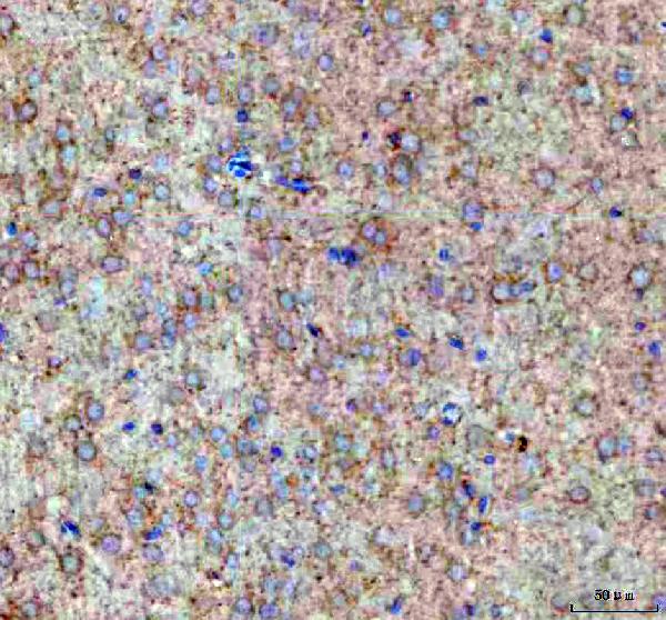
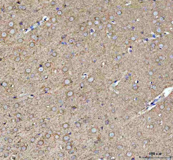
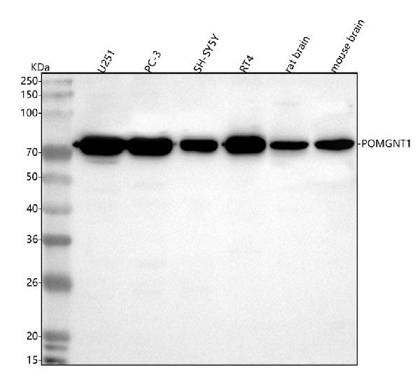
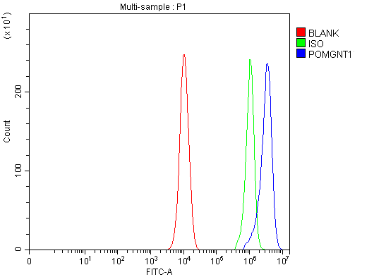
(Figure 4. Flow Cytometry analysis of PC-3 cells using anti-POMGNT1 antibody (AAA127182).Overlay histogram showing PC-3 cells stained with AAA127182 (Blue line). To facilitate intracellular staining, cells were fixed with 4% paraformaldehyde and permeabilized with permeabilization buffer. The cells were blocked with 10% normal goat serum. And then incubated with rabbit anti-POMGNT1 Antibody (AAA127182, 1ug/1x106 cells) for 30 min at 20 degree C. DyLight488 conjugated goat anti-rabbit IgG was used as secondary antibody for 30 minutes at 20 degree C. Isotype control antibody (Green line) was rabbit IgG (1ug/1x106) used under the same conditions. Unlabelled sample (Red line) was also used as a control.)
FCM/FACS (Flow Cytometry)
(Figure 4. Flow Cytometry analysis of PC-3 cells using anti-POMGNT1 antibody (AAA127182).Overlay histogram showing PC-3 cells stained with AAA127182 (Blue line). To facilitate intracellular staining, cells were fixed with 4% paraformaldehyde and permeabilized with permeabilization buffer. The cells were blocked with 10% normal goat serum. And then incubated with rabbit anti-POMGNT1 Antibody (AAA127182, 1ug/1x106 cells) for 30 min at 20 degree C. DyLight488 conjugated goat anti-rabbit IgG was used as secondary antibody for 30 minutes at 20 degree C. Isotype control antibody (Green line) was rabbit IgG (1ug/1x106) used under the same conditions. Unlabelled sample (Red line) was also used as a control.)
POMGNT1, Polyclonal Antibody (Cat# AAA127182)
Full Name
Anti-POMGNT1 Antibody Picoband
Gene Names
POMGNT1; MEB; GNTI.2; LGMD2O; GnT I.2; MGAT1.2; gnT-I.2
Reactivity
Human, Mouse, Rat
Applications
Western Blot, Immunohistochemistry, Flow Cytometry
Purity
Immunogen affinity purified.
Pricing
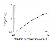 Standard Curve (Sample)
NUB1/NEDD8 Ultimate Buster 1, ELISA Kit (Cat# AAA196760)
Full Name
Human NUB1/NEDD8 Ultimate Buster 1 ELISA Kit
Gene Names
NUB1; BS4; NUB1L; NYREN18
Reactivity
Human
Pricing
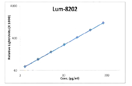 Standard Curve (Sample)
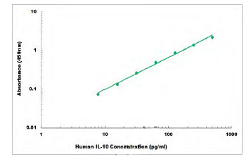 Standard Curve (Sample)
IL-10, ELISA Kit (Cat# AAA106076)
Full Name
Human IL-10 ELISA Kit
Gene Names
IL10; CSIF; TGIF; GVHDS; IL-10; IL10A
Reactivity
Human
Applications
Sandwich ELISA
Pricing
 SDS-PAGE
(Purity: Sample Purity Data. For specific information on a given lot, see related technical data sheet.)
SDS-PAGE
(Purity: Sample Purity Data. For specific information on a given lot, see related technical data sheet.)
RNF34 (CARP), Recombinant Protein (Cat# AAA73944)
Full Name
RNF34 (CARP), Active
Applications
Western Blot
Pricing
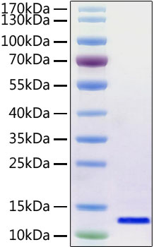 SDS-PAGE
(Figure-1:Recombinant 2019-nCoV envelope Protein was determined by SDS-PAGE with Coomassie Blue, showing a band at 12 kDa.)
SDS-PAGE
(Figure-1:Recombinant 2019-nCoV envelope Protein was determined by SDS-PAGE with Coomassie Blue, showing a band at 12 kDa.)
COVID 19 Envelope Coronavirus, Recombinant Protein (Cat# AAA78535)
Full Name
Recombinant SARS-CoV-2 Envelope Protein
Purity
>95% by SDS-PAGE.
Pricing