
Filters
Clonality
Type
Reactivity
Gene Name
Isotype
Host
Application
Clone
10000 results for "Recombinant Proteins" - showing 1200-1250


 FCM (Flow Cytometry)
(Figure 8. Flow Cytometry analysis of SiHa cells using anti-Astrin/Deepest/SPAG5 antibody (AAA19313).Overlay histogram showing SiHa cells stained with AAA19313 (Blue line). The cells were blocked with 10% normal goat serum. And then incubated with rabbit anti-Astrin/Deepest/SPAG5 Antibody (AAA19313, 1μg/1x106 cells) for 30 min at 20 degree C. DyLight®488 conjugated goat anti-rabbit IgG (5-10μg/1x106 cells) was used as secondary antibody for 30 minutes at 20 degree C. Isotype control antibody (Green line) was rabbit IgG (1μg/1x106) used under the same conditions. Unlabelled sample (Red line) was also used as a control.)
FCM (Flow Cytometry)
(Figure 8. Flow Cytometry analysis of SiHa cells using anti-Astrin/Deepest/SPAG5 antibody (AAA19313).Overlay histogram showing SiHa cells stained with AAA19313 (Blue line). The cells were blocked with 10% normal goat serum. And then incubated with rabbit anti-Astrin/Deepest/SPAG5 Antibody (AAA19313, 1μg/1x106 cells) for 30 min at 20 degree C. DyLight®488 conjugated goat anti-rabbit IgG (5-10μg/1x106 cells) was used as secondary antibody for 30 minutes at 20 degree C. Isotype control antibody (Green line) was rabbit IgG (1μg/1x106) used under the same conditions. Unlabelled sample (Red line) was also used as a control.)
Astrin/Deepest/SPAG5, Polyclonal Antibody (Cat# AAA19313)
Full Name
Anti-Astrin/Deepest/SPAG5 Antibody
Gene Names
SPAG5; MAP126; DEEPEST; hMAP126
Reactivity
Human
Applications
Western Blot, Immunohistochemistry, Immunocytochemistry, Immunofluorescence, Flow Cytometry, Direct ELISA
Purity
Immunogen affinity purified.
Pricing


 FCM (Flow Cytometry)
(Figure 7. Flow Cytometry analysis of Raji cells using anti-HIP1R antibody (AAA19533).Overlay histogram showing Raji cells stained with AAA19533 (Blue line). The cells were blocked with 10% normal goat serum. And then incubated with rabbit anti-HIP1R Antibody (AAA19533, 1 ug/1x10^6 cells) for 30 min at 20 degree C. DyLight488 conjugated goat anti-rabbit IgG was used as secondary antibody for 30 minutes at 20 degree C. Isotype control antibody (Green line) was rabbit IgG (1 ug/1x10^6) used under the same conditions. Unlabelled sample (Red line) was also used as a control.)
FCM (Flow Cytometry)
(Figure 7. Flow Cytometry analysis of Raji cells using anti-HIP1R antibody (AAA19533).Overlay histogram showing Raji cells stained with AAA19533 (Blue line). The cells were blocked with 10% normal goat serum. And then incubated with rabbit anti-HIP1R Antibody (AAA19533, 1 ug/1x10^6 cells) for 30 min at 20 degree C. DyLight488 conjugated goat anti-rabbit IgG was used as secondary antibody for 30 minutes at 20 degree C. Isotype control antibody (Green line) was rabbit IgG (1 ug/1x10^6) used under the same conditions. Unlabelled sample (Red line) was also used as a control.)
HIP1R, Polyclonal Antibody (Cat# AAA19533)
Full Name
Anti-HIP1R Antibody Picoband
Gene Names
HIP1R; HIP3; HIP12; ILWEQ
Reactivity
Human, Mouse, Rat, Monkey
Applications
Western Blot, Immunohistochemistry, Immunocytochemistry, Immunofluorescence, Flow Cytometry
Purity
Immunogen affinity purified.
Pricing
 Standard Curve (Sample)
NGF beta, ELISA Kit (Cat# AAA17907)
Full Name
Mouse NGF beta ELISA Kit
Gene Names
Ngf; Ngfb
Reactivity
Mouse
Applications
Sandwich ELISA
Pricing


 Application Data
(Detection limit for recombinant GST tagged RPS23 is ~0.3ng/ml as a capture antibody.)
Application Data
(Detection limit for recombinant GST tagged RPS23 is ~0.3ng/ml as a capture antibody.)
RPS23, Monoclonal Antibody (Cat# AAA25231)
Full Name
RPS23 (40S Ribosomal Protein S23, FLJ35016) (FITC)
Gene Names
RPS23; S23; BTDD; uS12; MABAS; MCINS; PAMAS
Reactivity
Human, Mouse, Rat
Applications
Immunofluorescence, Western Blot
Purity
Purified by Protein A Affinity Chromatography.
Pricing


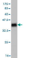
 WB (Western Blot)
(RAB7B monoclonal antibody. Western Blot analysis of RAB7B expression in A-431.)
WB (Western Blot)
(RAB7B monoclonal antibody. Western Blot analysis of RAB7B expression in A-431.)
RAB7B, Monoclonal Antibody (Cat# AAA24629)
Full Name
RAB7B (Ras-related Protein Rab-7b, RAB7) APC
Gene Names
RAB7B; RAB7
Reactivity
Human
Applications
Immunoprecipitation, Western Blot
Purity
Purified by Protein A Affinity Chromatography.
Pricing


 IF (Immunofluorescence)
(Immunofluorescence of monoclonal antibody to TOMM20 on HeLa cell. [antibody concentration 10ug/ml])
IF (Immunofluorescence)
(Immunofluorescence of monoclonal antibody to TOMM20 on HeLa cell. [antibody concentration 10ug/ml])
TOMM20, Monoclonal Antibody (Cat# AAA25866)
Full Name
TOMM20 (Mitochondrial Import Receptor Subunit TOM20 Homolog, TOM20, Mitochondrial 20kD Outer Membrane Protein, Outer Mitochondrial Membrane Receptor Tom20, KIAA0016, MAS20, MOM19)
Gene Names
TOMM20; MAS20; MOM19; TOM20
Reactivity
Human, Mouse, Rat
Applications
Immunofluorescence, Immunohistochemistry, Western Blot
Purity
Purified by Protein A Affinity Chromatography.
Pricing


 WB (Western Blot)
(Anti-ANP32A rabbit polyclonal antibody at 1:500 dilution Lane A: ANP32A konckout Hela Whole Cell Lysate Lane B: Hela Whole Cell Lysate Lysates/proteins at 20 ug per lane. Secondary Goat Anti-Rabbit IgG (H+L)/HRP at 1/10000 dilution. Developed using the ECL technique. Performed under reducing conditions. Predicted band size:28 kDa Observed band size:28 kDa)
WB (Western Blot)
(Anti-ANP32A rabbit polyclonal antibody at 1:500 dilution Lane A: ANP32A konckout Hela Whole Cell Lysate Lane B: Hela Whole Cell Lysate Lysates/proteins at 20 ug per lane. Secondary Goat Anti-Rabbit IgG (H+L)/HRP at 1/10000 dilution. Developed using the ECL technique. Performed under reducing conditions. Predicted band size:28 kDa Observed band size:28 kDa)
ANP32A, Polyclonal Antibody (Cat# AAA27748)
Full Name
Anti-ANP32A Antibody, Rabbit Polyclonal
Gene Names
ANP32A; LANP; MAPM; PP32; HPPCn; PHAP1; PHAPI; I1PP2A; C15orf1
Reactivity
Human
Applications
Western Blot, Immunohistochemistry, Immunocytochemistry, Immunofluorescence, Immunoprecipitation
Purity
Protein A & Antigen Affinity
Pricing


 IHC (Immunohistochemistry)
(Figure 8. IHC analysis of TAGLN/Transgelin using anti-TAGLN/Transgelin antibody (AAA19286).TAGLN/Transgelin was detected in paraffin-embedded section of rat testis tissue. Heat mediated antigen retrieval was performed in EDTA buffer (pH8. 0, epitope retrieval solution). The tissue section was blocked with 10% goat serum. The tissue section was then incubated with 2μg/ml rabbit anti-TAGLN/Transgelin Antibody (AAA19286) overnight at 4 degree C. Biotinylated goat anti-rabbit IgG was used as secondary antibody and incubated for 30 minutes at 37 degree C. The tissue section was developed using Strepavidin-Biotin-Complex (SABC) (Catalog # with DAB as the chromogen.)
IHC (Immunohistochemistry)
(Figure 8. IHC analysis of TAGLN/Transgelin using anti-TAGLN/Transgelin antibody (AAA19286).TAGLN/Transgelin was detected in paraffin-embedded section of rat testis tissue. Heat mediated antigen retrieval was performed in EDTA buffer (pH8. 0, epitope retrieval solution). The tissue section was blocked with 10% goat serum. The tissue section was then incubated with 2μg/ml rabbit anti-TAGLN/Transgelin Antibody (AAA19286) overnight at 4 degree C. Biotinylated goat anti-rabbit IgG was used as secondary antibody and incubated for 30 minutes at 37 degree C. The tissue section was developed using Strepavidin-Biotin-Complex (SABC) (Catalog # with DAB as the chromogen.)
TAGLN/Transgelin, Polyclonal Antibody (Cat# AAA19286)
Full Name
Anti-TAGLN/Transgelin Antibody
Gene Names
TAGLN; SM22; SMCC; TAGLN1; WS3-10
Reactivity
Human, Mouse, Rat
Applications
Western Blot, Immunohistochemistry, Immunocytochemistry, Immunofluorescence, Flow Cytometry, Direct ELISA
Purity
Immunogen affinity purified.
Pricing


 FCM (Flow Cytometry)
(Figure 8. Flow Cytometry analysis of U937 cells using anti-SEC14L3/TAP2 antibody (AAA19342).Overlay histogram showing U937 cells stained with AAA19342 (Blue line). The cells were blocked with 10% normal goat serum. And then incubated with rabbit anti-SEC14L3/TAP2 Antibody (AAA19342, 1μg/1x106 cells) for 30 min at 20 degree C. DyLight®488 conjugated goat anti-rabbit IgG (5-10μg/1x106 cells) was used as secondary antibody for 30 minutes at 20 degree C. Isotype control antibody (Green line) was rabbit IgG (1μg/1x106) used under the same conditions. Unlabelled sample (Red line) was also used as a control.)
FCM (Flow Cytometry)
(Figure 8. Flow Cytometry analysis of U937 cells using anti-SEC14L3/TAP2 antibody (AAA19342).Overlay histogram showing U937 cells stained with AAA19342 (Blue line). The cells were blocked with 10% normal goat serum. And then incubated with rabbit anti-SEC14L3/TAP2 Antibody (AAA19342, 1μg/1x106 cells) for 30 min at 20 degree C. DyLight®488 conjugated goat anti-rabbit IgG (5-10μg/1x106 cells) was used as secondary antibody for 30 minutes at 20 degree C. Isotype control antibody (Green line) was rabbit IgG (1μg/1x106) used under the same conditions. Unlabelled sample (Red line) was also used as a control.)
SEC14L3/TAP2, Polyclonal Antibody (Cat# AAA19342)
Full Name
Anti-SEC14L3/TAP2 Antibody
Gene Names
SEC14L3; TAP2
Reactivity
Human, Mouse, Rat
Applications
Western Blot, Immunohistochemistry, Immunocytochemistry, Immunofluorescence, Flow Cytometry, Direct ELISA
Purity
Immunogen affinity purified.
Pricing

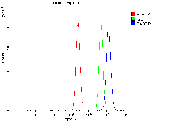 FCM (Flow Cytometry)
(Figure 6. Flow Cytometry analysis of MCF-7 cells using anti-RAB3IP antibody (AAA19919).Overlay histogram showing MCF-7 cells stained with AAA19919 (Blue line). To facilitate intracellular staining, cells were fixed with 4% paraformaldehyde and permeabilized with permeabilization buffer. The cells were blocked with 10% normal goat serum. And then incubated with rabbit anti-RAB3IP Antibody (AAA19919, 1ug/1x106 cells) for 30 min at 20 degree C. DyLight488 conjugated goat anti-rabbit IgG was used as secondary antibody for 30 minutes at 20 degree C. Isotype control antibody (Green line) was rabbit IgG (1ug/1x106) used under the same conditions. Unlabelled sample (Red line) was also used as a control.)
FCM (Flow Cytometry)
(Figure 6. Flow Cytometry analysis of MCF-7 cells using anti-RAB3IP antibody (AAA19919).Overlay histogram showing MCF-7 cells stained with AAA19919 (Blue line). To facilitate intracellular staining, cells were fixed with 4% paraformaldehyde and permeabilized with permeabilization buffer. The cells were blocked with 10% normal goat serum. And then incubated with rabbit anti-RAB3IP Antibody (AAA19919, 1ug/1x106 cells) for 30 min at 20 degree C. DyLight488 conjugated goat anti-rabbit IgG was used as secondary antibody for 30 minutes at 20 degree C. Isotype control antibody (Green line) was rabbit IgG (1ug/1x106) used under the same conditions. Unlabelled sample (Red line) was also used as a control.)
RAB3IP, Polyclonal Antibody (Cat# AAA19919)
Full Name
Anti-RAB3IP Antibody Picoband
Gene Names
RAB3IP; RABIN3
Reactivity
Human, Mouse, Rat
Applications
Western Blot, Immunohistochemistry, Flow Cytometry
Purity
Immunogen affinity purified.
Pricing

 FCM (Flow Cytometry)
(Figure 7. Flow Cytometry analysis of HEL cells using anti-LYPLA2 antibody (AAA19947).Overlay histogram showing HEL cells stained with AAA19947 (Blue line). To facilitate intracellular staining, cells were fixed with 4% paraformaldehyde and permeabilized with permeabilization buffer. The cells were blocked with 10% normal goat serum. And then incubated with rabbit anti-LYPLA2 Antibody (AAA19947, 1ug/1x106 cells) for 30 min at 20 degree C. DyLight488 conjugated goat anti-rabbit IgG was used as secondary antibody for 30 minutes at 20 degree C. Isotype control antibody (Green line) was rabbit IgG (1ug/1x106) used under the same conditions. Unlabelled sample (Red line) was also used as a control.)
FCM (Flow Cytometry)
(Figure 7. Flow Cytometry analysis of HEL cells using anti-LYPLA2 antibody (AAA19947).Overlay histogram showing HEL cells stained with AAA19947 (Blue line). To facilitate intracellular staining, cells were fixed with 4% paraformaldehyde and permeabilized with permeabilization buffer. The cells were blocked with 10% normal goat serum. And then incubated with rabbit anti-LYPLA2 Antibody (AAA19947, 1ug/1x106 cells) for 30 min at 20 degree C. DyLight488 conjugated goat anti-rabbit IgG was used as secondary antibody for 30 minutes at 20 degree C. Isotype control antibody (Green line) was rabbit IgG (1ug/1x106) used under the same conditions. Unlabelled sample (Red line) was also used as a control.)
LYPLA2, Polyclonal Antibody (Cat# AAA19947)
Full Name
Anti-LYPLA2 Antibody Picoband
Gene Names
LYPLA2; APT2; APT-2; DJ886K2.4
Reactivity
Human, Mouse, Rat
Applications
Western Blot, Immunohistochemistry, Immunocytochemistry, Immunofluorescence, Flow Cytometry
Purity
Immunogen affinity purified.
Pricing

 FCM (Flow Cytometry)
(Figure 8. Flow Cytometry analysis of JK cells using anti-LGI4 antibody (AAA19971).Overlay histogram showing JK cells stained with AAA19971 (Blue line). The cells were fixed with 4% paraformaldehyde and blocked with 10% normal goat serum. And then incubated with rabbit anti-LGI4 Antibody (AAA19971, 1ug/1x106 cells) for 30 min at 20 degree C. DyLight488 conjugated goat anti-rabbit IgG was used as secondary antibody for 30 minutes at 20 degree C. Isotype control antibody (Green line) was rabbit IgG (1ug/1x106) used under the same conditions. Unlabelled sample (Red line) was also used as a control.)
FCM (Flow Cytometry)
(Figure 8. Flow Cytometry analysis of JK cells using anti-LGI4 antibody (AAA19971).Overlay histogram showing JK cells stained with AAA19971 (Blue line). The cells were fixed with 4% paraformaldehyde and blocked with 10% normal goat serum. And then incubated with rabbit anti-LGI4 Antibody (AAA19971, 1ug/1x106 cells) for 30 min at 20 degree C. DyLight488 conjugated goat anti-rabbit IgG was used as secondary antibody for 30 minutes at 20 degree C. Isotype control antibody (Green line) was rabbit IgG (1ug/1x106) used under the same conditions. Unlabelled sample (Red line) was also used as a control.)
LGI4, Polyclonal Antibody (Cat# AAA19971)
Full Name
Anti-LGI4 Antibody Picoband
Gene Names
LGI4; LGIL3
Reactivity
Human, Mouse, Rat
Applications
Immunofluorescence, Immunohistochemistry, Immunocytochemistry, Western Blot, Flow Cytometry
Purity
Immunogen affinity purified.
Pricing

 FCM (Flow Cytometry)
(Figure 9. Flow Cytometry analysis of A431 cells using anti-BAP31/BCAP31 antibody (AAA19799).Overlay histogram showing A431 cells stained with AAA19799 (Blue line). To facilitate intracellular staining, cells were fixed with 4% paraformaldehyde and permeabilized with permeabilization buffer. The cells were blocked with 10% normal goat serum. And then incubated with rabbit anti-BAP31/BCAP31 Antibody (AAA19799, 1ug/1x106 cells) for 30 min at 20 degree C. DyLight488 conjugated goat anti-rabbit IgG was used as secondary antibody for 30 minutes at 20 degree C. Isotype control antibody (Green line) was rabbit IgG (1ug/1x106) used under the same conditions. Unlabelled sample without incubation with primary antibody and secondary antibody (Red line) was used as a blank control.)
FCM (Flow Cytometry)
(Figure 9. Flow Cytometry analysis of A431 cells using anti-BAP31/BCAP31 antibody (AAA19799).Overlay histogram showing A431 cells stained with AAA19799 (Blue line). To facilitate intracellular staining, cells were fixed with 4% paraformaldehyde and permeabilized with permeabilization buffer. The cells were blocked with 10% normal goat serum. And then incubated with rabbit anti-BAP31/BCAP31 Antibody (AAA19799, 1ug/1x106 cells) for 30 min at 20 degree C. DyLight488 conjugated goat anti-rabbit IgG was used as secondary antibody for 30 minutes at 20 degree C. Isotype control antibody (Green line) was rabbit IgG (1ug/1x106) used under the same conditions. Unlabelled sample without incubation with primary antibody and secondary antibody (Red line) was used as a blank control.)
BAP31/BCAP31, Polyclonal Antibody (Cat# AAA19799)
Full Name
Anti-BAP31/BCAP31 Antibody Picoband
Gene Names
BCAP31; CDM; BAP31; 6C6-AG; DXS1357E
Reactivity
Human, Mouse, Rat
Applications
Western Blot, Immunohistochemistry, Immunocytochemistry, Immunofluorescence, Immunoprecipitation, Flow Cytometry
Purity
Immunogen affinity purified.
Pricing


 IHC (Immunohistchemistry)
(Figure 7. IHC analysis of FOX2/RBM9/RBFOX2 using anti-FOX2/RBM9/RBFOX2 antibody (AAA19843).FOX2/RBM9/RBFOX2 was detected in a paraffin-embedded section of mouse kidney tissue. Heat mediated antigen retrieval was performed in EDTA buffer (pH 8.0, epitope retrieval solution). The tissue section was blocked with 10% goat serum. The tissue section was then incubated with 2ug/ml rabbit anti-FOX2/RBM9/RBFOX2 Antibody (AAA19843) overnight at 4 degree C. Peroxidase Conjugated Goat Anti-rabbit IgG was used as secondary antibody and incubated for 30 minutes at 37 degree C. The tissue section was developed using HRP Conjugated Rabbit IgG Super Vision Assay Kit ( epitope retrieval solution). The tissue section was blocked with 10% goat serum. The tissue section was then incubated with 2ug/ml rabbit anti-FOX2/RBM9/RBFOX2 Antibody (AAA19843) overnight at 4 degree C. Peroxidase Conjugated Goat Anti-rabbit IgG was used as secondary antibody and incubated for 30 minutes at 37 degree C. The tissue section was developed using HRP Conjugated Rabbit IgG Super Vision Assay Kit ( cells were fixed with 4% paraformaldehyde and permeabilized with permeabilization buffer. The cells were blocked with 10% normal goat serum. And then incubated with rabbit anti-FOX2/RBM9/RBFOX2 Antibody (AAA19843, 1ug/1x106 cells) for 30 min at 20 degree C. DyLight488 conjugated goat anti-rabbit IgG was used as secondary antibody for 30 minutes at 20 degree C. Isotype control antibody (Green line) was rabbit IgG (1ug/1x106) used under the same conditions. Unlabelled sample (Red line) was also used as a control.)
IHC (Immunohistchemistry)
(Figure 7. IHC analysis of FOX2/RBM9/RBFOX2 using anti-FOX2/RBM9/RBFOX2 antibody (AAA19843).FOX2/RBM9/RBFOX2 was detected in a paraffin-embedded section of mouse kidney tissue. Heat mediated antigen retrieval was performed in EDTA buffer (pH 8.0, epitope retrieval solution). The tissue section was blocked with 10% goat serum. The tissue section was then incubated with 2ug/ml rabbit anti-FOX2/RBM9/RBFOX2 Antibody (AAA19843) overnight at 4 degree C. Peroxidase Conjugated Goat Anti-rabbit IgG was used as secondary antibody and incubated for 30 minutes at 37 degree C. The tissue section was developed using HRP Conjugated Rabbit IgG Super Vision Assay Kit ( epitope retrieval solution). The tissue section was blocked with 10% goat serum. The tissue section was then incubated with 2ug/ml rabbit anti-FOX2/RBM9/RBFOX2 Antibody (AAA19843) overnight at 4 degree C. Peroxidase Conjugated Goat Anti-rabbit IgG was used as secondary antibody and incubated for 30 minutes at 37 degree C. The tissue section was developed using HRP Conjugated Rabbit IgG Super Vision Assay Kit ( cells were fixed with 4% paraformaldehyde and permeabilized with permeabilization buffer. The cells were blocked with 10% normal goat serum. And then incubated with rabbit anti-FOX2/RBM9/RBFOX2 Antibody (AAA19843, 1ug/1x106 cells) for 30 min at 20 degree C. DyLight488 conjugated goat anti-rabbit IgG was used as secondary antibody for 30 minutes at 20 degree C. Isotype control antibody (Green line) was rabbit IgG (1ug/1x106) used under the same conditions. Unlabelled sample (Red line) was also used as a control.)
FOX2/RBM9/RBFOX2, Polyclonal Antibody (Cat# AAA19843)
Full Name
Anti-FOX2/RBM9/RBFOX2 Antibody Picoband
Reactivity
Human, Mouse, Rat
Applications
Flow Cytometry, Immunofluorescence, Immunohistochemistry, Western Blot
Purity
Immunogen affinity purified.
Pricing
 IHC (Immunohistochemistry)
(Figure 15. IHC analysis of NRTN using anti-NRTN antibody (AAA19855).NRTN was detected in a paraffin-embedded section of rat brain tissue. Heat mediated antigen retrieval was performed in EDTA buffer (pH 8.0, epitope retrieval solution). The tissue section was blocked with 10% goat serum. The tissue section was then incubated with 2ug/ml rabbit anti-NRTN Antibody (AAA19855) overnight at 4 degree C. Peroxidase Conjugated Goat Anti-rabbit IgG was used as secondary antibody and incubated for 30 minutes at 37 degree C. The tissue section was developed using HRP Conjugated Rabbit IgG Super Vision Assay Kit ( epitope retrieval solution). The tissue section was blocked with 10% goat serum. The tissue section was then incubated with 2ug/ml rabbit anti-NRTN Antibody (AAA19855) overnight at 4 degree C. Peroxidase Conjugated Goat Anti-rabbit IgG was used as secondary antibody and incubated for 30 minutes at 37 degree C. The tissue section was developed using HRP Conjugated Rabbit IgG Super Vision Assay Kit ( epitope retrieval solution). The tissue section was blocked with 10% goat serum. The tissue section was then incubated with 2ug/ml rabbit anti-NRTN Antibody (AAA19855) overnight at 4 degree C. Peroxidase Conjugated Goat Anti-rabbit IgG was used as secondary antibody and incubated for 30 minutes at 37 degree C. The tissue section was developed using HRP Conjugated Rabbit IgG Super Vision Assay Kit ( epitope retrieval solution). The tissue section was blocked with 10% goat serum. The tissue section was then incubated with 2ug/ml rabbit anti-NRTN Antibody (AAA19855) overnight at 4 degree C. Peroxidase Conjugated Goat Anti-rabbit IgG was used as secondary antibody and incubated for 30 minutes at 37 degree C. The tissue section was developed using HRP Conjugated Rabbit IgG Super Vision Assay Kit ( epitope retrieval solution). The tissue section was blocked with 10% goat serum. The tissue section was then incubated with 2ug/ml rabbit anti-NRTN Antibody (AAA19855) overnight at 4 degree C. Peroxidase Conjugated Goat Anti-rabbit IgG was used as secondary antibody and incubated for 30 minutes at 37 degree C. The tissue section was developed using HRP Conjugated Rabbit IgG Super Vision Assay Kit ( epitope retrieval solution). The tissue section was blocked with 10% goat serum. The tissue section was then incubated with 2ug/ml rabbit anti-NRTN Antibody (AAA19855) overnight at 4 degree C. Peroxidase Conjugated Goat Anti-rabbit IgG was used as secondary antibody and incubated for 30 minutes at 37 degree C. The tissue section was developed using HRP Conjugated Rabbit IgG Super Vision Assay Kit ( epitope retrieval solution). The tissue section was blocked with 10% goat serum. The tissue section was then incubated with 2ug/ml rabbit anti-NRTN Antibody (AAA19855) overnight at 4 degree C. Peroxidase Conjugated Goat Anti-rabbit IgG was used as secondary antibody and incubated for 30 minutes at 37 degree C. The tissue section was developed using HRP Conjugated Rabbit IgG Super Vision Assay Kit ( epitope retrieval solution). The tissue section was blocked with 10% goat serum. The tissue section was then incubated with 2ug/ml rabbit anti-NRTN Antibody (AAA19855) overnight at 4 degree C. Peroxidase Conjugated Goat Anti-rabbit IgG was used as secondary antibody and incubated for 30 minutes at 37 degree C. The tissue section was developed using HRP Conjugated Rabbit IgG Super Vision Assay Kit ( epitope retrieval solution). The tissue section was blocked with 10% goat serum. The tissue section was then incubated with 2ug/ml rabbit anti-NRTN Antibody (AAA19855) overnight at 4 degree C. Peroxidase Conjugated Goat Anti-rabbit IgG was used as secondary antibody and incubated for 30 minutes at 37 degree C. The tissue section was developed using HRP Conjugated Rabbit IgG Super Vision Assay Kit ( epitope retrieval solution). The tissue section was blocked with 10% goat serum. The tissue section was then incubated with 2ug/ml rabbit anti-NRTN Antibody (AAA19855) overnight at 4 degree C. Peroxidase Conjugated Goat Anti-rabbit IgG was used as secondary antibody and incubated for 30 minutes at 37 degree C. The tissue section was developed using HRP Conjugated Rabbit IgG Super Vision Assay Kit ( epitope retrieval solution). The tissue section was blocked with 10% goat serum. The tissue section was then incubated with 2ug/ml rabbit anti-NRTN Antibody (AAA19855) overnight at 4 degree C. Peroxidase Conjugated Goat Anti-rabbit IgG was used as secondary antibody and incubated for 30 minutes at 37 degree C. The tissue section was developed using HRP Conjugated Rabbit IgG Super Vision Assay Kit ( epitope retrieval solution). The tissue section was blocked with 10% goat serum. The tissue section was then incubated with 2ug/ml rabbit anti-NRTN Antibody (AAA19855) overnight at 4 degree C. Peroxidase Conjugated Goat Anti-rabbit IgG was used as secondary antibody and incubated for 30 minutes at 37 degree C. The tissue section was developed using HRP Conjugated Rabbit IgG Super Vision Assay Kit ( epitope retrieval solution). The tissue section was blocked with 10% goat serum. The tissue section was then incubated with 2ug/ml rabbit anti-NRTN Antibody (AAA19855) overnight at 4 degree C. Peroxidase Conjugated Goat Anti-rabbit IgG was used as secondary antibody and incubated for 30 minutes at 37 degree C. The tissue section was developed using HRP Conjugated Rabbit IgG Super Vision Assay Kit ( epitope retrieval solution). The tissue section was blocked with 10% goat serum. The tissue section was then incubated with 2ug/ml rabbit anti-NRTN Antibody (AAA19855) overnight at 4 degree C. Peroxidase Conjugated Goat Anti-rabbit IgG was used as secondary antibody and incubated for 30 minutes at 37 degree C. The tissue section was developed using HRP Conjugated Rabbit IgG Super Vision Assay Kit (Lane 2: human 293T whole cell lysates,Lane 3: mouse heart tissue lysates.After electrophoresis, proteins were transferred to a nitrocellulose membrane at 150 mA for 50-90 minutes. Blocked the membrane with 5% non-fat milk/TBS for 1.5 hour at RT. The membrane was incubated with rabbit anti-NRTN antigen affinity purified polyclonal antibody (#AAA19855) at 0.5ug/mL overnight at 4 degree C, then washed with TBS-0.1%Tween 3 times with 5 minutes each and probed with a goat anti-rabbit IgG-HRP secondary antibody at a dilution of 1:5000 for 1.5 hour at RT. The signal is developed using an Enhanced Chemiluminescent detection (ECL) kit with Tanon 5200 system. A specific band was detected for NRTN at approximately 22 kDa. The expected band size for NRTN is at 22 kDa.)
IHC (Immunohistochemistry)
(Figure 15. IHC analysis of NRTN using anti-NRTN antibody (AAA19855).NRTN was detected in a paraffin-embedded section of rat brain tissue. Heat mediated antigen retrieval was performed in EDTA buffer (pH 8.0, epitope retrieval solution). The tissue section was blocked with 10% goat serum. The tissue section was then incubated with 2ug/ml rabbit anti-NRTN Antibody (AAA19855) overnight at 4 degree C. Peroxidase Conjugated Goat Anti-rabbit IgG was used as secondary antibody and incubated for 30 minutes at 37 degree C. The tissue section was developed using HRP Conjugated Rabbit IgG Super Vision Assay Kit ( epitope retrieval solution). The tissue section was blocked with 10% goat serum. The tissue section was then incubated with 2ug/ml rabbit anti-NRTN Antibody (AAA19855) overnight at 4 degree C. Peroxidase Conjugated Goat Anti-rabbit IgG was used as secondary antibody and incubated for 30 minutes at 37 degree C. The tissue section was developed using HRP Conjugated Rabbit IgG Super Vision Assay Kit ( epitope retrieval solution). The tissue section was blocked with 10% goat serum. The tissue section was then incubated with 2ug/ml rabbit anti-NRTN Antibody (AAA19855) overnight at 4 degree C. Peroxidase Conjugated Goat Anti-rabbit IgG was used as secondary antibody and incubated for 30 minutes at 37 degree C. The tissue section was developed using HRP Conjugated Rabbit IgG Super Vision Assay Kit ( epitope retrieval solution). The tissue section was blocked with 10% goat serum. The tissue section was then incubated with 2ug/ml rabbit anti-NRTN Antibody (AAA19855) overnight at 4 degree C. Peroxidase Conjugated Goat Anti-rabbit IgG was used as secondary antibody and incubated for 30 minutes at 37 degree C. The tissue section was developed using HRP Conjugated Rabbit IgG Super Vision Assay Kit ( epitope retrieval solution). The tissue section was blocked with 10% goat serum. The tissue section was then incubated with 2ug/ml rabbit anti-NRTN Antibody (AAA19855) overnight at 4 degree C. Peroxidase Conjugated Goat Anti-rabbit IgG was used as secondary antibody and incubated for 30 minutes at 37 degree C. The tissue section was developed using HRP Conjugated Rabbit IgG Super Vision Assay Kit ( epitope retrieval solution). The tissue section was blocked with 10% goat serum. The tissue section was then incubated with 2ug/ml rabbit anti-NRTN Antibody (AAA19855) overnight at 4 degree C. Peroxidase Conjugated Goat Anti-rabbit IgG was used as secondary antibody and incubated for 30 minutes at 37 degree C. The tissue section was developed using HRP Conjugated Rabbit IgG Super Vision Assay Kit ( epitope retrieval solution). The tissue section was blocked with 10% goat serum. The tissue section was then incubated with 2ug/ml rabbit anti-NRTN Antibody (AAA19855) overnight at 4 degree C. Peroxidase Conjugated Goat Anti-rabbit IgG was used as secondary antibody and incubated for 30 minutes at 37 degree C. The tissue section was developed using HRP Conjugated Rabbit IgG Super Vision Assay Kit ( epitope retrieval solution). The tissue section was blocked with 10% goat serum. The tissue section was then incubated with 2ug/ml rabbit anti-NRTN Antibody (AAA19855) overnight at 4 degree C. Peroxidase Conjugated Goat Anti-rabbit IgG was used as secondary antibody and incubated for 30 minutes at 37 degree C. The tissue section was developed using HRP Conjugated Rabbit IgG Super Vision Assay Kit ( epitope retrieval solution). The tissue section was blocked with 10% goat serum. The tissue section was then incubated with 2ug/ml rabbit anti-NRTN Antibody (AAA19855) overnight at 4 degree C. Peroxidase Conjugated Goat Anti-rabbit IgG was used as secondary antibody and incubated for 30 minutes at 37 degree C. The tissue section was developed using HRP Conjugated Rabbit IgG Super Vision Assay Kit ( epitope retrieval solution). The tissue section was blocked with 10% goat serum. The tissue section was then incubated with 2ug/ml rabbit anti-NRTN Antibody (AAA19855) overnight at 4 degree C. Peroxidase Conjugated Goat Anti-rabbit IgG was used as secondary antibody and incubated for 30 minutes at 37 degree C. The tissue section was developed using HRP Conjugated Rabbit IgG Super Vision Assay Kit ( epitope retrieval solution). The tissue section was blocked with 10% goat serum. The tissue section was then incubated with 2ug/ml rabbit anti-NRTN Antibody (AAA19855) overnight at 4 degree C. Peroxidase Conjugated Goat Anti-rabbit IgG was used as secondary antibody and incubated for 30 minutes at 37 degree C. The tissue section was developed using HRP Conjugated Rabbit IgG Super Vision Assay Kit ( epitope retrieval solution). The tissue section was blocked with 10% goat serum. The tissue section was then incubated with 2ug/ml rabbit anti-NRTN Antibody (AAA19855) overnight at 4 degree C. Peroxidase Conjugated Goat Anti-rabbit IgG was used as secondary antibody and incubated for 30 minutes at 37 degree C. The tissue section was developed using HRP Conjugated Rabbit IgG Super Vision Assay Kit ( epitope retrieval solution). The tissue section was blocked with 10% goat serum. The tissue section was then incubated with 2ug/ml rabbit anti-NRTN Antibody (AAA19855) overnight at 4 degree C. Peroxidase Conjugated Goat Anti-rabbit IgG was used as secondary antibody and incubated for 30 minutes at 37 degree C. The tissue section was developed using HRP Conjugated Rabbit IgG Super Vision Assay Kit ( epitope retrieval solution). The tissue section was blocked with 10% goat serum. The tissue section was then incubated with 2ug/ml rabbit anti-NRTN Antibody (AAA19855) overnight at 4 degree C. Peroxidase Conjugated Goat Anti-rabbit IgG was used as secondary antibody and incubated for 30 minutes at 37 degree C. The tissue section was developed using HRP Conjugated Rabbit IgG Super Vision Assay Kit (Lane 2: human 293T whole cell lysates,Lane 3: mouse heart tissue lysates.After electrophoresis, proteins were transferred to a nitrocellulose membrane at 150 mA for 50-90 minutes. Blocked the membrane with 5% non-fat milk/TBS for 1.5 hour at RT. The membrane was incubated with rabbit anti-NRTN antigen affinity purified polyclonal antibody (#AAA19855) at 0.5ug/mL overnight at 4 degree C, then washed with TBS-0.1%Tween 3 times with 5 minutes each and probed with a goat anti-rabbit IgG-HRP secondary antibody at a dilution of 1:5000 for 1.5 hour at RT. The signal is developed using an Enhanced Chemiluminescent detection (ECL) kit with Tanon 5200 system. A specific band was detected for NRTN at approximately 22 kDa. The expected band size for NRTN is at 22 kDa.)
NRTN, Polyclonal Antibody (Cat# AAA19855)
Full Name
Anti-NRTN Antibody Picoband
Gene Names
NRTN; NTN
Reactivity
Human, Mouse, Rat
Applications
Immunohistochemistry, Western Blot
Purity
Immunogen affinity purified.
Pricing
 Standard Curve (Sample)
Flt3 Ligand, ELISA Kit (Cat# AAA17895)
Full Name
Mouse Flt3 Ligand ELISA Kit
Gene Names
Flt3l; Ly72L; Flt3lg
Reactivity
Mouse
Applications
Sandwich ELISA
Pricing


 IHC (Immunohistchemistry)
(Immunohistochemistry of paraffin-embedded human colon carcinoma using TEFM antibody at dilution of 1:100 (40x lens).)
IHC (Immunohistchemistry)
(Immunohistochemistry of paraffin-embedded human colon carcinoma using TEFM antibody at dilution of 1:100 (40x lens).)
TEFM, Polyclonal Antibody (Cat# AAA28194)
Full Name
TEFM Polyclonal Antibody
Gene Names
TEFM; C17orf42
Reactivity
Human, Mouse, Rat
Applications
Western Blot
Purity
Affinity Purification
Pricing


 IF (Immunofluorescence)
(Immunofluorescence of monoclonal antibody to RPL36A on HeLa cell. [antibody concentration 10 ug/ml])
IF (Immunofluorescence)
(Immunofluorescence of monoclonal antibody to RPL36A on HeLa cell. [antibody concentration 10 ug/ml])
RPL36A, Monoclonal Antibody (Cat# AAA26522)
Full Name
RPL36A (Ribosomal Protein L36a, L44L, MGC72020, MIG6, RPL44) (HRP)
Gene Names
RPL36A; L36A; L44L; MIG6; RPL44
Applications
Immunofluorescence, Western Blot
Purity
Purified
Pricing


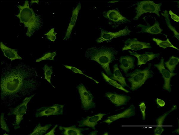 IF (Immunofluorescence)
(Immunofluorescence of monoclonal antibody to RPL36A on HeLa cell. [antibody concentration 10 ug/ml])
IF (Immunofluorescence)
(Immunofluorescence of monoclonal antibody to RPL36A on HeLa cell. [antibody concentration 10 ug/ml])
RPL36A, Monoclonal Antibody (Cat# AAA26389)
Full Name
RPL36A (Ribosomal Protein L36a, L44L, MGC72020, MIG6, RPL44) (FITC)
Gene Names
RPL36A; L36A; L44L; MIG6; RPL44
Applications
Immunofluorescence, Western Blot
Purity
Purified
Pricing


 WB (Western Blot)
(Western Blot detection against Immunogen (34.21kD).)
WB (Western Blot)
(Western Blot detection against Immunogen (34.21kD).)
SMAD5, Monoclonal Antibody (Cat# AAA24956)
Full Name
SMAD5 (Mothers Against Decapentaplegic Homolog 5, Mothers Against DPP Homolog 5, MAD Homolog 5, Mad5, SMAD Family Member 5, SMAD 5, Smad5, JV5-1, MADH5, DKFZp781C1895, DKFZp781O1323) (Biotin)
Gene Names
SMAD5; DWFC; JV5-1; MADH5
Reactivity
Human
Applications
Immunofluorescence, Western Blot
Purity
Purified by Protein A Affinity Chromatography.
Pricing


 IHC (Immunohistochemistry)
(Immunohistochemistry of paraffin-embedded mouse heart using CSRP3 Antibody at dilution of 1:100 (40x lens).)
IHC (Immunohistochemistry)
(Immunohistochemistry of paraffin-embedded mouse heart using CSRP3 Antibody at dilution of 1:100 (40x lens).)
CSRP3, Polyclonal Antibody (Cat# AAA28177)
Full Name
CSRP3 Polyclonal Antibody
Gene Names
CSRP3; CLP; MLP; CRP3; LMO4; CMD1M; CMH12
Reactivity
Human, Mouse
Applications
Western Blot, Immunohistochemistry, Immunofluorescence
Purity
Affinity Purification
Pricing
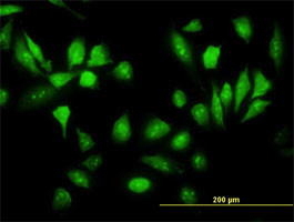


 Application Data
(Detection limit for recombinant GST tagged RPS5 is ~3ng/ml as a capture antibody.)
Application Data
(Detection limit for recombinant GST tagged RPS5 is ~3ng/ml as a capture antibody.)
RPS5, Monoclonal Antibody (Cat# AAA25526)
Full Name
RPS5 (40S Ribosomal Protein S5) (HRP)
Gene Names
RPS5; S5
Reactivity
Human, Mouse, Rat
Applications
Western Blot
Purity
Purified by Protein A Affinity Chromatography.
Pricing


 WB (Western Blot)
(DDX56 monoclonal antibody. Western Blot analysis of DDX56 expression in PC-12.)
WB (Western Blot)
(DDX56 monoclonal antibody. Western Blot analysis of DDX56 expression in PC-12.)
DDX56, Monoclonal Antibody (Cat# AAA24780)
Full Name
DDX56 (Probable ATP-dependent RNA Helicase DDX56, ATP-dependent 61kD Nucleolar RNA Helicase, DEAD Box Protein 21, DEAD Box Protein 56, DDX21, NOH61) (Biotin)
Reactivity
Human, Rat
Applications
Immunofluorescence, Immunohistochemistry, Western Blot
Purity
Purified by Protein A Affinity Chromatography.
Pricing


 Application Data
(Proximity Ligation Analysis (PLA) of protein-protein interactions between CASP3 and HCLS1. HeLa cells were stained with anti-CASP3 rabbit purified polyclonal 1:1200 and anti-HCLS1 mouse monoclonal antibody 1:50. Signals were detected by Duolink® 30 Detection Kit 613 (red), and nuclei were counterstained with DAPI (blue). Each red dot represents the detection of protein-protein interaction complex.)
Application Data
(Proximity Ligation Analysis (PLA) of protein-protein interactions between CASP3 and HCLS1. HeLa cells were stained with anti-CASP3 rabbit purified polyclonal 1:1200 and anti-HCLS1 mouse monoclonal antibody 1:50. Signals were detected by Duolink® 30 Detection Kit 613 (red), and nuclei were counterstained with DAPI (blue). Each red dot represents the detection of protein-protein interaction complex.)
HCLS1, Monoclonal Antibody (Cat# AAA24530)
Full Name
HCLS1 (Hematopoietic Lineage Cell-specific Protein, Hematopoietic Cell-specific LYN Substrate 1, LckBP1, p75, HS1) APC
Gene Names
HCLS1; HS1; p75; CTTNL; lckBP1
Reactivity
Human
Applications
Immunohistochemistry, Western Blot
Purity
Purified by Protein A Affinity Chromatography.
Pricing




 WB (Western Blot)
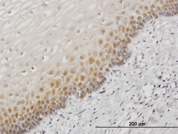
(HSPA1B monoclonal antibody (M05), clone 2D11. Western Blot analysis of HSPA1B expression in PC-12.)
WB (Western Blot)
(HSPA1B monoclonal antibody (M05), clone 2D11. Western Blot analysis of HSPA1B expression in PC-12.)
HSPA1B, Monoclonal Antibody (Cat# AAA26511)
Full Name
HSPA1B (Heat Shock 70kD Protein 1B, FLJ54328, HSP70-1B, HSP70-2, HSPA1A) (HRP)
Gene Names
HSPA1A; HSP72; HSPA1; HSP70I; HSP70-1; HSP70.1; HSP70-1A; HEL-S-103
Applications
Immunohistochemistry, Western Blot
Purity
Purified
Pricing


 IHC (Immunohistchemistry)
(Immunohistochemistry analysis of paraffin-embedded Human tonsil tissue using CD105/Endoglin Rabbit mAb (AAA28466) at a dilution of 1:10000 (40x lens). High pressure antigen retrieval performed with 0.01M Tris-EDTA Buffer (pH 9.0) prior to IHC staining.)
IHC (Immunohistchemistry)
(Immunohistochemistry analysis of paraffin-embedded Human tonsil tissue using CD105/Endoglin Rabbit mAb (AAA28466) at a dilution of 1:10000 (40x lens). High pressure antigen retrieval performed with 0.01M Tris-EDTA Buffer (pH 9.0) prior to IHC staining.)
CD105/Endoglin, Monoclonal Antibody (Cat# AAA28466)
Full Name
CD105/Endoglin Rabbit mAb
Reactivity
Human
Applications
Western Blot, Immunohistochemistry
Purity
Affinity purification
Pricing

 FCM (Flow Cytometry)
(Figure 7. Flow Cytometry analysis of MCF-7 cells using anti-PFKP antibody (AAA19883).Overlay histogram showing MCF-7 cells stained with AAA19883 (Blue line). To facilitate intracellular staining, cells were fixed with 4% paraformaldehyde and permeabilized with permeabilization buffer. The cells were blocked with 10% normal goat serum. And then incubated with rabbit anti-PFKP Antibody (AAA19883, 1ug/1x106 cells) for 30 min at 20 degree C. DyLight488 conjugated goat anti-rabbit IgG was used as secondary antibody for 30 minutes at 20 degree C. Isotype control antibody (Green line) was rabbit IgG (1ug/1x106) used under the same conditions. Unlabelled sample (Red line) was also used as a control.)
FCM (Flow Cytometry)
(Figure 7. Flow Cytometry analysis of MCF-7 cells using anti-PFKP antibody (AAA19883).Overlay histogram showing MCF-7 cells stained with AAA19883 (Blue line). To facilitate intracellular staining, cells were fixed with 4% paraformaldehyde and permeabilized with permeabilization buffer. The cells were blocked with 10% normal goat serum. And then incubated with rabbit anti-PFKP Antibody (AAA19883, 1ug/1x106 cells) for 30 min at 20 degree C. DyLight488 conjugated goat anti-rabbit IgG was used as secondary antibody for 30 minutes at 20 degree C. Isotype control antibody (Green line) was rabbit IgG (1ug/1x106) used under the same conditions. Unlabelled sample (Red line) was also used as a control.)
PFKP, Polyclonal Antibody (Cat# AAA19883)
Full Name
Anti-PFKP Antibody Picoband
Gene Names
PFKP; PFKF; PFK-C
Reactivity
Human
Applications
Western Blot, Immunohistochemistry, Flow Cytometry
Purity
Immunogen affinity purified.
Pricing


 FCM (Flow Cytometry)
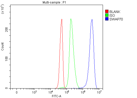
(Figure 6. Flow Cytometry analysis of Jurkat cells using anti-SWAP70 antibody (AAA19880).Overlay histogram showing Jurkat cells stained with AAA19880 (Blue line). To facilitate intracellular staining, cells were fixed with 4% paraformaldehyde and permeabilized with permeabilization buffer. The cells were blocked with 10% normal goat serum. And then incubated with rabbit anti-SWAP70 Antibody (AAA19880, 1ug/1x106 cells) for 30 min at 20 degree C. DyLight488 conjugated goat anti-rabbit IgG was used as secondary antibody for 30 minutes at 20 degree C. Isotype control antibody (Green line) was rabbit IgG (1ug/1x106) used under the same conditions. Unlabelled sample without incubation with primary antibody and secondary antibody (Red line) was used as a blank control.)
FCM (Flow Cytometry)
(Figure 6. Flow Cytometry analysis of Jurkat cells using anti-SWAP70 antibody (AAA19880).Overlay histogram showing Jurkat cells stained with AAA19880 (Blue line). To facilitate intracellular staining, cells were fixed with 4% paraformaldehyde and permeabilized with permeabilization buffer. The cells were blocked with 10% normal goat serum. And then incubated with rabbit anti-SWAP70 Antibody (AAA19880, 1ug/1x106 cells) for 30 min at 20 degree C. DyLight488 conjugated goat anti-rabbit IgG was used as secondary antibody for 30 minutes at 20 degree C. Isotype control antibody (Green line) was rabbit IgG (1ug/1x106) used under the same conditions. Unlabelled sample without incubation with primary antibody and secondary antibody (Red line) was used as a blank control.)
SWAP70, Polyclonal Antibody (Cat# AAA19880)
Full Name
Anti-SWAP70 Antibody Picoband
Gene Names
SWAP70; HSPC321; SWAP-70
Reactivity
Human
Applications
Flow Cytometry, Immunofluorescence, Immunohistochemistry, Western Blot
Purity
Immunogen affinity purified.
Pricing


 FCM (Flow Cytometry)
(Figure 6. Flow Cytometry analysis of MCF-7 cells using anti-PSAP antibody (AAA19741).Overlay histogram showing MCF-7 cells stained with AAA19741 (Blue line). To facilitate intracellular staining, cells were fixed with 4% paraformaldehyde and permeabilized with permeabilization buffer. The cells were blocked with 10% normal goat serum. And then incubated with rabbit anti-MCF-7 Antibody (AAA19741, 1ug/1x106 cells) for 30 min at 20 degree C. DyLight488 conjugated goat anti-rabbit IgG was used as secondary antibody for 30 minutes at 20 degree C. Isotype control antibody (Green line) was rabbit IgG (1ug/1x106) used under the same conditions. Unlabelled sample (Red line) was also used as a control.)
FCM (Flow Cytometry)
(Figure 6. Flow Cytometry analysis of MCF-7 cells using anti-PSAP antibody (AAA19741).Overlay histogram showing MCF-7 cells stained with AAA19741 (Blue line). To facilitate intracellular staining, cells were fixed with 4% paraformaldehyde and permeabilized with permeabilization buffer. The cells were blocked with 10% normal goat serum. And then incubated with rabbit anti-MCF-7 Antibody (AAA19741, 1ug/1x106 cells) for 30 min at 20 degree C. DyLight488 conjugated goat anti-rabbit IgG was used as secondary antibody for 30 minutes at 20 degree C. Isotype control antibody (Green line) was rabbit IgG (1ug/1x106) used under the same conditions. Unlabelled sample (Red line) was also used as a control.)
PSAP, Polyclonal Antibody (Cat# AAA19741)
Full Name
Anti-PSAP Antibody Picoband
Gene Names
PSAP; GLBA; SAP1
Reactivity
Human
Applications
Western Blot, Immunohistochemistry, Immunocytochemistry, Immunofluorescence, Flow Cytometry
Purity
Immunogen affinity purified.
Pricing

 FCM (Flow Cytometry)
(Figure 6. Flow Cytometry analysis of JK cells using anti-GIMAP4 antibody (AAA19951).Overlay histogram showing JK cells stained with AAA19951 (Blue line). To facilitate intracellular staining, cells were fixed with 4% paraformaldehyde and permeabilized with permeabilization buffer. The cells were blocked with 10% normal goat serum. And then incubated with rabbit anti-GIMAP4 Antibody (AAA19951, 1ug/1x106 cells) for 30 min at 20 degree C. DyLight488 conjugated goat anti-rabbit IgG was used as secondary antibody for 30 minutes at 20 degree C. Isotype control antibody (Green line) was rabbit IgG (1ug/1x106) used under the same conditions. Unlabelled sample without incubation with primary antibody and secondary antibody (Red line) was used as a blank control.)
FCM (Flow Cytometry)
(Figure 6. Flow Cytometry analysis of JK cells using anti-GIMAP4 antibody (AAA19951).Overlay histogram showing JK cells stained with AAA19951 (Blue line). To facilitate intracellular staining, cells were fixed with 4% paraformaldehyde and permeabilized with permeabilization buffer. The cells were blocked with 10% normal goat serum. And then incubated with rabbit anti-GIMAP4 Antibody (AAA19951, 1ug/1x106 cells) for 30 min at 20 degree C. DyLight488 conjugated goat anti-rabbit IgG was used as secondary antibody for 30 minutes at 20 degree C. Isotype control antibody (Green line) was rabbit IgG (1ug/1x106) used under the same conditions. Unlabelled sample without incubation with primary antibody and secondary antibody (Red line) was used as a blank control.)
GIMAP4, Polyclonal Antibody (Cat# AAA19951)
Full Name
Anti-GIMAP4 Antibody Picoband
Gene Names
GIMAP4; IAN1; IAN-1; IMAP4; MSTP062
Reactivity
Human, Mouse, Rat
Applications
Western Blot, Immunohistochemistry, Flow Cytometry
Purity
Immunogen affinity purified.
Pricing




 IHC (Immunohistochemistry)
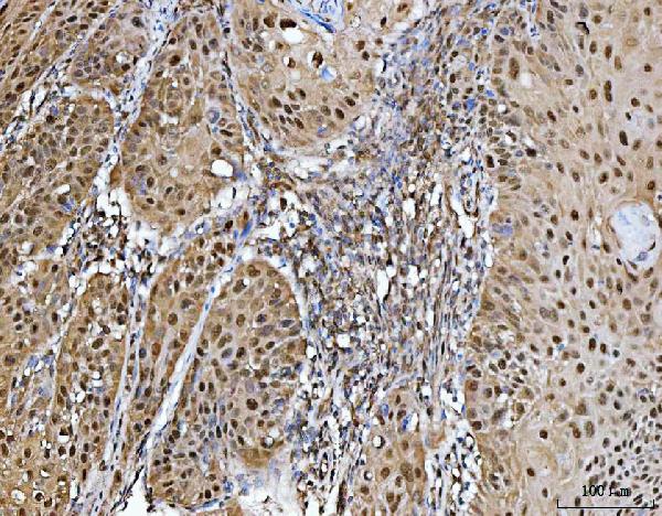
(Figure 8. IHC analysis of PRMT5 using anti-PRMT5 antibody (AAA19401).PRMT5 was detected in a paraffin-embedded section of rat brain tissue. Heat mediated antigen retrieval was performed in EDTA buffer (pH 8.0, epitope retrieval solution). The tissue section was blocked with 10% goat serum. The tissue section was then incubated with 2 ug/ml rabbit anti-PRMT5 Antibody (AAA19401) overnight at 4 degree C. Peroxidase Conjugated Goat Anti-rabbit IgG was used as secondary antibody and incubated for 30 minutes at 37 degree C. The tissue section was developed using HRP Conjugated Rabbit IgG Super Vision Assay Kit with DAB as the chromogen.)
IHC (Immunohistochemistry)
(Figure 8. IHC analysis of PRMT5 using anti-PRMT5 antibody (AAA19401).PRMT5 was detected in a paraffin-embedded section of rat brain tissue. Heat mediated antigen retrieval was performed in EDTA buffer (pH 8.0, epitope retrieval solution). The tissue section was blocked with 10% goat serum. The tissue section was then incubated with 2 ug/ml rabbit anti-PRMT5 Antibody (AAA19401) overnight at 4 degree C. Peroxidase Conjugated Goat Anti-rabbit IgG was used as secondary antibody and incubated for 30 minutes at 37 degree C. The tissue section was developed using HRP Conjugated Rabbit IgG Super Vision Assay Kit with DAB as the chromogen.)
PRMT5, Polyclonal Antibody (Cat# AAA19401)
Full Name
Anti-PRMT5 Antibody Picoband
Gene Names
PRMT5; JBP1; SKB1; IBP72; SKB1Hs; HRMT1L5
Reactivity
Human, Mouse, Rat
Applications
Western Blot, Immunohistochemistry
Purity
Immunogen affinity purified.
Pricing


 WB (Western Blot)
(RNPS1 monoclonal antibody (M05), clone 7G8. Western Blot analysis of RNPS1 expression in rat testis.)
WB (Western Blot)
(RNPS1 monoclonal antibody (M05), clone 7G8. Western Blot analysis of RNPS1 expression in rat testis.)
RNPS1, Monoclonal Antibody (Cat# AAA26387)
Full Name
RNPS1 (RNA Binding Protein S1, Serine-Rich Domain, E5.1, MGC117332) (FITC)
Gene Names
RNPS1; E5.1
Applications
Western Blot
Purity
Purified
Pricing

 SDS-PAGE
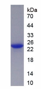
(SDS-PAGE)
SDS-PAGE
(SDS-PAGE)
Growth Hormone (GH), Recombinant Protein (Cat# AAA152135)
Full Name
Eukaryotic Growth Hormone (GH)
Gene Names
Gh; Gh1
Applications
Western Blot
Purity
Greater than 95% by SDS-PAGE
Pricing


 IHC (Immunohistochemistry)
(Immunohistochemistry of paraffin-embedded mouse heart using HIST1H2AG antibody at dilution of 1:100 (40x lens).)
IHC (Immunohistochemistry)
(Immunohistochemistry of paraffin-embedded mouse heart using HIST1H2AG antibody at dilution of 1:100 (40x lens).)
HIST1H2AG, Polyclonal Antibody (Cat# AAA28259)
Full Name
HIST1H2AG Polyclonal Antibody
Gene Names
HIST1H2AG; H2AG; H2A/p; H2AFP; H2A.1b; pH2A/f
Reactivity
Human, Mouse, Rat
Applications
Western Blot, Immunohistochemistry
Purity
Affinity Purification
Pricing


 WB (Western Blot)
(RRAS2 monoclonal antibody Western Blot analysis of RRAS2 expression in A-431.)
WB (Western Blot)
(RRAS2 monoclonal antibody Western Blot analysis of RRAS2 expression in A-431.)
RRAS2, Monoclonal Antibody (Cat# AAA25528)
Full Name
RRAS2 (Ras-related Protein R-Ras2, Ras-like Protein TC21, Teratocarcinoma Oncogene, TC21) (HRP)
Reactivity
Human, Mouse, Rat
Applications
Immunohistochemistry, Western Blot
Purity
Purified by Protein A Affinity Chromatography.
Pricing


 IHC (Immunohistchemistry)
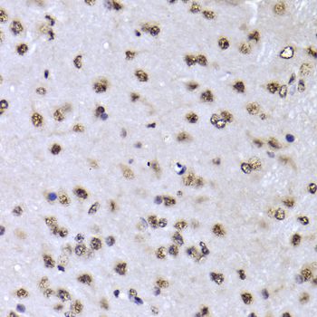
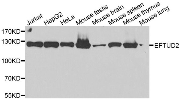
(Immunoprecipitation analysis of 100ug extracts of 293T cells using 3ug EFTUD2 antibody. Western blot was performed from the immunoprecipitate using EFTUD2 antibody at a dilition of 1:1000.)
IHC (Immunohistchemistry)
(Immunoprecipitation analysis of 100ug extracts of 293T cells using 3ug EFTUD2 antibody. Western blot was performed from the immunoprecipitate using EFTUD2 antibody at a dilition of 1:1000.)
EFTUD2, Polyclonal Antibody (Cat# AAA28125)
Full Name
EFTUD2 Polyclonal Antibody
Gene Names
EFTUD2; MFDM; MFDGA; Snu114; Snrp116; SNRNP116; U5-116KD
Reactivity
Human, Mouse, Rat
Applications
Western Blot, Immunohistochemistry, Immunofluorescence
Purity
Affinity Purification
Pricing


 WB (Western Blot)
(G3BP monoclonal antibody Western Blot analysis of G3BP expression in A-431)
WB (Western Blot)
(G3BP monoclonal antibody Western Blot analysis of G3BP expression in A-431)
G3BP1, Monoclonal Antibody (Cat# AAA25695)
Full Name
G3BP1 (Ras GTPase-activating Protein-binding Protein 1, G3BP-1, ATP-dependent DNA Helicase VIII, hDH VIII, GAP SH3 Domain-binding Protein 1, G3BP) (PE)
Gene Names
G3BP1; G3BP; HDH-VIII
Reactivity
Human
Applications
Immunofluorescence, Immunohistochemistry, Western Blot
Purity
Purified by Protein A Affinity Chromatography.
Pricing


 Application Data
(Proximity Ligation Analysis of protein-protein interactions between TP53 and MDM2 HeLa cells were stained with anti-TP53 rabbit purified polyclonal 1:1200 and anti-MDM2 mouse monoclonal antibody 1:50. Each red dot represents the detection of protein-protein interaction complex, and nuclei were counterstained with DAPI (blue).)
Application Data
(Proximity Ligation Analysis of protein-protein interactions between TP53 and MDM2 HeLa cells were stained with anti-TP53 rabbit purified polyclonal 1:1200 and anti-MDM2 mouse monoclonal antibody 1:50. Each red dot represents the detection of protein-protein interaction complex, and nuclei were counterstained with DAPI (blue).)
MDM2, Monoclonal Antibody (Cat# AAA24565)
Full Name
MDM2 (E3 Ubiquitin-protein Ligase Mdm2, Double Minute 2 Protein, Hdm2, Oncoprotein Mdm2, p53-binding Protein Mdm2) APC
Gene Names
MDM2; HDMX; hdm2; ACTFS
Reactivity
Human
Applications
Immunohistochemistry, Western Blot
Purity
Purified by Protein A Affinity Chromatography.
Pricing


 WB (Western Blot)
(Western Blot Analysis of human liver tissue lysate using Prohibitin Mouse Monoclonal Antibody (PHB/3194).)
WB (Western Blot)
(Western Blot Analysis of human liver tissue lysate using Prohibitin Mouse Monoclonal Antibody (PHB/3194).)
Prohibitin, Monoclonal Antibody (Cat# AAA23932)
Full Name
Prohibitin (Mitochondrial Marker)
Gene Names
PHB; PHB1; HEL-215; HEL-S-54e
Reactivity
Human
Applications
Western Blot, Immunofluorescence, Immunohistochemistry
Purity
Purified Ab with BSA and Azide at 200ug/ml OR Purified Ab WITHOUT BSA and Azide at 1.0mg/ml
Pricing


 IHC (Immunohistchemistry)
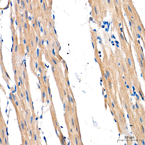
(Immunohistochemistry analysis of paraffin-embedded Rat heart tissue using AMPKa1/AMPKa2 Mouse mAb (AAA28599) at a dilution of 1:200 (40x lens). High pressure antigen retrieval performed with 0.01M Tris-EDTA Buffer (pH 9.0) prior to IHC staining.)
IHC (Immunohistchemistry)
(Immunohistochemistry analysis of paraffin-embedded Rat heart tissue using AMPKa1/AMPKa2 Mouse mAb (AAA28599) at a dilution of 1:200 (40x lens). High pressure antigen retrieval performed with 0.01M Tris-EDTA Buffer (pH 9.0) prior to IHC staining.)
AMPKa1/AMPKa2, Monoclonal Antibody (Cat# AAA28599)
Full Name
AMPKa1/AMPKa2 Mouse mAb
Reactivity
Human
Applications
Western Blot, Immunohistochemistry
Purity
Affinity purification
Pricing

 FCM (Flow Cytometry)
(Figure 7. Flow Cytometry analysis of PC-3 cells using anti-MTCH2 antibody (AAA19839).Overlay histogram showing PC-3 cells stained with AAA19839 (Blue line). To facilitate intracellular staining, cells were fixed with 4% paraformaldehyde and permeabilized with permeabilization buffer. The cells were blocked with 10% normal goat serum. And then incubated with rabbit anti-MTCH2 Antibody (AAA19839, 1ug/1x106 cells) for 30 min at 20 degree C. DyLight488 conjugated goat anti-rabbit IgG was used as secondary antibody for 30 minutes at 20 degree C. Isotype control antibody (Green line) was rabbit IgG (1ug/1x106) used under the same conditions. Unlabelled sample without incubation with primary antibody and secondary antibody (Red line) was used as a blank control.)
FCM (Flow Cytometry)
(Figure 7. Flow Cytometry analysis of PC-3 cells using anti-MTCH2 antibody (AAA19839).Overlay histogram showing PC-3 cells stained with AAA19839 (Blue line). To facilitate intracellular staining, cells were fixed with 4% paraformaldehyde and permeabilized with permeabilization buffer. The cells were blocked with 10% normal goat serum. And then incubated with rabbit anti-MTCH2 Antibody (AAA19839, 1ug/1x106 cells) for 30 min at 20 degree C. DyLight488 conjugated goat anti-rabbit IgG was used as secondary antibody for 30 minutes at 20 degree C. Isotype control antibody (Green line) was rabbit IgG (1ug/1x106) used under the same conditions. Unlabelled sample without incubation with primary antibody and secondary antibody (Red line) was used as a blank control.)
MTCH2, Polyclonal Antibody (Cat# AAA19839)
Full Name
Anti-MTCH2 Antibody Picoband
Gene Names
MTCH2; MIMP; HSPC032; SLC25A50
Reactivity
Human, Rat
Applications
Flow Cytometry, Immunoprecipitation, Immunofluorescence, Immunohistochemistry, Immunocytochemistry, Western Blot
Purity
Immunogen affinity purified.
Pricing
 IHC (Immunohistochemistry)
(Figure 7. IHC analysis of HOXD10 using anti-HOXD10 antibody (AAA19838).HOXD10 was detected in a paraffin-embedded section of human urothelial carcinoma tissue. Heat mediated antigen retrieval was performed in EDTA buffer (pH 8.0, epitope retrieval solution). The tissue section was blocked with 10% goat serum. The tissue section was then incubated with 2ug/ml rabbit anti-HOXD10 Antibody (AAA19838) overnight at 4 degree C. Peroxidase Conjugated Goat Anti-rabbit IgG was used as secondary antibody and incubated for 30 minutes at 37 degree C. The tissue section was developed using HRP Conjugated Rabbit IgG Super Vision Assay Kit ( epitope retrieval solution). The tissue section was blocked with 10% goat serum. The tissue section was then incubated with 2ug/ml rabbit anti-HOXD10 Antibody (AAA19838) overnight at 4 degree C. Peroxidase Conjugated Goat Anti-rabbit IgG was used as secondary antibody and incubated for 30 minutes at 37 degree C. The tissue section was developed using HRP Conjugated Rabbit IgG Super Vision Assay Kit ( epitope retrieval solution). The tissue section was blocked with 10% goat serum. The tissue section was then incubated with 2ug/ml rabbit anti-HOXD10 Antibody (AAA19838) overnight at 4 degree C. Peroxidase Conjugated Goat Anti-rabbit IgG was used as secondary antibody and incubated for 30 minutes at 37 degree C. The tissue section was developed using HRP Conjugated Rabbit IgG Super Vision Assay Kit ( epitope retrieval solution). The tissue section was blocked with 10% goat serum. The tissue section was then incubated with 2ug/ml rabbit anti-HOXD10 Antibody (AAA19838) overnight at 4 degree C. Peroxidase Conjugated Goat Anti-rabbit IgG was used as secondary antibody and incubated for 30 minutes at 37 degree C. The tissue section was developed using HRP Conjugated Rabbit IgG Super Vision Assay Kit ( epitope retrieval solution). The tissue section was blocked with 10% goat serum. The tissue section was then incubated with 2ug/ml rabbit anti-HOXD10 Antibody (AAA19838) overnight at 4 degree C. Peroxidase Conjugated Goat Anti-rabbit IgG was used as secondary antibody and incubated for 30 minutes at 37 degree C. The tissue section was developed using HRP Conjugated Rabbit IgG Super Vision Assay Kit ( epitope retrieval solution). The tissue section was blocked with 10% goat serum. The tissue section was then incubated with 2ug/ml rabbit anti-HOXD10 Antibody (AAA19838) overnight at 4 degree C. Peroxidase Conjugated Goat Anti-rabbit IgG was used as secondary antibody and incubated for 30 minutes at 37 degree C. The tissue section was developed using HRP Conjugated Rabbit IgG Super Vision Assay Kit ( proteins were transferred to a nitrocellulose membrane at 150 mA for 50-90 minutes. Blocked the membrane with 5% non-fat milk/TBS for 1.5 hour at RT. The membrane was incubated with rabbit anti-HOXD10 antigen affinity purified polyclonal antibody (#AAA19838) at 0.5ug/mL overnight at 4 degree C, then washed with TBS-0.1%Tween 3 times with 5 minutes each and probed with a goat anti-rabbit IgG-HRP secondary antibody at a dilution of 1:5000 for 1.5 hour at RT. The signal is developed using an Enhanced Chemiluminescent detection (ECL) kit with Tanon 5200 system. A specific band was detected for HOXD10 at approximately 40 kDa. The expected band size for HOXD10 is at 38 kDa.)
IHC (Immunohistochemistry)
(Figure 7. IHC analysis of HOXD10 using anti-HOXD10 antibody (AAA19838).HOXD10 was detected in a paraffin-embedded section of human urothelial carcinoma tissue. Heat mediated antigen retrieval was performed in EDTA buffer (pH 8.0, epitope retrieval solution). The tissue section was blocked with 10% goat serum. The tissue section was then incubated with 2ug/ml rabbit anti-HOXD10 Antibody (AAA19838) overnight at 4 degree C. Peroxidase Conjugated Goat Anti-rabbit IgG was used as secondary antibody and incubated for 30 minutes at 37 degree C. The tissue section was developed using HRP Conjugated Rabbit IgG Super Vision Assay Kit ( epitope retrieval solution). The tissue section was blocked with 10% goat serum. The tissue section was then incubated with 2ug/ml rabbit anti-HOXD10 Antibody (AAA19838) overnight at 4 degree C. Peroxidase Conjugated Goat Anti-rabbit IgG was used as secondary antibody and incubated for 30 minutes at 37 degree C. The tissue section was developed using HRP Conjugated Rabbit IgG Super Vision Assay Kit ( epitope retrieval solution). The tissue section was blocked with 10% goat serum. The tissue section was then incubated with 2ug/ml rabbit anti-HOXD10 Antibody (AAA19838) overnight at 4 degree C. Peroxidase Conjugated Goat Anti-rabbit IgG was used as secondary antibody and incubated for 30 minutes at 37 degree C. The tissue section was developed using HRP Conjugated Rabbit IgG Super Vision Assay Kit ( epitope retrieval solution). The tissue section was blocked with 10% goat serum. The tissue section was then incubated with 2ug/ml rabbit anti-HOXD10 Antibody (AAA19838) overnight at 4 degree C. Peroxidase Conjugated Goat Anti-rabbit IgG was used as secondary antibody and incubated for 30 minutes at 37 degree C. The tissue section was developed using HRP Conjugated Rabbit IgG Super Vision Assay Kit ( epitope retrieval solution). The tissue section was blocked with 10% goat serum. The tissue section was then incubated with 2ug/ml rabbit anti-HOXD10 Antibody (AAA19838) overnight at 4 degree C. Peroxidase Conjugated Goat Anti-rabbit IgG was used as secondary antibody and incubated for 30 minutes at 37 degree C. The tissue section was developed using HRP Conjugated Rabbit IgG Super Vision Assay Kit ( epitope retrieval solution). The tissue section was blocked with 10% goat serum. The tissue section was then incubated with 2ug/ml rabbit anti-HOXD10 Antibody (AAA19838) overnight at 4 degree C. Peroxidase Conjugated Goat Anti-rabbit IgG was used as secondary antibody and incubated for 30 minutes at 37 degree C. The tissue section was developed using HRP Conjugated Rabbit IgG Super Vision Assay Kit ( proteins were transferred to a nitrocellulose membrane at 150 mA for 50-90 minutes. Blocked the membrane with 5% non-fat milk/TBS for 1.5 hour at RT. The membrane was incubated with rabbit anti-HOXD10 antigen affinity purified polyclonal antibody (#AAA19838) at 0.5ug/mL overnight at 4 degree C, then washed with TBS-0.1%Tween 3 times with 5 minutes each and probed with a goat anti-rabbit IgG-HRP secondary antibody at a dilution of 1:5000 for 1.5 hour at RT. The signal is developed using an Enhanced Chemiluminescent detection (ECL) kit with Tanon 5200 system. A specific band was detected for HOXD10 at approximately 40 kDa. The expected band size for HOXD10 is at 38 kDa.)
HOXD10, Polyclonal Antibody (Cat# AAA19838)
Full Name
Anti-HOXD10 Antibody Picoband
Gene Names
HOXD10; HOX4; HOX4D; HOX4E; Hox-4.4
Reactivity
Human, Mouse
Applications
Western Blot, Immunohistochemistry
Purity
Immunogen affinity purified.
Pricing


 FCM (Flow Cytometry)
(Figure 8. Flow Cytometry analysis of Hela cells using anti-PITPNB antibody (AAA19957).Overlay histogram showing Hela cells stained with AAA19957 (Blue line). To facilitate intracellular staining, cells were fixed with 4% paraformaldehyde and permeabilized with permeabilization buffer. The cells were blocked with 10% normal goat serum. And then incubated with rabbit anti-PITPNB Antibody (AAA19957, 1ug/1x106 cells) for 30 min at 20 degree C. PE conjugated goat anti-rabbit IgG (5-10ug/1x106 cells) was used as secondary antibody for 30 minutes at 20 degree C. Isotype control antibody (Green line) was rabbit IgG (1ug/1x106) used under the same conditions. Unlabelled sample without incubation with primary antibody and secondary antibody (Red line) was used as a blank control.)
FCM (Flow Cytometry)
(Figure 8. Flow Cytometry analysis of Hela cells using anti-PITPNB antibody (AAA19957).Overlay histogram showing Hela cells stained with AAA19957 (Blue line). To facilitate intracellular staining, cells were fixed with 4% paraformaldehyde and permeabilized with permeabilization buffer. The cells were blocked with 10% normal goat serum. And then incubated with rabbit anti-PITPNB Antibody (AAA19957, 1ug/1x106 cells) for 30 min at 20 degree C. PE conjugated goat anti-rabbit IgG (5-10ug/1x106 cells) was used as secondary antibody for 30 minutes at 20 degree C. Isotype control antibody (Green line) was rabbit IgG (1ug/1x106) used under the same conditions. Unlabelled sample without incubation with primary antibody and secondary antibody (Red line) was used as a blank control.)
PITPNB, Polyclonal Antibody (Cat# AAA19957)
Full Name
Anti-PITPNB Antibody Picoband
Gene Names
PITPNB; VIB1B; PtdInsTP; PI-TP-beta
Reactivity
Human, Mouse, Rat
Applications
Flow Cytometry, Immunofluorescence, Immunohistochemistry, Western Blot
Purity
Immunogen affinity purified.
Pricing


 FCM (Flow Cytometry)
(Figure 6. Flow Cytometry analysis of Jurkat cells using anti-GSDMD antibody (AAA19705).Overlay histogram showing Jurkat cells stained with AAA19705 (Blue line). The cells were blocked with 10% normal goat serum. And then incubated with mouse anti-GSDMD Antibody (AAA19705, 1 ug/1x10^6 cells) for 30 min at 20 degree C. DyLight488 conjugated goat anti-mouse IgG was used as secondary antibody for 30 minutes at 20 degree C. Isotype control antibody (Green line) was mouse IgG (1 ug/1x10^6) used under the same conditions. Unlabelled sample (Red line) was also used as a control.)
FCM (Flow Cytometry)
(Figure 6. Flow Cytometry analysis of Jurkat cells using anti-GSDMD antibody (AAA19705).Overlay histogram showing Jurkat cells stained with AAA19705 (Blue line). The cells were blocked with 10% normal goat serum. And then incubated with mouse anti-GSDMD Antibody (AAA19705, 1 ug/1x10^6 cells) for 30 min at 20 degree C. DyLight488 conjugated goat anti-mouse IgG was used as secondary antibody for 30 minutes at 20 degree C. Isotype control antibody (Green line) was mouse IgG (1 ug/1x10^6) used under the same conditions. Unlabelled sample (Red line) was also used as a control.)
GSDMD, Monoclonal Antibody (Cat# AAA19705)
Full Name
Anti-GSDMD Antibody Picoband (monoclonal, 6D11)
Gene Names
GSDMD; DF5L; DFNA5L; FKSG10; GSDMDC1
Reactivity
Human
Applications
Western Blot, Immunohistochemistry, Immunocytochemistry, Immunofluorescence, Flow Cytometry
Purity
Immunogen affinity purified.
Pricing


 FCM (Flow Cytometry)
(Figure 6. Flow Cytometry analysis of HeLa cells using anti-RRP4/EXOSC2 antibody (AAA19615).Overlay histogram showing HeLa cells stained with AAA19615 (Blue line). The cells were blocked with 10% normal goat serum. And then incubated with rabbit anti-RRP4/EXOSC2 Antibody (AAA19615, 1 ug/1x10^6 cells) for 30 min at 20 degree C. DyLight488 conjugated goat anti-rabbit IgG was used as secondary antibody for 30 minutes at 20 degree C. Isotype control antibody (Green line) was rabbit IgG (1 ug/1x10^6) used under the same conditions. Unlabelled sample (Red line) was also used as a control.)
FCM (Flow Cytometry)
(Figure 6. Flow Cytometry analysis of HeLa cells using anti-RRP4/EXOSC2 antibody (AAA19615).Overlay histogram showing HeLa cells stained with AAA19615 (Blue line). The cells were blocked with 10% normal goat serum. And then incubated with rabbit anti-RRP4/EXOSC2 Antibody (AAA19615, 1 ug/1x10^6 cells) for 30 min at 20 degree C. DyLight488 conjugated goat anti-rabbit IgG was used as secondary antibody for 30 minutes at 20 degree C. Isotype control antibody (Green line) was rabbit IgG (1 ug/1x10^6) used under the same conditions. Unlabelled sample (Red line) was also used as a control.)
RRP4/EXOSC2, Polyclonal Antibody (Cat# AAA19615)
Full Name
Anti-RRP4/EXOSC2 Antibody Picoband
Gene Names
EXOSC2; p7; RRP4; Rrp4p; hRrp4p
Reactivity
Human
Applications
Western Blot, Immunohistochemistry, Flow Cytometry
Purity
Immunogen affinity purified.
Pricing
 IHC (Immunohistochemistry)
(Figure 7. IHC analysis of IRF2BPL using anti-IRF2BPL antibody (AAA19950).IRF2BPL was detected in a paraffin-embedded section of follicles of human thyroid cancer tissue. Heat mediated antigen retrieval was performed in EDTA buffer (pH 8.0, epitope retrieval solution). The tissue section was blocked with 10% goat serum. The tissue section was then incubated with 2ug/ml rabbit anti-IRF2BPL Antibody (AAA19950) overnight at 4 degree C. Peroxidase Conjugated Goat Anti-rabbit IgG was used as secondary antibody and incubated for 30 minutes at 37 degree C. The tissue section was developed using HRP Conjugated Rabbit IgG Super Vision Assay Kit ( epitope retrieval solution). The tissue section was blocked with 10% goat serum. The tissue section was then incubated with 2ug/ml rabbit anti-IRF2BPL Antibody (AAA19950) overnight at 4 degree C. Peroxidase Conjugated Goat Anti-rabbit IgG was used as secondary antibody and incubated for 30 minutes at 37 degree C. The tissue section was developed using HRP Conjugated Rabbit IgG Super Vision Assay Kit ( epitope retrieval solution). The tissue section was blocked with 10% goat serum. The tissue section was then incubated with 2ug/ml rabbit anti-IRF2BPL Antibody (AAA19950) overnight at 4 degree C. Peroxidase Conjugated Goat Anti-rabbit IgG was used as secondary antibody and incubated for 30 minutes at 37 degree C. The tissue section was developed using HRP Conjugated Rabbit IgG Super Vision Assay Kit ( epitope retrieval solution). The tissue section was blocked with 10% goat serum. The tissue section was then incubated with 2ug/ml rabbit anti-IRF2BPL Antibody (AAA19950) overnight at 4 degree C. Peroxidase Conjugated Goat Anti-rabbit IgG was used as secondary antibody and incubated for 30 minutes at 37 degree C. The tissue section was developed using HRP Conjugated Rabbit IgG Super Vision Assay Kit ( epitope retrieval solution). The tissue section was blocked with 10% goat serum. The tissue section was then incubated with 2ug/ml rabbit anti-IRF2BPL Antibody (AAA19950) overnight at 4 degree C. Peroxidase Conjugated Goat Anti-rabbit IgG was used as secondary antibody and incubated for 30 minutes at 37 degree C. The tissue section was developed using HRP Conjugated Rabbit IgG Super Vision Assay Kit ( epitope retrieval solution). The tissue section was blocked with 10% goat serum. The tissue section was then incubated with 2ug/ml rabbit anti-IRF2BPL Antibody (AAA19950) overnight at 4 degree C. Peroxidase Conjugated Goat Anti-rabbit IgG was used as secondary antibody and incubated for 30 minutes at 37 degree C. The tissue section was developed using HRP Conjugated Rabbit IgG Super Vision Assay Kit (Lane 2: human SH-SY5Y whole cell lysates,Lane 3: human MCF-7whole cell lysates,Lane 4: human RT4 whole cell lysates.After electrophoresis, proteins were transferred to a nitrocellulose membrane at 150 mA for 50-90 minutes. Blocked the membrane with 5% non-fat milk/TBS for 1.5 hour at RT. The membrane was incubated with rabbit anti-IRF2BPL antigen affinity purified polyclonal antibody (#AAA19950) at 0.25ug/mL overnight at 4 degree C, then washed with TBS-0.1%Tween 3 times with 5 minutes each and probed with a goat anti-rabbit IgG-HRP secondary antibody at a dilution of 1:5000 for 1.5 hour at RT. The signal is developed using an Enhanced Chemiluminescent detection (ECL) kit with Tanon 5200 system. A specific band was detected for IRF2BPL at approximately 95 kDa. The expected band size for IRF2BPL is at 83 kDa.)
IHC (Immunohistochemistry)
(Figure 7. IHC analysis of IRF2BPL using anti-IRF2BPL antibody (AAA19950).IRF2BPL was detected in a paraffin-embedded section of follicles of human thyroid cancer tissue. Heat mediated antigen retrieval was performed in EDTA buffer (pH 8.0, epitope retrieval solution). The tissue section was blocked with 10% goat serum. The tissue section was then incubated with 2ug/ml rabbit anti-IRF2BPL Antibody (AAA19950) overnight at 4 degree C. Peroxidase Conjugated Goat Anti-rabbit IgG was used as secondary antibody and incubated for 30 minutes at 37 degree C. The tissue section was developed using HRP Conjugated Rabbit IgG Super Vision Assay Kit ( epitope retrieval solution). The tissue section was blocked with 10% goat serum. The tissue section was then incubated with 2ug/ml rabbit anti-IRF2BPL Antibody (AAA19950) overnight at 4 degree C. Peroxidase Conjugated Goat Anti-rabbit IgG was used as secondary antibody and incubated for 30 minutes at 37 degree C. The tissue section was developed using HRP Conjugated Rabbit IgG Super Vision Assay Kit ( epitope retrieval solution). The tissue section was blocked with 10% goat serum. The tissue section was then incubated with 2ug/ml rabbit anti-IRF2BPL Antibody (AAA19950) overnight at 4 degree C. Peroxidase Conjugated Goat Anti-rabbit IgG was used as secondary antibody and incubated for 30 minutes at 37 degree C. The tissue section was developed using HRP Conjugated Rabbit IgG Super Vision Assay Kit ( epitope retrieval solution). The tissue section was blocked with 10% goat serum. The tissue section was then incubated with 2ug/ml rabbit anti-IRF2BPL Antibody (AAA19950) overnight at 4 degree C. Peroxidase Conjugated Goat Anti-rabbit IgG was used as secondary antibody and incubated for 30 minutes at 37 degree C. The tissue section was developed using HRP Conjugated Rabbit IgG Super Vision Assay Kit ( epitope retrieval solution). The tissue section was blocked with 10% goat serum. The tissue section was then incubated with 2ug/ml rabbit anti-IRF2BPL Antibody (AAA19950) overnight at 4 degree C. Peroxidase Conjugated Goat Anti-rabbit IgG was used as secondary antibody and incubated for 30 minutes at 37 degree C. The tissue section was developed using HRP Conjugated Rabbit IgG Super Vision Assay Kit ( epitope retrieval solution). The tissue section was blocked with 10% goat serum. The tissue section was then incubated with 2ug/ml rabbit anti-IRF2BPL Antibody (AAA19950) overnight at 4 degree C. Peroxidase Conjugated Goat Anti-rabbit IgG was used as secondary antibody and incubated for 30 minutes at 37 degree C. The tissue section was developed using HRP Conjugated Rabbit IgG Super Vision Assay Kit (Lane 2: human SH-SY5Y whole cell lysates,Lane 3: human MCF-7whole cell lysates,Lane 4: human RT4 whole cell lysates.After electrophoresis, proteins were transferred to a nitrocellulose membrane at 150 mA for 50-90 minutes. Blocked the membrane with 5% non-fat milk/TBS for 1.5 hour at RT. The membrane was incubated with rabbit anti-IRF2BPL antigen affinity purified polyclonal antibody (#AAA19950) at 0.25ug/mL overnight at 4 degree C, then washed with TBS-0.1%Tween 3 times with 5 minutes each and probed with a goat anti-rabbit IgG-HRP secondary antibody at a dilution of 1:5000 for 1.5 hour at RT. The signal is developed using an Enhanced Chemiluminescent detection (ECL) kit with Tanon 5200 system. A specific band was detected for IRF2BPL at approximately 95 kDa. The expected band size for IRF2BPL is at 83 kDa.)
IRF2BPL, Polyclonal Antibody (Cat# AAA19950)
Full Name
Anti-IRF2BPL Antibody Picoband
Gene Names
IRF2BPL; EAP1; C14orf4
Reactivity
Human
Applications
Western Blot, Immunohistochemistry, Flow Cytometry
Purity
Immunogen affinity purified.
Pricing




 FCM (Flow Cytometry)
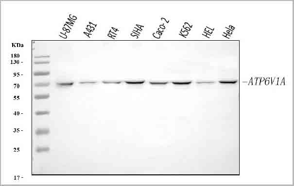
(Figure 12. Flow Cytometry analysis of U251 cells using anti-ATP6V1A antibody (AAA19619).Overlay histogram showing U251 cells stained with AAA19619 (Blue line). The cells were blocked with 10% normal goat serum. And then incubated with rabbit anti-ATP6V1A Antibody (AAA19619, 1 ug/1x106 cells) for 30 min at 20 degree C. DyLight488 conjugated goat anti-rabbit IgG (BA1127, 5-10 ug/1x106 cells) was used as secondary antibody for 30 minutes at 20 degree C. Isotype control antibody (Green line) was rabbit IgG (1 ug/1x106) used under the same conditions. Unlabelled sample (Red line) was also used as a control.)
FCM (Flow Cytometry)
(Figure 12. Flow Cytometry analysis of U251 cells using anti-ATP6V1A antibody (AAA19619).Overlay histogram showing U251 cells stained with AAA19619 (Blue line). The cells were blocked with 10% normal goat serum. And then incubated with rabbit anti-ATP6V1A Antibody (AAA19619, 1 ug/1x106 cells) for 30 min at 20 degree C. DyLight488 conjugated goat anti-rabbit IgG (BA1127, 5-10 ug/1x106 cells) was used as secondary antibody for 30 minutes at 20 degree C. Isotype control antibody (Green line) was rabbit IgG (1 ug/1x106) used under the same conditions. Unlabelled sample (Red line) was also used as a control.)
ATP6V1A, Polyclonal Antibody (Cat# AAA19619)
Full Name
Anti-ATP6V1A Antibody Picoband
Gene Names
ATP6V1A; HO68; VA68; VPP2; Vma1; ATP6A1; ATP6V1A1
Reactivity
Human, Mouse, Rat
Applications
Western Blot, Immunohistochemistry, Immunocytochemistry, Immunofluorescence, Flow Cytometry
Purity
Immunogen affinity purified.
Pricing


 FCM (Flow Cytometry)
(Figure 9. Flow Cytometry analysis of U87 cells using anti-SOS1 antibody (AAA19738).Overlay histogram showing U87 cells stained with AAA19738 (Blue line). The cells were fixed with 4% paraformaldehyde and blocked with 10% normal goat serum. And then incubated with rabbit anti-SOS1 Antibody (AAA19738, 1ug/1x106 cells) for 30 min at 20 degree C. DyLight488 conjugated goat anti-rabbit IgG was used as secondary antibody for 30 minutes at 20 degree C. Isotype control antibody (Green line) was rabbit IgG (1ug/1x106) used under the same conditions. Unlabelled sample without incubation with primary antibody and secondary antibody (Red line) was used as a blank control.)
FCM (Flow Cytometry)
(Figure 9. Flow Cytometry analysis of U87 cells using anti-SOS1 antibody (AAA19738).Overlay histogram showing U87 cells stained with AAA19738 (Blue line). The cells were fixed with 4% paraformaldehyde and blocked with 10% normal goat serum. And then incubated with rabbit anti-SOS1 Antibody (AAA19738, 1ug/1x106 cells) for 30 min at 20 degree C. DyLight488 conjugated goat anti-rabbit IgG was used as secondary antibody for 30 minutes at 20 degree C. Isotype control antibody (Green line) was rabbit IgG (1ug/1x106) used under the same conditions. Unlabelled sample without incubation with primary antibody and secondary antibody (Red line) was used as a blank control.)
SOS1, Polyclonal Antibody (Cat# AAA19738)
Full Name
Anti-SOS1 Antibody Picoband
Gene Names
SOS1; GF1; HGF; NS4; GGF1; GINGF
Reactivity
Human, Mouse, Rat
Applications
Western Blot, Immunohistochemistry, Flow Cytometry
Purity
Immunogen affinity purified.
Pricing
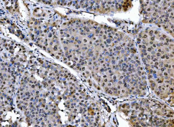


 FCM (Flow Cytometry)
(Figure 8. Flow Cytometry analysis of U2OS cells using anti-SAP18 antibody (AAA19568).Overlay histogram showing U2OS cells stained with AAA19568 (Blue line). The cells were blocked with 10% normal goat serum. And then incubated with rabbit anti-SAP18 Antibody (AAA19568, 1 ug/1x10^6 cells) for 30 min at 20 degree C. DyLight488 conjugated goat anti-rabbit IgG was used as secondary antibody for 30 minutes at 20 degree C. Isotype control antibody (Green line) was rabbit IgG (1 ug/1x10^6) used under the same conditions. Unlabelled sample (Red line) was also used as a control.)
FCM (Flow Cytometry)
(Figure 8. Flow Cytometry analysis of U2OS cells using anti-SAP18 antibody (AAA19568).Overlay histogram showing U2OS cells stained with AAA19568 (Blue line). The cells were blocked with 10% normal goat serum. And then incubated with rabbit anti-SAP18 Antibody (AAA19568, 1 ug/1x10^6 cells) for 30 min at 20 degree C. DyLight488 conjugated goat anti-rabbit IgG was used as secondary antibody for 30 minutes at 20 degree C. Isotype control antibody (Green line) was rabbit IgG (1 ug/1x10^6) used under the same conditions. Unlabelled sample (Red line) was also used as a control.)
SAP18, Polyclonal Antibody (Cat# AAA19568)
Full Name
Anti-SAP18 Antibody Picoband
Gene Names
SAP18; SAP18P; 2HOR0202
Reactivity
Human, Mouse, Rat
Applications
Flow Cytometry, Immunofluorescence, Immunohistochemistry, Immunocytochemistry, Western Blot
Purity
Immunogen affinity purified.
Pricing


 WB (Western Blot)
(RAB33B monoclonal antibody. Western Blot analysis of RAB33B expression in NIH/3T3.)
WB (Western Blot)
(RAB33B monoclonal antibody. Western Blot analysis of RAB33B expression in NIH/3T3.)
RAB33B, Monoclonal Antibody (Cat# AAA14775)
Full Name
RAB33B (Ras-related Protein Rab-33B, SMC2)
Reactivity
Human, Mouse, Rat
Applications
Western Blot
Purity
Affinity Purified
Purified by Protein A affinity chromatography.
Purified by Protein A affinity chromatography.
Pricing