Filters
Clonality
Type
Reactivity
Gene Name
Isotype
Host
Application
Clone
3200 results for "Antibodies" - showing 3100-3150


 WB (Western Blot)
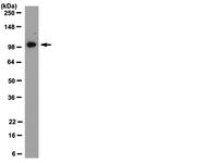
(Western Blot Analysis: Representative lot data. Lysate from HEK-293 cells was resolved by electrophoresis, transferred to PVDF and probed with anti-PI3 Kinase, p110beta (0.05 ug/mL). Proteins were visualized using donkey anti-rabbit secondary antibody conjugated to HRP and chemiluminescence detection. Arrow indicates PI3 Kinase, p110beta (~110kD).)
WB (Western Blot)
(Western Blot Analysis: Representative lot data. Lysate from HEK-293 cells was resolved by electrophoresis, transferred to PVDF and probed with anti-PI3 Kinase, p110beta (0.05 ug/mL). Proteins were visualized using donkey anti-rabbit secondary antibody conjugated to HRP and chemiluminescence detection. Arrow indicates PI3 Kinase, p110beta (~110kD).)
Phosphoinositide 3 Kinase, p110 beta, Polyclonal Antibody (Cat# AAA26902)
Full Name
Phosphoinositide 3 Kinase, p110 beta (DKFZp779K1237, MGC133043, p110beta, Phosphatidylinositol 3 Kinase Catalytic beta Polypeptide, Phosphatidylinositol-4,5-bisphosphate 3-kinase Catalytic Subunit beta Isoform, Phosphoinositide 3 Kinase Catalytic beta Pol
Gene Names
PIK3CA; MCM; CWS5; MCAP; PI3K; CLOVE; MCMTC; p110-alpha
Reactivity
Human
Applications
ICC, WB, IP, IHC
Purity
Purified by Immunoaffinity chromatography.
Pricing


 IHC (Immunohistchemistry)
(Figure 9 Immunohistochemistry Validation of hRIP3 in Human Colon Tissue Immunohistochemical analysis of paraffin-embedded human colon tissue using anti-hRIP3 antibody (8963) at 2 μg/ml. Tissue was fixed with formaldehyde and blocked with 10% serum for 1 h at RT; antigen retrieval was by heat mediation with a citrate buffer (pH6). Samples were incubated with primary antibody overnight at 4˚C. A goat anti-rabbit IgG H&L (HRP) at 1/250 was used as secondary. Counter stained with Hematoxylin.)
IHC (Immunohistchemistry)
(Figure 9 Immunohistochemistry Validation of hRIP3 in Human Colon Tissue Immunohistochemical analysis of paraffin-embedded human colon tissue using anti-hRIP3 antibody (8963) at 2 μg/ml. Tissue was fixed with formaldehyde and blocked with 10% serum for 1 h at RT; antigen retrieval was by heat mediation with a citrate buffer (pH6). Samples were incubated with primary antibody overnight at 4˚C. A goat anti-rabbit IgG H&L (HRP) at 1/250 was used as secondary. Counter stained with Hematoxylin.)
hRIP3, Polyclonal Antibody (Cat# AAA11031)
Full Name
hRIP3 Antibody
Gene Names
RIPK3; RIP3
Reactivity
Human
Applications
EIA, IF, IHC, WB
Purity
hRIP3 Antibody is Protein A purified.
Pricing

 WB (Western Blot)
(Western blot analysis of lysates from Hela, HepG2, MCF-7, SH-SY5Y, mouse NIH/3T3 cell line (from left to right), using FGFR1 Antibody (Center). AAA28687 was diluted at 1:1000 at each lane. A goat anti-rabbit IgG H&L(HRP) at 1:10000 dilution was used as the secondary antibody. Lysates at 20ug per lane.)
WB (Western Blot)
(Western blot analysis of lysates from Hela, HepG2, MCF-7, SH-SY5Y, mouse NIH/3T3 cell line (from left to right), using FGFR1 Antibody (Center). AAA28687 was diluted at 1:1000 at each lane. A goat anti-rabbit IgG H&L(HRP) at 1:10000 dilution was used as the secondary antibody. Lysates at 20ug per lane.)
FGFR1, Polyclonal Antibody (Cat# AAA28687)
Full Name
FGFR1 Antibody (Center)
Gene Names
FGFR1; CEK; FLG; HH2; OGD; FLT2; KAL2; BFGFR; CD331; FGFBR; FLT-2; HBGFR; N-SAM; FGFR-1; HRTFDS; bFGF-R-1
Reactivity
Human, mouse (Predicted Reactivity: Chicken, Rat)
Applications
FC/FACS, EIA, WB
Purity
Purified Rabbit Polyclonal Antibody (Pab)
Pricing


 WB (Western Blot)
(HSPA1B monoclonal antibody Western Blot analysis of HSPA1B expression in Raw 264.7.)
WB (Western Blot)
(HSPA1B monoclonal antibody Western Blot analysis of HSPA1B expression in Raw 264.7.)
HSPA1A, Monoclonal Antibody (Cat# AAA25129)
Full Name
HSPA1A (Heat Shock 70kD Protein 1A/1B, Heat Shock 70kD Protein 1/2, HSP70.1/HSP70.2, HSP70-1/HSP70-2, HSPA1, HSPA1B) (FITC)
Gene Names
HSPA1A; HSP72; HSPA1; HSP70I; HSP70-1; HSP70.1; HSP70-1A; HEL-S-103
Reactivity
Human, Mouse, Rat
Applications
EIA, IF, IHC, WB
Purity
Purified by Protein A Affinity Chromatography.
Pricing


 WB (Western Blot)
(HSPA1B monoclonal antibody Western Blot analysis of HSPA1B expression in Raw 264.7.)
WB (Western Blot)
(HSPA1B monoclonal antibody Western Blot analysis of HSPA1B expression in Raw 264.7.)
HSPA1A, Monoclonal Antibody (Cat# AAA24241)
Full Name
HSPA1A (Heat Shock 70kD Protein 1A/1B, Heat Shock 70kD Protein 1/2, HSP70.1/HSP70.2, HSP70-1/HSP70-2, HSPA1, HSPA1B) (AP)
Gene Names
HSPA1A; HSP72; HSPA1; HSP70I; HSP70-1; HSP70.1; HSP70-1A; HEL-S-103
Reactivity
Human, Mouse, Rat
Applications
EIA, IHC, WB
Purity
Purified by Protein A Affinity Chromatography.
Pricing


 IHC (Immunohistchemistry)
(Formalin-fixed, paraffin-embedded human Uterus stained with Vimentin Monoclonal Antibody (VM1170).)
IHC (Immunohistchemistry)
(Formalin-fixed, paraffin-embedded human Uterus stained with Vimentin Monoclonal Antibody (VM1170).)
Vimentin, Monoclonal Antibody (Cat# AAA13829)
Full Name
Vimentin (Mesenchymal Cell Marker) Mouse Monoclonal Antibody
Gene Names
VIM; HEL113; CTRCT30
Reactivity
Human.
Does not react with Mouse and Rat
Does not react with Mouse and Rat
Applications
FC/FACS, IF, WB, IHC
Pricing


 IHC (Immunohistochemistry)
(ATP5B Antibody (Center) flow cytometric analysis of WiDr cells (right histogram) compared to a negative control cell (left histogram).FITC-conjugated goat-anti-rabbit secondary antibodies were used for the analysis.)
IHC (Immunohistochemistry)
(ATP5B Antibody (Center) flow cytometric analysis of WiDr cells (right histogram) compared to a negative control cell (left histogram).FITC-conjugated goat-anti-rabbit secondary antibodies were used for the analysis.)
ATP5B, Polyclonal Antibody (Cat# AAA28724)
Full Name
ATP5B Antibody (Center)
Gene Names
ATP5B; ATPMB; ATPSB; HEL-S-271
Reactivity
Human (Predicted Reactivity: Bovine, Mouse, Rat)
Applications
EIA, IHC, IF, WB, FC/FACS
Purity
Peptide Affinity Purified Rabbit Polyclonal Antibody (Pab)
Pricing


 FCM (Flow Cytometry)
(CDK4 Antibody (C-term) flow cytometric analysis of HL60 cells (bottom histogram) compared to a negative control cell (top histogram).FITC-conjugated goat-anti-rabbit secondary antibodies were used for the analysis.)
FCM (Flow Cytometry)
(CDK4 Antibody (C-term) flow cytometric analysis of HL60 cells (bottom histogram) compared to a negative control cell (top histogram).FITC-conjugated goat-anti-rabbit secondary antibodies were used for the analysis.)
CDK4, Polyclonal Antibody (Cat# AAA28747)
Full Name
CDK4 Antibody (C-term)
Gene Names
CDK4; CMM3; PSK-J3
Reactivity
Human
Applications
FC/FACS, EIA, IHC, WB, IF
Purity
Purified Rabbit Polyclonal Antibody (Pab)
Pricing

 WB (Western Blot)
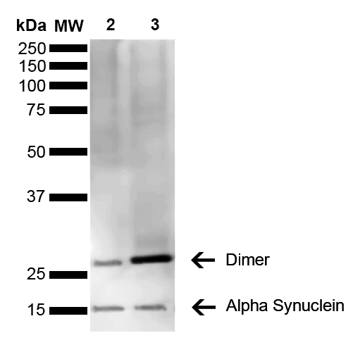
(Western Blot analysis of Mouse, Rat Brain showing detection of 14 kDa Alpha Synuclein protein using Mouse Anti-Alpha Synuclein Monoclonal Antibody, Clone 3C11. Lane 1: Molecular Weight Ladder (MW). Lane 2: Mouse brain cell lysate. Lane 3: Rat brain cell lysate. Load: 15 ug. Block: 5% Skim Milk in 1X TBST. Primary Antibody: Mouse Anti-Alpha Synuclein Monoclonal Antibody at 1:1000 for 2 hours at RT. Secondary Antibody: Goat Anti-Mouse HRP:IgG at 1:3000 for 1 hour at RT. Color Development: ECL solution (Super Signal West Pico) for 5 min in RT. Predicted/Observed Size: 14 kDa. Other Band(s): ~30 kDa (dimer).)
WB (Western Blot)
(Western Blot analysis of Mouse, Rat Brain showing detection of 14 kDa Alpha Synuclein protein using Mouse Anti-Alpha Synuclein Monoclonal Antibody, Clone 3C11. Lane 1: Molecular Weight Ladder (MW). Lane 2: Mouse brain cell lysate. Lane 3: Rat brain cell lysate. Load: 15 ug. Block: 5% Skim Milk in 1X TBST. Primary Antibody: Mouse Anti-Alpha Synuclein Monoclonal Antibody at 1:1000 for 2 hours at RT. Secondary Antibody: Goat Anti-Mouse HRP:IgG at 1:3000 for 1 hour at RT. Color Development: ECL solution (Super Signal West Pico) for 5 min in RT. Predicted/Observed Size: 14 kDa. Other Band(s): ~30 kDa (dimer).)
Alpha Synuclein, Monoclonal Antibody (Cat# AAA17810)
Full Name
Alpha Synuclein Antibody, Clone 3C11
Gene Names
SNCA; PD1; NACP; PARK1; PARK4
Reactivity
Human; Mouse; Rat
Applications
WB, DB, ICC, IF, EIA
Purity
Protein G Purified
Pricing


 Application Data
(C:FGFR2/isolectinB4 (C) and FGFR1/isolectinB4 (D) staining of apparent mesenchymal cells and the subpopulation of endothelial cells. Virtually all other dispersed apparent mesenchymal cells express FGFR1 and FGFR2 (merged image in E). F: FGFR2 (F) and FGFR1 (G) staining in clustered cells of epithelial origin (inferred by morphology here) demonstrating that epithelial cells express both FGFR1 and FGFR2 (merged image with DAPI staining in H).)
Application Data
(C:FGFR2/isolectinB4 (C) and FGFR1/isolectinB4 (D) staining of apparent mesenchymal cells and the subpopulation of endothelial cells. Virtually all other dispersed apparent mesenchymal cells express FGFR1 and FGFR2 (merged image in E). F: FGFR2 (F) and FGFR1 (G) staining in clustered cells of epithelial origin (inferred by morphology here) demonstrating that epithelial cells express both FGFR1 and FGFR2 (merged image with DAPI staining in H).)
FGFR2, Polyclonal Antibody (Cat# AAA26856)
Full Name
FGFR2, NT (FGFR2, BEK, KGFR, KSAM, Fibroblast growth factor receptor 2, K-sam, Keratinocyte growth factor receptor, CD332) (PE)
Gene Names
FGFR2; BEK; JWS; BBDS; CEK3; CFD1; ECT1; KGFR; TK14; TK25; BFR-1; CD332; K-SAM
Reactivity
Human, Monkey, Mouse, Rat
Applications
WB, IHC, IF, FC/FACS
Purity
Purified by Protein G Affinity Chromatography.
Pricing


 WB (Western Blot)
(Western Blot Analysis: Representative lot data. Lysate from HEK-293 cells was resolved by electrophoresis, transferred to PVDF and probed with anti-PI3 Kinase, p110beta (0.05 ug/mL). Proteins were visualized using donkey anti-rabbit secondary antibody conjugated to HRP and chemiluminescence detection. Arrow indicates PI3 Kinase, p110beta (~110kD).)
WB (Western Blot)
(Western Blot Analysis: Representative lot data. Lysate from HEK-293 cells was resolved by electrophoresis, transferred to PVDF and probed with anti-PI3 Kinase, p110beta (0.05 ug/mL). Proteins were visualized using donkey anti-rabbit secondary antibody conjugated to HRP and chemiluminescence detection. Arrow indicates PI3 Kinase, p110beta (~110kD).)
Phosphoinositide 3 Kinase, p110 beta, Polyclonal Antibody (Cat# AAA26895)
Full Name
Phosphoinositide 3 Kinase, p110 beta (DKFZp779K1237, MGC133043, p110beta, Phosphatidylinositol 3 Kinase Catalytic beta Polypeptide, Phosphatidylinositol-4,5-bisphosphate 3-kinase Catalytic Subunit beta Isoform, Phosphoinositide 3 Kinase Catalytic beta Pol
Gene Names
PIK3CA; MCM; CWS5; MCAP; PI3K; CLOVE; MCMTC; p110-alpha
Reactivity
Human
Applications
ICC, EIA, WB, IP, IHC
Purity
Purified by Immunoaffinity chromatography.
Pricing


 WB (Western Blot)
(Western Blot Analysis: Representative lot data. Lysate from HEK-293 cells was resolved by electrophoresis, transferred to PVDF and probed with anti-PI3 Kinase, p110beta (0.05 ug/mL). Proteins were visualized using donkey anti-rabbit secondary antibody conjugated to HRP and chemiluminescence detection. Arrow indicates PI3 Kinase, p110beta (~110kD).)
WB (Western Blot)
(Western Blot Analysis: Representative lot data. Lysate from HEK-293 cells was resolved by electrophoresis, transferred to PVDF and probed with anti-PI3 Kinase, p110beta (0.05 ug/mL). Proteins were visualized using donkey anti-rabbit secondary antibody conjugated to HRP and chemiluminescence detection. Arrow indicates PI3 Kinase, p110beta (~110kD).)
Phosphoinositide 3 Kinase, p110 beta, Polyclonal Antibody (Cat# AAA26901)
Full Name
Phosphoinositide 3 Kinase, p110 beta (DKFZp779K1237, MGC133043, p110beta, Phosphatidylinositol 3 Kinase Catalytic beta Polypeptide, Phosphatidylinositol-4,5-bisphosphate 3-kinase Catalytic Subunit beta Isoform, Phosphoinositide 3 Kinase Catalytic beta Pol
Gene Names
PIK3CA; MCM; CWS5; MCAP; PI3K; CLOVE; MCMTC; p110-alpha
Reactivity
Human
Applications
ICC, WB, IP, IHC
Purity
Purified by Immunoaffinity chromatography.
Pricing


 WB (Western Blot)
(Western Blot Analysis: Representative lot data. Lysate from HEK-293 cells was resolved by electrophoresis, transferred to PVDF and probed with anti-PI3 Kinase, p110beta (0.05 ug/mL). Proteins were visualized using donkey anti-rabbit secondary antibody conjugated to HRP and chemiluminescence detection. Arrow indicates PI3 Kinase, p110beta (~110kD).)
WB (Western Blot)
(Western Blot Analysis: Representative lot data. Lysate from HEK-293 cells was resolved by electrophoresis, transferred to PVDF and probed with anti-PI3 Kinase, p110beta (0.05 ug/mL). Proteins were visualized using donkey anti-rabbit secondary antibody conjugated to HRP and chemiluminescence detection. Arrow indicates PI3 Kinase, p110beta (~110kD).)
Phosphoinositide 3 Kinase, p110 beta, Polyclonal Antibody (Cat# AAA26899)
Full Name
Phosphoinositide 3 Kinase, p110 beta (DKFZp779K1237, MGC133043, p110beta, Phosphatidylinositol 3 Kinase Catalytic beta Polypeptide, Phosphatidylinositol-4,5-bisphosphate 3-kinase Catalytic Subunit beta Isoform, Phosphoinositide 3 Kinase Catalytic beta Pol
Gene Names
PIK3CA; MCM; CWS5; MCAP; PI3K; CLOVE; MCMTC; p110-alpha
Reactivity
Human
Applications
ICC, WB, IP, IHC
Purity
Purified by Immunoaffinity chromatography.
Pricing


 WB (Western Blot)
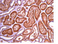
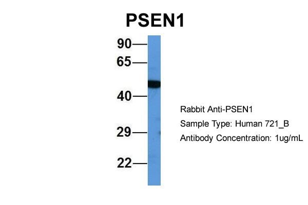
(WB Suggested Anti-PSEN1 Antibody Titration: 0.2-1 ug/mlELISA Titer: 1:312500Positive Control: Human brain)
WB (Western Blot)
(WB Suggested Anti-PSEN1 Antibody Titration: 0.2-1 ug/mlELISA Titer: 1:312500Positive Control: Human brain)
PSEN1, Polyclonal Antibody (Cat# AAA23585)
Full Name
PSEN1 antibody - middle region
Gene Names
PSEN1; AD3; FAD; PS1; PS-1; S182; ACNINV3
Reactivity
Cow, Dog, Guinea Pig, Horse, Human, Mouse, Rabbit, Rat
Applications
IHC, IP, WB
Purity
Affinity Purified
Pricing


 IP (Immunoprecipitation)
(Immunoprecipitation(IP) of CRYAB by using monoclonal anti-CRYAB antibodies (Negative control: IP without adding anti-CRYAB antibody.). For each experiment, 500ul of DDK tagged CRYAB overexpression lysates (at 1:5 dilution with HEK293T lysate), 2 ug of anti-CRYAB antibody and 20ul (0.1 mg) of goat anti-mouse conjugated magnetic beads were mixed and incubated overnight. After extensive wash to remove any non-specific binding, the immuno-precipitated products were analyzed with rabbit anti-DDK polyclonal antibody.)
IP (Immunoprecipitation)
(Immunoprecipitation(IP) of CRYAB by using monoclonal anti-CRYAB antibodies (Negative control: IP without adding anti-CRYAB antibody.). For each experiment, 500ul of DDK tagged CRYAB overexpression lysates (at 1:5 dilution with HEK293T lysate), 2 ug of anti-CRYAB antibody and 20ul (0.1 mg) of goat anti-mouse conjugated magnetic beads were mixed and incubated overnight. After extensive wash to remove any non-specific binding, the immuno-precipitated products were analyzed with rabbit anti-DDK polyclonal antibody.)
CRYAB / Alpha B Crystallin, Monoclonal Antibody (Cat# AAA12373)
Full Name
Mouse Monoclonal [clone 6D11] (IgG1) to Human CRYAB / Alpha B Crystallin
Gene Names
CRYAB; MFM2; CRYA2; CTPP2; HSPB5; CMD1II; CTRCT16; HEL-S-101
Reactivity
Human, Monkey, Rat
Applications
IHC - Paraffin, IF, WB, IP, FC/FACS
Purity
Protein A/G Purified
Pricing


 WB (Western Blot)
(HSPA1B monoclonal antibody Western Blot analysis of HSPA1B expression in Raw 264.7.)
WB (Western Blot)
(HSPA1B monoclonal antibody Western Blot analysis of HSPA1B expression in Raw 264.7.)
HSPA1A, Monoclonal Antibody (Cat# AAA24537)
Full Name
HSPA1A (Heat Shock 70kD Protein 1A/1B, Heat Shock 70kD Protein 1/2, HSP70.1/HSP70.2, HSP70-1/HSP70-2, HSPA1, HSPA1B) APC
Gene Names
HSPA1A; HSP72; HSPA1; HSP70I; HSP70-1; HSP70.1; HSP70-1A; HEL-S-103
Reactivity
Human, Mouse, Rat
Applications
EIA, IF, IHC, WB
Purity
Purified by Protein A Affinity Chromatography.
Pricing


 Application Data
(C:FGFR2/isolectinB4 (C) and FGFR1/isolectinB4 (D) staining of apparent mesenchymal cells and the subpopulation of endothelial cells. Virtually all other dispersed apparent mesenchymal cells express FGFR1 and FGFR2 (merged image in E). F: FGFR2 (F) and FGFR1 (G) staining in clustered cells of epithelial origin (inferred by morphology here) demonstrating that epithelial cells express both FGFR1 and FGFR2 (merged image with DAPI staining in H).)
Application Data
(C:FGFR2/isolectinB4 (C) and FGFR1/isolectinB4 (D) staining of apparent mesenchymal cells and the subpopulation of endothelial cells. Virtually all other dispersed apparent mesenchymal cells express FGFR1 and FGFR2 (merged image in E). F: FGFR2 (F) and FGFR1 (G) staining in clustered cells of epithelial origin (inferred by morphology here) demonstrating that epithelial cells express both FGFR1 and FGFR2 (merged image with DAPI staining in H).)
FGFR2, Polyclonal Antibody (Cat# AAA26851)
Full Name
FGFR2, NT (FGFR2, BEK, KGFR, KSAM, Fibroblast growth factor receptor 2, K-sam, Keratinocyte growth factor receptor, CD332) (AP)
Gene Names
FGFR2; BEK; JWS; BBDS; CEK3; CFD1; ECT1; KGFR; TK14; TK25; BFR-1; CD332; K-SAM
Reactivity
Human, Monkey, Mouse, Rat
Applications
IF, EIA, IHC, WB
Purity
Purified by Protein G Affinity Chromatography.
Pricing


 Application Data
(C:FGFR2/isolectinB4 (C) and FGFR1/isolectinB4 (D) staining of apparent mesenchymal cells and the subpopulation of endothelial cells. Virtually all other dispersed apparent mesenchymal cells express FGFR1 and FGFR2 (merged image in E). F: FGFR2 (F) and FGFR1 (G) staining in clustered cells of epithelial origin (inferred by morphology here) demonstrating that epithelial cells express both FGFR1 and FGFR2 (merged image with DAPI staining in H).)
Application Data
(C:FGFR2/isolectinB4 (C) and FGFR1/isolectinB4 (D) staining of apparent mesenchymal cells and the subpopulation of endothelial cells. Virtually all other dispersed apparent mesenchymal cells express FGFR1 and FGFR2 (merged image in E). F: FGFR2 (F) and FGFR1 (G) staining in clustered cells of epithelial origin (inferred by morphology here) demonstrating that epithelial cells express both FGFR1 and FGFR2 (merged image with DAPI staining in H).)
FGFR2, Polyclonal Antibody (Cat# AAA26855)
Full Name
FGFR2, NT (FGFR2, BEK, KGFR, KSAM, Fibroblast growth factor receptor 2, K-sam, Keratinocyte growth factor receptor, CD332) (Azide free) (HRP)
Gene Names
FGFR2; BEK; JWS; BBDS; CEK3; CFD1; ECT1; KGFR; TK14; TK25; BFR-1; CD332; K-SAM
Reactivity
Human, Monkey, Mouse, Rat
Applications
IHC, EIA, WB
Purity
Purified by Protein G Affinity Chromatography.
Pricing


 Application Data
(C:FGFR2/isolectinB4 (C) and FGFR1/isolectinB4 (D) staining of apparent mesenchymal cells and the subpopulation of endothelial cells. Virtually all other dispersed apparent mesenchymal cells express FGFR1 and FGFR2 (merged image in E). F: FGFR2 (F) and FGFR1 (G) staining in clustered cells of epithelial origin (inferred by morphology here) demonstrating that epithelial cells express both FGFR1 and FGFR2 (merged image with DAPI staining in H).)
Application Data
(C:FGFR2/isolectinB4 (C) and FGFR1/isolectinB4 (D) staining of apparent mesenchymal cells and the subpopulation of endothelial cells. Virtually all other dispersed apparent mesenchymal cells express FGFR1 and FGFR2 (merged image in E). F: FGFR2 (F) and FGFR1 (G) staining in clustered cells of epithelial origin (inferred by morphology here) demonstrating that epithelial cells express both FGFR1 and FGFR2 (merged image with DAPI staining in H).)
FGFR2, Polyclonal Antibody (Cat# AAA26853)
Full Name
FGFR2, NT (FGFR2, BEK, KGFR, KSAM, Fibroblast growth factor receptor 2, K-sam, Keratinocyte growth factor receptor, CD332) (Biotin)
Gene Names
FGFR2; BEK; JWS; BBDS; CEK3; CFD1; ECT1; KGFR; TK14; TK25; BFR-1; CD332; K-SAM
Reactivity
Human, Monkey, Mouse, Rat
Applications
FC/FACS, EIA, IF, IHC, WB
Purity
Purified by Protein G Affinity Chromatography.
Pricing


 Application Data
(Staining of KG1 lymphocytes with Mouse anti Human CD59:FITC)
Application Data
(Staining of KG1 lymphocytes with Mouse anti Human CD59:FITC)
CD59, Monoclonal Antibody (Cat# AAA12022)
Full Name
MOUSE ANTI HUMAN CD59:RPE
Gene Names
CD59; 1F5; EJ16; EJ30; EL32; G344; MIN1; MIN2; MIN3; MIRL; HRF20; MACIF; MEM43; MIC11; MSK21; 16.3A5; HRF-20; MAC-IP; p18-20
Applications
FC/FACS
Pricing


 WB (Western Blot)
(Figure 9 KD Validation of PUMA (Han et al., 2010)Immunoblot analyses of Tet-induced p53 cells treated with NOXA, Puma, Bim or non-targeting siRNAs that were utilized in this experiment. PUMA protein levels were markedly reduced in PUMA KD cells detected by anti-PUMA antibodies (3041).)
WB (Western Blot)
(Figure 9 KD Validation of PUMA (Han et al., 2010)Immunoblot analyses of Tet-induced p53 cells treated with NOXA, Puma, Bim or non-targeting siRNAs that were utilized in this experiment. PUMA protein levels were markedly reduced in PUMA KD cells detected by anti-PUMA antibodies (3041).)
PUMA, Polyclonal Antibody (Cat# AAA10943)
Full Name
PUMA Antibody
Gene Names
BBC3; JFY1; PUMA; JFY-1
Reactivity
Human, Mouse
Applications
EIA, WB, ICC, IF
Purity
PUMA Antibody is affinity chromatography purified via peptide column.
Pricing


 IF (Immunofluorescence)
(Immunofluorescent analysis of 4% paraformaldehyde-fixed, 0.1% Triton X-100 permeabilized MCF-7 (human breast cancer cell line) cells labeling Pdx1 with at 1:25 dilution, followed by DyLight 488-conjugated IgG goat anti-rabbit secondary antibody at 1:200 dilution (green). Immunofluorescence image showing cytoplasm staining on MCF-7 cell line. Cytoplasmic actin is detected with DyLight 554 Phalloidin (PD18466410) at 1:100 dilution (red). The nuclear counter stain is DAPI (blue).)
IF (Immunofluorescence)
(Immunofluorescent analysis of 4% paraformaldehyde-fixed, 0.1% Triton X-100 permeabilized MCF-7 (human breast cancer cell line) cells labeling Pdx1 with at 1:25 dilution, followed by DyLight 488-conjugated IgG goat anti-rabbit secondary antibody at 1:200 dilution (green). Immunofluorescence image showing cytoplasm staining on MCF-7 cell line. Cytoplasmic actin is detected with DyLight 554 Phalloidin (PD18466410) at 1:100 dilution (red). The nuclear counter stain is DAPI (blue).)
OPN-a/b, Polyclonal Antibody (Cat# AAA26861)
Full Name
OPN-a/b, NT (SPP1, BNSP, OPN, Osteopontin, Bone sialoprotein 1, Nephropontin, Secreted phosphoprotein 1, Urinary stone protein, Uropontin) (MaxLight 490)
Gene Names
SPP1; OPN; BNSP; BSPI; ETA-1
Reactivity
Human
Applications
WB, IHC, IF
Purity
Purified by Protein A and Peptide Affinity Chromatography.
Pricing


 IF (Immunofluorescence)
(Immunofluorescent analysis of 4% paraformaldehyde-fixed, 0.1% Triton X-100 permeabilized MCF-7 (human breast cancer cell line) cells labeling Pdx1 with at 1:25 dilution, followed by DyLight 488-conjugated IgG goat anti-rabbit secondary antibody at 1:200 dilution (green). Immunofluorescence image showing cytoplasm staining on MCF-7 cell line. Cytoplasmic actin is detected with DyLight 554 Phalloidin (PD18466410) at 1:100 dilution (red). The nuclear counter stain is DAPI (blue).)
IF (Immunofluorescence)
(Immunofluorescent analysis of 4% paraformaldehyde-fixed, 0.1% Triton X-100 permeabilized MCF-7 (human breast cancer cell line) cells labeling Pdx1 with at 1:25 dilution, followed by DyLight 488-conjugated IgG goat anti-rabbit secondary antibody at 1:200 dilution (green). Immunofluorescence image showing cytoplasm staining on MCF-7 cell line. Cytoplasmic actin is detected with DyLight 554 Phalloidin (PD18466410) at 1:100 dilution (red). The nuclear counter stain is DAPI (blue).)
OPN-a/b, Polyclonal Antibody (Cat# AAA26865)
Full Name
OPN-a/b, NT (SPP1, BNSP, OPN, Osteopontin, Bone sialoprotein 1, Nephropontin, Secreted phosphoprotein 1, Urinary stone protein, Uropontin) (PE)
Gene Names
SPP1; OPN; BNSP; BSPI; ETA-1
Reactivity
Human
Applications
WB, IHC, IF
Purity
Purified by Protein A and Peptide Affinity Chromatography.
Pricing


 WB (Western Blot)
(Western Blot Analysis: Representative lot data. Lysate from HEK-293 cells was resolved by electrophoresis, transferred to PVDF and probed with anti-PI3 Kinase, p110beta (0.05 ug/mL). Proteins were visualized using donkey anti-rabbit secondary antibody conjugated to HRP and chemiluminescence detection. Arrow indicates PI3 Kinase, p110beta (~110kD).)
WB (Western Blot)
(Western Blot Analysis: Representative lot data. Lysate from HEK-293 cells was resolved by electrophoresis, transferred to PVDF and probed with anti-PI3 Kinase, p110beta (0.05 ug/mL). Proteins were visualized using donkey anti-rabbit secondary antibody conjugated to HRP and chemiluminescence detection. Arrow indicates PI3 Kinase, p110beta (~110kD).)
Phosphoinositide 3 Kinase, p110 beta, Polyclonal Antibody (Cat# AAA26894)
Full Name
Phosphoinositide 3 Kinase, p110 beta (DKFZp779K1237, MGC133043, p110beta, Phosphatidylinositol 3 Kinase Catalytic beta Polypeptide, Phosphatidylinositol-4,5-bisphosphate 3-kinase Catalytic Subunit beta Isoform, Phosphoinositide 3 Kinase Catalytic beta Pol
Gene Names
PIK3CA; MCM; CWS5; MCAP; PI3K; CLOVE; MCMTC; p110-alpha
Reactivity
Human
Applications
ICC, EIA, WB, IP, IHC
Purity
Purified by Immunoaffinity chromatography.
Pricing


 WB (Western Blot)
(Western Blot Analysis: Representative lot data. Lysate from HEK-293 cells was resolved by electrophoresis, transferred to PVDF and probed with anti-PI3 Kinase, p110beta (0.05 ug/mL). Proteins were visualized using donkey anti-rabbit secondary antibody conjugated to HRP and chemiluminescence detection. Arrow indicates PI3 Kinase, p110beta (~110kD).)
WB (Western Blot)
(Western Blot Analysis: Representative lot data. Lysate from HEK-293 cells was resolved by electrophoresis, transferred to PVDF and probed with anti-PI3 Kinase, p110beta (0.05 ug/mL). Proteins were visualized using donkey anti-rabbit secondary antibody conjugated to HRP and chemiluminescence detection. Arrow indicates PI3 Kinase, p110beta (~110kD).)
Phosphoinositide 3 Kinase, p110 beta, Polyclonal Antibody (Cat# AAA26903)
Full Name
Phosphoinositide 3 Kinase, p110 beta (DKFZp779K1237, MGC133043, p110beta, Phosphatidylinositol 3 Kinase Catalytic beta Polypeptide, Phosphatidylinositol-4,5-bisphosphate 3-kinase Catalytic Subunit beta Isoform, Phosphoinositide 3 Kinase Catalytic beta Pol
Gene Names
PIK3CA; MCM; CWS5; MCAP; PI3K; CLOVE; MCMTC; p110-alpha
Reactivity
Human
Applications
ICC, WB, IP, IHC
Purity
Purified by Immunoaffinity chromatography.
Pricing


 WB (Western Blot)
(Western Blot Analysis: Representative lot data. Lysate from HEK-293 cells was resolved by electrophoresis, transferred to PVDF and probed with anti-PI3 Kinase, p110beta (0.05 ug/mL). Proteins were visualized using donkey anti-rabbit secondary antibody conjugated to HRP and chemiluminescence detection. Arrow indicates PI3 Kinase, p110beta (~110kD).)
WB (Western Blot)
(Western Blot Analysis: Representative lot data. Lysate from HEK-293 cells was resolved by electrophoresis, transferred to PVDF and probed with anti-PI3 Kinase, p110beta (0.05 ug/mL). Proteins were visualized using donkey anti-rabbit secondary antibody conjugated to HRP and chemiluminescence detection. Arrow indicates PI3 Kinase, p110beta (~110kD).)
Phosphoinositide 3 Kinase, p110 beta, Polyclonal Antibody (Cat# AAA26898)
Full Name
Phosphoinositide 3 Kinase, p110 beta (DKFZp779K1237, MGC133043, p110beta, Phosphatidylinositol 3 Kinase Catalytic beta Polypeptide, Phosphatidylinositol-4,5-bisphosphate 3-kinase Catalytic Subunit beta Isoform, Phosphoinositide 3 Kinase Catalytic beta Pol
Gene Names
PIK3CA; MCM; CWS5; MCAP; PI3K; CLOVE; MCMTC; p110-alpha
Reactivity
Human
Applications
ICC, EIA, WB, IP, IHC
Purity
Purified by Immunoaffinity chromatography.
Pricing


 WB (Western Blot)
(Western Blot Analysis: Representative lot data. Lysate from HEK-293 cells was resolved by electrophoresis, transferred to PVDF and probed with anti-PI3 Kinase, p110beta (0.05 ug/mL). Proteins were visualized using donkey anti-rabbit secondary antibody conjugated to HRP and chemiluminescence detection. Arrow indicates PI3 Kinase, p110beta (~110kD).)
WB (Western Blot)
(Western Blot Analysis: Representative lot data. Lysate from HEK-293 cells was resolved by electrophoresis, transferred to PVDF and probed with anti-PI3 Kinase, p110beta (0.05 ug/mL). Proteins were visualized using donkey anti-rabbit secondary antibody conjugated to HRP and chemiluminescence detection. Arrow indicates PI3 Kinase, p110beta (~110kD).)
Phosphoinositide 3 Kinase, p110 beta, Polyclonal Antibody (Cat# AAA26900)
Full Name
Phosphoinositide 3 Kinase, p110 beta (DKFZp779K1237, MGC133043, p110beta, Phosphatidylinositol 3 Kinase Catalytic beta Polypeptide, Phosphatidylinositol-4,5-bisphosphate 3-kinase Catalytic Subunit beta Isoform, Phosphoinositide 3 Kinase Catalytic beta Pol
Gene Names
PIK3CA; MCM; CWS5; MCAP; PI3K; CLOVE; MCMTC; p110-alpha
Reactivity
Human
Applications
ICC, WB, IP, IHC
Purity
Purified by Immunoaffinity chromatography.
Pricing


 WB (Western Blot)
(Western Blot Analysis: Representative lot data. Lysate from HEK-293 cells was resolved by electrophoresis, transferred to PVDF and probed with anti-PI3 Kinase, p110beta (0.05 ug/mL). Proteins were visualized using donkey anti-rabbit secondary antibody conjugated to HRP and chemiluminescence detection. Arrow indicates PI3 Kinase, p110beta (~110kD).)
WB (Western Blot)
(Western Blot Analysis: Representative lot data. Lysate from HEK-293 cells was resolved by electrophoresis, transferred to PVDF and probed with anti-PI3 Kinase, p110beta (0.05 ug/mL). Proteins were visualized using donkey anti-rabbit secondary antibody conjugated to HRP and chemiluminescence detection. Arrow indicates PI3 Kinase, p110beta (~110kD).)
Phosphoinositide 3 Kinase, p110 beta, Polyclonal Antibody (Cat# AAA26896)
Full Name
Phosphoinositide 3 Kinase, p110 beta (DKFZp779K1237, MGC133043, p110beta, Phosphatidylinositol 3 Kinase Catalytic beta Polypeptide, Phosphatidylinositol-4,5-bisphosphate 3-kinase Catalytic Subunit beta Isoform, Phosphoinositide 3 Kinase Catalytic beta Pol
Gene Names
PIK3CA; MCM; CWS5; MCAP; PI3K; CLOVE; MCMTC; p110-alpha
Reactivity
Human
Applications
ICC, EIA, WB, IP, IHC
Purity
Purified by Immunoaffinity chromatography.
Pricing


 WB (Western Blot)
(Western Blot Analysis: Representative lot data. Lysate from HEK-293 cells was resolved by electrophoresis, transferred to PVDF and probed with anti-PI3 Kinase, p110beta (0.05 ug/mL). Proteins were visualized using donkey anti-rabbit secondary antibody conjugated to HRP and chemiluminescence detection. Arrow indicates PI3 Kinase, p110beta (~110kD).)
WB (Western Blot)
(Western Blot Analysis: Representative lot data. Lysate from HEK-293 cells was resolved by electrophoresis, transferred to PVDF and probed with anti-PI3 Kinase, p110beta (0.05 ug/mL). Proteins were visualized using donkey anti-rabbit secondary antibody conjugated to HRP and chemiluminescence detection. Arrow indicates PI3 Kinase, p110beta (~110kD).)
Phosphoinositide 3 Kinase, p110 beta, Polyclonal Antibody (Cat# AAA26897)
Full Name
Phosphoinositide 3 Kinase, p110 beta (DKFZp779K1237, MGC133043, p110beta, Phosphatidylinositol 3 Kinase Catalytic beta Polypeptide, Phosphatidylinositol-4,5-bisphosphate 3-kinase Catalytic Subunit beta Isoform, Phosphoinositide 3 Kinase Catalytic beta Pol
Gene Names
PIK3CA; MCM; CWS5; MCAP; PI3K; CLOVE; MCMTC; p110-alpha
Reactivity
Human
Applications
ICC, EIA, WB, IP, IHC
Purity
Purified by Immunoaffinity chromatography.
Pricing


 WB (Western Blot)
(Western Blot Analysis: Representative lot data. Lysate from HEK-293 cells was resolved by electrophoresis, transferred to PVDF and probed with anti-PI3 Kinase, p110beta (0.05 ug/mL). Proteins were visualized using donkey anti-rabbit secondary antibody conjugated to HRP and chemiluminescence detection. Arrow indicates PI3 Kinase, p110beta (~110kD).)
WB (Western Blot)
(Western Blot Analysis: Representative lot data. Lysate from HEK-293 cells was resolved by electrophoresis, transferred to PVDF and probed with anti-PI3 Kinase, p110beta (0.05 ug/mL). Proteins were visualized using donkey anti-rabbit secondary antibody conjugated to HRP and chemiluminescence detection. Arrow indicates PI3 Kinase, p110beta (~110kD).)
Phosphoinositide 3 Kinase, p110 beta, Polyclonal Antibody (Cat# AAA26904)
Full Name
Phosphoinositide 3 Kinase, p110 beta (DKFZp779K1237, MGC133043, p110beta, Phosphatidylinositol 3 Kinase Catalytic beta Polypeptide, Phosphatidylinositol-4,5-bisphosphate 3-kinase Catalytic Subunit beta Isoform, Phosphoinositide 3 Kinase Catalytic beta Pol
Gene Names
PIK3CA; MCM; CWS5; MCAP; PI3K; CLOVE; MCMTC; p110-alpha
Reactivity
Human
Applications
ICC, EIA, WB, IP, IHC
Purity
Purified by Immunoaffinity chromatography.
Pricing


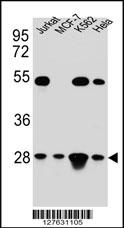 WB (Western Blot)
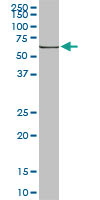
(TFAM Antibody (C-term) western blot analysis in Hela,Jurkat,K562,MCF-7 cell line lysates (35ug/lane).This demonstrates the TFAM antibody detected the TFAM protein (arrow).)
WB (Western Blot)
(TFAM Antibody (C-term) western blot analysis in Hela,Jurkat,K562,MCF-7 cell line lysates (35ug/lane).This demonstrates the TFAM antibody detected the TFAM protein (arrow).)
TFAM, Polyclonal Antibody (Cat# AAA28696)
Full Name
TFAM Antibody (C-term)
Gene Names
TFAM; TCF6; MTTF1; MTTFA; TCF6L1; TCF6L2; TCF6L3
Reactivity
Human
Applications
EIA, IHC, FC/FACS, IF, WB
Purity
Peptide Affinity Purified Rabbit Polyclonal Antibody (Pab)
Pricing


 Application Data
(Staining of KG1 lymphocytes with Mouse anti Human CD59:FITC)
Application Data
(Staining of KG1 lymphocytes with Mouse anti Human CD59:FITC)
CD59, Monoclonal Antibody (Cat# AAA12068)
Full Name
MOUSE ANTI HUMAN CD59:FITC
Gene Names
CD59; 1F5; EJ16; EJ30; EL32; G344; MIN1; MIN2; MIN3; MIRL; HRF20; MACIF; MEM43; MIC11; MSK21; 16.3A5; HRF-20; MAC-IP; p18-20
Applications
FC/FACS
Pricing


 WB (Western Blot)
(HSPA1B monoclonal antibody Western Blot analysis of HSPA1B expression in Raw 264.7.)
WB (Western Blot)
(HSPA1B monoclonal antibody Western Blot analysis of HSPA1B expression in Raw 264.7.)
HSPA1A, Monoclonal Antibody (Cat# AAA25423)
Full Name
HSPA1A (Heat Shock 70kD Protein 1A/1B, Heat Shock 70kD Protein 1/2, HSP70.1/HSP70.2, HSP70-1/HSP70-2, HSPA1, HSPA1B) (HRP)
Gene Names
HSPA1A; HSP72; HSPA1; HSP70I; HSP70-1; HSP70.1; HSP70-1A; HEL-S-103
Reactivity
Human, Mouse, Rat
Applications
EIA, IHC, WB
Purity
Purified by Protein A Affinity Chromatography.
Pricing


 Application Data
(Staining of KG1 lymphocytes with Mouse anti Human CD59:FITC)
Application Data
(Staining of KG1 lymphocytes with Mouse anti Human CD59:FITC)
CD59, Monoclonal Antibody (Cat# AAA11857)
Full Name
MOUSE ANTI HUMAN CD59:FITC
Gene Names
CD59; 1F5; EJ16; EJ30; EL32; G344; MIN1; MIN2; MIN3; MIRL; HRF20; MACIF; MEM43; MIC11; MSK21; 16.3A5; HRF-20; MAC-IP; p18-20
Applications
FC/FACS
Pricing


 WB (Western Blot)
(HSPA1B monoclonal antibody Western Blot analysis of HSPA1B expression in Raw 264.7.)
WB (Western Blot)
(HSPA1B monoclonal antibody Western Blot analysis of HSPA1B expression in Raw 264.7.)
HSPA1A, Monoclonal Antibody (Cat# AAA25719)
Full Name
HSPA1A (Heat Shock 70kD Protein 1A/1B, Heat Shock 70kD Protein 1/2, HSP70.1/HSP70.2, HSP70-1/HSP70-2, HSPA1, HSPA1B) (PE)
Gene Names
HSPA1A; HSP72; HSPA1; HSP70I; HSP70-1; HSP70.1; HSP70-1A; HEL-S-103
Reactivity
Human, Mouse, Rat
Applications
EIA, IF, IHC, WB
Purity
Purified by Protein A Affinity Chromatography.
Pricing


 FCM (Flow Cytometry)
(HAS2 Antibody (Center) (AAA28714) flow cytometric analysis of K562 cells (bottom histogram) compared to a Rabbit IgG Isotype control (negative control-top histogram).FITC-conjugated goat-anti-rabbit secondary antibodies were used for the analysis.)
FCM (Flow Cytometry)
(HAS2 Antibody (Center) (AAA28714) flow cytometric analysis of K562 cells (bottom histogram) compared to a Rabbit IgG Isotype control (negative control-top histogram).FITC-conjugated goat-anti-rabbit secondary antibodies were used for the analysis.)
HAS2, Polyclonal Antibody (Cat# AAA28714)
Full Name
HAS2 Antibody (Center)
Reactivity
Human, mouse
Applications
WB, EIA, IHC-P, FC/FACS
Purity
Peptide Affinity Purified Rabbit Polyclonal Antibody (Pab)
Pricing


 WB (Western Blot)
(Western Blot Analysis: Representative lot data. Lysate from HEK-293 cells was resolved by electrophoresis, transferred to PVDF and probed with anti-PI3 Kinase, p110β (0.05 ug/mL). Proteins were visualized using donkey anti-rabbit secondary antibody conjugated to HRP and chemiluminescence detection. Arrow indicates PI3 Kinase, p110β (~110kD).)
WB (Western Blot)
(Western Blot Analysis: Representative lot data. Lysate from HEK-293 cells was resolved by electrophoresis, transferred to PVDF and probed with anti-PI3 Kinase, p110β (0.05 ug/mL). Proteins were visualized using donkey anti-rabbit secondary antibody conjugated to HRP and chemiluminescence detection. Arrow indicates PI3 Kinase, p110β (~110kD).)
Phosphoinositide 3 Kinase, p110 beta, Polyclonal Antibody (Cat# AAA14720)
Full Name
Phosphoinositide 3 Kinase, p110 beta (DKFZp779K1237, MGC133043, p110beta, Phosphatidylinositol 3 Kinase Catalytic beta Polypeptide, Phosphatidylinositol-4,5-bisphosphate 3-kinase Catalytic Subunit beta Isoform, Phosphoinositide 3 Kinase Catalytic beta Pol
Gene Names
PIK3CB; PI3K; PIK3C1; P110BETA; PI3KBETA; MGC133043; DKFZp779K1237
Reactivity
Human
Applications
EL/EIA, WB, IP, IHC, ICC
Purity
Affinity Purified
Purified by immunoaffinity chromatography.
Purified by immunoaffinity chromatography.
Pricing


 Application Data
(C:FGFR2/isolectinB4 (C) and FGFR1/isolectinB4 (D) staining of apparent mesenchymal cells and the subpopulation of endothelial cells. Virtually all other dispersed apparent mesenchymal cells express FGFR1 and FGFR2 (merged image in E). F: FGFR2 (F) and FGFR1 (G) staining in clustered cells of epithelial origin (inferred by morphology here) demonstrating that epithelial cells express both FGFR1 and FGFR2 (merged image with DAPI staining in H).)
Application Data
(C:FGFR2/isolectinB4 (C) and FGFR1/isolectinB4 (D) staining of apparent mesenchymal cells and the subpopulation of endothelial cells. Virtually all other dispersed apparent mesenchymal cells express FGFR1 and FGFR2 (merged image in E). F: FGFR2 (F) and FGFR1 (G) staining in clustered cells of epithelial origin (inferred by morphology here) demonstrating that epithelial cells express both FGFR1 and FGFR2 (merged image with DAPI staining in H).)
FGFR2, Polyclonal Antibody (Cat# AAA26852)
Full Name
FGFR2, NT (FGFR2, BEK, KGFR, KSAM, Fibroblast growth factor receptor 2, K-sam, Keratinocyte growth factor receptor, CD332) (APC)
Gene Names
FGFR2; BEK; JWS; BBDS; CEK3; CFD1; ECT1; KGFR; TK14; TK25; BFR-1; CD332; K-SAM
Reactivity
Human, Monkey, Mouse, Rat
Applications
FC/FACS, IF, IHC, WB
Purity
Purified by Protein G Affinity Chromatography.
Pricing


 IHC (Immunohistchemistry)
(Figure 6. IHC analysis of AMD1 using anti-AMD1 antibody (AAA19177).AMD1 was detected in paraffin-embedded section of human mammary cancer tissue. Heat mediated antigen retrieval was performed in citrate buffer (pH6, epitope retrieval solution) for 20 mins. The tissue section was blocked with 10% goat serum. The tissue section was then incubated with 1ug/ml rabbit anti-AMD1 Antibody (AAA19177) overnight at 4 degree C. Biotinylated goat anti-rabbit IgG was used as secondary antibody and incubated for 30 minutes at 37 degree C. The tissue section was developed using Strepavidin-Biotin-Complex (SABC) with DAB as the chromogen.)
IHC (Immunohistchemistry)
(Figure 6. IHC analysis of AMD1 using anti-AMD1 antibody (AAA19177).AMD1 was detected in paraffin-embedded section of human mammary cancer tissue. Heat mediated antigen retrieval was performed in citrate buffer (pH6, epitope retrieval solution) for 20 mins. The tissue section was blocked with 10% goat serum. The tissue section was then incubated with 1ug/ml rabbit anti-AMD1 Antibody (AAA19177) overnight at 4 degree C. Biotinylated goat anti-rabbit IgG was used as secondary antibody and incubated for 30 minutes at 37 degree C. The tissue section was developed using Strepavidin-Biotin-Complex (SABC) with DAB as the chromogen.)
AMD1/Adometdc, Polyclonal Antibody (Cat# AAA19177)
Full Name
Anti-AMD1/Adometdc Antibody
Gene Names
AMD1; AMD; SAMDC; ADOMETDC
Reactivity
Human, Mouse, Rat
No cross reactivity with other proteins.
No cross reactivity with other proteins.
Applications
IHC, WB
Purity
Immunogen affinity purified
Pricing


 ICC (Immunocytochemistry)
(Figure 10 Immunocytochemistry Validation of PD-L1Immunocytochemical analysis of 4% paraformaldehyde-fixed PD-L1 transfected 293 cells labeling PD-L1 with at 1 μg/ml, followed by Goat anti-mouse IgG secondary antibody at 1/250 dilution (red). Lower left: Use mouse IgG antibody at 1 μg/ml as control.)
ICC (Immunocytochemistry)
(Figure 10 Immunocytochemistry Validation of PD-L1Immunocytochemical analysis of 4% paraformaldehyde-fixed PD-L1 transfected 293 cells labeling PD-L1 with at 1 μg/ml, followed by Goat anti-mouse IgG secondary antibody at 1/250 dilution (red). Lower left: Use mouse IgG antibody at 1 μg/ml as control.)
PDL1, Monoclonal Antibody (Cat# AAA10992)
Full Name
PDL1 Antibody [6H10]
Gene Names
CD274; B7-H; B7H1; PDL1; PD-L1; PDCD1L1; PDCD1LG1
Reactivity
Human
Applications
EIA, WB, IHC, ICC, IF
Purity
Protein A purified
Pricing


 IP (Immunoprecipitation)
(Figure 11 Immunoprecipitation Validation in HEK293 cells (Kawai et al., 2004)HEK293 cells were transiently transfected with DYKDDDDK-IRF7. Ccell lysates were immunoprecipitated with control rabbit anti-mouse immunoglobulin serum (IgG) or anti-MyD88 (Ab1 and Ab2), followed by immunoblotting with anti-DYKDDDDK.)
IP (Immunoprecipitation)
(Figure 11 Immunoprecipitation Validation in HEK293 cells (Kawai et al., 2004)HEK293 cells were transiently transfected with DYKDDDDK-IRF7. Ccell lysates were immunoprecipitated with control rabbit anti-mouse immunoglobulin serum (IgG) or anti-MyD88 (Ab1 and Ab2), followed by immunoblotting with anti-DYKDDDDK.)
MYD88, Polyclonal Antibody (Cat# AAA10944)
Full Name
MYD88 Antibody
Gene Names
MYD88; MYD88D
Reactivity
Human, Mouse, Rat
Applications
EIA, WB
Purity
MYD88 Antibody is affinity chromatography purified via peptide column.
Pricing


 FCM (Flow Cytometry)
(Overlay Peak curve showing 293F cells transfected with GST stained with AAA28065 (red line) at 1:189. The cells were fixed in 4% formaldehyde and permeated by 0.2% TritonX-100. Then 10% normal goat serum was Incubated to block non-specific protein-protein interactions followed by the antibody (1?g/1*106cells) for 1 h at 4 degree C. The secondary antibody used was FITC-conjugated Goat Anti-Mouse IgG(H+L) at 1/100 dilution for 30min at 4 degree C. Isotype control antibody (green line) was mouse IgG2b (1?g/1*106cells) used under the same conditions. Acquisition of >10,000 events was performed.)
FCM (Flow Cytometry)
(Overlay Peak curve showing 293F cells transfected with GST stained with AAA28065 (red line) at 1:189. The cells were fixed in 4% formaldehyde and permeated by 0.2% TritonX-100. Then 10% normal goat serum was Incubated to block non-specific protein-protein interactions followed by the antibody (1?g/1*106cells) for 1 h at 4 degree C. The secondary antibody used was FITC-conjugated Goat Anti-Mouse IgG(H+L) at 1/100 dilution for 30min at 4 degree C. Isotype control antibody (green line) was mouse IgG2b (1?g/1*106cells) used under the same conditions. Acquisition of >10,000 events was performed.)
GST, Monoclonal Antibody (Cat# AAA28065)
Full Name
GST Monoclonal Antibody
Reactivity
All
Applications
EIA, WB, IF, FC/FACS, IP
Purity
>95%, Protein A purified
Pricing


 Application Data
(Formalin fixed, paraffin embedded human breast cancer biopsy stained with Mouse anti Human estrogen receptor beta5 antibody followed by HRP polymer detection and DAB substrate development following heat mediated antigen retrieval using citrate buffer at pH6.2 (low power))
Application Data
(Formalin fixed, paraffin embedded human breast cancer biopsy stained with Mouse anti Human estrogen receptor beta5 antibody followed by HRP polymer detection and DAB substrate development following heat mediated antigen retrieval using citrate buffer at pH6.2 (low power))
ESTROGEN RECEPTOR BETA 5, Monoclonal Antibody (Cat# AAA12216)
Full Name
MOUSE ANTI HUMAN ESTROGEN RECEPTOR BETA 5
Gene Names
ESR2; Erb; ESRB; ESTRB; NR3A2; ER-BETA; ESR-BETA
Applications
WB
Pricing


 IF (Immunofluorescence)
(Fluorescent confocal image of Hela cell stained with XRCC6 Antibody (C-term). Hela cells were fixed with 4% PFA (20 min), permeabilized with Triton X-100 (0.1%, 10 min), then incubated with XRCC6 primary antibody (1:25, 1 h at 37 degree). For secondary antibody, Alexa Fluor 488 conjugated donkey anti-rabbit antibody (green) was used (1:400, 50 min at 37 degree).Cytoplasmic actin was counterstained with Alexa Fluor 555 (red) conjugated Phalloidin (7units/ml, 1 h at 37 degree). Nuclei were counterstained with DAPI (blue) (10 ug/ml, 10 min). XRCC6 immunoreactivity is localized to nucleus significantly and Cytoplasm weakly.)
IF (Immunofluorescence)
(Fluorescent confocal image of Hela cell stained with XRCC6 Antibody (C-term). Hela cells were fixed with 4% PFA (20 min), permeabilized with Triton X-100 (0.1%, 10 min), then incubated with XRCC6 primary antibody (1:25, 1 h at 37 degree). For secondary antibody, Alexa Fluor 488 conjugated donkey anti-rabbit antibody (green) was used (1:400, 50 min at 37 degree).Cytoplasmic actin was counterstained with Alexa Fluor 555 (red) conjugated Phalloidin (7units/ml, 1 h at 37 degree). Nuclei were counterstained with DAPI (blue) (10 ug/ml, 10 min). XRCC6 immunoreactivity is localized to nucleus significantly and Cytoplasm weakly.)
XRCC6, Polyclonal Antibody (Cat# AAA28768)
Full Name
XRCC6 Antibody (C-term)
Gene Names
XRCC6; ML8; KU70; TLAA; CTC75; CTCBF; G22P1
Reactivity
Human
Applications
WB, EIA, IHC, FC/FACS, IF
Purity
Peptide Affinity Purified Rabbit Polyclonal Antibody (Pab)
Pricing


 IF (Immunofluorescence)
(Immunofluorescent analysis of 4% paraformaldehyde-fixed, 0.1% Triton X-100 permeabilized MCF-7 (human breast cancer cell line) cells labeling Pdx1 with at 1:25 dilution, followed by DyLight 488-conjugated IgG goat anti-rabbit secondary antibody at 1:200 dilution (green). Immunofluorescence image showing cytoplasm staining on MCF-7 cell line. Cytoplasmic actin is detected with DyLight 554 Phalloidin (PD18466410) at 1:100 dilution (red). The nuclear counter stain is DAPI (blue).)
IF (Immunofluorescence)
(Immunofluorescent analysis of 4% paraformaldehyde-fixed, 0.1% Triton X-100 permeabilized MCF-7 (human breast cancer cell line) cells labeling Pdx1 with at 1:25 dilution, followed by DyLight 488-conjugated IgG goat anti-rabbit secondary antibody at 1:200 dilution (green). Immunofluorescence image showing cytoplasm staining on MCF-7 cell line. Cytoplasmic actin is detected with DyLight 554 Phalloidin (PD18466410) at 1:100 dilution (red). The nuclear counter stain is DAPI (blue).)
OPN-a/b, Polyclonal Antibody (Cat# AAA26857)
Full Name
OPN-a/b, NT (SPP1, BNSP, OPN, Osteopontin, Bone sialoprotein 1, Nephropontin, Secreted phosphoprotein 1, Urinary stone protein, Uropontin) (APC)
Gene Names
SPP1; OPN; BNSP; BSPI; ETA-1
Reactivity
Human
Applications
WB, IHC, IF
Purity
Purified by Protein A and Peptide Affinity Chromatography.
Pricing


 IF (Immunofluorescence)
(Immunofluorescent analysis of 4% paraformaldehyde-fixed, 0.1% Triton X-100 permeabilized MCF-7 (human breast cancer cell line) cells labeling Pdx1 with at 1:25 dilution, followed by DyLight 488-conjugated IgG goat anti-rabbit secondary antibody at 1:200 dilution (green). Immunofluorescence image showing cytoplasm staining on MCF-7 cell line. Cytoplasmic actin is detected with DyLight 554 Phalloidin (PD18466410) at 1:100 dilution (red). The nuclear counter stain is DAPI (blue).)
IF (Immunofluorescence)
(Immunofluorescent analysis of 4% paraformaldehyde-fixed, 0.1% Triton X-100 permeabilized MCF-7 (human breast cancer cell line) cells labeling Pdx1 with at 1:25 dilution, followed by DyLight 488-conjugated IgG goat anti-rabbit secondary antibody at 1:200 dilution (green). Immunofluorescence image showing cytoplasm staining on MCF-7 cell line. Cytoplasmic actin is detected with DyLight 554 Phalloidin (PD18466410) at 1:100 dilution (red). The nuclear counter stain is DAPI (blue).)
OPN-a/b, Polyclonal Antibody (Cat# AAA26860)
Full Name
OPN-a/b, NT (SPP1, BNSP, OPN, Osteopontin, Bone sialoprotein 1, Nephropontin, Secreted phosphoprotein 1, Urinary stone protein, Uropontin) (MaxLight 405)
Gene Names
SPP1; OPN; BNSP; BSPI; ETA-1
Reactivity
Human
Applications
WB, IHC, IF
Purity
Purified by Protein A and Peptide Affinity Chromatography.
Pricing


 IF (Immunofluorescence)
(Immunofluorescent analysis of 4% paraformaldehyde-fixed, 0.1% Triton X-100 permeabilized MCF-7 (human breast cancer cell line) cells labeling Pdx1 with at 1:25 dilution, followed by DyLight 488-conjugated IgG goat anti-rabbit secondary antibody at 1:200 dilution (green). Immunofluorescence image showing cytoplasm staining on MCF-7 cell line. Cytoplasmic actin is detected with DyLight 554 Phalloidin (PD18466410) at 1:100 dilution (red). The nuclear counter stain is DAPI (blue).)
IF (Immunofluorescence)
(Immunofluorescent analysis of 4% paraformaldehyde-fixed, 0.1% Triton X-100 permeabilized MCF-7 (human breast cancer cell line) cells labeling Pdx1 with at 1:25 dilution, followed by DyLight 488-conjugated IgG goat anti-rabbit secondary antibody at 1:200 dilution (green). Immunofluorescence image showing cytoplasm staining on MCF-7 cell line. Cytoplasmic actin is detected with DyLight 554 Phalloidin (PD18466410) at 1:100 dilution (red). The nuclear counter stain is DAPI (blue).)
OPN-a/b, Polyclonal Antibody (Cat# AAA26859)
Full Name
OPN-a/b, NT (SPP1, BNSP, OPN, Osteopontin, Bone sialoprotein 1, Nephropontin, Secreted phosphoprotein 1, Urinary stone protein, Uropontin) (FITC)
Gene Names
SPP1; OPN; BNSP; BSPI; ETA-1
Reactivity
Human
Applications
WB, IHC, IF
Purity
Purified by Protein A and Peptide Affinity Chromatography.
Pricing


 IF (Immunofluorescence)
(Immunofluorescent analysis of 4% paraformaldehyde-fixed, 0.1% Triton X-100 permeabilized MCF-7 (human breast cancer cell line) cells labeling Pdx1 with at 1:25 dilution, followed by DyLight 488-conjugated IgG goat anti-rabbit secondary antibody at 1:200 dilution (green). Immunofluorescence image showing cytoplasm staining on MCF-7 cell line. Cytoplasmic actin is detected with DyLight 554 Phalloidin (PD18466410) at 1:100 dilution (red). The nuclear counter stain is DAPI (blue).)
IF (Immunofluorescence)
(Immunofluorescent analysis of 4% paraformaldehyde-fixed, 0.1% Triton X-100 permeabilized MCF-7 (human breast cancer cell line) cells labeling Pdx1 with at 1:25 dilution, followed by DyLight 488-conjugated IgG goat anti-rabbit secondary antibody at 1:200 dilution (green). Immunofluorescence image showing cytoplasm staining on MCF-7 cell line. Cytoplasmic actin is detected with DyLight 554 Phalloidin (PD18466410) at 1:100 dilution (red). The nuclear counter stain is DAPI (blue).)
OPN-a/b, Polyclonal Antibody (Cat# AAA26864)
Full Name
OPN-a/b, NT (SPP1, BNSP, OPN, Osteopontin, Bone sialoprotein 1, Nephropontin, Secreted phosphoprotein 1, Urinary stone protein, Uropontin) (MaxLight 750)
Gene Names
SPP1; OPN; BNSP; BSPI; ETA-1
Reactivity
Human
Applications
WB, IHC, IF
Purity
Purified by Protein A and Peptide Affinity Chromatography.
Pricing


 IF (Immunofluorescence)
(Immunofluorescent analysis of 4% paraformaldehyde-fixed, 0.1% Triton X-100 permeabilized MCF-7 (human breast cancer cell line) cells labeling Pdx1 with at 1:25 dilution, followed by DyLight 488-conjugated IgG goat anti-rabbit secondary antibody at 1:200 dilution (green). Immunofluorescence image showing cytoplasm staining on MCF-7 cell line. Cytoplasmic actin is detected with DyLight 554 Phalloidin (PD18466410) at 1:100 dilution (red). The nuclear counter stain is DAPI (blue).)
IF (Immunofluorescence)
(Immunofluorescent analysis of 4% paraformaldehyde-fixed, 0.1% Triton X-100 permeabilized MCF-7 (human breast cancer cell line) cells labeling Pdx1 with at 1:25 dilution, followed by DyLight 488-conjugated IgG goat anti-rabbit secondary antibody at 1:200 dilution (green). Immunofluorescence image showing cytoplasm staining on MCF-7 cell line. Cytoplasmic actin is detected with DyLight 554 Phalloidin (PD18466410) at 1:100 dilution (red). The nuclear counter stain is DAPI (blue).)
OPN-a/b, Polyclonal Antibody (Cat# AAA26863)
Full Name
OPN-a/b, NT (SPP1, BNSP, OPN, Osteopontin, Bone sialoprotein 1, Nephropontin, Secreted phosphoprotein 1, Urinary stone protein, Uropontin) (MaxLight 650)
Gene Names
SPP1; OPN; BNSP; BSPI; ETA-1
Reactivity
Human
Applications
WB, IHC, IF
Purity
Purified by Protein A and Peptide Affinity Chromatography.
Pricing


 IF (Immunofluorescence)
(Immunofluorescent analysis of 4% paraformaldehyde-fixed, 0.1% Triton X-100 permeabilized MCF-7 (human breast cancer cell line) cells labeling Pdx1 with at 1:25 dilution, followed by DyLight 488-conjugated IgG goat anti-rabbit secondary antibody at 1:200 dilution (green). Immunofluorescence image showing cytoplasm staining on MCF-7 cell line. Cytoplasmic actin is detected with DyLight 554 Phalloidin (PD18466410) at 1:100 dilution (red). The nuclear counter stain is DAPI (blue).)
IF (Immunofluorescence)
(Immunofluorescent analysis of 4% paraformaldehyde-fixed, 0.1% Triton X-100 permeabilized MCF-7 (human breast cancer cell line) cells labeling Pdx1 with at 1:25 dilution, followed by DyLight 488-conjugated IgG goat anti-rabbit secondary antibody at 1:200 dilution (green). Immunofluorescence image showing cytoplasm staining on MCF-7 cell line. Cytoplasmic actin is detected with DyLight 554 Phalloidin (PD18466410) at 1:100 dilution (red). The nuclear counter stain is DAPI (blue).)
OPN-a/b, Polyclonal Antibody (Cat# AAA26862)
Full Name
OPN-a/b, NT (SPP1, BNSP, OPN, Osteopontin, Bone sialoprotein 1, Nephropontin, Secreted phosphoprotein 1, Urinary stone protein, Uropontin) (MaxLight 550)
Gene Names
SPP1; OPN; BNSP; BSPI; ETA-1
Reactivity
Human
Applications
WB, IHC, IF
Purity
Purified by Protein A and Peptide Affinity Chromatography.
Pricing