Filters
▼Clonality
▼Type
▼Reactivity
▼Gene Name
▼Isotype
▼Host
▼Application
▼Clone
▼Monoclonal Antibodies
Get accurate results in your research with our Monoclonal Antibodies, which are specially made to target exactly what you require for your research, and will produce consistent, reliable performance in lab tests.
Viewing 4250-4300 of 27645 product results
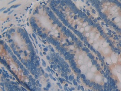 IHC (Immunohistochemistry)
(DAB staining on IHCP;Sample: Rat Intestine Tissue; Primary Ab: 30ug/ml Mouse AntiRat IL1a AntibodySecond Ab: 2ug/mL HRPLinked Caprine AntiMouse IgG Polyclonal Antibody(Catalog: SAA544Mu19))
IHC (Immunohistochemistry)
(DAB staining on IHCP;Sample: Rat Intestine Tissue; Primary Ab: 30ug/ml Mouse AntiRat IL1a AntibodySecond Ab: 2ug/mL HRPLinked Caprine AntiMouse IgG Polyclonal Antibody(Catalog: SAA544Mu19))
Interleukin 1 Alpha (IL1a), Monoclonal Antibody (Cat# AAA151458)
 IHC (Immunohistochemisry)
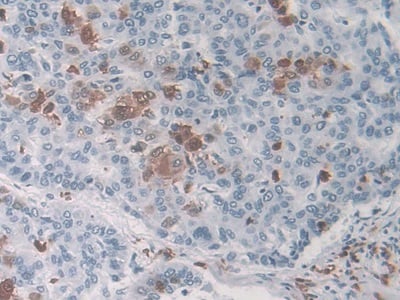
(DAB staining on IHCP;Sample: Human Colorectal cancer Tissue; Primary Ab: 30ug/ml Mouse AntiHuman MIP3a AntibodySecond Ab: 2ug/mL HRPLinked Caprine AntiMouse IgG Polyclonal Antibody(Catalog: SAA544Mu19))
IHC (Immunohistochemisry)
(DAB staining on IHCP;Sample: Human Colorectal cancer Tissue; Primary Ab: 30ug/ml Mouse AntiHuman MIP3a AntibodySecond Ab: 2ug/mL HRPLinked Caprine AntiMouse IgG Polyclonal Antibody(Catalog: SAA544Mu19))
Macrophage Inflammatory Protein 3 Alpha (MIP3a), Monoclonal Antibody (Cat# AAA151473)
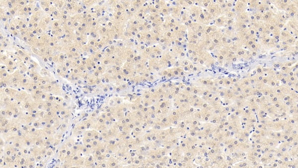 IHC (Immunohiostchemistry)
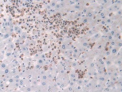
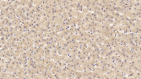
(DAB staining on IHCP;Sample: Human Liver Tissue; Primary Ab: 30ug/ml Mouse AntiHuman NT3 AntibodySecond Ab: 2ug/mL HRPLinked Caprine AntiMouse IgG Polyclonal Antibody(Catalog: SAA544Mu19))
IHC (Immunohiostchemistry)
(DAB staining on IHCP;Sample: Human Liver Tissue; Primary Ab: 30ug/ml Mouse AntiHuman NT3 AntibodySecond Ab: 2ug/mL HRPLinked Caprine AntiMouse IgG Polyclonal Antibody(Catalog: SAA544Mu19))
Neurotrophin 3 (NT3), Monoclonal Antibody (Cat# AAA151480)
 IHC (Immunohiostchemistry)
(DAB staining on IHCP;Sample: Human Liver Tissue; Primary Ab: 30ug/ml Mouse AntiHuman NT3 AntibodySecond Ab: 2ug/mL HRPLinked Caprine AntiMouse IgG Polyclonal Antibody(Catalog: SAA544Mu19))
IHC (Immunohiostchemistry)
(DAB staining on IHCP;Sample: Human Liver Tissue; Primary Ab: 30ug/ml Mouse AntiHuman NT3 AntibodySecond Ab: 2ug/mL HRPLinked Caprine AntiMouse IgG Polyclonal Antibody(Catalog: SAA544Mu19))
Neurotrophin 3 (NT3), Monoclonal Antibody (Cat# AAA151481)
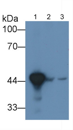
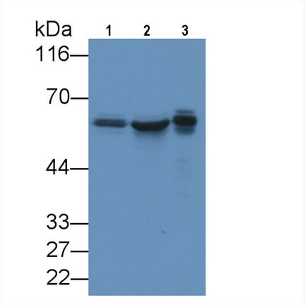
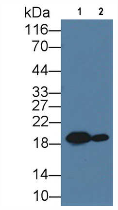
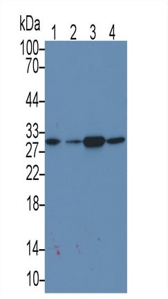
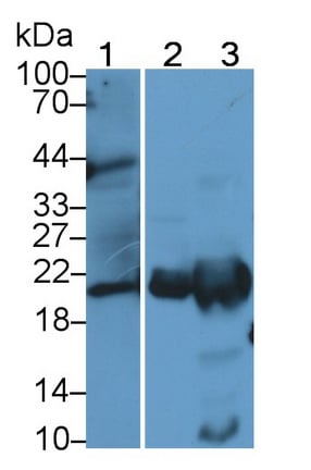 WB (Western Blot)
(Western Blot; Sample: Lane1: Human Placenta lysate; Lane2: Rat Liver lysate; Lane3: Rat Spleen lysate Primary Ab: 2ug/mL Mouse AntiHuman FTL Antibody Second Ab: 0.2ug/mL HRPLinked Caprine AntiMouse IgG Polyclonal Antibody (Catalog: SAA544Mu19))
WB (Western Blot)
(Western Blot; Sample: Lane1: Human Placenta lysate; Lane2: Rat Liver lysate; Lane3: Rat Spleen lysate Primary Ab: 2ug/mL Mouse AntiHuman FTL Antibody Second Ab: 0.2ug/mL HRPLinked Caprine AntiMouse IgG Polyclonal Antibody (Catalog: SAA544Mu19))
Ferritin, Light Polypeptide (FTL), Monoclonal Antibody (Cat# AAA151828)
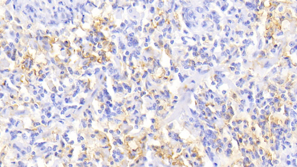
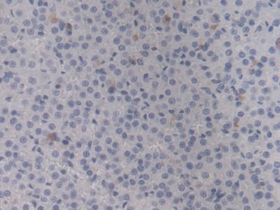
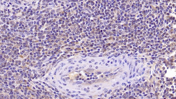
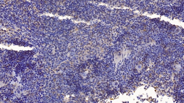
 IHC (Immunohiostchemistry)
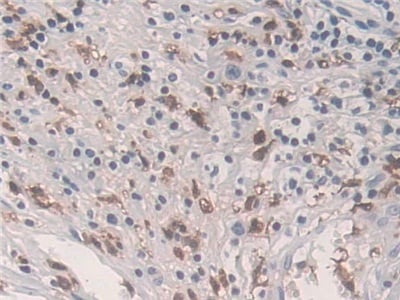
(DAB staining on IHCP;Sample: Human Lymph node Tissue; Primary Ab: 20ug/ml Mouse AntiHuman SQSTM1 AntibodySecond Ab: 2ug/mL HRPLinked Caprine AntiMouse IgG Polyclonal Antibody(Catalog: SAA544Mu19))
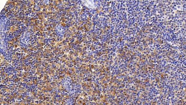
IHC (Immunohiostchemistry)
(DAB staining on IHCP;Sample: Human Lymph node Tissue; Primary Ab: 20ug/ml Mouse AntiHuman SQSTM1 AntibodySecond Ab: 2ug/mL HRPLinked Caprine AntiMouse IgG Polyclonal Antibody(Catalog: SAA544Mu19))
Sequestosome 1 (SQSTM1), Monoclonal Antibody (Cat# AAA151833)
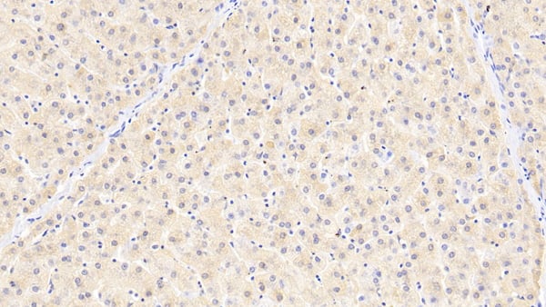
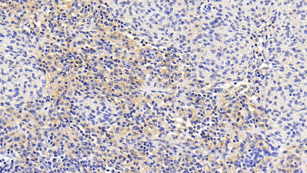
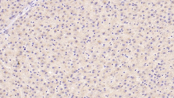
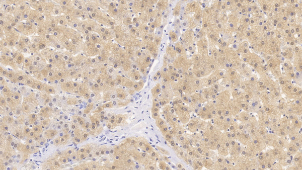 IHC (Immunohistochemisry)
(DAB staining on IHCP;Samples: Human Liver Tissue;Primary Ab: 30ug/ml Mouse AntiHuman HPD AntibodySecond Ab: 2ug/mL HRPLinked Caprine AntiMouse IgG Polyclonal Antibody(Catalog: SAA544Mu19))
IHC (Immunohistochemisry)
(DAB staining on IHCP;Samples: Human Liver Tissue;Primary Ab: 30ug/ml Mouse AntiHuman HPD AntibodySecond Ab: 2ug/mL HRPLinked Caprine AntiMouse IgG Polyclonal Antibody(Catalog: SAA544Mu19))
4Hydroxyphenylpyruvate Dioxygenase (HPD), Monoclonal Antibody (Cat# AAA151849)
 IHC (Immunohiostchemistry)
(DAB staining on IHCP;Sample: Human Liver Tissue; Primary Ab: 30ug/ml Mouse AntiHuman ROS1 AntibodySecond Ab: 2ug/mL HRPLinked Caprine AntiMouse IgG Polyclonal Antibody(Catalog: SAA544Mu19))
IHC (Immunohiostchemistry)
(DAB staining on IHCP;Sample: Human Liver Tissue; Primary Ab: 30ug/ml Mouse AntiHuman ROS1 AntibodySecond Ab: 2ug/mL HRPLinked Caprine AntiMouse IgG Polyclonal Antibody(Catalog: SAA544Mu19))
CRos Oncogene 1, Receptor Tyrosine Kinase (ROS1), Monoclonal Antibody (Cat# AAA151851)
 WB (Western Blot)
(Western Blot; Sample: Lane1: U2OS cell lysate; Lane2: Jurkat cell lysate; Lane3: 293T cell lysate Primary Ab: 3ug/ml Mouse AntiHuman KPNa2 Antibody Second Ab: 0.2ug/mL HRPLinked Caprine AntiMouse IgG Polyclonal Antibody (Catalog: SAA544Mu19))
WB (Western Blot)
(Western Blot; Sample: Lane1: U2OS cell lysate; Lane2: Jurkat cell lysate; Lane3: 293T cell lysate Primary Ab: 3ug/ml Mouse AntiHuman KPNa2 Antibody Second Ab: 0.2ug/mL HRPLinked Caprine AntiMouse IgG Polyclonal Antibody (Catalog: SAA544Mu19))
Karyopherin Alpha 2 (KPNa2), Monoclonal Antibody (Cat# AAA151858)
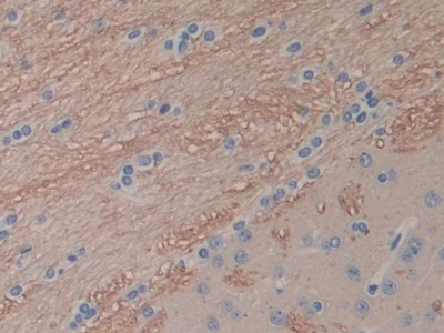
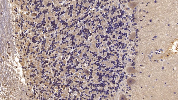
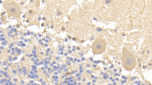 IHC (Immunohistochemisry)
(DAB staining on IHCP;Sample: Human Cerebellum Tissue; Primary Ab: 20ug/ml Mouse AntiHuman BAI3 AntibodySecond Ab: 2ug/mL HRPLinked Caprine AntiMouse IgG Polyclonal Antibody(Catalog: SAA544Mu19))
IHC (Immunohistochemisry)
(DAB staining on IHCP;Sample: Human Cerebellum Tissue; Primary Ab: 20ug/ml Mouse AntiHuman BAI3 AntibodySecond Ab: 2ug/mL HRPLinked Caprine AntiMouse IgG Polyclonal Antibody(Catalog: SAA544Mu19))
Brain Specific Angiogenesis Inhibitor 3 (BAI3), Monoclonal Antibody (Cat# AAA151866)
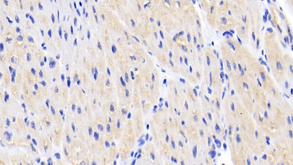
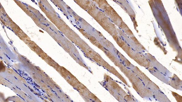
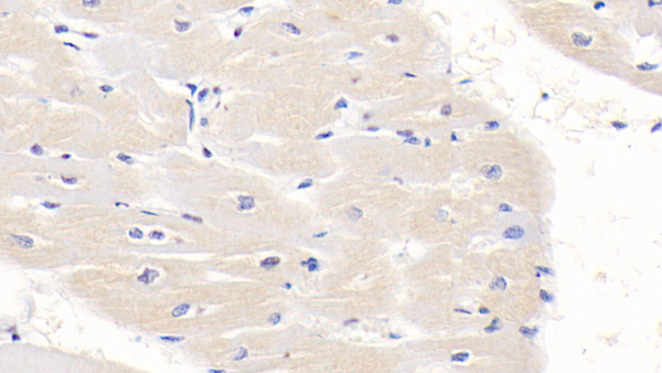
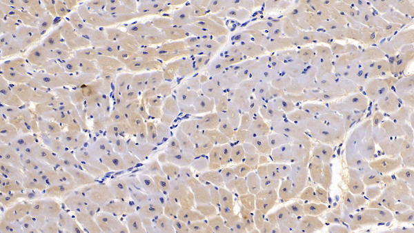
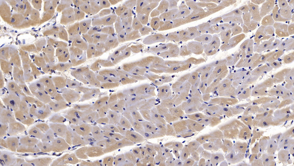
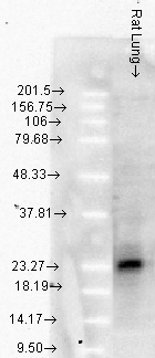
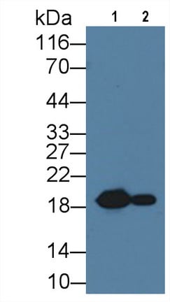 WB (Western Blot)
(Western Blot; Sample: Lane1: Rat Heart lysate; Lane1: Rat Skeletal muscle lysate Primary Ab: 1ug/ml Mouse AntiRat HSPb6 Antibody Second Ab: 0.2ug/mL HRPLinked Caprine AntiMouse IgG Polyclonal Antibody (Catalog: SAA544Mu19))
WB (Western Blot)
(Western Blot; Sample: Lane1: Rat Heart lysate; Lane1: Rat Skeletal muscle lysate Primary Ab: 1ug/ml Mouse AntiRat HSPb6 Antibody Second Ab: 0.2ug/mL HRPLinked Caprine AntiMouse IgG Polyclonal Antibody (Catalog: SAA544Mu19))
Heat Shock Protein Beta 6 (HSPb6), Monoclonal Antibody (Cat# AAA151867)
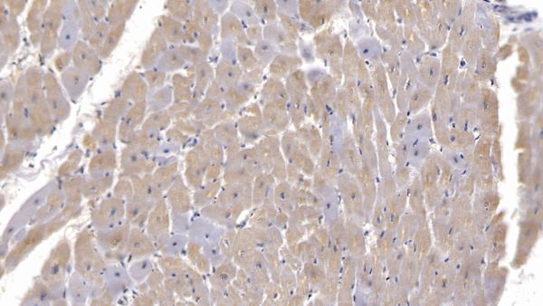
 WB (Western Blot)
(Western Blot; Sample: Lane1: Rat Heart lysate; Lane1: Rat Skeletal muscle lysate Primary Ab: 1ug/ml Mouse AntiRat HSPb6 Antibody Second Ab: 0.2ug/mL HRPLinked Caprine AntiMouse IgG Polyclonal Antibody (Catalog: SAA544Mu19))
WB (Western Blot)
(Western Blot; Sample: Lane1: Rat Heart lysate; Lane1: Rat Skeletal muscle lysate Primary Ab: 1ug/ml Mouse AntiRat HSPb6 Antibody Second Ab: 0.2ug/mL HRPLinked Caprine AntiMouse IgG Polyclonal Antibody (Catalog: SAA544Mu19))
Heat Shock Protein Beta 6 (HSPb6), Monoclonal Antibody (Cat# AAA151868)
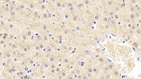
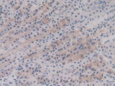
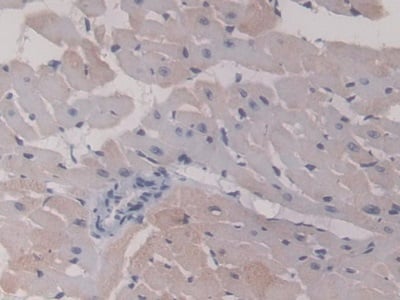
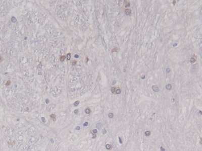
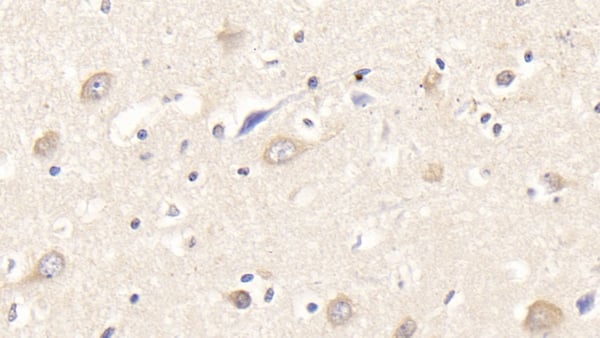
 IHC (Immunohiostchemistry)
(DAB staining on IHCP;Sample: Human Liver Tissue; Primary Ab: 40ug/ml Mouse AntiHuman CDNF AntibodySecond Ab: 2ug/mL HRPLinked Caprine AntiMouse IgG Polyclonal Antibody(Catalog: SAA544Mu19))
IHC (Immunohiostchemistry)
(DAB staining on IHCP;Sample: Human Liver Tissue; Primary Ab: 40ug/ml Mouse AntiHuman CDNF AntibodySecond Ab: 2ug/mL HRPLinked Caprine AntiMouse IgG Polyclonal Antibody(Catalog: SAA544Mu19))
Cerebral Dopamine Neurotrophic Factor (CDNF), Monoclonal Antibody (Cat# AAA151874)
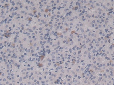
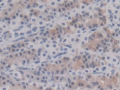
 IHC (Immunohiostchemistry)
(DAB staining on IHCP;Sample: Mouse Spleen Tissue; Primary Ab: 10ug/ml Mouse AntiMouse CDNF AntibodySecond Ab: 2ug/mL HRPLinked Caprine AntiMouse IgG Polyclonal Antibody(Catalog: SAA544Mu19))
IHC (Immunohiostchemistry)
(DAB staining on IHCP;Sample: Mouse Spleen Tissue; Primary Ab: 10ug/ml Mouse AntiMouse CDNF AntibodySecond Ab: 2ug/mL HRPLinked Caprine AntiMouse IgG Polyclonal Antibody(Catalog: SAA544Mu19))
Cerebral Dopamine Neurotrophic Factor (CDNF), Monoclonal Antibody (Cat# AAA151875)
 WB (Western Blot)
(Western Blot; Sample: Lane1: Rat Thymus lysate; Lane2: Hela cell lysate; Lane3: Jurkat cell lysate; Lane4: A431 cell lysate Primary Ab: 3ug/ml Mouse AntiHuman MAPRE1 Antibody Second Ab: 0.2ug/mL HRPLinked Caprine AntiMouse IgG Polyclonal Antibody (Catalog: SAA544Mu19))
WB (Western Blot)
(Western Blot; Sample: Lane1: Rat Thymus lysate; Lane2: Hela cell lysate; Lane3: Jurkat cell lysate; Lane4: A431 cell lysate Primary Ab: 3ug/ml Mouse AntiHuman MAPRE1 Antibody Second Ab: 0.2ug/mL HRPLinked Caprine AntiMouse IgG Polyclonal Antibody (Catalog: SAA544Mu19))
Microtubule Associated Protein RP/EB Family, Member 1 (MAPRE1), Monoclonal Antibody (Cat# AAA151876)
 WB (Western Blot)
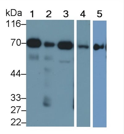
(Western Blot; Sample: Lane1: Mouse Heart lysate; Lane2: Mouse Liver lysate; Lane3: Mouse Cerebrum lysate; Lane4: Mouse Kidney lysate; Lane5: HepG2 cell lysate Primary Ab: 0.1ug/ml Mouse AntiMouse SDHA Antibody Second Ab: 0.2ug/mL HRPLinked Caprine AntiMouse IgG Polyclonal Antibody (Catalog: SAA54)
WB (Western Blot)
(Western Blot; Sample: Lane1: Mouse Heart lysate; Lane2: Mouse Liver lysate; Lane3: Mouse Cerebrum lysate; Lane4: Mouse Kidney lysate; Lane5: HepG2 cell lysate Primary Ab: 0.1ug/ml Mouse AntiMouse SDHA Antibody Second Ab: 0.2ug/mL HRPLinked Caprine AntiMouse IgG Polyclonal Antibody (Catalog: SAA54)
Succinate Dehydrogenase Complex Subunit A (SDHA), Monoclonal Antibody (Cat# AAA151898)
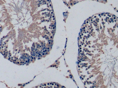
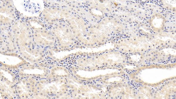
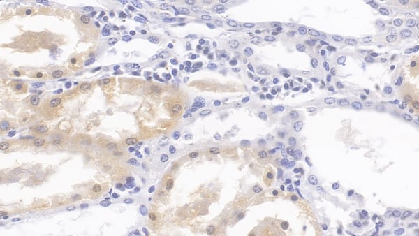
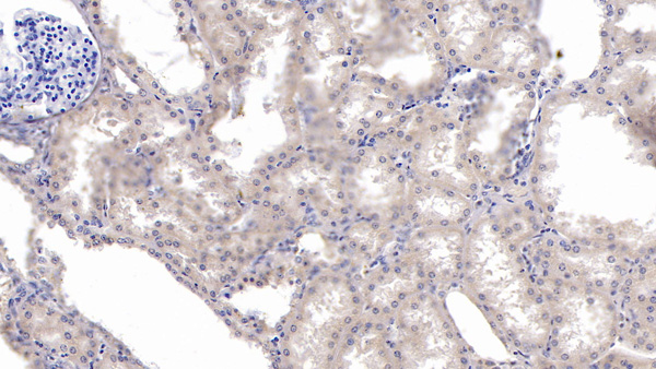
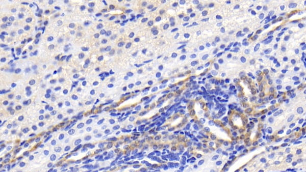
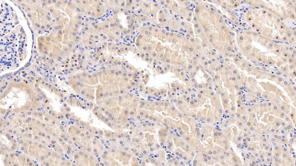
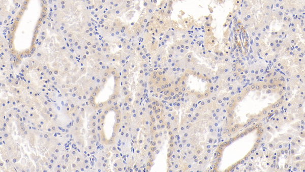
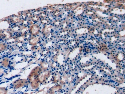 IHC (Immunohiostchemistry)
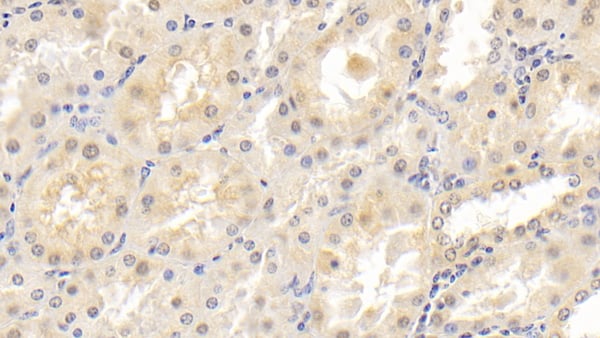
(DAB staining on IHCP;Sample: Rat Kidney Tissue; Primary Ab: 10ug/ml Mouse AntiRat FGF15 AntibodySecond Ab: 2ug/mL HRPLinked Caprine AntiMouse IgG Polyclonal Antibody(Catalog: SAA544Mu19))
IHC (Immunohiostchemistry)
(DAB staining on IHCP;Sample: Rat Kidney Tissue; Primary Ab: 10ug/ml Mouse AntiRat FGF15 AntibodySecond Ab: 2ug/mL HRPLinked Caprine AntiMouse IgG Polyclonal Antibody(Catalog: SAA544Mu19))
Fibroblast Growth Factor 15 (FGF15), Monoclonal Antibody (Cat# AAA151901)
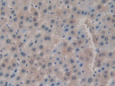
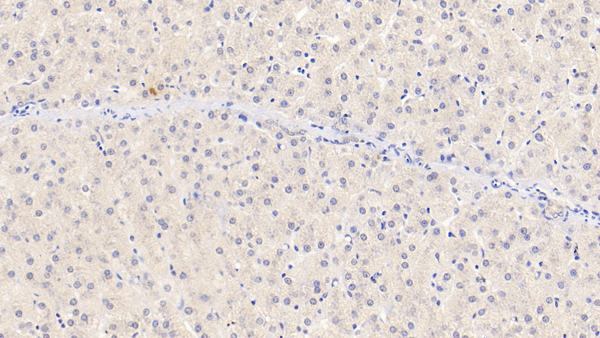 IHC (Immunohistochemistry)
(DAB staining on IHCP;Sample: Human Liver Tissue; Primary Ab: 20ug/ml Mouse AntiHuman SEMA3A AntibodySecond Ab: 2ug/mL HRPLinked Caprine AntiMouse IgG Polyclonal Antibody(Catalog: SAA544Mu19))
IHC (Immunohistochemistry)
(DAB staining on IHCP;Sample: Human Liver Tissue; Primary Ab: 20ug/ml Mouse AntiHuman SEMA3A AntibodySecond Ab: 2ug/mL HRPLinked Caprine AntiMouse IgG Polyclonal Antibody(Catalog: SAA544Mu19))
Semaphorin 3A (SEMA3A), Monoclonal Antibody (Cat# AAA151904)
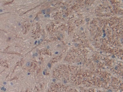
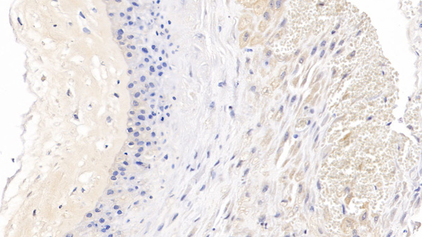
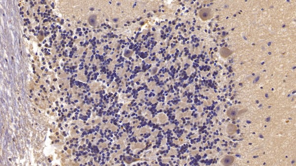 IHC (Immunohiostchemistry)
(DAB staining on IHCP;Sample: Human Cerebellum Tissue; Primary Ab: 10ug/ml Mouse AntiHuman SEMA5B AntibodySecond Ab: 2ug/mL HRPLinked Caprine AntiMouse IgG Polyclonal Antibody(Catalog: SAA544Mu19))
IHC (Immunohiostchemistry)
(DAB staining on IHCP;Sample: Human Cerebellum Tissue; Primary Ab: 10ug/ml Mouse AntiHuman SEMA5B AntibodySecond Ab: 2ug/mL HRPLinked Caprine AntiMouse IgG Polyclonal Antibody(Catalog: SAA544Mu19))
Semaphorin 5B (SEMA5B), Monoclonal Antibody (Cat# AAA151907)
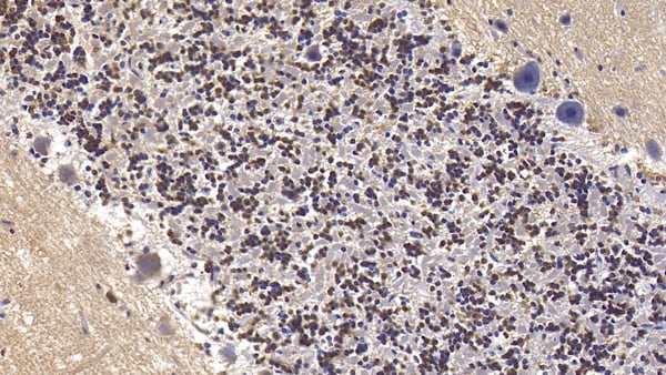 IHC (Immunohiostchemistry)
(DAB staining on IHCP;Sample: Human Cerebellum Tissue; Primary Ab: 10ug/ml Mouse AntiHuman SEMA5B AntibodySecond Ab: 2ug/mL HRPLinked Caprine AntiMouse IgG Polyclonal Antibody(Catalog: SAA544Mu19))
IHC (Immunohiostchemistry)
(DAB staining on IHCP;Sample: Human Cerebellum Tissue; Primary Ab: 10ug/ml Mouse AntiHuman SEMA5B AntibodySecond Ab: 2ug/mL HRPLinked Caprine AntiMouse IgG Polyclonal Antibody(Catalog: SAA544Mu19))
Semaphorin 5B (SEMA5B), Monoclonal Antibody (Cat# AAA151908)
 IHC (Immunohiostchemistry)
(DAB staining on IHCP;Sample: Human Cerebellum Tissue; Primary Ab: 10ug/ml Mouse AntiHuman SEMA5B AntibodySecond Ab: 2ug/mL HRPLinked Caprine AntiMouse IgG Polyclonal Antibody(Catalog: SAA544Mu19))
IHC (Immunohiostchemistry)
(DAB staining on IHCP;Sample: Human Cerebellum Tissue; Primary Ab: 10ug/ml Mouse AntiHuman SEMA5B AntibodySecond Ab: 2ug/mL HRPLinked Caprine AntiMouse IgG Polyclonal Antibody(Catalog: SAA544Mu19))
Semaphorin 5B (SEMA5B), Monoclonal Antibody (Cat# AAA151909)
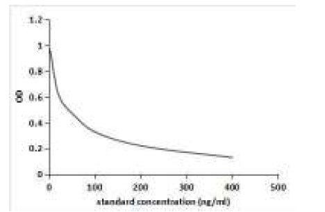 ELISA
(Competitive inhibition ELISA; Coated protein: AFB1 conjugated to BSAStandard: Series DilutedAFB1; Primary Ab: 0.79ug/mlMouse Anti AFB1 AntibodySecond Ab: 2ug/mL HRP-Linked Caprine Anti-mouse IgGPolyclonal AntibodyIC50=25ng/ml)
ELISA
(Competitive inhibition ELISA; Coated protein: AFB1 conjugated to BSAStandard: Series DilutedAFB1; Primary Ab: 0.79ug/mlMouse Anti AFB1 AntibodySecond Ab: 2ug/mL HRP-Linked Caprine Anti-mouse IgGPolyclonal AntibodyIC50=25ng/ml)
Aflatoxin B1 (AFB1), Monoclonal Antibody (Cat# AAA151914)
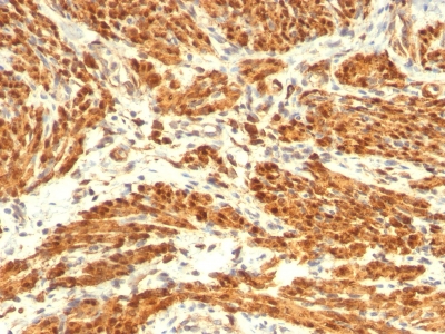
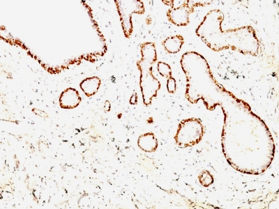 IHC (Immunohistochemisry)
(Formalin-fixed, paraffin-embedded Rat Uterus stained with Calponin-1 Monoclonal Antibody (CNN1/832 + CALP).)
IHC (Immunohistochemisry)
(Formalin-fixed, paraffin-embedded Rat Uterus stained with Calponin-1 Monoclonal Antibody (CNN1/832 + CALP).)
Calponin-1, Monoclonal Antibody (Cat# AAA62819)
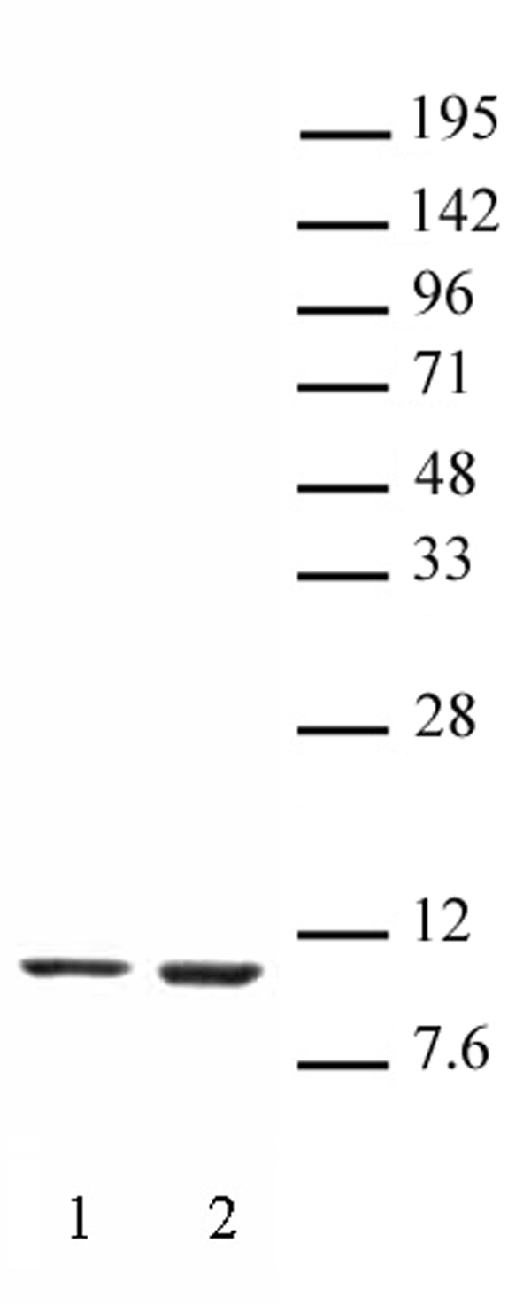
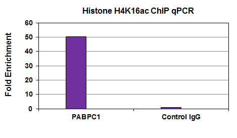
 Application Data
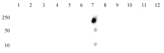
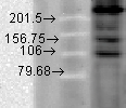
(Histone H4K16ac antibody (mAb) specificity tested by peptide array analysis. Peptide array analysis was used to confirm the specificity of this antibody for its intended modification. Histone H4K16ac antibody (mAb) was applied at a dilution of 0.7 ug/ml to MODified Histone Peptide Array tested by dot blot analysis. Dot blot analysis was used to confirm the specificity of Histone H4K16ac antibody (mAb) for acetyl-Lys16 histone H4. Acetylated peptides corresponding to the immunogen and related peptides were spotted onto PVDF and probed with Histone H4K16ac antibody (mAb) at a dilution of 2 ug/ml. The amount of peptide (picomoles) spotted is indicated next to each row. Lane 1: acetyl-Lys5 peptide. Lane 2: unmodified Lys5 peptide. Lane 3: acetyl-Lys8 peptide. Lane 4: unmodified Lys8 peptide. Lane 5: acetyl-Lys12 peptide. Lane 6: unmodified Lys12 peptide. Lane 7: acetyl-Lys16 peptide. Lane 8: unmodified Lys16 peptide. Lane 9: acetyl-Lys20 peptide. Lane 10: unmodified Lys20 peptide. Lane 11: acetyl-Lys31 peptide. Lane 12: unmodified Lys31 peptide.)
Application Data
(Histone H4K16ac antibody (mAb) specificity tested by peptide array analysis. Peptide array analysis was used to confirm the specificity of this antibody for its intended modification. Histone H4K16ac antibody (mAb) was applied at a dilution of 0.7 ug/ml to MODified Histone Peptide Array tested by dot blot analysis. Dot blot analysis was used to confirm the specificity of Histone H4K16ac antibody (mAb) for acetyl-Lys16 histone H4. Acetylated peptides corresponding to the immunogen and related peptides were spotted onto PVDF and probed with Histone H4K16ac antibody (mAb) at a dilution of 2 ug/ml. The amount of peptide (picomoles) spotted is indicated next to each row. Lane 1: acetyl-Lys5 peptide. Lane 2: unmodified Lys5 peptide. Lane 3: acetyl-Lys8 peptide. Lane 4: unmodified Lys8 peptide. Lane 5: acetyl-Lys12 peptide. Lane 6: unmodified Lys12 peptide. Lane 7: acetyl-Lys16 peptide. Lane 8: unmodified Lys16 peptide. Lane 9: acetyl-Lys20 peptide. Lane 10: unmodified Lys20 peptide. Lane 11: acetyl-Lys31 peptide. Lane 12: unmodified Lys31 peptide.)
Histone H4K16ac, Monoclonal Antibody (Cat# AAA60045)


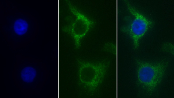
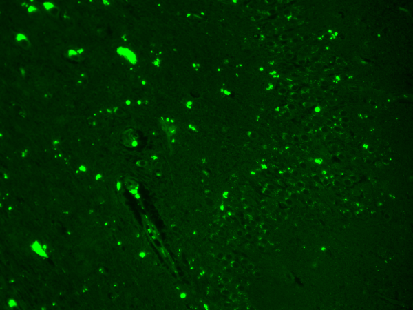
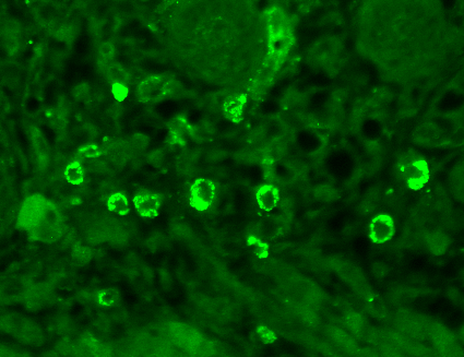
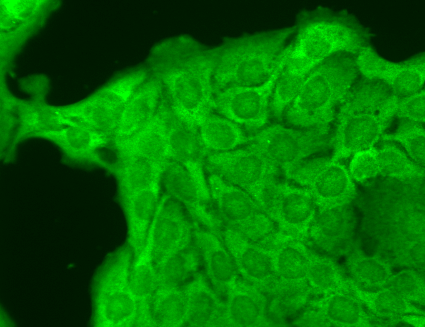
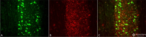
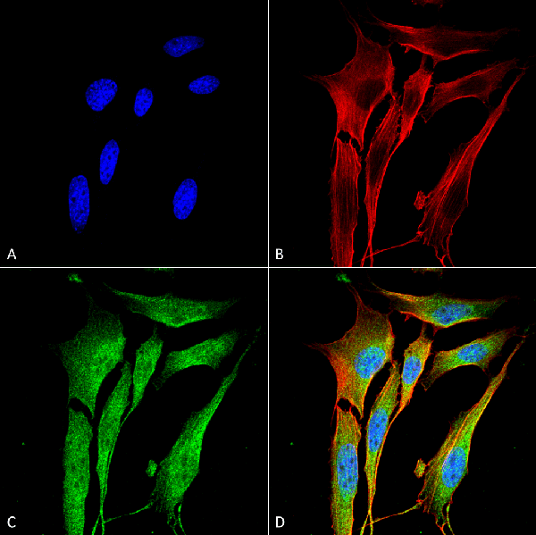
 IHC (Immunohistochemisry)
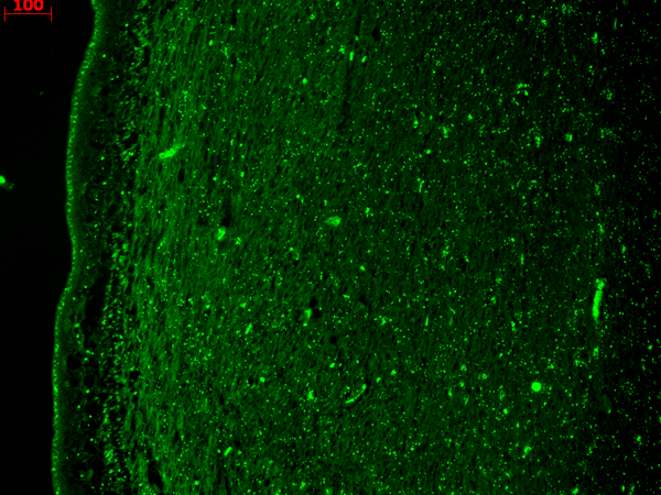
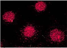
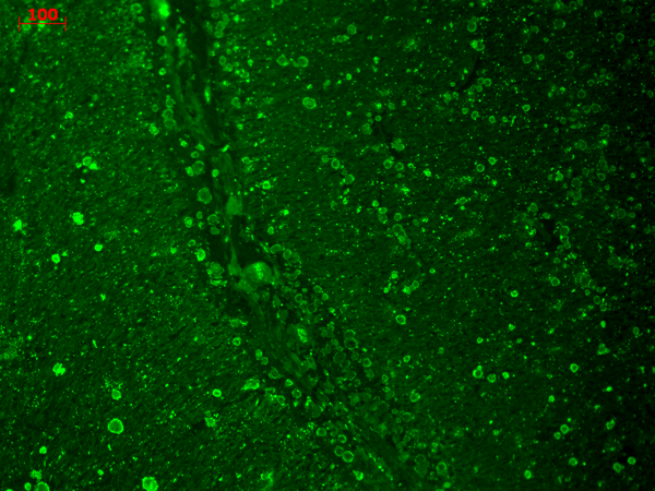
(Immunohistochemistry analysis using Mouse Anti-KCNQ4 Monoclonal Antibody, Clone S43-6. Tissue: hippocampus. Species: Human. Fixation: Bouin's Fixative and paraffin-embedded. Primary Antibody: Mouse Anti-KCNQ4 Monoclonal Antibody at 1:1000 for 1 hour at RT. Secondary Antibody: FITC Goat Anti-Mouse (green) at 1:50 for 1 hour at RT.)
IHC (Immunohistochemisry)
(Immunohistochemistry analysis using Mouse Anti-KCNQ4 Monoclonal Antibody, Clone S43-6. Tissue: hippocampus. Species: Human. Fixation: Bouin's Fixative and paraffin-embedded. Primary Antibody: Mouse Anti-KCNQ4 Monoclonal Antibody at 1:1000 for 1 hour at RT. Secondary Antibody: FITC Goat Anti-Mouse (green) at 1:50 for 1 hour at RT.)
KCNQ4, Monoclonal Antibody (Cat# AAA103158)
 WB (Western Blot)
(Western Blot analysis of Human Cell lysates showing detection of TrpM7 protein using Mouse Anti-TrpM7 Monoclonal Antibody, Clone S74-25. Load: 15 ug. Block: 1.5% BSA for 30 minutes at RT. Primary Antibody: Mouse Anti-TrpM7 Monoclonal Antibody at 1:1000 for 2 hours at RT. Secondary Antibody: Sheep Anti-Mouse IgG: HRP for 1 hour at RT.)
WB (Western Blot)
(Western Blot analysis of Human Cell lysates showing detection of TrpM7 protein using Mouse Anti-TrpM7 Monoclonal Antibody, Clone S74-25. Load: 15 ug. Block: 1.5% BSA for 30 minutes at RT. Primary Antibody: Mouse Anti-TrpM7 Monoclonal Antibody at 1:1000 for 2 hours at RT. Secondary Antibody: Sheep Anti-Mouse IgG: HRP for 1 hour at RT.)
TrpM7, Monoclonal Antibody (Cat# AAA103162)
 WB (Western Blot)
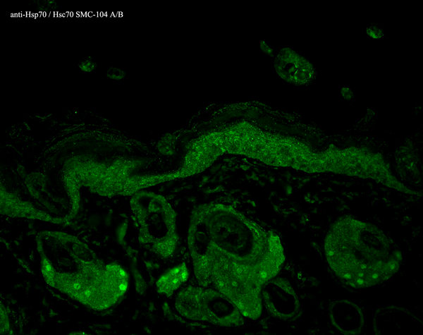
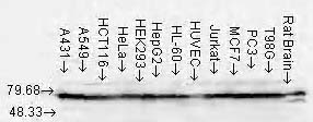
(Western Blot analysis of Human Cell lysates showing detection of Hsp70 protein using Mouse Anti-Hsp70 Monoclonal Antibody, Clone N27. Load: 15 ug. Block: 1.5% BSA for 30 minutes at RT. Primary Antibody: Mouse Anti-Hsp70 Monoclonal Antibody at 1:1000 for 2 hours at RT. Secondary Antibody: Sheep Anti-Mouse IgG: HRP for 1 hour at RT.)
WB (Western Blot)
(Western Blot analysis of Human Cell lysates showing detection of Hsp70 protein using Mouse Anti-Hsp70 Monoclonal Antibody, Clone N27. Load: 15 ug. Block: 1.5% BSA for 30 minutes at RT. Primary Antibody: Mouse Anti-Hsp70 Monoclonal Antibody at 1:1000 for 2 hours at RT. Secondary Antibody: Sheep Anti-Mouse IgG: HRP for 1 hour at RT.)
HSP70/HSC70, Monoclonal Antibody (Cat# AAA103169)
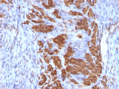
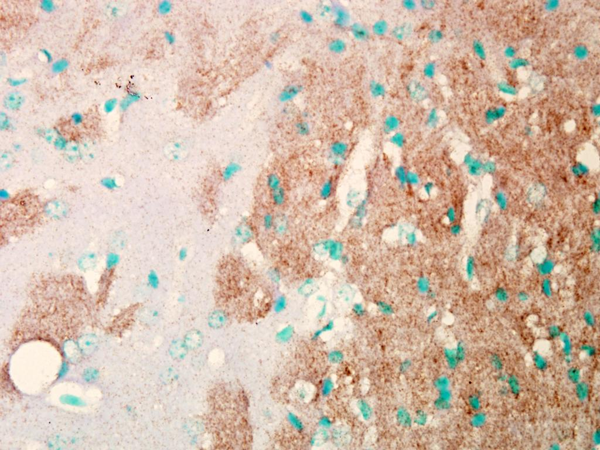
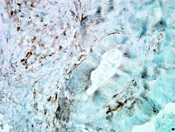
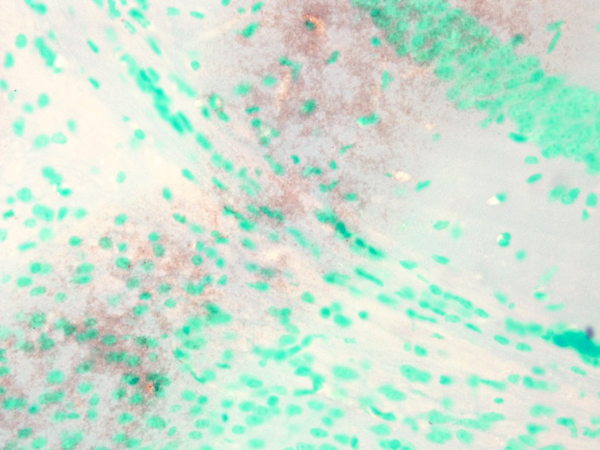
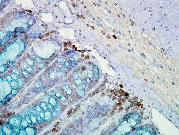 IHC (Immunohiostchemistry)
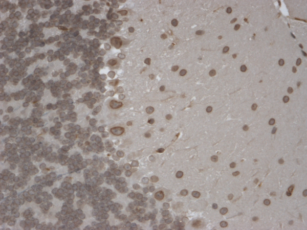
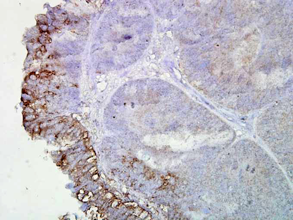
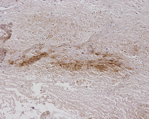
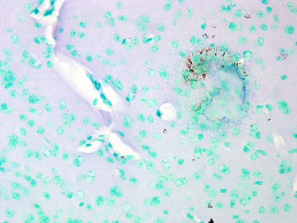
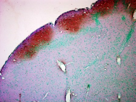
(Immunohistochemistry analysis using Mouse Anti-Hsp90 Monoclonal Antibody, Clone AC-16. Tissue: inflamed colon. Species: Mouse. Fixation: Formalin. Primary Antibody: Mouse Anti-Hsp90 Monoclonal Antibody at 1:2000 for 12 hours at 4 degree C. Secondary Antibody: Biotin Goat Anti-Mouse at 1:2000 for 1 hour at RT. Counterstain: Mayer Hematoxylin (purple/blue) nuclear stain at 200 ul for 2 minutes at RT. Localization: Inflammatory cells. Magnification: 40x. Mostly inflammatory cells, some mucosa.)
IHC (Immunohiostchemistry)
(Immunohistochemistry analysis using Mouse Anti-Hsp90 Monoclonal Antibody, Clone AC-16. Tissue: inflamed colon. Species: Mouse. Fixation: Formalin. Primary Antibody: Mouse Anti-Hsp90 Monoclonal Antibody at 1:2000 for 12 hours at 4 degree C. Secondary Antibody: Biotin Goat Anti-Mouse at 1:2000 for 1 hour at RT. Counterstain: Mayer Hematoxylin (purple/blue) nuclear stain at 200 ul for 2 minutes at RT. Localization: Inflammatory cells. Magnification: 40x. Mostly inflammatory cells, some mucosa.)
Hsp90, Monoclonal Antibody (Cat# AAA103177)
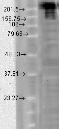
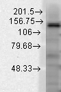
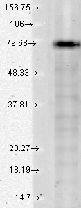
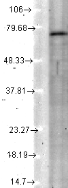
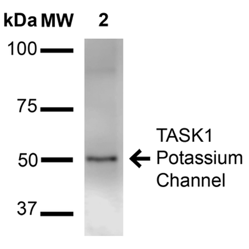 WB (Western Blot)
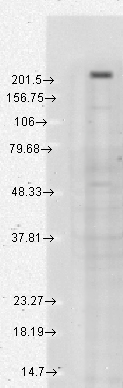
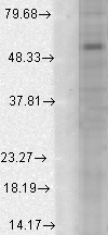
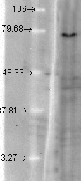
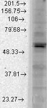
(Western Blot analysis of Rat Brain Membrane showing detection of ~50 kDa TASK1 Potassium Channel protein using Mouse Anti-TASK1 Potassium Channel Monoclonal Antibody, Clone S374-48 . Lane 1: Molecular Weight Ladder (MW). Lane 2: Rat brain membrane. Load: 15 ug. Block: 2% BSA and 2% Skim Milk in 1X TBST. Primary Antibody: Mouse Anti-TASK1 Potassium Channel Monoclonal Antibody at 1:1000 for 16 hours at 4 degree C. Secondary Antibody: Goat Anti-Mouse IgG: HRP at 1:2000 for 60 min at RT. Color Development: ECL solution for 6 min at RT. Predicted/Observed Size: ~50 kDa.)
WB (Western Blot)
(Western Blot analysis of Rat Brain Membrane showing detection of ~50 kDa TASK1 Potassium Channel protein using Mouse Anti-TASK1 Potassium Channel Monoclonal Antibody, Clone S374-48 . Lane 1: Molecular Weight Ladder (MW). Lane 2: Rat brain membrane. Load: 15 ug. Block: 2% BSA and 2% Skim Milk in 1X TBST. Primary Antibody: Mouse Anti-TASK1 Potassium Channel Monoclonal Antibody at 1:1000 for 16 hours at 4 degree C. Secondary Antibody: Goat Anti-Mouse IgG: HRP at 1:2000 for 60 min at RT. Color Development: ECL solution for 6 min at RT. Predicted/Observed Size: ~50 kDa.)
TASK1 Potassium Channel, Monoclonal Antibody (Cat# AAA103185)
 WB (Western Blot)
(Western Blot analysis of Rat tissue lysate showing detection of KDEL Receptor protein using Mouse Anti-KDEL Receptor Monoclonal Antibody, Clone KR-10. Load: 15 ug. Block: 1.5% BSA for 30 minutes at RT. Primary Antibody: Mouse Anti-KDEL Receptor Monoclonal Antibody at 1:1000 for 2 hours at RT. Secondary Antibody: Sheep Anti-Mouse IgG: HRP for 1 hour at RT.)
WB (Western Blot)
(Western Blot analysis of Rat tissue lysate showing detection of KDEL Receptor protein using Mouse Anti-KDEL Receptor Monoclonal Antibody, Clone KR-10. Load: 15 ug. Block: 1.5% BSA for 30 minutes at RT. Primary Antibody: Mouse Anti-KDEL Receptor Monoclonal Antibody at 1:1000 for 2 hours at RT. Secondary Antibody: Sheep Anti-Mouse IgG: HRP for 1 hour at RT.)
KDEL, Monoclonal Antibody (Cat# AAA103206)
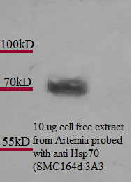
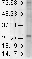
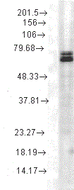 WB (Western Blot)
(Western Blot analysis of Rat cell lysates showing detection of Hsp70 protein using Mouse Anti-Hsp70 Monoclonal Antibody, Clone 3A3. Load: 15 ug. Block: 1.5% BSA for 30 minutes at RT. Primary Antibody: Mouse Anti-Hsp70 Monoclonal Antibody at 1:1000 for 2 hours at RT. Secondary Antibody: Sheep Anti-Mouse IgG: HRP for 1 hour at RT.)
WB (Western Blot)
(Western Blot analysis of Rat cell lysates showing detection of Hsp70 protein using Mouse Anti-Hsp70 Monoclonal Antibody, Clone 3A3. Load: 15 ug. Block: 1.5% BSA for 30 minutes at RT. Primary Antibody: Mouse Anti-Hsp70 Monoclonal Antibody at 1:1000 for 2 hours at RT. Secondary Antibody: Sheep Anti-Mouse IgG: HRP for 1 hour at RT.)
Hsp70, Monoclonal Antibody (Cat# AAA103211)
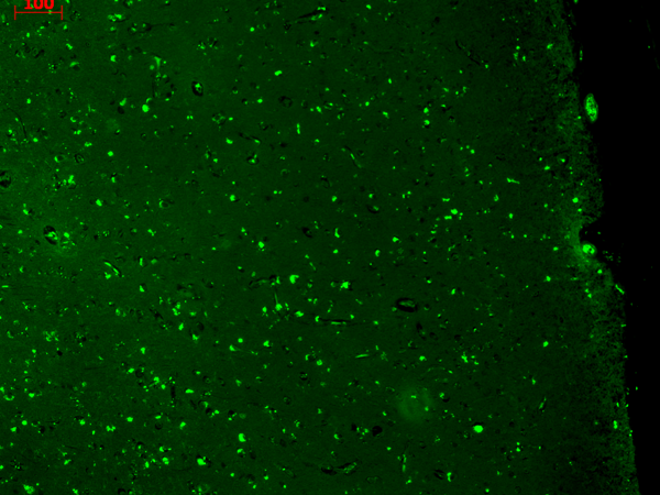
 IHC (Immunohistochemistry)
(Immunohistochemistry analysis using Mouse Anti-Nav1.7 Sodium Channel Monoclonal Antibody, Clone S68-6. Tissue: backskin. Species: Mouse. Fixation: Bouin's Fixative and paraffin-embedded. Primary Antibody: Mouse Anti-Nav1.7 Sodium Channel Monoclonal Antibody at 1:100 for 1 hour at RT. Secondary Antibody: FITC Goat Anti-Mouse (green) at 1:50 for 1 hour at RT.)
IHC (Immunohistochemistry)
(Immunohistochemistry analysis using Mouse Anti-Nav1.7 Sodium Channel Monoclonal Antibody, Clone S68-6. Tissue: backskin. Species: Mouse. Fixation: Bouin's Fixative and paraffin-embedded. Primary Antibody: Mouse Anti-Nav1.7 Sodium Channel Monoclonal Antibody at 1:100 for 1 hour at RT. Secondary Antibody: FITC Goat Anti-Mouse (green) at 1:50 for 1 hour at RT.)
Nav1.7, Monoclonal Antibody (Cat# AAA103213)
 WB (Western Blot)
(Western Blot analysis of Human Cell line lysates showing detection of GABA A Receptor protein using Mouse Anti-GABA A Receptor Monoclonal Antibody, Clone S95-35. Load: 15 ug. Block: 1.5% BSA for 30 minutes at RT. Primary Antibody: Mouse Anti-GABA A Receptor Monoclonal Antibody at 1:1000 for 2 hours at RT. Secondary Antibody: Sheep Anti-Mouse IgG: HRP for 1 hour at RT.)
WB (Western Blot)
(Western Blot analysis of Human Cell line lysates showing detection of GABA A Receptor protein using Mouse Anti-GABA A Receptor Monoclonal Antibody, Clone S95-35. Load: 15 ug. Block: 1.5% BSA for 30 minutes at RT. Primary Antibody: Mouse Anti-GABA A Receptor Monoclonal Antibody at 1:1000 for 2 hours at RT. Secondary Antibody: Sheep Anti-Mouse IgG: HRP for 1 hour at RT.)
GABA(A) Receptor Alpha1, Monoclonal Antibody (Cat# AAA103214)
 WB (Western Blot)
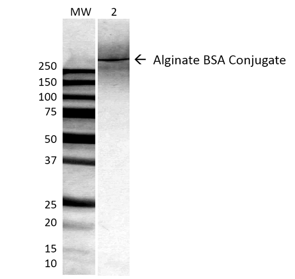
(Western Blot analysis of ALL BSA-Alginate Conjugate showing detection of ~250 kDa Alginate protein using Mouse Anti-Alginate Monoclonal Antibody, Clone 4B10-1C5. Lane 1: MW ladder. Lane 2: 0.625ug BSA:Alginate. Load: 0.625 ug. Block: 5% milk + TBST for 1 hour at RT. Primary Antibody: Mouse Anti-Alginate Monoclonal Antibody at 1:500 for 1 hour at RT. Secondary Antibody: HRP Goat Anti-Mouse at 1:100 for 1 hour at RT. Color Development: TMB solution for 2 min at RT. Predicted/Observed Size: ~250 kDa.)
WB (Western Blot)
(Western Blot analysis of ALL BSA-Alginate Conjugate showing detection of ~250 kDa Alginate protein using Mouse Anti-Alginate Monoclonal Antibody, Clone 4B10-1C5. Lane 1: MW ladder. Lane 2: 0.625ug BSA:Alginate. Load: 0.625 ug. Block: 5% milk + TBST for 1 hour at RT. Primary Antibody: Mouse Anti-Alginate Monoclonal Antibody at 1:500 for 1 hour at RT. Secondary Antibody: HRP Goat Anti-Mouse at 1:100 for 1 hour at RT. Color Development: TMB solution for 2 min at RT. Predicted/Observed Size: ~250 kDa.)
Alginate, Monoclonal Antibody (Cat# AAA103217)
 WB (Western Blot)
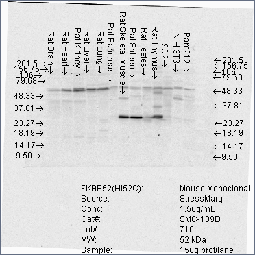
(Western Blot analysis of Human Cell lysates showing detection of FKBP52 protein using Mouse Anti-FKBP52 Monoclonal Antibody, Clone Hi52C. Load: 15 ug. Block: 1.5% BSA for 30 minutes at RT. Primary Antibody: Mouse Anti-FKBP52 Monoclonal Antibody at 1.5 ug/mL for 2 hours at RT. Secondary Antibody: Sheep Anti-Mouse IgG: HRP for 1 hour at RT.)
WB (Western Blot)
(Western Blot analysis of Human Cell lysates showing detection of FKBP52 protein using Mouse Anti-FKBP52 Monoclonal Antibody, Clone Hi52C. Load: 15 ug. Block: 1.5% BSA for 30 minutes at RT. Primary Antibody: Mouse Anti-FKBP52 Monoclonal Antibody at 1.5 ug/mL for 2 hours at RT. Secondary Antibody: Sheep Anti-Mouse IgG: HRP for 1 hour at RT.)
FKBP52, Monoclonal Antibody (Cat# AAA103225)
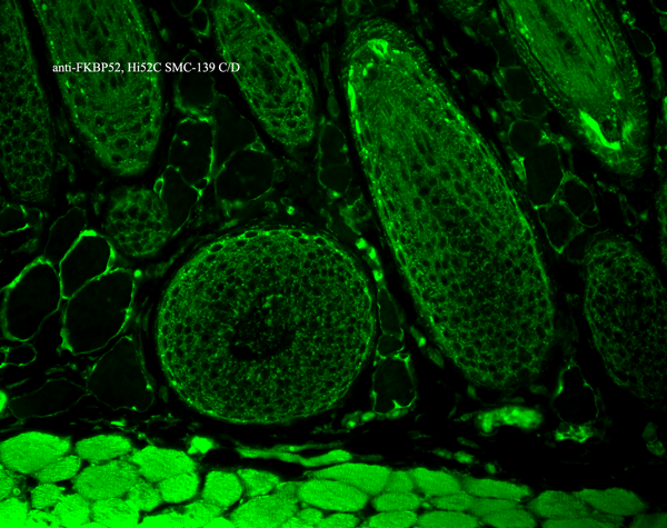
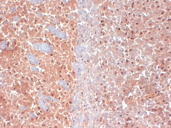
 IHC (Immunohistochemistry)
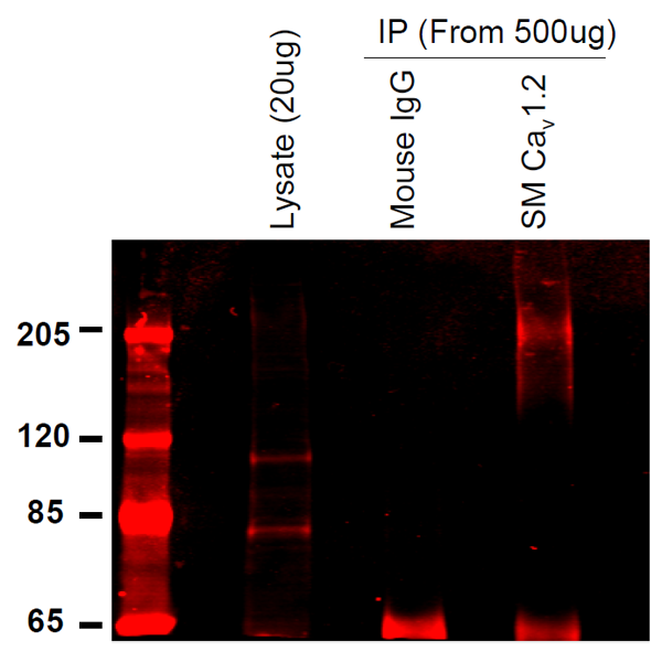
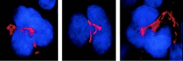
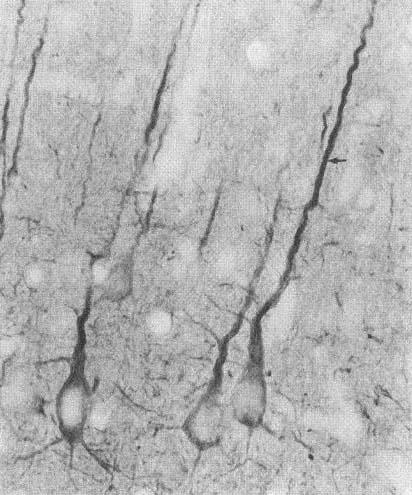
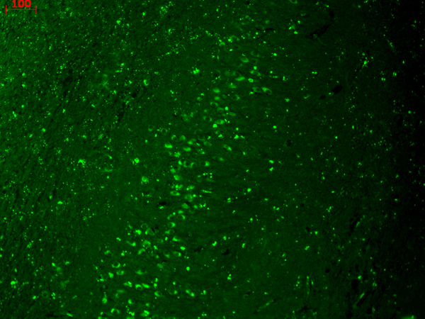
(Immunohistochemistry analysis using Mouse Anti-CaV1.2 Calcium channel Monoclonal Antibody, Clone S57-47. Tissue: Brain Tissue. Species: Mouse. Fixation: Formalin. Primary Antibody: Mouse Anti-CaV1.2 Calcium channel Monoclonal Antibody at 1:10000 for 12 hours at 4 degree C. Secondary Antibody: Biotin Goat Anti-Mouse at 1:2000 for 1 hour at RT. Counterstain: Mayer Hematoxylin (purple/blue) nuclear stain at 200 ul for 2 minutes at RT. Magnification: 40x.)
IHC (Immunohistochemistry)
(Immunohistochemistry analysis using Mouse Anti-CaV1.2 Calcium channel Monoclonal Antibody, Clone S57-47. Tissue: Brain Tissue. Species: Mouse. Fixation: Formalin. Primary Antibody: Mouse Anti-CaV1.2 Calcium channel Monoclonal Antibody at 1:10000 for 12 hours at 4 degree C. Secondary Antibody: Biotin Goat Anti-Mouse at 1:2000 for 1 hour at RT. Counterstain: Mayer Hematoxylin (purple/blue) nuclear stain at 200 ul for 2 minutes at RT. Magnification: 40x.)
Cav1.2, Monoclonal Antibody (Cat# AAA103227)
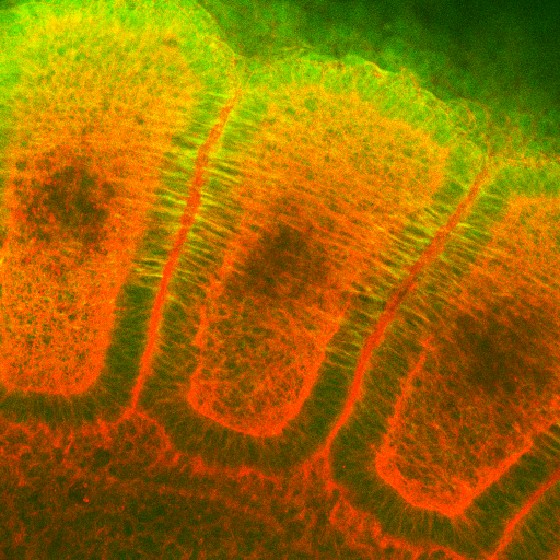
 IHC (Immunohistochemisry)
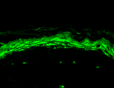
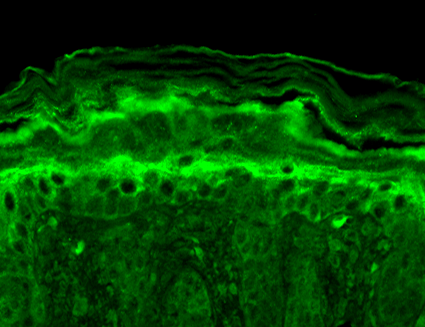
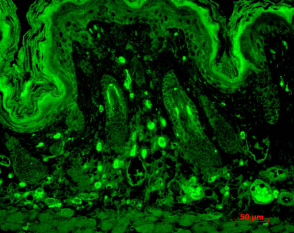
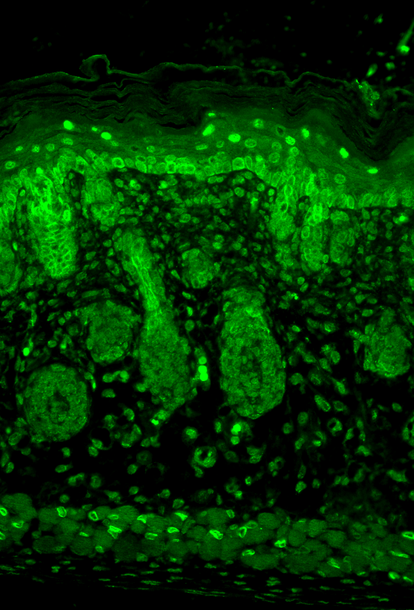
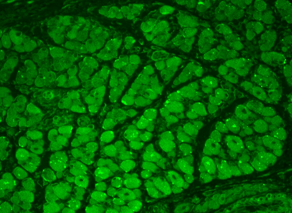
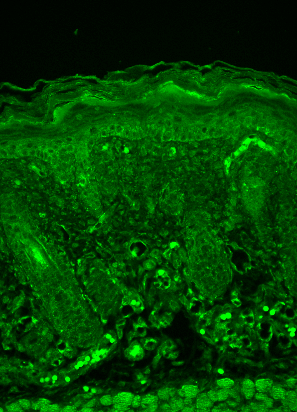
(Immunohistochemistry analysis using Mouse Anti-Nav1.8 Monoclonal Antibody, Clone S134-12. Tissue: backskin. Species: Mouse. Fixation: Bouin's Fixative and paraffin-embedded. Primary Antibody: Mouse Anti-Nav1.8 Monoclonal Antibody at 1:100 for 1 hour at RT. Secondary Antibody: FITC Goat Anti-Mouse (green) at 1:50 for 1 hour at RT. Localization: Heavy filaggrin-like staining, lower epidermal cells have some staining.)
IHC (Immunohistochemisry)
(Immunohistochemistry analysis using Mouse Anti-Nav1.8 Monoclonal Antibody, Clone S134-12. Tissue: backskin. Species: Mouse. Fixation: Bouin's Fixative and paraffin-embedded. Primary Antibody: Mouse Anti-Nav1.8 Monoclonal Antibody at 1:100 for 1 hour at RT. Secondary Antibody: FITC Goat Anti-Mouse (green) at 1:50 for 1 hour at RT. Localization: Heavy filaggrin-like staining, lower epidermal cells have some staining.)
Nav1.8, Monoclonal Antibody (Cat# AAA103229)
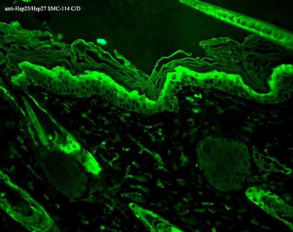
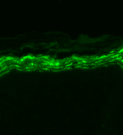
 IHC (Immunohistochemistry)
(Immunohistochemistry analysis using Mouse Anti-Hsp27 Monoclonal Antibody, Clone 8A7. Tissue: backskin. Species: Mouse. Fixation: Bouin's Fixative and paraffin-embedded. Primary Antibody: Mouse Anti-Hsp27 Monoclonal Antibody at 1:100 for 1 hour at RT. Secondary Antibody: FITC Goat Anti-Mouse (green) at 1:50 for 1 hour at RT. Localization: Epidermis.)
IHC (Immunohistochemistry)
(Immunohistochemistry analysis using Mouse Anti-Hsp27 Monoclonal Antibody, Clone 8A7. Tissue: backskin. Species: Mouse. Fixation: Bouin's Fixative and paraffin-embedded. Primary Antibody: Mouse Anti-Hsp27 Monoclonal Antibody at 1:100 for 1 hour at RT. Secondary Antibody: FITC Goat Anti-Mouse (green) at 1:50 for 1 hour at RT. Localization: Epidermis.)
Hsp25/Hsp27, Monoclonal Antibody (Cat# AAA103236)
 WB (Western Blot)
(Western Blot analysis of Rat brain membrane lysate showing detection of VGLUT1 protein using Mouse Anti-VGLUT1 Monoclonal Antibody, Clone S28-9. Primary Antibody: Mouse Anti-VGLUT1 Monoclonal Antibody at 1:1000.)
WB (Western Blot)
(Western Blot analysis of Rat brain membrane lysate showing detection of VGLUT1 protein using Mouse Anti-VGLUT1 Monoclonal Antibody, Clone S28-9. Primary Antibody: Mouse Anti-VGLUT1 Monoclonal Antibody at 1:1000.)
VGLUT1, Monoclonal Antibody (Cat# AAA103240)
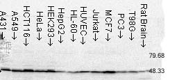
 WB (Western Blot)
(Western Blot analysis of Human Cervical cancer cell line (HeLa) lysate showing detection of FKBP51 protein using Mouse Anti-FKBP51 Monoclonal Antibody, Clone Hi51B. Load: 15 ug. Block: 1.5% BSA for 30 minutes at RT. Primary Antibody: Mouse Anti-FKBP51 Monoclonal Antibody at 1:1000 for 2 hours at RT. Secondary Antibody: Sheep Anti-Mouse IgG: HRP for 1 hour at RT.)
WB (Western Blot)
(Western Blot analysis of Human Cervical cancer cell line (HeLa) lysate showing detection of FKBP51 protein using Mouse Anti-FKBP51 Monoclonal Antibody, Clone Hi51B. Load: 15 ug. Block: 1.5% BSA for 30 minutes at RT. Primary Antibody: Mouse Anti-FKBP51 Monoclonal Antibody at 1:1000 for 2 hours at RT. Secondary Antibody: Sheep Anti-Mouse IgG: HRP for 1 hour at RT.)
FKBP51, Monoclonal Antibody (Cat# AAA103248)

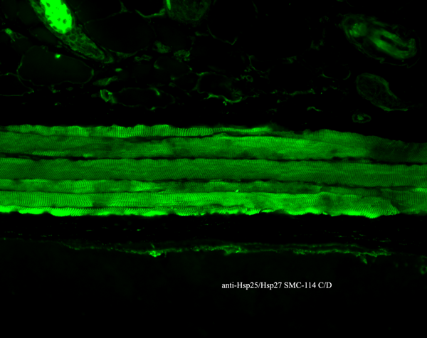
 IHC (Immunohistochemisry)
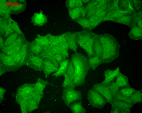
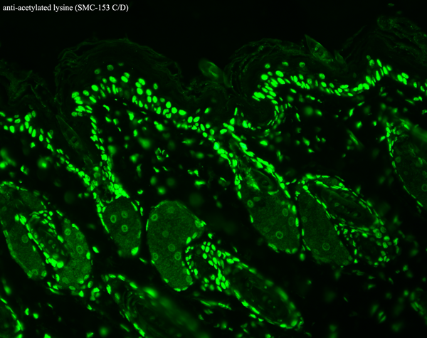
(Immunohistochemistry analysis using Mouse Anti-Slo2.2 Potassium Channel Monoclonal Antibody, Clone S3-26. Tissue: backskin. Species: Mouse. Fixation: Bouin's Fixative and paraffin-embedded. Primary Antibody: Mouse Anti-Slo2.2 Potassium Channel Monoclonal Antibody at 1:100 for 1 hour at RT. Secondary Antibody: FITC Goat Anti-Mouse (green) at 1:50 for 1 hour at RT. Localization: Suprabasal epidermal staining. Hair follicles negative.)
IHC (Immunohistochemisry)
(Immunohistochemistry analysis using Mouse Anti-Slo2.2 Potassium Channel Monoclonal Antibody, Clone S3-26. Tissue: backskin. Species: Mouse. Fixation: Bouin's Fixative and paraffin-embedded. Primary Antibody: Mouse Anti-Slo2.2 Potassium Channel Monoclonal Antibody at 1:100 for 1 hour at RT. Secondary Antibody: FITC Goat Anti-Mouse (green) at 1:50 for 1 hour at RT. Localization: Suprabasal epidermal staining. Hair follicles negative.)
Slo2.2, Monoclonal Antibody (Cat# AAA103258)


 WB (Western Blot)
(Western Blot analysis of Rat brain membrane lysate showing detection of SHANK3 protein using Mouse Anti-SHANK3 Monoclonal Antibody, Clone S69-46. Load: 15 ug. Block: 1.5% BSA for 30 minutes at RT. Primary Antibody: Mouse Anti-SHANK3 Monoclonal Antibody at 1:1000 for 2 hours at RT. Secondary Antibody: Sheep Anti-Mouse IgG: HRP for 1 hour at RT.)
WB (Western Blot)
(Western Blot analysis of Rat brain membrane lysate showing detection of SHANK3 protein using Mouse Anti-SHANK3 Monoclonal Antibody, Clone S69-46. Load: 15 ug. Block: 1.5% BSA for 30 minutes at RT. Primary Antibody: Mouse Anti-SHANK3 Monoclonal Antibody at 1:1000 for 2 hours at RT. Secondary Antibody: Sheep Anti-Mouse IgG: HRP for 1 hour at RT.)
SHANK3, Monoclonal Antibody (Cat# AAA103263)
 WB (Western Blot)
(Western Blot analysis of hamster T-CHO cell lysate showing detection of KCNQ1 protein using Mouse Anti-KCNQ1 Monoclonal Antibody, Clone S37A-10. Load: 15 ug. Block: 1.5% BSA for 30 minutes at RT. Primary Antibody: Mouse Anti-KCNQ1 Monoclonal Antibody at 1:1000 for 2 hours at RT. Secondary Antibody: Sheep Anti-Mouse IgG: HRP for 1 hour at RT.)
WB (Western Blot)
(Western Blot analysis of hamster T-CHO cell lysate showing detection of KCNQ1 protein using Mouse Anti-KCNQ1 Monoclonal Antibody, Clone S37A-10. Load: 15 ug. Block: 1.5% BSA for 30 minutes at RT. Primary Antibody: Mouse Anti-KCNQ1 Monoclonal Antibody at 1:1000 for 2 hours at RT. Secondary Antibody: Sheep Anti-Mouse IgG: HRP for 1 hour at RT.)
KCNQ1, Monoclonal Antibody (Cat# AAA103275)
 WB (Western Blot)
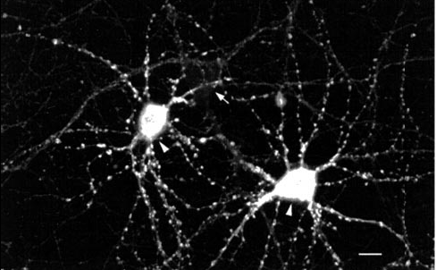
(Western Blot analysis of Rat brain membrane lysate showing detection of PSD95 protein using Mouse Anti-PSD95 Monoclonal Antibody, Clone 7E3. Primary Antibody: Mouse Anti-PSD95 Monoclonal Antibody at 1:1000.)
WB (Western Blot)
(Western Blot analysis of Rat brain membrane lysate showing detection of PSD95 protein using Mouse Anti-PSD95 Monoclonal Antibody, Clone 7E3. Primary Antibody: Mouse Anti-PSD95 Monoclonal Antibody at 1:1000.)
PSD95, Monoclonal Antibody (Cat# AAA103288)
 IHC (Immunohistochemisry)
(Immunohistochemistry analysis using Mouse Anti-Kir2.1 Potassium Channel Monoclonal Antibody, Clone S112B-14. Tissue: hippocampus. Species: Human. Fixation: Bouin's Fixative and paraffin-embedded. Primary Antibody: Mouse Anti-Kir2.1 Potassium Channel Monoclonal Antibody at 1:1000 for 1 hour at RT. Secondary Antibody: FITC Goat Anti-Mouse (green) at 1:50 for 1 hour at RT.)
IHC (Immunohistochemisry)
(Immunohistochemistry analysis using Mouse Anti-Kir2.1 Potassium Channel Monoclonal Antibody, Clone S112B-14. Tissue: hippocampus. Species: Human. Fixation: Bouin's Fixative and paraffin-embedded. Primary Antibody: Mouse Anti-Kir2.1 Potassium Channel Monoclonal Antibody at 1:1000 for 1 hour at RT. Secondary Antibody: FITC Goat Anti-Mouse (green) at 1:50 for 1 hour at RT.)
Kir2.1, Monoclonal Antibody (Cat# AAA103301)
 WB (Western Blot)
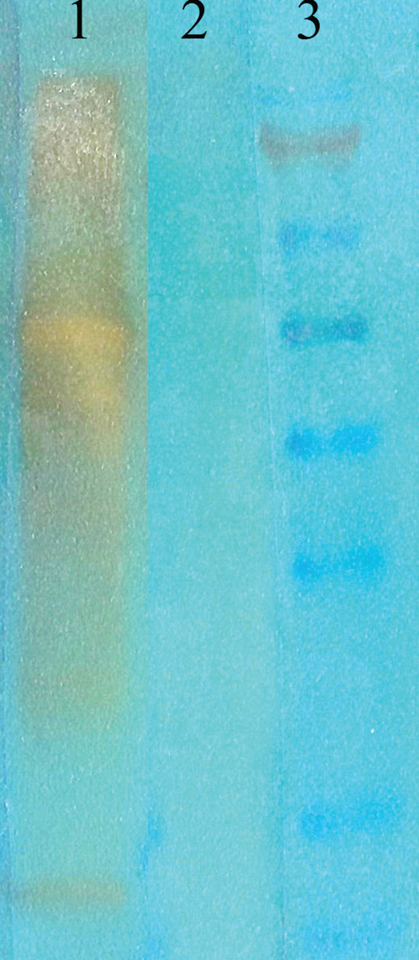
(Western Blot analysis of acetylated lysine showing detection of Acetylated Lysine protein using Mouse Anti-Acetylated Lysine Monoclonal Antibody, Clone 7F8. Primary Antibody: Mouse Anti-Acetylated Lysine Monoclonal Antibody at 1:1000. (1) acetylated BSA (75ng of protein), (2) non-acetylated BSA, and (3) marker.)
WB (Western Blot)
(Western Blot analysis of acetylated lysine showing detection of Acetylated Lysine protein using Mouse Anti-Acetylated Lysine Monoclonal Antibody, Clone 7F8. Primary Antibody: Mouse Anti-Acetylated Lysine Monoclonal Antibody at 1:1000. (1) acetylated BSA (75ng of protein), (2) non-acetylated BSA, and (3) marker.)
Acetylated Lysine, Monoclonal Antibody (Cat# AAA103322)
 WB (Western Blot)
(Western Blot analysis of Mouse Ventricle lysates showing detection of CaMKII protein using Mouse Anti-CaMKII Monoclonal Antibody, Clone 22B1. Primary Antibody: Mouse Anti-CaMKII Monoclonal Antibody at 1:1000. Analysis of CaMKII and NFAT phosphorylation in ventricles of 14 day old mice over-expressing CaMK.)
WB (Western Blot)
(Western Blot analysis of Mouse Ventricle lysates showing detection of CaMKII protein using Mouse Anti-CaMKII Monoclonal Antibody, Clone 22B1. Primary Antibody: Mouse Anti-CaMKII Monoclonal Antibody at 1:1000. Analysis of CaMKII and NFAT phosphorylation in ventricles of 14 day old mice over-expressing CaMK.)
CaMKII, Monoclonal Antibody (Cat# AAA103082)
 IHC (Immunohistochemisry)
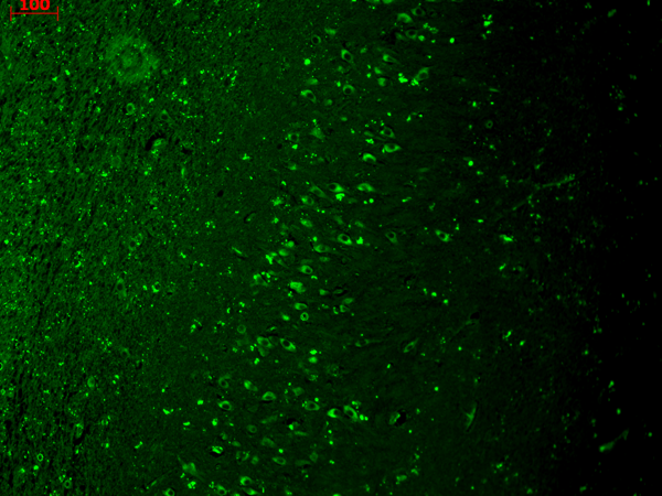
(Immunohistochemistry analysis using Mouse Anti-KCNQ4 Monoclonal Antibody, Clone S43-6. Tissue: hippocampus. Species: Human. Fixation: Bouin's Fixative and paraffin-embedded. Primary Antibody: Mouse Anti-KCNQ4 Monoclonal Antibody at 1:1000 for 1 hour at RT. Secondary Antibody: FITC Goat Anti-Mouse (green) at 1:50 for 1 hour at RT.)
IHC (Immunohistochemisry)
(Immunohistochemistry analysis using Mouse Anti-KCNQ4 Monoclonal Antibody, Clone S43-6. Tissue: hippocampus. Species: Human. Fixation: Bouin's Fixative and paraffin-embedded. Primary Antibody: Mouse Anti-KCNQ4 Monoclonal Antibody at 1:1000 for 1 hour at RT. Secondary Antibody: FITC Goat Anti-Mouse (green) at 1:50 for 1 hour at RT.)
KCNQ4, Monoclonal Antibody (Cat# AAA103094)
 WB (Western Blot)
(Western Blot analysis of Human Cell lysates showing detection of Rhodopsin protein using Mouse Anti-Rhodopsin Monoclonal Antibody, Clone 1D4. Load: 15 ug. Block: 1.5% BSA for 30 minutes at RT. Primary Antibody: Mouse Anti-Rhodopsin Monoclonal Antibody at 1:1000 for 2 hours at RT. Secondary Antibody: Sheep Anti-Mouse IgG: HRP for 1 hour at RT.)
WB (Western Blot)
(Western Blot analysis of Human Cell lysates showing detection of Rhodopsin protein using Mouse Anti-Rhodopsin Monoclonal Antibody, Clone 1D4. Load: 15 ug. Block: 1.5% BSA for 30 minutes at RT. Primary Antibody: Mouse Anti-Rhodopsin Monoclonal Antibody at 1:1000 for 2 hours at RT. Secondary Antibody: Sheep Anti-Mouse IgG: HRP for 1 hour at RT.)
Rhodopsin, Monoclonal Antibody (Cat# AAA103102)
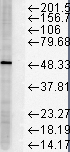
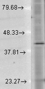
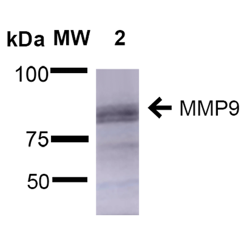 WB (Western Blot)
(Western Blot analysis of Rat Brain showing detection of ~92 kDa and ~82 kDa (pro and active) MMP9 protein using Mouse Anti-MMP9 Monoclonal Antibody, Clone S51-82 . Lane 1: Molecular Weight Ladder (MW). Lane 2: Rat Brain. Load: 15 ug. Block: 5% Skim Milk in 1X TBST. Primary Antibody: Mouse Anti-MMP9 Monoclonal Antibody at 1:1000 for 2 hours at RT. Secondary Antibody: Goat Anti-Mouse IgG: HRP at 1:2000 for 60 min at RT. Color Development: ECL solution for 5 min at RT. Predicted/Observed Size: ~92 kDa and ~82 kDa (pro and active).)
WB (Western Blot)
(Western Blot analysis of Rat Brain showing detection of ~92 kDa and ~82 kDa (pro and active) MMP9 protein using Mouse Anti-MMP9 Monoclonal Antibody, Clone S51-82 . Lane 1: Molecular Weight Ladder (MW). Lane 2: Rat Brain. Load: 15 ug. Block: 5% Skim Milk in 1X TBST. Primary Antibody: Mouse Anti-MMP9 Monoclonal Antibody at 1:1000 for 2 hours at RT. Secondary Antibody: Goat Anti-Mouse IgG: HRP at 1:2000 for 60 min at RT. Color Development: ECL solution for 5 min at RT. Predicted/Observed Size: ~92 kDa and ~82 kDa (pro and active).)
MMP9, Monoclonal Antibody (Cat# AAA103108)
What are Monoclonal Antibodies?
Monoclonal antibodies are specialized laboratory-produced proteins developed for binding to specific biological antigens or other molecular targets. Since they come from a single cell (or clone), they are especially consistent and accurate in the data they are involved in producing.
This type of antibody material has been shown to be a powerful tool in finding and subsequently destroying harmful cells in an organism, such as those found in cancers or various autoimmune diseases. This makes them excellent aids in medical testing and research, which is why they are so widely used.
AAA Biotech offers a comprehensive range of high-quality monoclonal antibodies that perform effectively in various laboratory tests, including (amongst others) ELISA, western blotting, immunohistochemistry, and flow cytometry. All of the products in our catalog are thoroughly quality tested to make sure that they are reliable and will consistently perform well in your research.
What Are The Uses of Monoclonal Antibodies
Monoclonal antibodies are used in many lab tests, including (amongst others) ELISA, western blotting, immunohistochemistry, and flow cytometry.
ELISA is a test that helps detect a specific substance/analyte in a sample. It uses antibodies (often monoclonal) bound to a solid surface (such as the well of a microplate) to “capture” the substance/analyte in the sample and immobilize it so that the detection antibody component can then bind to it and produce a signal, which can then be measured.
Western blotting identifies specific proteins in a sample. The sample is first separated on a gel, and then antibodies are applied that will typically bind to the target, which will all be localized to a single band in a lane.
Immunohistochemistry helps locate specific proteins in cells or tissue samples using antibodies.
Flow cytometry looks at and sorts cells. It uses antibodies that are conjugated to reporter molecules called “fluorophores”, which, under special lights, emit light themselves, which can then be measured by a detector instrument. For a deeper understanding of these techniques, explore our complete guide to monoclonal antibodies and their benefits.
How Monoclonal Antibodies Are Used as Medicine?
Please note that all of the products listed in AAA Biotech’s also known as AAA Bio or AAABio catalog are strictly for research-use only (RUO).
Monoclonal antibodies can also be used as therapeutic/medical treatments, particularly in the context of cancers. They are designed to find and bind to specific cells or proteins, helping the immune system recognize and attack the cancer. These treatments work in different ways, such as:
- Radioimmunotherapy attaches a small amount of radioactive molecule to the antibody, so it delivers the radiation directly to the cancer cells that the antibody is specifically binding to.
- Antibody-directed enzyme prodrug therapy uses antibodies that are specifically bound to special enzymes. These enzymes activate a harmless drug in the body and turn it into a cancer-killing drug only near the cancer cells—this helps avoid harming healthy cells.
- Immunoliposomes are tiny “bubbles” filled with medicine/drug and coated with antibodies. They carry the drug straight to the cancer cells.
Why Buy Monoclonal Antibodies From Us?
At AAA Biotech, we provide high-performance monoclonal antibodies designed to support a wide range of research needs.
1. Validated for Versatile Applications
The antibodies in our catalog are extensively validated and compatible with multiple techniques, including (but not limited to) ELISA, flow cytometry (FC), immunocytochemistry (ICC), immunofluorescence (IF), immunohistochemistry (IHC), immunoprecipitation (IP), and western blotting (WB).
2. Wide Selection & Specialized Options
We offer antibodies for common and rare species, that are available in various conjugated forms, and also in recombinant formats. Essentially, there is almost anything one might need to meet their experimental model’s requirements.
3. High-Quality Proteins
Our proteins meet high purity standards—90% or more as confirmed by SDS-PAGE. Many are available with tags like His, Flag, GST, or MBP, and we also supply native and biologically active proteins for functional studies.
Frequently Asked Questions
1. Are your monoclonal antibodies validated for specific applications?
Yes, our antibodies are tested and validated for use in methods such as ELISA, western blot, IHC, flow cytometry, and more. Refer to specific product pages or datasheets for individual product information.
2. How do I choose the right monoclonal antibody for my application?
Review the product details directly for application validation, species reactivity, and target information. You may also contact our support team at any time for help.
3. How quickly can I receive my order?
Most orders are processed and shipped within 1–3 business days, depending on product availability and your shipping location.