Filters
▼Clonality
▼Type
▼Reactivity
▼Gene Name
▼Isotype
▼Host
▼Application
▼Clone
▼Monoclonal Antibodies
Get accurate results in your research with our Monoclonal Antibodies, which are specially made to target exactly what you require for your research, and will produce consistent, reliable performance in lab tests.
Viewing 4400-4450 of 27645 product results
 WB (Western Blot)
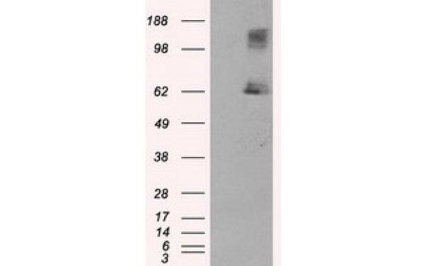
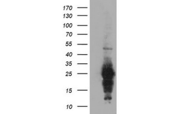
(HeLa cells were subjected to SDS PAGE followed by western blot with AAA102712 (BORIS antibody) at dilution of 1:1000)
WB (Western Blot)
(HeLa cells were subjected to SDS PAGE followed by western blot with AAA102712 (BORIS antibody) at dilution of 1:1000)
BORIS, Monoclonal Antibody (Cat# AAA102712)
Protein A+G purification
 WB (Western Blot)
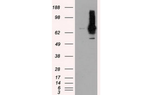
(Raji cells were subjected to SDS PAGE followed by western blot with AAA102733 (CD23,FCER2 antibody) at dilution of 1:2000)
WB (Western Blot)
(Raji cells were subjected to SDS PAGE followed by western blot with AAA102733 (CD23,FCER2 antibody) at dilution of 1:2000)
CD23, Monoclonal Antibody (Cat# AAA102733)
Protein A+G purification
 WB (Western Blot)
(Jurkat cells were subjected to SDS PAGE followed by western blot with AAA102739 (CD7 antibody) at dilution of 1:1000)
WB (Western Blot)
(Jurkat cells were subjected to SDS PAGE followed by western blot with AAA102739 (CD7 antibody) at dilution of 1:1000)
CD7, Monoclonal Antibody (Cat# AAA102739)
Protein A+G purification
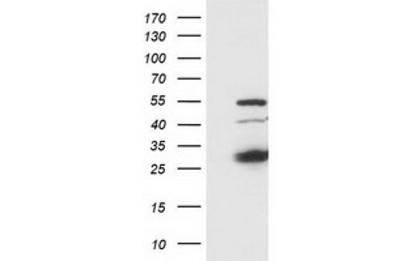
 WB (Western Blot)
(HeLa cells were subjected to SDS PAGE followed by western blot with AAA102745 (CHK1 Antibody) at dilution of 1:1000)
WB (Western Blot)
(HeLa cells were subjected to SDS PAGE followed by western blot with AAA102745 (CHK1 Antibody) at dilution of 1:1000)
Chk1, Monoclonal Antibody (Cat# AAA102745)
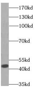
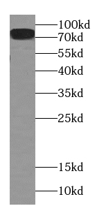 WB (Western Blot)
(human brain tissue were subjected to SDS PAGE followed by western blot with AAA102758 (CPT1C antibody) at dilution of 1:500)
WB (Western Blot)
(human brain tissue were subjected to SDS PAGE followed by western blot with AAA102758 (CPT1C antibody) at dilution of 1:500)
CPT1C, Monoclonal Antibody (Cat# AAA102758)
Protein A+G purification
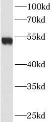
 WB (Western Blot)
(human skeletal muscle tissue were subjected to SDS PAGE followed by western blot with AAA102759 (CKM-Specific antibody) at dilution of 1:500)
WB (Western Blot)
(human skeletal muscle tissue were subjected to SDS PAGE followed by western blot with AAA102759 (CKM-Specific antibody) at dilution of 1:500)
Creatine Kinase MM, Monoclonal Antibody (Cat# AAA102759)
Protein A+G purification
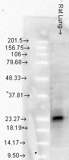
 WB (Western Blot)
(Serum from mouse injected with bacteria were subjected to SDS PAGE followed by western blot with AAA102761 (CRP Antibody) at dilution of 1:2000)
WB (Western Blot)
(Serum from mouse injected with bacteria were subjected to SDS PAGE followed by western blot with AAA102761 (CRP Antibody) at dilution of 1:2000)
CRP, Monoclonal Antibody (Cat# AAA102761)
Protein A+G purification
 WB (Western Blot)
(Jurkat cells were subjected to SDS PAGE followed by western blot with AAA102763 (CTAGE1 antibody) at dilution of 1:500)
WB (Western Blot)
(Jurkat cells were subjected to SDS PAGE followed by western blot with AAA102763 (CTAGE1 antibody) at dilution of 1:500)
CTAGE1, Monoclonal Antibody (Cat# AAA102763)
Protein A+G purification
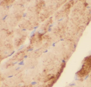
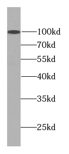
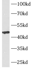
 WB (Western Blot)
(human heart tissue were subjected to SDS PAGE followed by western blot with AAA102771 (CYGB antibody) at dilution of 1:1000)
WB (Western Blot)
(human heart tissue were subjected to SDS PAGE followed by western blot with AAA102771 (CYGB antibody) at dilution of 1:1000)
Cytoglobin, Monoclonal Antibody (Cat# AAA102771)
Protein A+G purification
 WB (Western Blot)
(HeLa cells were subjected to SDS PAGE followed by western blot with AAA102773 (KRT18 Antibody) at dilution of 1:20000)
WB (Western Blot)
(HeLa cells were subjected to SDS PAGE followed by western blot with AAA102773 (KRT18 Antibody) at dilution of 1:20000)
Cytokeratin 18, Monoclonal Antibody (Cat# AAA102773)
Protein A+G purification
 WB (Western Blot)
(HeLa cells were subjected to SDS PAGE followed by western blot with AAA102777 (DDB1 antibody) at dilution of 1:1000)
WB (Western Blot)
(HeLa cells were subjected to SDS PAGE followed by western blot with AAA102777 (DDB1 antibody) at dilution of 1:1000)
DDB1, Monoclonal Antibody (Cat# AAA102777)
Protein A+G purification
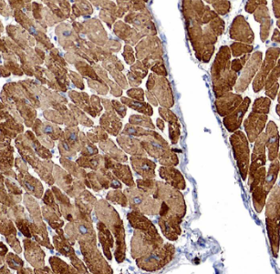
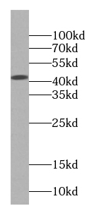
 WB (Western Blot)
(human heart tissue were subjected to SDS PAGE followed by western blot with AAA102778 (DES antibody) at dilution of 1:1000)
WB (Western Blot)
(human heart tissue were subjected to SDS PAGE followed by western blot with AAA102778 (DES antibody) at dilution of 1:1000)
Desmin, Monoclonal Antibody (Cat# AAA102778)
Protein A+G purification
 WB (Western Blot)
(COLO 320 cells were subjected to SDS PAGE followed by western blot with AAA102780 (DSE antibody) at dilution of 1:300)
WB (Western Blot)
(COLO 320 cells were subjected to SDS PAGE followed by western blot with AAA102780 (DSE antibody) at dilution of 1:300)
DSE, Monoclonal Antibody (Cat# AAA102780)
Protein A+G purification
 WB (Western Blot)
(HeLa cells were subjected to SDS PAGE followed by western blot with AAA102788 (ERK1/2 Antibody) at dilution of 1:1000)
WB (Western Blot)
(HeLa cells were subjected to SDS PAGE followed by western blot with AAA102788 (ERK1/2 Antibody) at dilution of 1:1000)
ERK1/2, Monoclonal Antibody (Cat# AAA102788)
Protein A+G purification
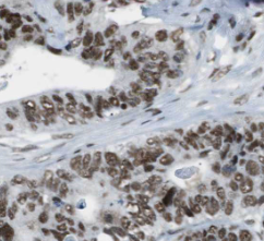
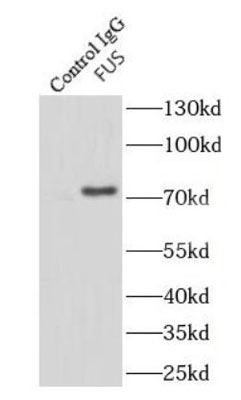 Application Data
(IP Result of anti-FUS/TLS (IP: AAA102805, 4ug; Detection: AAA102805 1:10000) with HeLa cells lysate 920ug.)
Application Data
(IP Result of anti-FUS/TLS (IP: AAA102805, 4ug; Detection: AAA102805 1:10000) with HeLa cells lysate 920ug.)
FUS/TLS, Monoclonal Antibody (Cat# AAA102805)
Purification: Protein A+G purification
 WB (Western Blot)
(pig brain tissue were subjected to SDS PAGE followed by western blot with AAA102807 (FUT9 Antibody) at dilution of 1:1000)
WB (Western Blot)
(pig brain tissue were subjected to SDS PAGE followed by western blot with AAA102807 (FUT9 Antibody) at dilution of 1:1000)
FUT9, Monoclonal Antibody (Cat# AAA102807)
Protein A+G purification
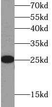
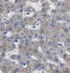
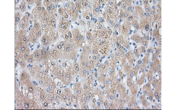
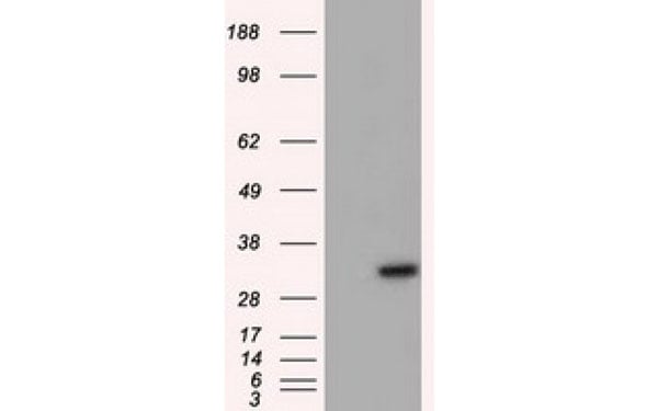
 WB (Western Blot)
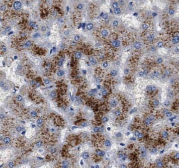
(human brain tissue were subjected to SDS PAGE followed by western blot with AAA102818 (GOT1 Antibody) at dilution of 1:1000)
WB (Western Blot)
(human brain tissue were subjected to SDS PAGE followed by western blot with AAA102818 (GOT1 Antibody) at dilution of 1:1000)
GOT1, Monoclonal Antibody (Cat# AAA102818)
Protein A+G purification
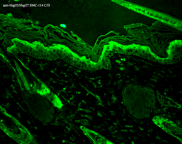
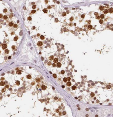
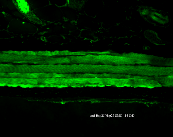 IHC (Immunohistochemistry)
(Immunohistochemistry analysis using Mouse Anti-Hsp27 Monoclonal Antibody, Clone 8A7. Tissue: backskin. Species: Mouse. Fixation: Bouin's Fixative and paraffin-embedded. Primary Antibody: Mouse Anti-Hsp27 Monoclonal Antibody at 1:100 for 1 hour at RT. Secondary Antibody: FITC Goat Anti-Mouse (green) at 1:50 for 1 hour at RT. Localization: Epidermis.)
IHC (Immunohistochemistry)
(Immunohistochemistry analysis using Mouse Anti-Hsp27 Monoclonal Antibody, Clone 8A7. Tissue: backskin. Species: Mouse. Fixation: Bouin's Fixative and paraffin-embedded. Primary Antibody: Mouse Anti-Hsp27 Monoclonal Antibody at 1:100 for 1 hour at RT. Secondary Antibody: FITC Goat Anti-Mouse (green) at 1:50 for 1 hour at RT. Localization: Epidermis.)
Hsp25/Hsp27, Monoclonal Antibody (Cat# AAA102826)
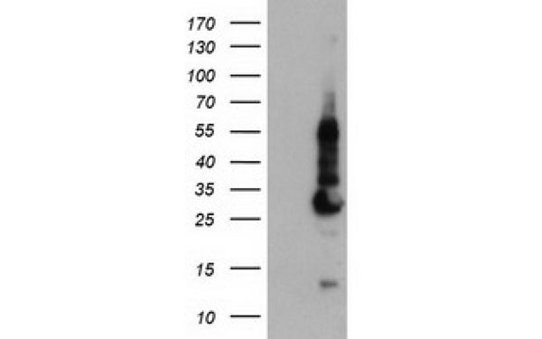
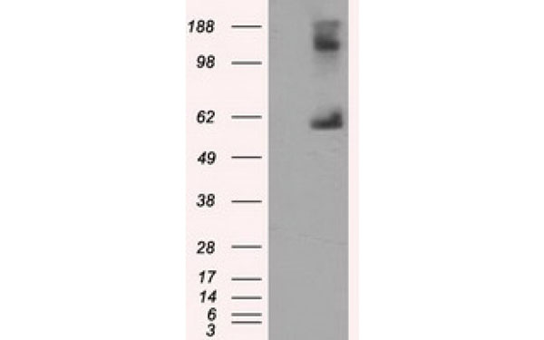
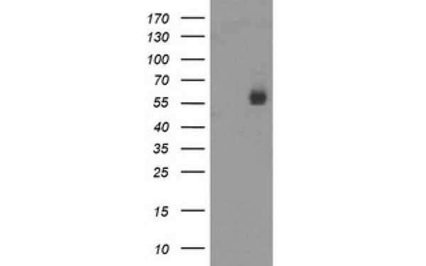
 WB (Western Blot)
(human testis tissue were subjected to SDS PAGE followed by western blot with AAA102669 (ACPP antibody) at dilution of 1:400)
WB (Western Blot)
(human testis tissue were subjected to SDS PAGE followed by western blot with AAA102669 (ACPP antibody) at dilution of 1:400)
ACPP, Monoclonal Antibody (Cat# AAA102669)
Protein A+G purification
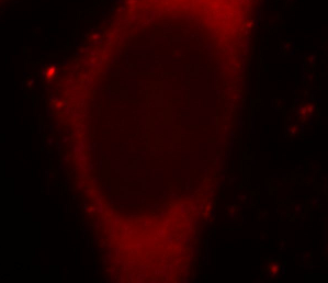
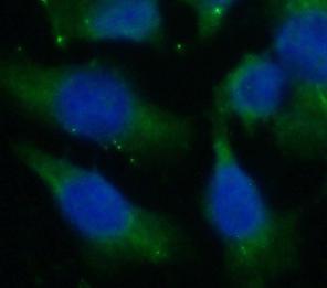
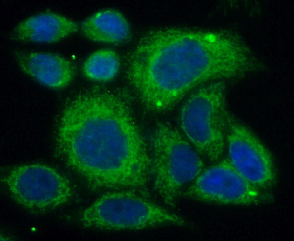
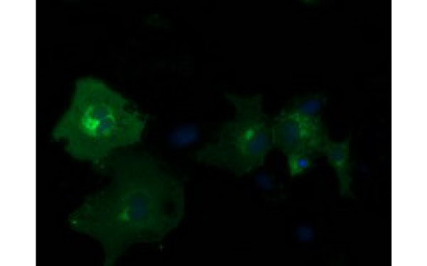
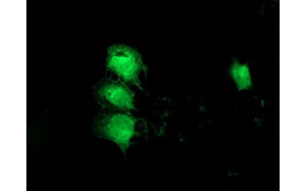
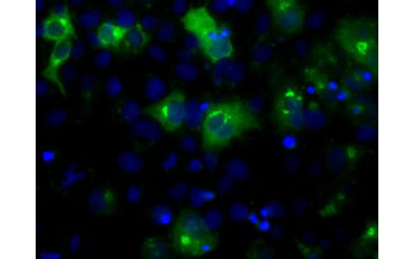
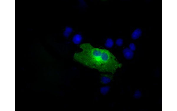
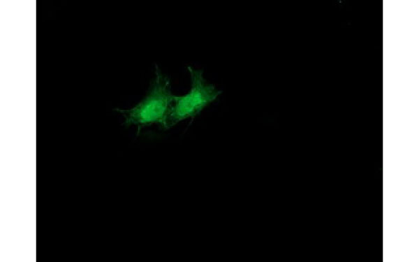
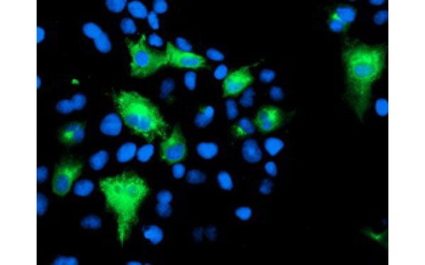
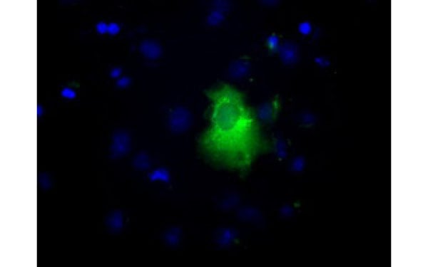
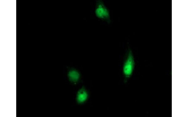
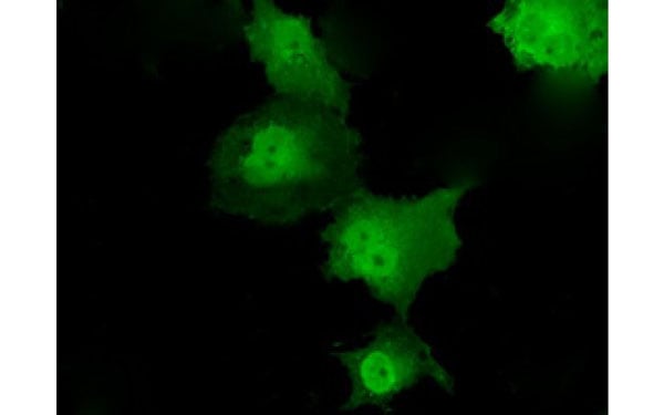
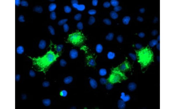
 IF (Immunofluorescence)
(Immunofluorescent staining of COS7 cells transiently transfected with recombinant NME4 protein using NME4 antibody)
IF (Immunofluorescence)
(Immunofluorescent staining of COS7 cells transiently transfected with recombinant NME4 protein using NME4 antibody)
NME4, Monoclonal Antibody (Cat# AAA107544)
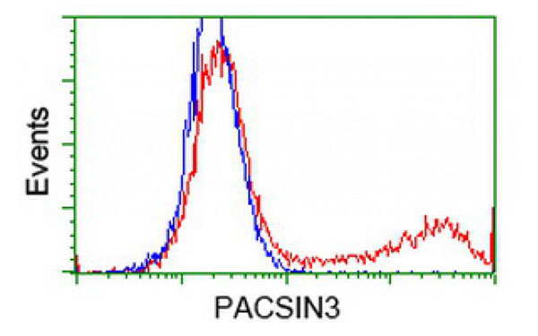
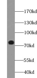
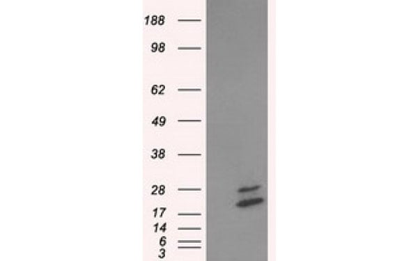
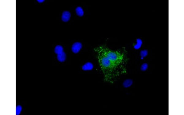
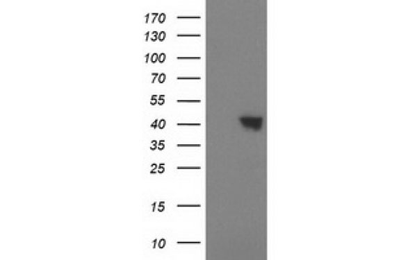
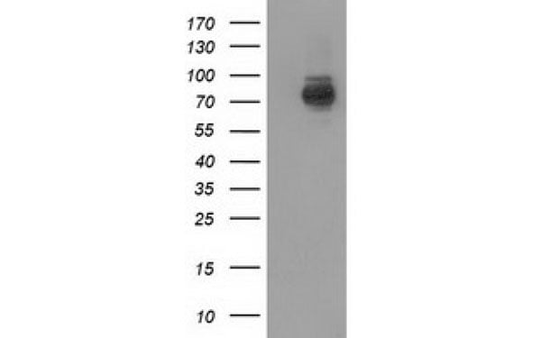
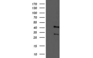
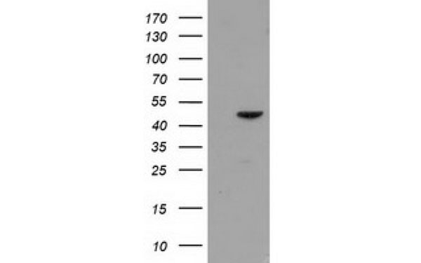 WB (Western Blot)
(Western Blot analysis of HEK293T cell lysates (5 ug) transfected with either recombinant PACSIN3 protein (Right) or empty vector (Left) detected with PACSIN3 antibody)
WB (Western Blot)
(Western Blot analysis of HEK293T cell lysates (5 ug) transfected with either recombinant PACSIN3 protein (Right) or empty vector (Left) detected with PACSIN3 antibody)
PACSIN3, Monoclonal Antibody (Cat# AAA107550)
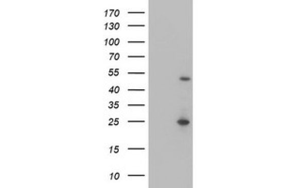
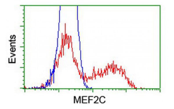
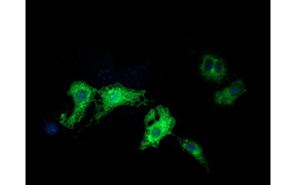
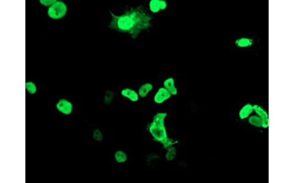 IF (Immunofluorescence)
(Immunofluorescent staining of COS7 cells transiently transfected with recombinant MEF2C protein using MEF2C antibody)
IF (Immunofluorescence)
(Immunofluorescent staining of COS7 cells transiently transfected with recombinant MEF2C protein using MEF2C antibody)
MEF2C, Monoclonal Antibody (Cat# AAA107552)
Influenza A HA H1, Monoclonal Antibody (Cat# AAA107325)
70 kDa Neurofilament, Monoclonal Antibody (Cat# AAA107329)
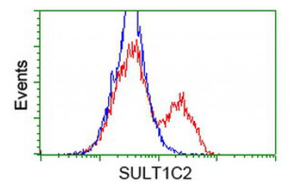
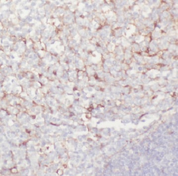
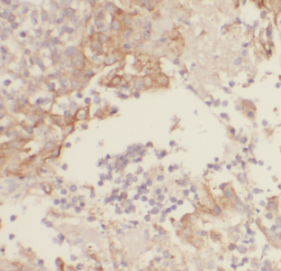
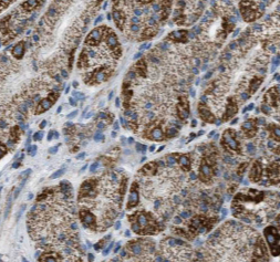
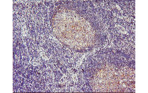
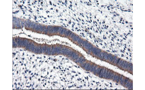
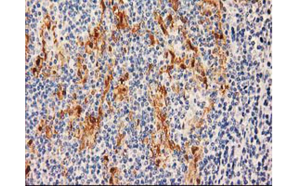 IHC (Immunohistochemisry)
(Immunohistochemical analysis of SulT1C2 protein in paraffin embedded Human lymphoma tissue using SulT1C2 antibody)
IHC (Immunohistochemisry)
(Immunohistochemical analysis of SulT1C2 protein in paraffin embedded Human lymphoma tissue using SulT1C2 antibody)
SulT1C2, Monoclonal Antibody (Cat# AAA107330)
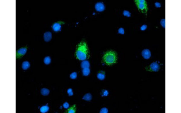
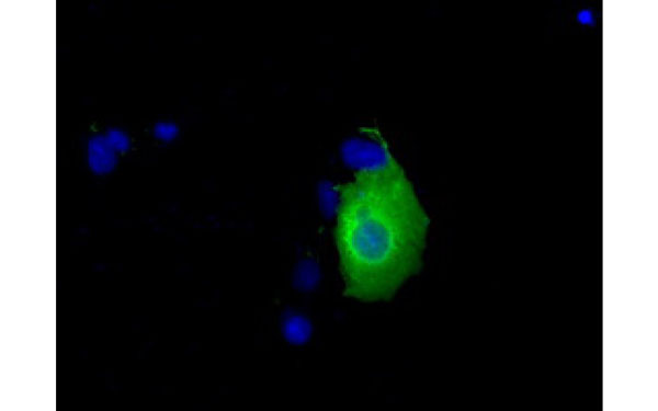
 IF (Immunofluorescence)
(Immunofluorescent staining of COS7 cells transiently transfected with recombinant PDE2A protein using PDE2A antibody)
IF (Immunofluorescence)
(Immunofluorescent staining of COS7 cells transiently transfected with recombinant PDE2A protein using PDE2A antibody)
PDE2A, Monoclonal Antibody (Cat# AAA107333)
Norovirus G1, Monoclonal Antibody (Cat# AAA107335)
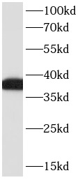
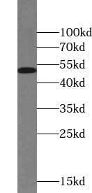
 WB (Western Blot)
(Western Blot analysis of HEK293T cell lysates (5 ug) transfected with either recombinant MAOA protein (Right) or empty vector (Left) detected with MAOA antibody)
WB (Western Blot)
(Western Blot analysis of HEK293T cell lysates (5 ug) transfected with either recombinant MAOA protein (Right) or empty vector (Left) detected with MAOA antibody)
MAOA, Monoclonal Antibody (Cat# AAA107343)
 IF (Immunofluorescence)
(Immunofluorescent staining of COS7 cells transiently transfected with recombinant MGLL protein using MGLL antibody)
IF (Immunofluorescence)
(Immunofluorescent staining of COS7 cells transiently transfected with recombinant MGLL protein using MGLL antibody)
MGLL, Monoclonal Antibody (Cat# AAA107345)
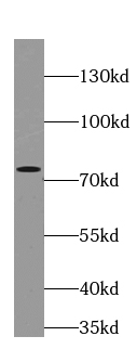
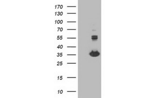
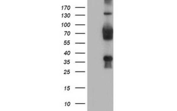
 WB (Western Blot)
(Western Blot analysis of HEK293T cell lysates (5 ug) transfected with either recombinant PIK3AP1 protein (Right) or empty vector (Left) detected with PIK3AP1 antibody)
WB (Western Blot)
(Western Blot analysis of HEK293T cell lysates (5 ug) transfected with either recombinant PIK3AP1 protein (Right) or empty vector (Left) detected with PIK3AP1 antibody)
PIK3AP1, Monoclonal Antibody (Cat# AAA107359)
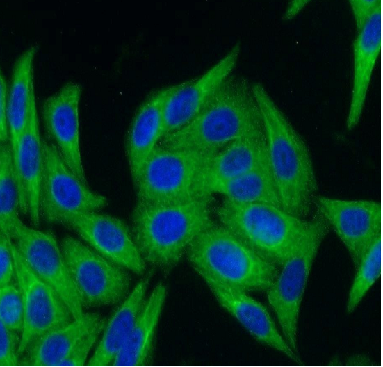
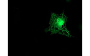
 IF (Immunofluorescence)
(Immunofluorescent staining of COS7 cells transiently transfected with recombinant LIPG protein using LIPG antibody)
IF (Immunofluorescence)
(Immunofluorescent staining of COS7 cells transiently transfected with recombinant LIPG protein using LIPG antibody)
LIPG, Monoclonal Antibody (Cat# AAA107369)
 IF (Immunofluorescence)
(Immunofluorescent staining of COS7 cells transiently transfected with recombinant RALBP1 protein using RALBP1 antibody)
IF (Immunofluorescence)
(Immunofluorescent staining of COS7 cells transiently transfected with recombinant RALBP1 protein using RALBP1 antibody)
RALBP1, Monoclonal Antibody (Cat# AAA107375)
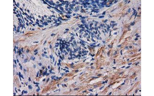
 IHC (Immunohistochemisry)
(Immunohistochemical analysis of TMOD1 protein in paraffin embedded Human tonsil tissue using TMOD1 antibody)
IHC (Immunohistochemisry)
(Immunohistochemical analysis of TMOD1 protein in paraffin embedded Human tonsil tissue using TMOD1 antibody)
TMOD1, Monoclonal Antibody (Cat# AAA107376)
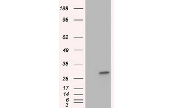
 WB (Western Blot)
(Western Blot analysis of HEK293T cell lysates (5 ug) transfected with either recombinant NIT2 protein (Right) or empty vector (Left) detected with NIT2 antibody)
WB (Western Blot)
(Western Blot analysis of HEK293T cell lysates (5 ug) transfected with either recombinant NIT2 protein (Right) or empty vector (Left) detected with NIT2 antibody)
NIT2, Monoclonal Antibody (Cat# AAA107381)
EB1+EB2+EB3, Monoclonal Antibody (Cat# AAA107383)
 IF (Immunofluorescence)
(Immunofluorescent staining of COS7 cells transiently transfected with recombinant SulT1A1 protein using SulT1A1 antibody)
IF (Immunofluorescence)
(Immunofluorescent staining of COS7 cells transiently transfected with recombinant SulT1A1 protein using SulT1A1 antibody)
SulT1A1, Monoclonal Antibody (Cat# AAA107386)
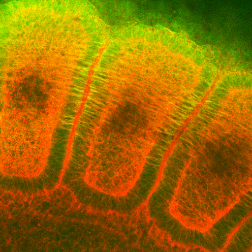
 IF (Immunofluorescence)
(Immunofluorescent staining of COS7 cells transiently transfected with recombinant ILVBL protein using ILVBL antibody)
IF (Immunofluorescence)
(Immunofluorescent staining of COS7 cells transiently transfected with recombinant ILVBL protein using ILVBL antibody)
ILVBL, Monoclonal Antibody (Cat# AAA107397)
 IF (Immunofluorescence)
(Immunofluorescent staining of COS7 cells transiently transfected with recombinant KCNJ3 protein using KCNJ3 antibody)
IF (Immunofluorescence)
(Immunofluorescent staining of COS7 cells transiently transfected with recombinant KCNJ3 protein using KCNJ3 antibody)
KCNJ3, Monoclonal Antibody (Cat# AAA107402)
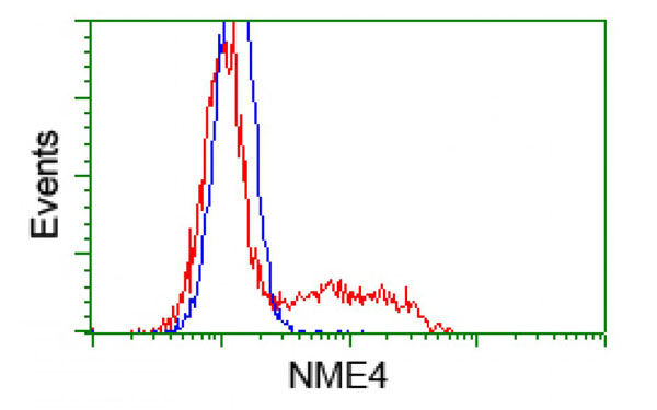
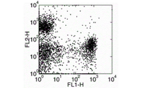
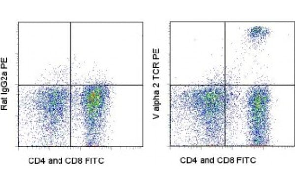 FCM/FACS (Flow Cytometry)
(Staining of C57Bl/6 lymph node cells with CD4 antibody and CD8a antibody and 0.125 ug of Rat IgG2a K Isotype Control (FITC) (left) or 0.125 ug of V alpha 2 TCR antibody (FITC) (right). Cells in the lymphocyte gate were used for analysis.)
FCM/FACS (Flow Cytometry)
(Staining of C57Bl/6 lymph node cells with CD4 antibody and CD8a antibody and 0.125 ug of Rat IgG2a K Isotype Control (FITC) (left) or 0.125 ug of V alpha 2 TCR antibody (FITC) (right). Cells in the lymphocyte gate were used for analysis.)
V alpha 2 TCR, Monoclonal Antibody (Cat# AAA107245)
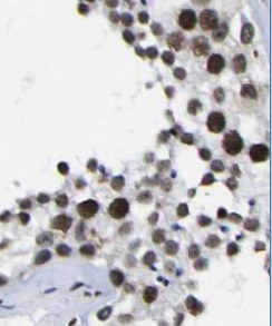
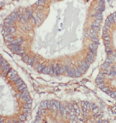
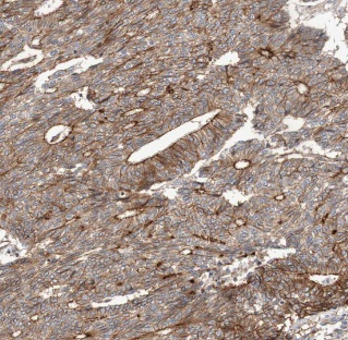
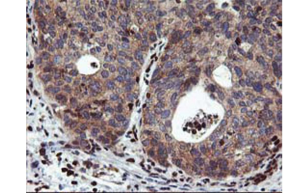
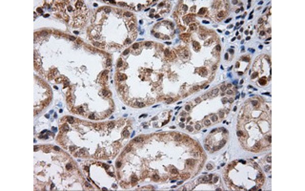
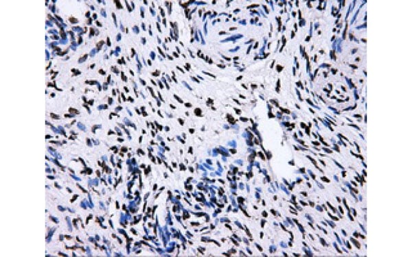
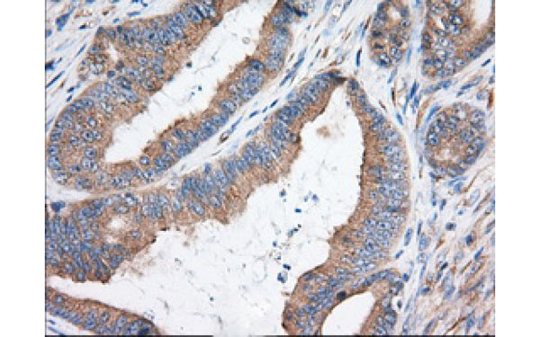 IHC (Immunohiostchemistry)
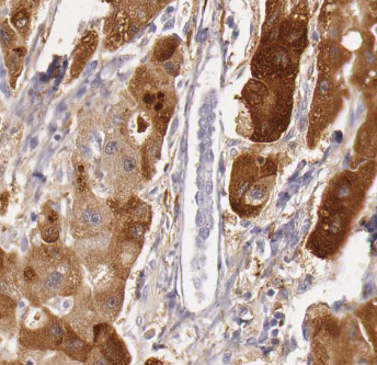
(Immunohistochemical analysis of KHK protein in paraffin embedded Adenocarcinoma of Human colon tissue using KHK antibody)
IHC (Immunohiostchemistry)
(Immunohistochemical analysis of KHK protein in paraffin embedded Adenocarcinoma of Human colon tissue using KHK antibody)
KHK, Monoclonal Antibody (Cat# AAA107252)
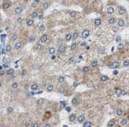
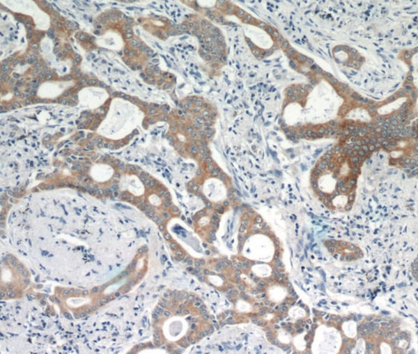
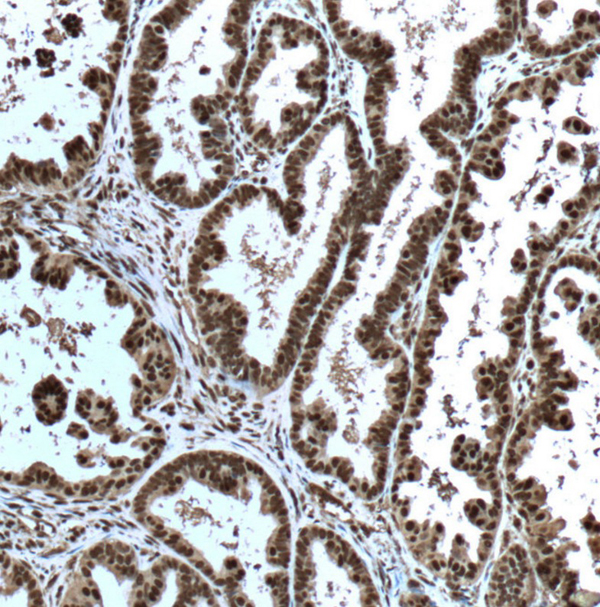
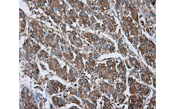
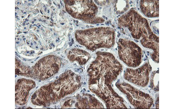
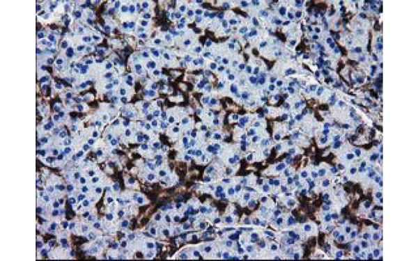
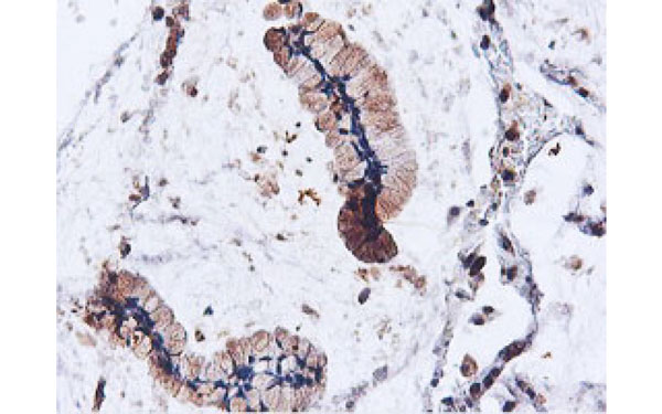
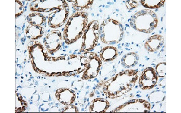
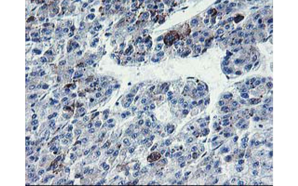
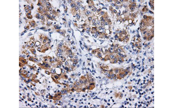 IHC (Immunohiostchemistry)
(Immunohistochemical analysis of LIPG protein in paraffin embedded Carcinoma of Human liver tissue using LIPG antibody)
IHC (Immunohiostchemistry)
(Immunohistochemical analysis of LIPG protein in paraffin embedded Carcinoma of Human liver tissue using LIPG antibody)
LIPG, Monoclonal Antibody (Cat# AAA107257)
 IF (Immunofluorescence)
(Immunofluorescent staining of COS7 cells transiently transfected with recombinant LIPG protein using LIPG antibody)
IF (Immunofluorescence)
(Immunofluorescent staining of COS7 cells transiently transfected with recombinant LIPG protein using LIPG antibody)
LIPG, Monoclonal Antibody (Cat# AAA107279)
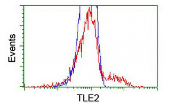
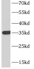
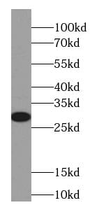
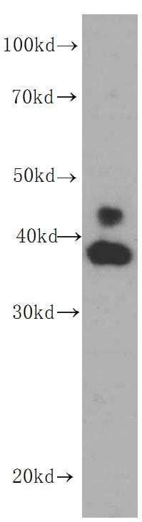
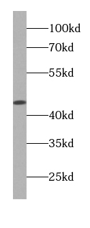
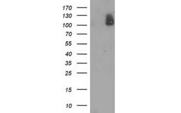
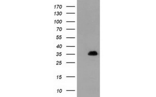
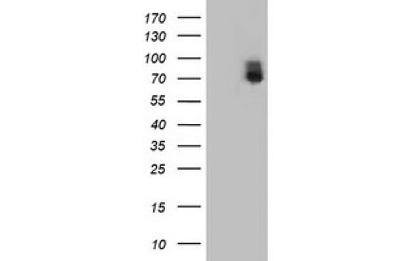
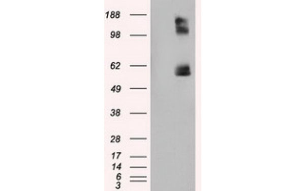
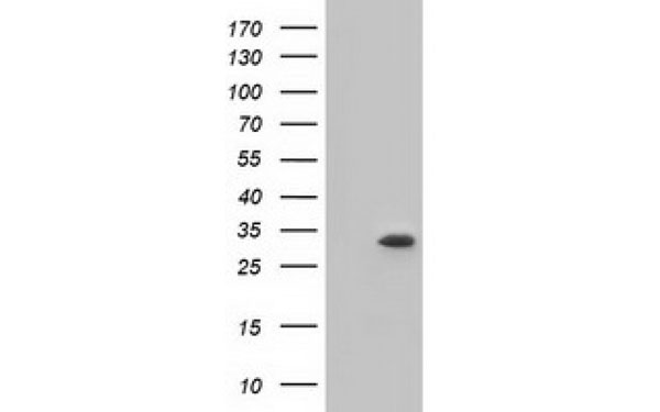
 WB (Western Blot)
(Western Blot analysis of HEK293T cell lysates (5 ug) transfected with either recombinant TLE2 protein (Right) or empty vector (Left) detected with TLE2 antibody)
WB (Western Blot)
(Western Blot analysis of HEK293T cell lysates (5 ug) transfected with either recombinant TLE2 protein (Right) or empty vector (Left) detected with TLE2 antibody)
TLE2, Monoclonal Antibody (Cat# AAA107283)
 IHC (Immunohistochemisry)
(Immunohistochemical analysis of RGS16 protein in paraffin embedded Human endometrium tissue using RGS16 antibody)
IHC (Immunohistochemisry)
(Immunohistochemical analysis of RGS16 protein in paraffin embedded Human endometrium tissue using RGS16 antibody)
RGS16, Monoclonal Antibody (Cat# AAA107290)
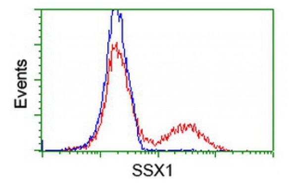
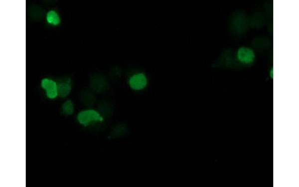 IF (Immunofluorescence)
(Immunofluorescent staining of COS7 cells transiently transfected with recombinant SSX1 protein using SSX1 antibody)
IF (Immunofluorescence)
(Immunofluorescent staining of COS7 cells transiently transfected with recombinant SSX1 protein using SSX1 antibody)
SSX1, Monoclonal Antibody (Cat# AAA107305)
MHC Class II, Monoclonal Antibody (Cat# AAA107156)
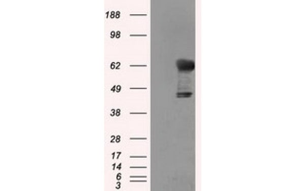
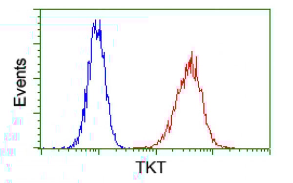
 IF (Immunofluorescence)
(Immunofluorescent staining of COS7 cells transiently transfected with recombinant TKT protein using TKT antibody)
IF (Immunofluorescence)
(Immunofluorescent staining of COS7 cells transiently transfected with recombinant TKT protein using TKT antibody)
TKT, Monoclonal Antibody (Cat# AAA107175)
 IF (Immunofluorescence)
(Immunofluorescent staining of COS7 cells transiently transfected with recombinant KHK protein using KHK antibody)
IF (Immunofluorescence)
(Immunofluorescent staining of COS7 cells transiently transfected with recombinant KHK protein using KHK antibody)
KHK, Monoclonal Antibody (Cat# AAA107177)
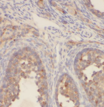
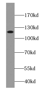
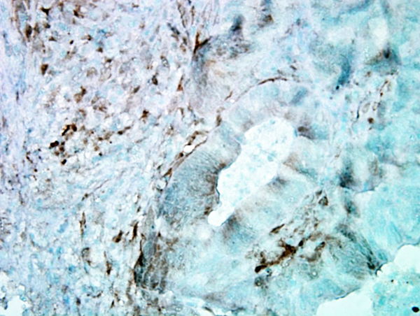
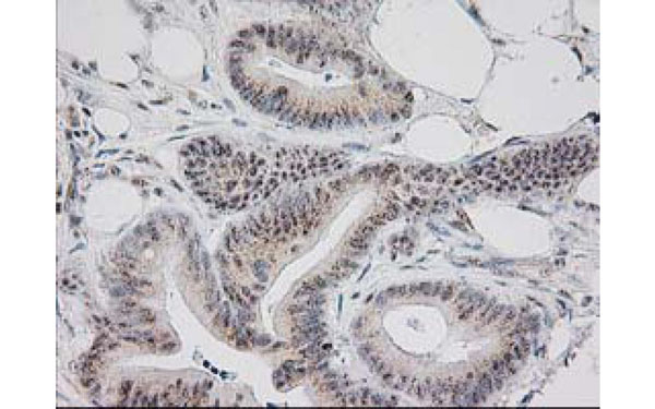
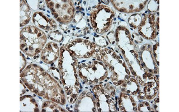
 IHC (Immunohistochemisry)
(Immunohistochemical analysis of PVRL1 protein in paraffin embedded Carcinoma of Human liver tissue using PVRL1 antibody)
IHC (Immunohistochemisry)
(Immunohistochemical analysis of PVRL1 protein in paraffin embedded Carcinoma of Human liver tissue using PVRL1 antibody)
PVRL1, Monoclonal Antibody (Cat# AAA107180)
 FCM/FACS (Flow Cytometry)
(Staining of mouse bone marrow with Ly-6G antibody (FITC) and CD45R antibody (PE). Total viable cells were used for analysis)
FCM/FACS (Flow Cytometry)
(Staining of mouse bone marrow with Ly-6G antibody (FITC) and CD45R antibody (PE). Total viable cells were used for analysis)
Ly6G, Monoclonal Antibody (Cat# AAA107189)
What are Monoclonal Antibodies?
Monoclonal antibodies are specialized laboratory-produced proteins developed for binding to specific biological antigens or other molecular targets. Since they come from a single cell (or clone), they are especially consistent and accurate in the data they are involved in producing.
This type of antibody material has been shown to be a powerful tool in finding and subsequently destroying harmful cells in an organism, such as those found in cancers or various autoimmune diseases. This makes them excellent aids in medical testing and research, which is why they are so widely used.
AAA Biotech offers a comprehensive range of high-quality monoclonal antibodies that perform effectively in various laboratory tests, including (amongst others) ELISA, western blotting, immunohistochemistry, and flow cytometry. All of the products in our catalog are thoroughly quality tested to make sure that they are reliable and will consistently perform well in your research.
What Are The Uses of Monoclonal Antibodies
Monoclonal antibodies are used in many lab tests, including (amongst others) ELISA, western blotting, immunohistochemistry, and flow cytometry.
ELISA is a test that helps detect a specific substance/analyte in a sample. It uses antibodies (often monoclonal) bound to a solid surface (such as the well of a microplate) to “capture” the substance/analyte in the sample and immobilize it so that the detection antibody component can then bind to it and produce a signal, which can then be measured.
Western blotting identifies specific proteins in a sample. The sample is first separated on a gel, and then antibodies are applied that will typically bind to the target, which will all be localized to a single band in a lane.
Immunohistochemistry helps locate specific proteins in cells or tissue samples using antibodies.
Flow cytometry looks at and sorts cells. It uses antibodies that are conjugated to reporter molecules called “fluorophores”, which, under special lights, emit light themselves, which can then be measured by a detector instrument. For a deeper understanding of these techniques, explore our complete guide to monoclonal antibodies and their benefits.
How Monoclonal Antibodies Are Used as Medicine?
Please note that all of the products listed in AAA Biotech’s also known as AAA Bio or AAABio catalog are strictly for research-use only (RUO).
Monoclonal antibodies can also be used as therapeutic/medical treatments, particularly in the context of cancers. They are designed to find and bind to specific cells or proteins, helping the immune system recognize and attack the cancer. These treatments work in different ways, such as:
- Radioimmunotherapy attaches a small amount of radioactive molecule to the antibody, so it delivers the radiation directly to the cancer cells that the antibody is specifically binding to.
- Antibody-directed enzyme prodrug therapy uses antibodies that are specifically bound to special enzymes. These enzymes activate a harmless drug in the body and turn it into a cancer-killing drug only near the cancer cells—this helps avoid harming healthy cells.
- Immunoliposomes are tiny “bubbles” filled with medicine/drug and coated with antibodies. They carry the drug straight to the cancer cells.
Why Buy Monoclonal Antibodies From Us?
At AAA Biotech, we provide high-performance monoclonal antibodies designed to support a wide range of research needs.
1. Validated for Versatile Applications
The antibodies in our catalog are extensively validated and compatible with multiple techniques, including (but not limited to) ELISA, flow cytometry (FC), immunocytochemistry (ICC), immunofluorescence (IF), immunohistochemistry (IHC), immunoprecipitation (IP), and western blotting (WB).
2. Wide Selection & Specialized Options
We offer antibodies for common and rare species, that are available in various conjugated forms, and also in recombinant formats. Essentially, there is almost anything one might need to meet their experimental model’s requirements.
3. High-Quality Proteins
Our proteins meet high purity standards—90% or more as confirmed by SDS-PAGE. Many are available with tags like His, Flag, GST, or MBP, and we also supply native and biologically active proteins for functional studies.
Frequently Asked Questions
1. Are your monoclonal antibodies validated for specific applications?
Yes, our antibodies are tested and validated for use in methods such as ELISA, western blot, IHC, flow cytometry, and more. Refer to specific product pages or datasheets for individual product information.
2. How do I choose the right monoclonal antibody for my application?
Review the product details directly for application validation, species reactivity, and target information. You may also contact our support team at any time for help.
3. How quickly can I receive my order?
Most orders are processed and shipped within 1–3 business days, depending on product availability and your shipping location.