Get accurate results in your research with our Monoclonal Antibodies, which are specially made to target exactly what you require for your research, and will produce consistent, reliable performance in lab tests.
FCM (Flow Cytometry) (Flow cytometric analysis of Jurkat cells with DIS3L2 antibody at 1/100 dilution (green) compared with an unlabelled control (cells without incubation with primary antibody; red).)
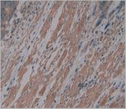
IHC (Immunohistochemistry) (Immunohistochemical analysis of paraffin-embedded human bladder cancer tissue using anti-DIS3L2 antibody. Counter stained with hematoxylin.)
IHC (Immunohistochemistry) (Immunohistochemical analysis of paraffin-embedded human endometrial cancer tissue using anti-DIS3L2 antibody. Counter stained with hematoxylin.)
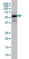
WB (Western Blot) (Western blot analysis of DIS3L2 on Hela (1) and HepG2 (2) cell lysate using anti-DIS3L2 antibody at 1/1, 000 dilution.)
WB (Western Blot) (Western blot analysis of DIS3L2 on HEK293 (1) and DIS3L2-hIgGFc transfected HEK293 (2) cell lysate using anti-DIS3L2 antibody at 1/1, 000 dilution.)
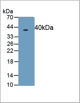
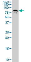
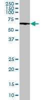
WB (Western Blot) (Western blot analysis of DIS3L2 on human DIS3L2 recombinant protein using anti-DIS3L2 antibody at 1/1, 000 dilution.)
FCM (Flow Cytometry) (Flow cytometry analysis of TREM2 in Hep3B cell line, staining at 2-5ug for 1x10^6cells. The secondary antibody used goat anti-mouse IgG Alexa fluor 488 conjugate.)
IF (Immunofluorescence) (ICC/IF analysis of TREM2 in Raw264.7 cells. The cell was stained with AAA11730 (1:100). The secondary antibody (green) was used Alexa Fluor 488. DAPI was stained the cell nucleus (blue).)
IF (Immunofluorescence) (ICC/IF analysis of TREM2 in HepG2 cells. The cell was stained with AAA11730 (1:100). The secondary antibody (green) was used Alexa Fluor 488. DAPI was stained the cell nucleus (blue).)
SDS-PAGE (The cell lysates(40ug) were resolved by SDS-PAGE, transferred to PVDF membrane and probed with anti-human TREM-2 antibody (1:1000). Proteins were visualized using a goat anti-mouse secondary antibody conjugated to HRP and an ECL detection system.Lane 1 : 293T cell lysateLane 2 : TREM2 19-161aa Transfected 293T cell lysate)
WB (Western Blot) (Cell lysates of HeLa, MCF7, LNCaP and mouse brain (30ug) were resolved by SDS-PAGE, transferred to NC membrane and probed with anti-human TREM2 (1:500). Proteins were visualized using a goat anti-mouse secondary antibody conjugated to HRP and an ECL detection system.)
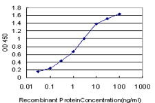
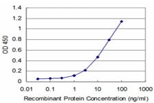
Application Data (Detection limit for recombinant GST tagged HNRPM is ~0.1ng/ml as a capture antibody.)
IF (Immunofluorescence) (Immunofluorescence of monoclonal antibody to HNRPM on HepG2 cell. [antibody concentration 10ug/ml])
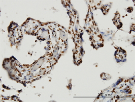
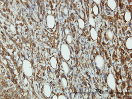
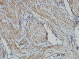
IHC (Immunohistochemistry) (Immunoperoxidase of monoclonal antibody to HNRPM on formalin-fixed paraffin-embedded human thyroid nodular goiter. [antibody concentration 3ug/ml.)
WB (Western Blot) (HNRPM monoclonal antibody. Western Blot analysis of HNRPM expression in NIH/3T3.)
WB (Western Blot) (HNRPM monoclonal antibody Western Blot analysis of HNRPM expression in HepG2.)
WB (Western Blot) (HNRPM monoclonal antibody Western Blot analysis of HNRPM expression in PC-12.)
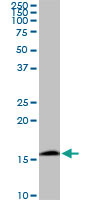
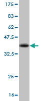
WB (Western Blot) (Western Blot detection against Immunogen (36.3kD).)
WB (Western Blot) (F3 monoclonal antibody, Western Blot analysis of F3 expression in A-431.)
Application Data (Proximity Ligation Analysis (PLA) of protein-protein interactions between F7 and F3. HeLa cells were stained with F7 rabbit purified polyclonal 1:1200 and F3 mouse monoclonal antibody 1:50. Signals were detected by 30 Detection Kit 613 (red), and nuclei were counterstained with DAPI (blue). Each red dot represents the detection of protein-protein interaction complex)
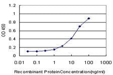
Application Data (Detection limit for recombinant GST tagged F3 is ~0.03ng/ml as a capture antibody.)
IP (Immunoprecipitation) (Immunoprecipitation of F3 transfected lysate using F3 monoclonal antibody and Protein A Magnetic Bead and immunoblotted with F3 rabbit polyclonal antibody.)
WB (Western Blot) (Western Blot analysis of F3 expression in transfected 293T cell line by F3 monoclonal antibody. Lane 1: F3 transfected lysate (33.1kD). Lane 2: Non-transfected lysate.)
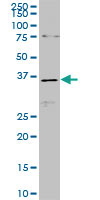
WB (Western Blot) (Western Blot detection against Immunogen (37.84kD).)
WB (Western Blot) (Western Blot detection against Immunogen (37kD).)
WB (Western Blot) (RBM9 monoclonal antibody Western Blot analysis of RBM9 expression in NIH/3T3.)
WB (Western Blot) (Western blot analysis of RBM9 over-expressed 293 cell line, cotransfected with RBM9 Validated Chimera RNAi (Lane 2) or non-transfected control (Lane 1). Blot probed with RBM9 monoclonal antibody. GAPDH (36.1kD) used as specificity and loading control.)
Application Data (Detection limit for recombinant GST tagged RBM9 is ~0.1ng/ml as a capture antibody.)
IF (Immunofluorescence) (Immunofluorescence of monoclonal antibody to RBM9 on HeLa cell. [antibody concentration 10ug/ml])
IHC (Immunohistochemistry) (Immunoperoxidase of monoclonal antibody to RBM9 on formalin-fixed paraffin-embedded human placenta. [antibody concentration 3ug/ml])
WB (Western Blot) (Western Blot analysis of RBM9 expression in transfected 293T cell line by RBM9 monoclonal antibody Lane 1: RBM9 transfected lysate (40.4kD). Lane 2: Non-transfected lysate.)
Application Data (Detection limit for recombinant GST tagged STAT5B is approximately 0.3ng/ml as a capture antibody.)
IHC (Immunohistochemistry) (Immunoperoxidase of monoclonal antibody to STAT5B on formalin-fixed paraffin-embedded human lymph node. [antibody concentration 1.5 ug/ml])
WB (Western Blot) (STAT5B monoclonal antibody (M03), clone 2D1 Western Blot analysis of STAT5B expression in HeLa.)
IF (Immunofluorescence) (Immunofluorescence of monoclonal antibody to STAT5B on HeLa cell. [antibody concentration 10 ug/ml])
Application Data (Detection limit for recombinant GST tagged TYK2 is approximately 1ng/ml as a capture antibody.)
WB (Western Blot) (TYK2 monoclonal antibody (M03), clone 6H1 Western Blot analysis of TYK2 expression in Hela S3 NE.)
IHC (Immunohistochemistry) (Immunoperoxidase of monoclonal antibody to TYK2 on formalin-fixed paraffin-embedded human lymphoma. [antibody concentration 3 ug/ml])
IHC (Immunohistochemistry) (Immunoperoxidase of monoclonal antibody to TYK2 on formalin-fixed paraffin-embedded human small Intestine. [antibody concentration 3 ug/ml])
Application Data (Immunoperoxidase of monoclonal antibody to TYK2 on formalin-fixed paraffin-embedded human small Intestine. [antibody concentration 3 ug/ml])
FCM (Flow Cytometry) (Flow cytometric analysis of HepG2 cells with PARK7 antibody at 1/50 dilution (red) compared with an unlabelled control (cells without incubation with primary antibody; black). Alexa Fluor 488-conjugated goat anti rabbit IgG was used as the secondary antibody.)
ICC (Immunocytochemistry) (ICC staining PARK7 in MCF-7 cells (green). The nuclear counter stain is DAPI (blue). Cells were fixed in paraformaldehyde, permeabilised with 0.25% Triton X100/PBS.)
ICC (Immunocytochemistry) (ICC staining PARK7 in HepG2 cells (green). The nuclear counter stain is DAPI (blue). Cells were fixed in paraformaldehyde, permeabilised with 0.25% Triton X100/PBS.)
IHC (Immunohistchemistry) (Immunohistochemical analysis of paraffin-embedded mouse pancreas tissue using anti-PARK7 antibody. Counter stained with hematoxylin.)
IHC (Immunohistochemistry) (Immunohistochemical analysis of paraffin-embedded mouse prostate tissue using anti-PARK7 antibody. Counter stained with hematoxylin.)
IHC (Immunohistochemistry) (Immunohistochemical analysis of paraffin-embedded human pancreas tissue using anti-PARK7 antibody. Counter stained with hematoxylin.)
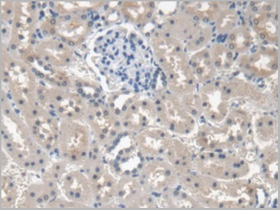
IHC (Immunohistochemistry) (Immunohistochemical analysis of paraffin-embedded human kidney tissue using anti-PARK7 antibody. Counter stained with hematoxylin.)
IHC (Immunohistochemistry) (Immunohistochemical analysis of paraffin-embedded human breast tissue using anti-PARK7 antibody. Counter stained with hematoxylin.)
WB (Western Blot) (Western blot analysis of PARK7 on different lysates using anti-PARK7 antibody at 1/1, 000 dilution. Positive control: Lane 1: Hela Lane 2: Jurkat Lane 3: NIH/3T3)
WB (Western Blot) (SLC25A13 monoclonal antibody Western Blot analysis of SLC25A13 expression in HepG2)
WB (Western Blot) (Western blot analysis of SLC25A13 over-expressed 293 cell line, cotransfected with SLC25A13 Validated Chimera RNAi (Lane 2) or non-transfected control (Lane 1). Blot probed with SLC25A13 monoclonal antibody GAPDH (36.1kD) used as specificity and loading control.)
Application Data (Detection limit for recombinant GST tagged SLC25A13 is ~0.3ng/ml as a capture antibody.)
IF (Immunofluorescence) (Immunofluorescence of monoclonal antibody to SLC25A13 on HepG2 cell. [antibody concentration 10ug/ml])
WB (Western Blot) (Western Blot analysis of SLC25A13 expression in transfected 293T cell line by SLC25A13 monoclonal antibodyLane 1: SLC25A13 transfected lysate (74.2kD).Lane 2: Non-transfected lysate.)
WB (Western Blot) (Western Blot detection against Immunogen (34.8kD).)
WB (Western Blot) (EHD3 monoclonal antibody. Western Blot analysis of EHD3 expression in COLO 320 HSR.)
WB (Western Blot) (EHD3 monoclonal antibody, Western Blot analysis of EHD3 expression in IMR-32.)
Application Data (Detection limit for recombinant GST tagged EHD3 is ~0.1ng/ml as a capture antibody.)
IF (Immunofluorescence) (Immunofluorescence of monoclonal antibody to EHD3 on HeLa cell. [antibody concentration 25ug/ml].)
IHC (Immunohistochemistry) (Immunoperoxidase of monoclonal antibody to EHD3 on formalin-fixed paraffin-embedded human liver. [antibody concentration 3ug/ml].)
WB (Western Blot) (EHD3 monoclonal antibody. Western Blot analysis of EHD3 expression in MCF-7.)
WB (Western Blot) (Western Blot detection against Immunogen (31.61kD).)
WB (Western Blot) (CBS monoclonal antibody. Western Blot analysis of CBS expression in MCF-7.)
WB (Western Blot) (CBS monoclonal antibody, Western Blot analysis of CBS expression in HeLa.)
WB (Western Blot) (Western blot analysis of CBS over-expressed 293 cell line, cotransfected with CBS Validated Chimera RNAi (Lane 2) or non-transfected control (Lane 1). Blot probed with 124401. GAPDH (36.1kD) used as specificity and loading control.)
Application Data (Detection limit for recombinant GST tagged CBS is ~0.1ng/ml as a capture antibody.)
IP (Immunoprecipitation) (Immunoprecipitation of CBS transfected lysate using 124401and Protein A Magnetic Bead and immunoblotted with CBS rabbit polyclonal antibody.)
IHC (Immunohistochemistry) (Immunoperoxidase of formalin-fixed paraffin-embedded human hepatocellular carcinoma using 124401 (3ug/ml).)
WB (Western Blot) (Western Blot analysis of CBS expression in transfected 293T cell line by 124401. Lane 1: CBS transfected lysate (61kD). Lane 2: Non-transfected lysate.)
WB (Western Blot) (SPRY2 monoclonal antibody Western Blot analysis of SPRY2 expression in C32)
Application Data (Detection limit for recombinant GST tagged SPRY2 is ~0.1ng/ml as a capture antibody.)
IF (Immunofluorescence) (Immunofluorescence of monoclonal antibody to SPRY2 on HeLa cell. [antibody concentration 25ug/ml])
IHC (Immunohistochemistry) (Immunoperoxidase of monoclonal antibody to SPRY2 on formalin-fixed paraffin-embedded human lymphoma. [antibody concentration 3ug/ml])
WB (Western Blot) (Western Blot analysis of SPRY2 expression in transfected 293T cell line by SPRY2 monoclonal antibody Lane 1: SPRY2 transfected lysate (34.7kD). Lane 2: Non-transfected lysate.)
WB (Western Blot) (Western Blot detection against Immunogen (60.76kD).)
WB (Western Blot) (Western blot analysis of ENO3 over-expressed 293 cell line, cotransfected with ENO3 Validated Chimera RNAi (Lane 2) or non-transfected control (Lane 1). Blot probed with ENO3 monoclonal antibody. GAPDH (36.1kD) used as specificity and loading control.)
Application Data (Detection limit for recombinant GST tagged ENO3 is ~0.03ng/ml as a capture antibody.)
IF (Immunofluorescence) (Immunofluorescence of monoclonal antibody to ENO3 on HeLa cell. [antibody concentration 10ug/ml].)
IHC (Immunohistochemistry) (Immunoperoxidase of monoclonal antibody to ENO3 on formalin-fixed paraffin-embedded human colon. [antibody concentration 3ug/ml].)
IHC (Immunohistochemistry) (Immunoperoxidase of monoclonal antibody to ENO3 on formalin-fixed paraffin-embedded human stomach. [antibody concentration 1.5ug/ml].)
WB (Western Blot) (Western Blot analysis of ENO3 expression in transfected 293T cell line by ENO3 monoclonal antibody. Lane 1: ENO3 transfected lysate (46.9kD). Lane 2: Non-transfected lysate.)
WB (Western Blot) (Western Blot detection against Immunogen (31.24kD).)
WB (Western Blot) (RNF12 monoclonal antibody, Western Blot analysis of RNF12 expression in HeLa NE.)
Application Data (Detection limit for recombinant GST tagged RNF12 is ~0.03ng/ml as a capture antibody.)
IP (Immunoprecipitation) (Immunoprecipitation of RNF12 transfected lysate using RNF12 monoclonal antibody and Protein A Magnetic Bead and immunoblotted with RNF12 rabbit polyclonal antibody.)
IF (Immunofluorescence) (Immunofluorescence of monoclonal antibody to RNF12 on HeLa cell. [antibody concentration 30ug/ml].)
WB (Western Blot) (Western Blot analysis of RNF12 expression in transfected 293T cell line by RNF12 monoclonal antibody. Lane 1: RNF12 transfected lysate (68.5kD). Lane 2: Non-transfected lysate.)
WB (Western Blot) (Western Blot detection against Immunogen (35.24kD).)
IF (Immunofluorescence) (Confocal immunofluorescent analysis of CD138 Antibody (C-term) (Ascites) with T47D cell followed by Alexa Fluor 488-conjugated goat anti-rabbit lgG (green).DAPI was used to stain the cell nuclear (blue).)
IF (Immunofluorescence) (Confocal immunofluorescent analysis of CD138 Antibody (C-term) (Ascites) with HepG2 cell followed by Alexa Fluor 488-conjugated goat anti-rabbit lgG (green).DAPI was used to stain the cell nuclear (blue).)
IF (Immunofluorescence) (Confocal immunofluorescent analysis of CD138 Antibody (C-term) (Ascites) with U266 cell followed by Alexa Fluor 488-conjugated goat anti-rabbit lgG (green).DAPI was used to stain the cell nuclear (blue).)
FCM (Flow Cytometry) (CD138 Antibody (C-term) (Ascites) flow cytometric analysis of T47D cells (right histogram) compared to a negative control cell (left histogram).Alexa Fluor 488-conjugated donkey anti-mouse lgG secondary antibodies were used for the analysis)
FCM (Flow Cytometry) (CD138 Antibody (C-term) (Ascites) flow cytometric analysis of HepG2 cells (right histogram) compared to a negative control cell (left histogram).Alexa Fluor 488-conjugated donkey anti-mouse lgG secondary antibodies were used for the analysis)
FCM (Flow Cytometry) (CD138 Antibody (C-term) (Ascites) flow cytometric analysis of U266 cells (right histogram) compared to a negative control cell (left histogram).Alexa Fluor 488-conjugated donkey anti-mouse lgG secondary antibodies were used for the analysis)
WB (Western Blot) (CD138 Antibody (C-term)(Ascites) western blot analysis in HepG2 cell line lysates (35ug/lane).This demonstrates the CD138 antibody detected the CD138 protein (arrow).)
FCM (Flow Cytometry) (Flow cytometric analysis of Hela cells with BMI1 antibody at 1/100 dilution (blue) compared with an unlabelled control (cells without incubation with primary antibody; red). Goat anti mouse IgG (FITC) was used as the secondary antibody.)
ICC (Immunocytochemistry) (ICC staining Bmi1 in Hela cells (red). Cells were fixed in paraformaldehyde, permeabilised with 0.25% Triton X100/PBS.)
ICC (Immunocytochemistry) (ICC staining Bmi1 in Lovo cells (red). Cells were fixed in paraformaldehyde, permeabilised with 0.25% Triton X100/PBS.)
ICC (Immunocytochemistry) (ICC staining Bmi1 in A549 cells (red). Cells were fixed in paraformaldehyde, permeabilised with 0.25% Triton X100/PBS.)
IHC (Immunohistochemistry) (Immunohistochemical analysis of paraffin-embedded human breast cancer tissue using anti-BMI1 antibody. Counter stained with hematoxylin.)
IHC (Immunohistochemistry) (Immunohistochemical analysis of paraffin-embedded human colon cancer tissue using anti-Bmi1 antibody. Counter stained with hematoxylin.)
IHC (Immunohistochemistry) (Immunohistochemical analysis of paraffin-embedded human tonsil tissue using anti-Bmi1 antibody. Counter stained with hematoxylin.)
WB (Western Blot) (Western blot analysis of Bmi1 on different lysates using anti-Bmi1 antibody at 1/1, 000 dilution. Positive control: Lane 1: 293T Lane 2: Jurkat Lane 3: Hela Lane 4: MCF-7 Lane 5: HepG2 Lane 6: NIH/3T3 Lane 7: PC12 Lane 8: Mouse kidney Lane 9: Human kidney Lane 10: K562 Lane 11: Human brain)
FCM (Flow Cytometry) (Flow cytometric analysis of SH-SY-5Y cells with CRMP1 antibody at 1/50 dilution (red) compared with an unlabelled control (cells without incubation with primary antibody; black). Alexa Fluor 488-conjugated goat anti rabbit IgG was used as the secondary antibody.)
ICC (Immunocytochemistry) (ICC staining CRMP1 in SH-SY5Y cells (green). The nuclear counter stain is DAPI (blue). Cells were fixed in paraformaldehyde, permeabilised with 0.25% Triton X100/PBS.)
ICC (Immunocytochemistry) (ICC staining CRMP1 in SHG-44 cells (green). The nuclear counter stain is DAPI (blue). Cells were fixed in paraformaldehyde, permeabilised with 0.25% Triton X100/PBS.)
ICC (Immunocytochemistry) (ICC staining CRMP1 in N2A cells (green). The nuclear counter stain is DAPI (blue). Cells were fixed in paraformaldehyde, permeabilised with 0.25% Triton X100/PBS.)
IHC (Immunohistochemistry) (Immunohistochemical analysis of paraffin-embedded mouse cerebellum tissue using anti-CRMP1 antibody. Counter stained with hematoxylin.)
IHC (Immunohistochemistry) (Immunohistochemical analysis of paraffin-embedded mouse brain tissue using anti-CRMP1 antibody. Counter stained with hematoxylin.)
IHC (Immunohistochemistry) (Immunohistochemical analysis of paraffin-embedded rat brain tissue using anti-CRMP1 antibody. Counter stained with hematoxylin.)
WB (Western Blot) (Western blot analysis of CRMP1 on mouse brain tissue lysate using anti-CRMP1 antibody at 1/500 dilution.)
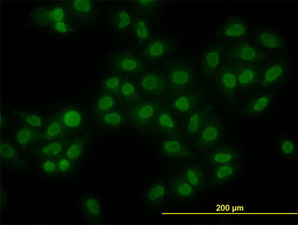
ICC (Immunocytochemistry) (ICC staining ADAR in Hela cells (green). The nuclear counter stain is DAPI (blue). Cells were fixed in paraformaldehyde, permeabilised with 0.25% Triton X100/PBS.)
IHC (Immunohistochemistry) (Immunohistochemical analysis of paraffin-embedded human pancreas tissue using anti-ADAR antibody. Counter stained with hematoxylin.)
IHC (Immunohistochemistry) (Immunohistochemical analysis of paraffin-embedded human lung cancer tissue using anti-ADAR antibody. Counter stained with hematoxylin.)
IHC (Immunohistochemistry) (Immunohistochemical analysis of paraffin-embedded human kidney tissue using anti-ADAR antibody. Counter stained with hematoxylin.)
IHC (Immunohistochemistry) (Immunohistochemical analysis of paraffin-embedded human colon cancer tissue using anti-ADAR antibody. Counter stained with hematoxylin.)
WB (Western Blot) (Western blot analysis of ADAR on SiHa cell using anti-ADAR antibody at 1/500 dilution.)
WB (Western Blot) (Western Blot detection against Immunogen (36.89kD).)
Application Data (Detection limit for recombinant GST tagged RICTOR is ~0.03ng/ml as a capture antibody.)
IP (Immunoprecipitation) (Immunoprecipitation of RICTOR transfected lysate using RICTOR monoclonal antibody and Protein A Magnetic Bead, and immunoblotted with RICTOR rabbit polyclonal antibody)
IHC (Immunohistochemistry) (Immunoperoxidase of monoclonal antibody to RICTOR on formalin-fixed paraffin-embedded human small Intestine. [antibody concentration 3ug/ml].)
WB (Western Blot) (Western Blot analysis of RICTOR expression in transfected 293T cell line by RICTOR monoclonal antibody. Lane 1: RICTOR transfected lysate (29.8kD). Lane 2: Non-transfected lysate.)
WB (Western Blot) (RICTOR monoclonal antibody Western Blot analysis of RICTOR expression in Hela NE.)
FCM (Flow Cytometry) (Flow cytometric analysis of Hela cells with PBK antibody at 1/100 dilution (green) compared with an unlabelled control (cells without incubation with primary antibody; red).)
ICC (Immunocytochemistry) (ICC staining PBK (green) and actin filaments (red) in Hela cells. The nuclear counter stain is DAPI (blue). Cells were fixed in paraformaldehyde, permeabilised with 0.25% Triton X100/PBS.)
IHC (Immunohistochemistry) (Immunohistochemical analysis of paraffin-embedded human colon cancer tissue using anti-PBK antibody. Counter stained with hematoxylin.)
IHC (Immunohistochemistry) (Immunohistochemical analysis of paraffin-embedded human ovarian cancer tissue using anti-PBK antibody. Counter stained with hematoxylin.)
WB (Western Blot) (Western blot analysis of PBK on A431 cell lysate using anti-PBK antibody at 1/1, 000 dilution.)
WB (Western Blot) (Western blot analysis of PBK on human PBK recombinant protein using anti-PBK antibody at 1/1, 000 dilution.)
Application Data (Staining of human peripheral blood lymphocytes with Mouse anti Human CD43: FITC)
Application Data (Imunofluorescence staining of human tonsil cryosection with Mouse anti Human CD43, clone DFT-1, red in A and Mouse anti Human CD21 , green in B. C is the merged image with nuclei counterstained blue using DAPI. High power)
Application Data (Immunoperoxidase staining of human tonsil cryosection with Mouse anti Human CD43 antibody, clone DFT-1 followed by the HISTAR detection system . Medium power)
Application Data (Staining of human peripheral blood monocytes with Mouse anti Human CD43)
Application Data (Staining of human peripheral blood granulocytes with Mouse anti Human CD43:Biotin)
Application Data (Immunoperoxidase staining of human tonsil cryosection with Mouse anti Human CD43 antibody, clone DFT-1 followed by the HISTAR detection system . Low power)
Application Data (Staining of human peripheral blood lymphocytes with Mouse anti Human CD43:Azide Free)
Application Data (Imunofluorescence staining of human tonsil cryosection with Mouse anti Human CD43, clone DFT-1, red in A and Mouse anti Human CD21 , green in B. C is the merged image with nuclei counterstained blue using DAPI. Medium power)
Application Data (Immunoperoxidase staining of human tonsil cryosection with Mouse anti Human CD43 antibody, clone DFT-1 followed by the HISTAR detection system . Medium power)
Application Data (Imunofluorescence staining of human tonsil cryosection with Mouse anti Human CD43, clone DFT-1, red in A and Mouse anti Human CD21 , green in B. C is the merged image with nuclei counterstained blue using DAPI. Low power)
WB (Western Blot) (EIF2S1 monoclonal antibody, Western Blot analysis of EIF2S1 expression in HeLa.)
WB (Western Blot) (EIF2S1 monoclonal antibody. Western Blot analysis of EIF2S1 expression in Hela NE.)
WB (Western Blot) (Western blot analysis of EIF2S1 over-expressed 293 cell line, cotransfected with EIF2S1 Validated Chimera RNAi (Lane 2) or non-transfected control (Lane 1). Blot probed with EIF2S1 monoclonal antibody. GAPDH (36.1kD) used as specificity and loading control.)
Application Data (Detection limit for recombinant GST tagged EIF2S1 is ~0.3ng/ml as a capture antibody.)
IF (Immunofluorescence) (Immunofluorescence of monoclonal antibody to EIF2S1 on HeLa cell. [antibody concentration 10ug/ml].)
WB (Western Blot) (Western Blot analysis of EIF2S1 expression in transfected 293T cell line by EIF2S1 monoclonal antibody. Lane 1: EIF2S1 transfected lysate (36.1kD). Lane 2: Non-transfected lysate.)
WB (Western Blot) (Western Blot detection against Immunogen (60.39kD).)
Application Data (Detection limit for recombinant GST tagged PGR is approximately 0.03ng/ml as a capture antibody.)
WB (Western Blot) (PGR monoclonal antibody (M08), clone 4E9 Western Blot analysis of PGR expression in MCF-7 (Cat # L046V1).)
IF (Immunofluorescence) (Immunofluorescence of monoclonal antibody to PGR on MCF-7 cell. [antibody concentration 10 ug/ml])
IHC (Immunohistochemistry) (Immunoperoxidase of monoclonal antibody to PGR on formalin-fixed paraffin-embedded human breast cancer. [antibody concentration 3 ug/ml])
Application Data (Immunoperoxidase of monoclonal antibody to PGR on formalin-fixed paraffin-embedded human breast cancer. [antibody concentration 3 ug/ml])
Application Data (Publised customer image:Mouse anti Human CD163 antibody, clone EDHu-1 used for the identification of perivascular macrophages in human brain by immunofluorescence.Image caption:Images demonstrating immunohistological stainings of amylin and double immunofluorescence staining against NG2/amylin, laminin/amylin and CD163/amylin in the hippocampus of the patient with AD and T2D.Amylin cell inclusions are indicated with arrows in (a) and shown in a higher magnification in (b). Pericytes with round cell bodies and NG2-positive coverage of the microvessel surface (green in c), without amylin cell inclusions (red in d), displayed round DAPI-positive cell nuclei (blue in e). The images in (c), (d) and (e) are merged in (f). Cells with more diffuse and weak NG2 staining (indicated by the arrowhead, green in g) and cytosolic amylin cell inclusions (red in h) showed altered cell nuclei (indicated with arrow, blue in i).The adjacent unaffected NG2-positive cell is indicated with an arrowhead in (i). The images in (g), (h) and (i) are merged in (j). Cells enclosed by laminin (green in K) contained amylin grains (red in l) and fragmented DAPI-positive cell nuclei (indicated with an arrow blue in m). The images in (k), (l) and (m) are merged in (n).Loss of NG2 coverage (green in o) was associated with polarized amylin cell inclusion (red in p) and fragmented DAPI-positive cell nuclei (indicated with an arrow, blue in q). The images in (o), (p) and (q) are merged in (r). Staining against macrophage marker CD163 (green in s) did not co-localize with amylin cell inclusions (red in t). The cell nucleus was stained with DAPI (blue in U). The images in (s), (t) and (u) are merged in (v). Scale bars: (a) 50 mum, (b), (c) to (v) 5 mum.From: Schultz, N. et al. (2016).Amylin alters human brain pericyte viability and NG2 expression.J Cereb Blood Flow & Metab. Jun 28 [Epub ahead of print]This is from an open access article distributed under the terms of the Creative Commons Attribution License.)
Application Data (Published customer image:Mouse anti Human CD163 antibody, clone EDHu-1 used for the identification of infiltrating macrophages in the pulmonary tissues of cattle by immunohistochemistry on formalin fixed tissue sections.Image caption:Immunohistochemical Labeling of Leukocytes in the Lungs.Shown are the results of IHC labeling for CD3-like immunoreactivity (CD3-li), CD163-li, IBA-1-li, and IL-17-li in the lung of uninfected control and representative infected Holstein calves.Note: CD3-li, CD163-li, and IL-17-li is significantly increased in the lungs of the infected calf.Furthermore, CD3+, CD163+, IBA-1+ and IL-17+ mononuclear cells are components of vasculitis lesions. Vascular endothelial cells consistently exhibit pronounced IL-17-li in the infected calf, but endothelial IL-17-li is rare in the control calf. Scale bar: 200 mum.)
IF (Immunofluorescence) (Immunofluorescence staining of a human tonsil cryosection with Mouse anti Human CD163 antibody, clone EDHu-1 , red in A and Mouse anti Human CD21 antibody, clone LB21 , green in B. C is the merged image with nuclei counterstained blue using DAPI. High power.)
IF (Immunofluorescence) (Immunofluorescence staining of a human tonsil cryosection with Mouse anti Human CD163 antibody, clone EDHu-1 , red in A and Mouse anti Human CD21 antibody, clone LB21 , green in B. C is the merged image with nuclei counterstained blue using DAPI. Medium power.)
IF (Immunofluorescence) (Immunofluorescence staining of a human tonsil cryosection with Mouse anti Human CD163 antibody, clone EDHu-1 , red in A and Mouse anti Human CD21 antibody, clone LB21 , green in B. C is the merged image with nuclei counterstained blue using DAPI. Low power.)
Application Data (Immunoperoxidase staining of a human tonsil cryosection with Mouse anti Human CD163 antibody, clone EDHu-1 followed by the Histar detection system . High power.)
Application Data (Immunoperoxidase staining of a human tonsil cryosection with Mouse anti Human CD163 antibody, clone EDHu-1 followed by the Histar detection system . Medium power.)
Application Data (Immunoperoxidase staining of a human tonsil cryosection with Mouse anti Human CD163 antibody, clone EDHu-1 followed by the Histar detection system . Low power.)
Application Data (Figure A. Alexa Fluor700 conjugated mouse anti human CD14 and RPE conjugated mouse IgG1 isotype control . Figure B. Alexa Fluor700 conjugated mouse anti human CD14 and RPE conjugated mouse anti human CD163 . All experiments performed on red cell lysed human blood gated on monocytes in the presence of Human Seroblock (BUF070A). Data acquired on the ZE5 cell analyzer.)
Application Data (Figure A. RPE conjugated mouse anti CD14 and FITC conjugated mouse IgG1 isotype control . Figure B. RPE conjugated mouse anti CD14 and FITC conjugated mouse anti human CD163 . All experiments performed on human peripheral blood mononuclear cells in the presence of Human SeroBlock (BUF070A).)
Application Data (Figure A. Alexa Fluor700 conjugated mouse anti human CD14 and Alexa Fluor488 conjugated mouse IgG1 isotype control . Figure B. Alexa Fluor700 conjugated mouse anti human CD14 and Alexa Fluor488 conjugated mouse anti human CD163 . All experiments performed on red cell lysed human blood gated on mononuclear cells in the presence of Human Seroblock (BUF070A). Data acquired on the ZE5 cell analyzer.)
WB (Western Blot) (NEK11 monoclonal antibody. Western Blot analysis of NEK11 expression in A-431.)
WB (Western Blot) (NEK11 monoclonal antibody, Western Blot analysis of NEK11 expression in HL-60.)
Application Data (Detection limit for recombinant GST tagged NEK11 is 0.1ng/ml as a capture antibody.)
IF (Immunofluorescence) (Immunofluorescence of monoclonal antibody to NEK11 on HeLa cell. [antibody concentration 10ug/ml].)
IHC (Immunohistochemistry) (Immunoperoxidase of monoclonal antibody to NEK11 on formalin-fixed paraffin-embedded human endometrium tissue. [antibody concentration 3ug/ml].)
WB (Western Blot) (Western Blot analysis of NEK11 expression in transfected 293T cell line by NEK11 monoclonal antibody. Lane 1: NEK11 transfected lysate (55.5kD). Lane 2: Non-transfected lysate.)
WB (Western Blot) (Western Blot detection against Immunogen (79.13kD).)
WB (Western Blot) (Western blot analysis of IRAK1 over-expressed 293 cell line, cotransfected with IRAK1 Validated Chimera RNAi ((Lane 2) or non-transfected control (Lane 1). Blot probed with IRAK1 monoclonal antibody. GAPDH (36.1kD) used as specificity and loading control.)
Application Data (Detection limit for recombinant GST tagged IRAK1 is ~0.03ng/ml as a capture antibody.)
IP (Immunoprecipitation) (Immunoprecipitation of IRAK1 transfected lysate using IRAK1 monoclonal antibody and Protein A Magnetic Bead, and immunoblotted with IRAK1 rabbit polyclonal antibody.)
IF (Immunofluorescence) (Immunofluorescence of monoclonal antibody to IRAK1 on HeLa cell. [antibody concentration 10ug/ml])
IHC (Immunohistochemistry) (Immunoperoxidase of monoclonal antibody to IRAK1 on formalin-fixed paraffin-embedded human lung. [antibody concentration 3ug/ml])
WB (Western Blot) (Western Blot detection against Immunogen (43.45kD).)
WB (Western Blot) (Western Blot detection against Immunogen (37.11kD).)
WB (Western Blot) (FGL2 monoclonal antibody Western Blot analysis of FGL2 expression in PC-12.)
Application Data (Detection limit for recombinant GST tagged FGL2 is ~0.1ng/ml as a capture antibody.)
IP (Immunoprecipitation) (Immunoprecipitation of FGL2 transfected lysate using FGL2 monoclonal antibody and Protein A Magnetic Bead and immunoblotted with FGL2 rabbit polyclonal antibody.)
IHC (Immunohistochemistry) (Immunoperoxidase of monoclonal antibody to FGL2 on formalin-fixed paraffin-embedded human stomach. [antibody concentration 0.5ug/ml].)
WB (Western Blot) (Western Blot analysis of FGL2 expression in transfected 293T cell line by FGL2 monoclonal antibody Lane 1: FGL2 transfected lysate (50kD). Lane 2: Non-transfected lysate.)
WB (Western Blot) (FGL2 monoclonal antibody Western Blot analysis of FGL2 expression in Raw 264.7.)
WB (Western Blot) (RRAS2 monoclonal antibody Western Blot analysis of RRAS2 expression in A-431.)
WB (Western Blot) (RRAS2 monoclonal antibody Western Blot analysis of RRAS2 expression in NIH/3T3)
IF (Immunofluorescence) (Immunofluorescence of monoclonal antibody to RRAS2 on A-431 cell. [antibody concentration 10ug/ml])
IHC (Immunohistochemistry) (Immunoperoxidase of monoclonal antibody to RRAS2 on formalin-fixed paraffin-embedded human dysgerminoma tissue. [antibody concentration 5ug/ml])
WB (Western Blot) (Western Blot analysis of RRAS2 expression in transfected 293T cell line by RRAS2 monoclonal antibody Lane 1: RRAS2 transfected lysate (Predicted MW: 23.4kD. Lane 2: Non-transfected lysate.)
WB (Western Blot) (RRAS2 monoclonal antibody Western Blot analysis of RRAS2 expression in HeLa.)
WB (Western Blot) (Western Blot detection against Immunogen (48.55kD).)
Application Data (Published customer image: Mouse anti Human CD49d antibody, clone HP2/1 used for binding efficiency determinationImage caption:Binding efficiencies (BE) of different a4beta7 molecules composed of distinct a4 mutants to monoclonal antibodies against a4, beta7 or the a4beta7 heterodimer. Binding efficiency is determined by the ratio between the mean fluorescence of antibody binding to each a4 molecule and of the binding in a mock-transfected cell culture (see Materials and Methods for details). Dark gray bars represent binding to the human (wild type) a4 clone, whereas light gray bars are those of binding to the different a4 mutants (as shown in the x-axis). a4 mutants which included substitutions at codon 201 are boxed. A, binding of anti-a4 2b4 antibody. B, binding of anti-a4 HP2/1 antibody. C, BE of different anti-a4 and beta7 antibodies to the human a4 and the quintuple a4 mutant (5 aa mut). Bars represent the range of standard errors deduced from triplicate experiments. p-values of Student's t tests are shown above each comparison. NS, non-significant (> 0.05).From:Darc M, Hait SH, Soares EA, Cicala C, Seuanez HN, et al. (2011) Polymorphisms in the a4 Integrin of Neotropical Primates: Insights for Binding of Natural Ligands and HIV-1 gp120 to the Human a4beta7. PLoS ONE 6(9): e24461.)
Application Data (Published customer image: Mouse anti Human CD49d antibody, clone HP2/1 used for blocking studies.Image caption: Blocking experiments with anti-beta7, anti-a4 and anti-aVbeta3 antibodies for adhesion to HS-5 stromal cells. Panel A, Blocking with anti-beta7. HMCLs were stimulated with Pam3CSK4 for 24 hours and then treated with anti-beta7 antibody before adhesion to HS5-coated wells. Panel B, Blocking experiments with anti-a4 and anti-aVbeta3 antibodies for adhesion to HS-5. HMCLs were stimulated with Pam3CSK4 for 24 hours and then treated with anti-a4 and anti-aVbeta3 antibodies before adhesion to HS5-coated wells. The results are the statistical analyses of data in 3 separate experiments expressed as mean +/- SEM, *P)
Application Data (Published customer image: Mouse anti Human CD49d antibody, clone HP2/1 used for immunoprecipitationImage caption:All anti-CD81, anti-CD82 and anti-CD151 clones co-precipitate beta1 and beta3 integrins from normal and leukemic proerythroblasts. A. CD81 clones. B. CD82 clones. C. CD151 clones. ERB, day 6 proerythroblasts. a4, HP2/1; a5, IIA1; aL, TS1/22; aIIb, PAB-1. Integrins in 7.5% NR gels, tetraspanins in 12% non-reduced gels. More beta3 integrins are co-precipitated from HEL cells as they express aIIbbeta3 and aVbeta3; proerythroblasts express only aIIbbeta3.From: Spring FA, Griffiths RE, Mankelow TJ, Agnew C, Parsons SF, et al. (2013) Tetraspanins CD81 and CD82 Facilitate a4beta1-Mediated Adhesion of Human Erythroblasts to Vascular Cell Adhesion Molecule-1. PLoS ONE 8(5): e62654.)
Application Data (Published customer image: Mouse anti Human CD49d antibody, clone HP2/1 used for western blotting and immunoprecipitationImage caption:Tetraspanins CD81, CD82 and CD151 are associated with a4beta1 throughout erythroid maturation and with beta3 in proerythroblasts and basophilic erythroblasts. A. CD81, CD82 and CD151 precipitates from Mn2+-activated proerythroblasts (ProEB, day 5), basophilic (BasoEB, day 8) and polychromatic (PolyEB, day 12) erythroblasts were successively probed with anti-a4, anti-beta1 and anti-beta3 antibodies; tetraspanin controls from each time point are also illustrated. All tetraspanins co-precipitated a4 and beta1 from erythroblasts B. Tetraspanin precipitates from day 6 proerythroblasts (ProEB) solubilised in the presence of EDTA or different cations, and from Mn2+-activated basophilic erythroblasts (BasoEB, day 8) were probed with a mix of antibodies to a5, beta1, beta2 and beta3 integrins while the control samples were probed with the relevant tetraspanin antibodies. For clarity, integrin controls are illustrated for the EDTA blot but were present on all blots. beta1 and beta3 integrins were precipitated well only in the presence of Mn2+. C. CD81 and CD82 precipitates from day 5 proerythroblasts were successively probed with different anti-integrin subunit antibodies and demonstrate co-precipitation of beta1 and beta3 but not a5 or beta2 integrins. D. CD81 (454720) and CD82 (53H5) precipitates from day 6 proerythroblasts (ProEB) and HEL cells (HEL) solubilised in the presence of EDTA, Ca2++Mg2+ or Mn2+ probed with anti-CD82 and anti-CD81 antibodies. Each tetraspanin co-precipitates the other most strongly in the presence of Mn2+ from proerythroblasts while any cation permits co-precipitation in HEL cells. Integrins were analysed on 7.5% gels, tetraspanins on 12% gels; non-reducing conditions. Unless stated, the following clones were used: CD53, MEM-53; CD63, MEM-259; CD81, 454720; CD82, TS82b; CD151, IIG5a; a4, HP2/1; a5, IIA1; aL, TS1/22; aIIb, PAB-1. All day 5 and 6 cultures comprised 90 -95% proerythroblasts; day 8 culture comprised 5% proerythroblasts, 81% basophilic erythroblasts and 14% polychromatic erythroblasts; day 12 culture comprised 41% polychromatic erythroblasts, 15% orthochromatic erythroblasts and 41% reticulocytes. In the day 5 and 6 cultures 15 -34% of cells were GPA+ and 28 -35% of cells were aIIb+. Day 8 and day 12 cultures had 77% and 97% GPA+ cells, respectively, and 9% and 0% aIIb+ cells, respectively.From: Spring FA, Griffiths RE, Mankelow TJ, Agnew C, Parsons SF, et al. (2013) Tetraspanins CD81 and CD82 Facilitate a4beta1-Mediated Adhesion of Human Erythroblasts to Vascular Cell Adhesion Molecule-1. PLoS ONE 8(5): e62654.)
Application Data (Published customer image: Mouse anti Human CD49d antibody, clone HP2/1 used for immunoprecipitationImage caption:Several anti-tetraspanin antibodies co-precipitate beta1 integrins from HEL cells solubilised in Brij-97. Precipitates were prepared from HEL cells solubilised in different detergents in the presence of Mn2+. CD53, MEM-53; CD63, MEM-259; CD81, 454720; CD82, TS82b; CD151, IIG5a; a4, HP2/1; aL, TS1/22; aIIb, PAB-1. Precipitates were run on 7.5% non-reduced gels.From: Spring FA, Griffiths RE, Mankelow TJ, Agnew C, Parsons SF, et al. (2013) Tetraspanins CD81 and CD82 Facilitate a4beta1-Mediated Adhesion of Human Erythroblasts to Vascular Cell Adhesion Molecule-1. PLoS ONE 8(5): e62654.)
Application Data (Hela Cells were treated with BrdU for 3 hours and stained with Mouse anti BrdU antibody, clone Bu20a . Goat anti Mouse IgG (H/L) DyLight 549 conjugated antibody (red) was used as the secondary antibody. Cytoplasm was stained with Rabbit anti GAPDH antibody . As the secondary antibody, Sheep anti Rabbit IgG DyLight 488 conjugated antibody (green) was used. PureBlu DAPI (1351303) was used as nuclear counterstain (blue).)
Application Data (HeLa Cells were treated with 10 mug BrdU for 1 hour (B) or left untreated (A). Cells were stained with Mouse anti BrdU antibody, clone Bu20a at a dilution of 1/25. As a secondary antibody, Goat anti Mouse IgG (H/L) DyLight 549 conjugated antibody (red) was used at a 1/50 dilution. Cytoplasm was stained with Rabbit anti GAPDH antibody at a dilution of 1/100. As a secondary antibody, Sheep anti Rabbit IgG DyLight 488 conjugated antibody (green) was used at a 1/50 dilution. PureBlu DAPI (1351303) was used as nuclear counterstain.)
Application Data (Published Customer Image:Mouse Anti Bromodeoxyuridine antibody, clone Bu20a used to stain dividing crypt cells in rat small intestine by immunohistochemistry on formalin fixed, paraffin embedded tissue sections.Image caption:BrdU/ Bu20a/ Small intestine/ Rat.From: Furukawa S, Nagaike M, Ozaki K.Databases for technical aspects of immunohistochemistry.J Toxicol Pathol. 2017 Jan;30(1):79-107.)
Application Data (Published Customer image:Mouse anti Bromodeoxyuridine antibody, clone Bu20a used for the identification of proliferating cells in the rat dentate gyrus by immunofluorescence.Image caption:Ghrelin does not significantly inhibit the rate of stem cell self-renewal in the SGZ of the DG of adult rats. Representative images identifying triple positive (BrdU+/Sox2+/S100B+) new adult-born astrocytes (arrows) and double-positive (BrdU+/Sox2+/S100B-) new adult-born stem cells (arrowheads). Statistical analysis was performed using one-way ANOVA with Bonferroni's post hoc test, n = 12 rats per group. Scale bar = 20mum.From: Kent BA, Beynon AL, Hornsby AK, Bekinschtein P, Bussey TJ, Davies JS, Saksida LM.The orexigenic hormone acyl-ghrelin increases adult hippocampal neurogenesis and enhances pattern separation.Psychoneuroendocrinology. 2015 Jan;51:431-9.)
Application Data (Mouse anti Bromodeoxyuridine antibody, clone Bu20a used for the identification of proliferating cells in the rat dentate gyrus by immunofluorescence.Image caption:Ghrelin enhances spatial pattern separation and adult hippocampal neurogenesis in adult rats.(D) Representative images of DCX+ immature neurones and new adult-born DG neurones (white arrows) co-expressing NeuN+ and BrdU+ (yellow). Scale bar = 200mum.From: Kent BA, Beynon AL, Hornsby AK, Bekinschtein P, Bussey TJ, Davies JS, Saksida LM.The orexigenic hormone acyl-ghrelin increases adult hippocampal neurogenesis and enhances pattern separation.Psychoneuroendocrinology. 2015 Jan;51:431-9.)
Application Data (Mouse Anti Bromodeoxyuridine antibody, clone Bu20a used to label DNA synthesis in cultured mouse astrocytes by immunofluorescence.Image caption:Astrocyte response to injury involves proliferation which is attenuated by TTX and KB-R7943. (a) GFAP positive cultured rat cortical astrocytes (green) exhibit BrdU immunolabelling (red), seen in resting cells (top row) and scratched cells (second row). There is an increase in BrdU-positive cells along the edge of a scratch, which is attenuated with TTX and KB-R7943 treatment (third and fourth rows, respectively). Scale bar 200 mum. (b) After 24 h, there was a significant 77 +/- 9% increase in proliferation amongst cells along the edge of a wound (n=7) compared to cells in unscratched cultures (n=8), as measured by BrdU staining. Compared to untreated cells, TTX decreased proliferation by 54 +/- 4%, while KB-R7943 decreased proliferation by 46 +/- 9%. *p)
Application Data (Brdu-labeled human lymphoma cells were stained with Mouse anti BrdU antibody, clone BU20a, at a 1/100 dilution for 1 hour at room temperature. Goat anti Mouse IgG (H/L) DyLight 649 conjugated was used as the secondary detection reagent at a 1/50 dilution. PUREBLU Hoechst 33342 (1351304) was used to stain total DNA. B. BrdU-labeled human lymphoma cells were stained with Mouse anti BrdU antibody, clone BU20a, at a 1/100 dilution overnight at 4 degree C. Goat anti Mouse IgG (H/L) FITC conjugated antibody was used at a 1/50 dilution. READIDROP Propidium Iodide (1351101) was used to stain total DNA.)
WB (Western Blot) (TRIM28 monoclonal antibody Western Blot analysis of TRIM28 expression in Hela NE)
WB (Western Blot) (TRIM28 monoclonal antibody Western Blot analysis of TRIM28 expression in PC-12)
WB (Western Blot) (Western blot analysis of TRIM28 over-expressed 293 cell line, cotransfected with TRIM28 Validated Chimera RNAi (Lane 2) or non-transfected control (Lane 1). Blot probed with TRIM28 monoclonal antibody GAPDH (36.1kD) used as specificity and loading control.)
IF (Immunofluorescence) (Immunofluorescence of monoclonal antibody to TRIM28 on HeLa cell. [antibody concentration 10ug/ml])
IHC (Immunohistochemistry) (Immunoperoxidase of monoclonal antibody to TRIM28 on formalin-fixed paraffin-embedded human small Intestine. [antibody concentration 3ug/ml])
WB (Western Blot) (Western Blot analysis of TRIM28 expression in transfected 293T cell line by TRIM28 monoclonal antibody Lane 1: TRIM28 transfected lysate (88.5kD). Lane 2: Non-transfected lysate)
WB (Western Blot) (Western Blot detection against Immunogen (41.69kD).)
KAP-1 (Transcription Intermediary Factor 1-beta, TIF1-beta, E3 SUMO-protein Ligase TRIM28, KRAB-associated Protein 1, KRAB-interacting Protein 1, KRIP-1, Nuclear Corepressor KAP-1, RING Finger Protein 96, Tripartite Motif-containing Protein 28, TRIM28, KA
Gene Names
TRIM28; KAP1; TF1B; RNF96; TIF1B; PPP1R157
Reactivity
Human, Mouse, Rat
Applications
ELISA (EIA), Immunofluorescence (IF), Immunohistochemistry (IHC), Western Blot (WB)
WB (Western Blot) (CLK3 monoclonal antibody. Western Blot analysis of CLK3 expression in Hela NE.)
WB (Western Blot) (Western blot analysis of CLK3 over-expressed 293 cell line, cotransfected with CLK3 Validated Chimera RNAi (Lane 2) or non-transfected control (Lane 1). Blot probed with CLK3 monoclonal antibody. GAPDH (36.1kD) used as specificity and loading control.)
IF (Immunofluorescence) (Immunofluorescence of monoclonal antibody to CLK3 on HeLa cell. [antibody concentration 8ug/ml].)
IHC (Immunohistochemistry) (Immunoperoxidase of monoclonal antibody to CLK3 on formalin-fixed paraffin-embedded human placenta. [antibody concentration 0.5ug/ml].)
WB (Western Blot) (Western Blot analysis of CLK3 expression in transfected 293T cell line by CLK3 monoclonal antibody. Lane 1: CLK3 transfected lysate (58.6kD). Lane 2: Non-transfected lysate.)
WB (Western Blot) (Western Blot detection against Immunogen (36.74kD).)
WB (Western Blot) (Western blot analysis of TCF19 over-expressed 293 cell line, cotransfected with TCF19 Validated Chimera RNAi (Lane 2) or non-transfected control (Lane 1). Blot probed with TCF19 monoclonal antibody. GAPDH (36.1kD) used as specificity and loading control.)
Application Data (Detection limit for recombinant GST tagged TCF19 is ~0.1ng/ml as a capture antibody.)
IF (Immunofluorescence) (Immunofluorescence of monoclonal antibody to TCF19 on HeLa cell. [antibody concentration 10ug/ml])
IHC (Immunohistochemistry) (Immunoperoxidase of monoclonal antibody to TCF19 on formalin-fixed paraffin-embedded human testis. [antibody concentration 3ug/ml])
WB (Western Blot) (Western Blot analysis of TCF19 expression in transfected 293T cell line by TCF19 monoclonal antibody. Lane 1: TCF19 transfected lysate (37.2kD). Lane 2: Non-transfected lysate.)
WB (Western Blot) (TCF19 monoclonal antibody Western Blot analysis of TCF19 expression in Hela NE.)
Application Data (Detection limit for recombinant GST tagged RPS5 is ~3ng/ml as a capture antibody.)
IF (Immunofluorescence) (Immunofluorescence of monoclonal antibody to RPS5 on HeLa cell. [antibody concentration 10ug/ml])
WB (Western Blot) (Western Blot analysis of RPS5 expression in transfected 293T cell line by RPS5 monoclonal antibody. Lane 1: RPS5 transfected lysate (22.9kD). Lane 2: Non-transfected lysate.)
WB (Western Blot) (RPS5 monoclonal antibody. Western Blot analysis of RPS5 expression in NIH/3T3.)
WB (Western Blot) (RPS5 monoclonal antibody. Western Blot analysis of RPS5 expression in Raw 264.7.)
WB (Western Blot) (RPS5 monoclonal antibody. Western Blot analysis of RPS5 expression in PC-12.)
WB (Western Blot) (RPS5 monoclonal antibody, Western Blot analysis of RPS5 expression in HeLa.)
Application Data (Detection limit for recombinant GST tagged TFAP4 is approximately 0.1ng/ml as a capture antibody.)
IHC (Immunohistochemistry) (Immunoperoxidase of monoclonal antibody to TFAP4 on formalin-fixed paraffin-embedded human uterine cervix. [antibody concentration 1 ug/ml])
IF (Immunofluorescence) (Immunofluorescence of monoclonal antibody to TFAP4 on HeLa cell. [antibody concentration 10 ug/ml])
WB (Western Blot) (TFAP4 monoclonal antibody (M01), clone 6B1 Western Blot analysis of TFAP4 expression in Hela S3 NE (Cat # L013V3).)
Application Data (Detection limit for recombinant GST tagged PGR is approximately 0.1ng/ml as a capture antibody.)
WB (Western Blot) (PGR monoclonal antibody (M04), clone 2C2 Western Blot analysis of PGR expression in A-431 (Cat # L015V1).)
IF (Immunofluorescence) (Immunofluorescence of monoclonal antibody to PGR on A-431 cell. [antibody concentration 10 ug/ml])
IHC (Immunohistochemistry) (Immunoperoxidase of monoclonal antibody to PGR on formalin-fixed paraffin-embedded human endometrium. [antibody concentration 1.5 ug/ml])
Application Data (Immunoperoxidase of monoclonal antibody to PGR on formalin-fixed paraffin-embedded human endometrium. [antibody concentration 1.5 ug/ml])
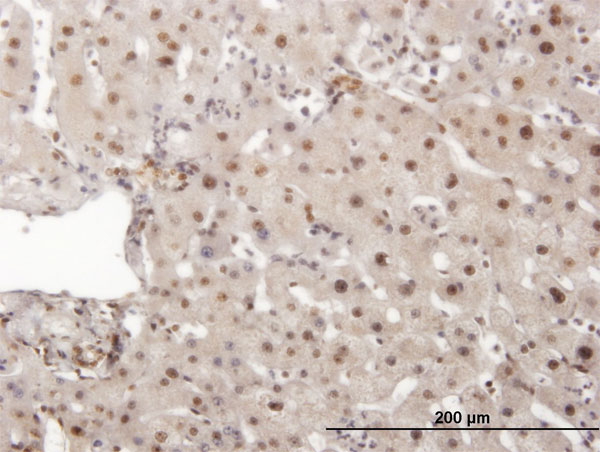
IHC (Immunohistchemistry) (PINK1 Monoclonal Antibody immunohistochemistry analysis in formalin fixed and paraffin embedded human kidney tissue followed by peroxidase conjugation of the secondary antibody and DAB staining. This data demonstrates the use of the PINK1 Monoclonal Antibody for immunohistochemistry. Clinical relevance has not been evaluated.)
WB (Western Blot) (Western blot analysis of PINK (arrow) using mouse monoclonal PINK antibody. 293 cell lysates (2 ug/lane) either nontransfected (Lane 1) or transiently transfected with the PINK gene (Lane 2) (Origene Technologies))
WB (Western Blot) (Western blot analysis of lysates from A431 cell line, mouse brain tissue lysate(from left to right), using Pink1 Antibody(115-213). AAA28638 was diluted at 1:1000 at each lane. A goat anti-rabbit IgG H&L(HRP) at 1:10000 dilution was used as the secondary antibody. Lysates at 20ug per lane.)
IF (Immunofluorescence) (Fluorescent image of PC12 cells stained with Pink1(115-213) Antibody. AAA28638 was diluted at 1:25 dilution. An Alexa Fluor 488-conjugated goat anti-mouse lgG at 1:400 dilution was used as the secondary antibody (green). Cytoplasmic actin was counterstained with Alexa Fluor 555 conjugated with Phalloidin (red).)
IHC (Immunohistochemistry) (Immunohistochemical analysis of paraffin-embedded H. heart section using Pink1(115-213). AAA28638 was diluted at 1:25 dilution. A undiluted biotinylated goat polyvalent antibody was used as the secondary, followed by DAB staining.)
IHC (Immunohistochemistry) (Immunohistochemical analysis of paraffin-embedded H. stomach section using Pink1(115-213). AAA28638 was diluted at 1:25 dilution. A undiluted biotinylated goat polyvalent antibody was used as the secondary, followed by DAB staining.)
WB (Western Blot) (NEK10 monoclonal antibody, Western Blot analysis of NEK10 expression in HeLa.)
WB (Western Blot) (Western Blot detection against Immunogen (36.01kD).)
Application Data (Detection limit for recombinant GST tagged NEK10 is ~0.03ng/ml as a capture antibody.)
IF (Immunofluorescence) (Immunofluorescence of monoclonal antibody to NEK10 on HeLa cell. [antibody concentration 10ug/ml].)
WB (Western Blot) (Western Blot analysis of NEK10 expression in transfected 293T cell line by NEK10 monoclonal antibody. Lane 1: NEK10 transfected lysate (53.1kD). Lane 2: Non-transfected lysate.)
WB (Western Blot) (NEK10 monoclonal antibody. Western Blot analysis of NEK10 expression in NIH/3T3.)
WB (Western Blot) (NEK10 monoclonal antibody. Western Blot analysis of NEK10 expression in Raw 264.7.)
IF (Immunofluorescence) (Immunofluorescence of HeLa cells using 126766 (10ug/ml).)
IHC (Immunohistochemistry) (Immunohistochemistry of formalin-fixed paraffin-embedded human tonsil using 1267661 (3ug/ml) and Immunoperoxidase.)
IP (Immunoprecipitation) (Immunoprecipitation of FGF1 transfected lysate using 126766 and Protein A Magnetic Bead and immunoblotted with FGF1 rabbit polyclonal antibody.)
WB (Western Blot) (Western Blot analysis of FGF1 expression in transfected 293T cell line using 126766. Lane 1: transfected lysate (17.5kD). Lane 2: Non-transfected lysate.)
Application Data (Detection limit for recombinant GST tagged FGF1 is ~10ng/ml using 126766 as a capture antibody.)
ELISA (EIA), Immunofluorescence (IF), Immunohistochemistry (IHC) Paraffin, Immunoprecipitation (IP), Western Blot (WB)
Purity
Purified by Protein A Affinity Chromatography.
Pricing
What are Monoclonal Antibodies?
Monoclonal antibodies are specialized laboratory-produced proteins developed for binding to specific biological antigens or other molecular targets. Since they come from a single cell (or clone), they are especially consistent and accurate in the data they are involved in producing.
This type of antibody material has been shown to be a powerful tool in finding and subsequently destroying harmful cells in an organism, such as those found in cancers or various autoimmune diseases. This makes them excellent aids in medical testing and research, which is why they are so widely used.
AAA Biotech offers a comprehensive range of high-quality monoclonal antibodies that perform effectively in various laboratory tests, including (amongst others) ELISA, western blotting, immunohistochemistry, and flow cytometry. All of the products in our catalog are thoroughly quality tested to make sure that they are reliable and will consistently perform well in your research.
What Are The Uses of Monoclonal Antibodies
Monoclonal antibodies are used in many lab tests, including (amongst others) ELISA, western blotting, immunohistochemistry, and flow cytometry.
ELISA is a test that helps detect a specific substance/analyte in a sample. It uses antibodies (often monoclonal) bound to a solid surface (such as the well of a microplate) to “capture” the substance/analyte in the sample and immobilize it so that the detection antibody component can then bind to it and produce a signal, which can then be measured.
Western blotting identifies specific proteins in a sample. The sample is first separated on a gel, and then antibodies are applied that will typically bind to the target, which will all be localized to a single band in a lane.
Immunohistochemistry helps locate specific proteins in cells or tissue samples using antibodies.
Flow cytometry looks at and sorts cells. It uses antibodies that are conjugated to reporter molecules called “fluorophores”, which, under special lights, emit light themselves, which can then be measured by a detector instrument.
How Monoclonal Antibodies Are Used as Medicine?
Please note that all of the products listed in AAA Biotech’s catalog are strictly for research-use only (RUO).
Monoclonal antibodies can also be used as therapeutic/medical treatments, particularly in the context of cancers. They are designed to find and bind to specific cells or proteins, helping the immune system recognize and attack the cancer. These treatments work in different ways, such as:
Radioimmunotherapy attaches a small amount of radioactive molecule to the antibody, so it delivers the radiation directly to the cancer cells that the antibody is specifically binding to.
Antibody-directed enzyme prodrug therapy uses antibodies that are specifically bound to special enzymes. These enzymes activate a harmless drug in the body and turn it into a cancer-killing drug only near the cancer cells—this helps avoid harming healthy cells.
Immunoliposomes are tiny “bubbles” filled with medicine/drug and coated with antibodies. They carry the drug straight to the cancer cells.
Why Buy Monoclonal Antibodies From Us?
At AAA Biotech, we provide high-performance monoclonal antibodies designed to support a wide range of research needs.
1. Validated for Versatile Applications
The antibodies in our catalog are extensively validated and compatible with multiple techniques, including (but not limited to) ELISA, flow cytometry (FC), immunocytochemistry (ICC), immunofluorescence (IF), immunohistochemistry (IHC), immunoprecipitation (IP), and western blotting (WB).
2. Wide Selection & Specialized Options
We offer antibodies for common and rare species, that are available in various conjugated forms, and also in recombinant formats. Essentially, there is almost anything one might need to meet their experimental model’s requirements.
3. High-Quality Proteins
Our proteins meet high purity standards—90% or more as confirmed by SDS-PAGE. Many are available with tags like His, Flag, GST, or MBP, and we also supply native and biologically active proteins for functional studies.
Frequently Asked Questions
1. Are your monoclonal antibodies validated for specific applications?
Yes, our antibodies are tested and validated for use in methods such as ELISA, western blot, IHC, flow cytometry, and more. Refer to specific product pages or datasheets for individual product information.
2. How do I choose the right monoclonal antibody for my application?
Review the product details directly for application validation, species reactivity, and target information. You may also contact our support team at any time for help.
3. How quickly can I receive my order?
Most orders are processed and shipped within 1–3 business days, depending on product availability and your shipping location.
Submit a Question
Please complete the form below and a representative will contact you as soon as possible.
Request more Information
Please complete the form below and a representative will contact you as soon as possible.
Request a Manual
Please complete the form below and a representative will contact you as soon as possible.
Request a Quote
Please complete the form below and a representative will contact you as soon as possible.