Filters
▼Clonality
▼Type
▼Reactivity
▼Gene Name
▼Isotype
▼Host
▼Application
▼Clone
▼Monoclonal Antibodies
Get accurate results in your research with our Monoclonal Antibodies, which are specially made to target exactly what you require for your research, and will produce consistent, reliable performance in lab tests.
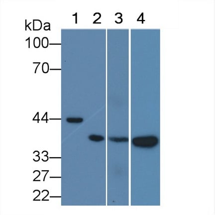
Viewing 4500-4550 of 27645 product results
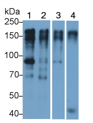
 WB (Western Blot)
(Western Blot;Sample:Lane 1: Porcine Thymus lysate;Lane 2: A431 cell lysate;Lane 3: Jurkat cell lysate;Lane 4: THP1 cell lysatePrimary Ab: 1ug/ml Mouse Anti-Porcine IL1b AntibodySecond Ab: 0.2ug/mL HRP-Linked Caprine Anti-Mouse IgG Polyclonal Antibody)
WB (Western Blot)
(Western Blot;Sample:Lane 1: Porcine Thymus lysate;Lane 2: A431 cell lysate;Lane 3: Jurkat cell lysate;Lane 4: THP1 cell lysatePrimary Ab: 1ug/ml Mouse Anti-Porcine IL1b AntibodySecond Ab: 0.2ug/mL HRP-Linked Caprine Anti-Mouse IgG Polyclonal Antibody)
Interleukin 1 Beta, Monoclonal Antibody (Cat# AAA141364)
Bone Morphogenetic Protein 10 (BMP10), Monoclonal Antibody (Cat# AAA148009)
Interferon Alpha (IFNa), Monoclonal Antibody (Cat# AAA152556)
Lipopolysaccharide (LPS), Monoclonal Antibody (Cat# AAA152557)
Programmed Cell Death Protein 1 Ligand 2 (PDL2), Monoclonal Antibody (Cat# AAA152559)
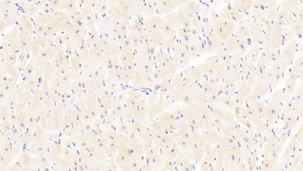
 IHC (Immunohiostchemistry)
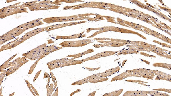
(DAB staining on IHC-P;Sample: Mouse Cardiac Muscle Tissue; Primary Ab: 20ug/ml Mouse Anti-human EPOR AntibodySecond Ab: 2ug/mL HRP-Linked Caprine Anti-Mouse IgG Polyclonal Antibody)
IHC (Immunohiostchemistry)
(DAB staining on IHC-P;Sample: Mouse Cardiac Muscle Tissue; Primary Ab: 20ug/ml Mouse Anti-human EPOR AntibodySecond Ab: 2ug/mL HRP-Linked Caprine Anti-Mouse IgG Polyclonal Antibody)
Erythropoietin Receptor (EPOR), Monoclonal Antibody (Cat# AAA152561)
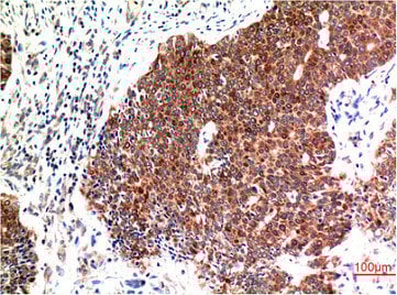
 IHC (Immunohiostchemistry)
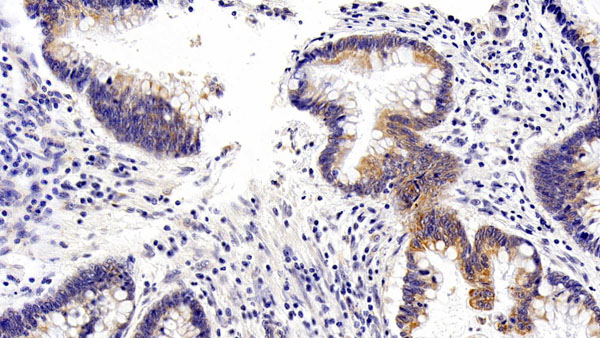
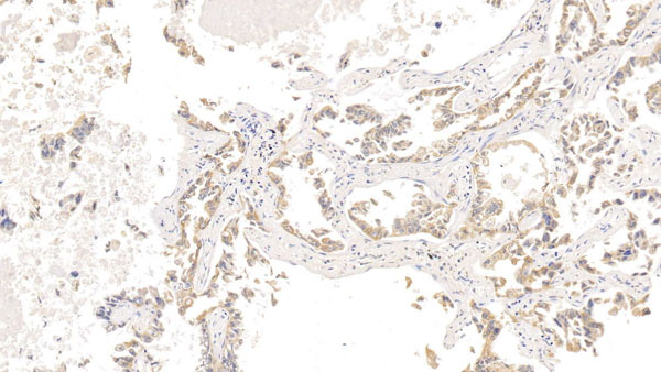
(DAB staining on IHC-P;Sample: human Lung cancer Tissue; Primary Ab: 20ug/ml Mouse Anti-human VEGFC AntibodySecond Ab: 2ug/mL HRP-Linked Caprine Anti-Mouse IgG Polyclonal Antibody)
IHC (Immunohiostchemistry)
(DAB staining on IHC-P;Sample: human Lung cancer Tissue; Primary Ab: 20ug/ml Mouse Anti-human VEGFC AntibodySecond Ab: 2ug/mL HRP-Linked Caprine Anti-Mouse IgG Polyclonal Antibody)
Vasular Endothelial Growth Factor C (VEGFC), Monoclonal Antibody (Cat# AAA152566)
 WB (Western Blot)
(Western Blot; Sample: Jurkat cell lysate Primary Ab: 0.6ug/ml Mouse Anti-human CD86 Antibody Second Ab: 0.2ug/mL HRP-Linked Caprine Anti-Mouse IgG Polyclonal Antibody)
WB (Western Blot)
(Western Blot; Sample: Jurkat cell lysate Primary Ab: 0.6ug/ml Mouse Anti-human CD86 Antibody Second Ab: 0.2ug/mL HRP-Linked Caprine Anti-Mouse IgG Polyclonal Antibody)
Cluster Of Differentiation 86 (CD86), Monoclonal Antibody (Cat# AAA152571)
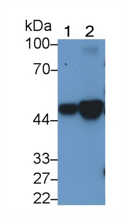 WB (Western Blot)
(Western Blot; Sample: Lane1: K562 Cell lysate; Lane2: Hela cell lysate Primary Ab: 2ug/ml Mouse Anti-human KRT18 Antibody Second Ab: 0.2ug/mL HRP-Linked Caprine Anti-Mouse IgG Polyclonal Antibody)
WB (Western Blot)
(Western Blot; Sample: Lane1: K562 Cell lysate; Lane2: Hela cell lysate Primary Ab: 2ug/ml Mouse Anti-human KRT18 Antibody Second Ab: 0.2ug/mL HRP-Linked Caprine Anti-Mouse IgG Polyclonal Antibody)
Cytokeratin 18 (CK18), Monoclonal Antibody (Cat# AAA152572)
 WB (Western Blot)
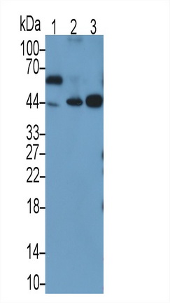
(Western Blot; Sample: Lane1: human SeuM; Lane2: human Plasma; Lane3: human Lung lysate Primary Ab: 0.2ug/ml Mouse Anti-human a1AT Antibody Second Ab: 0.2ug/mL HRP-Linked Caprine Anti-Mouse IgG Polyclonal Antibody)
WB (Western Blot)
(Western Blot; Sample: Lane1: human SeuM; Lane2: human Plasma; Lane3: human Lung lysate Primary Ab: 0.2ug/ml Mouse Anti-human a1AT Antibody Second Ab: 0.2ug/mL HRP-Linked Caprine Anti-Mouse IgG Polyclonal Antibody)
Alpha-1-Antitrypsin (a1AT), Monoclonal Antibody (Cat# AAA152576)
 WB (Western Blot)
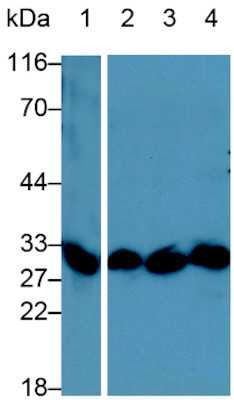
(Western Blot; Sample: Rat Liver lysate Primary Ab: 0.2ug/ml Mouse Anti-Rat COMT Antibody Second Ab: 0.2ug/mL HRP-Linked Caprine Anti-Mouse IgG Polyclonal Antibody)
WB (Western Blot)
(Western Blot; Sample: Rat Liver lysate Primary Ab: 0.2ug/ml Mouse Anti-Rat COMT Antibody Second Ab: 0.2ug/mL HRP-Linked Caprine Anti-Mouse IgG Polyclonal Antibody)
Catechol-O-Methyltransferase (COMT), Monoclonal Antibody (Cat# AAA152585)
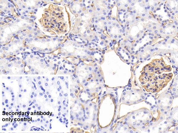 IHC (Immunohistochemistry)
(DAB staining on IHC-P;Sample: Rat Kidney TissuePrimary Ab: 20ug/ml Mouse Anti-Rat PODXL AntibodyControl: Used PBS instead of primary antibodySecond Ab: 2ug/ml HRP-Linked Caprine Anti-Mouse IgG Polyclonal Antibody)
IHC (Immunohistochemistry)
(DAB staining on IHC-P;Sample: Rat Kidney TissuePrimary Ab: 20ug/ml Mouse Anti-Rat PODXL AntibodyControl: Used PBS instead of primary antibodySecond Ab: 2ug/ml HRP-Linked Caprine Anti-Mouse IgG Polyclonal Antibody)
Podocalyxin (PODXL), Monoclonal Antibody (Cat# AAA152588)
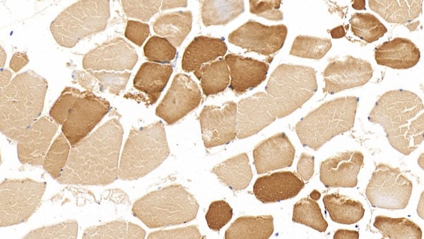
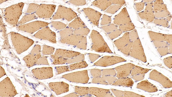
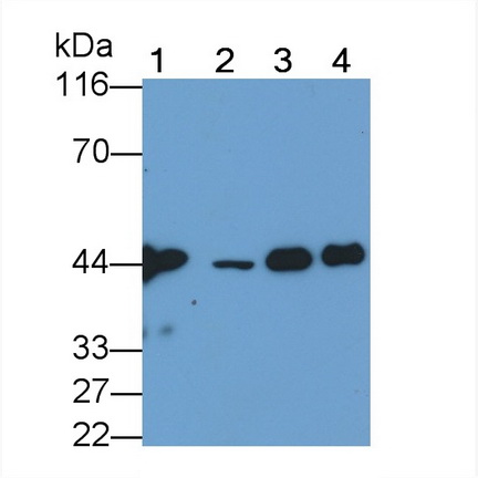
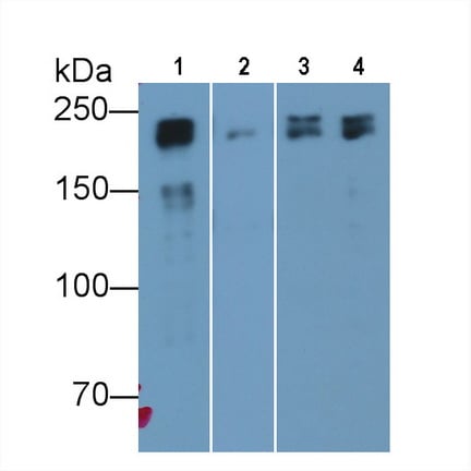 WB (Western Blot)
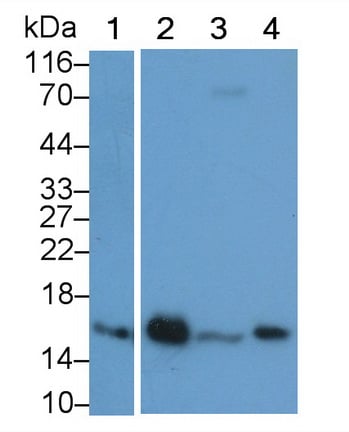
(Western Blot; Sample: Lane1: Porcine Skeletal muscle lysate; Lane2: Porcine Esophagus lysate; Lane3: Rat Skeletal muscle lysate; Lane4: Mouse Skeletal muscle lysate Primary Ab: 0.01ug/ml Mouse Anti-human MYH8 Antibody Second Ab: 0.2ug/mL HRP-Linked Caprine Anti-Mouse IgG Polyclonal Antibody)
WB (Western Blot)
(Western Blot; Sample: Lane1: Porcine Skeletal muscle lysate; Lane2: Porcine Esophagus lysate; Lane3: Rat Skeletal muscle lysate; Lane4: Mouse Skeletal muscle lysate Primary Ab: 0.01ug/ml Mouse Anti-human MYH8 Antibody Second Ab: 0.2ug/mL HRP-Linked Caprine Anti-Mouse IgG Polyclonal Antibody)
Myosin Heavy Chain 8, Skeletal Muscle, Perinatal (MYH8), Monoclonal Antibody (Cat# AAA152592)
 WB (Western Blot)
(Western Blot; Sample: Lane1: human SeuM; Lane2: human Plasma Primary Ab: 0.1ug/ml Mouse Anti-human F2 Antibody Second Ab: 0.2ug/mL HRP-Linked Caprine Anti-Mouse IgG Polyclonal Antibody)
WB (Western Blot)
(Western Blot; Sample: Lane1: human SeuM; Lane2: human Plasma Primary Ab: 0.1ug/ml Mouse Anti-human F2 Antibody Second Ab: 0.2ug/mL HRP-Linked Caprine Anti-Mouse IgG Polyclonal Antibody)
Coaulation Factor II (F2), Monoclonal Antibody (Cat# AAA152598)
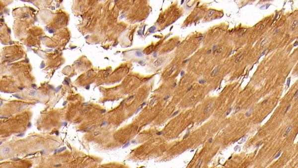
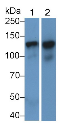
 WB (Western Blot)
(Western Blot; Sample: Lane1: Rat Heart lysate; Lane2: Rat Skeletal muscle lysate Primary Ab: 1ug/ml Mouse Anti-human MYO Antibody Second Ab: 0.2ug/mL HRP-Linked Caprine Anti-Mouse IgG Polyclonal Antibody)
WB (Western Blot)
(Western Blot; Sample: Lane1: Rat Heart lysate; Lane2: Rat Skeletal muscle lysate Primary Ab: 1ug/ml Mouse Anti-human MYO Antibody Second Ab: 0.2ug/mL HRP-Linked Caprine Anti-Mouse IgG Polyclonal Antibody)
Myoglobin (MYO), Monoclonal Antibody (Cat# AAA152600)
 WB (Western Blot)
(Western Blot; Sample: Lane1: Porcine Lymph node lysate; Lane2: Porcine Heart lysate; Lane3: Bovine Lymph node lysatePrimary Ab: 0.2ug/ml Mouse Anti-human FABP4 AntibodySecond Ab: 0.2ug/mL HRP-Linked Caprine Anti-Mouse IgG Polyclonal Antibody)
WB (Western Blot)
(Western Blot; Sample: Lane1: Porcine Lymph node lysate; Lane2: Porcine Heart lysate; Lane3: Bovine Lymph node lysatePrimary Ab: 0.2ug/ml Mouse Anti-human FABP4 AntibodySecond Ab: 0.2ug/mL HRP-Linked Caprine Anti-Mouse IgG Polyclonal Antibody)
Fatty Acid Binding Protein 4 (FABP4), Monoclonal Antibody (Cat# AAA152602)
 WB (Western Blot)
(Western Blot; Sample: Lane1: Hela cell lysate; Lane2: MCF7 cell lysate; Lane3: A431 cell lysate; Lane4: human Placenta lysate Primary Ab: 0.02ug/ml Mouse Anti-human EGFR Antibody Second Ab: 0.2ug/mL HRP-Linked Caprine Anti-Mouse IgG Polyclonal Antibody)
WB (Western Blot)
(Western Blot; Sample: Lane1: Hela cell lysate; Lane2: MCF7 cell lysate; Lane3: A431 cell lysate; Lane4: human Placenta lysate Primary Ab: 0.02ug/ml Mouse Anti-human EGFR Antibody Second Ab: 0.2ug/mL HRP-Linked Caprine Anti-Mouse IgG Polyclonal Antibody)
Epidermal Growth Factor Receptor (EGFR), Monoclonal Antibody (Cat# AAA152604)
 WB (Western Blot)
(Western Blot; Sample: HepG2 cell lysate Primary Ab: 5ug/ml Mouse Anti-Rat APOB Antibody Second Ab: 0.2ug/mL HRP-Linked Caprine Anti-Mouse IgG Polyclonal Antibody)
WB (Western Blot)
(Western Blot; Sample: HepG2 cell lysate Primary Ab: 5ug/ml Mouse Anti-Rat APOB Antibody Second Ab: 0.2ug/mL HRP-Linked Caprine Anti-Mouse IgG Polyclonal Antibody)
Apolipoprotein B (APOB), Monoclonal Antibody (Cat# AAA152613)
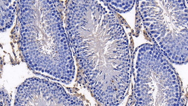
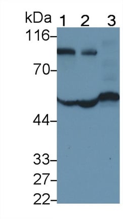
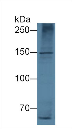
 WB (Western Blot)
(Western Blot; Sample: Lane1: human Lung lysate; Lane2: Rat Testis lysate; Lane3: Hela cell lysate Primary Ab: 2 ug/ml Mouse Anti-human DMD Antibody Second Ab: 0.2ug/mL HRP-Linked Caprine Anti-Mouse IgG Polyclonal Antibody)
WB (Western Blot)
(Western Blot; Sample: Lane1: human Lung lysate; Lane2: Rat Testis lysate; Lane3: Hela cell lysate Primary Ab: 2 ug/ml Mouse Anti-human DMD Antibody Second Ab: 0.2ug/mL HRP-Linked Caprine Anti-Mouse IgG Polyclonal Antibody)
Dystrophin (DMD), Monoclonal Antibody (Cat# AAA152617)
 WB (Western Blot)
(Western Blot; Sample: Rat Heart lysate Primary Ab: 2ug/ml Mouse Anti-human RNLS Antibody Second Ab: 0.2ug/mL HRP-Linked Caprine Anti-Mouse IgG Polyclonal Antibody)
WB (Western Blot)
(Western Blot; Sample: Rat Heart lysate Primary Ab: 2ug/ml Mouse Anti-human RNLS Antibody Second Ab: 0.2ug/mL HRP-Linked Caprine Anti-Mouse IgG Polyclonal Antibody)
Renalase (RNLS), Monoclonal Antibody (Cat# AAA152619)
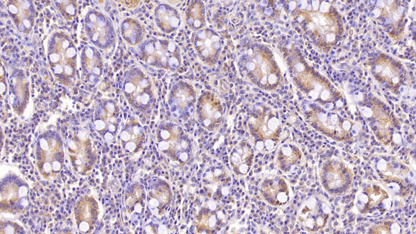
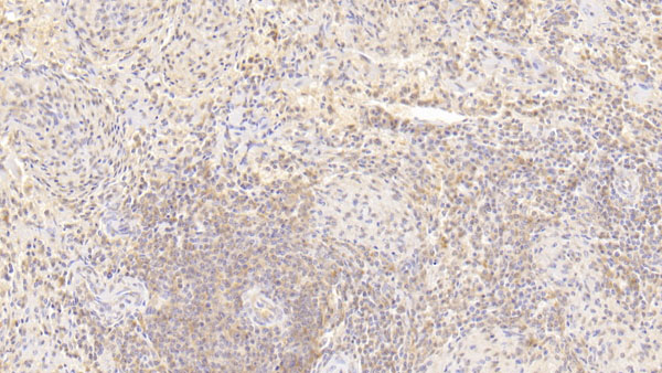
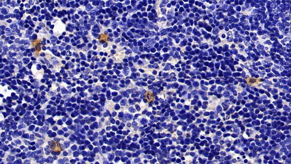
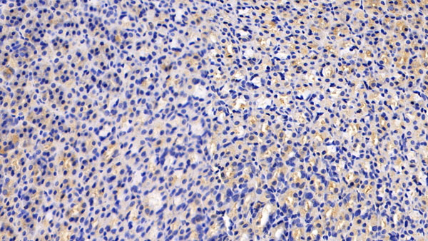
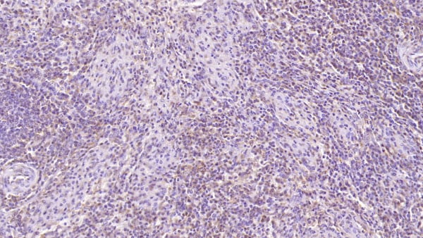 IHC (Immunohiostchemistry)
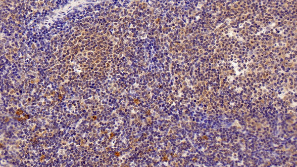
(DAB staining on IHC-P;Sample: human Spleen Tissue; Primary Ab: 40ug/ml Mouse Anti-human CD97 AntibodySecond Ab: 2ug/mL HRP-Linked Caprine Anti-Mouse IgG Polyclonal Antibody)
IHC (Immunohiostchemistry)
(DAB staining on IHC-P;Sample: human Spleen Tissue; Primary Ab: 40ug/ml Mouse Anti-human CD97 AntibodySecond Ab: 2ug/mL HRP-Linked Caprine Anti-Mouse IgG Polyclonal Antibody)
Cluster Of Differentiation 97 (CD97), Monoclonal Antibody (Cat# AAA152699)
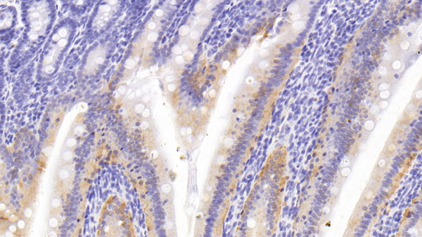
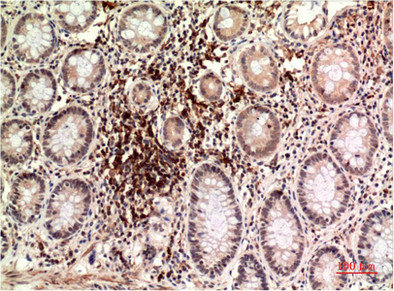
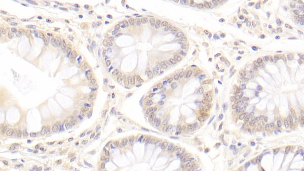 IHC (Immunohiostchemistry)
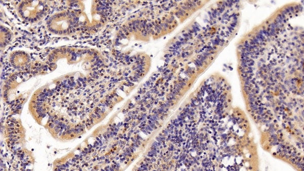
(DAB staining on IHC-P;Sample: human Colon Tissue; Primary Ab: 20ug/ml Mouse Anti-human SCNN1g AntibodySecond Ab: 2ug/mL HRP-Linked Caprine Anti-Mouse IgG Polyclonal Antibody)
IHC (Immunohiostchemistry)
(DAB staining on IHC-P;Sample: human Colon Tissue; Primary Ab: 20ug/ml Mouse Anti-human SCNN1g AntibodySecond Ab: 2ug/mL HRP-Linked Caprine Anti-Mouse IgG Polyclonal Antibody)
Amiloride Sensitive SoduM Channel Subunit Gamma (SCNN1g), Monoclonal Antibody (Cat# AAA152700)
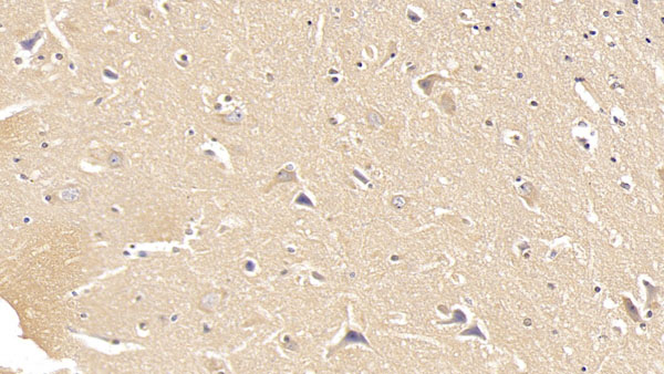
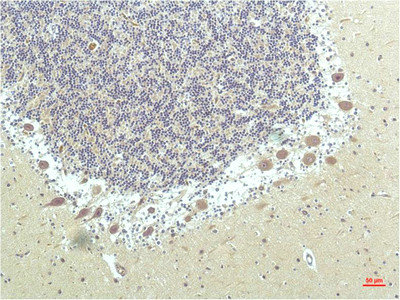
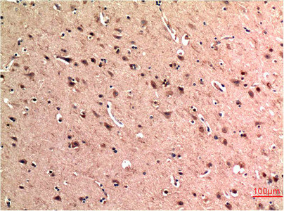
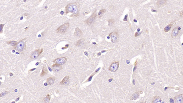 IHC (Immunohistochemistry)
(DAB staining on IHC-P;Sample: human CerebuM Tissue; Primary Ab: 40ug/ml Mouse Anti-human ASPH AntibodySecond Ab: 2ug/mL HRP-Linked Caprine Anti-Mouse IgG Polyclonal Antibody)
IHC (Immunohistochemistry)
(DAB staining on IHC-P;Sample: human CerebuM Tissue; Primary Ab: 40ug/ml Mouse Anti-human ASPH AntibodySecond Ab: 2ug/mL HRP-Linked Caprine Anti-Mouse IgG Polyclonal Antibody)
Aspartate Beta Hydroxylase (ASPH), Monoclonal Antibody (Cat# AAA152702)
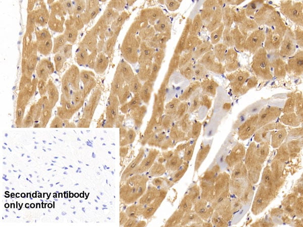
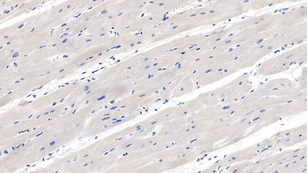
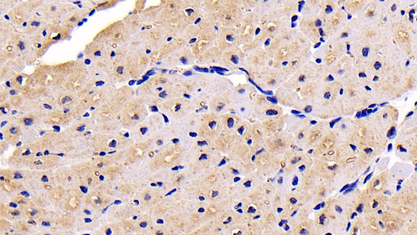 IHC (Immunohistochemistry)
(DAB staining on IHC-P;Sample: human Cardiac Muscle Tissue; Primary Ab: 30ug/ml Mouse Anti-human NRP1 AntibodySecond Ab: 2ug/mL HRP-Linked Caprine Anti-Mouse IgG Polyclonal Antibody)
IHC (Immunohistochemistry)
(DAB staining on IHC-P;Sample: human Cardiac Muscle Tissue; Primary Ab: 30ug/ml Mouse Anti-human NRP1 AntibodySecond Ab: 2ug/mL HRP-Linked Caprine Anti-Mouse IgG Polyclonal Antibody)
Neuropilin 1 (NRP1), Monoclonal Antibody (Cat# AAA152705)
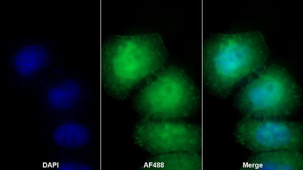
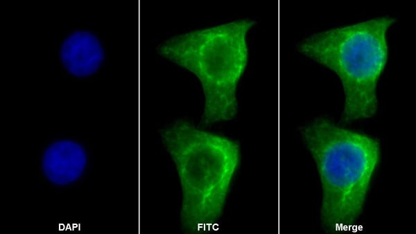
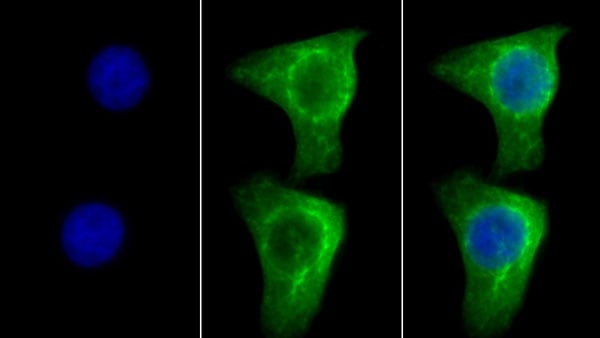
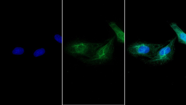
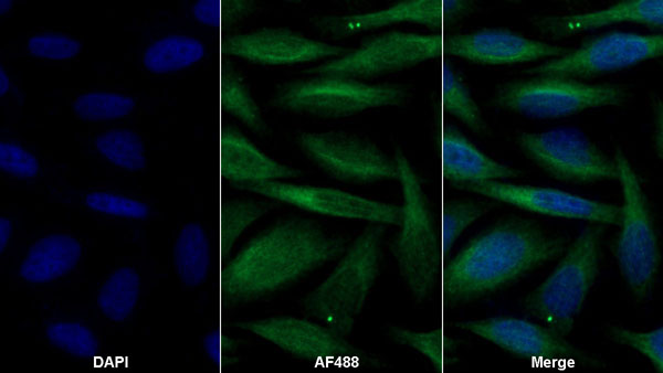
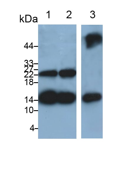
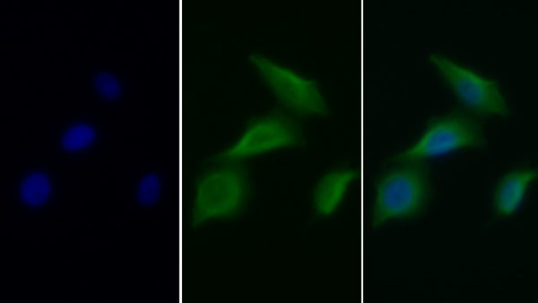
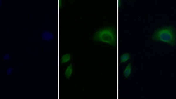
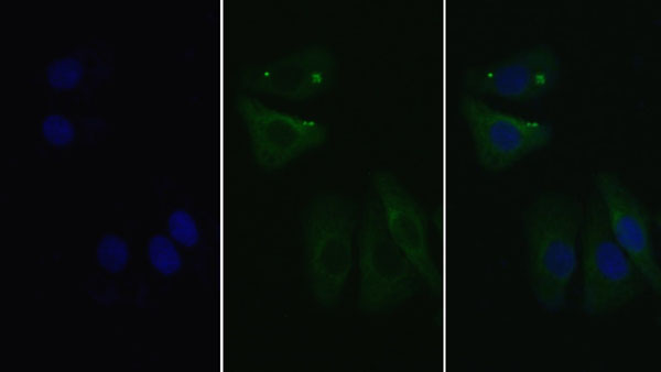
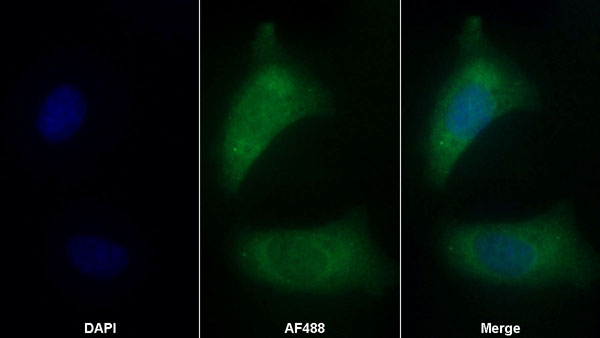
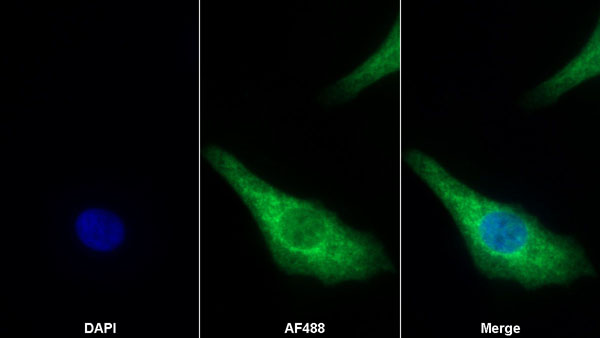
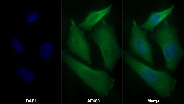
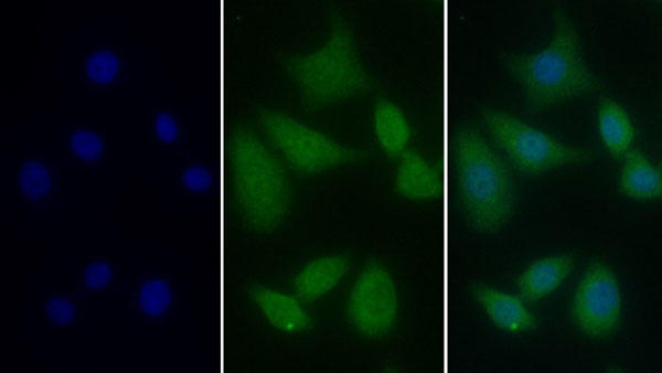
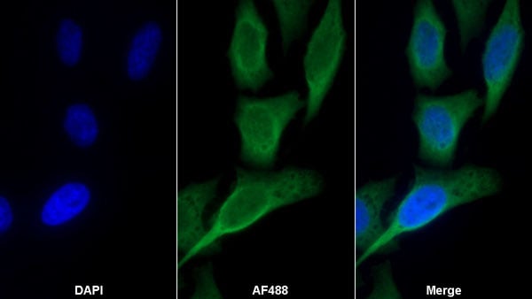 IF (Immunofluorescence)
(AF488 staining on IF;Sample: HepG2 cellPrimary Ab: 30ug/ml Mouse Anti-human TIMP1 AntibodySecond Ab: 2 ug/ml AF488-Linked Caprine Anti-Mouse IgG Polyclonal Antibody)
IF (Immunofluorescence)
(AF488 staining on IF;Sample: HepG2 cellPrimary Ab: 30ug/ml Mouse Anti-human TIMP1 AntibodySecond Ab: 2 ug/ml AF488-Linked Caprine Anti-Mouse IgG Polyclonal Antibody)
Tissue Inhibitors Of Metalloproteinase 1 (TIMP1), Monoclonal Antibody (Cat# AAA152706)
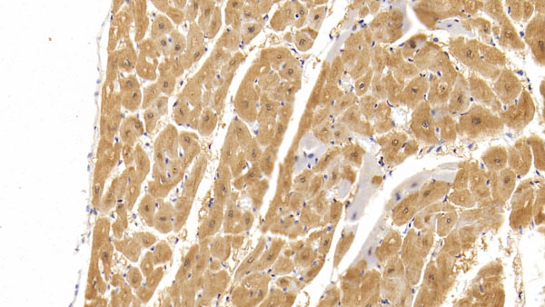
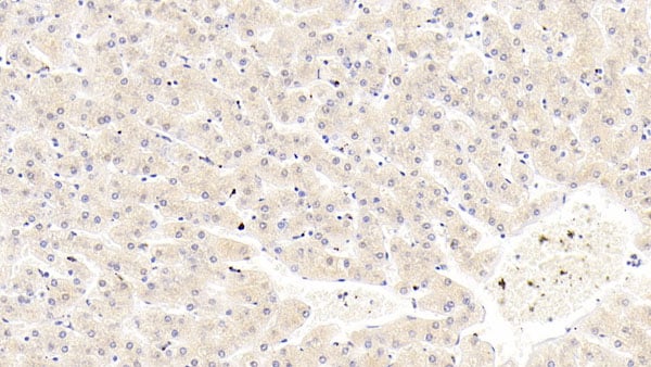
 IHC (Immunohistochemisry)
(DAB staining on IHC-P;Sample: human Cardiac Muscle Tissue; Primary Ab: 20ug/ml Mouse Anti-human IL18 AntibodySecond Ab: 2ug/mL HRP-Linked Caprine Anti-Mouse IgG Polyclonal Antibody)
IHC (Immunohistochemisry)
(DAB staining on IHC-P;Sample: human Cardiac Muscle Tissue; Primary Ab: 20ug/ml Mouse Anti-human IL18 AntibodySecond Ab: 2ug/mL HRP-Linked Caprine Anti-Mouse IgG Polyclonal Antibody)
Interleukin 18 (IL18), Monoclonal Antibody (Cat# AAA152707)

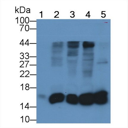
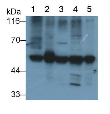
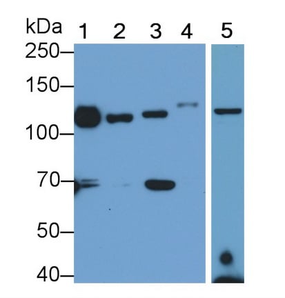 WB (Western Blot)
(Western Blot; Sample: Lane1: Rat CerebuM lysate; Lane2: Rat Liver lysate; Lane3: Rat Testis lysate; Lane4: Porcine CerebuM lysate; Lane5: human Placenta lysatePrimary Ab: 0.2ug/ml Mouse Anti-human ITGaV AntibodySecond Ab: 0.2ug/mL HRP-Linked Caprine Anti-Mouse IgG Polyclonal Antibody)
WB (Western Blot)
(Western Blot; Sample: Lane1: Rat CerebuM lysate; Lane2: Rat Liver lysate; Lane3: Rat Testis lysate; Lane4: Porcine CerebuM lysate; Lane5: human Placenta lysatePrimary Ab: 0.2ug/ml Mouse Anti-human ITGaV AntibodySecond Ab: 0.2ug/mL HRP-Linked Caprine Anti-Mouse IgG Polyclonal Antibody)
Integrin Alpha V (ITGaV), Monoclonal Antibody (Cat# AAA152722)
 WB (Western Blot)
(Western Blot; Sample: Rat Lung lysate Primary Ab: 0.1ug/ml Mouse Anti-Mouse GZMK Antibody Second Ab: 0.2ug/mL HRP-Linked Caprine Anti-Mouse IgG Polyclonal Antibody)
WB (Western Blot)
(Western Blot; Sample: Rat Lung lysate Primary Ab: 0.1ug/ml Mouse Anti-Mouse GZMK Antibody Second Ab: 0.2ug/mL HRP-Linked Caprine Anti-Mouse IgG Polyclonal Antibody)
Granzyme K (GZMK), Monoclonal Antibody (Cat# AAA152731)
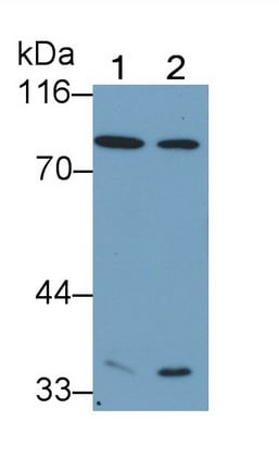
 WB (Western Blot)
(Western Blot; Sample: Lane1: human SeuM; Lane2: Hela cell lysate; Lane3: Rat Heart lysate; Lane4: Rat Skeletal muscle lysate Primary Ab: 0.1ug/ml Mouse Anti-human CKM Antibody Second Ab: 0.2ug/mL HRP-Linked Caprine Anti-Mouse IgG Polyclonal Antibody)
WB (Western Blot)
(Western Blot; Sample: Lane1: human SeuM; Lane2: Hela cell lysate; Lane3: Rat Heart lysate; Lane4: Rat Skeletal muscle lysate Primary Ab: 0.1ug/ml Mouse Anti-human CKM Antibody Second Ab: 0.2ug/mL HRP-Linked Caprine Anti-Mouse IgG Polyclonal Antibody)
Creatine Kinase, Muscle (CKM), Monoclonal Antibody (Cat# AAA152735)
 WB (Western Blot)
(Western Blot; Sample: Lane1: Rat Placenta lysate; Lane2: Rat Lung lysatePrimary Ab: 0.2ug/ml Mouse Anti-Rat CDH5 AntibodySecond Ab: 0.2ug/mL HRP-Linked Caprine Anti-Mouse IgG Polyclonal Antibody)
WB (Western Blot)
(Western Blot; Sample: Lane1: Rat Placenta lysate; Lane2: Rat Lung lysatePrimary Ab: 0.2ug/ml Mouse Anti-Rat CDH5 AntibodySecond Ab: 0.2ug/mL HRP-Linked Caprine Anti-Mouse IgG Polyclonal Antibody)
Cadherin 5 (CDH5), Monoclonal Antibody (Cat# AAA152742)
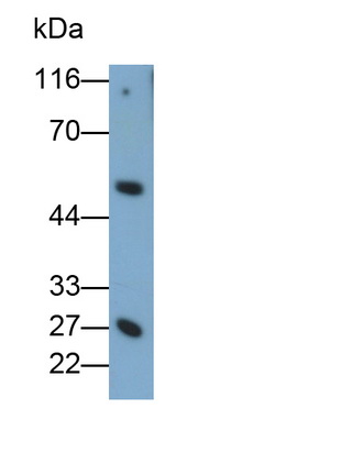
 WB (Western Blot)
(Western Blot; Sample: human SeuM Primary Ab: 0.2ug/ml Mouse Anti-human C1s Antibody Second Ab: 0.2ug/mL HRP-Linked Caprine Anti-Mouse IgG Polyclonal Antibody)
WB (Western Blot)
(Western Blot; Sample: human SeuM Primary Ab: 0.2ug/ml Mouse Anti-human C1s Antibody Second Ab: 0.2ug/mL HRP-Linked Caprine Anti-Mouse IgG Polyclonal Antibody)
Complement Component 1, S Subcomponent (C1s), Monoclonal Antibody (Cat# AAA152743)
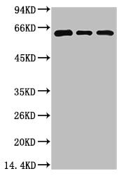
 WB (Western Blot)
(Western Blot; Sample: Lane1: Porcine Kidney lysate; Lane2: Bovine Kidney lysate; Lane3: Caprine Kidney lysatePrimary Ab: 1ug/ml Mouse Anti-human TBP AntibodySecond Ab: 0.2ug/mL HRP-Linked Caprine Anti-Mouse IgG Polyclonal Antibody)
WB (Western Blot)
(Western Blot; Sample: Lane1: Porcine Kidney lysate; Lane2: Bovine Kidney lysate; Lane3: Caprine Kidney lysatePrimary Ab: 1ug/ml Mouse Anti-human TBP AntibodySecond Ab: 0.2ug/mL HRP-Linked Caprine Anti-Mouse IgG Polyclonal Antibody)
TATA Binding Protein (TBP), Monoclonal Antibody (Cat# AAA152752)
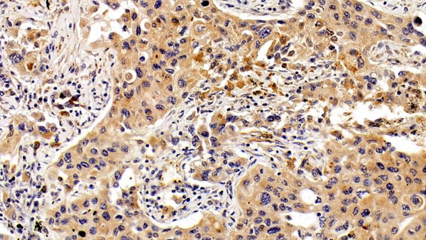
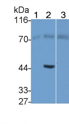
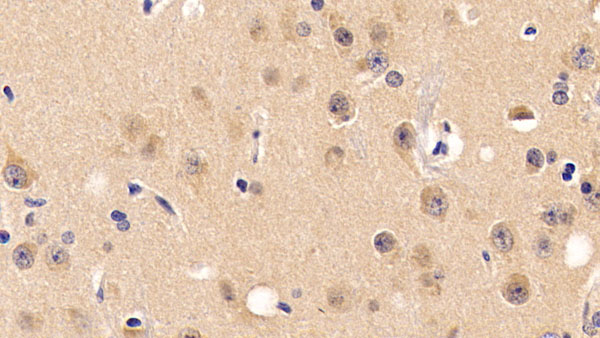
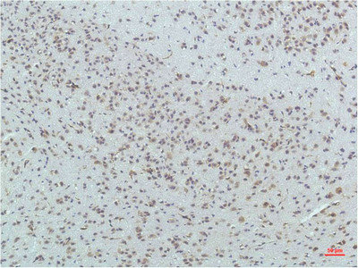
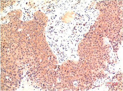
 IHC (Immunohistochemistry)
(DAB staining on IHC-P;Sample: human CerebeluM Tissue; Primary Ab: 20ug/ml Mouse Anti-human TGFb1 AntibodySecond Ab: 2ug/mL HRP-Linked Caprine Anti-Mouse IgG Polyclonal Antibody)
IHC (Immunohistochemistry)
(DAB staining on IHC-P;Sample: human CerebeluM Tissue; Primary Ab: 20ug/ml Mouse Anti-human TGFb1 AntibodySecond Ab: 2ug/mL HRP-Linked Caprine Anti-Mouse IgG Polyclonal Antibody)
Transforming Growth Factor Beta 1 (TGFb1), Monoclonal Antibody (Cat# AAA152766)
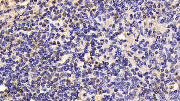 IHC (Immunohiostchemistry)
(DAB staining on IHC-P;Sample: Caprine Spleen Tissue; Primary Ab: 20ug/ml Mouse Anti-Caprine TLR4 AntibodySecond Ab: 2ug/mL HRP-Linked Caprine Anti-Mouse IgG Polyclonal Antibody)
IHC (Immunohiostchemistry)
(DAB staining on IHC-P;Sample: Caprine Spleen Tissue; Primary Ab: 20ug/ml Mouse Anti-Caprine TLR4 AntibodySecond Ab: 2ug/mL HRP-Linked Caprine Anti-Mouse IgG Polyclonal Antibody)
Toll Like Receptor 4 (TLR4), Monoclonal Antibody (Cat# AAA152771)
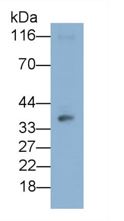
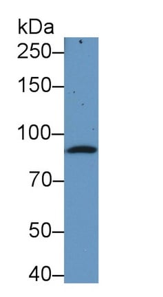
 WB (Western Blot)
(Western Blot; Sample: PC3 cell lysate Primary Ab: 0.8ug/ml Mouse Anti-human GAL8 Antibody Second Ab: 0.2ug/mL HRP-Linked Caprine Anti-Mouse IgG Polyclonal Antibody)
WB (Western Blot)
(Western Blot; Sample: PC3 cell lysate Primary Ab: 0.8ug/ml Mouse Anti-human GAL8 Antibody Second Ab: 0.2ug/mL HRP-Linked Caprine Anti-Mouse IgG Polyclonal Antibody)
Galectin 8 (GAL8), Monoclonal Antibody (Cat# AAA152779)
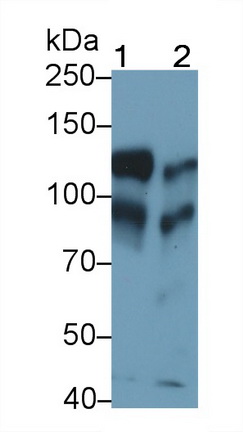
 WB (Western Blot)
(Western Blot; Sample: Rat CerebuM lysate Primary Ab: 0.8ug/ml Mouse Anti-human GAL8 Antibody Second Ab: 0.2ug/mL HRP-Linked Caprine Anti-Mouse IgG Polyclonal Antibody)
WB (Western Blot)
(Western Blot; Sample: Rat CerebuM lysate Primary Ab: 0.8ug/ml Mouse Anti-human GAL8 Antibody Second Ab: 0.2ug/mL HRP-Linked Caprine Anti-Mouse IgG Polyclonal Antibody)
Galectin 8 (GAL8), Monoclonal Antibody (Cat# AAA152786)
 WB (Western Blot)
(Western Blot; Sample: Porcine Liver lysate Primary Ab: 0.3ug/ml Mouse Anti-human F11 Antibody Second Ab: 0.2ug/mL HRP-Linked Caprine Anti-Mouse IgG Polyclonal Antibody)
WB (Western Blot)
(Western Blot; Sample: Porcine Liver lysate Primary Ab: 0.3ug/ml Mouse Anti-human F11 Antibody Second Ab: 0.2ug/mL HRP-Linked Caprine Anti-Mouse IgG Polyclonal Antibody)
Coaulation Factor XI (F11), Monoclonal Antibody (Cat# AAA152787)
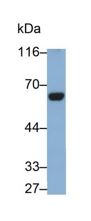
 WB (Western Blot)
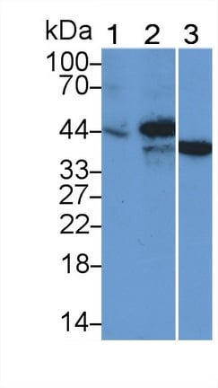
(Western Blot; Sample: Lane1: Porcine Liver lysate; Lane2: Rat Liver lysate; Lane3: HepG2 cell lysate Primary Ab: 0.2ug/ml Mouse Anti-human HPD Antibody Second Ab: 0.2ug/mL HRP-Linked Caprine Anti-Mouse IgG Polyclonal Antibody)
WB (Western Blot)
(Western Blot; Sample: Lane1: Porcine Liver lysate; Lane2: Rat Liver lysate; Lane3: HepG2 cell lysate Primary Ab: 0.2ug/ml Mouse Anti-human HPD Antibody Second Ab: 0.2ug/mL HRP-Linked Caprine Anti-Mouse IgG Polyclonal Antibody)
4-Hydroxyphenylpyruvate Dioxygenase (HPD), Monoclonal Antibody (Cat# AAA152789)
 WB (Western Blot)
(Western Blot; Sample: Lane1: human Lung lysate; Lane2: Porcine Testis lysate; Lane3: Hela cell lysate; Lane4: U2OS cell lysate; Lane5: A549 cell lysate Primary Ab: 0.3ug/ml Mouse Anti-human CCT2 Antibody Second Ab: 0.2ug/mL HRP-Linked Caprine Anti-Mouse IgG Polyclonal Antibody)
WB (Western Blot)
(Western Blot; Sample: Lane1: human Lung lysate; Lane2: Porcine Testis lysate; Lane3: Hela cell lysate; Lane4: U2OS cell lysate; Lane5: A549 cell lysate Primary Ab: 0.3ug/ml Mouse Anti-human CCT2 Antibody Second Ab: 0.2ug/mL HRP-Linked Caprine Anti-Mouse IgG Polyclonal Antibody)
Chaperonin Containing TCP1, Subunit 2 (CCT2), Monoclonal Antibody (Cat# AAA152795)
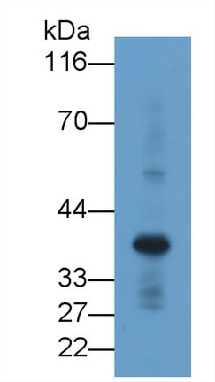
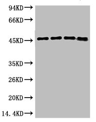
 WB (Western Blot)
(Western Blot; Sample: Lane1: Rat Heart lysate; Lane2: Rat Liver lysate; Lane3: Rat Lung lysate; Lane4: Rat CerebuM lysate Primary Ab: 0.04ug/ml Mouse Anti-Rat SOD1 Antibody Second Ab: 0.2ug/mL HRP-Linked Caprine Anti-Mouse IgG Polyclonal Antibody)
WB (Western Blot)
(Western Blot; Sample: Lane1: Rat Heart lysate; Lane2: Rat Liver lysate; Lane3: Rat Lung lysate; Lane4: Rat CerebuM lysate Primary Ab: 0.04ug/ml Mouse Anti-Rat SOD1 Antibody Second Ab: 0.2ug/mL HRP-Linked Caprine Anti-Mouse IgG Polyclonal Antibody)
Superoxide Dismutase 1 (SOD1), Monoclonal Antibody (Cat# AAA152802)
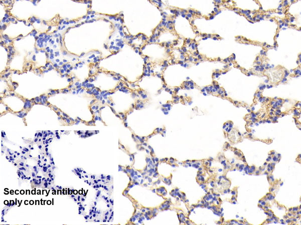
 WB (Western Blot)
(Western Blot; Sample: Lane1: Rat Liver lysate; Lane2: Hela cell lysate; Lane3: HL60 cell lysate; Lane4: HepG2 cell lysate Primary Ab: 0.2ug/ml Mouse Anti-Rat CALR Antibody Second Ab: 0.2ug/mL HRP-Linked Caprine Anti-Mouse IgG Polyclonal Antibody)
WB (Western Blot)
(Western Blot; Sample: Lane1: Rat Liver lysate; Lane2: Hela cell lysate; Lane3: HL60 cell lysate; Lane4: HepG2 cell lysate Primary Ab: 0.2ug/ml Mouse Anti-Rat CALR Antibody Second Ab: 0.2ug/mL HRP-Linked Caprine Anti-Mouse IgG Polyclonal Antibody)
Calretiulin (CALR), Monoclonal Antibody (Cat# AAA152803)
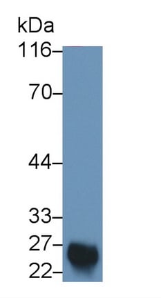
 WB (Western Blot)
(Western Blot; Sample: Porcine Heart lysate Primary Ab: 1ug/ml Mouse Anti-human PLS3 Antibody Second Ab: 0.2ug/mL HRP-Linked Caprine Anti-Mouse IgG Polyclonal Antibody)
WB (Western Blot)
(Western Blot; Sample: Porcine Heart lysate Primary Ab: 1ug/ml Mouse Anti-human PLS3 Antibody Second Ab: 0.2ug/mL HRP-Linked Caprine Anti-Mouse IgG Polyclonal Antibody)
Plastin 3 (PLS3), Monoclonal Antibody (Cat# AAA152819)
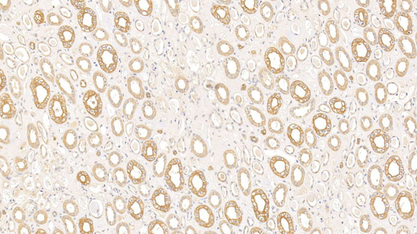
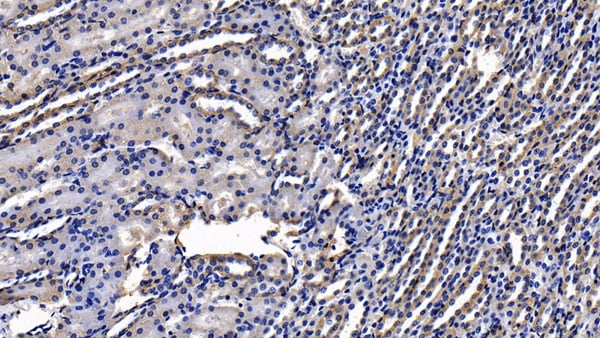
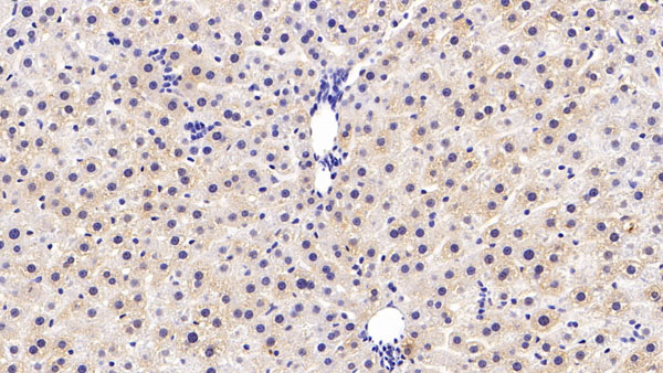
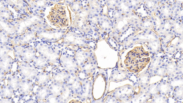
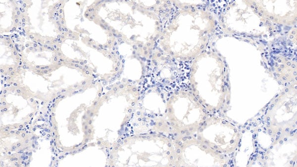
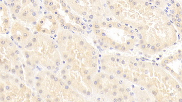
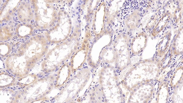
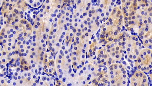
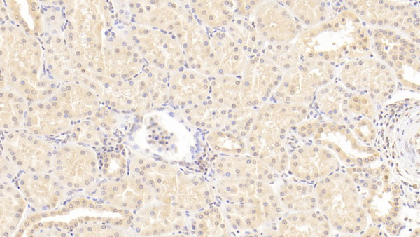
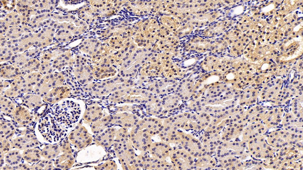
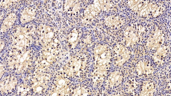
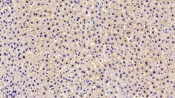
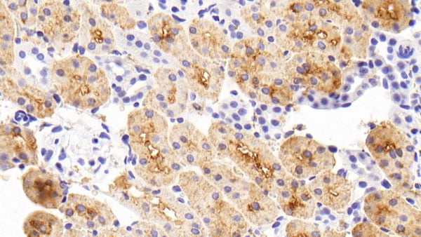 IHC (Immunohiostchemistry)
(DAB staining on IHC-P;Sample: Rat Kidney Tissue; Primary Ab: 30ug/ml Mouse Anti-Rat CYPA AntibodySecond Ab: 2ug/mL HRP-Linked Caprine Anti-Mouse IgG Polyclonal Antibody)
IHC (Immunohiostchemistry)
(DAB staining on IHC-P;Sample: Rat Kidney Tissue; Primary Ab: 30ug/ml Mouse Anti-Rat CYPA AntibodySecond Ab: 2ug/mL HRP-Linked Caprine Anti-Mouse IgG Polyclonal Antibody)
Cyclophilin A (CYPA), Monoclonal Antibody (Cat# AAA152838)
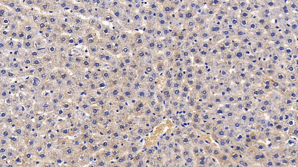
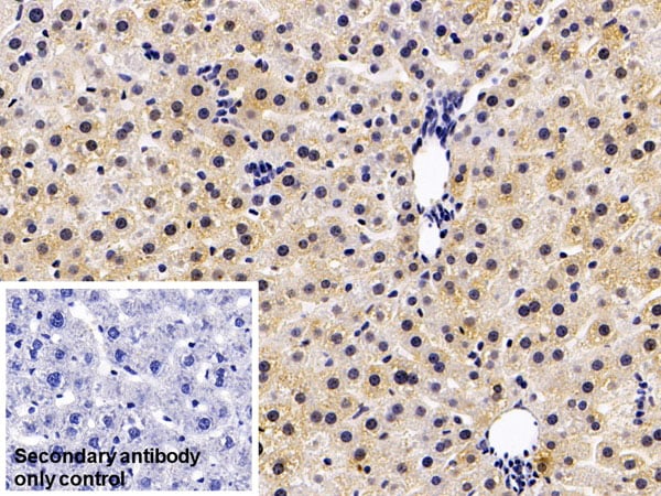
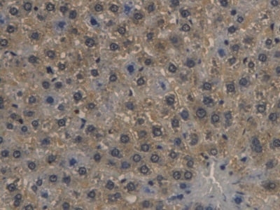
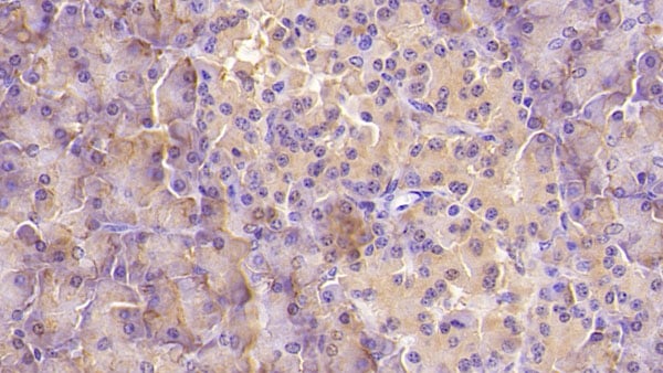
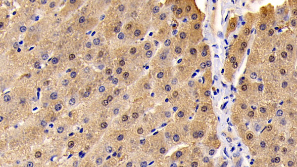
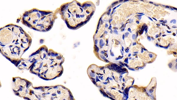
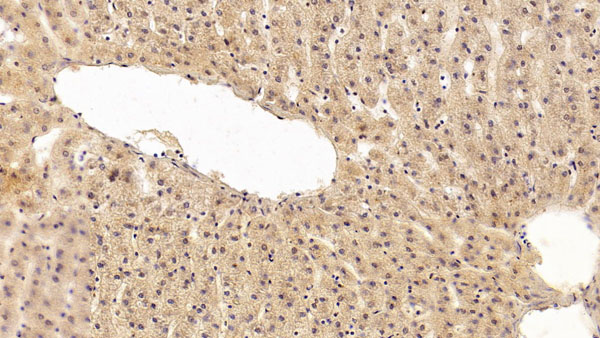
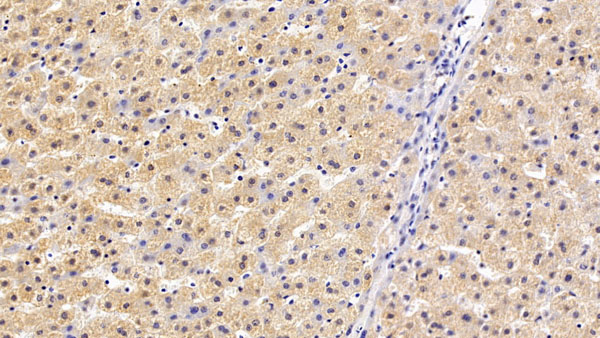
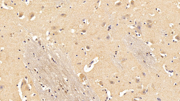
 IHC (Immunohiostchemistry)
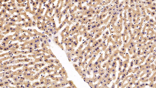
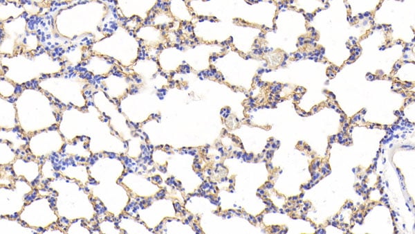
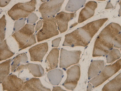
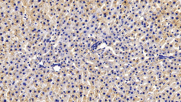
(DAB staining on IHC-P;Sample: Mouse Liver Tissue; Primary Ab: 20ug/ml Mouse Anti-human EPOR AntibodySecond Ab: 2ug/mL HRP-Linked Caprine Anti-Mouse IgG Polyclonal Antibody)
IHC (Immunohiostchemistry)
(DAB staining on IHC-P;Sample: Mouse Liver Tissue; Primary Ab: 20ug/ml Mouse Anti-human EPOR AntibodySecond Ab: 2ug/mL HRP-Linked Caprine Anti-Mouse IgG Polyclonal Antibody)
Erythropoietin Receptor (EPOR), Monoclonal Antibody (Cat# AAA152839)
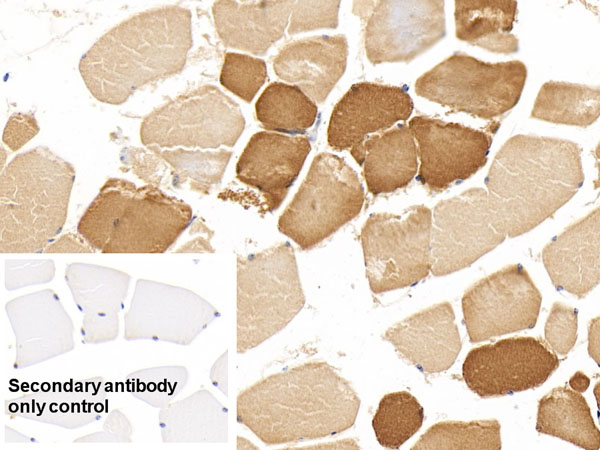
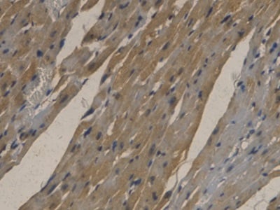
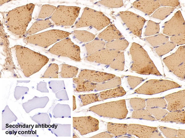
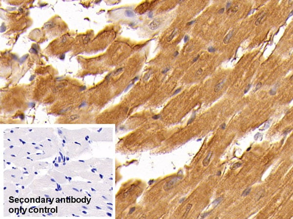
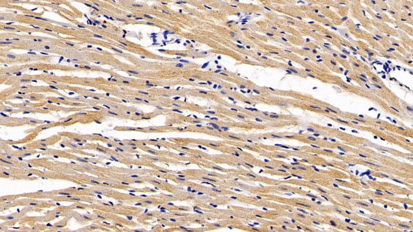
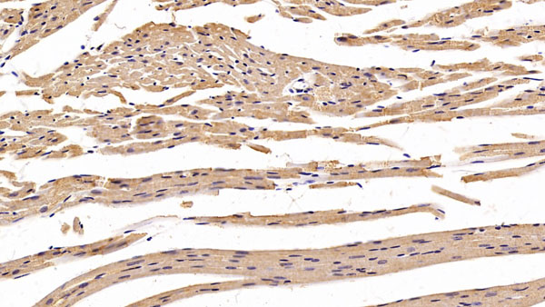
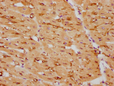 IHC (Immunohiostchemistry)
(IHC image of CSB-RA005655A0HU diluted at 1:105 and staining in paraffin-embedded human heart tissue performed on a Leica BondTM system. After dewaxing and hydration, antigen retrieval was mediated by high pressure in a citrate buffer (pH 6.0). Section was blocked with 10% normal goat serum 30min at RT. Then primary antibody (1% BSA) was incubated at 4 degree C overnight. The primary is detected by a biotinylated secondary antibody and visualized using an HRP conjugated SP system.)
IHC (Immunohiostchemistry)
(IHC image of CSB-RA005655A0HU diluted at 1:105 and staining in paraffin-embedded human heart tissue performed on a Leica BondTM system. After dewaxing and hydration, antigen retrieval was mediated by high pressure in a citrate buffer (pH 6.0). Section was blocked with 10% normal goat serum 30min at RT. Then primary antibody (1% BSA) was incubated at 4 degree C overnight. The primary is detected by a biotinylated secondary antibody and visualized using an HRP conjugated SP system.)
CNN1, Monoclonal Recombinant Antibody (Cat# AAA235533)
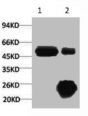
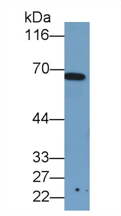
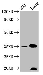
 WB (Western Blot)
(Western blot analysis of 1) Hela, 2) MCF7, 3) 293T, diluted at 1:2000.)
WB (Western Blot)
(Western blot analysis of 1) Hela, 2) MCF7, 3) 293T, diluted at 1:2000.)
KRT17, Monoclonal Antibody (Cat# AAA243585)
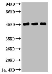
 WB (Western Blot)
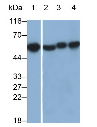
(Western blot analysis of 1) Hela, 2) 293T, 3) Mouse Brain Tissue, 4) Rat Brain Tissue using GAP-43 Monoclonal Antibody.)
WB (Western Blot)
(Western blot analysis of 1) Hela, 2) 293T, 3) Mouse Brain Tissue, 4) Rat Brain Tissue using GAP-43 Monoclonal Antibody.)
GAP43, Monoclonal Antibody (Cat# AAA243608)
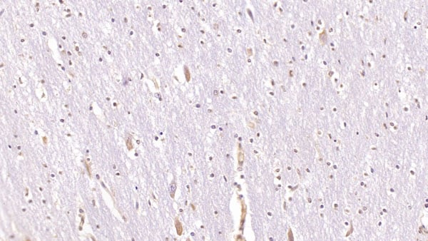
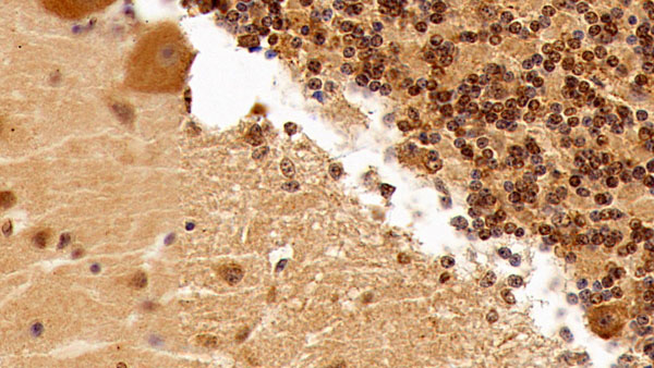
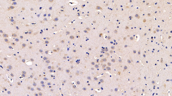
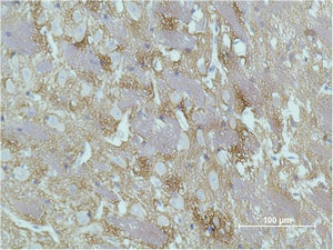
 IHC (Immunohiostchemistry)
(Immunohistochemical analysis of paraffin-embedded Mouse Brain Tissue using PPAR Delta Mouse mAb diluted at 1:200.)
IHC (Immunohiostchemistry)
(Immunohistochemical analysis of paraffin-embedded Mouse Brain Tissue using PPAR Delta Mouse mAb diluted at 1:200.)
PPARD, Monoclonal Antibody (Cat# AAA243629)
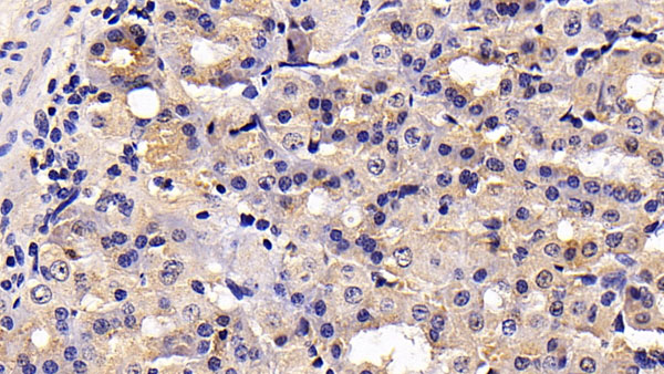
 IHC (Immunohiostchemistry)
(Immunohistochemical analysis of paraffin-embedded Human Colon Carcinoma Tissue using Epsilon Tubulin Mouse mAb diluted at 1:200.)
IHC (Immunohiostchemistry)
(Immunohistochemical analysis of paraffin-embedded Human Colon Carcinoma Tissue using Epsilon Tubulin Mouse mAb diluted at 1:200.)
TUBE1, Monoclonal Antibody (Cat# AAA243637)
 WB (Western Blot)
(Western blot analysis of 1) 293T Cell Lysate, 2) C2C12 Cell Lysate, 3) Rat Brain Tissue Lysate using Beclin-1 Mouse mAb diluted at 1:2000.)
WB (Western Blot)
(Western blot analysis of 1) 293T Cell Lysate, 2) C2C12 Cell Lysate, 3) Rat Brain Tissue Lysate using Beclin-1 Mouse mAb diluted at 1:2000.)
BECN1, Monoclonal Antibody (Cat# AAA243640)
What are Monoclonal Antibodies?
Monoclonal antibodies are specialized laboratory-produced proteins developed for binding to specific biological antigens or other molecular targets. Since they come from a single cell (or clone), they are especially consistent and accurate in the data they are involved in producing.
This type of antibody material has been shown to be a powerful tool in finding and subsequently destroying harmful cells in an organism, such as those found in cancers or various autoimmune diseases. This makes them excellent aids in medical testing and research, which is why they are so widely used.
AAA Biotech offers a comprehensive range of high-quality monoclonal antibodies that perform effectively in various laboratory tests, including (amongst others) ELISA, western blotting, immunohistochemistry, and flow cytometry. All of the products in our catalog are thoroughly quality tested to make sure that they are reliable and will consistently perform well in your research.
What Are The Uses of Monoclonal Antibodies
Monoclonal antibodies are used in many lab tests, including (amongst others) ELISA, western blotting, immunohistochemistry, and flow cytometry.
ELISA is a test that helps detect a specific substance/analyte in a sample. It uses antibodies (often monoclonal) bound to a solid surface (such as the well of a microplate) to “capture” the substance/analyte in the sample and immobilize it so that the detection antibody component can then bind to it and produce a signal, which can then be measured.
Western blotting identifies specific proteins in a sample. The sample is first separated on a gel, and then antibodies are applied that will typically bind to the target, which will all be localized to a single band in a lane.
Immunohistochemistry helps locate specific proteins in cells or tissue samples using antibodies.
Flow cytometry looks at and sorts cells. It uses antibodies that are conjugated to reporter molecules called “fluorophores”, which, under special lights, emit light themselves, which can then be measured by a detector instrument. For a deeper understanding of these techniques, explore our complete guide to monoclonal antibodies and their benefits.
How Monoclonal Antibodies Are Used as Medicine?
Please note that all of the products listed in AAA Biotech’s also known as AAA Bio or AAABio catalog are strictly for research-use only (RUO).
Monoclonal antibodies can also be used as therapeutic/medical treatments, particularly in the context of cancers. They are designed to find and bind to specific cells or proteins, helping the immune system recognize and attack the cancer. These treatments work in different ways, such as:
- Radioimmunotherapy attaches a small amount of radioactive molecule to the antibody, so it delivers the radiation directly to the cancer cells that the antibody is specifically binding to.
- Antibody-directed enzyme prodrug therapy uses antibodies that are specifically bound to special enzymes. These enzymes activate a harmless drug in the body and turn it into a cancer-killing drug only near the cancer cells—this helps avoid harming healthy cells.
- Immunoliposomes are tiny “bubbles” filled with medicine/drug and coated with antibodies. They carry the drug straight to the cancer cells.
Why Buy Monoclonal Antibodies From Us?
At AAA Biotech, we provide high-performance monoclonal antibodies designed to support a wide range of research needs.
1. Validated for Versatile Applications
The antibodies in our catalog are extensively validated and compatible with multiple techniques, including (but not limited to) ELISA, flow cytometry (FC), immunocytochemistry (ICC), immunofluorescence (IF), immunohistochemistry (IHC), immunoprecipitation (IP), and western blotting (WB).
2. Wide Selection & Specialized Options
We offer antibodies for common and rare species, that are available in various conjugated forms, and also in recombinant formats. Essentially, there is almost anything one might need to meet their experimental model’s requirements.
3. High-Quality Proteins
Our proteins meet high purity standards—90% or more as confirmed by SDS-PAGE. Many are available with tags like His, Flag, GST, or MBP, and we also supply native and biologically active proteins for functional studies.
Frequently Asked Questions
1. Are your monoclonal antibodies validated for specific applications?
Yes, our antibodies are tested and validated for use in methods such as ELISA, western blot, IHC, flow cytometry, and more. Refer to specific product pages or datasheets for individual product information.
2. How do I choose the right monoclonal antibody for my application?
Review the product details directly for application validation, species reactivity, and target information. You may also contact our support team at any time for help.
3. How quickly can I receive my order?
Most orders are processed and shipped within 1–3 business days, depending on product availability and your shipping location.