Filters
▼Clonality
▼Type
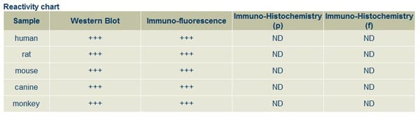
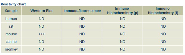
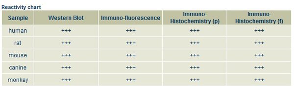
▼Reactivity
▼Gene Name
▼Isotype
▼Host
▼Application
▼Clone
▼Polyclonal Antibodies
At AAA Biotech also known as AAA Bio or AAABio, we provide a broad range of purified polyclonal antibodies (pAbs) that are able to all be browsed online through our website. Due to their high specificity and strong binding affinity, these antibodies are ideal for wide swathes of research and experimental applications.
Our polyclonal antibodies can easily support your work, whether you use them for Western Blotting, Immunocytochemistry (with or without Immunofluorescence used in conjunction), Immunohistochemistry, Immunoprecipitation, and ELISA tests. We highly encourage you to browse our range of pAbs and choose the one that best suits your experimental model.
Viewing 50-100 of 118597 product results
 IF (Immunofluorescence)
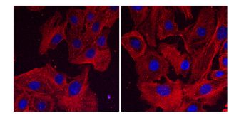
(Immunofluorescence using hCEC cells, 1st Ab (anti-beta-Actin at 1/250) and 2nd Ab (anti-mouse AB27550 at 1/1,000); cells were fixed with methanol;)
IF (Immunofluorescence)
(Immunofluorescence using hCEC cells, 1st Ab (anti-beta-Actin at 1/250) and 2nd Ab (anti-mouse AB27550 at 1/1,000); cells were fixed with methanol;)
IgG, Polyclonal Secondary Antibody (Cat# AAA63219)

 ICC (Immunocytochemistry)
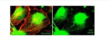
(Immunocytochemical labeling of Talin phosphorylation relative to F-actin in chick fibroblasts. The cells were labeled with rabbit polyclonal Talin (Ser-425) antibody (TP4171), then the antibody was detected using appropriate secondary antibody (Green). This labeling is compared to F-actin staining (Red, Left). (Image provided by Dr. Gianluca Gallo at Drexel University).)
ICC (Immunocytochemistry)
(Immunocytochemical labeling of Talin phosphorylation relative to F-actin in chick fibroblasts. The cells were labeled with rabbit polyclonal Talin (Ser-425) antibody (TP4171), then the antibody was detected using appropriate secondary antibody (Green). This labeling is compared to F-actin staining (Red, Left). (Image provided by Dr. Gianluca Gallo at Drexel University).)
Talin, Polyclonal Antibody (Cat# AAA71722)
 WB (Western Blot)
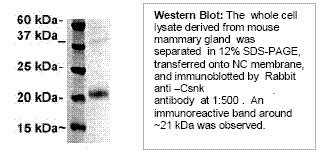
Casein (kappa), Polyclonal Antibody (Cat# AAA71432)
 ICC (Immunocytochemistry)
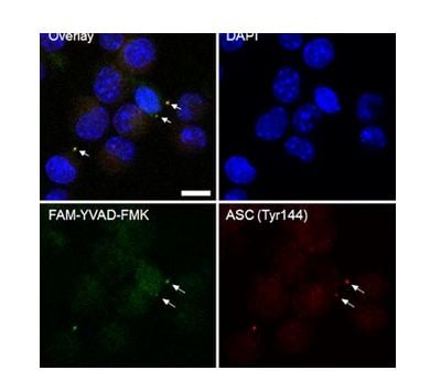
(Immunocytochemical labeling of Asc (Tyr-144) in inflammasomes. Paraformaldehyde fixed J774 cells were primed with LPS and treated with nigericin. Cells were co-labeled with DAPI, a caspase-1 inhibitor (FAM-YVAD-FMK), and anti-Asc (Tyr-144) phosphospecific antibody detected with AlexaFluor 568 secondary. (Image provided by Jordan Yaron, Center for Biosignatures Discovery Automation, Arizona State University))
ICC (Immunocytochemistry)
(Immunocytochemical labeling of Asc (Tyr-144) in inflammasomes. Paraformaldehyde fixed J774 cells were primed with LPS and treated with nigericin. Cells were co-labeled with DAPI, a caspase-1 inhibitor (FAM-YVAD-FMK), and anti-Asc (Tyr-144) phosphospecific antibody detected with AlexaFluor 568 secondary. (Image provided by Jordan Yaron, Center for Biosignatures Discovery Automation, Arizona State University))
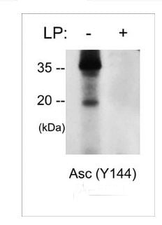
Asc, Polyclonal Antibody (Cat# AAA71570)
 ICC (Immunocytochemistry)
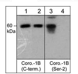
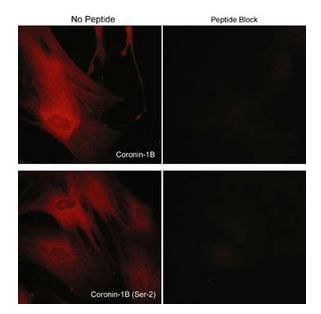
(Immunocytochemical labeling of coronin-1B in rabbit spleen fibroblasts treated with Calyculin A. The cells were labeled with rabbit polyclonal Coronin-1B (C-terminus) and Coronin-1B (Ser-2) antibodies, then detected using appropriate secondary antibodies conjugated to Cy3. The antibodies were used in the absence (left) or presence (right) of their respective blocking peptide (CX2585 or CX2625).)
ICC (Immunocytochemistry)
(Immunocytochemical labeling of coronin-1B in rabbit spleen fibroblasts treated with Calyculin A. The cells were labeled with rabbit polyclonal Coronin-1B (C-terminus) and Coronin-1B (Ser-2) antibodies, then detected using appropriate secondary antibodies conjugated to Cy3. The antibodies were used in the absence (left) or presence (right) of their respective blocking peptide (CX2585 or CX2625).)
Coronin-1B, Polyclonal Antibody (Cat# AAA71606)
 ICC (Immunocytochemistry)
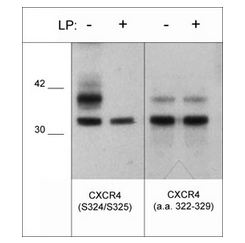
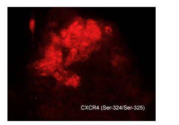
(Immunocytochemical labeling of CXCR4 in chick pluripotent cells. The cells were labeled with rabbit polyclonal CXCR4 (S324/S325) antibody (CP4251), then detected using appropriate secondary antibody (Red). (Image provided by Dr. Yangqing Lu at the Regenerative Bioscience Center, University of Georgia).)
ICC (Immunocytochemistry)
(Immunocytochemical labeling of CXCR4 in chick pluripotent cells. The cells were labeled with rabbit polyclonal CXCR4 (S324/S325) antibody (CP4251), then detected using appropriate secondary antibody (Red). (Image provided by Dr. Yangqing Lu at the Regenerative Bioscience Center, University of Georgia).)
CXCR4, Polyclonal Antibody (Cat# AAA71615)
 Application Data
PLCg1 (Tyr-775), Polyclonal Antibody (Cat# AAA71528)
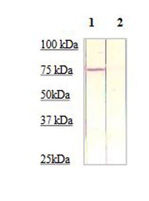 WB (Western Blot)
(The cell lysate derived from HELA was immunoprobedbyeither Rabbit anti-beta-Catenin (pS675) (Lane 1) or theantibody pre-incubatedby immunization peptide (lane 2) at 1:500 dilution.)
WB (Western Blot)
(The cell lysate derived from HELA was immunoprobedbyeither Rabbit anti-beta-Catenin (pS675) (Lane 1) or theantibody pre-incubatedby immunization peptide (lane 2) at 1:500 dilution.)
Catenin-b, Polyclonal Antibody (Cat# AAA71321)
 Application Data
Nitrotyrosine, Polyclonal Antibody (Cat# AAA71786)
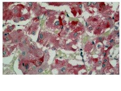 IHC (Immunohiostchemistry)
(AAA61062 (5ug/ml) staining of paraffin embedded Human Adrenal Gland. Steamed antigen retrieval with citratebuffer pH 6, AP-staining.)
IHC (Immunohiostchemistry)
(AAA61062 (5ug/ml) staining of paraffin embedded Human Adrenal Gland. Steamed antigen retrieval with citratebuffer pH 6, AP-staining.)
FOXL2/BPES, Polyclonal Antibody (Cat# AAA61062)
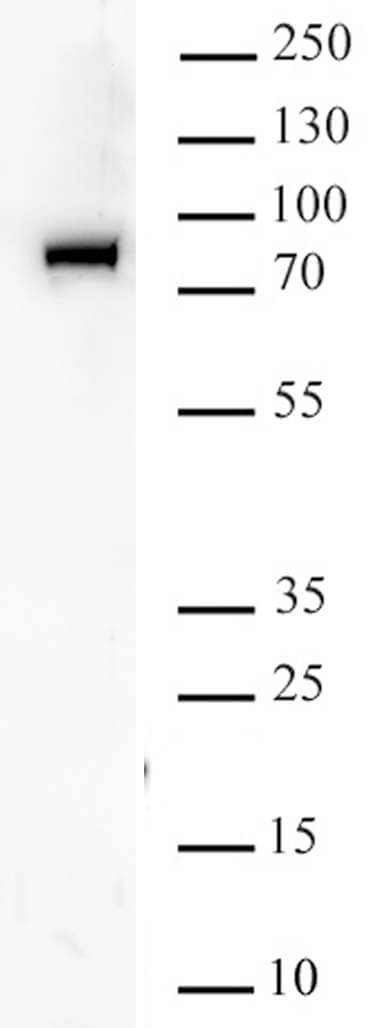
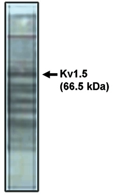 WB (Western Blot)
(Western blot analysis using Kv1.5 antibody on rat brain lysate)
WB (Western Blot)
(Western blot analysis using Kv1.5 antibody on rat brain lysate)
Kv1.5 Potassium Channel, Polyclonal Antibody (Cat# AAA60494)
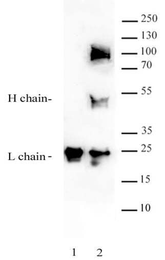
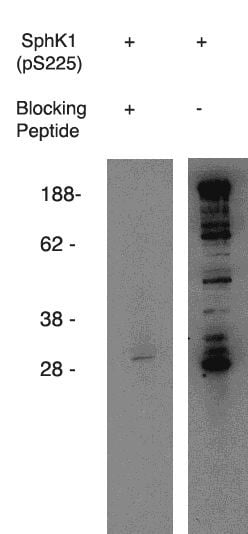 WB (Western Blot)
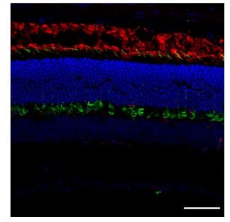
(Western blot using anti-mouse SphK1 (pS225) on mouse B-cell lysate. Antibody used at 1 ug/ml with phosphorylated blocking peptide (lane A) and without (laneB).)
WB (Western Blot)
(Western blot using anti-mouse SphK1 (pS225) on mouse B-cell lysate. Antibody used at 1 ug/ml with phosphorylated blocking peptide (lane A) and without (laneB).)
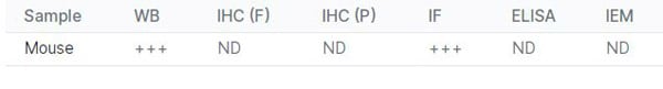
phospho-Sphingosine Kinase 1,(pS225), Mouse Reactive, Polyclonal Antibody (Cat# AAA60497)
Purification: Antigen Immunoaffiinity Purification
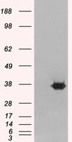
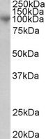 WB (Western Blot)
((1ug/ml) staining of Human Bone Marrow lysate (35ug protein in RIPA buffer). Primary incubation was 1 hour. Detected by chemiluminescence.)
WB (Western Blot)
((1ug/ml) staining of Human Bone Marrow lysate (35ug protein in RIPA buffer). Primary incubation was 1 hour. Detected by chemiluminescence.)
MS2/ADAM8/CD156, Polyclonal Antibody (Cat# AAA60821)
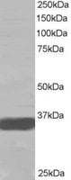 WB (Western Blot)
(Staining (0.2ug/ml) of A431 lysate (RIPA buffer, 35ug total protein per lane). Primary incubated for 1 hour. Detected by western blot using chemiluminescence.)
WB (Western Blot)
(Staining (0.2ug/ml) of A431 lysate (RIPA buffer, 35ug total protein per lane). Primary incubated for 1 hour. Detected by western blot using chemiluminescence.)
MRGX, Polyclonal Antibody (Cat# AAA60831)
 Application Data
BRD9, Polyclonal Antibody (Cat# AAA60351)
 IF (Immunofluorescence)
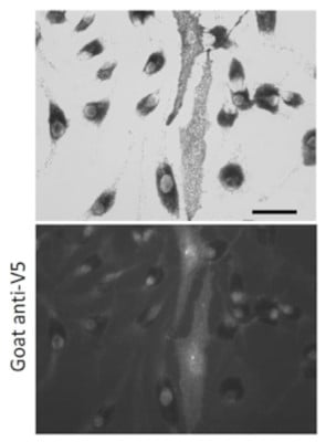
(Immunofluorescence – anti-V5 Ab using hCEC cells transduced with V5-mCherry-Rab11a; cells were fixed with methanol and anti-V5 at 1/100;)
IF (Immunofluorescence)
(Immunofluorescence – anti-V5 Ab using hCEC cells transduced with V5-mCherry-Rab11a; cells were fixed with methanol and anti-V5 at 1/100;)
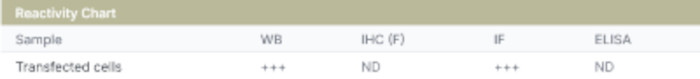
V5, Polyclonal Antibody (Cat# AAA63062)
 Application Data
Rab27a, Polyclonal Antibody (Cat# AAA63067)
 Application Data
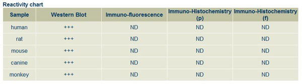
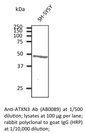
ATXN3, Polyclonal Antibody (Cat# AAA63075)
 Application Data
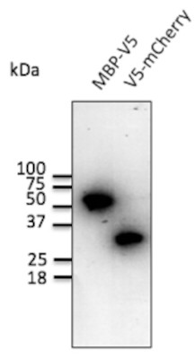
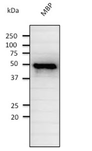
(Anti-MBP Ab at 1/2000 dilution; rabbit polyclonal to goat IgG (HRP) at 1/10,000 dilution.)
Application Data
(Anti-MBP Ab at 1/2000 dilution; rabbit polyclonal to goat IgG (HRP) at 1/10,000 dilution.)
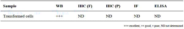
MBP, Polyclonal Antibody (Cat# AAA63077)
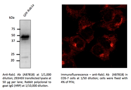 Application Data
Rab1, Polyclonal Antibody (Cat# AAA63086)
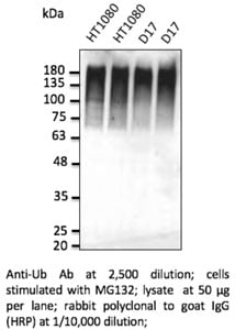 WB (Western Blot)
Ubiquitin, Polyclonal Antibody (Cat# AAA63096)
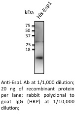
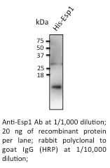 Application Data
Esp1, Polyclonal Antibody (Cat# AAA63108)
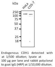
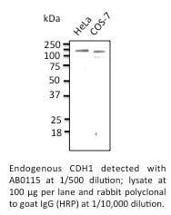 Application Data
CDH1, Polyclonal Antibody (Cat# AAA63110)
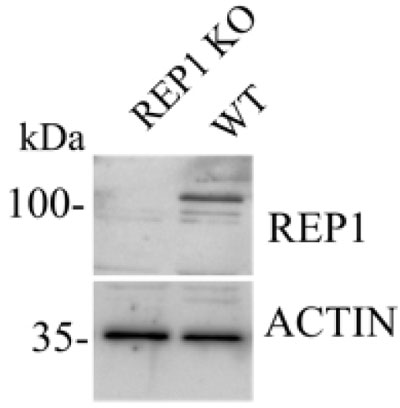
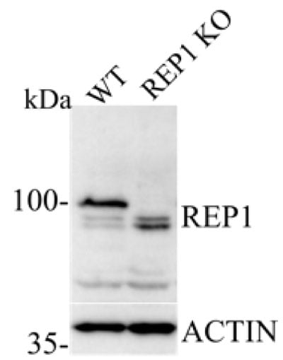
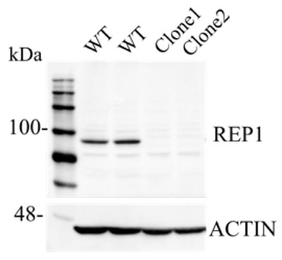
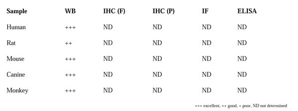
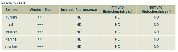
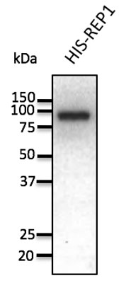 WB (Western Blot)
(Anti-REP1 Ab at 1/1,000 dilution; 50 ng of recombinant protein per lane; rabbit polyclonal to goat IgG (HRP) at 1/10,000 dilution.)
WB (Western Blot)
(Anti-REP1 Ab at 1/1,000 dilution; 50 ng of recombinant protein per lane; rabbit polyclonal to goat IgG (HRP) at 1/10,000 dilution.)
REP1, Polyclonal Antibody (Cat# AAA63112)
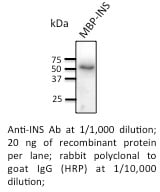 Application Data
Insulin, Polyclonal Antibody (Cat# AAA63118)
 Application Data
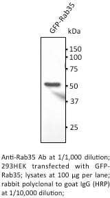
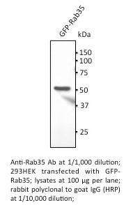
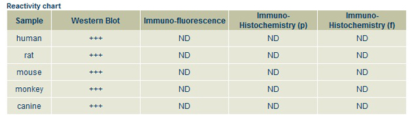
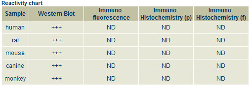
Rab35, Polyclonal Antibody (Cat# AAA63122)
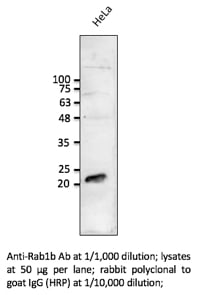 WB (Western Blot)
Rab1b, Polyclonal Antibody (Cat# AAA63129)
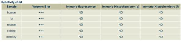
 WB (Western Blot)
Rab9a, Polyclonal Antibody (Cat# AAA63132)
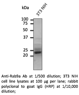
 IF (Immunofluorescence)
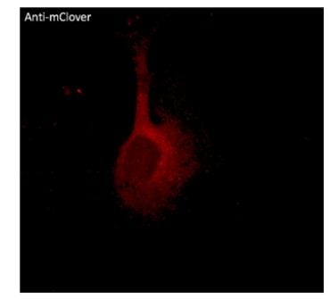
(Immunofluorescence - anti-mClover Ab using NIH3T3 cells transduced with mClover-Rab5a; cells were fixed with methanol and anti-mClover at 1/250;)
IF (Immunofluorescence)
(Immunofluorescence - anti-mClover Ab using NIH3T3 cells transduced with mClover-Rab5a; cells were fixed with methanol and anti-mClover at 1/250;)
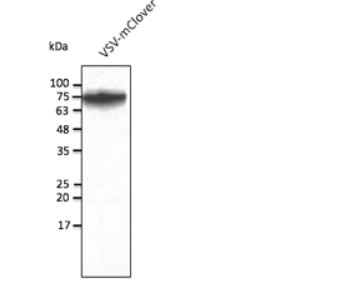
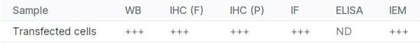
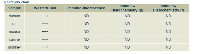
mClover, Polyclonal Antibody (Cat# AAA63255)
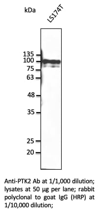 WB (Western Blot)
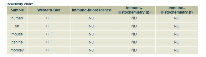
PTK2, Polyclonal Antibody (Cat# AAA63154)
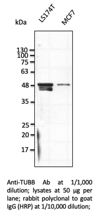 WB (Western Blot)
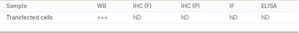
TUBB, Polyclonal Antibody (Cat# AAA63155)
 Application Data
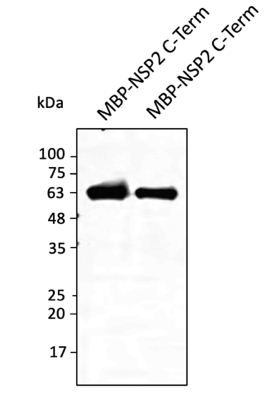
(Anti-NSP2 protein Ab at 1/2,500 dilution; lane with 30 ng of recombinant fusion protein (480 aa - stop); rabbit polyclonal to goat IgG (HRP) at 1/10,000 dilution;)
Application Data
(Anti-NSP2 protein Ab at 1/2,500 dilution; lane with 30 ng of recombinant fusion protein (480 aa - stop); rabbit polyclonal to goat IgG (HRP) at 1/10,000 dilution;)
COVID 19 NSP2 Coronavirus, Polyclonal Antibody (Cat# AAA63180)

 Application Data
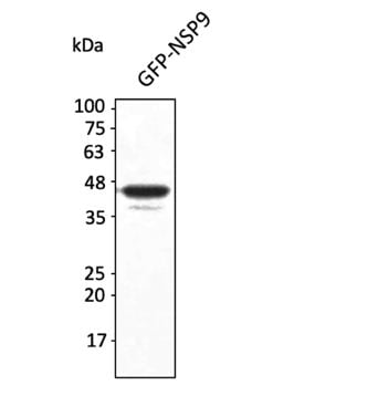
(Anti-NSP9 protein Ab at 1/2,500 dilution; lane with 30 ng of recombinant fusion protein; rabbit polyclonal to goat IgG (HRP) at 1/10,000 dilution;)
Application Data
(Anti-NSP9 protein Ab at 1/2,500 dilution; lane with 30 ng of recombinant fusion protein; rabbit polyclonal to goat IgG (HRP) at 1/10,000 dilution;)
COVID 19 NSP9 Coronavirus, Polyclonal Antibody (Cat# AAA63185)
 Application Data
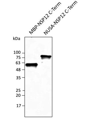
(Anti-NSP12 protein Ab at 1/2,500 dilution; lane with 30 ng of recombinant fusion protein (820 aa - stop); rabbit polyclonal to goat IgG (HRP) at 1/10,000 dilution;)
Application Data
(Anti-NSP12 protein Ab at 1/2,500 dilution; lane with 30 ng of recombinant fusion protein (820 aa - stop); rabbit polyclonal to goat IgG (HRP) at 1/10,000 dilution;)
COVID 19 NSP12 Coronavirus, Polyclonal Antibody (Cat# AAA63187)
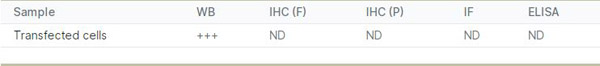
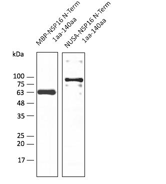 Application Data
(Anti-NSP16 Ab at 1/2,500 dilution; lane with 30 ng of recombinant fusion protein; rabbit polyclonal to goat IgG (HRP) at 1/10,000 dilution;)
Application Data
(Anti-NSP16 Ab at 1/2,500 dilution; lane with 30 ng of recombinant fusion protein; rabbit polyclonal to goat IgG (HRP) at 1/10,000 dilution;)
COVID 19 NSP16 Coronavirus, Polyclonal Antibody (Cat# AAA63191)
 Application Data
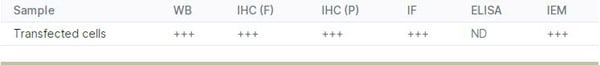
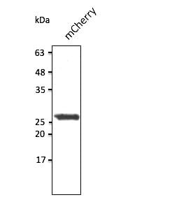
(Anti-mCherry Ab at 1/2,500 dilution; 293HEK cells transduced with mCherry Ad; lysates at 50 ug per lane; rabbit polyclonal to goat IgG (HRP) at 1/10,000 dilution;)
Application Data
(Anti-mCherry Ab at 1/2,500 dilution; 293HEK cells transduced with mCherry Ad; lysates at 50 ug per lane; rabbit polyclonal to goat IgG (HRP) at 1/10,000 dilution;)
mCherry, Polyclonal Antibody (Cat# AAA63205)
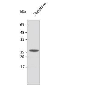
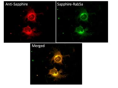

 IF (Immunofluorescence)
(Immunofluorescence - anti-Sapphire Ab using NIH3T3 cells transduced with Sapphire-Rab5a; cells were fixed with methanol and anti-Sapphire at 1/100;)
IF (Immunofluorescence)
(Immunofluorescence - anti-Sapphire Ab using NIH3T3 cells transduced with Sapphire-Rab5a; cells were fixed with methanol and anti-Sapphire at 1/100;)
Sapphire, Polyclonal Antibody (Cat# AAA63213)
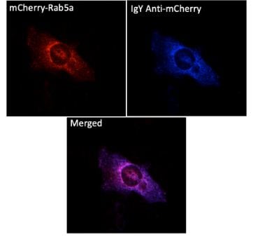
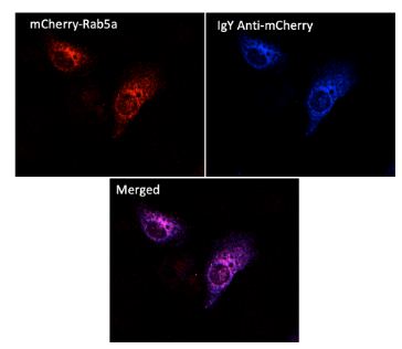 IF (Immunofluorescence)
(Immunofluorescence - anti-mCherry Ab (AB9770) using hCEC cells transduced with mCherry-Rab5a; cells were fixed with methanol and mCherry Ab at 1/250; 2nd Ab goat anti-IgY (AB307405) at 1:1,000;)
IF (Immunofluorescence)
(Immunofluorescence - anti-mCherry Ab (AB9770) using hCEC cells transduced with mCherry-Rab5a; cells were fixed with methanol and mCherry Ab at 1/250; 2nd Ab goat anti-IgY (AB307405) at 1:1,000;)
IgY, Polyclonal Secondary Antibody (Cat# AAA63223)
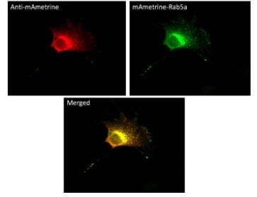
 IF (Immunofluorescence)
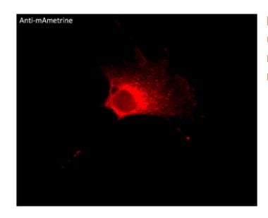
(Immunofluorescence -anti-mAmetrine Ab usingNIH3T3 cells transduced with mAmetrine-Rab5a; cells were fixed with methanol and anti-mAmetrine at 1/250;)
IF (Immunofluorescence)
(Immunofluorescence -anti-mAmetrine Ab usingNIH3T3 cells transduced with mAmetrine-Rab5a; cells were fixed with methanol and anti-mAmetrine at 1/250;)
mAmetrine, Polyclonal Antibody (Cat# AAA63234)

 Application Data
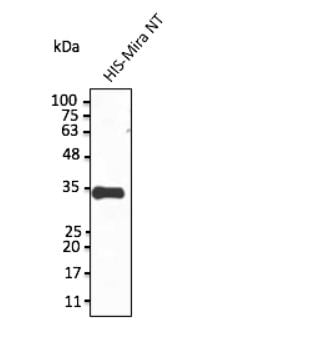
(Anti-Miranda Ab at 1/1,000 dilution; protein at 20 ng per lane; rabbit polyclonal to goat IgG (HRP) at 1/10,000 dilution;)
Application Data
(Anti-Miranda Ab at 1/1,000 dilution; protein at 20 ng per lane; rabbit polyclonal to goat IgG (HRP) at 1/10,000 dilution;)
Miranda, Polyclonal Antibody (Cat# AAA63235)
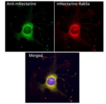
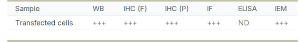
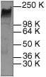
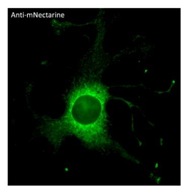 IF (Immunofluorescence)
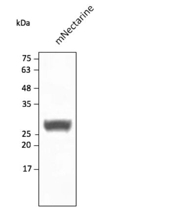
(Immunofluorescence -anti-mNectarine Ab using hCEC cells transduced with mNectarine-Rab5a; cells were fixed with methanol and anti-mNectarine at 1/250;)
IF (Immunofluorescence)
(Immunofluorescence -anti-mNectarine Ab using hCEC cells transduced with mNectarine-Rab5a; cells were fixed with methanol and anti-mNectarine at 1/250;)
mNectarine, Polyclonal Antibody (Cat# AAA63236)
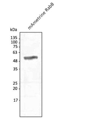
 WB (Western Blot)
Neu, Polyclonal Antibody (Cat# AAA63011)
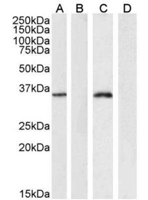
 WB (Western Blot)
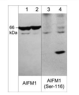
(Western blot image of human jurkat cells untreated (lanes 1 & 3) or treated with calyculin A (100nM, 30 min.) (lanes 2 and 4). The blot was probed with rabbit polyclonals anti-AIFM1 (C-terminal region) at 1:500 (lanes 1 & 2) and anti-AIFM1 (Ser-116) phospho-specific antibody at 1:1000 (lanes 3 & 4).)
WB (Western Blot)
(Western blot image of human jurkat cells untreated (lanes 1 & 3) or treated with calyculin A (100nM, 30 min.) (lanes 2 and 4). The blot was probed with rabbit polyclonals anti-AIFM1 (C-terminal region) at 1:500 (lanes 1 & 2) and anti-AIFM1 (Ser-116) phospho-specific antibody at 1:1000 (lanes 3 & 4).)
AIFM1, Polyclonal Antibody (Cat# AAA71569)
 ICC (Immunocytochemistry)
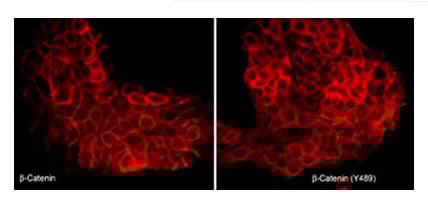
(Immunocytochemical labeling of beta-Catenin in pervanadate-treated A431 cells. The cells were labeled with mouse monoclonal beta-Catenin (CM1181) or rabbit polyclonal beta-Catenin (Tyr-489) antibodies, then the antibodies were detected using appropriate secondary antibodies conjugated to Cy3.)
ICC (Immunocytochemistry)
(Immunocytochemical labeling of beta-Catenin in pervanadate-treated A431 cells. The cells were labeled with mouse monoclonal beta-Catenin (CM1181) or rabbit polyclonal beta-Catenin (Tyr-489) antibodies, then the antibodies were detected using appropriate secondary antibodies conjugated to Cy3.)
beta-Catenin, Polyclonal Antibody (Cat# AAA71609)
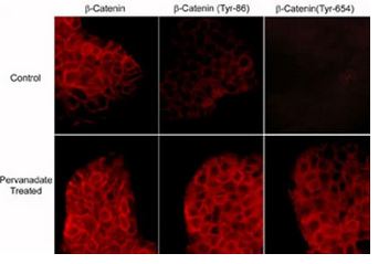 ICC (Immunocytochemistry)
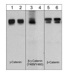
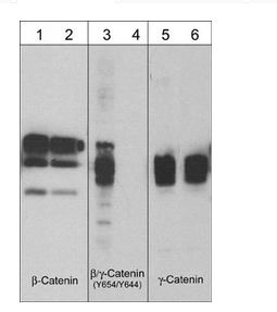
(Immunocytochemical labeling of phosphorylated beta-Catenin in control and pervanadate-treated A431 cells. The cells were labeled with mouse monoclonal beta-Catenin (CM1181) or rabbit polyclonal beta-Catenin (Tyr-86) or beta-Catenin (Y654) antibodies, then the antibodies were detected using appropriate secondary antibodies conjugated to Cy3.)
ICC (Immunocytochemistry)
(Immunocytochemical labeling of phosphorylated beta-Catenin in control and pervanadate-treated A431 cells. The cells were labeled with mouse monoclonal beta-Catenin (CM1181) or rabbit polyclonal beta-Catenin (Tyr-86) or beta-Catenin (Y654) antibodies, then the antibodies were detected using appropriate secondary antibodies conjugated to Cy3.)
beta-Catenin, Polyclonal Antibody (Cat# AAA71610)
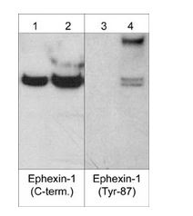
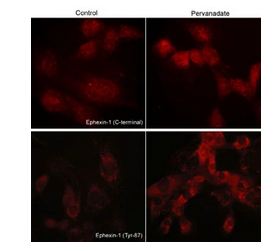 ICC (Immunocytochemistry)
(Immunocytochemical labeling of phosphorylated Exphexin-1 in pervanadate-treated mouse C2C12. The cells were labeled with rabbit polyclonal Ephexin-1 (C-terminal region) and Ephexin-1 (Tyr-87) antibodies, then the antibodies were detected using appropriate secondary antibodies conjugated to Cy3.)
ICC (Immunocytochemistry)
(Immunocytochemical labeling of phosphorylated Exphexin-1 in pervanadate-treated mouse C2C12. The cells were labeled with rabbit polyclonal Ephexin-1 (C-terminal region) and Ephexin-1 (Tyr-87) antibodies, then the antibodies were detected using appropriate secondary antibodies conjugated to Cy3.)
Ephexin-1, Polyclonal Antibody (Cat# AAA71631)
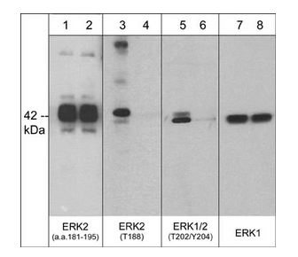
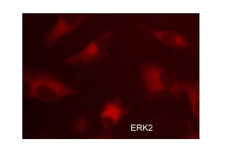 ICC (Immunocytochemistry)
(Immunocytochemical labeling of ERK2 in aldehyde-fixed and NP-40 permeabilized human NCI-H1915 lung carcinoma cells. The cells were labeled with rabbit polyclonal anti-ERK2 (EP4071) antibody. The antibody was detected using appropriate secondary antibody conjugated to DyLight 594.)
ICC (Immunocytochemistry)
(Immunocytochemical labeling of ERK2 in aldehyde-fixed and NP-40 permeabilized human NCI-H1915 lung carcinoma cells. The cells were labeled with rabbit polyclonal anti-ERK2 (EP4071) antibody. The antibody was detected using appropriate secondary antibody conjugated to DyLight 594.)
ERK2, Polyclonal Antibody (Cat# AAA71633)
 Application Data
Cofilin 1 (Ser-3), Polyclonal Antibody (Cat# AAA71517)
 Application Data
Neuropilin-1 (a1 CUB Domain), Polyclonal Antibody (Cat# AAA71524)
 ICC (Immunocytochemistry)
(Immunocytochemical labeling of phosphorylated WAVE in pervanadate-treated mouse C2C12. The cells were labeled with rabbit polyclonal WAVE1 (N-terminal region) and WAVE (Tyr-125) antibodies, then the antibodies were detected using appropriate secondary antibodies conjugated to Cy3.)
ICC (Immunocytochemistry)
(Immunocytochemical labeling of phosphorylated WAVE in pervanadate-treated mouse C2C12. The cells were labeled with rabbit polyclonal WAVE1 (N-terminal region) and WAVE (Tyr-125) antibodies, then the antibodies were detected using appropriate secondary antibodies conjugated to Cy3.)
WAVE1 (N-terminal region), Polyclonal Antibody (Cat# AAA71534)
What are Polyclonal Antibodies?
Polyclonal antibodies are antibodies that come from multiple B cell clones of a host animal. The typical hosts used for the majority of polyclonal antibody production are rabbits, goats, sheep, and donkeys. These polyclonal antibodies, once having identified their target, will bind to different epitopes located at different regions or sequences on the same protein/antigen. This ability to bind multiple epitopes is what makes polyclonal antibodies highly sensitive, as explained in our detailed guide on polyclonal antibodies and why they matter.
As a result, they are ideal at locating and binding to the target, even if the target is in very low concentrations (due to many different antibodies being able to bind to the same target molecule, which allows for significant amplification of a downstream signal).
Polyclonal antibodies are typically produced by injecting an antigen into a host animal, which causes the animal’s immune system to attack the foreign antigen by mass generating antibodies against it. After a period of time, serum is collected from the animal and purified using physicochemical fractionation, class-specific affinity purification, and/or antigen-affinity purification.
Key Uses of Polyclonal Antibodies
- Western Blotting: This method is used to find specific proteins in biological samples after separating them by size.
- Immunohistochemistry: IHC helps visualize the location of proteins in tissue sections using various staining techniques.
- ELISA: (Enzyme-Linked Immunosorbent Assay) is typically used to identify specific protein quantities in a sample. ELISAs can be either “Quantitative” or “Qualitative”.
- Flow Cytometry: technique that identifies and measures the specific protein on the surface or inside the cells in a fluid suspension.
- Immunoprecipitation: IP isolates and studies a specific protein from a complex mixture using antibodies.
Why Buy Polyclonal Antibodies from AAA Biotech?
1. Ideal for Various Applications
Our antibodies are generally going to be validated for use in multiple types of assays, including ELISA, Western Blotting, Immunohistochemistry, Immunoprecipitation, amongst others. They are ideal for a wide range of research applications.
2. Rigorous Quality Control
All of the antibodies in our catalog undergo strict quality testing to ensure specificity, sensitivity, and consistent performance. We are confident in the ability of our antibodies to provide you with accurate results.
3. Wide Assortment of Antibodies
Antibodies in our catalog can be found for both common and exotic species, and these antibodies are also available in both conjugated and recombinant forms to suit many diverse experimental needs.
4. Highly Purified
Our antibodies are available in purified forms with over 85% purity, as confirmed by SDS-PAGE. They are also available with tags such as His, Flag, GST, or MBP. We cater to customers worldwide.
FAQ
1. How are polyclonal antibodies produced?
Traditionally, polyclonal antibodies are produced by injecting an antigen into a host animal (such as a rabbit or goat), which then triggers an immune response from the host animal. The animal’s B cells produce antibodies that will recognize different parts of the injected antigen. These antibodies are then collected from the animal’s blood and purified for use.
2. How do polyclonal antibodies differ from monoclonal antibodies?
Polyclonal antibodies are a mix of antibodies that bind to different locations (epitopes) of the same antigen, while monoclonal antibodies are identical and bind to just one specific epitope. This makes polyclonal antibodies more versatile and better at detecting proteins that may be present in low quantities or in altered/modified forms.
3. How should I store polyclonal antibodies?
Polyclonal antibodies should be stored at 4°C for short-term use (up to a few weeks) and at -20°C or -80°C for long-term storage. Avoid repeated freeze-thaw cycles by dividing them into small aliquots. Always check the datasheet for specific storage instructions.