Filters
▼Clonality
▼Type
▼Reactivity
▼Gene Name
▼Isotype
▼Host
▼Application
▼Clone
▼Polyclonal Antibodies
At AAA Biotech also known as AAA Bio or AAABio, we provide a broad range of purified polyclonal antibodies (pAbs) that are able to all be browsed online through our website. Due to their high specificity and strong binding affinity, these antibodies are ideal for wide swathes of research and experimental applications.
Our polyclonal antibodies can easily support your work, whether you use them for Western Blotting, Immunocytochemistry (with or without Immunofluorescence used in conjunction), Immunohistochemistry, Immunoprecipitation, and ELISA tests. We highly encourage you to browse our range of pAbs and choose the one that best suits your experimental model.
Viewing 250-300 of 118597 product results
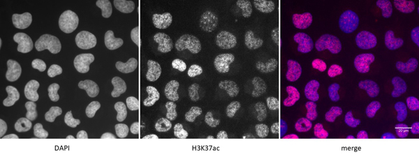 IF (Immunofluorescence)
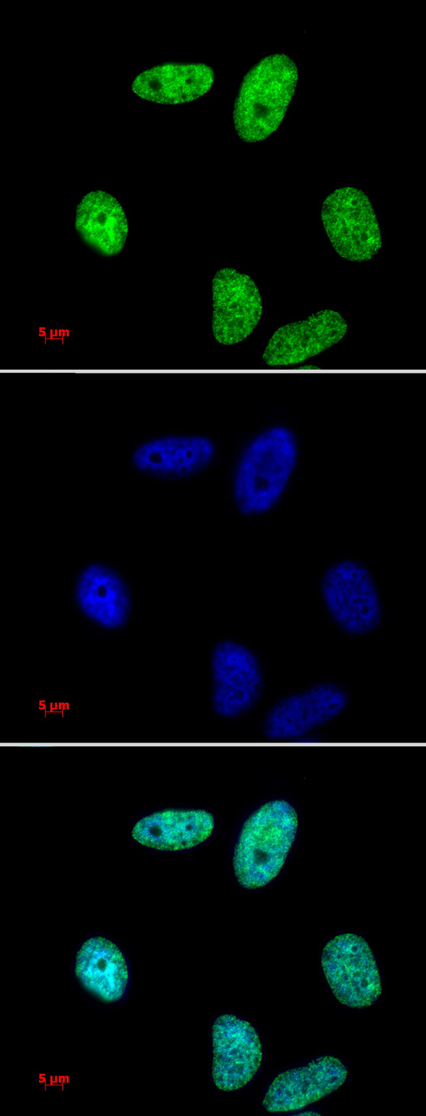
(Detection of H3K37ac by immunofluorescence U2OS cells were stained with H3K37ac antibody at a dilution of 1:500. Left panel: DAPI. Middle panel: H3K37ac antibody staining. Right panel: merge.)
IF (Immunofluorescence)
(Detection of H3K37ac by immunofluorescence U2OS cells were stained with H3K37ac antibody at a dilution of 1:500. Left panel: DAPI. Middle panel: H3K37ac antibody staining. Right panel: merge.)
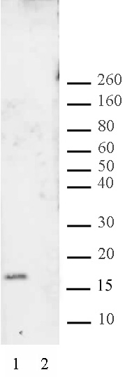
Histone H3K37ac, Polyclonal Antibody (Cat# AAA60051)
M2 (A/California/06/2009)(H1N1), Polyclonal Antibody (Cat# AAA62129)
 Application Data
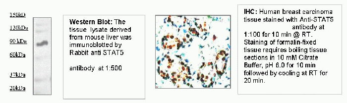
STAT5, Polyclonal Antibody (Cat# AAA71415)

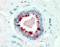
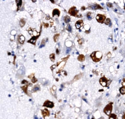 IHC (Immunohiostchemistry)
(Immunohistochemistry: Human lymphomacarcinoma (FFPE) stained with Rabbit anti-CD117/kit (Cat# AAA71440) at 1:200 for 10 min @ RT. Staining of formalin-fixed tissue requires boiling tissue sections in 10 mM Citrate Buffer, pH 6.0 for 10 min followed by cooling at RT for 20 min.)
IHC (Immunohiostchemistry)
(Immunohistochemistry: Human lymphomacarcinoma (FFPE) stained with Rabbit anti-CD117/kit (Cat# AAA71440) at 1:200 for 10 min @ RT. Staining of formalin-fixed tissue requires boiling tissue sections in 10 mM Citrate Buffer, pH 6.0 for 10 min followed by cooling at RT for 20 min.)
CD117 (kit), Polyclonal Antibody (Cat# AAA71440)
 IHC (Immunohistochemistry)
(Human lung tissue was immunce-stained with Rabbit anti IRAK-M antibody (AAA71324) at 1:200 for 10 min @ RT. Staining of formalin-fixed tissue requires boiling tissue sections in 10mM Citrate Buffer, pH 6.0 for 10 min followed by cooling at RT for 20 min.)
IHC (Immunohistochemistry)
(Human lung tissue was immunce-stained with Rabbit anti IRAK-M antibody (AAA71324) at 1:200 for 10 min @ RT. Staining of formalin-fixed tissue requires boiling tissue sections in 10mM Citrate Buffer, pH 6.0 for 10 min followed by cooling at RT for 20 min.)
IRAK-M, Polyclonal Antibody (Cat# AAA71324)
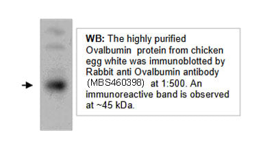 WB (Western Blot)
Ovalbumin, Polyclonal Antibody (Cat# AAA71335)
 WB (Western Blot)
(WB: The cell lysate derived from EGF-stimulated A431was resolved onto 12% SDS-PAGE and immunoblotted by Rabbit anti Phosphoserine (Cat#AAA71345) at 1:500. A panel of phosphorylated proteins was observed.)
WB (Western Blot)
(WB: The cell lysate derived from EGF-stimulated A431was resolved onto 12% SDS-PAGE and immunoblotted by Rabbit anti Phosphoserine (Cat#AAA71345) at 1:500. A panel of phosphorylated proteins was observed.)
Phosphoserine (pSer), Polyclonal Antibody (Cat# AAA71345)
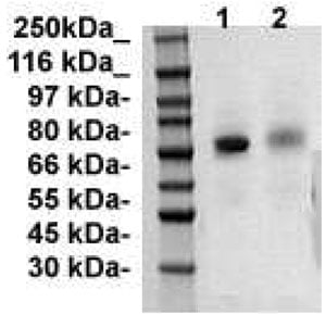
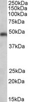
 WB (Western Blot)
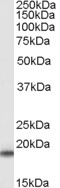
(Western Blot: The Cell lysatederived from HT-29 wereresolved onto 10% SDSPAGE, transferred onto NCmembrane, and immunoblotted by Rabbit anti 15-LOX2 at1:500. An immunoreactiveband around ~75 kDa wasobserved.)
WB (Western Blot)
(Western Blot: The Cell lysatederived from HT-29 wereresolved onto 10% SDSPAGE, transferred onto NCmembrane, and immunoblotted by Rabbit anti 15-LOX2 at1:500. An immunoreactiveband around ~75 kDa wasobserved.)
15-Lox-2, Polyclonal Antibody (Cat# AAA71353)
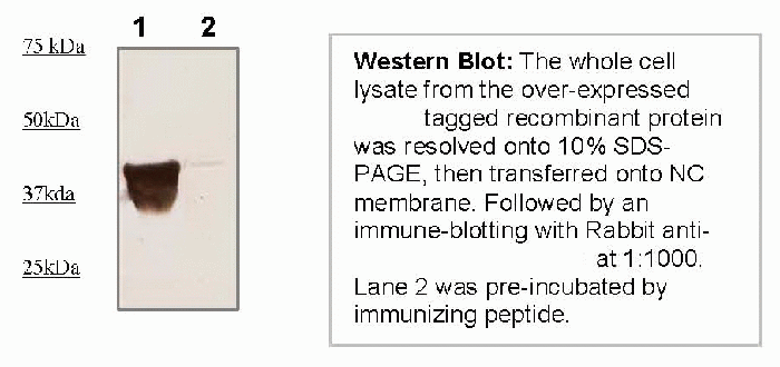
 WB (Western Blot)
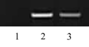
(Western Blot: Rabbit anti c-Myc Tag antibody staining on c-Myc tagged recombinant proteinlane 1:loading protein: 1ug/laneLane 2: loading protein: 0.1ug/lane.)
WB (Western Blot)
(Western Blot: Rabbit anti c-Myc Tag antibody staining on c-Myc tagged recombinant proteinlane 1:loading protein: 1ug/laneLane 2: loading protein: 0.1ug/lane.)
Myc-Tag, Polyclonal Antibody (Cat# AAA71379)
 Application Data
DYKDDDDK-tag, Polyclonal Antibody (Cat# AAA71384)
GST A1-1, Polyclonal Antibody (Cat# AAA71785)
 Application Data
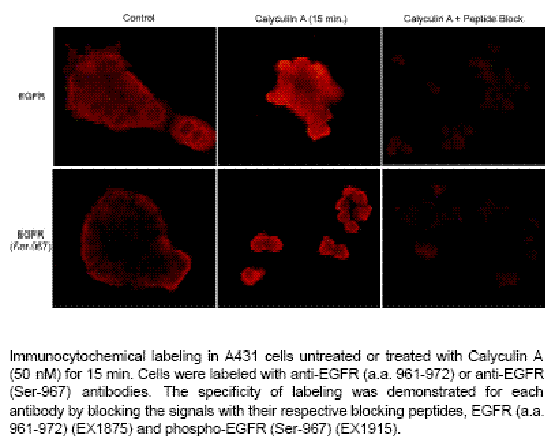
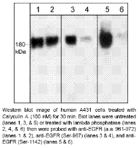
EGFR (Ser-967), Polyclonal Antibody (Cat# AAA71520)
 WB (Western Blot)
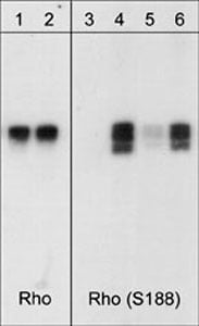
(Western blot analysis of human RhoA GST fusion recombinant unphosphorylated (lanes 1 & 3) or phosphorylated with PKA (lanes 2, 4, 5 & 6). The blots were probed with anti-Rho ( lanes 1 & 2) or with anti-RhoA (Ser-188) (AAA71530; lanes 3-6). The latter antibody was used in the presence of no peptide (lanes 3 & 4), phospho-RhoA (Ser-188) peptide (lane 5), or a non-specific phosphoserine peptide (lane 6).)
WB (Western Blot)
(Western blot analysis of human RhoA GST fusion recombinant unphosphorylated (lanes 1 & 3) or phosphorylated with PKA (lanes 2, 4, 5 & 6). The blots were probed with anti-Rho ( lanes 1 & 2) or with anti-RhoA (Ser-188) (AAA71530; lanes 3-6). The latter antibody was used in the presence of no peptide (lanes 3 & 4), phospho-RhoA (Ser-188) peptide (lane 5), or a non-specific phosphoserine peptide (lane 6).)
RhoA (Ser-188), Polyclonal Antibody (Cat# AAA71530)
 Application Data
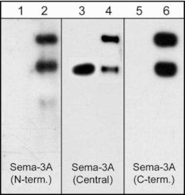
(Formalin fixed, citric acid treated parafin sections of adult Rat cerebellum. Sections were probed with anti-Sema3A (AAA71531) then anti-RabbitHRP before detection using DAB. (Images provided by Carl Hobbs and Dr. Pat Doherty at Wolfson Centre for Age-Related Diseases, King's College London).)
Application Data
(Formalin fixed, citric acid treated parafin sections of adult Rat cerebellum. Sections were probed with anti-Sema3A (AAA71531) then anti-RabbitHRP before detection using DAB. (Images provided by Carl Hobbs and Dr. Pat Doherty at Wolfson Centre for Age-Related Diseases, King's College London).)
Semaphorin-3A (Central Region), Polyclonal Antibody (Cat# AAA71531)
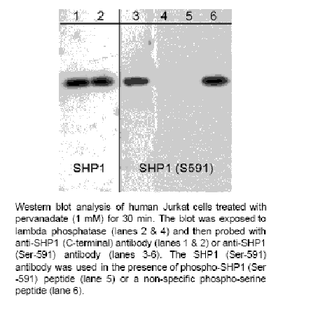 Application Data
SHP1 (Ser-591), Polyclonal Antibody (Cat# AAA71532)
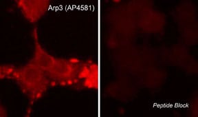 ICC (Immunocytochemistry)
(Immunocytochemical labeling of Arp3 in aldehyde-fixed and NP-40-permeabilized rat PC12 cells differentiated with NGF. The cells were labeled with rabbit polyclonal anti-Arp3 (C-terminal region) (AP4581) in the absence (left) or presence (right) of blocking peptide (AX4585). The antibody was detected using appropriate secondary antibody conjugated to DyLight 594.)
ICC (Immunocytochemistry)
(Immunocytochemical labeling of Arp3 in aldehyde-fixed and NP-40-permeabilized rat PC12 cells differentiated with NGF. The cells were labeled with rabbit polyclonal anti-Arp3 (C-terminal region) (AP4581) in the absence (left) or presence (right) of blocking peptide (AX4585). The antibody was detected using appropriate secondary antibody conjugated to DyLight 594.)
Arp3, Polyclonal Antibody (Cat# AAA71549)
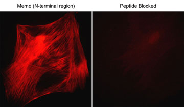 ICC (Immunocytochemistry)
(Immunocytochemical labeling of Memo in rabbit spleen fibroblasts that were fixed in paraformaldehyde and permeabilized with NP-40. The cells were probed with the Memo (N-terminal Region) MP3721, then the antibody was detected using goat anti-rabbit DyLight 594. The antibody was used in the absence (left) or presence (right) of it's blocking peptide (MX3725).)
ICC (Immunocytochemistry)
(Immunocytochemical labeling of Memo in rabbit spleen fibroblasts that were fixed in paraformaldehyde and permeabilized with NP-40. The cells were probed with the Memo (N-terminal Region) MP3721, then the antibody was detected using goat anti-rabbit DyLight 594. The antibody was used in the absence (left) or presence (right) of it's blocking peptide (MX3725).)
Memo, Polyclonal Antibody (Cat# AAA71551)
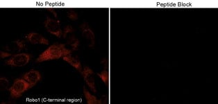 ICC (Immunocytochemistry)
(Immunocytochemical labeling of Robo1 in mouse C2C12. The cells were labeled with rabbit polyclonal Robo1 (C-terminal region) in the absence or presence of blocking peptide (RX2795). The antibody was then detected using appropriate secondary antibodies conjugated to Cy3.)
ICC (Immunocytochemistry)
(Immunocytochemical labeling of Robo1 in mouse C2C12. The cells were labeled with rabbit polyclonal Robo1 (C-terminal region) in the absence or presence of blocking peptide (RX2795). The antibody was then detected using appropriate secondary antibodies conjugated to Cy3.)
Robo1, Polyclonal Antibody (Cat# AAA71552)
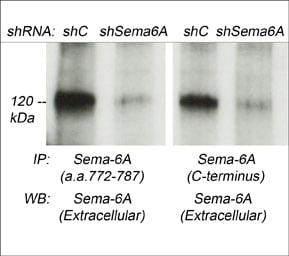
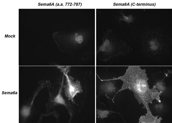 ICC (Immunocytochemistry)
(Immunocytochemical labeling of Sema-6A in COS7 cells that were mock transfected (top images) or Sema-6A transfected (bottom images). The cells were labeled with anti-Sema-6A (a.a. 772-787) (Left top and bottom image) or anti-Sema-6A (C-terminus) (Right top and bottom image). The antibodies were detected using anti-rabbit fluorescent secondary antibody. (Images provided by Dr. Luca Tamagnone from the IRCC, University of Torino, Italy).)
ICC (Immunocytochemistry)
(Immunocytochemical labeling of Sema-6A in COS7 cells that were mock transfected (top images) or Sema-6A transfected (bottom images). The cells were labeled with anti-Sema-6A (a.a. 772-787) (Left top and bottom image) or anti-Sema-6A (C-terminus) (Right top and bottom image). The antibodies were detected using anti-rabbit fluorescent secondary antibody. (Images provided by Dr. Luca Tamagnone from the IRCC, University of Torino, Italy).)
Semaphorin-6A, Polyclonal Antibody (Cat# AAA71554)
 WB (Western Blot)
(Western blot analysis of human recombinant AIM2 full length sequence with N-terminal GST tag (62kDa). The blot was probed with rabbit polyclonal anti-AIM2 (N-terminal region) antibody at 1:250 (lane 1) and 1:1000 (lane 2).)
WB (Western Blot)
(Western blot analysis of human recombinant AIM2 full length sequence with N-terminal GST tag (62kDa). The blot was probed with rabbit polyclonal anti-AIM2 (N-terminal region) antibody at 1:250 (lane 1) and 1:1000 (lane 2).)
AIM2, Polyclonal Antibody (Cat# AAA71566)
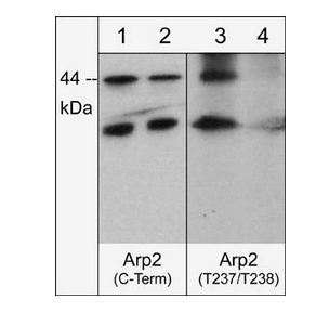
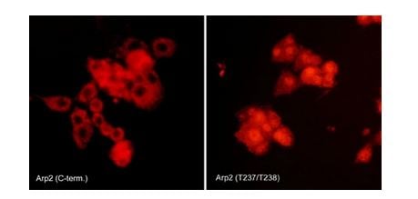 ICC (Immunocytochemistry)
(Immunocytochemical labeling of Arp2 phosphorylation in rat PC12 cells differentiated with NGF. The cells were probed with Arp2 (C-terminal region) and Arp2 (Thr-237/Thr-238) rabbit polyclonal antibodies, then the antibodies were detected using appropriate secondary antibody conjugated to Cy3.)
ICC (Immunocytochemistry)
(Immunocytochemical labeling of Arp2 phosphorylation in rat PC12 cells differentiated with NGF. The cells were probed with Arp2 (C-terminal region) and Arp2 (Thr-237/Thr-238) rabbit polyclonal antibodies, then the antibodies were detected using appropriate secondary antibody conjugated to Cy3.)
Arp2, Polyclonal Antibody (Cat# AAA71567)
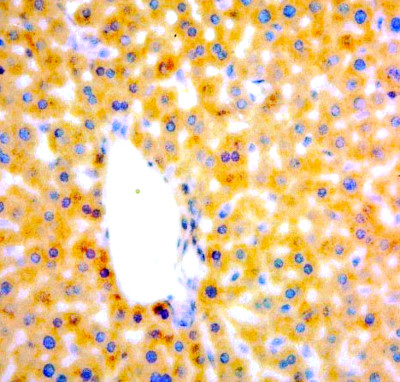 IHC (Immunohistochemistry)
(Immunohistochemistry: Human Liver carcinoma (FFPE) stained with Rabbit anti-HBV core antigen (Cat# AAA71307 ) at 1:200 for 10 min @ RT. Staining of formalin-fixed tissue requires boiling tissue sections in 10 mM Citrate Buffer, pH 6.0 for 10 min followed by cooling at RT for 20 min.)
IHC (Immunohistochemistry)
(Immunohistochemistry: Human Liver carcinoma (FFPE) stained with Rabbit anti-HBV core antigen (Cat# AAA71307 ) at 1:200 for 10 min @ RT. Staining of formalin-fixed tissue requires boiling tissue sections in 10 mM Citrate Buffer, pH 6.0 for 10 min followed by cooling at RT for 20 min.)
Hepatitis B virus core Ag, Polyclonal Antibody (Cat# AAA71307)
 IHC (Immunohistochemistry)
(Immunohistochemistry: Human esophagus tissue (FFPE) stained with Rabbit Anti-GLUT1 antibody (Cat# AAA71309) at 1:50 for 10 min @ RT. Staining of formalin-fixed tissue requires boiling tissue sections in 10 mM Citrate Buffer, pH 6.0 for 10 min followed by cooling at RT for 20 min.)
IHC (Immunohistochemistry)
(Immunohistochemistry: Human esophagus tissue (FFPE) stained with Rabbit Anti-GLUT1 antibody (Cat# AAA71309) at 1:50 for 10 min @ RT. Staining of formalin-fixed tissue requires boiling tissue sections in 10 mM Citrate Buffer, pH 6.0 for 10 min followed by cooling at RT for 20 min.)
GLUT-1, Polyclonal Antibody (Cat# AAA71309)
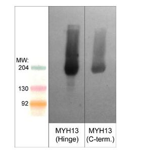 WB (Western Blot)
(Western blot analysis of MYH13 in mouse extraocular muscle. The blot was probed on the left with anti-MYH13/Extraocular myosin (Hinge region) antibody (AAA71667) and on the right with anti-MYH13/Extraocular myosin (C-terminal region) antibody .)
WB (Western Blot)
(Western blot analysis of MYH13 in mouse extraocular muscle. The blot was probed on the left with anti-MYH13/Extraocular myosin (Hinge region) antibody (AAA71667) and on the right with anti-MYH13/Extraocular myosin (C-terminal region) antibody .)
Myosin 13/Extraocular Myosin, Polyclonal Antibody (Cat# AAA71667)
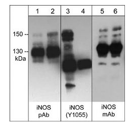
 ICC (Immunocytochemistry)
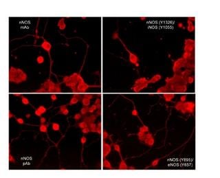
(Immunocytochemical labeling of nNOS phosphorylation in rat PC12 cells differentiated with NGF. The cells were probed with mouse monoclonal (mAb) nNOS (NM4011), and rabbit polyclonal (pAb) nNOS (C-terminal region), nNOS (Tyr-895)/eNOS (Tyr-657), and nNOS (Tyr-1326)/iNOS (Tyr-1055). The antibodies were detected using appropriate secondary antibody conjugated to DyLight 594.)
ICC (Immunocytochemistry)
(Immunocytochemical labeling of nNOS phosphorylation in rat PC12 cells differentiated with NGF. The cells were probed with mouse monoclonal (mAb) nNOS (NM4011), and rabbit polyclonal (pAb) nNOS (C-terminal region), nNOS (Tyr-895)/eNOS (Tyr-657), and nNOS (Tyr-1326)/iNOS (Tyr-1055). The antibodies were detected using appropriate secondary antibody conjugated to DyLight 594.)
iNOS, Polyclonal Antibody (Cat# AAA71676)
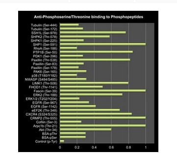 Application Data
(Bar grapHshowing anti-Phosphoserine/threonine (PP2551) binding to a variety of phosphoserine and phosphothreonine peptides, but not control peptide containing unphosphorylated serine or phosphotyrosine.)
Application Data
(Bar grapHshowing anti-Phosphoserine/threonine (PP2551) binding to a variety of phosphoserine and phosphothreonine peptides, but not control peptide containing unphosphorylated serine or phosphotyrosine.)
Phosphoserine/threonine, Polyclonal Antibody (Cat# AAA71694)
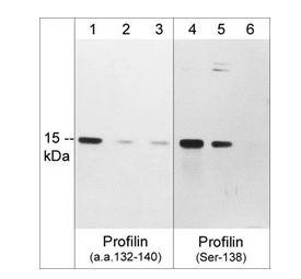
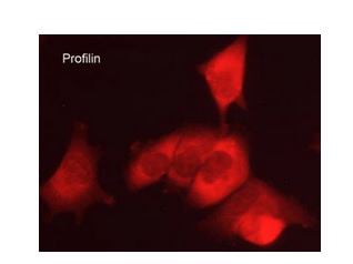 ICC (Immunocytochemistry)
(Immunocytochemical labeling of Profilin in aldehyde-fixed and NP-40 permeabilized human NCI-H1915 lung carcinoma cells. The cells were labeled with rabbit polyclonal anti-Profilin (PP4821) antibody. The antibody was detected using appropriate secondary antibody conjugated to DyLight 594.)
ICC (Immunocytochemistry)
(Immunocytochemical labeling of Profilin in aldehyde-fixed and NP-40 permeabilized human NCI-H1915 lung carcinoma cells. The cells were labeled with rabbit polyclonal anti-Profilin (PP4821) antibody. The antibody was detected using appropriate secondary antibody conjugated to DyLight 594.)
Profilin, Polyclonal Antibody (Cat# AAA71698)
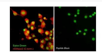 ICC (Immunocytochemistry)
(Immunocytochemical labeling of nSMase2 in aldehyde-fixed and NP-40-permeabilized differentiated PC12 cells. The cells were labeled with rabbit polyclonal anti-nSMase2 (SP4061) antibody in the absence (Left) or presence (Right) of blocking peptide (SX4065). The antibody was detected using appropriate secondary antibody conjugated to DyLight 594. The cells were counterstained with Sytox green to label nuclei.)
ICC (Immunocytochemistry)
(Immunocytochemical labeling of nSMase2 in aldehyde-fixed and NP-40-permeabilized differentiated PC12 cells. The cells were labeled with rabbit polyclonal anti-nSMase2 (SP4061) antibody in the absence (Left) or presence (Right) of blocking peptide (SX4065). The antibody was detected using appropriate secondary antibody conjugated to DyLight 594. The cells were counterstained with Sytox green to label nuclei.)
nSMase2, Polyclonal Antibody (Cat# AAA71714)
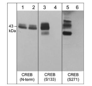
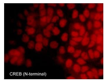 ICC (Immunocytochemistry)
(Immunocytochemical labeling of CREB in A431 that were fixed in paraformaldehyde and permeabilized using NP-40. The cells were labeled with rabbit polyclonal CREB (N-terminal region). The antibody was detected using goat anti-rabbit DyLight 594.)
ICC (Immunocytochemistry)
(Immunocytochemical labeling of CREB in A431 that were fixed in paraformaldehyde and permeabilized using NP-40. The cells were labeled with rabbit polyclonal CREB (N-terminal region). The antibody was detected using goat anti-rabbit DyLight 594.)
CREB, Polyclonal Antibody (Cat# AAA71611)
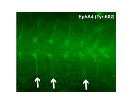
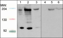
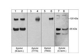 WB (Western Blot)
(Western blot analysis of humanuMbilical vein endothelial cells untreated (lanes 1, 3, 5, & 7) or treated with pervanadate (1mM) for 30 min. (lanes 2, 4, 6, & 8). The blot was probed with anti-EphA4 (N-terminal region) (lanes 1 & 2), anti-EphA4 (Tyr-779) (lanes 3 & 4), anti-EphA4 (Tyr-602) (lanes 5 & 6), or anti-EphA4 (C-terminal region) (lanes 7 & 8).)
WB (Western Blot)
(Western blot analysis of humanuMbilical vein endothelial cells untreated (lanes 1, 3, 5, & 7) or treated with pervanadate (1mM) for 30 min. (lanes 2, 4, 6, & 8). The blot was probed with anti-EphA4 (N-terminal region) (lanes 1 & 2), anti-EphA4 (Tyr-779) (lanes 3 & 4), anti-EphA4 (Tyr-602) (lanes 5 & 6), or anti-EphA4 (C-terminal region) (lanes 7 & 8).)
EphA4, Polyclonal Antibody (Cat# AAA71630)
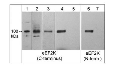
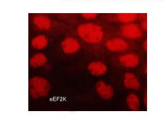 ICC (Immunocytochemistry)
(Immunocytochemical labeling of eEF2K in paraformaldehyde fixed and NP-40 permeabilized A431 cells. The cells were labeled with rabbit polyclonal anti-eEF2K (EP4661). The antibody was detected using goat anti-rabbit DyLight 594.)
ICC (Immunocytochemistry)
(Immunocytochemical labeling of eEF2K in paraformaldehyde fixed and NP-40 permeabilized A431 cells. The cells were labeled with rabbit polyclonal anti-eEF2K (EP4661). The antibody was detected using goat anti-rabbit DyLight 594.)
eEF2K, Polyclonal Antibody (Cat# AAA71635)
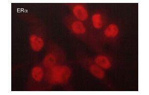 ICC (Immunocytochemistry)
(Immunocytochemical labeling of Estrogen Receptor alpha in paraformaldehyde fixed and NP-40 permeabilized MDA-MB-231 cells. The cells were labeled with rabbit polyclonal anti-Estrogen Receptor alpha (EP5431). The antibody was detected using goat anti-rabbit DyLight 594.)
ICC (Immunocytochemistry)
(Immunocytochemical labeling of Estrogen Receptor alpha in paraformaldehyde fixed and NP-40 permeabilized MDA-MB-231 cells. The cells were labeled with rabbit polyclonal anti-Estrogen Receptor alpha (EP5431). The antibody was detected using goat anti-rabbit DyLight 594.)
Estrogen Receptor alpha, Polyclonal Antibody (Cat# AAA71636)

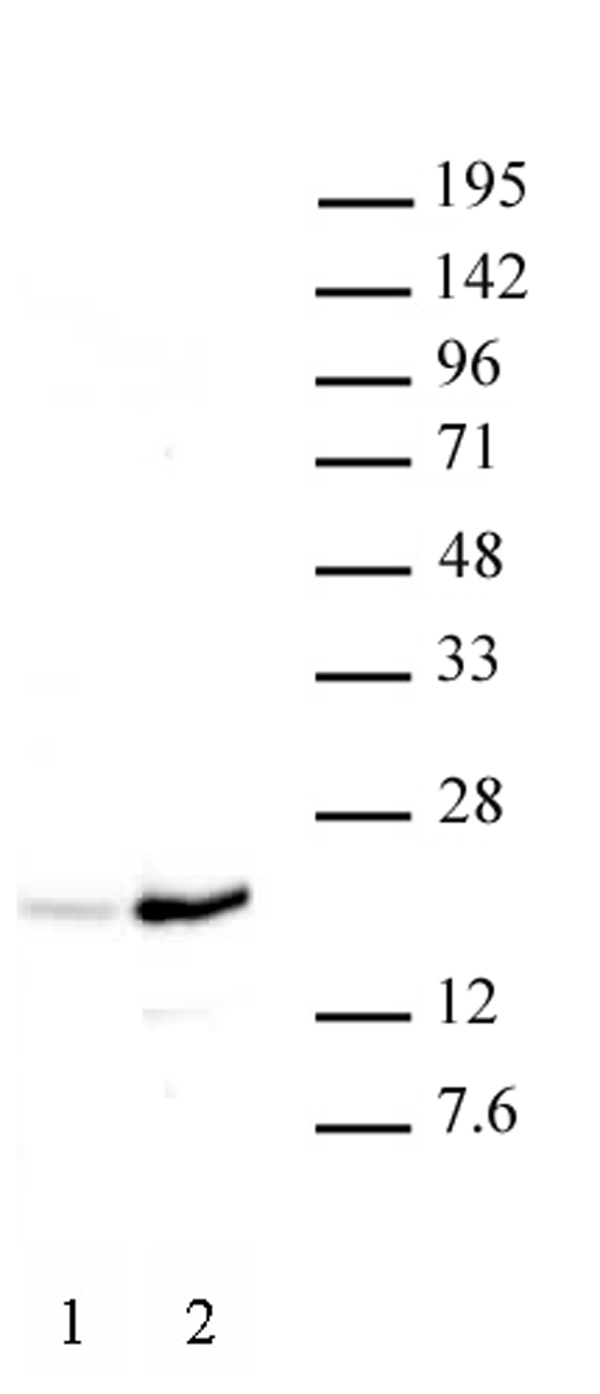
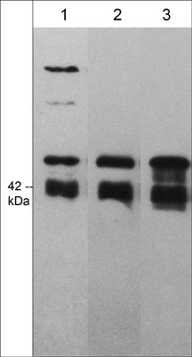
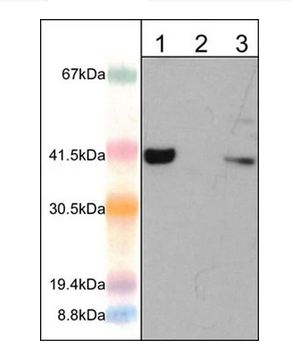
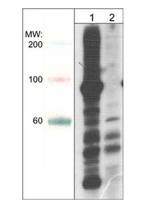
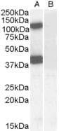
 WB (Western Blot)
(STAT2 antibody (pAb) tested by Western blot. Whole cell extract (20 ug) of Jurkat cells (Lane 1) or HeLa cells (Lane 2) probed with STAT2 antibody at a 1:1,000 dilution.)
WB (Western Blot)
(STAT2 antibody (pAb) tested by Western blot. Whole cell extract (20 ug) of Jurkat cells (Lane 1) or HeLa cells (Lane 2) probed with STAT2 antibody at a 1:1,000 dilution.)
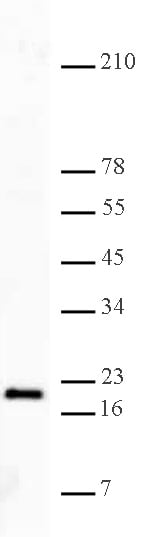
STAT2, Polyclonal Antibody (Cat# AAA60068)
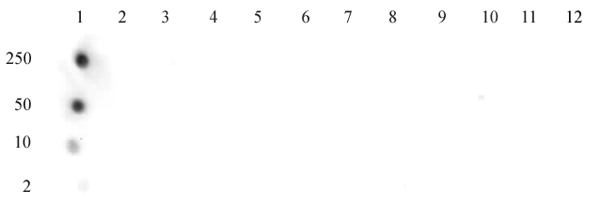
 DB (Dot Blot)
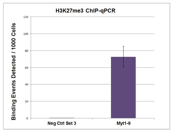
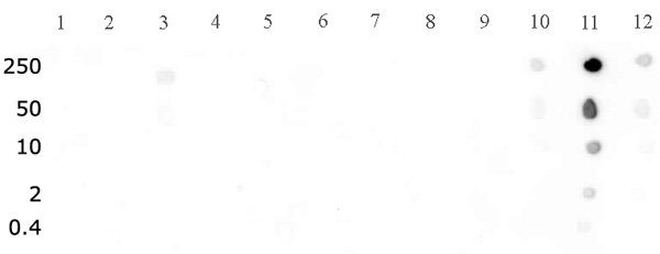
(Histone H3K27me3 antibody (pAb) tested by dot blot analysis. Dot blot analysis was used to confirm the specificity of Histone H3 trimethyl Lys27 antibody for trimethyl-lysine 27 of histone H3. Peptides corresponding to regions around major sites of histone H3 methylation (lysine 4, lysine 9, lysine 27) were spotted onto PVDF and probed with the antibody at a dilution of 1:5,000. The amount of peptide (in picomoles) spotted is indicated next to each row. Lane 1: Unmodified Lys4 peptide. Lane 2: Monomethyl Lys4 peptide. Lane 3: Dimethyl Lys4 peptide. Lane 4: Trimethyl Lys4 peptide. Lane 5: Monomethyl Lys9 peptide. Lane 6: Unmodified Lys9 peptide. Lane 7: Dimethyl Lys9 peptide. Lane 8: Trimethyl Lys9 peptide. Lane 9: Unmodified Lys27 peptide. Lane 10: Monomethyl Lys27 peptide. Lane 11: Dimethyl Lys27 peptide. Lane 12: Trimethyl Lys27 peptide.)
DB (Dot Blot)
(Histone H3K27me3 antibody (pAb) tested by dot blot analysis. Dot blot analysis was used to confirm the specificity of Histone H3 trimethyl Lys27 antibody for trimethyl-lysine 27 of histone H3. Peptides corresponding to regions around major sites of histone H3 methylation (lysine 4, lysine 9, lysine 27) were spotted onto PVDF and probed with the antibody at a dilution of 1:5,000. The amount of peptide (in picomoles) spotted is indicated next to each row. Lane 1: Unmodified Lys4 peptide. Lane 2: Monomethyl Lys4 peptide. Lane 3: Dimethyl Lys4 peptide. Lane 4: Trimethyl Lys4 peptide. Lane 5: Monomethyl Lys9 peptide. Lane 6: Unmodified Lys9 peptide. Lane 7: Dimethyl Lys9 peptide. Lane 8: Trimethyl Lys9 peptide. Lane 9: Unmodified Lys27 peptide. Lane 10: Monomethyl Lys27 peptide. Lane 11: Dimethyl Lys27 peptide. Lane 12: Trimethyl Lys27 peptide.)
Histone H3K27me3, Polyclonal Antibody (Cat# AAA59843)

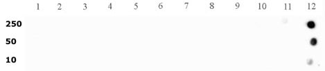
 DB (Dot Blot)
(Histone H3 dimethyl Lys27 antibody tested by dot blot analysis. Dot blot analysis was used to confirm the specificity of Histone H3 dimethyl Lys27 antibody for dimethyl Lys27 histone H3. Methylated peptides corresponding to the immunogen and related sequences derived from histone H3 were spotted onto PVDF and probed with the antibody at a 1:5,000 dilution. The amount of peptide (picomoles) spotted is indicated next to each row. Lane 1: Unmodified lysine 4 peptide. Lane 2: Monomethyl lysine 4 peptide. Lane 3: Dimethyl lysine 4 peptide. Lane 4: Trimethyl lysine 4 peptide. Lane 5: Unmodified lysine 9 peptide. Lane 6: Monomethyl lysine 9 peptide. Lane 7: Dimethyl lysine 9 peptide. Lane 8: Trimethyl lysine 9 peptide. Lane 9: Unmodified lysine 27 peptide. Lane 10: Monomethyl lysine 27 peptide. Lane 11: Dimethyl lysine 27 peptide. Lane 12: Trimethyl lysine 27 peptide.)
DB (Dot Blot)
(Histone H3 dimethyl Lys27 antibody tested by dot blot analysis. Dot blot analysis was used to confirm the specificity of Histone H3 dimethyl Lys27 antibody for dimethyl Lys27 histone H3. Methylated peptides corresponding to the immunogen and related sequences derived from histone H3 were spotted onto PVDF and probed with the antibody at a 1:5,000 dilution. The amount of peptide (picomoles) spotted is indicated next to each row. Lane 1: Unmodified lysine 4 peptide. Lane 2: Monomethyl lysine 4 peptide. Lane 3: Dimethyl lysine 4 peptide. Lane 4: Trimethyl lysine 4 peptide. Lane 5: Unmodified lysine 9 peptide. Lane 6: Monomethyl lysine 9 peptide. Lane 7: Dimethyl lysine 9 peptide. Lane 8: Trimethyl lysine 9 peptide. Lane 9: Unmodified lysine 27 peptide. Lane 10: Monomethyl lysine 27 peptide. Lane 11: Dimethyl lysine 27 peptide. Lane 12: Trimethyl lysine 27 peptide.)
Histone H3K27me2, Polyclonal Antibody (Cat# AAA59850)
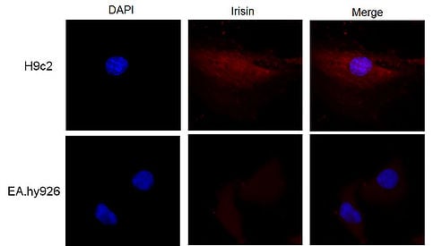
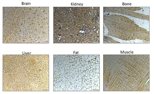
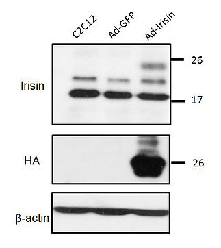
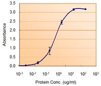
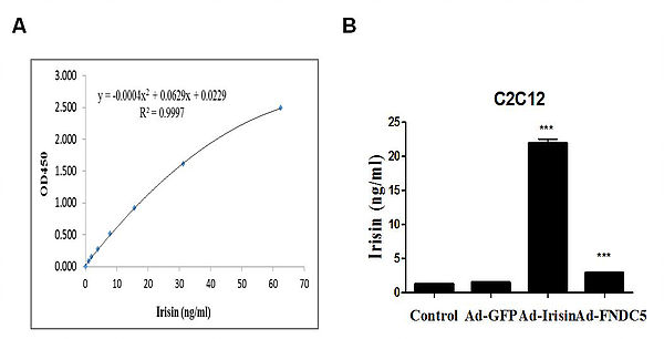 ELISA
(ELISA: Data demonstrates detection of irisin after gene delivery following ELISA protocol)
ELISA
(ELISA: Data demonstrates detection of irisin after gene delivery following ELISA protocol)
Irisin (FNDC5), Polyclonal Antibody (Cat# AAA60453)
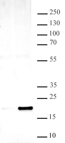
 Application Data
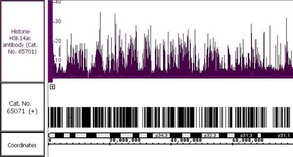
Histone H3K14ac, Polyclonal Antibody (Cat# AAA60350)
Mammaglobin B/SCGB2A1, Polyclonal Antibody (Cat# AAA60818)
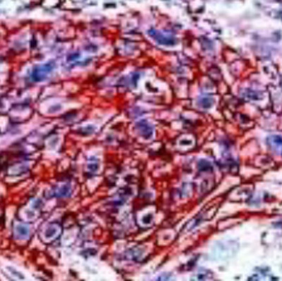
 IHC (Immunohiostchemistry)
((3.8ug/ml) staining of paraffin embedded Human Skeletal Muscle. Steamed antigen retrieval with citrate buffer pH 6, AP-staining.)
IHC (Immunohiostchemistry)
((3.8ug/ml) staining of paraffin embedded Human Skeletal Muscle. Steamed antigen retrieval with citrate buffer pH 6, AP-staining.)
AS160/TBC1D4, Polyclonal Antibody (Cat# AAA60819)

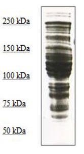
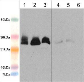
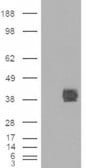
 WB (Western Blot)
(AAA61135 HEK293 overexpressing Dudulin4 (RC216917) and probed (mock transfection in first lane), tested by Origene.)
WB (Western Blot)
(AAA61135 HEK293 overexpressing Dudulin4 (RC216917) and probed (mock transfection in first lane), tested by Origene.)
STEAP4/Dudulin4, Polyclonal Antibody (Cat# AAA61135)
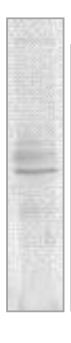
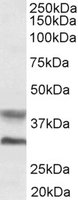
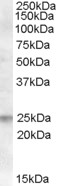
 WB (Western Blot)
((0.3ug/ml) staining of Human Brain (Cerebellum) lysate (35ug protein in RIPA buffer). Primary incubation was 1 hour. Detected by chemiluminescence.)
WB (Western Blot)
((0.3ug/ml) staining of Human Brain (Cerebellum) lysate (35ug protein in RIPA buffer). Primary incubation was 1 hour. Detected by chemiluminescence.)
SEC61A1, Polyclonal Antibody (Cat# AAA61138)
 IHC (Immunohiostchemistry)
((2.5ug/ml) staining of paraffin embedded Human Uterus. Steamed antigen retrieval with citrate buffer pH 6, AP-staining.)
IHC (Immunohiostchemistry)
((2.5ug/ml) staining of paraffin embedded Human Uterus. Steamed antigen retrieval with citrate buffer pH 6, AP-staining.)
EPB41L2/4.1G, Polyclonal Antibody (Cat# AAA61197)
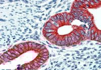
 IHC (Immunohiostchemistry)
((2.5ug/ml) staining of paraffin embedded Human Breast. Steamed antigen retrieval with citrate buffer pH 6, AP-staining.)
IHC (Immunohiostchemistry)
((2.5ug/ml) staining of paraffin embedded Human Breast. Steamed antigen retrieval with citrate buffer pH 6, AP-staining.)
GCDFP15/PIP, Polyclonal Antibody (Cat# AAA60868)
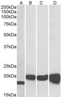
 WB (Western Blot)
((1ug/ml) staining of Human Adipose lysate (35ug protein in RIPA buffer). Primary incubation was 1 hour. Detected by chemiluminescence.)
WB (Western Blot)
((1ug/ml) staining of Human Adipose lysate (35ug protein in RIPA buffer). Primary incubation was 1 hour. Detected by chemiluminescence.)
UCP1, Polyclonal Antibody (Cat# AAA60873)
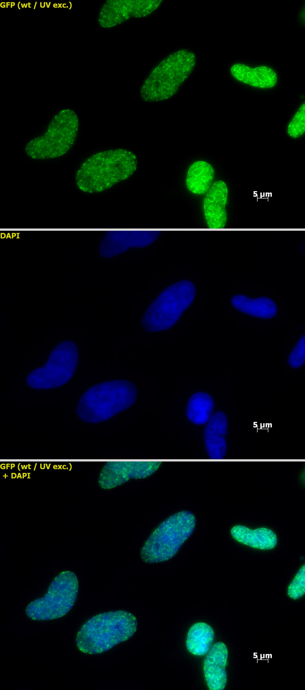
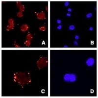 IF (Immunofluorescence)
(Staining of ATDC5 cells (Panels A and C) and DAPI (panels B and D). Data gratefuly received from Dr. Shiro Ikegawa, SNP Research Center, RIKEN, Japan.)
IF (Immunofluorescence)
(Staining of ATDC5 cells (Panels A and C) and DAPI (panels B and D). Data gratefuly received from Dr. Shiro Ikegawa, SNP Research Center, RIKEN, Japan.)
Asporin/ASPN, Polyclonal Antibody (Cat# AAA60886)
 IHC (Immunohiostchemistry)
((2.5ug/ml) staining of paraffin embedded Human Placenta. Steamed antigen retrieval with citrate buffer pH 6, AP-staining.)
IHC (Immunohiostchemistry)
((2.5ug/ml) staining of paraffin embedded Human Placenta. Steamed antigen retrieval with citrate buffer pH 6, AP-staining.)
FAIM1, Polyclonal Antibody (Cat# AAA60934)
PDX1, Polyclonal Antibody (Cat# AAA60496)
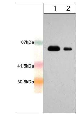
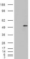
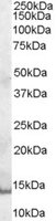
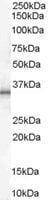
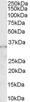
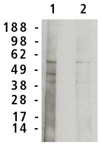 WB (Western Blot)
(Western blot analysis using acid sphingomyelinase antibody on normal human brain lysate (7 ug/lane). Antibody used at 1 ug/ml (1) and 0.5 ug/ml (2) and detected using mouse anti-rabbit antibody at 1:75k dilution and visualized using Pierce West Femto substrate.)
WB (Western Blot)
(Western blot analysis using acid sphingomyelinase antibody on normal human brain lysate (7 ug/lane). Antibody used at 1 ug/ml (1) and 0.5 ug/ml (2) and detected using mouse anti-rabbit antibody at 1:75k dilution and visualized using Pierce West Femto substrate.)
Acid Sphingomyelinase, Polyclonal Antibody (Cat# AAA60506)
 Application Data
((1.5ug/ml) as the reporter with as the capture rabbit antibody (5ug/ml).)
Application Data
((1.5ug/ml) as the reporter with as the capture rabbit antibody (5ug/ml).)
SOD1, Polyclonal Antibody (Cat# AAA61049)
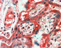
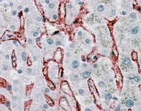 IHC (Immunohistochemisry)
((3.8ug/ml) staining of paraffin embedded Human Liver. Steamed antigen retrieval with citrate buffer pH 6, AP-staining.)
IHC (Immunohistochemisry)
((3.8ug/ml) staining of paraffin embedded Human Liver. Steamed antigen retrieval with citrate buffer pH 6, AP-staining.)
CD32/FCGR2B, Polyclonal Antibody (Cat# AAA61115)
What are Polyclonal Antibodies?
Polyclonal antibodies are antibodies that come from multiple B cell clones of a host animal. The typical hosts used for the majority of polyclonal antibody production are rabbits, goats, sheep, and donkeys. These polyclonal antibodies, once having identified their target, will bind to different epitopes located at different regions or sequences on the same protein/antigen. This ability to bind multiple epitopes is what makes polyclonal antibodies highly sensitive, as explained in our detailed guide on polyclonal antibodies and why they matter.
As a result, they are ideal at locating and binding to the target, even if the target is in very low concentrations (due to many different antibodies being able to bind to the same target molecule, which allows for significant amplification of a downstream signal).
Polyclonal antibodies are typically produced by injecting an antigen into a host animal, which causes the animal’s immune system to attack the foreign antigen by mass generating antibodies against it. After a period of time, serum is collected from the animal and purified using physicochemical fractionation, class-specific affinity purification, and/or antigen-affinity purification.
Key Uses of Polyclonal Antibodies
- Western Blotting: This method is used to find specific proteins in biological samples after separating them by size.
- Immunohistochemistry: IHC helps visualize the location of proteins in tissue sections using various staining techniques.
- ELISA: (Enzyme-Linked Immunosorbent Assay) is typically used to identify specific protein quantities in a sample. ELISAs can be either “Quantitative” or “Qualitative”.
- Flow Cytometry: technique that identifies and measures the specific protein on the surface or inside the cells in a fluid suspension.
- Immunoprecipitation: IP isolates and studies a specific protein from a complex mixture using antibodies.
Why Buy Polyclonal Antibodies from AAA Biotech?
1. Ideal for Various Applications
Our antibodies are generally going to be validated for use in multiple types of assays, including ELISA, Western Blotting, Immunohistochemistry, Immunoprecipitation, amongst others. They are ideal for a wide range of research applications.
2. Rigorous Quality Control
All of the antibodies in our catalog undergo strict quality testing to ensure specificity, sensitivity, and consistent performance. We are confident in the ability of our antibodies to provide you with accurate results.
3. Wide Assortment of Antibodies
Antibodies in our catalog can be found for both common and exotic species, and these antibodies are also available in both conjugated and recombinant forms to suit many diverse experimental needs.
4. Highly Purified
Our antibodies are available in purified forms with over 85% purity, as confirmed by SDS-PAGE. They are also available with tags such as His, Flag, GST, or MBP. We cater to customers worldwide.
FAQ
1. How are polyclonal antibodies produced?
Traditionally, polyclonal antibodies are produced by injecting an antigen into a host animal (such as a rabbit or goat), which then triggers an immune response from the host animal. The animal’s B cells produce antibodies that will recognize different parts of the injected antigen. These antibodies are then collected from the animal’s blood and purified for use.
2. How do polyclonal antibodies differ from monoclonal antibodies?
Polyclonal antibodies are a mix of antibodies that bind to different locations (epitopes) of the same antigen, while monoclonal antibodies are identical and bind to just one specific epitope. This makes polyclonal antibodies more versatile and better at detecting proteins that may be present in low quantities or in altered/modified forms.
3. How should I store polyclonal antibodies?
Polyclonal antibodies should be stored at 4°C for short-term use (up to a few weeks) and at -20°C or -80°C for long-term storage. Avoid repeated freeze-thaw cycles by dividing them into small aliquots. Always check the datasheet for specific storage instructions.