Filters
▼Clonality
▼Type
▼Reactivity
▼Gene Name
▼Isotype
▼Host
▼Application
▼Clone
▼Polyclonal Antibodies
At AAA Biotech also known as AAA Bio or AAABio, we provide a broad range of purified polyclonal antibodies (pAbs) that are able to all be browsed online through our website. Due to their high specificity and strong binding affinity, these antibodies are ideal for wide swathes of research and experimental applications.
Our polyclonal antibodies can easily support your work, whether you use them for Western Blotting, Immunocytochemistry (with or without Immunofluorescence used in conjunction), Immunohistochemistry, Immunoprecipitation, and ELISA tests. We highly encourage you to browse our range of pAbs and choose the one that best suits your experimental model.
Viewing 200-250 of 118597 product results
 WB (Western Blot)
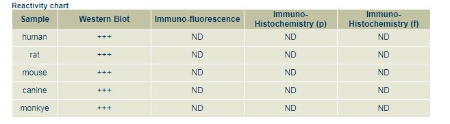
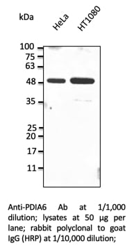
PDIA6, Polyclonal Antibody (Cat# AAA63160)
 WB (Western Blot)
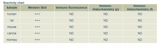
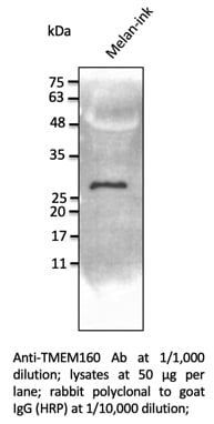
TMEM160, Polyclonal Antibody (Cat# AAA63161)
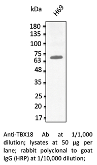 WB (Western Blot)
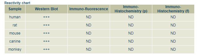
TBX18, Polyclonal Antibody (Cat# AAA63165)
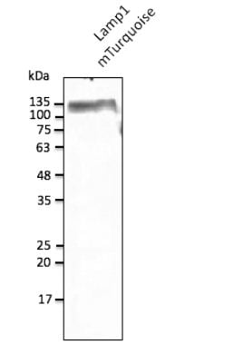
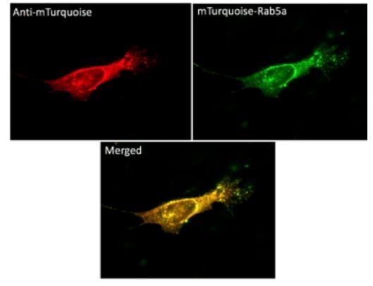
 IF (Immunofluorescence)
(Immunofluorescence - anti-mTurquoise Ab using NIH3T3 cells transduced with mTurquoise-Rab5a; cells were fixed with methanol and anti-mTurquoise at 1/100;)
IF (Immunofluorescence)
(Immunofluorescence - anti-mTurquoise Ab using NIH3T3 cells transduced with mTurquoise-Rab5a; cells were fixed with methanol and anti-mTurquoise at 1/100;)
mTurquoise, Polyclonal Antibody (Cat# AAA63227)
 IF (Immunofluorescence)
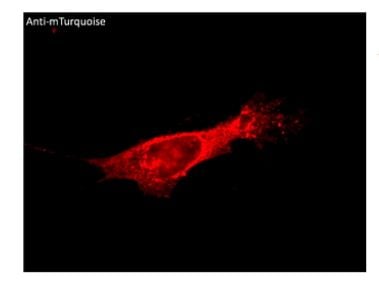
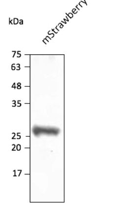
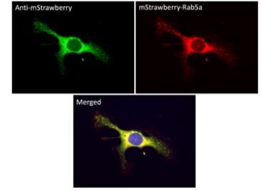
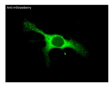
(Immunofluorescence -anti-mStrawberry Ab using hCEC cells transduced with mStrawberry-Rab5a; cells were fixed with methanol and anti-mStrawberry at 1/250;)
IF (Immunofluorescence)
(Immunofluorescence -anti-mStrawberry Ab using hCEC cells transduced with mStrawberry-Rab5a; cells were fixed with methanol and anti-mStrawberry at 1/250;)
mStrawberry, Polyclonal Antibody (Cat# AAA63238)
 Application Data
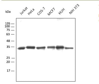
(Anti-GAPDH Ab conjugated to DyLight550 (AB49550) at 1:2,000 using lysates at 30 ug per lane;)
Application Data
(Anti-GAPDH Ab conjugated to DyLight550 (AB49550) at 1:2,000 using lysates at 30 ug per lane;)
GAPDH, Polyclonal Antibody (Cat# AAA63240)
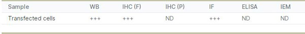
 Application Data
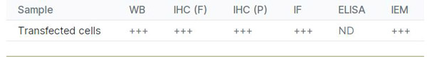
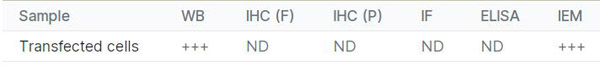
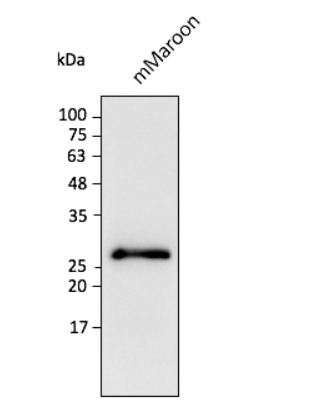
(Anti-mMaroon Ab at 1/2,500 dilution using HEK293 transfected cell lysates at 40 ug per lane; rabbit polyclonal to goat IgG (HRP) at 1/10,000 dilution;)
Application Data
(Anti-mMaroon Ab at 1/2,500 dilution using HEK293 transfected cell lysates at 40 ug per lane; rabbit polyclonal to goat IgG (HRP) at 1/10,000 dilution;)
mMaroon, Polyclonal Antibody (Cat# AAA63245)
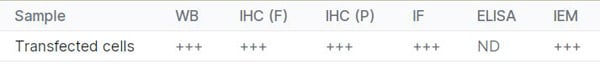
 IF (Immunofluorescence)
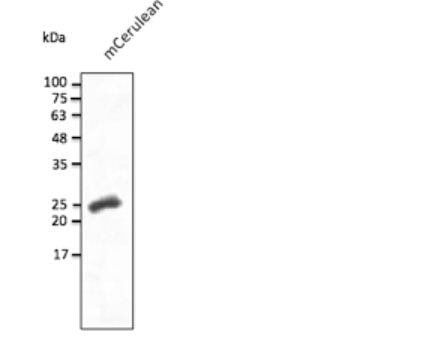
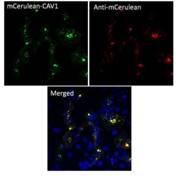
(Immunofluorescence - anti-mCerulean Ab using COS7 cells transduced with mCerulean-CAV1; cells were fixed with methanol and anti-mCerulean at 1/250;)
IF (Immunofluorescence)
(Immunofluorescence - anti-mCerulean Ab using COS7 cells transduced with mCerulean-CAV1; cells were fixed with methanol and anti-mCerulean at 1/250;)
mCerulean, Polyclonal Antibody (Cat# AAA63254)
 IF (Immunofluorescence)
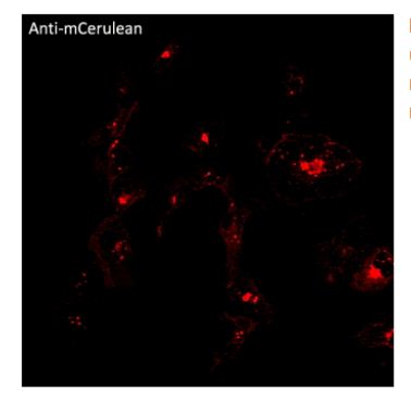
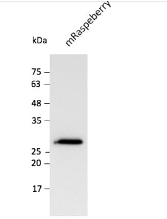
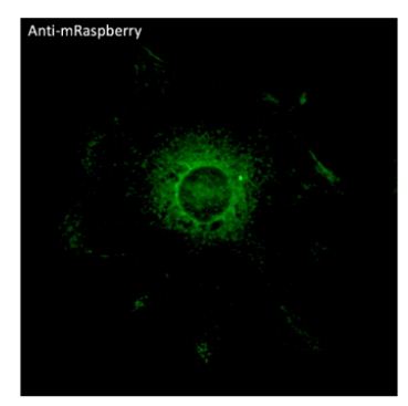
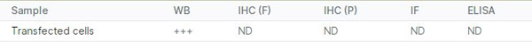
(Immunofluorescence - anti-mRaspberry Ab using hCEC cells transduced with mRaspberry-Rab5a; cells were fixed with methanol and anti-mRaspberry at 1/250;)
IF (Immunofluorescence)
(Immunofluorescence - anti-mRaspberry Ab using hCEC cells transduced with mRaspberry-Rab5a; cells were fixed with methanol and anti-mRaspberry at 1/250;)
mRaspberry, Polyclonal Antibody (Cat# AAA63260)
 Application Data
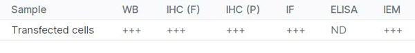
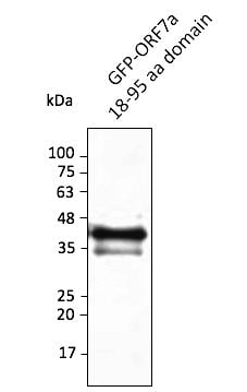
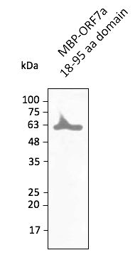
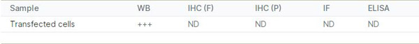
(Anti-ORF7a Ab at 1/2,500 dilution; lane with 30 ng of recombinant fusion protein; rabbit polyclonal to goat IgG (HRP) at 1/10,000 dilution;)
Application Data
(Anti-ORF7a Ab at 1/2,500 dilution; lane with 30 ng of recombinant fusion protein; rabbit polyclonal to goat IgG (HRP) at 1/10,000 dilution;)
COVID 19 ORF7a Coronavirus, Polyclonal Antibody (Cat# AAA63177)
 Application Data
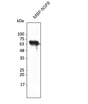
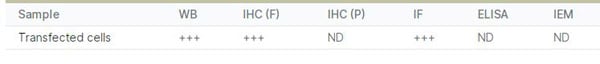
(Anti-NSP8 Ab at 1/2,500 dilution; lane with 30 ng of recombinant fusion protein; rabbit polyclonal to goat IgG (HRP) at 1/10,000 dilution;)
Application Data
(Anti-NSP8 Ab at 1/2,500 dilution; lane with 30 ng of recombinant fusion protein; rabbit polyclonal to goat IgG (HRP) at 1/10,000 dilution;)
COVID 19 NSP8 Coronavirus, Polyclonal Antibody (Cat# AAA63184)
 IF (Immunofluorescence)
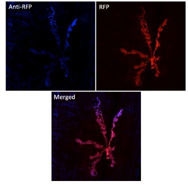
(Immunofluorescence in Drosophila larvaenmJ expressing RFP in neurons (DvglutRFP), muscle 6/7 usingAnti-RFP conjugated to DyLight405 at 1/500;)
IF (Immunofluorescence)
(Immunofluorescence in Drosophila larvaenmJ expressing RFP in neurons (DvglutRFP), muscle 6/7 usingAnti-RFP conjugated to DyLight405 at 1/500;)
RFP, Polyclonal Antibody (Cat# AAA63200)
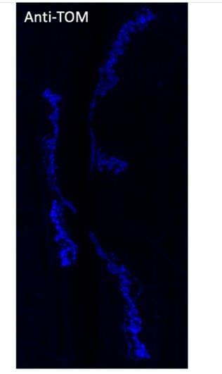 IF (Immunofluorescence)
(Immunofluorescence in Drosophila larvae expressing tdTomato in neurons usingAnti-tdTomato conjugated to DyLight405 at 1/500;)
IF (Immunofluorescence)
(Immunofluorescence in Drosophila larvae expressing tdTomato in neurons usingAnti-tdTomato conjugated to DyLight405 at 1/500;)
tdTomato, Polyclonal Antibody (Cat# AAA63207)
 IF (Immunofluorescence)
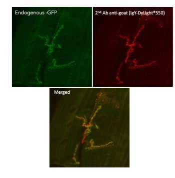
(Immunofluorescence in Drosophila larvaenmJ muscle 6/7 expressing GluRIIA-GFP in neurons (GluRIIA is a post-synaptic protein) using 1st Ab anti-GFP at 1/1,000 and 2nd Ab anti-goat IgY conjugated to DyLight550 at 1/500;)
IF (Immunofluorescence)
(Immunofluorescence in Drosophila larvaenmJ muscle 6/7 expressing GluRIIA-GFP in neurons (GluRIIA is a post-synaptic protein) using 1st Ab anti-GFP at 1/1,000 and 2nd Ab anti-goat IgY conjugated to DyLight550 at 1/500;)
IgG, Polyclonal Secondary Antibody (Cat# AAA63211)
 IF (Immunofluorescence)
(IF of C3c on a FFPE Lupus Positice Tissue)
IF (Immunofluorescence)
(IF of C3c on a FFPE Lupus Positice Tissue)
C3c, Polyclonal Antibody (Cat# AAA59322)
 WB (Western Blot)
((0.5ug/ml) staining of Rat Kidney lysate (35ug protein in RIPA buffer). Primary incubation was 1 hour. Detected by chemiluminescence.)
WB (Western Blot)
((0.5ug/ml) staining of Rat Kidney lysate (35ug protein in RIPA buffer). Primary incubation was 1 hour. Detected by chemiluminescence.)
Renalase, Polyclonal Antibody (Cat# AAA60960)

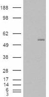
 WB (Western Blot)
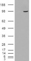
(HEK293 overexpressing SMEK1 (RC221040) and probed (mock transfection in first lane), tested by Origene.)
WB (Western Blot)
(HEK293 overexpressing SMEK1 (RC221040) and probed (mock transfection in first lane), tested by Origene.)
SMEK1/KIAA2010, Polyclonal Antibody (Cat# AAA60983)
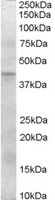
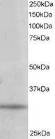 WB (Western Blot)
(Staining (1ug/ml) of 293 lysate (RIPA buffer, 35ug total protein per lane). Primary incubated for 1 hour. Detected by western blot using chemiluminescence.)
WB (Western Blot)
(Staining (1ug/ml) of 293 lysate (RIPA buffer, 35ug total protein per lane). Primary incubated for 1 hour. Detected by western blot using chemiluminescence.)
PITPN/PITP alpha, Polyclonal Antibody (Cat# AAA60988)
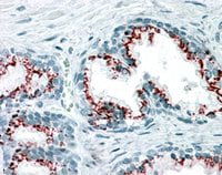
 IHC (Immunohistochemistry)
((3.8ug/ml) staining of paraffin embedded Human Prostate. Steamed antigen retrieval with citrate buffer pH 6, AP-staining.)
IHC (Immunohistochemistry)
((3.8ug/ml) staining of paraffin embedded Human Prostate. Steamed antigen retrieval with citrate buffer pH 6, AP-staining.)
ZNF217, Polyclonal Antibody (Cat# AAA60999)
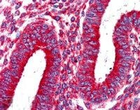 IHC (Immunohistochemisry)
((2.5ug/ml) staining of paraffin embedded Human Uterus. Steamed antigen retrieval with citrate buffer pH 6, AP-staining. Please note this data was obtained with a previous batch.)
IHC (Immunohistochemisry)
((2.5ug/ml) staining of paraffin embedded Human Uterus. Steamed antigen retrieval with citrate buffer pH 6, AP-staining. Please note this data was obtained with a previous batch.)
Kinesin 1/UKHC, Polyclonal Antibody (Cat# AAA61010)
 WB (Western Blot)
((2ug/ml) staining of Human T-lymphocyte lysate (35ug protein in RIPA buffer). Primary incubation was 1 hour. Detected by chemiluminescence. Data provided by anonymous customer.)
WB (Western Blot)
((2ug/ml) staining of Human T-lymphocyte lysate (35ug protein in RIPA buffer). Primary incubation was 1 hour. Detected by chemiluminescence. Data provided by anonymous customer.)
Munc13-4/UNC13D, Polyclonal Antibody (Cat# AAA61013)
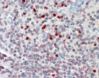 IHC (Immunohiostchemistry)
((3.8ug/ml) staining of paraffin embedded Human Spleen. Steamed antigen retrieval with citrate buffer pH 6, AP-staining.)
IHC (Immunohiostchemistry)
((3.8ug/ml) staining of paraffin embedded Human Spleen. Steamed antigen retrieval with citrate buffer pH 6, AP-staining.)
FLAP/ALOX5AP, Polyclonal Antibody (Cat# AAA61035)
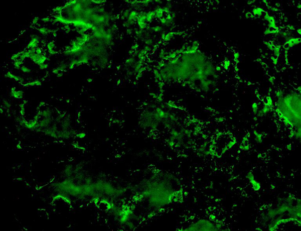
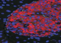 IF (Immunofluorescence)
((50ug/ml) staining of islets in Mouse Pancreas. Citrate antigen retrieval. Detected by immunofluorescence.)
IF (Immunofluorescence)
((50ug/ml) staining of islets in Mouse Pancreas. Citrate antigen retrieval. Detected by immunofluorescence.)
UNC5C, Polyclonal Antibody (Cat# AAA61362)
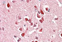
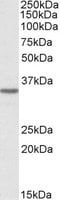
 WB (Western Blot)
((0.3ug/ml) staining of Rat Brain lysate (35ug protein in RIPA buffer). Primary incubation was 1 hour. Detected by chemiluminescence.)
WB (Western Blot)
((0.3ug/ml) staining of Rat Brain lysate (35ug protein in RIPA buffer). Primary incubation was 1 hour. Detected by chemiluminescence.)
Clusterin/ApoJ, Polyclonal Antibody (Cat# AAA61410)
 WB (Western Blot)
((0.3ug/ml) staining of HeLa cell lysate (35ug protein in RIPA buffer). Primary incubation was 1 hour. Detected by chemiluminescence.)
WB (Western Blot)
((0.3ug/ml) staining of HeLa cell lysate (35ug protein in RIPA buffer). Primary incubation was 1 hour. Detected by chemiluminescence.)
MICS1/GHITM, Polyclonal Antibody (Cat# AAA61416)
GLIPR1/RTVP-1, Polyclonal Antibody (Cat# AAA61317)
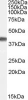
 WB (Western Blot)
(HEK293 overexpressing GCNT3 (RC202007) and probed (mock transfection in first lane), tested by Origene.)
WB (Western Blot)
(HEK293 overexpressing GCNT3 (RC202007) and probed (mock transfection in first lane), tested by Origene.)
C2GnT-M, Polyclonal Antibody (Cat# AAA61318)
 IHC (Immunohiostchemistry)
((3ug/ml) staining of paraffin embedded Human Pancreas. Microwaved antigen retrieval with citrate buffer pH 6, HRP-staining)
IHC (Immunohiostchemistry)
((3ug/ml) staining of paraffin embedded Human Pancreas. Microwaved antigen retrieval with citrate buffer pH 6, HRP-staining)
MEPI/SERPINI2, Polyclonal Antibody (Cat# AAA60879)
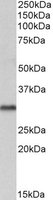
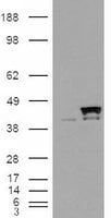 WB (Western Blot)
(HEK293 overexpressing CyP-40 (RC206039) and probed (mock transfection in first lane), tested by Origene.)
WB (Western Blot)
(HEK293 overexpressing CyP-40 (RC206039) and probed (mock transfection in first lane), tested by Origene.)
PPID/CyP-40, Polyclonal Antibody (Cat# AAA60904)
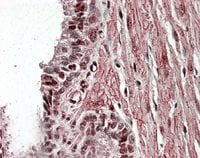
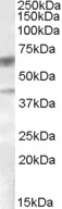
 IHC (Immunohiostchemistry)
((3.8ug/ml) staining of paraffin embedded Human Prostate. Steamed antigen retrieval with citrate buffer pH 6, AP-staining.)
IHC (Immunohiostchemistry)
((3.8ug/ml) staining of paraffin embedded Human Prostate. Steamed antigen retrieval with citrate buffer pH 6, AP-staining.)
VPS37C, Polyclonal Antibody (Cat# AAA60928)
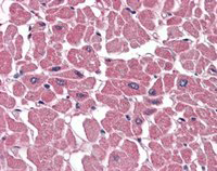 IHC (Immunohiostchemistry)
((3.8ug/ml) staining of paraffin embedded Human Heart. Steamed antigen retrieval with citrate buffer pH 6, AP-staining.)
IHC (Immunohiostchemistry)
((3.8ug/ml) staining of paraffin embedded Human Heart. Steamed antigen retrieval with citrate buffer pH 6, AP-staining.)
BPOZ/ABTB1, Polyclonal Antibody (Cat# AAA60936)
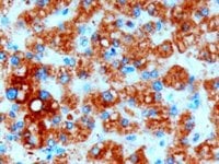
 IHC (Immunohiostchemistry)
((2ug/ml) staining of paraffin embedded Human Pituitary Gland. Steamed antigen retrieval with Tris/EDTA buffer pH 9, HRP-staining.)
IHC (Immunohiostchemistry)
((2ug/ml) staining of paraffin embedded Human Pituitary Gland. Steamed antigen retrieval with Tris/EDTA buffer pH 9, HRP-staining.)
Proopiomelanocortin/POMC, Polyclonal Antibody (Cat# AAA60939)
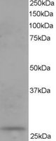
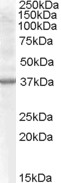
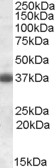
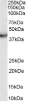
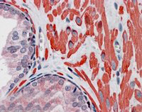
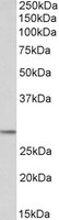
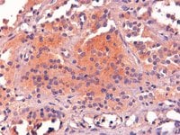
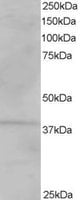
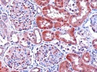 IHC (Immunohiostchemistry)
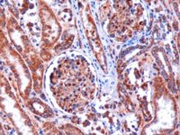
((1ug/ml) staining of paraffin embedded human kidney. Microwaved antigen retrieval with citrate buffer pH 6, HRP-staining.)
IHC (Immunohiostchemistry)
((1ug/ml) staining of paraffin embedded human kidney. Microwaved antigen retrieval with citrate buffer pH 6, HRP-staining.)
Dicarbonyl Reductase, Polyclonal Antibody (Cat# AAA61053)
 WB (Western Blot)
(HEK293 overexpressing VPS26A (RC205353) and probed (mock transfection in first lane), tested by Origene.)
WB (Western Blot)
(HEK293 overexpressing VPS26A (RC205353) and probed (mock transfection in first lane), tested by Origene.)
VPS26A, Polyclonal Antibody (Cat# AAA61067)
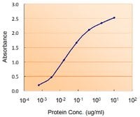 Application Data
((0.5ug/ml) as the reporter with as the capture rabbit antibody (2.5ug/ml).)
Application Data
((0.5ug/ml) as the reporter with as the capture rabbit antibody (2.5ug/ml).)
DGAT2, Polyclonal Antibody (Cat# AAA60812)
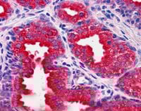
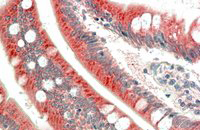 IHC (Immunohiostchemistry)
((5ug/ml) staining of paraffin embedded Human Small Intestine. Steamed antigen retrieval with citrate buffer pH 6, AP-staining.)
IHC (Immunohiostchemistry)
((5ug/ml) staining of paraffin embedded Human Small Intestine. Steamed antigen retrieval with citrate buffer pH 6, AP-staining.)
MIC1/C18orf8, Polyclonal Antibody (Cat# AAA60823)
 IHC (Immunohiostchemistry)
((3.8ug/ml) staining of paraffin embedded Human Prostate. Steamed antigen retrieval with citrate buffer pH 6, AP-staining.)
IHC (Immunohiostchemistry)
((3.8ug/ml) staining of paraffin embedded Human Prostate. Steamed antigen retrieval with citrate buffer pH 6, AP-staining.)
GOLPH2, Polyclonal Antibody (Cat# AAA60834)
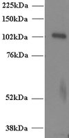
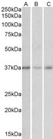
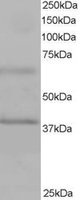
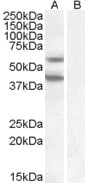
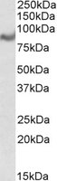
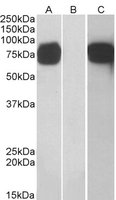
 WB (Western Blot)
((1ug/ml) staining of Human Brain (Cerebellum) lysate (35ug protein in RIPA buffer). Primary incubation was 1 hour. Detected by chemiluminescence.)
WB (Western Blot)
((1ug/ml) staining of Human Brain (Cerebellum) lysate (35ug protein in RIPA buffer). Primary incubation was 1 hour. Detected by chemiluminescence.)
VDAC2, Polyclonal Antibody (Cat# AAA60842)

 IHC (Immunohiostchemistry)
((3.8ug/ml) staining of paraffin embedded Human Prostate. Steamed antigen retrieval with citrate buffer pH 6, AP-staining.)
IHC (Immunohiostchemistry)
((3.8ug/ml) staining of paraffin embedded Human Prostate. Steamed antigen retrieval with citrate buffer pH 6, AP-staining.)
DUSP1, Polyclonal Antibody (Cat# AAA61226)
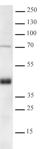
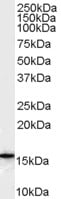
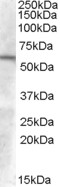
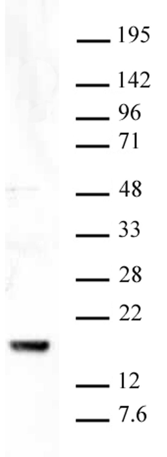
 WB (Western Blot)
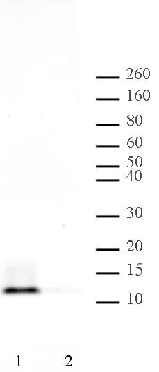
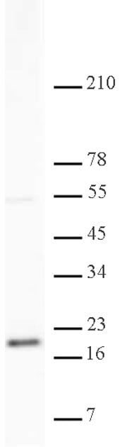
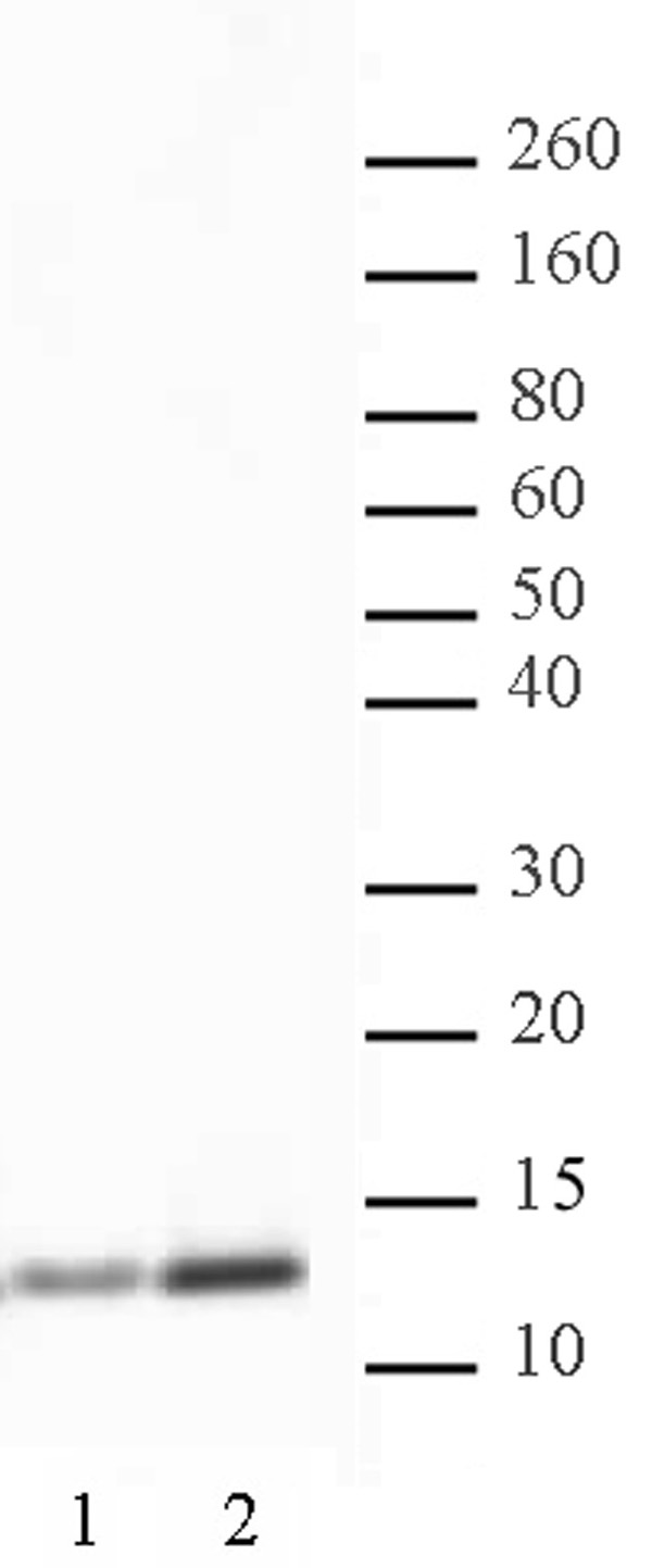
(Bcl7 antibody (pAb) tested by Western blot 20 ug of HEK293 whole cell extract was run on SDS-PAGE and probed with antibody at a dilution of 1:500. The calculated M.W. of Bcl7A is 23 kDa, whereas it's observed M.W. on SDS-PAGE is 45 kDa.)
WB (Western Blot)
(Bcl7 antibody (pAb) tested by Western blot 20 ug of HEK293 whole cell extract was run on SDS-PAGE and probed with antibody at a dilution of 1:500. The calculated M.W. of Bcl7A is 23 kDa, whereas it's observed M.W. on SDS-PAGE is 45 kDa.)
Bcl7A, Polyclonal Antibody (Cat# AAA60096)
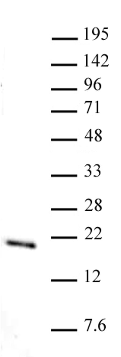
 DB (Dot Blot)
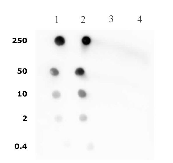
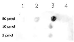
(Histone H4 pan-acetyl antibody (pAb) tested by dot blot analysis. Dot blot analysis was used to confirm the specificity of Histone H4 pan-acetyl pAb for acetyl histone H4. Decreasing amounts of acetylated peptides corresponding to the immunogen, a related sequence, as well as unacetylated histone H4 peptides were spotted onto PVDF and probed with the antibody at a 1:10,000 dilution. Lane 1: AGG[acK]GG[acK]GMG[acK]VGA[acK]RHSC Lane 2: AGG[acK]GG[acK]GG[acK]GG[acK]GG[acK]GGC Lane 3: SGRGKGGKGLC Lane 4: GKGGAKRHRKC)
DB (Dot Blot)
(Histone H4 pan-acetyl antibody (pAb) tested by dot blot analysis. Dot blot analysis was used to confirm the specificity of Histone H4 pan-acetyl pAb for acetyl histone H4. Decreasing amounts of acetylated peptides corresponding to the immunogen, a related sequence, as well as unacetylated histone H4 peptides were spotted onto PVDF and probed with the antibody at a 1:10,000 dilution. Lane 1: AGG[acK]GG[acK]GMG[acK]VGA[acK]RHSC Lane 2: AGG[acK]GG[acK]GG[acK]GG[acK]GG[acK]GGC Lane 3: SGRGKGGKGLC Lane 4: GKGGAKRHRKC)
Histone H4ac, Polyclonal Antibody (Cat# AAA59803)
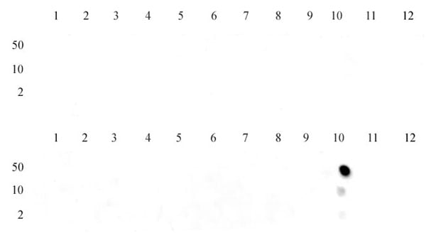
 DB (Dot Blot)
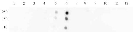
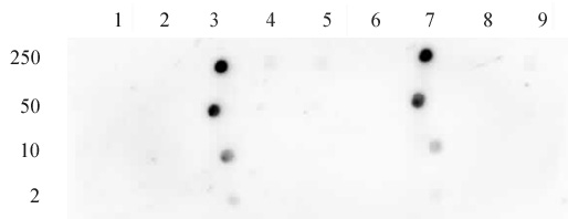
(Histone H3K9me3 tested by dot blot analysis to confirm the specificity of Histone H3K9me3. Peptides corresponding to regions around major sites of histone H3 methylation were spotted onto PVDF and probed with antibody at 2 ug/ml. The amount of peptide (picomoles) spotted is indicated next to each row. Top row Lane 1: unmodified Lys4. Lane 2: H3K4me1. Lane 3: H3K4me1. Lane 4: H3K4me2. Lane 5: H3K4me3. Lane 6: unmodified K9. Lane 7: H3K9me1. Lane 8: H3K9me2. Lane 9: H3K9me3. Bottom row, Lane 1: unmodified K27. Lane 2: H3k27me1. Lane 3: H3K27me2. Lane 4: H3K27me3. Lane 5: Unmod K36. Lane 6: H3K36me1. Lane 7: H3K36me2. Lane 8: H3K36me3.)
DB (Dot Blot)
(Histone H3K9me3 tested by dot blot analysis to confirm the specificity of Histone H3K9me3. Peptides corresponding to regions around major sites of histone H3 methylation were spotted onto PVDF and probed with antibody at 2 ug/ml. The amount of peptide (picomoles) spotted is indicated next to each row. Top row Lane 1: unmodified Lys4. Lane 2: H3K4me1. Lane 3: H3K4me1. Lane 4: H3K4me2. Lane 5: H3K4me3. Lane 6: unmodified K9. Lane 7: H3K9me1. Lane 8: H3K9me2. Lane 9: H3K9me3. Bottom row, Lane 1: unmodified K27. Lane 2: H3k27me1. Lane 3: H3K27me2. Lane 4: H3K27me3. Lane 5: Unmod K36. Lane 6: H3K36me1. Lane 7: H3K36me2. Lane 8: H3K36me3.)
Histone H3K9me3, Polyclonal Antibody (Cat# AAA59813)

 DB (Dot Blot)
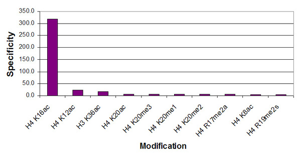
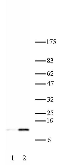
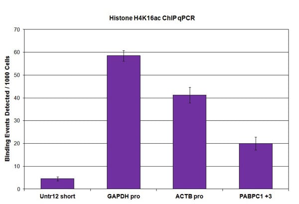
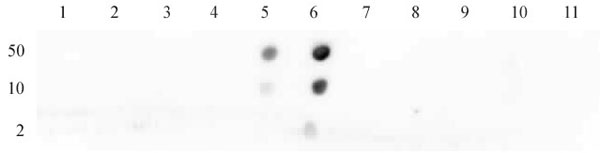
(Histone H4K16ac antibody (pAb) tested by dot blot analysis. Dot blot analysis was used to confirm the specificity of Histone H4 acetyl Lys16 pAb for histone H4 acetyl Lys16. Acetylated peptides corresponding to the immunogen and related H4 sequences were spotted onto PVDF and probed with the antibody at 1:4,000. The amount of peptide (picomoles) spotted is indicated next to each row. Lane 1: Unmodified Lys8 peptide. Lane 2: Acetyl Lys5 peptide. Lane 3: Acetyl Lys8 peptide. Lane 4: Acetyl Lys12 peptide. Lane 5: Unmodified Lys16 peptide. Lane 6: Acetyl Lys16 peptide. Lane 7: Acetyl Lys20 peptide. Lane 8: Unmodified Lys31 peptide. Lane 9: Acetyl Lys31 peptide. Lane 10: Acetyl Lys59 peptide. Lane 11: Acetyl Lys91 peptide.)
DB (Dot Blot)
(Histone H4K16ac antibody (pAb) tested by dot blot analysis. Dot blot analysis was used to confirm the specificity of Histone H4 acetyl Lys16 pAb for histone H4 acetyl Lys16. Acetylated peptides corresponding to the immunogen and related H4 sequences were spotted onto PVDF and probed with the antibody at 1:4,000. The amount of peptide (picomoles) spotted is indicated next to each row. Lane 1: Unmodified Lys8 peptide. Lane 2: Acetyl Lys5 peptide. Lane 3: Acetyl Lys8 peptide. Lane 4: Acetyl Lys12 peptide. Lane 5: Unmodified Lys16 peptide. Lane 6: Acetyl Lys16 peptide. Lane 7: Acetyl Lys20 peptide. Lane 8: Unmodified Lys31 peptide. Lane 9: Acetyl Lys31 peptide. Lane 10: Acetyl Lys59 peptide. Lane 11: Acetyl Lys91 peptide.)
Histone H4K16ac, Polyclonal Antibody (Cat# AAA59815)

 DB (Dot Blot)
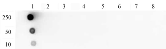
(Histone H3 dimethyl Lys79 antibody tested by dot blot analysis. Dot blot analysis was used to confirm the specificity of Histone H3 dimethyl Lys79 antibody for dimethyl lysine 79 of histone H3. Decreasing amounts of peptides corresponding to the region around lysine 79 of histone H3 were spotted onto PVDF and probed with the antibody at a 1:10,000 dilution. Lane 1: Unmodified Lys79 peptide. Lane 2: Monomethyl Lys79 peptide. Lane 3: Dimethyl Lys79 peptide. Lane 4: Trimethyl Lys79 peptide.)
DB (Dot Blot)
(Histone H3 dimethyl Lys79 antibody tested by dot blot analysis. Dot blot analysis was used to confirm the specificity of Histone H3 dimethyl Lys79 antibody for dimethyl lysine 79 of histone H3. Decreasing amounts of peptides corresponding to the region around lysine 79 of histone H3 were spotted onto PVDF and probed with the antibody at a 1:10,000 dilution. Lane 1: Unmodified Lys79 peptide. Lane 2: Monomethyl Lys79 peptide. Lane 3: Dimethyl Lys79 peptide. Lane 4: Trimethyl Lys79 peptide.)
Histone H3K79me2, Polyclonal Antibody (Cat# AAA59838)
 DB (Dot Blot)
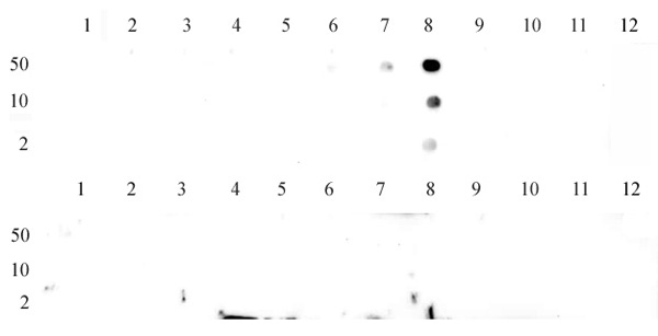
(STAT3 phospho Ser727 pAb tested by dot blot analysis. Dot blot analysis was used to confirm the specificity of STAT3 phospho Ser727 pAb for STAT2 phospho Ser727. Phosphorylated peptides corresponding to the immunogen and related peptides were spotted onto PVDF and probed with the antibody at 1:30,000. The amount of peptide (picomoles) spotted is indicated next to each row. Lane 1: Unmodified Ser727 STAT1 peptide. Lane 2: Phospho Ser727 STAT1 peptide. Lane 3: Unmodified Tyr689 STAT2 peptide. Lane 4: Phospho Tyr689 STAT2 peptide. Lane 5: Unmodified Ser727 STAT3 peptide. Lane 6: Phospho Ser727 STAT3 peptide. Lane 7: Unmodified Tyr705 STAT3 peptide. Lane 8: Phospho Tyr705 STAT3 peptide. Lane 9: Unmodified Ser726 STAT5A/Ser731 STAT5B peptide. Lane 10: Phospho Ser726 STAT5A/Ser731 STAT5B peptide. Lane 11: Unmodified Tyr694 STAT5A/Tyr699 STAT5B peptide. Lane 12: Phospho Tyr694 STAT5A/Tyr699 STAT5B peptide.)
DB (Dot Blot)
(STAT3 phospho Ser727 pAb tested by dot blot analysis. Dot blot analysis was used to confirm the specificity of STAT3 phospho Ser727 pAb for STAT2 phospho Ser727. Phosphorylated peptides corresponding to the immunogen and related peptides were spotted onto PVDF and probed with the antibody at 1:30,000. The amount of peptide (picomoles) spotted is indicated next to each row. Lane 1: Unmodified Ser727 STAT1 peptide. Lane 2: Phospho Ser727 STAT1 peptide. Lane 3: Unmodified Tyr689 STAT2 peptide. Lane 4: Phospho Tyr689 STAT2 peptide. Lane 5: Unmodified Ser727 STAT3 peptide. Lane 6: Phospho Ser727 STAT3 peptide. Lane 7: Unmodified Tyr705 STAT3 peptide. Lane 8: Phospho Tyr705 STAT3 peptide. Lane 9: Unmodified Ser726 STAT5A/Ser731 STAT5B peptide. Lane 10: Phospho Ser726 STAT5A/Ser731 STAT5B peptide. Lane 11: Unmodified Tyr694 STAT5A/Tyr699 STAT5B peptide. Lane 12: Phospho Tyr694 STAT5A/Tyr699 STAT5B peptide.)
STAT3 phospho Ser727, Polyclonal Antibody (Cat# AAA59894)

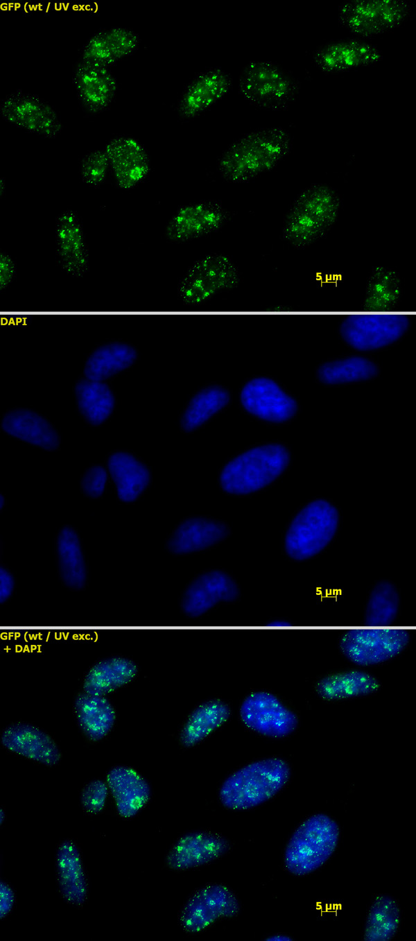
 IF (Immunofluorescence)
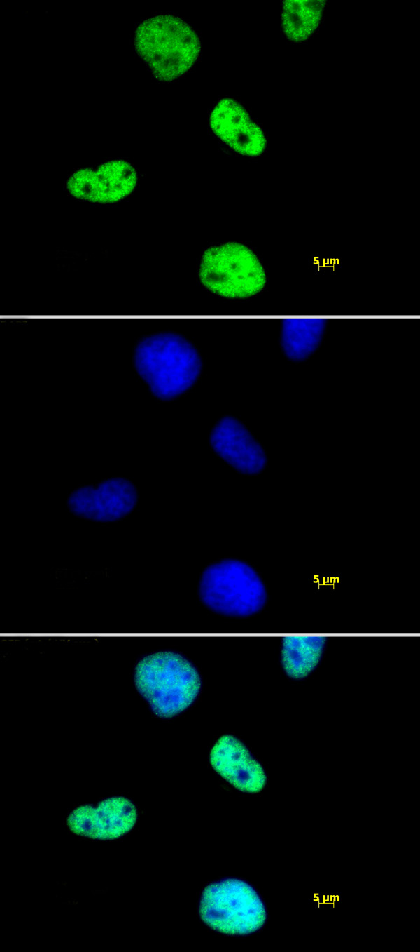
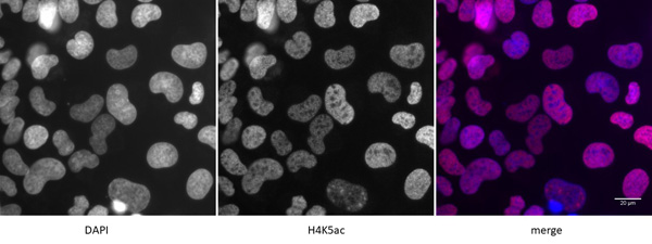
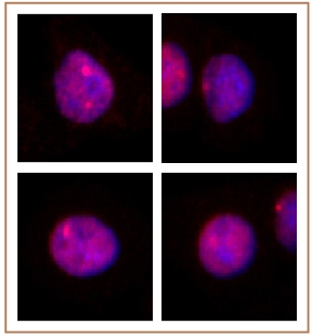
(Detection of H4K5ac by immunofluorescence. U2OS cells were stained with H4K5ac antibody at a dilution of 1:500. Left panel: DAPI. Middle panel: H4K5ac antibody staining. Right panel: merge.)
IF (Immunofluorescence)
(Detection of H4K5ac by immunofluorescence. U2OS cells were stained with H4K5ac antibody at a dilution of 1:500. Left panel: DAPI. Middle panel: H4K5ac antibody staining. Right panel: merge.)
Histone H4K5ac, Polyclonal Antibody (Cat# AAA59919)

 DB (Dot Blot)
(Histone H3 acetyl Lys18 antibody tested by dot blot analysis. Dot blot analysis was used to confirm the specificity of Histone H3 acetyl Lys18 antibody for acetyl Lys18 histone H3. Acetylated peptides corresponding to the immunogen and related peptides were spotted onto PVDF and probed with the antibody at a dilution of 0.1 ug/ml. The amount of peptide (picomoles) spotted is indicated next to each row. Lane 1: acetyl-Lys4 peptide. Lane 2: unmodified Lys4 peptide. Lane 3: acetyl-Lys18 peptide. Lane 4: unmodified Lys18 peptide. Lane 5: acetyl-Lys9 peptide. Lane 6: acetyl-Lys14 peptide. Lane 7: acetyl-Lys18 peptide 2. Lane 8: acetyl-Lys23 peptide. Lane 9: acetyl-Lys27 peptide.)
DB (Dot Blot)
(Histone H3 acetyl Lys18 antibody tested by dot blot analysis. Dot blot analysis was used to confirm the specificity of Histone H3 acetyl Lys18 antibody for acetyl Lys18 histone H3. Acetylated peptides corresponding to the immunogen and related peptides were spotted onto PVDF and probed with the antibody at a dilution of 0.1 ug/ml. The amount of peptide (picomoles) spotted is indicated next to each row. Lane 1: acetyl-Lys4 peptide. Lane 2: unmodified Lys4 peptide. Lane 3: acetyl-Lys18 peptide. Lane 4: unmodified Lys18 peptide. Lane 5: acetyl-Lys9 peptide. Lane 6: acetyl-Lys14 peptide. Lane 7: acetyl-Lys18 peptide 2. Lane 8: acetyl-Lys23 peptide. Lane 9: acetyl-Lys27 peptide.)
Histone H3K18ac, Polyclonal Antibody (Cat# AAA59926)
 WB (Western Blot)
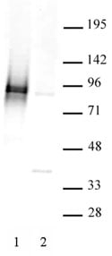
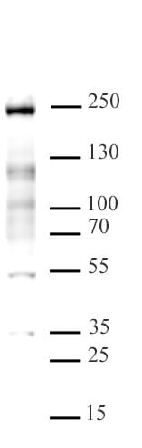
(MLL/HRX antibody (pAb) tested by Western blot. HeLa whole cell extract extract (20 ug) probed with MLL/HRX antibody (pAb) at 2 ug/ml.)
WB (Western Blot)
(MLL/HRX antibody (pAb) tested by Western blot. HeLa whole cell extract extract (20 ug) probed with MLL/HRX antibody (pAb) at 2 ug/ml.)
MLL/HRX, Polyclonal Antibody (Cat# AAA60005)
 DB (Dot Blot)
(Specificity Data: Dot blot analysis was used to confirm the specificity of Histone H3 monomethyl Lys36 antibody for monomethyl-lysine 36 of histone H3. Peptides corresponding to regions around major sites of histone H3 methylation were spotted onto PVDF and probed with the antibody at a dilution of 1:1,000. The amount of peptide (in picomoles) spotted is indicated next to each row. Top panel - Lane 1: unmodified Lys4. Lane 2: monomethyl Lys4. Lane 3: dimethyl Lys4. Lane 4: trimethyl Lys4. Lane 5: unmodified Lys9. Lane 6: monomethyl Lys9. Lane 7: dimethyl Lys9. Lane 8: trimethyl Lys9. Lane 9: unmodified Lys79. Lane 10: monomethyl Lys79. Lane 11: dimethyl Lys79. Lane 12: trimethyl Lys79. Bottom panel - Lane 1: Unmodified Lys23. Lane 2: Monomethyl Lys23. Lane 3: Dimethyl Lys23. Lane 4: Trimethyl Lys23. Lane 5: unmodified Lys27. Lane 6: monomethyl Lys27. Lane 7: dimethyl Lys27. Lane 8: trimethyl Lys27. Lane 9: unmodified Lys36. Lane 10: monomethyl Lys36. Lane 11: dimethyl Lys36. Lane 12: trimethyl Lys36.)
DB (Dot Blot)
(Specificity Data: Dot blot analysis was used to confirm the specificity of Histone H3 monomethyl Lys36 antibody for monomethyl-lysine 36 of histone H3. Peptides corresponding to regions around major sites of histone H3 methylation were spotted onto PVDF and probed with the antibody at a dilution of 1:1,000. The amount of peptide (in picomoles) spotted is indicated next to each row. Top panel - Lane 1: unmodified Lys4. Lane 2: monomethyl Lys4. Lane 3: dimethyl Lys4. Lane 4: trimethyl Lys4. Lane 5: unmodified Lys9. Lane 6: monomethyl Lys9. Lane 7: dimethyl Lys9. Lane 8: trimethyl Lys9. Lane 9: unmodified Lys79. Lane 10: monomethyl Lys79. Lane 11: dimethyl Lys79. Lane 12: trimethyl Lys79. Bottom panel - Lane 1: Unmodified Lys23. Lane 2: Monomethyl Lys23. Lane 3: Dimethyl Lys23. Lane 4: Trimethyl Lys23. Lane 5: unmodified Lys27. Lane 6: monomethyl Lys27. Lane 7: dimethyl Lys27. Lane 8: trimethyl Lys27. Lane 9: unmodified Lys36. Lane 10: monomethyl Lys36. Lane 11: dimethyl Lys36. Lane 12: trimethyl Lys36.)
Histone H3K36me1, Polyclonal Antibody (Cat# AAA60010)
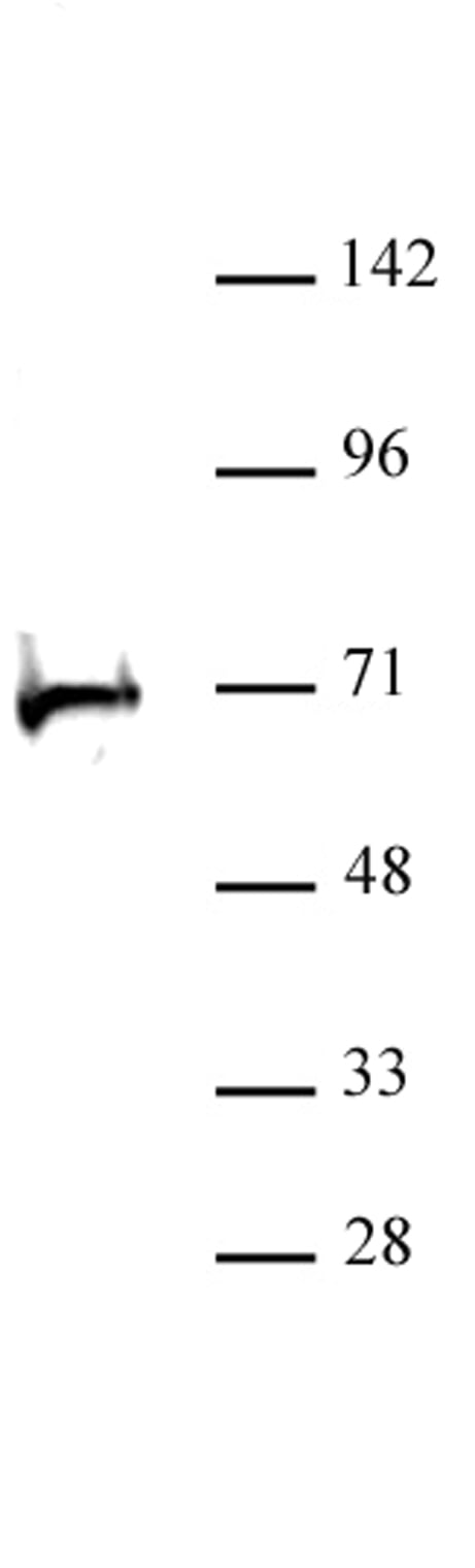 WB (Western Blot)
(RBBP5 antibody (pAb) tested by Western blot. MCF-7 whole-cell extract (20 ug) probed with RBBP5 antibody at a dilution of 1:1,000.)
WB (Western Blot)
(RBBP5 antibody (pAb) tested by Western blot. MCF-7 whole-cell extract (20 ug) probed with RBBP5 antibody at a dilution of 1:1,000.)
RBBP5, Polyclonal Antibody (Cat# AAA60022)
What are Polyclonal Antibodies?
Polyclonal antibodies are antibodies that come from multiple B cell clones of a host animal. The typical hosts used for the majority of polyclonal antibody production are rabbits, goats, sheep, and donkeys. These polyclonal antibodies, once having identified their target, will bind to different epitopes located at different regions or sequences on the same protein/antigen. This ability to bind multiple epitopes is what makes polyclonal antibodies highly sensitive, as explained in our detailed guide on polyclonal antibodies and why they matter.
As a result, they are ideal at locating and binding to the target, even if the target is in very low concentrations (due to many different antibodies being able to bind to the same target molecule, which allows for significant amplification of a downstream signal).
Polyclonal antibodies are typically produced by injecting an antigen into a host animal, which causes the animal’s immune system to attack the foreign antigen by mass generating antibodies against it. After a period of time, serum is collected from the animal and purified using physicochemical fractionation, class-specific affinity purification, and/or antigen-affinity purification.
Key Uses of Polyclonal Antibodies
- Western Blotting: This method is used to find specific proteins in biological samples after separating them by size.
- Immunohistochemistry: IHC helps visualize the location of proteins in tissue sections using various staining techniques.
- ELISA: (Enzyme-Linked Immunosorbent Assay) is typically used to identify specific protein quantities in a sample. ELISAs can be either “Quantitative” or “Qualitative”.
- Flow Cytometry: technique that identifies and measures the specific protein on the surface or inside the cells in a fluid suspension.
- Immunoprecipitation: IP isolates and studies a specific protein from a complex mixture using antibodies.
Why Buy Polyclonal Antibodies from AAA Biotech?
1. Ideal for Various Applications
Our antibodies are generally going to be validated for use in multiple types of assays, including ELISA, Western Blotting, Immunohistochemistry, Immunoprecipitation, amongst others. They are ideal for a wide range of research applications.
2. Rigorous Quality Control
All of the antibodies in our catalog undergo strict quality testing to ensure specificity, sensitivity, and consistent performance. We are confident in the ability of our antibodies to provide you with accurate results.
3. Wide Assortment of Antibodies
Antibodies in our catalog can be found for both common and exotic species, and these antibodies are also available in both conjugated and recombinant forms to suit many diverse experimental needs.
4. Highly Purified
Our antibodies are available in purified forms with over 85% purity, as confirmed by SDS-PAGE. They are also available with tags such as His, Flag, GST, or MBP. We cater to customers worldwide.
FAQ
1. How are polyclonal antibodies produced?
Traditionally, polyclonal antibodies are produced by injecting an antigen into a host animal (such as a rabbit or goat), which then triggers an immune response from the host animal. The animal’s B cells produce antibodies that will recognize different parts of the injected antigen. These antibodies are then collected from the animal’s blood and purified for use.
2. How do polyclonal antibodies differ from monoclonal antibodies?
Polyclonal antibodies are a mix of antibodies that bind to different locations (epitopes) of the same antigen, while monoclonal antibodies are identical and bind to just one specific epitope. This makes polyclonal antibodies more versatile and better at detecting proteins that may be present in low quantities or in altered/modified forms.
3. How should I store polyclonal antibodies?
Polyclonal antibodies should be stored at 4°C for short-term use (up to a few weeks) and at -20°C or -80°C for long-term storage. Avoid repeated freeze-thaw cycles by dividing them into small aliquots. Always check the datasheet for specific storage instructions.