Filters
▼Clonality
▼Type
▼Reactivity
▼Gene Name
▼Isotype
▼Host
▼Application
▼Clone
▼Polyclonal Antibodies
At AAA Biotech also known as AAA Bio or AAABio, we provide a broad range of purified polyclonal antibodies (pAbs) that are able to all be browsed online through our website. Due to their high specificity and strong binding affinity, these antibodies are ideal for wide swathes of research and experimental applications.
Our polyclonal antibodies can easily support your work, whether you use them for Western Blotting, Immunocytochemistry (with or without Immunofluorescence used in conjunction), Immunohistochemistry, Immunoprecipitation, and ELISA tests. We highly encourage you to browse our range of pAbs and choose the one that best suits your experimental model.
Viewing 100-150 of 118597 product results
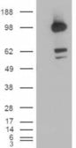
S1 (MERS-CoV), Polyclonal Antibody (Cat# AAA62217)
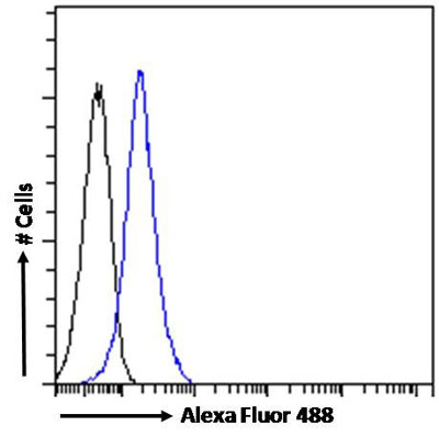 Application Data
(Flow cytometric analysis of paraformaldehyde fixed K562 cells (blue line), permeabilized with 0.5% Triton. Primary incubation 1hr (10ug/ml) followed by Alexa Fluor 488 secondary antibody (1ug/ml). IgG control: Unimmunized goat IgG (black line) followed by Alexa Fluor 488 secondary antibody.)
Application Data
(Flow cytometric analysis of paraformaldehyde fixed K562 cells (blue line), permeabilized with 0.5% Triton. Primary incubation 1hr (10ug/ml) followed by Alexa Fluor 488 secondary antibody (1ug/ml). IgG control: Unimmunized goat IgG (black line) followed by Alexa Fluor 488 secondary antibody.)
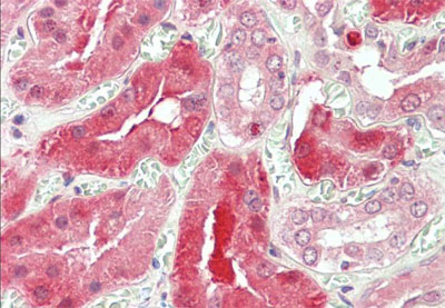
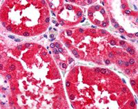
STAT3, Polyclonal Antibody (Cat# AAA61036)
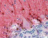
 IHC (Immunohiostchemistry)
((3.8ug/ml) staining of paraffin embedded Human Kidney. Steamed antigen retrieval with citrate buffer pH 6, AP-staining.)
IHC (Immunohiostchemistry)
((3.8ug/ml) staining of paraffin embedded Human Kidney. Steamed antigen retrieval with citrate buffer pH 6, AP-staining.)
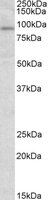
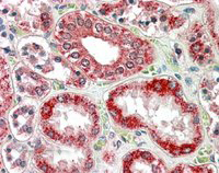
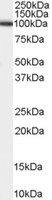
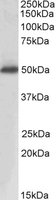
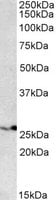
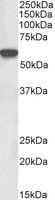
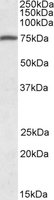
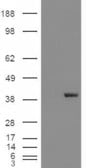
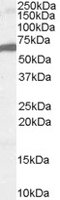
RANBP1, Polyclonal Antibody (Cat# AAA61041)
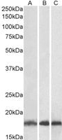
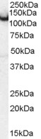
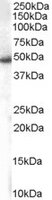
 WB (Western Blot)
((0. 01ug/ml) staining of HEK293 (A), HepG2 (B) and Jurkat (C) lysates (35ug protein in RIPA buffer). Primary incubation was 1 hour. Detected by chemiluminescence.)
WB (Western Blot)
((0. 01ug/ml) staining of HEK293 (A), HepG2 (B) and Jurkat (C) lysates (35ug protein in RIPA buffer). Primary incubation was 1 hour. Detected by chemiluminescence.)
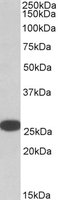
UBE2L3, Polyclonal Antibody (Cat# AAA61050)
 IHC (Immunohiostchemistry)
((2.5ug/ml) staining of paraffin embedded Human Cerebellum. Steamed antigen retrieval with citrate buffer pH 6, AP-staining.)
IHC (Immunohiostchemistry)
((2.5ug/ml) staining of paraffin embedded Human Cerebellum. Steamed antigen retrieval with citrate buffer pH 6, AP-staining.)
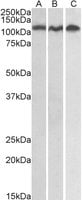
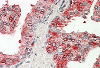
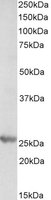
Amphiphysin/AMPH, Polyclonal Antibody (Cat# AAA61079)
 WB (Western Blot)
(HEK293 overexpressing PLA2G1B (RC216089) and probed (mock transfection in first lane), tested by Origene.)
WB (Western Blot)
(HEK293 overexpressing PLA2G1B (RC216089) and probed (mock transfection in first lane), tested by Origene.)
PLA2G1B, Polyclonal Antibody (Cat# AAA61089)

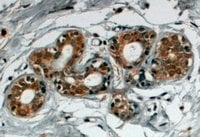
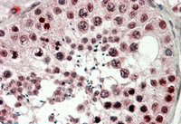
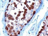 IHC (Immunohiostchemistry)
((10ug/ml) staining of paraffin embedded Human Testis. Microwaved antigen retrieval with Tris/EDTA buffer pH9, HRP-staining.)
IHC (Immunohiostchemistry)
((10ug/ml) staining of paraffin embedded Human Testis. Microwaved antigen retrieval with Tris/EDTA buffer pH9, HRP-staining.)
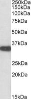
TCP1, Polyclonal Antibody (Cat# AAA61095)
 WB (Western Blot)
(HEK293 overexpressing PDE5A (RC218974) and probed (mock transfection in first lane), tested by Origene.)
WB (Western Blot)
(HEK293 overexpressing PDE5A (RC218974) and probed (mock transfection in first lane), tested by Origene.)
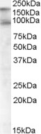
PDE5A, Polyclonal Antibody (Cat# AAA61113)
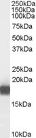
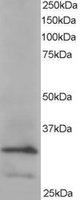
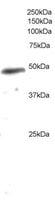
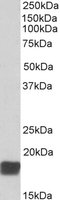
 WB (Western Blot)
((1ug/ml) staining of human spleen lysate (35ug protein in RIPA buffer). Primary incubation was 1 hour. Detected by chemiluminescence.)
WB (Western Blot)
((1ug/ml) staining of human spleen lysate (35ug protein in RIPA buffer). Primary incubation was 1 hour. Detected by chemiluminescence.)
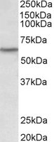
LXR alpha/LXR beta, Polyclonal Antibody (Cat# AAA61120)
NOTCH2, Polyclonal Antibody (Cat# AAA61149)
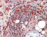
 IHC (Immunohiostchemistry)
((4ug/ml) staining of paraffin embedded Human Breast. Steamed antigen retrieval with citrate buffer pH 6, HRP-staining.)
IHC (Immunohiostchemistry)
((4ug/ml) staining of paraffin embedded Human Breast. Steamed antigen retrieval with citrate buffer pH 6, HRP-staining.)
SNX5, Polyclonal Antibody (Cat# AAA61150)
 IHC (Immunohiostchemistry)
((3.8ug/ml) staining of paraffin embedded Human Spleen. Steamed antigen retrieval with citrate buffer pH 6, AP-staining.)
IHC (Immunohiostchemistry)
((3.8ug/ml) staining of paraffin embedded Human Spleen. Steamed antigen retrieval with citrate buffer pH 6, AP-staining.)
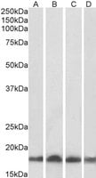
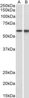
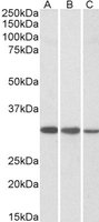
ARPC2, Polyclonal Antibody (Cat# AAA61165)
 WB (Western Blot)
(Staining (0.3ug/ml) of Daudi (A), Jurkat (B) and HeLa (C) lysates (35ug total protein in RIPA buffer). Primary incubated for 1 hour. Detected by chemiluminescence.)
WB (Western Blot)
(Staining (0.3ug/ml) of Daudi (A), Jurkat (B) and HeLa (C) lysates (35ug total protein in RIPA buffer). Primary incubated for 1 hour. Detected by chemiluminescence.)
ADAM17/TACE, Polyclonal Antibody (Cat# AAA61169)
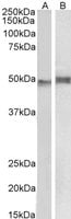 WB (Western Blot)
(1ug/ml) staining of Mouse Thymus (A) and Pig Spleen (B) lysate (35ug protein in RIPA buffer). Primary incubation was 1 hour. Detected by chemiluminescence.)
WB (Western Blot)
(1ug/ml) staining of Mouse Thymus (A) and Pig Spleen (B) lysate (35ug protein in RIPA buffer). Primary incubation was 1 hour. Detected by chemiluminescence.)
NCF1/p47phox, Polyclonal Antibody (Cat# AAA61191)
ATG4D, Polyclonal Antibody (Cat# AAA61442)
GPR139, Polyclonal Antibody (Cat# AAA61450)
 IHC (Immunohiostchemistry)
((3.8ug/ml) staining of paraffin embedded Human Prostate. Steamed antigen retrieval with citrate buffer pH 6, AP-staining.)
IHC (Immunohiostchemistry)
((3.8ug/ml) staining of paraffin embedded Human Prostate. Steamed antigen retrieval with citrate buffer pH 6, AP-staining.)
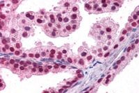
ACPP, Polyclonal Antibody (Cat# AAA61464)
 IHC (Immunohiostchemistry)
((3.8ug/ml) staining of paraffin embedded Human Testis. Steamed antigen retrieval with citrate buffer pH 6, AP-staining.)
IHC (Immunohiostchemistry)
((3.8ug/ml) staining of paraffin embedded Human Testis. Steamed antigen retrieval with citrate buffer pH 6, AP-staining.)
CIRBP, Polyclonal Antibody (Cat# AAA61515)
 WB (Western Blot)
((0.3ug/ml) staining of Mouse Liver lysate (35ug protein in RIPA buffer). Primary incubation was 1 hour. Detected by chemiluminescence.)
WB (Western Blot)
((0.3ug/ml) staining of Mouse Liver lysate (35ug protein in RIPA buffer). Primary incubation was 1 hour. Detected by chemiluminescence.)
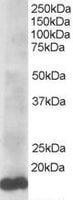
PSMB4, Polyclonal Antibody (Cat# AAA61520)
 WB (Western Blot)
((0. 01ug/ml) staining of Mouse Kidney lysate (35ug protein in RIPA buffer). Primary incubation was 1 hour. Detected by chemiluminescence.)
WB (Western Blot)
((0. 01ug/ml) staining of Mouse Kidney lysate (35ug protein in RIPA buffer). Primary incubation was 1 hour. Detected by chemiluminescence.)
Aspa, Polyclonal Antibody (Cat# AAA61544)
 WB (Western Blot)
(HEK293 overexpressing CDCP1 (RC220633) and probed (mock transfection in first lane), tested by Origene.)
WB (Western Blot)
(HEK293 overexpressing CDCP1 (RC220633) and probed (mock transfection in first lane), tested by Origene.)
CDCP1, Polyclonal Antibody (Cat# AAA61295)
NY-ESO-1, Polyclonal Antibody (Cat# AAA61339)
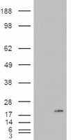
 WB (Western Blot)
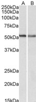
(AAA61583 (0.3ug/ml) staining of A549 (A) and NIH3T3 (B) lysates (35ug protein in RIPA buffer). Primary incubation was 1 hour. Detected by chemiluminescence.)
WB (Western Blot)
(AAA61583 (0.3ug/ml) staining of A549 (A) and NIH3T3 (B) lysates (35ug protein in RIPA buffer). Primary incubation was 1 hour. Detected by chemiluminescence.)
CB1, Polyclonal Antibody (Cat# AAA61583)
 WB (Western Blot)
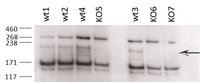
((0.2ug/ml) staining of different Testis lysates from wildtype 8-9 week old C57BL/6 (wt) and knock-out (KO) mice (35ug protein in RIPA buffer). Primary incubation was 2 hours. Detected by chemiluminescence. Data obtained from an anonymous customer.)
WB (Western Blot)
((0.2ug/ml) staining of different Testis lysates from wildtype 8-9 week old C57BL/6 (wt) and knock-out (KO) mice (35ug protein in RIPA buffer). Primary incubation was 2 hours. Detected by chemiluminescence. Data obtained from an anonymous customer.)
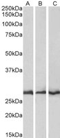
Terminal Uridylyltransferase 4, Polyclonal Antibody (Cat# AAA61599)
 WB (Western Blot)
((1ug/ml) staining of Pig Bone Marrow (A) and Spleen (B) lysates (35ug protein in RIPA buffer). Primary incubation was 1 hour. Detected by chemiluminescence.)
WB (Western Blot)
((1ug/ml) staining of Pig Bone Marrow (A) and Spleen (B) lysates (35ug protein in RIPA buffer). Primary incubation was 1 hour. Detected by chemiluminescence.)
LCK, Polyclonal Antibody (Cat# AAA61620)
 WB (Western Blot)
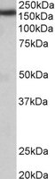
((0.3ug/ml) staining of Human Liver lysate (35ug protein in RIPA buffer). Primary incubation was 1 hour. Detected by chemiluminescence.)
WB (Western Blot)
((0.3ug/ml) staining of Human Liver lysate (35ug protein in RIPA buffer). Primary incubation was 1 hour. Detected by chemiluminescence.)
SLC46A1/PCFT, Polyclonal Antibody (Cat# AAA61638)
 WB (Western Blot)
((1ug/ml) staining of Human Peripheral Blood Mononucleocytes lysate (35ug protein in RIPA buffer). Primary incubation was 1 hour. Detected by chemiluminescence.)
WB (Western Blot)
((1ug/ml) staining of Human Peripheral Blood Mononucleocytes lysate (35ug protein in RIPA buffer). Primary incubation was 1 hour. Detected by chemiluminescence.)
URP2/kindlin-3, Polyclonal Antibody (Cat# AAA61402)
 WB (Western Blot)
((0.1ug/ml) staining of Mouse (A), Rat (B) and Pig (C) Heart lysates (35ug protein in RIPA buffer). Primary incubation was 1 hour. Detected by chemiluminescence.)
WB (Western Blot)
((0.1ug/ml) staining of Mouse (A), Rat (B) and Pig (C) Heart lysates (35ug protein in RIPA buffer). Primary incubation was 1 hour. Detected by chemiluminescence.)
CAPZB, Polyclonal Antibody (Cat# AAA61258)
 IHC (Immunohistochemistry)
((10ug/ml) staining of paraffin embedded Human Pancreas. Microwaved antigen retrieval with Tris/EDTA buffer pH9, HRP-staining.)
IHC (Immunohistochemistry)
((10ug/ml) staining of paraffin embedded Human Pancreas. Microwaved antigen retrieval with Tris/EDTA buffer pH9, HRP-staining.)
MDA5/IFIH1, Polyclonal Antibody (Cat# AAA61206)
 IHC (Immunohistochemisry)
((2.5g/ml) staining of paraffin embedded Human Kidney. Steamed antigen retrieval with citrate buffer pH 6, AP-staining.)
IHC (Immunohistochemisry)
((2.5g/ml) staining of paraffin embedded Human Kidney. Steamed antigen retrieval with citrate buffer pH 6, AP-staining.)
Aldehyde Reductase, Polyclonal Antibody (Cat# AAA61210)
 IHC (Immunohistochemisry)
(AAA61217 (3.8ug/ml) staining of paraffin embedded Human Prostate. Steamed antigen retrieval with citrate buffer pH 6, AP-staining.)
IHC (Immunohistochemisry)
(AAA61217 (3.8ug/ml) staining of paraffin embedded Human Prostate. Steamed antigen retrieval with citrate buffer pH 6, AP-staining.)
FOXA2/HNF3B, Polyclonal Antibody (Cat# AAA61217)
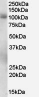
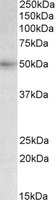
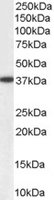
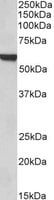
PTH [1-34] (Human), Polyclonal Antibody (Cat# AAA58833)
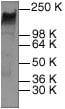 WB (Western Blot)
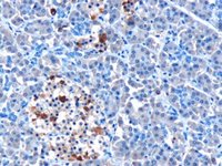
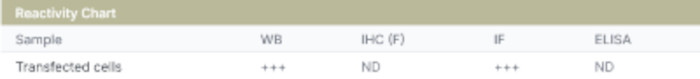
Neu, Polyclonal Antibody (Cat# AAA63011)
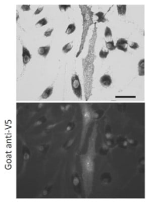
 IF (Immunofluorescence)
(Immunofluorescence – anti-V5 Ab using hCEC cells transduced with V5-mCherry-Rab11a; cells were fixed with methanol and anti-V5 at 1/100;)
IF (Immunofluorescence)
(Immunofluorescence – anti-V5 Ab using hCEC cells transduced with V5-mCherry-Rab11a; cells were fixed with methanol and anti-V5 at 1/100;)
V5, Polyclonal Antibody (Cat# AAA63062)
 Application Data
Rab27a, Polyclonal Antibody (Cat# AAA63067)
 Application Data
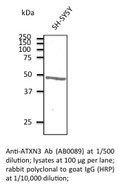
ATXN3, Polyclonal Antibody (Cat# AAA63075)
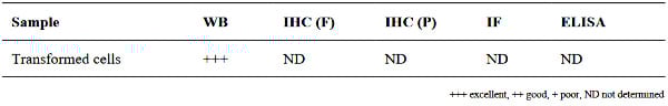
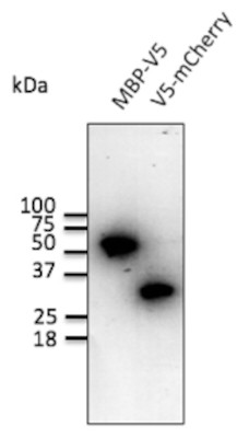
 Application Data
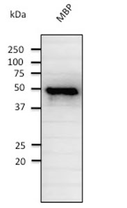
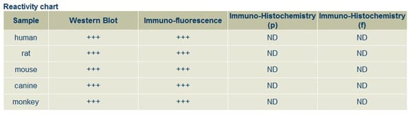
(Anti-MBP Ab at 1/2000 dilution; rabbit polyclonal to goat IgG (HRP) at 1/10,000 dilution.)
Application Data
(Anti-MBP Ab at 1/2000 dilution; rabbit polyclonal to goat IgG (HRP) at 1/10,000 dilution.)
MBP, Polyclonal Antibody (Cat# AAA63077)
 Application Data
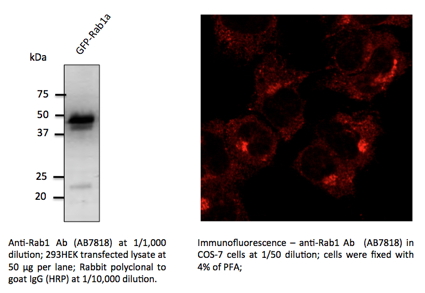
Rab1, Polyclonal Antibody (Cat# AAA63086)
 WB (Western Blot)
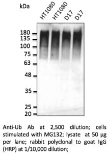
Ubiquitin, Polyclonal Antibody (Cat# AAA63096)
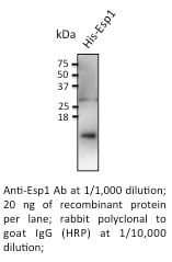 Application Data
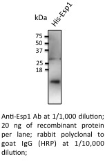
Esp1, Polyclonal Antibody (Cat# AAA63108)
 Application Data
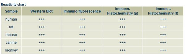
CDH1, Polyclonal Antibody (Cat# AAA63110)
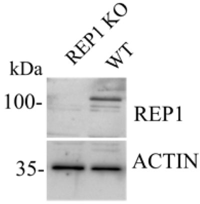
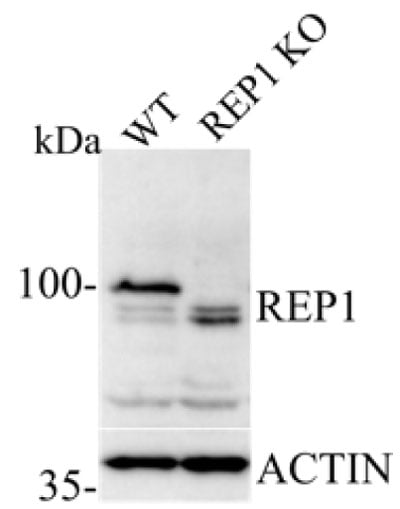
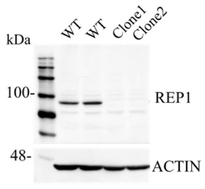
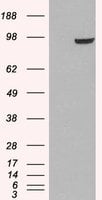
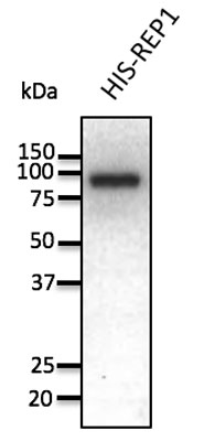 WB (Western Blot)
(Anti-REP1 Ab at 1/1,000 dilution; 50 ng of recombinant protein per lane; rabbit polyclonal to goat IgG (HRP) at 1/10,000 dilution.)
WB (Western Blot)
(Anti-REP1 Ab at 1/1,000 dilution; 50 ng of recombinant protein per lane; rabbit polyclonal to goat IgG (HRP) at 1/10,000 dilution.)
REP1, Polyclonal Antibody (Cat# AAA63112)
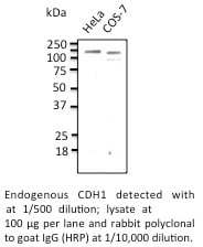
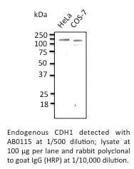
 Application Data
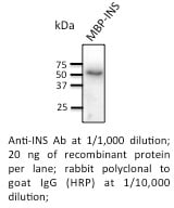
Insulin, Polyclonal Antibody (Cat# AAA63118)
 Application Data
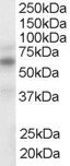
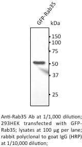
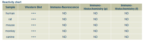
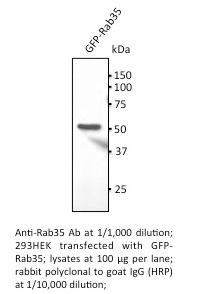
Rab35, Polyclonal Antibody (Cat# AAA63122)
 WB (Western Blot)
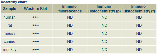
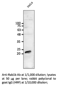
Rab1b, Polyclonal Antibody (Cat# AAA63129)
 WB (Western Blot)
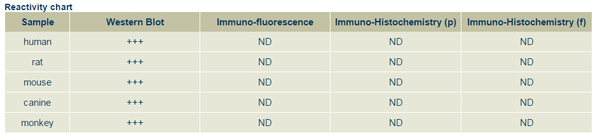
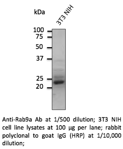
Rab9a, Polyclonal Antibody (Cat# AAA63132)
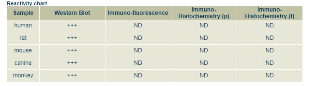
 WB (Western Blot)
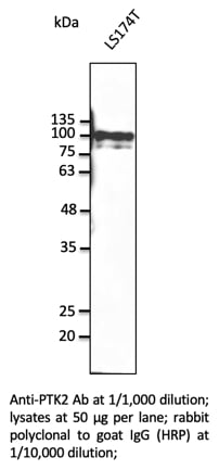
PTK2, Polyclonal Antibody (Cat# AAA63154)
 WB (Western Blot)
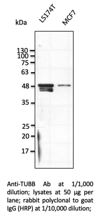
TUBB, Polyclonal Antibody (Cat# AAA63155)
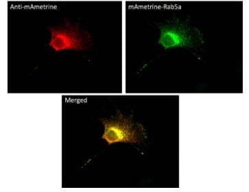
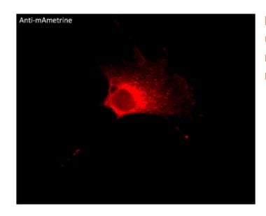 IF (Immunofluorescence)
(Immunofluorescence -anti-mAmetrine Ab usingNIH3T3 cells transduced with mAmetrine-Rab5a; cells were fixed with methanol and anti-mAmetrine at 1/250;)
IF (Immunofluorescence)
(Immunofluorescence -anti-mAmetrine Ab usingNIH3T3 cells transduced with mAmetrine-Rab5a; cells were fixed with methanol and anti-mAmetrine at 1/250;)
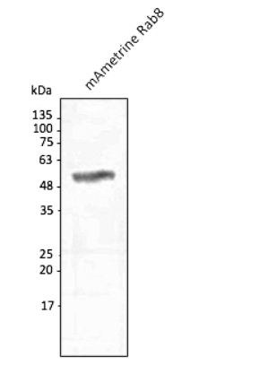
mAmetrine, Polyclonal Antibody (Cat# AAA63234)

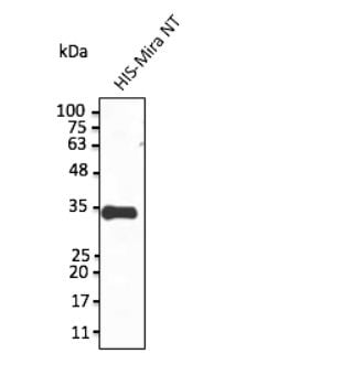 Application Data
(Anti-Miranda Ab at 1/1,000 dilution; protein at 20 ng per lane; rabbit polyclonal to goat IgG (HRP) at 1/10,000 dilution;)
Application Data
(Anti-Miranda Ab at 1/1,000 dilution; protein at 20 ng per lane; rabbit polyclonal to goat IgG (HRP) at 1/10,000 dilution;)
Miranda, Polyclonal Antibody (Cat# AAA63235)
What are Polyclonal Antibodies?
Polyclonal antibodies are antibodies that come from multiple B cell clones of a host animal. The typical hosts used for the majority of polyclonal antibody production are rabbits, goats, sheep, and donkeys. These polyclonal antibodies, once having identified their target, will bind to different epitopes located at different regions or sequences on the same protein/antigen. This ability to bind multiple epitopes is what makes polyclonal antibodies highly sensitive, as explained in our detailed guide on polyclonal antibodies and why they matter.
As a result, they are ideal at locating and binding to the target, even if the target is in very low concentrations (due to many different antibodies being able to bind to the same target molecule, which allows for significant amplification of a downstream signal).
Polyclonal antibodies are typically produced by injecting an antigen into a host animal, which causes the animal’s immune system to attack the foreign antigen by mass generating antibodies against it. After a period of time, serum is collected from the animal and purified using physicochemical fractionation, class-specific affinity purification, and/or antigen-affinity purification.
Key Uses of Polyclonal Antibodies
- Western Blotting: This method is used to find specific proteins in biological samples after separating them by size.
- Immunohistochemistry: IHC helps visualize the location of proteins in tissue sections using various staining techniques.
- ELISA: (Enzyme-Linked Immunosorbent Assay) is typically used to identify specific protein quantities in a sample. ELISAs can be either “Quantitative” or “Qualitative”.
- Flow Cytometry: technique that identifies and measures the specific protein on the surface or inside the cells in a fluid suspension.
- Immunoprecipitation: IP isolates and studies a specific protein from a complex mixture using antibodies.
Why Buy Polyclonal Antibodies from AAA Biotech?
1. Ideal for Various Applications
Our antibodies are generally going to be validated for use in multiple types of assays, including ELISA, Western Blotting, Immunohistochemistry, Immunoprecipitation, amongst others. They are ideal for a wide range of research applications.
2. Rigorous Quality Control
All of the antibodies in our catalog undergo strict quality testing to ensure specificity, sensitivity, and consistent performance. We are confident in the ability of our antibodies to provide you with accurate results.
3. Wide Assortment of Antibodies
Antibodies in our catalog can be found for both common and exotic species, and these antibodies are also available in both conjugated and recombinant forms to suit many diverse experimental needs.
4. Highly Purified
Our antibodies are available in purified forms with over 85% purity, as confirmed by SDS-PAGE. They are also available with tags such as His, Flag, GST, or MBP. We cater to customers worldwide.
FAQ
1. How are polyclonal antibodies produced?
Traditionally, polyclonal antibodies are produced by injecting an antigen into a host animal (such as a rabbit or goat), which then triggers an immune response from the host animal. The animal’s B cells produce antibodies that will recognize different parts of the injected antigen. These antibodies are then collected from the animal’s blood and purified for use.
2. How do polyclonal antibodies differ from monoclonal antibodies?
Polyclonal antibodies are a mix of antibodies that bind to different locations (epitopes) of the same antigen, while monoclonal antibodies are identical and bind to just one specific epitope. This makes polyclonal antibodies more versatile and better at detecting proteins that may be present in low quantities or in altered/modified forms.
3. How should I store polyclonal antibodies?
Polyclonal antibodies should be stored at 4°C for short-term use (up to a few weeks) and at -20°C or -80°C for long-term storage. Avoid repeated freeze-thaw cycles by dividing them into small aliquots. Always check the datasheet for specific storage instructions.