Filters
▼Clonality
▼Type
▼Reactivity
▼Gene Name
▼Isotype
▼Host
▼Application
▼Clone
▼Recombinant Antibodies
Also available for purchase in our catalog, we have flexible recombinant antibodies that are excellent tools in many different types of experiments, and well-suited for those that are large-scale or long-term. Our collection of recombinant antibodies includes different forms/formats of said antibodies, such as labeled or purified versions, and can be made utilizing different species’ isotypes to suit various experimental requirements.
Viewing 2750-2800 of 2821 product results


 WB (Western Blot)
(Detection of human Phospho-MCM2 (S53) by western blot. Samples: Whole cell lysate (10 ug) from HDLM-2, HeLa, Jurkat, MCF-7, MOLT-4, Malme-3M, HEK293T, and Raji cells prepared using NETN lysis buffer. Antibody: Rabbit anti-Phospho-MCM2 (S53) recombinant monoclonal antibody (AAA23835 lot 1) used at 1:1000. Secondary: HRP-conjugated goat anti-rabbit IgG . Detection: Chemiluminescence with an exposure time of 30 seconds. Lower Panel: Rabbit anti-Actin recombinant monoclonal .)
WB (Western Blot)
(Detection of human Phospho-MCM2 (S53) by western blot. Samples: Whole cell lysate (10 ug) from HDLM-2, HeLa, Jurkat, MCF-7, MOLT-4, Malme-3M, HEK293T, and Raji cells prepared using NETN lysis buffer. Antibody: Rabbit anti-Phospho-MCM2 (S53) recombinant monoclonal antibody (AAA23835 lot 1) used at 1:1000. Secondary: HRP-conjugated goat anti-rabbit IgG . Detection: Chemiluminescence with an exposure time of 30 seconds. Lower Panel: Rabbit anti-Actin recombinant monoclonal .)
MCM2, Monoclonal Recombinant Antibody (Cat# AAA23835)


 WB (Western Blot)
(Detection of human SMAD2 by western blot. Samples: Whole cell lysate (50 ug) from Jurkat, HEK293T, K-562, A-549, RKO, HeLa, and MCF-7 cells prepared using NETN lysis buffer. Antibody: Rabbit anti-SMAD2 recombinant monoclonal antibody (AAA23843 lot 1) used at 1:1000. Secondary: HRP-conjugated goat anti-rabbit IgG . Detection: Chemiluminescence with an exposure time of 30 seconds.)
WB (Western Blot)
(Detection of human SMAD2 by western blot. Samples: Whole cell lysate (50 ug) from Jurkat, HEK293T, K-562, A-549, RKO, HeLa, and MCF-7 cells prepared using NETN lysis buffer. Antibody: Rabbit anti-SMAD2 recombinant monoclonal antibody (AAA23843 lot 1) used at 1:1000. Secondary: HRP-conjugated goat anti-rabbit IgG . Detection: Chemiluminescence with an exposure time of 30 seconds.)
SMAD2, Monoclonal Recombinant Antibody (Cat# AAA23843)


 WB (Western Blot)
(Detection of mouse EpCAM by western blot. Samples: Whole cell lysate (50 ug) from BW5147.3, CTLL-2, EL4, and mIMCD-3 cells prepared using NETN lysis buffer. Antibody: Rabbit anti-EpCAM recombinant monoclonal antibody (AAA23822 lot 1) used at 1:1000. Secondary: HRP-conjugated goat anti-rabbit IgG . Detection: Chemiluminescence with an exposure time of 30 seconds.)
WB (Western Blot)
(Detection of mouse EpCAM by western blot. Samples: Whole cell lysate (50 ug) from BW5147.3, CTLL-2, EL4, and mIMCD-3 cells prepared using NETN lysis buffer. Antibody: Rabbit anti-EpCAM recombinant monoclonal antibody (AAA23822 lot 1) used at 1:1000. Secondary: HRP-conjugated goat anti-rabbit IgG . Detection: Chemiluminescence with an exposure time of 30 seconds.)
EpCAM, Monoclonal Recombinant Antibody (Cat# AAA23822)


 WB (Western Blot)
(Detection of human and mouse USP7/HAUSP by western blot. Samples: Whole cell lysate (50 ug) from RKO, HEK293T, K-562, Hep-G2, MCF-7, Jurkat, HeLa, U2OS, NIH 3T3, and TCMK-1 cells prepared using NETN lysis buffer. Antibody: Rabbit anti-USP7/HAUSP recombinant monoclonal antibody (AAA23820 lot 1) used at 1:1000. Secondary: HRP-conjugated goat anti-rabbit IgG . Detection: Chemiluminescence with an exposure time of 3 seconds. Lower Panel: Rabbit anti-eEF2 .)
WB (Western Blot)
(Detection of human and mouse USP7/HAUSP by western blot. Samples: Whole cell lysate (50 ug) from RKO, HEK293T, K-562, Hep-G2, MCF-7, Jurkat, HeLa, U2OS, NIH 3T3, and TCMK-1 cells prepared using NETN lysis buffer. Antibody: Rabbit anti-USP7/HAUSP recombinant monoclonal antibody (AAA23820 lot 1) used at 1:1000. Secondary: HRP-conjugated goat anti-rabbit IgG . Detection: Chemiluminescence with an exposure time of 3 seconds. Lower Panel: Rabbit anti-eEF2 .)
USP7/HAUSP, Monoclonal Recombinant Antibody (Cat# AAA23820)


 WB (Western Blot)
(Detection of human and mouse MEK2 by western blot. Samples: Whole cell lysate (50 ug) from Hep-G2, HEK293T, A-549, Jurkat, RKO, U2OS, LNCaP, HeLa, NIH 3T3, and TCMK-1 cells prepared using NETN lysis buffer. Antibody: Rabbit anti-MEK2 recombinant monoclonal antibody (AAA23823 lot 1) used at 1:1000. Secondary: HRP-conjugated goat anti-rabbit IgG . Detection: Chemiluminescence with an exposure time of 3 minutes. Lower Panel: Rabbit anti-GAPDH .)
WB (Western Blot)
(Detection of human and mouse MEK2 by western blot. Samples: Whole cell lysate (50 ug) from Hep-G2, HEK293T, A-549, Jurkat, RKO, U2OS, LNCaP, HeLa, NIH 3T3, and TCMK-1 cells prepared using NETN lysis buffer. Antibody: Rabbit anti-MEK2 recombinant monoclonal antibody (AAA23823 lot 1) used at 1:1000. Secondary: HRP-conjugated goat anti-rabbit IgG . Detection: Chemiluminescence with an exposure time of 3 minutes. Lower Panel: Rabbit anti-GAPDH .)
MEK2, Monoclonal Recombinant Antibody (Cat# AAA23823)


 WB (Western Blot)
(Detection of human CEACAM5 by western blot. Samples: Whole cell lysate (50 ug) from KM12 (10 ug), MCF-7, HEK293T, TT (2 ug), and HT-29 cells. Antibody: Rabbit anti-CEACAM5 recombinant monoclonal antibody (AAA23864 lot 1) used at 1:1000. Secondary: HRP-conjugated goat anti-rabbit IgG . Detection: Chemiluminescence with an exposure time of 75 seconds. Lower Panel: Rabbit anti-COPB2 antibody .)
WB (Western Blot)
(Detection of human CEACAM5 by western blot. Samples: Whole cell lysate (50 ug) from KM12 (10 ug), MCF-7, HEK293T, TT (2 ug), and HT-29 cells. Antibody: Rabbit anti-CEACAM5 recombinant monoclonal antibody (AAA23864 lot 1) used at 1:1000. Secondary: HRP-conjugated goat anti-rabbit IgG . Detection: Chemiluminescence with an exposure time of 75 seconds. Lower Panel: Rabbit anti-COPB2 antibody .)
CEACAM5, Monoclonal Recombinant Antibody (Cat# AAA23864)


 WB (Western Blot)
(Detection of mouse UCHL1 by western blot. Samples: Whole cell lysate (50 ug) from TCMK-1, NIH 3T3, ND7/23 (10 ug), RenCa, and EL4 (10 ug) cells prepared using NETN lysis buffer. Antibody: Rabbit anti-UCHL1 recombinant monoclonal antibody (AAA23869 lot 1) used at 1:1000. Secondary: HRP-conjugated goat anti-rabbit IgG . Detection: Chemiluminescence with an exposure time of 30 seconds. Lower Panel: Rabbit anti-Actin recombinant monoclonal antibody .)
WB (Western Blot)
(Detection of mouse UCHL1 by western blot. Samples: Whole cell lysate (50 ug) from TCMK-1, NIH 3T3, ND7/23 (10 ug), RenCa, and EL4 (10 ug) cells prepared using NETN lysis buffer. Antibody: Rabbit anti-UCHL1 recombinant monoclonal antibody (AAA23869 lot 1) used at 1:1000. Secondary: HRP-conjugated goat anti-rabbit IgG . Detection: Chemiluminescence with an exposure time of 30 seconds. Lower Panel: Rabbit anti-Actin recombinant monoclonal antibody .)
UCHL1, Monoclonal Recombinant Antibody (Cat# AAA23869)


 WB (Western Blot)
(Detection of human PCNA by western blot. Samples: Whole cell lysate (50 ug) from A-549, Jurkat, HEK293T, RKO, K-562, MCF-7, HeLa, U2OS, TCMK-1, and NIH 3T3 cells prepared using NETN lysis buffer. Antibody: Rabbit anti-PCNA recombinant monoclonal antibody (AAA23821 lot 1) used at 1:1000. Secondary: HRP-conjugated goat anti-rabbit IgG . Detection: Chemiluminescence with an exposure time of 30 seconds. Lower Panel: Rabbit anti-GAPDH .)
WB (Western Blot)
(Detection of human PCNA by western blot. Samples: Whole cell lysate (50 ug) from A-549, Jurkat, HEK293T, RKO, K-562, MCF-7, HeLa, U2OS, TCMK-1, and NIH 3T3 cells prepared using NETN lysis buffer. Antibody: Rabbit anti-PCNA recombinant monoclonal antibody (AAA23821 lot 1) used at 1:1000. Secondary: HRP-conjugated goat anti-rabbit IgG . Detection: Chemiluminescence with an exposure time of 30 seconds. Lower Panel: Rabbit anti-GAPDH .)
PCNA, Monoclonal Recombinant Antibody (Cat# AAA23821)


 WB (Western Blot)
(Detection of mouse CD8-alpha by western blot. Samples: Whole cell lysate (50 ug) from NIH 3T3, CT26, mouse thymus, TCMK-1, and BW5147.3 cells prepared using NETN lysis buffer. Antibody: Rabbit anti-CD8-alpha recombinant monoclonal antibody (AAA23850 lot 1) used at 1:1000. Secondary: HRP-conjugated goat anti-rabbit IgG . Detection: Chemiluminescence with an exposure time of 3 seconds. Lower Panel: Rabbit anti-Actin recombinant monoclonal antibody .)
WB (Western Blot)
(Detection of mouse CD8-alpha by western blot. Samples: Whole cell lysate (50 ug) from NIH 3T3, CT26, mouse thymus, TCMK-1, and BW5147.3 cells prepared using NETN lysis buffer. Antibody: Rabbit anti-CD8-alpha recombinant monoclonal antibody (AAA23850 lot 1) used at 1:1000. Secondary: HRP-conjugated goat anti-rabbit IgG . Detection: Chemiluminescence with an exposure time of 3 seconds. Lower Panel: Rabbit anti-Actin recombinant monoclonal antibody .)
CD8 alpha, Monoclonal Recombinant Antibody (Cat# AAA23850)


 WB (Western Blot)
(Detection of human BRD7 by western blot. Samples: Whole cell lysate (25 ug) from HeLa, HEK293T, Jurkat, Hep-G2, and LNCaP cells prepared using NETN lysis buffer. Antibody: Rabbit anti-BRD7 recombinant monoclonal antibody (AAA23872 lot 1) used at 1:1000. Secondary: HRP-conjugated goat anti-rabbit IgG . Detection: Chemiluminescence with an exposure time of 30 seconds. Lower Panel: Rabbit anti-Actin recombinant monoclonal antibody .)
WB (Western Blot)
(Detection of human BRD7 by western blot. Samples: Whole cell lysate (25 ug) from HeLa, HEK293T, Jurkat, Hep-G2, and LNCaP cells prepared using NETN lysis buffer. Antibody: Rabbit anti-BRD7 recombinant monoclonal antibody (AAA23872 lot 1) used at 1:1000. Secondary: HRP-conjugated goat anti-rabbit IgG . Detection: Chemiluminescence with an exposure time of 30 seconds. Lower Panel: Rabbit anti-Actin recombinant monoclonal antibody .)
BRD7, Monoclonal Recombinant Antibody (Cat# AAA23872)


 WB (Western Blot)
(Detection of mouse F4/80 by western blot. Samples: Whole cell lysate (5 ug) from RAW 264.7, NIH 3T3, Ba/F3, CH27, and TCMK-1 cells prepared using NETN lysis buffer. Antibody: Rabbit anti-F4/80 recombinant monoclonal antibody (AAA23866 lot 1) used at 1:1000. Secondary: HRP-conjugated goat anti-rabbit IgG . Detection: Chemiluminescence with an exposure time of 3 seconds. Lower Panel: Rabbit anti-COPB2 antibody .)
WB (Western Blot)
(Detection of mouse F4/80 by western blot. Samples: Whole cell lysate (5 ug) from RAW 264.7, NIH 3T3, Ba/F3, CH27, and TCMK-1 cells prepared using NETN lysis buffer. Antibody: Rabbit anti-F4/80 recombinant monoclonal antibody (AAA23866 lot 1) used at 1:1000. Secondary: HRP-conjugated goat anti-rabbit IgG . Detection: Chemiluminescence with an exposure time of 3 seconds. Lower Panel: Rabbit anti-COPB2 antibody .)
F4/80, Monoclonal Recombinant Antibody (Cat# AAA23866)
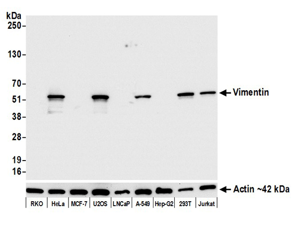


 WB (Western Blot)
(Detection of mouse Vimentin by western blot. Samples: Whole cell lysate (5 ug) from CH27, C2C12, 70Z/3, BW5147.3, CTLL-2, RenCa, J774A1, NIH 3T3, RAW 264.7, and TCMK-1 cells prepared using NETN lysis buffer. Antibody: Rabbit anti-Vimentin recombinant monoclonal antibody (AAA23833 lot 1) used at 1:1000. Secondary: HRP-conjugated goat anti-rabbit IgG . Detection: Chemiluminescence with an exposure time of 1 second. Lower Panel: Rabbit anti-Actin recombinant monoclonal antibody .)
WB (Western Blot)
(Detection of mouse Vimentin by western blot. Samples: Whole cell lysate (5 ug) from CH27, C2C12, 70Z/3, BW5147.3, CTLL-2, RenCa, J774A1, NIH 3T3, RAW 264.7, and TCMK-1 cells prepared using NETN lysis buffer. Antibody: Rabbit anti-Vimentin recombinant monoclonal antibody (AAA23833 lot 1) used at 1:1000. Secondary: HRP-conjugated goat anti-rabbit IgG . Detection: Chemiluminescence with an exposure time of 1 second. Lower Panel: Rabbit anti-Actin recombinant monoclonal antibody .)
Vimentin, Monoclonal Recombinant Antibody (Cat# AAA23833)


 WB (Western Blot)
(Detection of human MET by western blot. Samples: Whole cell lysate (10 ug) from GaMG, T-47D, HT-29, OVCAR-8, and 786-O cells prepared using NETN lysis buffer. Antibody: Rabbit anti-MET recombinant monoclonal antibody (AAA23881 lot 1) used at 1:1000. Secondary: HRP-conjugated goat anti-rabbit IgG . Detection: Chemiluminescence with an exposure time of 30 seconds. Lower Panel: Rabbit anti-Actin recombinant monoclonal antibody .)
WB (Western Blot)
(Detection of human MET by western blot. Samples: Whole cell lysate (10 ug) from GaMG, T-47D, HT-29, OVCAR-8, and 786-O cells prepared using NETN lysis buffer. Antibody: Rabbit anti-MET recombinant monoclonal antibody (AAA23881 lot 1) used at 1:1000. Secondary: HRP-conjugated goat anti-rabbit IgG . Detection: Chemiluminescence with an exposure time of 30 seconds. Lower Panel: Rabbit anti-Actin recombinant monoclonal antibody .)
Met, Monoclonal Recombinant Antibody (Cat# AAA23881)


 WB (Western Blot)
(Detection of mouse N-Cadherin by western blot. Samples: Whole cell lysate (50 ug) from C2C12, C2C12 treated with IFNgamma, BW5147.3, CTLL-2, EL4, RenCa, NIH 3T3, TCMK-1, A20, Ba/F3, and CH27 cells prepared using NETN lysis buffer. Antibody: Rabbit anti-N-Cadherin recombinant monoclonal antibody (AAA23831 lot 1) used at 1:1000. Secondary: HRP-conjugated goat anti-rabbit IgG . Detection: Chemiluminescence with an exposure time of 30 seconds. Lower Panel: Rabbit anti-COPB2 antibody .)
WB (Western Blot)
(Detection of mouse N-Cadherin by western blot. Samples: Whole cell lysate (50 ug) from C2C12, C2C12 treated with IFNgamma, BW5147.3, CTLL-2, EL4, RenCa, NIH 3T3, TCMK-1, A20, Ba/F3, and CH27 cells prepared using NETN lysis buffer. Antibody: Rabbit anti-N-Cadherin recombinant monoclonal antibody (AAA23831 lot 1) used at 1:1000. Secondary: HRP-conjugated goat anti-rabbit IgG . Detection: Chemiluminescence with an exposure time of 30 seconds. Lower Panel: Rabbit anti-COPB2 antibody .)
N-Cadherin, Monoclonal Recombinant Antibody (Cat# AAA23831)


 WB (Western Blot)
(Detection of human CD79B by western blot. Samples: Whole cell lysate (50 ug) from Daudi, HeLa, Raji, and Jurkat cells prepared using NETN lysis buffer. Antibody: Rabbit anti-CD79B recombinant monoclonal antibody (AAA23876 lot 1) used at 1:1000. Secondary: HRP-conjugated goat anti-rabbit IgG . Detection: Chemiluminescence with an exposure time of 10 seconds. Lower Panel: Rabbit anti-Actin recombinant monoclonal antibody .)
WB (Western Blot)
(Detection of human CD79B by western blot. Samples: Whole cell lysate (50 ug) from Daudi, HeLa, Raji, and Jurkat cells prepared using NETN lysis buffer. Antibody: Rabbit anti-CD79B recombinant monoclonal antibody (AAA23876 lot 1) used at 1:1000. Secondary: HRP-conjugated goat anti-rabbit IgG . Detection: Chemiluminescence with an exposure time of 10 seconds. Lower Panel: Rabbit anti-Actin recombinant monoclonal antibody .)
CD79B, Monoclonal Recombinant Antibody (Cat# AAA23876)


 WB (Western Blot)
(Detection of human CD38 by western blot. Samples: Whole cell lysate (10 ug) from HeLa, HEK293T, Jurkat, MOLT-4, RPMI-8226, CCRF-CEM, A-549, Ramos, Raji, and KG-1 cells prepared using NETN lysis buffer. Antibody: Rabbit anti-CD38 recombinant monoclonal antibody (AAA23840 lot 1) used at 1:1000. Secondary: HRP-conjugated goat anti-rabbit IgG . Detection: Chemiluminescence with an exposure time of 10 seconds. Lower Panel: Rabbit anti-COPB2 antibody .)
WB (Western Blot)
(Detection of human CD38 by western blot. Samples: Whole cell lysate (10 ug) from HeLa, HEK293T, Jurkat, MOLT-4, RPMI-8226, CCRF-CEM, A-549, Ramos, Raji, and KG-1 cells prepared using NETN lysis buffer. Antibody: Rabbit anti-CD38 recombinant monoclonal antibody (AAA23840 lot 1) used at 1:1000. Secondary: HRP-conjugated goat anti-rabbit IgG . Detection: Chemiluminescence with an exposure time of 10 seconds. Lower Panel: Rabbit anti-COPB2 antibody .)
CD38, Monoclonal Recombinant Antibody (Cat# AAA23840)


 WB (Western Blot)
(Detection of human STAT5b by western blot. Samples: Whole cell lysate (10 ug) from My-La CD4+, Hep-G2, HEK293T, Jurkat, and K-562 cells prepared using NETN lysis buffer. Antibody: Rabbit anti-STAT5b recombinant monoclonal antibody (AAA23847 lot 1) used at 1:1000. Secondary: HRP-conjugated goat anti-rabbit IgG . Detection: Chemiluminescence with an exposure time of 75 seconds. Lower Panel: Rabbit anti-COPB2 antibody .)
WB (Western Blot)
(Detection of human STAT5b by western blot. Samples: Whole cell lysate (10 ug) from My-La CD4+, Hep-G2, HEK293T, Jurkat, and K-562 cells prepared using NETN lysis buffer. Antibody: Rabbit anti-STAT5b recombinant monoclonal antibody (AAA23847 lot 1) used at 1:1000. Secondary: HRP-conjugated goat anti-rabbit IgG . Detection: Chemiluminescence with an exposure time of 75 seconds. Lower Panel: Rabbit anti-COPB2 antibody .)
STAT5b, Monoclonal Recombinant Antibody (Cat# AAA23847)


 WB (Western Blot)
(Detection of mouse P-Selectin/CD62P by western blot. Samples: Whole cell lysate (50 ug) from NIH 3T3, bEnd.3 (10 ug), CH27, Spleen, and CT26 cells prepared using NETN lysis buffer. Antibody: Rabbit anti-P-Selectin/CD62P recombinant monoclonal antibody (AAA23880 lot 1) used at 1:1000. Secondary: HRP-conjugated goat anti-rabbit IgG . Detection: Chemiluminescence with an exposure time of 10 seconds. Lower Panel: Rabbit anti-Actin recombinant monoclonal antibody .)
WB (Western Blot)
(Detection of mouse P-Selectin/CD62P by western blot. Samples: Whole cell lysate (50 ug) from NIH 3T3, bEnd.3 (10 ug), CH27, Spleen, and CT26 cells prepared using NETN lysis buffer. Antibody: Rabbit anti-P-Selectin/CD62P recombinant monoclonal antibody (AAA23880 lot 1) used at 1:1000. Secondary: HRP-conjugated goat anti-rabbit IgG . Detection: Chemiluminescence with an exposure time of 10 seconds. Lower Panel: Rabbit anti-Actin recombinant monoclonal antibody .)
P-Selectin/CD62P, Monoclonal Recombinant Antibody (Cat# AAA23880)


 FCM (Flow Cytometry)
(Figure-6: Epitope binding study by flow cytometric analysis. MCF-7 cells expressing HER2 antigen were treated with Either Herceptin or Samceptin (1 & 2 ug/10^6 Cells). Surface staining was done using FITC conjugated antibodies.)
FCM (Flow Cytometry)
(Figure-6: Epitope binding study by flow cytometric analysis. MCF-7 cells expressing HER2 antigen were treated with Either Herceptin or Samceptin (1 & 2 ug/10^6 Cells). Surface staining was done using FITC conjugated antibodies.)
ErbB2/HER2, Monoclonal Recombinant Antibody (Cat# AAA14881)


 WB (Western Blot)
(Detection of human EpCAM by western blot. Samples: Whole cell lysate (10 ug) from MCF-7, LNCaP, HeLa, SW620, and HT-29 cells prepared using NETN lysis buffer. Antibody: Rabbit anti-EpCAM recombinant monoclonal antibody (AAA23861 lot 1) used at 1:1000. Secondary: HRP-conjugated goat anti-rabbit IgG . Detection: Chemiluminescence with an exposure time of 10 seconds. Lower Panel: Rabbit anti-COPB2 antibody .)
WB (Western Blot)
(Detection of human EpCAM by western blot. Samples: Whole cell lysate (10 ug) from MCF-7, LNCaP, HeLa, SW620, and HT-29 cells prepared using NETN lysis buffer. Antibody: Rabbit anti-EpCAM recombinant monoclonal antibody (AAA23861 lot 1) used at 1:1000. Secondary: HRP-conjugated goat anti-rabbit IgG . Detection: Chemiluminescence with an exposure time of 10 seconds. Lower Panel: Rabbit anti-COPB2 antibody .)
EpCAM, Monoclonal Recombinant Antibody (Cat# AAA23861)


 WB (Western Blot)
(Detection of human E-Cadherin by western blot. Samples: Whole cell lysate (5 ug) from GaMG, HCT 116, U2OS, OVCAR-4, Jurkat, 22Rv1, HeLa, MCF-7, and K-562 cells prepared using NETN lysis buffer. Antibody: Rabbit anti-E-Cadherin recombinant monoclonal antibody (AAA23828 lot 1) used at 1:1000. Secondary: HRP-conjugated goat anti-rabbit IgG . Detection: Chemiluminescence with an exposure time of 10 seconds. Lower Panel: Rabbit anti-Actin recombinant monoclonal antibody .)
WB (Western Blot)
(Detection of human E-Cadherin by western blot. Samples: Whole cell lysate (5 ug) from GaMG, HCT 116, U2OS, OVCAR-4, Jurkat, 22Rv1, HeLa, MCF-7, and K-562 cells prepared using NETN lysis buffer. Antibody: Rabbit anti-E-Cadherin recombinant monoclonal antibody (AAA23828 lot 1) used at 1:1000. Secondary: HRP-conjugated goat anti-rabbit IgG . Detection: Chemiluminescence with an exposure time of 10 seconds. Lower Panel: Rabbit anti-Actin recombinant monoclonal antibody .)
E-Cadherin, Monoclonal Recombinant Antibody (Cat# AAA23828)
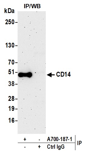


 WB (Western Blot)
(Detection of human CD14 by western blot. Samples: Whole cell lysate (50 ug) from THP-1 +LPS, HEK293T, MUTZ-3, HeLa, and Jurkat cells prepared using NETN lysis buffer. Antibody: Rabbit anti-CD14 recombinant monoclonal antibody (AAA23858 lot 1) used at 1:1000. Secondary: HRP-conjugated goat anti-rabbit IgG . Detection: Chemiluminescence with an exposure time of 3 minutes. Lower Panel: Rabbit anti-COPB2 antibody .)
WB (Western Blot)
(Detection of human CD14 by western blot. Samples: Whole cell lysate (50 ug) from THP-1 +LPS, HEK293T, MUTZ-3, HeLa, and Jurkat cells prepared using NETN lysis buffer. Antibody: Rabbit anti-CD14 recombinant monoclonal antibody (AAA23858 lot 1) used at 1:1000. Secondary: HRP-conjugated goat anti-rabbit IgG . Detection: Chemiluminescence with an exposure time of 3 minutes. Lower Panel: Rabbit anti-COPB2 antibody .)
CD14, Monoclonal Recombinant Antibody (Cat# AAA23858)


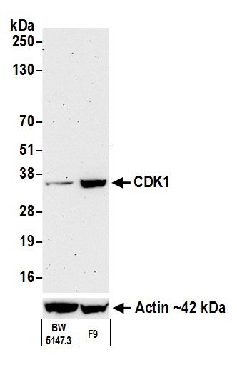 WB (Western Blot)
(Detection of mouse CDK1 by western blot. Samples: Whole cell lysate (50 ug) from BW5147.3 and F9 cells prepared using NETN lysis buffer. Antibody: Rabbit anti-CDK1 recombinant monoclonal antibody (AAA23826 lot 1) used at 1:1000. Secondary: HRP-conjugated goat anti-rabbit IgG . Detection: Chemiluminescence with an exposure time of 3 minutes. Lower Panel: Rabbit anti-Actin .)
WB (Western Blot)
(Detection of mouse CDK1 by western blot. Samples: Whole cell lysate (50 ug) from BW5147.3 and F9 cells prepared using NETN lysis buffer. Antibody: Rabbit anti-CDK1 recombinant monoclonal antibody (AAA23826 lot 1) used at 1:1000. Secondary: HRP-conjugated goat anti-rabbit IgG . Detection: Chemiluminescence with an exposure time of 3 minutes. Lower Panel: Rabbit anti-Actin .)
CDK1, Monoclonal Recombinant Antibody (Cat# AAA23826)


 WB (Western Blot)
(Western blot analysis of extracts from serum-starved Hela treated with TPA (100 nM, 15 min), using Phospho-CREB (Ser133) rabbit monoclonal Antibody (#AAA27785) at 1:10000, 1:100000, 1:500000 dilution.(Validation Experiment))
WB (Western Blot)
(Western blot analysis of extracts from serum-starved Hela treated with TPA (100 nM, 15 min), using Phospho-CREB (Ser133) rabbit monoclonal Antibody (#AAA27785) at 1:10000, 1:100000, 1:500000 dilution.(Validation Experiment))
CREB, Monoclonal Recombinant Antibody (Cat# AAA27785)


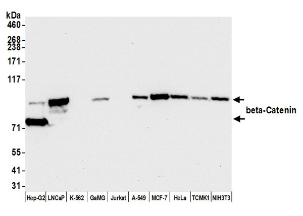 WB (Western Blot)
(Detection of human and mouse beta-Catenin by western blot. Samples: Whole cell lysate (50 ug) from Hep-G2, LNCaP, K-562, GaMG, Jurkat, A-549, MCF-7, HeLa, TCMK-1, and NIH 3T3 cells prepared using NETN lysis buffer. Antibody: Rabbit anti-beta-Catenin recombinant monoclonal antibody (AAA23827 lot 1) used at 1:1000. Secondary: HRP-conjugated goat anti-rabbit IgG . Detection: Chemiluminescence with an exposure time of 10 seconds.)
WB (Western Blot)
(Detection of human and mouse beta-Catenin by western blot. Samples: Whole cell lysate (50 ug) from Hep-G2, LNCaP, K-562, GaMG, Jurkat, A-549, MCF-7, HeLa, TCMK-1, and NIH 3T3 cells prepared using NETN lysis buffer. Antibody: Rabbit anti-beta-Catenin recombinant monoclonal antibody (AAA23827 lot 1) used at 1:1000. Secondary: HRP-conjugated goat anti-rabbit IgG . Detection: Chemiluminescence with an exposure time of 10 seconds.)
beta Catenin, Monoclonal Recombinant Antibody (Cat# AAA23827)


 FCM (Flow Cytometry)
(Figure-6: Epitope binding study by flow cytometric analysis. MCF-7 cells expressing HER2 antigen were treated with Herceptin and Samceptin (2 ug/10^6 Cells). Surface staining was done using FITC conjugated antibodies.)
FCM (Flow Cytometry)
(Figure-6: Epitope binding study by flow cytometric analysis. MCF-7 cells expressing HER2 antigen were treated with Herceptin and Samceptin (2 ug/10^6 Cells). Surface staining was done using FITC conjugated antibodies.)
ErbB2/HER2, Monoclonal Recombinant Antibody (Cat# AAA14887)


 WB (Western Blot)
(Detection of mouse FOX2/RBM9 by western blot. Samples: Whole cell lysate (50 ug) from NIH 3T3 and CT26 cells prepared using NETN lysis buffer. Antibody: Rabbit anti-FOX2/RBM9 recombinant monoclonal antibody (AAA23868 lot 1) used at 1:1000. Secondary: HRP-conjugated goat anti-rabbit IgG . Detection: Chemiluminescence with an exposure time of 75 seconds. Lower Panel: Rabbit anti-Actin recombinant monoclonal antibody .)
WB (Western Blot)
(Detection of mouse FOX2/RBM9 by western blot. Samples: Whole cell lysate (50 ug) from NIH 3T3 and CT26 cells prepared using NETN lysis buffer. Antibody: Rabbit anti-FOX2/RBM9 recombinant monoclonal antibody (AAA23868 lot 1) used at 1:1000. Secondary: HRP-conjugated goat anti-rabbit IgG . Detection: Chemiluminescence with an exposure time of 75 seconds. Lower Panel: Rabbit anti-Actin recombinant monoclonal antibody .)
FOX2/RBM9, Monoclonal Recombinant Antibody (Cat# AAA23868)


 WB (Western Blot)
(Detection of human T-bet/TBX21 by western blot. Samples: Whole cell lysate (50 ug) from MCF-7, HEK293T, MJ/G11, HeLa, Jurkat, NK-92, Hep-G2, and U2OS cells prepared using NETN lysis buffer. Antibody: Rabbit anti-T-bet/TBX21 recombinant monoclonal antibody (AAA23837 lot 1) used at 1:1000. Secondary: HRP-conjugated goat anti-rabbit IgG . Detection: Chemiluminescence with an exposure time of 30 seconds. Lower Panel: Rabbit anti-Actin recombinant monoclonal .)
WB (Western Blot)
(Detection of human T-bet/TBX21 by western blot. Samples: Whole cell lysate (50 ug) from MCF-7, HEK293T, MJ/G11, HeLa, Jurkat, NK-92, Hep-G2, and U2OS cells prepared using NETN lysis buffer. Antibody: Rabbit anti-T-bet/TBX21 recombinant monoclonal antibody (AAA23837 lot 1) used at 1:1000. Secondary: HRP-conjugated goat anti-rabbit IgG . Detection: Chemiluminescence with an exposure time of 30 seconds. Lower Panel: Rabbit anti-Actin recombinant monoclonal .)
T-bet/TBX21, Monoclonal Recombinant Antibody (Cat# AAA23837)


 WB (Western Blot)
(Detection of human CD206/Mannose Receptor by western blot. Samples: Whole cell lysate (50 ug) from HeLa, HEK293T, MUTZ-3, Hep-G2, and MCF-7 cells prepared using NETN lysis buffer. Antibody: Rabbit anti-CD206/Mannose Receptor recombinant monoclonal antibody (AAA23836 lot 1) used at 1:1000. Secondary: HRP-conjugated goat anti-rabbit IgG . Detection: Chemiluminescence with an exposure time of 1 second. Lower Panel: Rabbit anti-COPB2 antibody .)
WB (Western Blot)
(Detection of human CD206/Mannose Receptor by western blot. Samples: Whole cell lysate (50 ug) from HeLa, HEK293T, MUTZ-3, Hep-G2, and MCF-7 cells prepared using NETN lysis buffer. Antibody: Rabbit anti-CD206/Mannose Receptor recombinant monoclonal antibody (AAA23836 lot 1) used at 1:1000. Secondary: HRP-conjugated goat anti-rabbit IgG . Detection: Chemiluminescence with an exposure time of 1 second. Lower Panel: Rabbit anti-COPB2 antibody .)
CD206/Mannose Receptor, Monoclonal Recombinant Antibody (Cat# AAA23836)


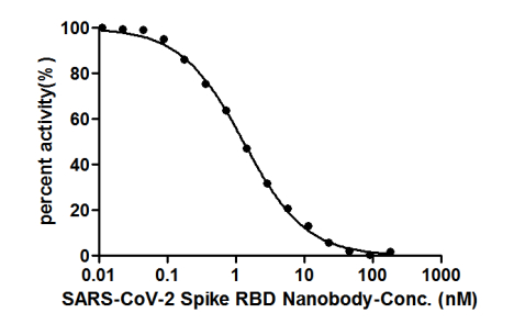


 Application Data
(ELISA: Immobilize various types of SARS proteins at concentration of 2mg/ml on solid substrate, then react with SARS-CoV-2 Spike RBD Nanobody at concentration of 100mg/ml, 10mg/ml and 1mg/ml. It shows the SARS-CoV-2 Spike RBD Nanobody (AAA27051) is specific for SARS-CoV-2-S1-RBD protein, without any cross-reactivity with MERS-CoV, SARS-CoV, HCoV-OC43 or HCoV-229E.)
Application Data
(ELISA: Immobilize various types of SARS proteins at concentration of 2mg/ml on solid substrate, then react with SARS-CoV-2 Spike RBD Nanobody at concentration of 100mg/ml, 10mg/ml and 1mg/ml. It shows the SARS-CoV-2 Spike RBD Nanobody (AAA27051) is specific for SARS-CoV-2-S1-RBD protein, without any cross-reactivity with MERS-CoV, SARS-CoV, HCoV-OC43 or HCoV-229E.)
COVID 19 Spike RBD Coronavirus, Monoclonal Recombinant Antibody (Cat# AAA27051)


 IF (Immunofluorescence)
(Immunofluorescence analysis of NIH/3T3 cells using Recombinant GAPDH Monoclonal Antibody at dilution of 1:1000)
IF (Immunofluorescence)
(Immunofluorescence analysis of NIH/3T3 cells using Recombinant GAPDH Monoclonal Antibody at dilution of 1:1000)
GAPDH, Monoclonal Recombinant Antibody (Cat# AAA22336)


 WB (Western Blot)
(Detection of human Nectin-2/CD112 by western blot. Samples: Whole cell lysate (50 ug) from OVCAR-8, Jurkat, U2OS, GaMG, OVCAR-4, 786-O, A-549, and SK-MEL-28 cells prepared using NETN lysis buffer. Antibody: Rabbit anti-Nectin-2/CD112 recombinant monoclonal antibody (AAA23819 lot 1) used at 1:1000. Secondary: HRP-conjugated goat anti-rabbit IgG . Detection: Chemiluminescence with an exposure time of 30 seconds. Lower panel: Rabbit anti-GAPDH .)
WB (Western Blot)
(Detection of human Nectin-2/CD112 by western blot. Samples: Whole cell lysate (50 ug) from OVCAR-8, Jurkat, U2OS, GaMG, OVCAR-4, 786-O, A-549, and SK-MEL-28 cells prepared using NETN lysis buffer. Antibody: Rabbit anti-Nectin-2/CD112 recombinant monoclonal antibody (AAA23819 lot 1) used at 1:1000. Secondary: HRP-conjugated goat anti-rabbit IgG . Detection: Chemiluminescence with an exposure time of 30 seconds. Lower panel: Rabbit anti-GAPDH .)
Nectin-2/CD112, Monoclonal Recombinant Antibody (Cat# AAA23819)


 WB (Western Blot)
(Detection of human PU.1 by western blot. Samples: Whole cell lysate (50 ug) from THP-1, Jurkat, K-562, HEK293T, and KG-1 cells prepared using NETN lysis buffer. Antibody: Rabbit anti-PU.1 recombinant monoclonal antibody (AAA23853 lot 1) used at 1:1000. Secondary: HRP-conjugated goat anti-rabbit IgG . Detection: Chemiluminescence with an exposure time of 10 seconds.)
WB (Western Blot)
(Detection of human PU.1 by western blot. Samples: Whole cell lysate (50 ug) from THP-1, Jurkat, K-562, HEK293T, and KG-1 cells prepared using NETN lysis buffer. Antibody: Rabbit anti-PU.1 recombinant monoclonal antibody (AAA23853 lot 1) used at 1:1000. Secondary: HRP-conjugated goat anti-rabbit IgG . Detection: Chemiluminescence with an exposure time of 10 seconds.)
PU.1, Monoclonal Recombinant Antibody (Cat# AAA23853)


 WB (Western Blot)
(Detection of human TJP1/ZO-1 by western blot. Samples: Whole cell lysate (25 ug) from A-549, HeLa, Jurkat, MCF-7, and RKO cells prepared using NETN lysis buffer. Antibody: Rabbit anti-TJP1/ZO-1 recombinant monoclonal antibody (AAA23830A lot 3) used at 1:1000. Secondary: HRP-conjugated goat anti-rabbit IgG . Detection: Chemiluminescence with an exposure time of 75 seconds. Lower Panel: Rabbit anti-COPB2 antibody .)
WB (Western Blot)
(Detection of human TJP1/ZO-1 by western blot. Samples: Whole cell lysate (25 ug) from A-549, HeLa, Jurkat, MCF-7, and RKO cells prepared using NETN lysis buffer. Antibody: Rabbit anti-TJP1/ZO-1 recombinant monoclonal antibody (AAA23830A lot 3) used at 1:1000. Secondary: HRP-conjugated goat anti-rabbit IgG . Detection: Chemiluminescence with an exposure time of 75 seconds. Lower Panel: Rabbit anti-COPB2 antibody .)
TJP1/ZO-1, Monoclonal Recombinant Antibody (Cat# AAA23830)


 WB (Western Blot)
(Detection of human OX40/CD134 by western blot. Samples: Whole cell lysate (50 ug) from HH, HeLa, MJ, HEK293T, Hep-G2, C5/MJ, Jurkat, and MOLT-4 cells prepared using NETN lysis buffer. Antibody: Rabbit anti-OX40/CD134 recombinant monoclonal antibody (AAA23810 lot 1) used at 1:1000. Secondary: HRP-conjugated goat anti-rabbit IgG . Detection: Chemiluminescence with an exposure time of 30 seconds. Lower panel: Rabbit anti-eEF2 .)
WB (Western Blot)
(Detection of human OX40/CD134 by western blot. Samples: Whole cell lysate (50 ug) from HH, HeLa, MJ, HEK293T, Hep-G2, C5/MJ, Jurkat, and MOLT-4 cells prepared using NETN lysis buffer. Antibody: Rabbit anti-OX40/CD134 recombinant monoclonal antibody (AAA23810 lot 1) used at 1:1000. Secondary: HRP-conjugated goat anti-rabbit IgG . Detection: Chemiluminescence with an exposure time of 30 seconds. Lower panel: Rabbit anti-eEF2 .)
OX40/CD134, Monoclonal Recombinant Antibody (Cat# AAA23810)


 WB (Western Blot)
(Detection of human and mouse NAMPT/PBEF/Visfatin by western blot. Samples: Whole cell lysate (50 ug) from RKO, MCF-7, K-562, GaMG, HEK293T, HeLa, U2OS, LNCaP, Hep-G2, Jurkat, and NIH 3T3 cells prepared using NETN lysis buffer. Antibody: Rabbit anti-NAMPT/PBEF/Visfatin recombinant monoclonal antibody (AAA23815 lot 1) used at 1:1000. Secondary: HRP-conjugated goat anti-rabbit IgG . Detection: Chemiluminescence with an exposure time of 30 seconds. Lower Panel: Rabbit anti-GAPDH .)
WB (Western Blot)
(Detection of human and mouse NAMPT/PBEF/Visfatin by western blot. Samples: Whole cell lysate (50 ug) from RKO, MCF-7, K-562, GaMG, HEK293T, HeLa, U2OS, LNCaP, Hep-G2, Jurkat, and NIH 3T3 cells prepared using NETN lysis buffer. Antibody: Rabbit anti-NAMPT/PBEF/Visfatin recombinant monoclonal antibody (AAA23815 lot 1) used at 1:1000. Secondary: HRP-conjugated goat anti-rabbit IgG . Detection: Chemiluminescence with an exposure time of 30 seconds. Lower Panel: Rabbit anti-GAPDH .)
NAMPT/PBEF/Visfatin, Monoclonal Recombinant Antibody (Cat# AAA23815)


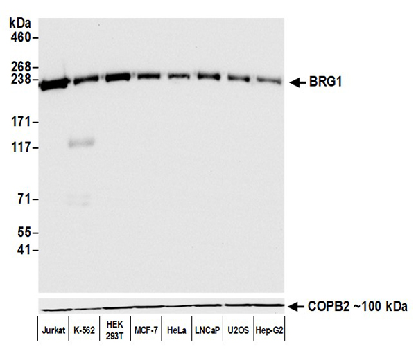 WB (Western Blot)
(Detection of human BRG1 by western blot. Samples: Whole cell lysate (50 ug) from Jurkat, K-562, HEK293T, MCF-7, HeLa, LNCaP, U2OS, and Hep-G2 cells prepared using NETN lysis buffer. Antibody: Rabbit anti-BRG1 recombinant monoclonal antibody (AAA23834 lot 1) used at 1:1000. Secondary: HRP-conjugated goat anti-rabbit IgG . Detection: Chemiluminescence with an exposure time of 30 seconds. Lower Panel: Rabbit anti-COPB2 antibody .)
WB (Western Blot)
(Detection of human BRG1 by western blot. Samples: Whole cell lysate (50 ug) from Jurkat, K-562, HEK293T, MCF-7, HeLa, LNCaP, U2OS, and Hep-G2 cells prepared using NETN lysis buffer. Antibody: Rabbit anti-BRG1 recombinant monoclonal antibody (AAA23834 lot 1) used at 1:1000. Secondary: HRP-conjugated goat anti-rabbit IgG . Detection: Chemiluminescence with an exposure time of 30 seconds. Lower Panel: Rabbit anti-COPB2 antibody .)
BRG1, Monoclonal Recombinant Antibody (Cat# AAA23834)


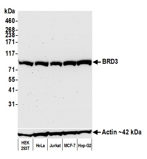 WB (Western Blot)
(Detection of human BRD3 by western blot. Samples: Whole cell lysate (50 ug) from HEK293T, HeLa, Jurkat, MCF-7, and Hep-G2 cells prepared using NETN lysis buffer. Antibody: Rabbit anti-BRD3 recombinant monoclonal antibody (AAA23818 lot 2) used at 1:1000. Secondary: HRP-conjugated goat anti-rabbit IgG . Detection: Chemiluminescence with an exposure time of 75 seconds. Lower Panel: Rabbit anti-Actin recombinant monoclonal antibody .)
WB (Western Blot)
(Detection of human BRD3 by western blot. Samples: Whole cell lysate (50 ug) from HEK293T, HeLa, Jurkat, MCF-7, and Hep-G2 cells prepared using NETN lysis buffer. Antibody: Rabbit anti-BRD3 recombinant monoclonal antibody (AAA23818 lot 2) used at 1:1000. Secondary: HRP-conjugated goat anti-rabbit IgG . Detection: Chemiluminescence with an exposure time of 75 seconds. Lower Panel: Rabbit anti-Actin recombinant monoclonal antibody .)
BRD3, Monoclonal Recombinant Antibody (Cat# AAA23818)


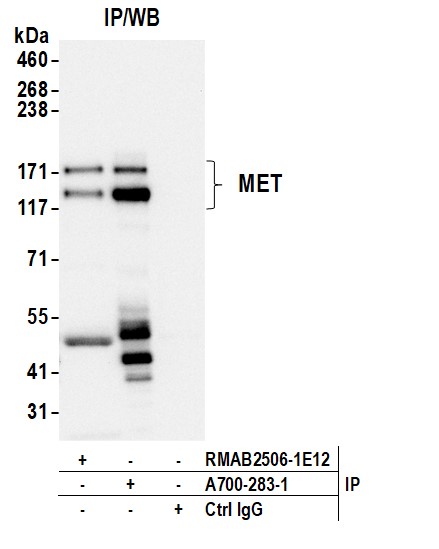
 IP (Immunoprecipitation)
(Tubulin was immunoprecipitated using: Lane A:0.5 mg HepG2 Whole Cell Lysate Lane B:0.5 mg Hela Whole Cell Lysate Lane C:0.5 mg Raw246.7 Whole Cell Lysate Lane D:0.5 mg Jurkat Whole Cell Lysate 4 uL anti-Tubulin rabbit monoclonal antibody and 60ug of Immunomagnetic beads Protein A/G. Primary antibody: Anti-Tubulin rabbit monoclonal antibody,at 1:100 dilution Secondary antibody: Clean-Blot IP Detection Reagent (HRP) at 1:1000dilution Developed using the ECL technique. Performed under reducing conditions. Predicted band size: 50 kDa Observed band size :54 kDa)
IP (Immunoprecipitation)
(Tubulin was immunoprecipitated using: Lane A:0.5 mg HepG2 Whole Cell Lysate Lane B:0.5 mg Hela Whole Cell Lysate Lane C:0.5 mg Raw246.7 Whole Cell Lysate Lane D:0.5 mg Jurkat Whole Cell Lysate 4 uL anti-Tubulin rabbit monoclonal antibody and 60ug of Immunomagnetic beads Protein A/G. Primary antibody: Anti-Tubulin rabbit monoclonal antibody,at 1:100 dilution Secondary antibody: Clean-Blot IP Detection Reagent (HRP) at 1:1000dilution Developed using the ECL technique. Performed under reducing conditions. Predicted band size: 50 kDa Observed band size :54 kDa)
Beta-Tubulin, Monoclonal Recombinant Antibody (Cat# AAA27784)


 WB (Western Blot)
(Detection of human BMI1 by western blot. Samples: Whole cell lysate (10 ug) from MOLT-4, HeLa, HEK293T, Jurkat, LNCaP, RKO, Hep-G2, and U2OS cells prepared using NETN lysis buffer. Antibody: Rabbit anti-BMI1 recombinant monoclonal antibody (AAA23838 lot 1) used at 1:1000. Secondary: HRP-conjugated goat anti-rabbit IgG . Detection: Chemiluminescence with an exposure time of 10 seconds. Lower Panel: Rabbit anti-COPB2 antibody .)
WB (Western Blot)
(Detection of human BMI1 by western blot. Samples: Whole cell lysate (10 ug) from MOLT-4, HeLa, HEK293T, Jurkat, LNCaP, RKO, Hep-G2, and U2OS cells prepared using NETN lysis buffer. Antibody: Rabbit anti-BMI1 recombinant monoclonal antibody (AAA23838 lot 1) used at 1:1000. Secondary: HRP-conjugated goat anti-rabbit IgG . Detection: Chemiluminescence with an exposure time of 10 seconds. Lower Panel: Rabbit anti-COPB2 antibody .)
BMI1, Monoclonal Recombinant Antibody (Cat# AAA23838)


 IHC (Immunohistochemistry)
(Immunohistochemistry of paraffin-embedded mouse ovary using Recombinant beta actin Monoclonal Antibody at dilution of 1:1000.)
IHC (Immunohistochemistry)
(Immunohistochemistry of paraffin-embedded mouse ovary using Recombinant beta actin Monoclonal Antibody at dilution of 1:1000.)
beta actin, Monoclonal Recombinant Antibody (Cat# AAA22337)


 WB (Western Blot)
(Detection of human STAT3 by western blot. Samples: Whole cell lysate (50 ug) from RKO, NCI-H226, HEK293T, GaMG, LNCaP, HeLa, Ramos, MOLT-4, and Jurkat cells prepared using NETN lysis buffer. Antibody: Rabbit anti-STAT3 recombinant monoclonal antibody (AAA23832 lot 1) used at 1:1000. Secondary: HRP-conjugated goat anti-rabbit IgG . Detection: Chemiluminescence with an exposure time of 10 seconds. Lower Panel: Rabbit anti-Actin recombinant monoclonal antibody .)
WB (Western Blot)
(Detection of human STAT3 by western blot. Samples: Whole cell lysate (50 ug) from RKO, NCI-H226, HEK293T, GaMG, LNCaP, HeLa, Ramos, MOLT-4, and Jurkat cells prepared using NETN lysis buffer. Antibody: Rabbit anti-STAT3 recombinant monoclonal antibody (AAA23832 lot 1) used at 1:1000. Secondary: HRP-conjugated goat anti-rabbit IgG . Detection: Chemiluminescence with an exposure time of 10 seconds. Lower Panel: Rabbit anti-Actin recombinant monoclonal antibody .)
STAT3, Monoclonal Recombinant Antibody (Cat# AAA23832)


 WB (Western Blot)
(Detection of human AXL by western blot. Samples: Whole cell lysate (10 ug) from OVCAR-8, HEK293T, HeLa, Jurkat, and 786-O cells prepared using NETN lysis buffer. Antibody: Rabbit anti-AXL recombinant monoclonal antibody (AAA23873 lot 1) used at 1:1000. Secondary: HRP-conjugated goat anti-rabbit IgG . Detection: Chemiluminescence with an exposure time of 3 minutes.)
WB (Western Blot)
(Detection of human AXL by western blot. Samples: Whole cell lysate (10 ug) from OVCAR-8, HEK293T, HeLa, Jurkat, and 786-O cells prepared using NETN lysis buffer. Antibody: Rabbit anti-AXL recombinant monoclonal antibody (AAA23873 lot 1) used at 1:1000. Secondary: HRP-conjugated goat anti-rabbit IgG . Detection: Chemiluminescence with an exposure time of 3 minutes.)
AXL, Monoclonal Recombinant Antibody (Cat# AAA23873)


 WB (Western Blot)
(Detection of human gamma-H2AX by western blot. Samples: Whole cell lysate (50 ug) from Jurkat cells treated with 100 uM etoposide (+) or mock treated (-). Antibody: Rabbit anti-gamma-H2AX recombinant monoclonal antibody (AAA23813 lot 2) used at 1:1000. Secondary: HRP-conjugated goat anti-rabbit IgG . Detection: Chemiluminescence with an exposure time of 3 seconds.)
WB (Western Blot)
(Detection of human gamma-H2AX by western blot. Samples: Whole cell lysate (50 ug) from Jurkat cells treated with 100 uM etoposide (+) or mock treated (-). Antibody: Rabbit anti-gamma-H2AX recombinant monoclonal antibody (AAA23813 lot 2) used at 1:1000. Secondary: HRP-conjugated goat anti-rabbit IgG . Detection: Chemiluminescence with an exposure time of 3 seconds.)
gamma-H2AX, Monoclonal Recombinant Antibody (Cat# AAA23813)


 WB (Western Blot)
(Detection of mouse CD3E by western blot. Samples: Whole cell lysate (10 ug) from BW5147.3, NIH 3T3, TCMK-1, EL4, and CTLL-2 cells prepared using NETN lysis buffer. Antibody: Rabbit anti-CD3E recombinant monoclonal antibody (AAA23852 lot 1) used at 1:1000. Secondary: HRP-conjugated goat anti-rabbit IgG . Detection: Chemiluminescence with an exposure time of 10 seconds.)
WB (Western Blot)
(Detection of mouse CD3E by western blot. Samples: Whole cell lysate (10 ug) from BW5147.3, NIH 3T3, TCMK-1, EL4, and CTLL-2 cells prepared using NETN lysis buffer. Antibody: Rabbit anti-CD3E recombinant monoclonal antibody (AAA23852 lot 1) used at 1:1000. Secondary: HRP-conjugated goat anti-rabbit IgG . Detection: Chemiluminescence with an exposure time of 10 seconds.)
CD3E, Monoclonal Recombinant Antibody (Cat# AAA23852)


 WB (Western Blot)
(Detection of human Beta-III-Tubulin by western blot. Samples: Whole cell lysate (10 ug) from SK-N-SH, SK-N-BE(2), HEK293T, A-172, and H1975 cells prepared using NETN lysis buffer. Antibody: Rabbit anti-Beta-III-Tubulin recombinant monoclonal antibody (AAA23857 lot 1) used at 1:1000. Secondary: HRP-conjugated goat anti-rabbit IgG . Detection: Chemiluminescence with an exposure time of 3 seconds. Lower Panel: Rabbit anti-Actin recombinant monoclonal antibody .)
WB (Western Blot)
(Detection of human Beta-III-Tubulin by western blot. Samples: Whole cell lysate (10 ug) from SK-N-SH, SK-N-BE(2), HEK293T, A-172, and H1975 cells prepared using NETN lysis buffer. Antibody: Rabbit anti-Beta-III-Tubulin recombinant monoclonal antibody (AAA23857 lot 1) used at 1:1000. Secondary: HRP-conjugated goat anti-rabbit IgG . Detection: Chemiluminescence with an exposure time of 3 seconds. Lower Panel: Rabbit anti-Actin recombinant monoclonal antibody .)
beta-III Tubulin, Monoclonal Recombinant Antibody (Cat# AAA23857)


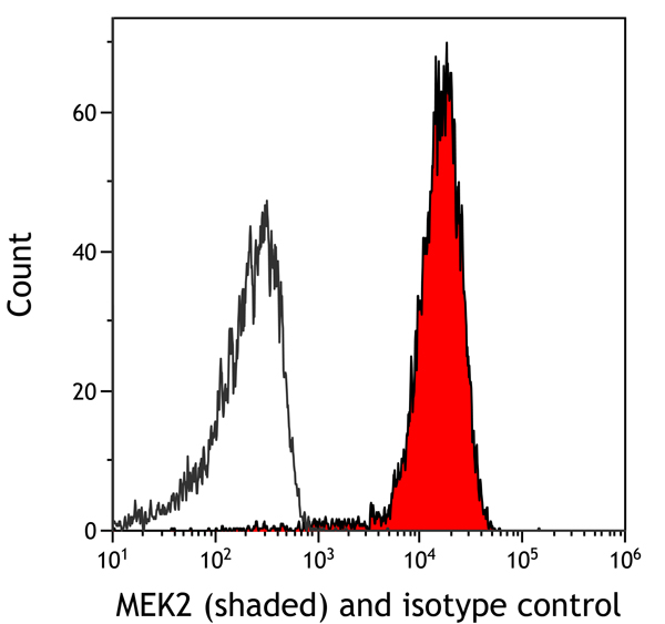
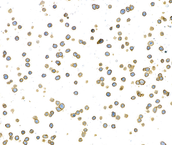
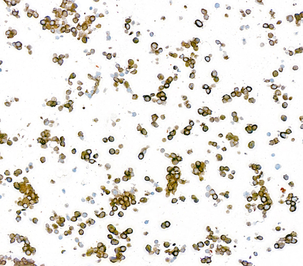
 Application Data
(Overlay histogram showing Raw264.7 cells stained with AAA27010 (red line) at 1:50. The cells were fixed with 70% Ethylalcohol (18h) and then permeabilized with 0.3% Triton X-100 for 2 min.The cells were then incubated in 1x PBS /10% normal goat serum to block non-specific protein-protein interactions followed by primary antibody for 1 h at 4 degree C.The secondary antibody used was FITC goat anti-rabbit IgG (H+L) at 1/200 dilution for 1 h at 4 degree C. Control antibody (green line) was used under the same conditions. Acquisition of >10, 000 events was performed.)
Application Data
(Overlay histogram showing Raw264.7 cells stained with AAA27010 (red line) at 1:50. The cells were fixed with 70% Ethylalcohol (18h) and then permeabilized with 0.3% Triton X-100 for 2 min.The cells were then incubated in 1x PBS /10% normal goat serum to block non-specific protein-protein interactions followed by primary antibody for 1 h at 4 degree C.The secondary antibody used was FITC goat anti-rabbit IgG (H+L) at 1/200 dilution for 1 h at 4 degree C. Control antibody (green line) was used under the same conditions. Acquisition of >10, 000 events was performed.)
CD163, Monoclonal Recombinant Antibody (Cat# AAA27010)


 WB (Western Blot)
(Detection of mouse NF-H by western blot. Samples: Whole cell lysate (50 ug) from CT26, NIH 3T3, ND7/23, TCMK-1 (10 ug), and CH27 cells prepared using NETN lysis buffer. Antibody: Rabbit anti-NF-H recombinant monoclonal antibody (AAA23859 lot 1) used at 1:1000. Secondary: HRP-conjugated goat anti-rabbit IgG . Detection: Chemiluminescence with an exposure time of 3 minutes. Lower Panel: Rabbit anti-COPB2 antibody .)
WB (Western Blot)
(Detection of mouse NF-H by western blot. Samples: Whole cell lysate (50 ug) from CT26, NIH 3T3, ND7/23, TCMK-1 (10 ug), and CH27 cells prepared using NETN lysis buffer. Antibody: Rabbit anti-NF-H recombinant monoclonal antibody (AAA23859 lot 1) used at 1:1000. Secondary: HRP-conjugated goat anti-rabbit IgG . Detection: Chemiluminescence with an exposure time of 3 minutes. Lower Panel: Rabbit anti-COPB2 antibody .)
NF-H, Monoclonal Recombinant Antibody (Cat# AAA23859)


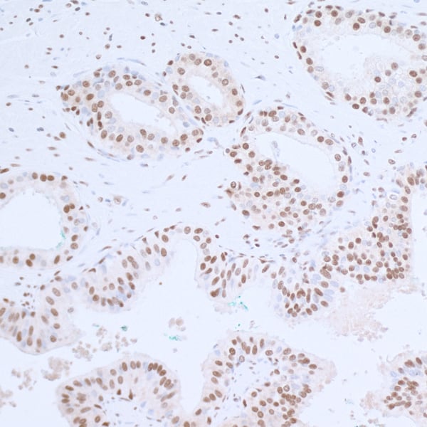
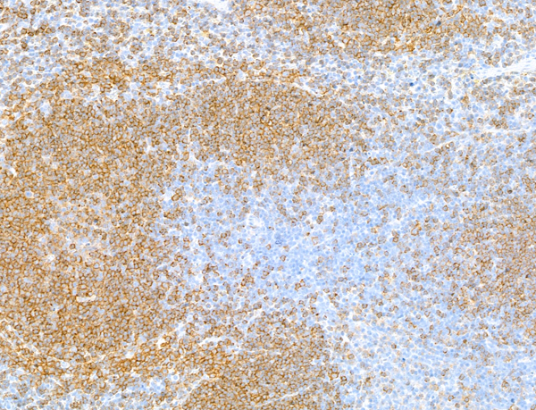
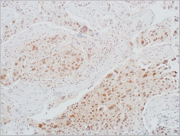
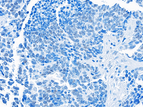
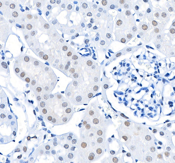
 IHC (Immunohistchemistry)
(Detection of human SMARCA2/BRM by immunohistochemistry. Sample: FFPE section of ovarian carcinoma. Antibody: Rabbit anti-SMARCA/BRM recombinant monoclonal antibody (AAA23882). Secondary: HRP-conjugated goat anti-rabbit IgG .)
IHC (Immunohistchemistry)
(Detection of human SMARCA2/BRM by immunohistochemistry. Sample: FFPE section of ovarian carcinoma. Antibody: Rabbit anti-SMARCA/BRM recombinant monoclonal antibody (AAA23882). Secondary: HRP-conjugated goat anti-rabbit IgG .)
SMARCA2/BRM, Monoclonal Recombinant Antibody (Cat# AAA23882)


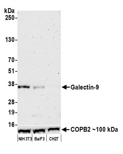 WB (Western Blot)
(Detection of mouse Galectin-9 by western blot. Samples: Whole cell lysate (50 ug) from NIH 3T3, Ba/F3, and CH27 cells prepared using NETN lysis buffer. Antibody: Rabbit anti-Galectin-9 recombinant monoclonal antibody [BL-1675A-4A5] (AAA23862 lot 1) used at 1:1000. Secondary: HRP-conjugated goat anti-rabbit IgG . Detection: Chemiluminescence with an exposure time of 3 minutes.)
WB (Western Blot)
(Detection of mouse Galectin-9 by western blot. Samples: Whole cell lysate (50 ug) from NIH 3T3, Ba/F3, and CH27 cells prepared using NETN lysis buffer. Antibody: Rabbit anti-Galectin-9 recombinant monoclonal antibody [BL-1675A-4A5] (AAA23862 lot 1) used at 1:1000. Secondary: HRP-conjugated goat anti-rabbit IgG . Detection: Chemiluminescence with an exposure time of 3 minutes.)
Galectin-9/Gal-9, Monoclonal Recombinant Antibody (Cat# AAA23862)
What are recombinant antibodies?
Recombinant antibodies are produced in the lab by utilizing a known genetic sequence (an amino acid and nucleotide sequence). This allows for high consistency in how each new lot will perform and how specific they will be for the target, regardless of how many times they are produced/reproduced.
They help solve the problems observed when traditional non-recombinant techniques are utilized, such as changes in cell behavior over time, differences between production batches, and accuracy in complex biological systems.
A powerful feature of recombinant antibodies is that they can also be modified through progressive genetic engineering and subsequent testing in order to make them perform better and/or more reliably.
Applications For Recombinant Antibodies
1. Basic Research
Recombinant antibodies are primarily utilized in laboratory experiments to study the function of various proteins in cells. They are helpful in finding and tracking specific molecules through their use in techniques such as ELISA, Western blotting, immunofluorescence, and flow cytometry.
2. Diagnostics
They are also key components in diagnostic tests used to detect diseases and infections. As recombinant antibodies are produced in a consistent and controlled way, they are able to provide highly reliable results across batches. Though recombinant antibodies are indeed quite useful in diagnostic applications, all of the products listed in AAA Biotech’s also known as AAA Bio or AAABio catalog are strictly for research-use only (RUO).
3. Therapeutics (Medical Treatments)
Please note that all of the products listed in AAA Biotech’s catalog are strictly for research-use only (RUO).
- Chimeric Antibodies: Commonly used in therapeutic treatment. They combine different pieces of mouse and human antibodies in order to reduce immune reactions.
- Humanized Antibodies: Modified non-human antibodies that are made to look the same as actual native human antibodies, making them safer and more effective.
- Antibody Fragments (Fab, scFv, VHH): Smaller pieces of antibody fragments that can reach deep into tissues and tumors. They are useful in cancer treatments and imaging.
- Bispecific Antibodies: Specially-engineered antibodies that can bind to two different target molecules at once. Each antibody has two “arms” that allow for two places where the target antigen can bind (both “arms” will recognize the same exact epitope). These bispecific antibodies are engineered so that each arm can detect a completely different epitope than the other. Used in many of the treatments for cancer, diabetes, Alzheimer’s disease, and more.
- Fc-Fusion Proteins: Used as drugs and research tools. They combine parts of antibodies with other proteins to improve stability and effectiveness.
4. Imaging and Detection
Small antibody fragments are also used as imaging agents in medical scans. Their small size helps them reach and report on the status of tumors quickly.
5. Drug Development and Delivery
Recombinant antibodies can also be designed and made to deliver drugs directly to diseased cells, especially in targeted cancer therapy.
6. Immunotherapy
These antibodies can aid the immune system recognize and fight diseases more effectively, which is often their role in cancer treatment.
Please note that all of the products listed in AAA Biotech’s catalog are strictly for research-use only (RUO).
Advantages of Recombinant Antibodies
- Reliable Results Every Time: Recombinant antibodies are exceptionally consistent from batch to batch. This means researchers can trust that results will remain the same each time they purchase and use them.
- More Specific and Accurate: Compared to polyclonal antibodies, recombinant antibodies are significantly less likely to exhibit non-specific binding, which promotes more accurate and clearer results.
- Easier to Develop: Making recombinant antibodies is simpler and quicker than the traditional method of producing monoclonal antibodies via hybridomas.
- No Genetic Drift: Over time, hybridoma cells can change or become unstable, which can result in the antibodies progressively losing their function/specificity. Recombinant antibodies avoid this problem because they are produced from a fixed template DNA sequence.
- Truly Monoclonal: Studies have shown that many hybridomas thought to be monoclonal actually are not so, which can lead to, in some cases, unreliable results. Recombinant antibodies are made using precise genetic sequence templates, ensuring they are truly specific to one target.
- Long-Term Stability and Safety: Since their amino acid and genetic sequences are known and stored, recombinant antibodies can be reproduced at any time with the same quality. This method protects against contamination, genetic changes over time, or even accidental loss/deterioration of cell lines.
Why Buy Recombinant Antibody from AAA Biotech?
- Highly Validated: The vast majority of our antibodies are thoroughly tested to ensure they work reliably across different applications and experimental platforms.
- Versatile Applications: Our antibodies can be used in a wide range of research techniques, including (but not limited to) immunocytochemistry (ICC), ELISA, immunofluorescence (IF), immunohistochemistry (IHC), flow cytometry (FC), immunoprecipitation (IP), and Western blotting (WB).
- Support for Rare Species: We offer antibodies specifically designed for rare or less commonly used laboratory species — something many other suppliers do not provide.
- Wide Selection of Conjugated and Recombinant Forms: Choose from various labeled (conjugated) options and browse our high-quality recombinant antibodies – we are confident that we will have materials to meet your research needs.
Please note that all of the products listed in AAA Biotech’s catalog are strictly for research-use only (RUO).
FAQ
1. What are recombinant antibodies used for?
Research (e.g., ELISA, Western blot, flow cytometry)
Disease diagnostics
Therapeutic treatments (e.g., cancer, autoimmune diseases)
Medical imaging
Targeted drug delivery
Vaccine development
Please note that all of the products listed in AAA Biotech’s catalog are strictly for research-use only (RUO).
2. What is the difference between traditional and recombinant antibodies?
Traditional antibodies: Produced using animal hosts (hybridomas are typically made from an original animal inoculation), slower, less consistent, risk of genetic drift
Recombinant antibodies: Produced using DNA template sequences via laboratory cell lines, faster, highly consistent, animal-free, easily customizable
3. What are antibodies used to treat?
Please note that all of the products listed in AAA Biotech’s catalog are strictly for research-use only (RUO).
Antibodies are proteins that your body makes to protect you from infections, allergies, and other potentially harmful substances. Scientists have used the intrinsic properties of antibodies to develop them into tools for scientific advancement, and can now create special lab-grown antibodies that aid in the treatment of diseases such as cancer, heart-related issues, and arthritis.
4. Why do we need recombinant proteins?
Recombinant proteins are lab-made proteins that have become essential in the advancement of science and medicine. They’re used in the lab for innumerable purposes, such as to help grow and support cells, especially for tasks like expanding cell colonies, helping them develop into other types, or turning them into stem cells.