
Filters
Clonality
Type
Reactivity
Gene Name
Isotype
Host
Application
Clone
10000 results for "Recombinant Proteins" - showing 5250-5300

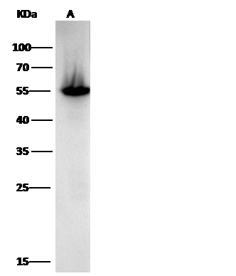 WB (Western Blot)
(Anti-ALDH1A1 mouse monoclonal antibody at 1:500 dilutionLane A: HepG2 Whole Cell LysateLysates/proteins at 30 ug per lane.SecondaryRabbit Anti-Mouse IgG F(ab)2/HRP at 1/10000 dilution.Developed using the ECL technique.Performed under reducing conditions.Predicted band size:55 kDaObserved band size:55 kDa)
WB (Western Blot)
(Anti-ALDH1A1 mouse monoclonal antibody at 1:500 dilutionLane A: HepG2 Whole Cell LysateLysates/proteins at 30 ug per lane.SecondaryRabbit Anti-Mouse IgG F(ab)2/HRP at 1/10000 dilution.Developed using the ECL technique.Performed under reducing conditions.Predicted band size:55 kDaObserved band size:55 kDa)
ALDH1A1, Monoclonal Antibody (Cat# AAA255410)
Full Name
Anti-ALDH1A1 Antibody, Mouse Monoclonal
Gene Names
ALDH1A1; ALDC; ALDH1; HEL-9; HEL12; PUMB1; ALDH11; RALDH1; ALDH-E1; HEL-S-53e
Reactivity
Human
Applications
Immunofluorescence, Immunocytochemistry, Flow Cytometry, Western Blot
Purity
Protein A
Pricing
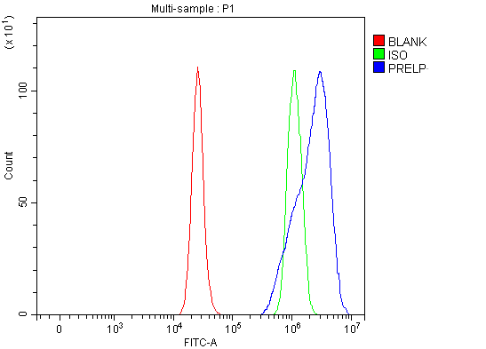 FCM/FACS (Flow Cytometry)
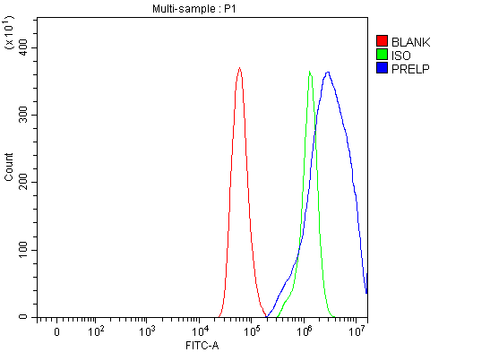
(Figure 3. Flow Cytometry analysis of RT4 cells using anti-PRELP antibody (AAA127465).Overlay histogram showing RT4 cells stained with AAA127465 (Blue line). The cells were fixed with 4% paraformaldehyde and blocked with 10% normal goat serum. And then incubated with rabbit anti-PRELP Antibody (AAA127465, 1ug/1x106 cells) for 30 min at 20 degree C. DyLight488 conjugated goat anti-rabbit IgG was used as secondary antibody for 30 minutes at 20 degree C. Isotype control antibody (Green line) was rabbit IgG (1ug/1x106) used under the same conditions. Unlabelled sample (Red line) was also used as a control.)
FCM/FACS (Flow Cytometry)
(Figure 3. Flow Cytometry analysis of RT4 cells using anti-PRELP antibody (AAA127465).Overlay histogram showing RT4 cells stained with AAA127465 (Blue line). The cells were fixed with 4% paraformaldehyde and blocked with 10% normal goat serum. And then incubated with rabbit anti-PRELP Antibody (AAA127465, 1ug/1x106 cells) for 30 min at 20 degree C. DyLight488 conjugated goat anti-rabbit IgG was used as secondary antibody for 30 minutes at 20 degree C. Isotype control antibody (Green line) was rabbit IgG (1ug/1x106) used under the same conditions. Unlabelled sample (Red line) was also used as a control.)
PRELP, Polyclonal Antibody (Cat# AAA127465)
Full Name
Anti-PRELP Antibody Picoband
Gene Names
PRELP; MST161; SLRR2A; MSTP161
Reactivity
Human, Mouse, Rat
Applications
Flow Cytometry, Western Blot
Purity
Immunogen affinity purified.
Pricing
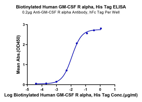
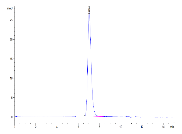
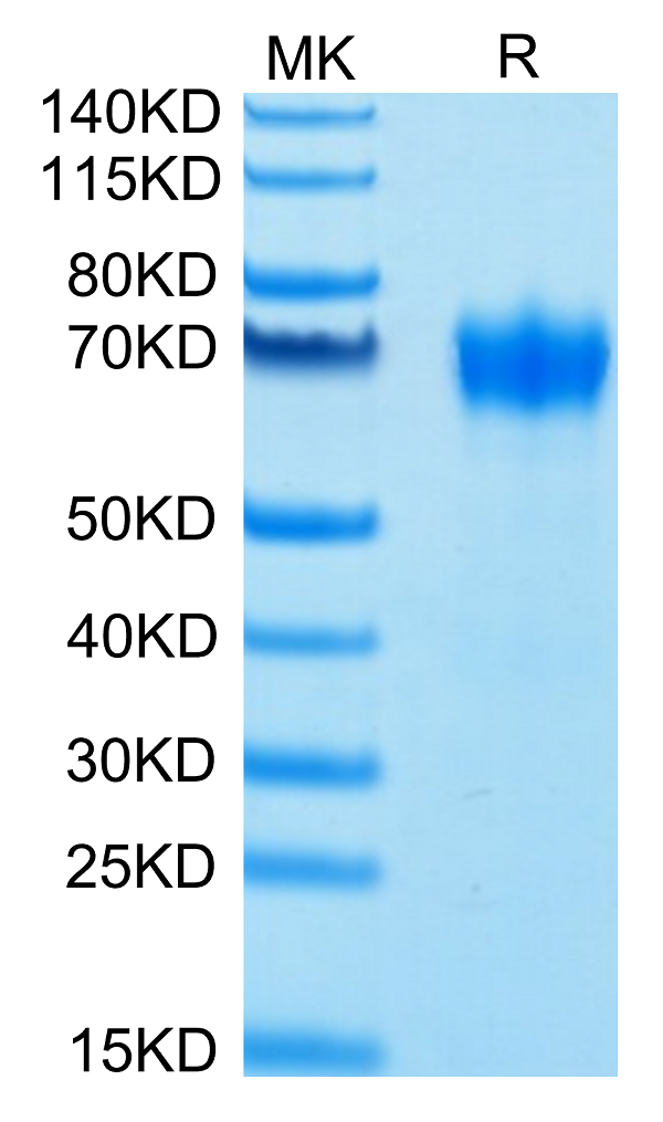
 Application Data
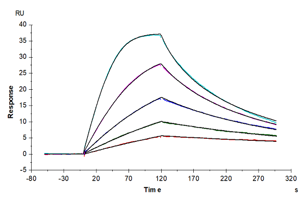
(Biotinylated Human GM-CSF R alpha Protein SPR DataBiotinylated Human GM-CSF R alpha, His-Avi Tag captured on CM5 Chip via Anti-His Antibody can bind Human GM-CSF, No Tag with an affinity constant of 1.68 nM as determined in SPR assay (Biacore T200).)
Application Data
(Biotinylated Human GM-CSF R alpha Protein SPR DataBiotinylated Human GM-CSF R alpha, His-Avi Tag captured on CM5 Chip via Anti-His Antibody can bind Human GM-CSF, No Tag with an affinity constant of 1.68 nM as determined in SPR assay (Biacore T200).)
GM-CSF R alpha, Recombinant Protein (Cat# AAA76284)
Full Name
Biotinylated Human GM-CSF R alpha Protein Recombinant
Gene Names
CSF2RA; GMR; CD116; CSF2R; SMDP4; CDw116; CSF2RX; CSF2RY; GMCSFR; CSF2RAX; CSF2RAY; GM-CSF-R-alpha
Purity
>95%, as determined by SDS-PAGE and HPLC
Pricing
 Bioactivity
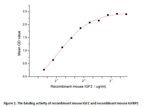
(Insulin-like growth factor 2 (IGF2) is one of three protein hormones that share structural similarity to insulin. It has growth-regulating, insulin-like and mitogenic activities. IGF2 exerts its effects by binding to the IGF-1 receptor and to the short isoform of the insulin receptor. IGF2 may also bind to the IGF2 receptor (also called the cation-independent mannose 6-phosphate receptor), which acts as a signalling antagonist; that is, to prevent IGF2 responses. Besides, Insulin Like Growth Factor Binding Protein 2 (IGFBP2) has been identified as an interactor of IGF2, thus a functional binding ELISA assay was conducted to detect the interaction of recombinant mouse IGF2 and recombinant mouse IGFBP2. Briefly, IGF2 were diluted serially in PBS, with 0.01% BSA (pH 7.4). Duplicate samples of 100 ul were then transferred to IGFBP2-coated microtiter wells and incubated for 2h at 37 degree C. Wells were washed with PBST and incubated for 1h with anti-IGF2 pAb, then aspirated and washed 3 times. After incubation with HRP labelled secondary antibody, wells were aspirated and washed 3 times. With the addition of substrate solution, wells were incubated 15-25 minutes at 37 degree C. Finally, add 50 uL stop solution to the wells and read at 450 nm immediately. The binding activity of recombinant mouse IGF2 and recombinant mouse IGFBP2 was shown in Figure 1, the EC50 for this effect is 0.009 ug/mL.)
Bioactivity
(Insulin-like growth factor 2 (IGF2) is one of three protein hormones that share structural similarity to insulin. It has growth-regulating, insulin-like and mitogenic activities. IGF2 exerts its effects by binding to the IGF-1 receptor and to the short isoform of the insulin receptor. IGF2 may also bind to the IGF2 receptor (also called the cation-independent mannose 6-phosphate receptor), which acts as a signalling antagonist; that is, to prevent IGF2 responses. Besides, Insulin Like Growth Factor Binding Protein 2 (IGFBP2) has been identified as an interactor of IGF2, thus a functional binding ELISA assay was conducted to detect the interaction of recombinant mouse IGF2 and recombinant mouse IGFBP2. Briefly, IGF2 were diluted serially in PBS, with 0.01% BSA (pH 7.4). Duplicate samples of 100 ul were then transferred to IGFBP2-coated microtiter wells and incubated for 2h at 37 degree C. Wells were washed with PBST and incubated for 1h with anti-IGF2 pAb, then aspirated and washed 3 times. After incubation with HRP labelled secondary antibody, wells were aspirated and washed 3 times. With the addition of substrate solution, wells were incubated 15-25 minutes at 37 degree C. Finally, add 50 uL stop solution to the wells and read at 450 nm immediately. The binding activity of recombinant mouse IGF2 and recombinant mouse IGFBP2 was shown in Figure 1, the EC50 for this effect is 0.009 ug/mL.)
Insulin Like Growth Factor 2 (IGF2), Active Protein (Cat# AAA161671)
Full Name
Active Insulin Like Growth Factor 2 (IGF2)
Gene Names
Igf2; Mpr; M6pr; Peg2; Igf-2; Igf-II; AL033362
Applications
Activity Assay, Cell Culture
Purity
Greater than 90% by SDS-PAGE
Pricing
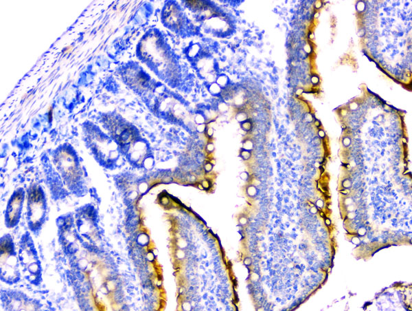 IHC (Immunohiostchemistry)
(Figure 3. IHC analysis of DDR2 using anti-DDR2 antibody.DDR2 was detected in paraffin-embedded section of rat small intestine tissue. Heat mediated antigen retrieval was performed in citrate buffer (pH6, epitope retrieval solution) for 20 mins. The tissue section was blocked with 10% goat serum. The tissue section was then incubated with 2ug/ml rabbit anti-DDR2 antibody.DDR2 was detected in paraffin-embedded section of rat small intestine tissue. Heat mediated antigen retrieval was performed in citrate buffer (pH6, epitope retrieval solution) for 20 mins. The tissue section was blocked with 10% goat serum. The tissue section was then incubated with 2ug/ml rabbit anti-DDR2 Antibody overnight at 4 degree C. Biotinylated goat anti-rabbit IgG was used as secondary antibody and incubated for 30 minutes at 37 degree C. The tissue section was developed using Strepavidin-Biotin-Complex (SABC) with DAB as the chromogen.)
IHC (Immunohiostchemistry)
(Figure 3. IHC analysis of DDR2 using anti-DDR2 antibody.DDR2 was detected in paraffin-embedded section of rat small intestine tissue. Heat mediated antigen retrieval was performed in citrate buffer (pH6, epitope retrieval solution) for 20 mins. The tissue section was blocked with 10% goat serum. The tissue section was then incubated with 2ug/ml rabbit anti-DDR2 antibody.DDR2 was detected in paraffin-embedded section of rat small intestine tissue. Heat mediated antigen retrieval was performed in citrate buffer (pH6, epitope retrieval solution) for 20 mins. The tissue section was blocked with 10% goat serum. The tissue section was then incubated with 2ug/ml rabbit anti-DDR2 Antibody overnight at 4 degree C. Biotinylated goat anti-rabbit IgG was used as secondary antibody and incubated for 30 minutes at 37 degree C. The tissue section was developed using Strepavidin-Biotin-Complex (SABC) with DAB as the chromogen.)
DDR2, Polyclonal Antibody (Cat# AAA124932)
Full Name
Anti-DDR2 Antibody
Gene Names
DDR2; TKT; MIG20a; NTRKR3; TYRO10
Reactivity
Human, Mouse, Rat
Applications
Direct ELISA, Immunohistochemistry, Western Blot
Purity
Immunogen Affinity Purified
Pricing
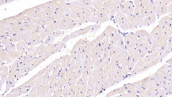
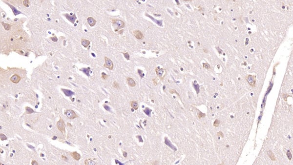 IHC (Immunohiostchemistry)
(DAB staining on IHCP;Sample: Human Cerebrum Tissue; Primary Ab: 40ug/ml Mouse AntiHuman FSTL1 AntibodySecond Ab: 2ug/mL HRPLinked Caprine AntiMouse IgG Polyclonal Antibody(Catalog: SAA544Mu19))
IHC (Immunohiostchemistry)
(DAB staining on IHCP;Sample: Human Cerebrum Tissue; Primary Ab: 40ug/ml Mouse AntiHuman FSTL1 AntibodySecond Ab: 2ug/mL HRPLinked Caprine AntiMouse IgG Polyclonal Antibody(Catalog: SAA544Mu19))
Follistatin Like Protein 1 (FSTL1), Monoclonal Antibody (Cat# AAA151888)
Full Name
Monoclonal Antibody to Follistatin Like Protein 1 (FSTL1)
Gene Names
FSTL1; FRP; FSL1; MIR198
Reactivity
Human, Pig
Applications
Immunoprecipitation, Immunocytochemistry, Immunohistochemistry, Western Blot
Purity
Protein A + Protein G affinity chromatography
Pricing
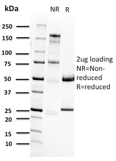
 Application Data
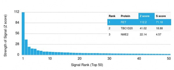
(Analysis of Protein Array containing more than 19,000 full-length human proteins using RET Mouse Monoclonal Antibody (RET/2662). Z- and S- Score: The Z-score represents the strength of a signal that a monoclonal antibody (Monoclonal Antibody) (in combination with a fluorescently-tagged anti-IgG secondary antibody) produces when binding to a particular protein on the HuProtTM array. Z-scores are described in units of standard deviations (SD's) above the mean value of all signals generated on that array. If targets on HuProtTM are arranged in descending order of the Z-score, the S-score is the difference (also in units of SD's) between the Z-score. S-score therefore represents the relative target specificity of a Monoclonal Antibody to its intended target. A Monoclonal Antibody is considered to specific to its intended target, if the Monoclonal Antibody has an S-score of at least 2.5. For example, if a Monoclonal Antibody binds to protein X with a Z-score of 43 and to protein Y with a Z-score of 14, then the S-score for the binding of that Monoclonal Antibody to protein X is equal to 29.)
Application Data
(Analysis of Protein Array containing more than 19,000 full-length human proteins using RET Mouse Monoclonal Antibody (RET/2662). Z- and S- Score: The Z-score represents the strength of a signal that a monoclonal antibody (Monoclonal Antibody) (in combination with a fluorescently-tagged anti-IgG secondary antibody) produces when binding to a particular protein on the HuProtTM array. Z-scores are described in units of standard deviations (SD's) above the mean value of all signals generated on that array. If targets on HuProtTM are arranged in descending order of the Z-score, the S-score is the difference (also in units of SD's) between the Z-score. S-score therefore represents the relative target specificity of a Monoclonal Antibody to its intended target. A Monoclonal Antibody is considered to specific to its intended target, if the Monoclonal Antibody has an S-score of at least 2.5. For example, if a Monoclonal Antibody binds to protein X with a Z-score of 43 and to protein Y with a Z-score of 14, then the S-score for the binding of that Monoclonal Antibody to protein X is equal to 29.)
RET Proto-oncogene, Monoclonal Antibody (Cat# AAA215180)
Full Name
RET Proto-oncogene
Gene Names
RET; PTC; MTC1; HSCR1; MEN2A; MEN2B; CDHF12; CDHR16; RET-ELE1
Reactivity
Human
Applications
ELISA
Purity
Purified Ab with BSA and Azide at 200ug/ml OR Purified Ab WITHOUT BSA and Azide at 1.0mg/ml
Pricing
 Application Data
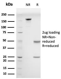
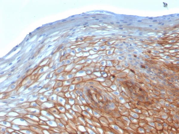
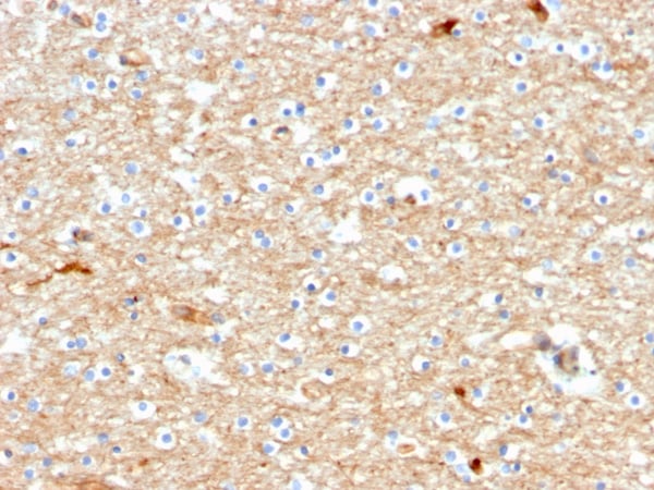
(Analysis of Protein Array containing more than 19,000 full-length human proteins using Drebrin-1 Mouse Monoclonal Antibody (DBN1/2880). Z- and S- Score: The Z-score represents the strength of a signal that a monoclonal antibody (MAb) (in combination with a fluorescently-tagged anti-IgG secondary antibody) produces when binding to a particular protein on the HuProtTM array. Z-scores are described in units of standard deviations (SD's) above the mean value of all signals generated on that array. If targets on HuProtTM are arranged in descending order of the Z-score, the S-score is the difference (also in units of SD's) between the Z-score. S-score therefore represents the relative target specificity of a MAb to its intended target. A MAb is considered to specific to its intended target, if the MAb has an S-score of at least 2.5. For example, if a MAb binds to protein X with a Z-score of 43 and to protein Y with a Z-score of 14, then the S-score for the binding of that MAb to protein X is equal to 29.)
Application Data
(Analysis of Protein Array containing more than 19,000 full-length human proteins using Drebrin-1 Mouse Monoclonal Antibody (DBN1/2880). Z- and S- Score: The Z-score represents the strength of a signal that a monoclonal antibody (MAb) (in combination with a fluorescently-tagged anti-IgG secondary antibody) produces when binding to a particular protein on the HuProtTM array. Z-scores are described in units of standard deviations (SD's) above the mean value of all signals generated on that array. If targets on HuProtTM are arranged in descending order of the Z-score, the S-score is the difference (also in units of SD's) between the Z-score. S-score therefore represents the relative target specificity of a MAb to its intended target. A MAb is considered to specific to its intended target, if the MAb has an S-score of at least 2.5. For example, if a MAb binds to protein X with a Z-score of 43 and to protein Y with a Z-score of 14, then the S-score for the binding of that MAb to protein X is equal to 29.)
Drebrin 1 (DBN1), Monoclonal Antibody (Cat# AAA214917)
Full Name
Drebrin 1 (DBN1)
Gene Names
DBN1; D0S117E
Reactivity
Human
Applications
Immunohistochemistry
Purity
Purified Ab with BSA and Azide at 200ug/ml OR Purified Ab WITHOUT BSA and Azide at 1.0mg/ml
Pricing
 FCM/FACS (Flow Cytometry)
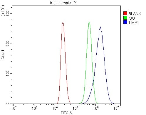
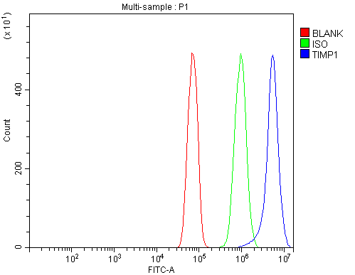
(Figure 3. Flow Cytometry analysis of RH35 cells using anti-TIMP1 antibody (AAA125559).Overlay histogram showing RH35 cells stained with AAA125559 (Blue line). The cells were blocked with 10% normal goat serum. And then incubated with rabbit anti-TIMP1 Antibody (AAA125559, 1μg/1x106 cells) for 30 min at 20 degree C. DyLight®488 conjugated goat anti-rabbit IgG (5-10μg/1x106 cells) was used as secondary antibody for 30 minutes at 20 degree C. Isotype control antibody (Green line) was rabbit IgG (1μg/1x106) used under the same conditions. Unlabelled sample (Red line) was also used as a control.)
FCM/FACS (Flow Cytometry)
(Figure 3. Flow Cytometry analysis of RH35 cells using anti-TIMP1 antibody (AAA125559).Overlay histogram showing RH35 cells stained with AAA125559 (Blue line). The cells were blocked with 10% normal goat serum. And then incubated with rabbit anti-TIMP1 Antibody (AAA125559, 1μg/1x106 cells) for 30 min at 20 degree C. DyLight®488 conjugated goat anti-rabbit IgG (5-10μg/1x106 cells) was used as secondary antibody for 30 minutes at 20 degree C. Isotype control antibody (Green line) was rabbit IgG (1μg/1x106) used under the same conditions. Unlabelled sample (Red line) was also used as a control.)
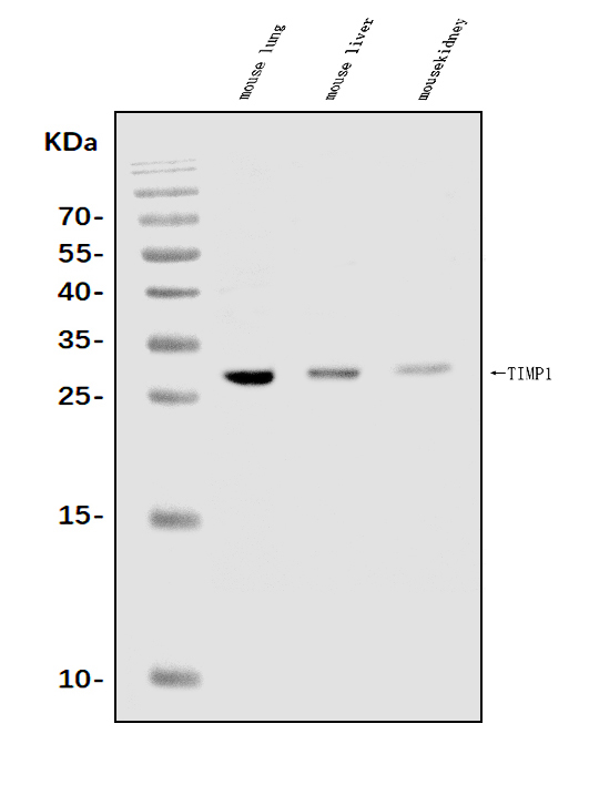
TIMP1, Polyclonal Antibody (Cat# AAA125559)
Full Name
Anti-TIMP1 Antibody
Gene Names
Timp1; EPA; Clgi; Timp; TIMP-1; TPA-S1
Reactivity
Mouse, Rat
Applications
Direct ELISA, Flow Cytometry, Western Blot
Purity
Immunogen affinity purified.
Pricing
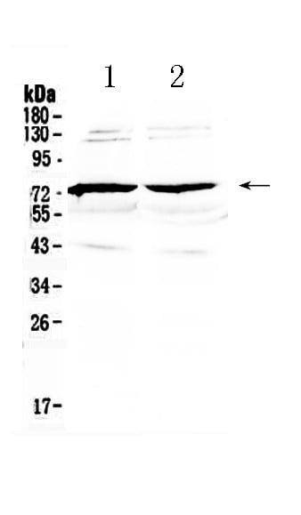
 IHC (Immunohistochemisry)
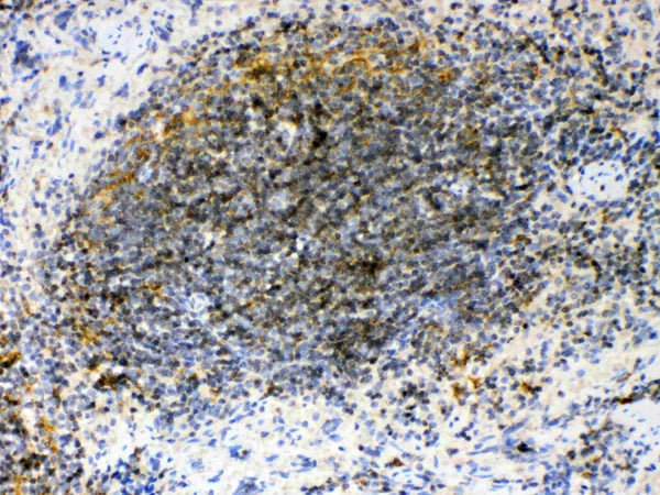
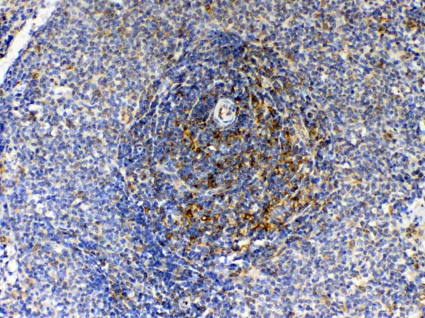
(Figure 3. IHC analysis of GAS 6 using anti- GAS 6 antibody (AAA124584).GAS 6 was detected in paraffin-embedded section of rat spleen tissues. Heat mediated antigen retrieval was performed in citrate buffer (pH6, epitope retrieval solution) for 20 mins. The tissue section was blocked with 10% goat serum. The tissue section was then incubated with 1ug/ml rabbit anti- GAS 6 Antibody (AAA124584) overnight at 4 degree C. Biotinylated goat anti-rabbit IgG was used as secondary antibody and incubated for 30 minutes at 37 degree C. The tissue section was developed using Strepavidin-Biotin-Complex (SABC) with DAB as the chromogen.)
IHC (Immunohistochemisry)
(Figure 3. IHC analysis of GAS 6 using anti- GAS 6 antibody (AAA124584).GAS 6 was detected in paraffin-embedded section of rat spleen tissues. Heat mediated antigen retrieval was performed in citrate buffer (pH6, epitope retrieval solution) for 20 mins. The tissue section was blocked with 10% goat serum. The tissue section was then incubated with 1ug/ml rabbit anti- GAS 6 Antibody (AAA124584) overnight at 4 degree C. Biotinylated goat anti-rabbit IgG was used as secondary antibody and incubated for 30 minutes at 37 degree C. The tissue section was developed using Strepavidin-Biotin-Complex (SABC) with DAB as the chromogen.)
GAS 6, Polyclonal Antibody (Cat# AAA124584)
Full Name
Anti-GAS 6 Picoband Antibody
Gene Names
GAS6; AXSF; AXLLG
Reactivity
Human, Mouse, Rat
No cross reactivity with other proteins.
No cross reactivity with other proteins.
Applications
Western Blot, Immunohistochemistry
Purity
Immunogen affinity purified
Pricing
 Application Data
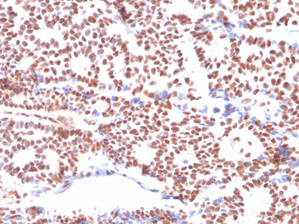
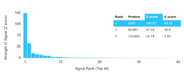
(Analysis of Protein Array containing more than 19,000 full-length human proteins using Estrogen Receptor alpha Mouse Monoclonal Antibody (ESR1/3564) Z- and S- Score: The Z-score represents the strength of a signal that a monoclonal antibody (MAb) (in combination with a fluorescently-tagged anti-IgG secondary antibody) produces when binding to a particular protein on the HuProtTM array. Z-scores are described in units of standard deviations (SD's) above the mean value of all signals generated on that array. If targets on HuProtTM are arranged in descending order of the Z-score, the S-score is the difference (also in units of SD's) between the Z-score. S-score therefore represents the relative target specificity of a MAb to its intended target. A MAb is considered to specific to its intended target, if the MAb has an S-score of at least 2.5. For example, if a MAb binds to protein X with a Z-score of 43 and to protein Y with a Z-score of 14, then the S-score for the binding of that MAb to protein X is equal to 29.)
Application Data
(Analysis of Protein Array containing more than 19,000 full-length human proteins using Estrogen Receptor alpha Mouse Monoclonal Antibody (ESR1/3564) Z- and S- Score: The Z-score represents the strength of a signal that a monoclonal antibody (MAb) (in combination with a fluorescently-tagged anti-IgG secondary antibody) produces when binding to a particular protein on the HuProtTM array. Z-scores are described in units of standard deviations (SD's) above the mean value of all signals generated on that array. If targets on HuProtTM are arranged in descending order of the Z-score, the S-score is the difference (also in units of SD's) between the Z-score. S-score therefore represents the relative target specificity of a MAb to its intended target. A MAb is considered to specific to its intended target, if the MAb has an S-score of at least 2.5. For example, if a MAb binds to protein X with a Z-score of 43 and to protein Y with a Z-score of 14, then the S-score for the binding of that MAb to protein X is equal to 29.)
Estrogen Receptor, alpha, Monoclonal Antibody (Cat# AAA215395)
Full Name
Estrogen Receptor, alpha (Marker of Estrogen Dependence)
Gene Names
ESR1; ER; ESR; Era; ESRA; ESTRR; NR3A1
Reactivity
Human
Applications
Immunohistochemistry
Purity
Purified Ab with BSA and Azide at 200ug/ml or Purified Ab with BSA and Azide at 200ug/ml or Purified Ab WITHOUT BSA and Azide at 1.0mg/ml
Pricing
 Application Data
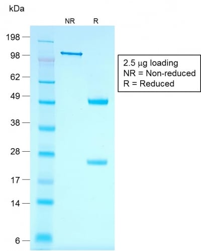
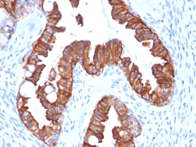
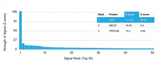
(Analysis of Protein Array containing more than 19, 000 full-length human proteins using Cytokeratin 7 Mouse Recombinant Monoclonal Antibody (rOV-TL12/30).Z- and S- Score: The Z-score represents the strength of a signal that a monoclonal antibody (MAb) (in combination with a fluorescently-tagged anti-IgG secondary antibody) produces when binding to a particular protein on the HuProtTM array. Z-scores are described in units of standard deviations (SD's) above the mean value of all signals generated on that array. If targets on HuProtTM are arranged in descending order of the Z-score, the S-score is the difference (also in units of SD's) between the Z-score. S-score therefore represents the relative target specificity of a MAb to its intended target. A MAb is considered to specific to its intended target, if the MAb has an S-score of at least 2.5. For example, if a MAb binds to protein X with a Z-score of 43 and to protein Y with a Z-score of 14, then the S-score for the binding of that MAb to protein X is equal to 29.)
Application Data
(Analysis of Protein Array containing more than 19, 000 full-length human proteins using Cytokeratin 7 Mouse Recombinant Monoclonal Antibody (rOV-TL12/30).Z- and S- Score: The Z-score represents the strength of a signal that a monoclonal antibody (MAb) (in combination with a fluorescently-tagged anti-IgG secondary antibody) produces when binding to a particular protein on the HuProtTM array. Z-scores are described in units of standard deviations (SD's) above the mean value of all signals generated on that array. If targets on HuProtTM are arranged in descending order of the Z-score, the S-score is the difference (also in units of SD's) between the Z-score. S-score therefore represents the relative target specificity of a MAb to its intended target. A MAb is considered to specific to its intended target, if the MAb has an S-score of at least 2.5. For example, if a MAb binds to protein X with a Z-score of 43 and to protein Y with a Z-score of 14, then the S-score for the binding of that MAb to protein X is equal to 29.)
Cytokeratin 7, Monoclonal Antibody (Cat# AAA214413)
Full Name
Cytokeratin 7 (Glandular and Transitional Epithelial Marker)
Gene Names
KRT7; K7; CK7; SCL; K2C7
Reactivity
Human. Others not known.
Applications
Immunohistochemistry, Immunofluorescence, Flow Cytometry
Pricing
 Application Data
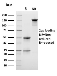
(Analysis of Protein Array containing more than 19,000 full-length human proteins using LSP1 Mouse Monoclonal Antibody (LSP1/3042). Z- and S- Score: The Z-score represents the strength of a signal that a monoclonal antibody (MAb) (in combination with a fluorescently-tagged anti-IgG secondary antibody) produces when binding to a particular protein on the HuProtTM array. Z-scores are described in units of standard deviations (SD's) above the mean value of all signals generated on that array. If targets on HuProtTM are arranged in descending order of the Z-score, the S-score is the difference (also in units of SD's) between the Z-score. S-score therefore represents the relative target specificity of a MAb to its intended target. A MAb is considered to specific to its intended target, if the MAb has an S-score of at least 2.5. For example, if a MAb binds to protein X with a Z-score of 43 and to protein Y with a Z-score of 14, then the S-score for the binding of that MAb to protein X is equal to 29.)
Application Data
(Analysis of Protein Array containing more than 19,000 full-length human proteins using LSP1 Mouse Monoclonal Antibody (LSP1/3042). Z- and S- Score: The Z-score represents the strength of a signal that a monoclonal antibody (MAb) (in combination with a fluorescently-tagged anti-IgG secondary antibody) produces when binding to a particular protein on the HuProtTM array. Z-scores are described in units of standard deviations (SD's) above the mean value of all signals generated on that array. If targets on HuProtTM are arranged in descending order of the Z-score, the S-score is the difference (also in units of SD's) between the Z-score. S-score therefore represents the relative target specificity of a MAb to its intended target. A MAb is considered to specific to its intended target, if the MAb has an S-score of at least 2.5. For example, if a MAb binds to protein X with a Z-score of 43 and to protein Y with a Z-score of 14, then the S-score for the binding of that MAb to protein X is equal to 29.)
Lymphocyte Specific Protein 1 (LSP1)/pp52, Monoclonal Antibody (Cat# AAA215072)
Full Name
Lymphocyte Specific Protein 1 (LSP1)/pp52
Gene Names
LSP1; WP34; pp52
Reactivity
Human
Applications
ELISA
Purity
Purified Ab with BSA and Azide at 200ug/ml OR Purified Ab WITHOUT BSA and Azide at 1.0mg/ml
Pricing
 Bioactivity
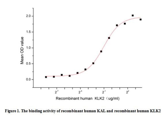
(Kallistatin (KAL), a member of serpin family, can Inhibit human amidolytic and kininogenase activities of tissue kallikrein. KAL is widely distributed in a variety of organs and body fluids and has biological effects such as anti-inflammatory, antioxidant stress, anti-angiogenesis and anti-tumor. Besides, Kallikrein 2 (KLK2) has been identified as an interactor of KAL, thus a functional binding ELISA assay was conducted to detect the interaction of recombinant human KAL and recombinant human KLK2. Briefly, KLK2 was diluted serially in PBS with 0.01% BSA (pH 7.4). Duplicate samples of 100 ul were then transferred to KAL-coated microtiter wells and incubated for 1h at 37 degree C. Wells were washed with PBST and incubated for 1h with anti-KLK2 pAb, then aspirated and washed 3 times. After incubation with HRP labelled secondary antibody for 1h at 37 degree C, wells were aspirated and washed 5 times. With the addition of substrate solution, wells were incubated 15-25 minutes at 37 degree C. Finally, add 50 uL stop solution to the wells and read at 450/630 nm immediately. When recombinant human KAL is immobilized at 2 ug/mL (100 uL/well), the concentration of KLK2 that produces 50% optimal binding response is found to be approximately 0.49 ug/mL.)
Bioactivity
(Kallistatin (KAL), a member of serpin family, can Inhibit human amidolytic and kininogenase activities of tissue kallikrein. KAL is widely distributed in a variety of organs and body fluids and has biological effects such as anti-inflammatory, antioxidant stress, anti-angiogenesis and anti-tumor. Besides, Kallikrein 2 (KLK2) has been identified as an interactor of KAL, thus a functional binding ELISA assay was conducted to detect the interaction of recombinant human KAL and recombinant human KLK2. Briefly, KLK2 was diluted serially in PBS with 0.01% BSA (pH 7.4). Duplicate samples of 100 ul were then transferred to KAL-coated microtiter wells and incubated for 1h at 37 degree C. Wells were washed with PBST and incubated for 1h with anti-KLK2 pAb, then aspirated and washed 3 times. After incubation with HRP labelled secondary antibody for 1h at 37 degree C, wells were aspirated and washed 5 times. With the addition of substrate solution, wells were incubated 15-25 minutes at 37 degree C. Finally, add 50 uL stop solution to the wells and read at 450/630 nm immediately. When recombinant human KAL is immobilized at 2 ug/mL (100 uL/well), the concentration of KLK2 that produces 50% optimal binding response is found to be approximately 0.49 ug/mL.)
Kallistatin (KAL), Active Protein (Cat# AAA161912)
Full Name
Active Kallistatin (KAL)
Gene Names
SERPINA4; KAL; KST; PI4; KLST; PI-4; kallistatin
Applications
Activity Assay, Cell Culture
Purity
Greater than 80% by SDS-PAGE
Pricing
 IHC (Immunohistochemisry)
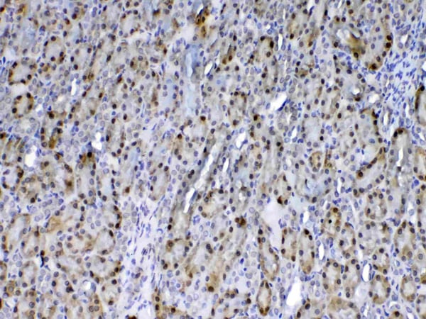
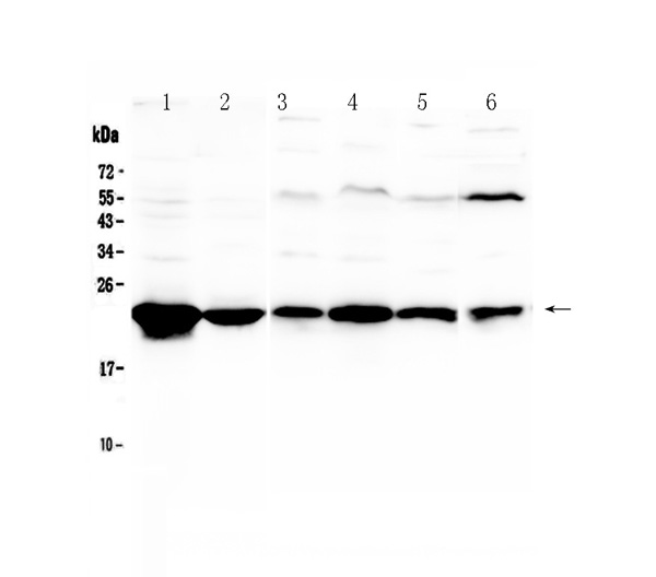
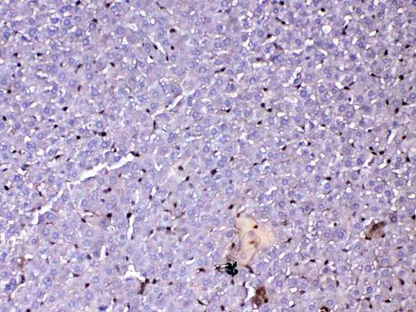
(Figure 3. IHC analysis of GST3 / GST pi using anti-GST3 / GST pi antibody (AAA124569).GST3 / GST pi was detected in paraffin-embedded section of rat liver tissue. Heat mediated antigen retrieval was performed in citrate buffer (pH6, epitope retrieval solution) for 20 mins. The tissue section was blocked with 10% goat serum. The tissue section was then incubated with 1ug/ml rabbit anti-GST3 / GST pi Antibody (AAA124569) overnight at 4 degree C. Biotinylated goat anti-rabbit IgG was used as secondary antibody and incubated for 30 minutes at 37 degree C. The tissue section was developed using Strepavidin-Biotin-Complex (SABC) with DAB as the chromogen.)
IHC (Immunohistochemisry)
(Figure 3. IHC analysis of GST3 / GST pi using anti-GST3 / GST pi antibody (AAA124569).GST3 / GST pi was detected in paraffin-embedded section of rat liver tissue. Heat mediated antigen retrieval was performed in citrate buffer (pH6, epitope retrieval solution) for 20 mins. The tissue section was blocked with 10% goat serum. The tissue section was then incubated with 1ug/ml rabbit anti-GST3 / GST pi Antibody (AAA124569) overnight at 4 degree C. Biotinylated goat anti-rabbit IgG was used as secondary antibody and incubated for 30 minutes at 37 degree C. The tissue section was developed using Strepavidin-Biotin-Complex (SABC) with DAB as the chromogen.)
GST3/GST pi, Polyclonal Antibody (Cat# AAA124569)
Full Name
Anti-GST3/GST pi Picoband Antibody
Gene Names
Gstp1; Gst3; Gstp; GST-P; Gstp2
Reactivity
Human, Mouse, Rat
No cross reactivity with other proteins.
No cross reactivity with other proteins.
Applications
Western Blot, Immunohistochemistry
Purity
Immunogen affinity purified
Pricing
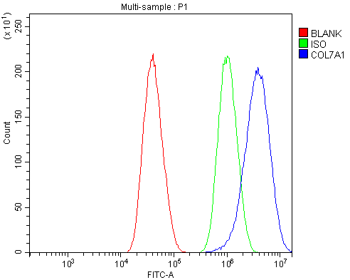
 IHC (Immunohistochemistry)
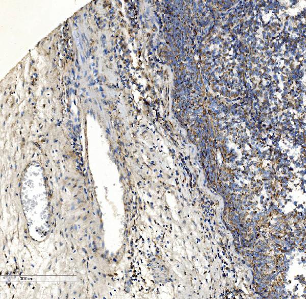
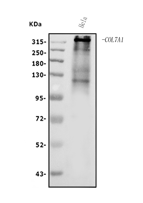
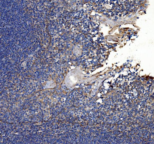
(Figure 4. IHC analysis of Collagen VII/COL7A1 using anti-Collagen VII/COL7A1 antibody (AAA125592).Collagen VII/COL7A1 was detected in paraffin-embedded section of human tonsil tissue. Heat mediated antigen retrieval was performed in EDTA buffer (pH8. 0, epitope retrieval solution). The tissue section was blocked with 10% goat serum. The tissue section was then incubated with 2μg/ml rabbit anti-Collagen VII/COL7A1 Antibody (AAA125592) overnight at 4 degree C. Biotinylated goat anti-rabbit IgG was used as secondary antibody and incubated for 30 minutes at 37 degree C. The tissue section was developed using Strepavidin-Biotin-Complex (SABC) with DAB as the chromogen.)
IHC (Immunohistochemistry)
(Figure 4. IHC analysis of Collagen VII/COL7A1 using anti-Collagen VII/COL7A1 antibody (AAA125592).Collagen VII/COL7A1 was detected in paraffin-embedded section of human tonsil tissue. Heat mediated antigen retrieval was performed in EDTA buffer (pH8. 0, epitope retrieval solution). The tissue section was blocked with 10% goat serum. The tissue section was then incubated with 2μg/ml rabbit anti-Collagen VII/COL7A1 Antibody (AAA125592) overnight at 4 degree C. Biotinylated goat anti-rabbit IgG was used as secondary antibody and incubated for 30 minutes at 37 degree C. The tissue section was developed using Strepavidin-Biotin-Complex (SABC) with DAB as the chromogen.)
Collagen VII/COL7A1, Polyclonal Antibody (Cat# AAA125592)
Full Name
Anti-Collagen VII/COL7A1 Antibody
Gene Names
COL7A1; EBD1; EBR1; EBDCT
Reactivity
Human
Applications
Direct ELISA, Flow Cytometry, Immunohistochemistry, Western Blot
Purity
Immunogen affinity purified.
Pricing
 Application Data
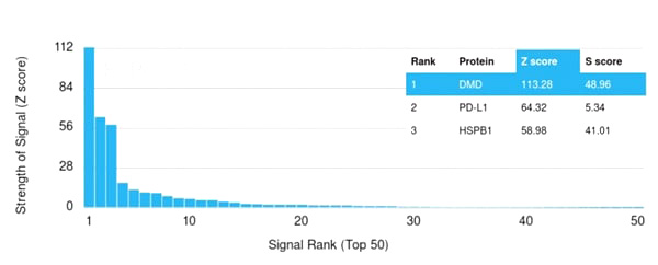
(Analysis of Protein Array containing more than 19,000 full-length human proteins using Dystrophin Monospecific Mouse Monoclonal Antibody (DMD/3245). Z- and S- Score: The Z-score represents the strength of a signal that a monoclonal antibody (Monoclonal Antibody) (in combination with a fluorescently-tagged anti-IgG secondary antibody) produces when binding to a particular protein on the HuProtTM array. Z-scores are described in units of standard deviations (SD’s) above the mean value of all signals generated on that array. If targets on HuProtTM are arranged in descending order of the Z-score, the S-score is the difference (also in units of SD’s) between the Z-score. S-score therefore represents the relative target specificity of a Monoclonal Antibody to its intended target. A Monoclonal Antibody is considered to specific to its intended target, if the Monoclonal Antibody has an S-score of at least 2.5. For example, if a Monoclonal Antibody binds to protein X with a Z-score of 43 and to protein Y with a Z-score of 14, then the S-score for the binding of that Monoclonal Antibody to protein X is equal to 29.)
Application Data
(Analysis of Protein Array containing more than 19,000 full-length human proteins using Dystrophin Monospecific Mouse Monoclonal Antibody (DMD/3245). Z- and S- Score: The Z-score represents the strength of a signal that a monoclonal antibody (Monoclonal Antibody) (in combination with a fluorescently-tagged anti-IgG secondary antibody) produces when binding to a particular protein on the HuProtTM array. Z-scores are described in units of standard deviations (SD’s) above the mean value of all signals generated on that array. If targets on HuProtTM are arranged in descending order of the Z-score, the S-score is the difference (also in units of SD’s) between the Z-score. S-score therefore represents the relative target specificity of a Monoclonal Antibody to its intended target. A Monoclonal Antibody is considered to specific to its intended target, if the Monoclonal Antibody has an S-score of at least 2.5. For example, if a Monoclonal Antibody binds to protein X with a Z-score of 43 and to protein Y with a Z-score of 14, then the S-score for the binding of that Monoclonal Antibody to protein X is equal to 29.)
Dystrophin (DMD), Monoclonal Antibody (Cat# AAA214926)
Full Name
Dystrophin (DMD) (Marker of Duchenne and Becker Muscular Dystrophy)
Gene Names
DMD; BMD; CMD3B; MRX85; DXS142; DXS164; DXS206; DXS230; DXS239; DXS268; DXS269; DXS270; DXS272
Reactivity
Human
Applications
Immunohistochemistry
Purity
Purified Ab with BSA and Azide at 200ug/ml OR Purified Ab WITHOUT BSA and Azide at 1.0mg/ml
Pricing
 WB (Western Blot)
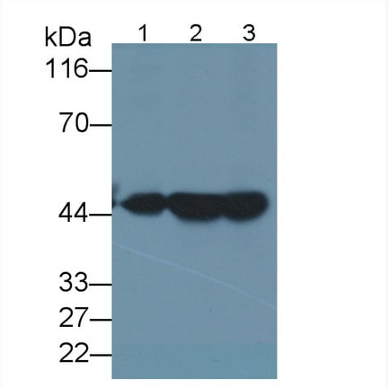
(Western Blot; Sample: Lane1: Rat Heart lysate; Lane2: Rat Liver lysate; Lane3: Rat Kidney lysate Primary Ab: 2ug/ml Mouse AntiHuman KRT18 Antibody Second Ab: 0.2ug/mL HRPLinked Caprine AntiMouse IgG Polyclonal Antibody (Catalog: SAA544Mu19))
WB (Western Blot)
(Western Blot; Sample: Lane1: Rat Heart lysate; Lane2: Rat Liver lysate; Lane3: Rat Kidney lysate Primary Ab: 2ug/ml Mouse AntiHuman KRT18 Antibody Second Ab: 0.2ug/mL HRPLinked Caprine AntiMouse IgG Polyclonal Antibody (Catalog: SAA544Mu19))
Cytokeratin 18 (CK18), Monoclonal Antibody (Cat# AAA151695)
Full Name
Monoclonal Antibody to Cytokeratin 18 (CK18)
Gene Names
KRT18; K18; CYK18
Reactivity
Human, Rat
Applications
Immunoprecipitation, Immunocytochemistry, Immunohistochemistry, Western Blot
Purity
Protein A + Protein G affinity chromatography
Pricing
 FCM/FACS (Flow Cytometry)
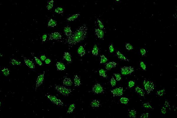
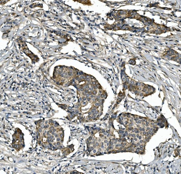
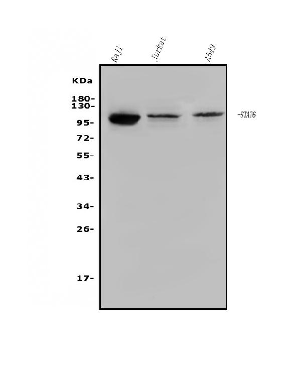
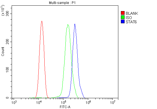
(Figure 4. Flow Cytometry analysis of THP-1 cells using anti-STAT6 antibody (AAA125555).Overlay histogram showing THP-1 cells stained with AAA125555 (Blue line). The cells were blocked with 10% normal goat serum. And then incubated with rabbit anti-STAT6 Antibody (AAA125555, 1μg/1x106 cells) for 30 min at 20 degree C. DyLight®488 conjugated goat anti-rabbit IgG (5-10μg/1x106 cells) was used as secondary antibody for 30 minutes at 20 degree C. Isotype control antibody (Green line) was rabbit IgG (1μg/1x106) used under the same conditions. Unlabelled sample (Red line) was also used as a control.)
FCM/FACS (Flow Cytometry)
(Figure 4. Flow Cytometry analysis of THP-1 cells using anti-STAT6 antibody (AAA125555).Overlay histogram showing THP-1 cells stained with AAA125555 (Blue line). The cells were blocked with 10% normal goat serum. And then incubated with rabbit anti-STAT6 Antibody (AAA125555, 1μg/1x106 cells) for 30 min at 20 degree C. DyLight®488 conjugated goat anti-rabbit IgG (5-10μg/1x106 cells) was used as secondary antibody for 30 minutes at 20 degree C. Isotype control antibody (Green line) was rabbit IgG (1μg/1x106) used under the same conditions. Unlabelled sample (Red line) was also used as a control.)
STAT6, Polyclonal Antibody (Cat# AAA125555)
Full Name
Anti-STAT6 Antibody
Gene Names
STAT6; STAT6B; STAT6C; D12S1644; IL-4-STAT
Reactivity
Human
Applications
Direct ELISA, Flow Cytometry, Immunofluorescence, Immunocytochemistry, Immunohistochemistry, Western Blot
Purity
Immunogen affinity purified.
Pricing
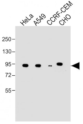 WB (Western Blot)
(All lanes : Anti-NR3C1 at dilutionLane 1: HeLa whole cell lysateLane 2: A549 whole cell lysateLane 3: CCRF-CEM whole cell lysateLane 4: CHO whole cell lysateLysates/proteins at 20 ug per lane.SecondaryGoat Anti-mouse IgG, (H+L), Peroxidase conjugated at 1/10000 dilution.Predicted band size : 86 kDaBlocking/Dilution buffer: 5% NFDM/TBST.)
WB (Western Blot)
(All lanes : Anti-NR3C1 at dilutionLane 1: HeLa whole cell lysateLane 2: A549 whole cell lysateLane 3: CCRF-CEM whole cell lysateLane 4: CHO whole cell lysateLysates/proteins at 20 ug per lane.SecondaryGoat Anti-mouse IgG, (H+L), Peroxidase conjugated at 1/10000 dilution.Predicted band size : 86 kDaBlocking/Dilution buffer: 5% NFDM/TBST.)
NR3C1, Monoclonal Antibody (Cat# AAA290964)
Full Name
NR3C1
Gene Names
NR3C1; GR; GCR; GRL; GCCR
Reactivity
Human, Mouse
Predicted : Human , Mouse
Predicted : Human , Mouse
Applications
Western Blot
Purity
This antibody is purified through a protein G column, followed by dialysis against PBS.
Pricing
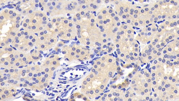
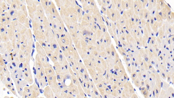
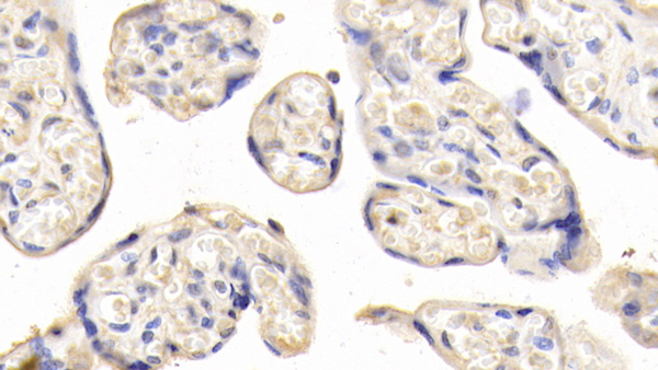
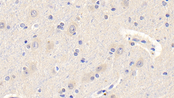 IHC (Immunohistochemistry)
(DAB staining on IHCP;Samples: Human Cerebrum Tissue; Primary Ab: 20ug/ml Mouse AntiHuman LEP AntibodySecond Ab: 2ug/mL HRPLinked Caprine AntiMouse IgG Polyclonal Antibody(Catalog: SAA544Mu19))
IHC (Immunohistochemistry)
(DAB staining on IHCP;Samples: Human Cerebrum Tissue; Primary Ab: 20ug/ml Mouse AntiHuman LEP AntibodySecond Ab: 2ug/mL HRPLinked Caprine AntiMouse IgG Polyclonal Antibody(Catalog: SAA544Mu19))
Leptin (LEP), Monoclonal Antibody (Cat# AAA151470)
Full Name
Monoclonal Antibody to Leptin (LEP)
Gene Names
LEP; OB; OBS; LEPD
Reactivity
Human, Pig
Applications
Immunoprecipitation, Immunocytochemistry, Immunohistochemistry, Western Blot
Purity
Protein A + Protein G affinity chromatography
Pricing
 FCM/FACS (Flow Cytometry)
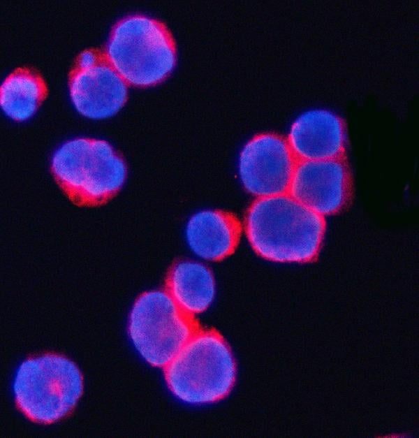
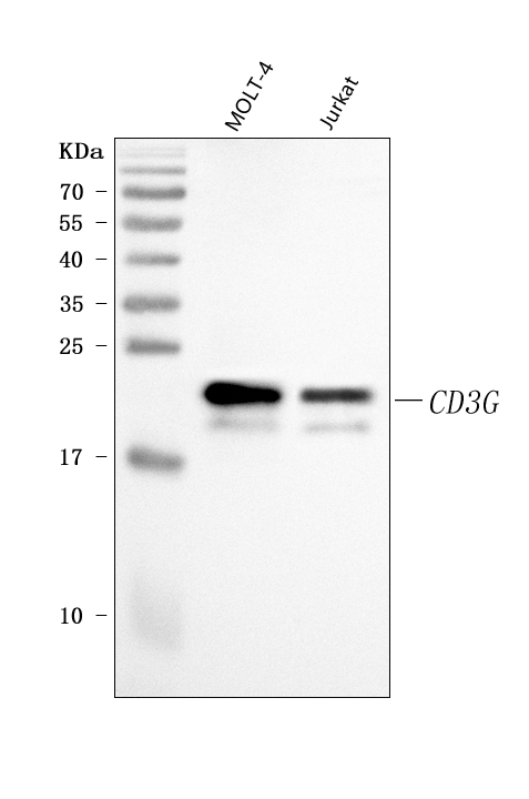
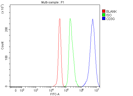
(Figure 3. Flow Cytometry analysis of JK cells using anti-CD3 Gamma/CD3G antibody (AAA127258).Overlay histogram showing JK cells stained with AAA127258 (Blue line). To facilitate intracellular staining, cells were fixed with 4% paraformaldehyde and permeabilized with permeabilization buffer. The cells were blocked with 10% normal goat serum. And then incubated with rabbit anti-CD3 Gamma/CD3G Antibody (AAA127258, 1ug/1x106 cells) for 30 min at 20 degree C. DyLight488 conjugated goat anti-rabbit IgG was used as secondary antibody for 30 minutes at 20 degree C. Isotype control antibody (Green line) was rabbit IgG (1ug/1x106) used under the same conditions. Unlabelled sample (Red line) was also used as a control.)
FCM/FACS (Flow Cytometry)
(Figure 3. Flow Cytometry analysis of JK cells using anti-CD3 Gamma/CD3G antibody (AAA127258).Overlay histogram showing JK cells stained with AAA127258 (Blue line). To facilitate intracellular staining, cells were fixed with 4% paraformaldehyde and permeabilized with permeabilization buffer. The cells were blocked with 10% normal goat serum. And then incubated with rabbit anti-CD3 Gamma/CD3G Antibody (AAA127258, 1ug/1x106 cells) for 30 min at 20 degree C. DyLight488 conjugated goat anti-rabbit IgG was used as secondary antibody for 30 minutes at 20 degree C. Isotype control antibody (Green line) was rabbit IgG (1ug/1x106) used under the same conditions. Unlabelled sample (Red line) was also used as a control.)
CD3 Gamma/CD3G, Polyclonal Antibody (Cat# AAA127258)
Full Name
Anti-CD3 Gamma/CD3G Antibody Picoband
Gene Names
CD3G; T3G; IMD17; CD3-GAMMA
Reactivity
Human
Applications
Flow Cytometry, Immunofluorescence, Immunocytochemistry, Western Blot
Purity
Immunogen affinity purified.
Pricing
 IF (Immunofluorescence)
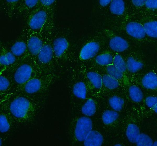
(Figure 2. IF analysis of PTH2R using anti-PTH2R antibody (AAA127649).PTH2R was detected in an immunocytochemical section of U2OS cells. Enzyme antigen retrieval was performed using IHC enzyme antigen retrieval reagent for 15 mins. The cells were blocked with 10% goat serum. And then incubated with 5ug/mL rabbit anti-PTH2R Antibody (AAA127649) overnight at 4 degree C. DyLight488 Conjugated Goat Anti-Rabbit IgG was used as secondary antibody at 1:500 dilution and incubated for 30 minutes at 37 degree C. The section was counterstained with DAPI. Visualize using a fluorescence microscope and filter sets appropriate for the label used.)
IF (Immunofluorescence)
(Figure 2. IF analysis of PTH2R using anti-PTH2R antibody (AAA127649).PTH2R was detected in an immunocytochemical section of U2OS cells. Enzyme antigen retrieval was performed using IHC enzyme antigen retrieval reagent for 15 mins. The cells were blocked with 10% goat serum. And then incubated with 5ug/mL rabbit anti-PTH2R Antibody (AAA127649) overnight at 4 degree C. DyLight488 Conjugated Goat Anti-Rabbit IgG was used as secondary antibody at 1:500 dilution and incubated for 30 minutes at 37 degree C. The section was counterstained with DAPI. Visualize using a fluorescence microscope and filter sets appropriate for the label used.)
PTH2R, Polyclonal Antibody (Cat# AAA127649)
Full Name
Anti-PTH2R Antibody Picoband
Gene Names
PTH2R; PTHR2
Reactivity
Human
Applications
Western Blot, Immunofluorescence, Immunocytochemistry
Purity
Immunogen affinity purified.
Pricing
 WB (Western Blot)
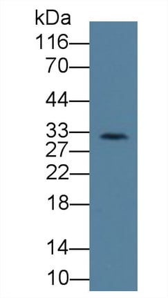
(Western Blot; Sample: Rat Lymph node lysate; Primary Ab: 3ug/ml Mouse AntiHuman KIR2DS4 Antibody Second Ab: 0.2ug/mL HRPLinked Caprine AntiMouse IgG Polyclonal Antibody (Catalog: SAA544Mu19))
WB (Western Blot)
(Western Blot; Sample: Rat Lymph node lysate; Primary Ab: 3ug/ml Mouse AntiHuman KIR2DS4 Antibody Second Ab: 0.2ug/mL HRPLinked Caprine AntiMouse IgG Polyclonal Antibody (Catalog: SAA544Mu19))
Killer Cell Immunoglobulin Like Receptor 2DS4 (KIR2DS4), Monoclonal Antibody (Cat# AAA151727)
Full Name
Monoclonal Antibody to Killer Cell Immunoglobulin Like Receptor 2DS4 (KIR2DS4)
Gene Names
KIR2DS4; KKA3; KIR1D; NKAT8; CD158I; KIR412; NKAT-8; KIR2DS1
Reactivity
Human, Rat
Applications
Immunoprecipitation, Immunocytochemistry, Immunohistochemistry, Western Blot
Purity
Protein A + Protein G affinity chromatography
Pricing
 FCM/FACS (Flow Cytometry)
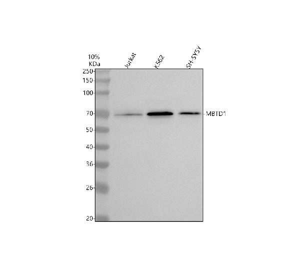
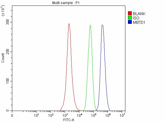
(Figure 3. Flow Cytometry analysis of SH-SY5Y cells using anti-MBTD1 antibody (AAA127741).Overlay histogram showing SH-SY5Y cells stained with AAA127741 (Blue line). To facilitate intracellular staining, cells were fixed with 4% paraformaldehyde and permeabilized with permeabilization buffer. The cells were blocked with 10% normal goat serum. And then incubated with rabbit anti-MBTD1 Antibody (AAA127741, 1ug/1x106 cells) for 30 min at 20 degree C. DyLight488 conjugated goat anti-rabbit IgG was used as secondary antibody for 30 minutes at 20 degree C. Isotype control antibody (Green line) was rabbit IgG (1ug/1x106) used under the same conditions. Unlabelled sample without incubation with primary antibody and secondary antibody (Red line) was used as a blank control.)
FCM/FACS (Flow Cytometry)
(Figure 3. Flow Cytometry analysis of SH-SY5Y cells using anti-MBTD1 antibody (AAA127741).Overlay histogram showing SH-SY5Y cells stained with AAA127741 (Blue line). To facilitate intracellular staining, cells were fixed with 4% paraformaldehyde and permeabilized with permeabilization buffer. The cells were blocked with 10% normal goat serum. And then incubated with rabbit anti-MBTD1 Antibody (AAA127741, 1ug/1x106 cells) for 30 min at 20 degree C. DyLight488 conjugated goat anti-rabbit IgG was used as secondary antibody for 30 minutes at 20 degree C. Isotype control antibody (Green line) was rabbit IgG (1ug/1x106) used under the same conditions. Unlabelled sample without incubation with primary antibody and secondary antibody (Red line) was used as a blank control.)
MBTD1, Polyclonal Antibody (Cat# AAA127741)
Full Name
Anti-MBTD1 Antibody Picoband
Gene Names
MBTD1; SA49P01
Reactivity
Human
Applications
Flow Cytometry, Immunofluorescence, Immunocytochemistry, Western Blot
Purity
Immunogen affinity purified.
Pricing
 ICC (Immunocytochemistry)
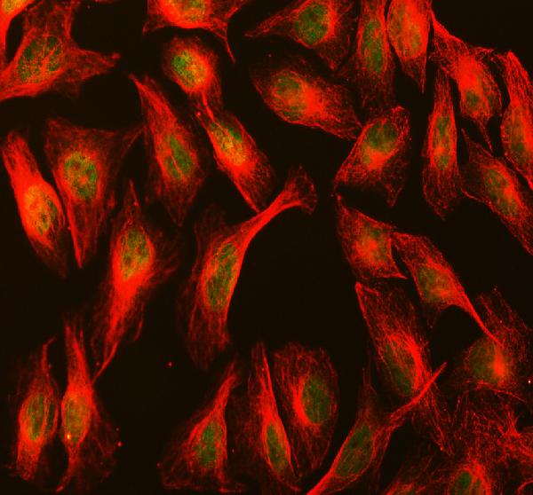
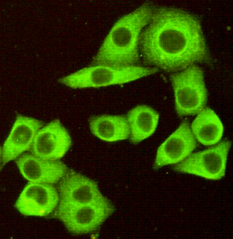
(Immunocytochemistry of HeLa cells fixed by Paraformaldehyde and using DR5 mouse mAb diluted 1:100.)
ICC (Immunocytochemistry)
(Immunocytochemistry of HeLa cells fixed by Paraformaldehyde and using DR5 mouse mAb diluted 1:100.)
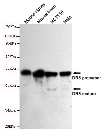
DR5, Monoclonal Antibody (Cat# AAA320363)
Full Name
DR5 Mouse mAb
Gene Names
TNFRSF10B; DR5; CD262; KILLER; TRICK2; TRICKB; ZTNFR9; TRAILR2; TRICK2A; TRICK2B; TRAIL-R2; KILLER/DR5
Reactivity
Human, Mouse
Applications
Immunocytochemistry, Western Blot
Purity
The antibody was affinity-purified from mouse ascites by affinity-chromatography using epitope-specific immunogen.
Pricing
 FCM/FACS (Flow Cytometry)
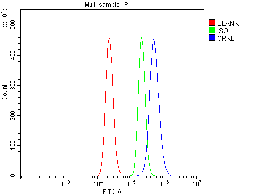
(Figure 3. Flow Cytometry analysis of RT4 cells using anti-CRKL antibody (AAA127100).Overlay histogram showing RT4 cells stained with AAA127100 (Blue line). To facilitate intracellular staining, cells were fixed with 4% paraformaldehyde and permeabilized with permeabilization buffer. The cells were blocked with 10% normal goat serum. And then incubated with rabbit anti-CRKL Antibody (AAA127100, 1ug/1x106 cells) for 30 min at 20 degree C. DyLight488 conjugated goat anti-rabbit IgG was used as secondary antibody for 30 minutes at 20 degree C. Isotype control antibody (Green line) was rabbit IgG (1ug/1x106) used under the same conditions. Unlabelled sample without incubation with primary antibody and secondary antibody (Red line) was used as a blank control.)
FCM/FACS (Flow Cytometry)
(Figure 3. Flow Cytometry analysis of RT4 cells using anti-CRKL antibody (AAA127100).Overlay histogram showing RT4 cells stained with AAA127100 (Blue line). To facilitate intracellular staining, cells were fixed with 4% paraformaldehyde and permeabilized with permeabilization buffer. The cells were blocked with 10% normal goat serum. And then incubated with rabbit anti-CRKL Antibody (AAA127100, 1ug/1x106 cells) for 30 min at 20 degree C. DyLight488 conjugated goat anti-rabbit IgG was used as secondary antibody for 30 minutes at 20 degree C. Isotype control antibody (Green line) was rabbit IgG (1ug/1x106) used under the same conditions. Unlabelled sample without incubation with primary antibody and secondary antibody (Red line) was used as a blank control.)
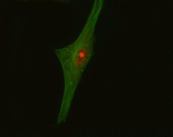
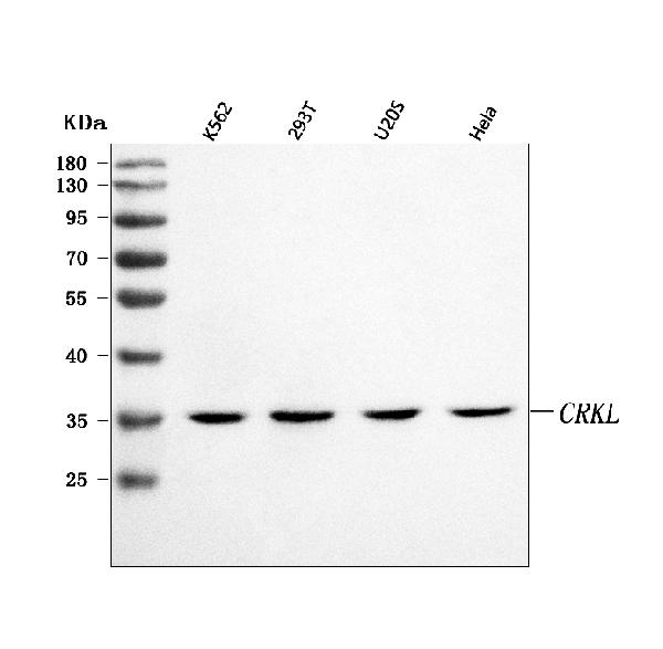
CRKL, Polyclonal Antibody (Cat# AAA127100)
Full Name
Anti-CRKL Antibody Picoband
Reactivity
Human
Applications
Flow Cytometry, Immunofluorescence, Immunocytochemistry, Western Blot
Purity
Immunogen affinity purified.
Pricing
 FCM/FACS (Flow Cytometry)
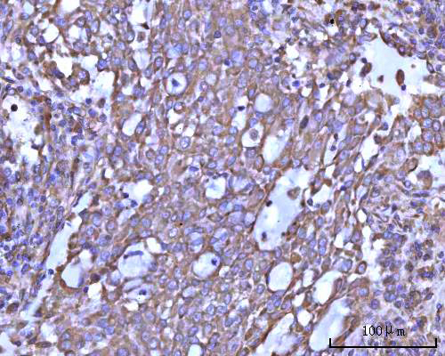
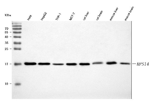
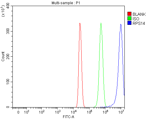
(Figure 3. Flow Cytometry analysis of HepG2 cells using anti-RPS14 antibody (AAA127195).Overlay histogram showing HepG2 cells stained with AAA127195 (Blue line). To facilitate intracellular staining, cells were fixed with 4% paraformaldehyde and permeabilized with permeabilization buffer. The cells were blocked with 10% normal goat serum. And then incubated with rabbit anti-RPS14 Antibody (AAA127195, 1ug/1x106 cells) for 30 min at 20 degree C. DyLight488 conjugated goat anti-rabbit IgG was used as secondary antibody for 30 minutes at 20 degree C. Isotype control antibody (Green line) was rabbit IgG (1ug/1x106) used under the same conditions. Unlabelled sample without incubation with primary antibody and secondary antibody (Red line) was used as a blank control.)
FCM/FACS (Flow Cytometry)
(Figure 3. Flow Cytometry analysis of HepG2 cells using anti-RPS14 antibody (AAA127195).Overlay histogram showing HepG2 cells stained with AAA127195 (Blue line). To facilitate intracellular staining, cells were fixed with 4% paraformaldehyde and permeabilized with permeabilization buffer. The cells were blocked with 10% normal goat serum. And then incubated with rabbit anti-RPS14 Antibody (AAA127195, 1ug/1x106 cells) for 30 min at 20 degree C. DyLight488 conjugated goat anti-rabbit IgG was used as secondary antibody for 30 minutes at 20 degree C. Isotype control antibody (Green line) was rabbit IgG (1ug/1x106) used under the same conditions. Unlabelled sample without incubation with primary antibody and secondary antibody (Red line) was used as a blank control.)
RPS14, Polyclonal Antibody (Cat# AAA127195)
Full Name
Anti-RPS14 Antibody Picoband
Gene Names
RPS14; S14; EMTB
Reactivity
Human, Mouse, Rat
Applications
Flow Cytometry, Immunohistochemistry, Western Blot
Purity
Immunogen affinity purified.
Pricing
 FCM/FACS (Flow Cytometry)
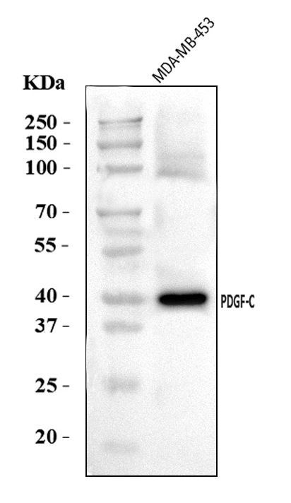
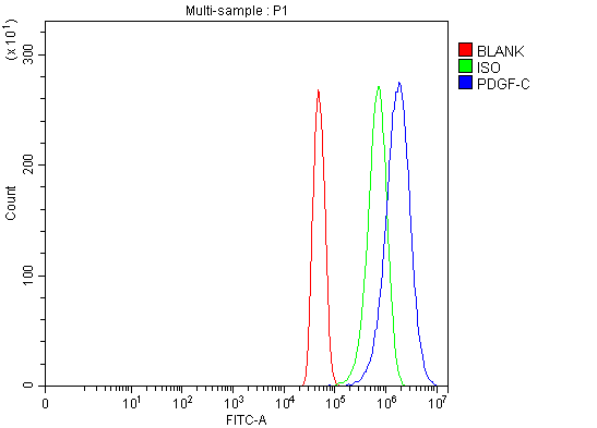
(Figure 2. Flow Cytometry analysis of U251 cells using anti-PDGFC antibody (AAA126281).Overlay histogram showing U251 cells stained with AAA126281 (Blue line). The cells were blocked with 10% normal goat serum. And then incubated with rabbit anti-PDGFC Antibody (AAA126281, 1 ug/1x10^6 cells) for 30 min at 20 degree C. DyLight488 conjugated goat anti-rabbit IgG was used as secondary antibody for 30 minutes at 20 degree C. Isotype control antibody (Green line) was rabbit IgG (1 ug/1x10^6) used under the same conditions. Unlabelled sample (Red line) was also used as a control.)
FCM/FACS (Flow Cytometry)
(Figure 2. Flow Cytometry analysis of U251 cells using anti-PDGFC antibody (AAA126281).Overlay histogram showing U251 cells stained with AAA126281 (Blue line). The cells were blocked with 10% normal goat serum. And then incubated with rabbit anti-PDGFC Antibody (AAA126281, 1 ug/1x10^6 cells) for 30 min at 20 degree C. DyLight488 conjugated goat anti-rabbit IgG was used as secondary antibody for 30 minutes at 20 degree C. Isotype control antibody (Green line) was rabbit IgG (1 ug/1x10^6) used under the same conditions. Unlabelled sample (Red line) was also used as a control.)
PDGFC, Polyclonal Antibody (Cat# AAA126281)
Full Name
Anti-PDGFC Antibody Picoband
Gene Names
PDGFC; SCDGF; FALLOTEIN
Reactivity
Human
Applications
Flow Cytometry, Western Blot
Purity
Immunogen affinity purified.
Pricing
 FCM/FACS (Flow Cytometry)
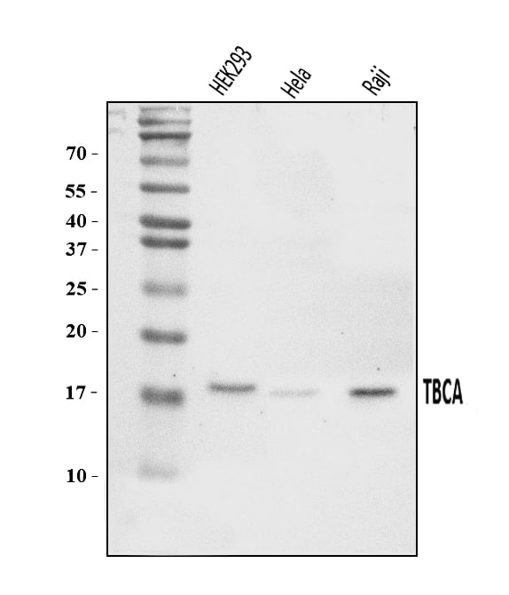
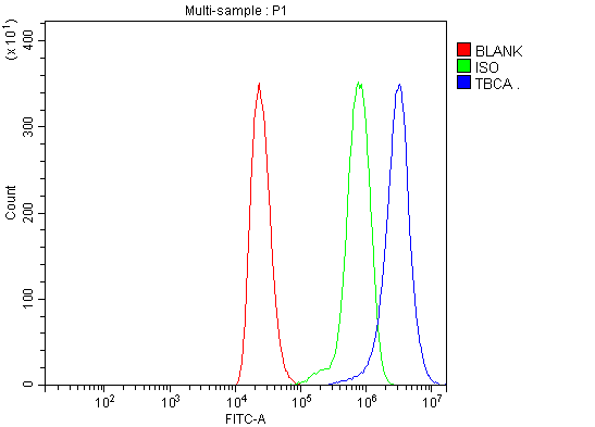
(Figure 2. Flow Cytometry analysis of THP-1 cells using anti-CFA/TBCA antibody (AAA126477).Overlay histogram showing THP-1 cells stained with AAA126477 (Blue line). The cells were blocked with 10% normal goat serum. And then incubated with rabbit anti-CFA/TBCA Antibody (AAA126477, 1 ug/1x10^6 cells) for 30 min at 20 degree C. DyLight488 conjugated goat anti-rabbit IgG was used as secondary antibody for 30 minutes at 20 degree C. Isotype control antibody (Green line) was rabbit IgG (1 ug/1x10^6) used under the same conditions. Unlabelled sample (Red line) was also used as a control.)
FCM/FACS (Flow Cytometry)
(Figure 2. Flow Cytometry analysis of THP-1 cells using anti-CFA/TBCA antibody (AAA126477).Overlay histogram showing THP-1 cells stained with AAA126477 (Blue line). The cells were blocked with 10% normal goat serum. And then incubated with rabbit anti-CFA/TBCA Antibody (AAA126477, 1 ug/1x10^6 cells) for 30 min at 20 degree C. DyLight488 conjugated goat anti-rabbit IgG was used as secondary antibody for 30 minutes at 20 degree C. Isotype control antibody (Green line) was rabbit IgG (1 ug/1x10^6) used under the same conditions. Unlabelled sample (Red line) was also used as a control.)
CFA/TBCA, Polyclonal Antibody (Cat# AAA126477)
Full Name
Anti-CFA/TBCA Antibody Picoband
Reactivity
Human
Applications
Flow Cytometry, Western Blot
Purity
Immunogen affinity purified.
Pricing
 FCM/FACS (Flow Cytometry)
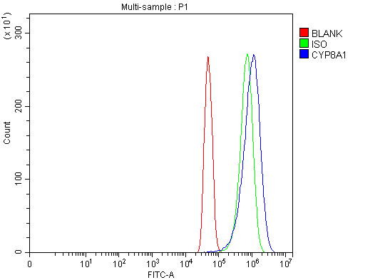
(Figure 2. Flow Cytometry analysis of U251 cells using anti-PTGIS/PGIS antibody (AAA126407).Overlay histogram showing U251 cells stained with AAA126407 (Blue line). The cells were blocked with 10% normal goat serum. And then incubated with rabbit anti-PTGIS/PGIS Antibody (AAA126407, 1 ug/1x10^6 cells) for 30 min at 20 degree C. DyLight488 conjugated goat anti-rabbit IgG was used as secondary antibody for 30 minutes at 20 degree C. Isotype control antibody (Green line) was rabbit IgG (1 ug/1x10^6) used under the same conditions. Unlabelled sample (Red line) was also used as a control.)
FCM/FACS (Flow Cytometry)
(Figure 2. Flow Cytometry analysis of U251 cells using anti-PTGIS/PGIS antibody (AAA126407).Overlay histogram showing U251 cells stained with AAA126407 (Blue line). The cells were blocked with 10% normal goat serum. And then incubated with rabbit anti-PTGIS/PGIS Antibody (AAA126407, 1 ug/1x10^6 cells) for 30 min at 20 degree C. DyLight488 conjugated goat anti-rabbit IgG was used as secondary antibody for 30 minutes at 20 degree C. Isotype control antibody (Green line) was rabbit IgG (1 ug/1x10^6) used under the same conditions. Unlabelled sample (Red line) was also used as a control.)
PTGIS/PGIS, Polyclonal Antibody (Cat# AAA126407)
Full Name
Anti-PTGIS/PGIS Antibody Picoband
Gene Names
PTGIS; CYP8; PGIS; PTGI; CYP8A1
Reactivity
Human, Mouse, Rat
Applications
Flow Cytometry, Western Blot
Purity
Immunogen affinity purified.
Pricing
 FCM/FACS (Flow Cytometry)
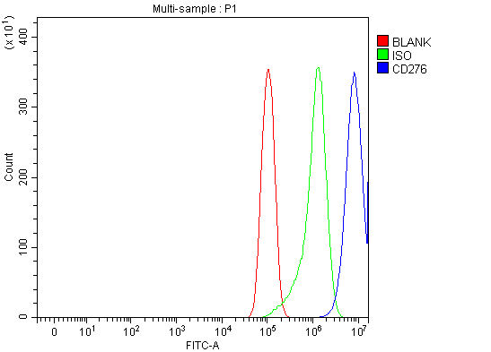
(Figure 5. Flow Cytometry analysis of PC-3 cells using anti-CD276 antibody (AAA127105).Overlay histogram showing PC-3 cells stained with AAA127105 (Blue line). The cells were fixed with 4% paraformaldehyde and blocked with 10% normal goat serum. And then incubated with rabbit anti-CD276 Antibody (AAA127105, 1ug/1x106 cells) for 30 min at 20 degree C. DyLight488 conjugated goat anti-rabbit IgG was used as secondary antibody for 30 minutes at 20 degree C. Isotype control antibody (Green line) was rabbit IgG (1ug/1x106) used under the same conditions. Unlabelled sample without incubation with primary antibody and secondary antibody (Red line) was used as a blank control.)
FCM/FACS (Flow Cytometry)
(Figure 5. Flow Cytometry analysis of PC-3 cells using anti-CD276 antibody (AAA127105).Overlay histogram showing PC-3 cells stained with AAA127105 (Blue line). The cells were fixed with 4% paraformaldehyde and blocked with 10% normal goat serum. And then incubated with rabbit anti-CD276 Antibody (AAA127105, 1ug/1x106 cells) for 30 min at 20 degree C. DyLight488 conjugated goat anti-rabbit IgG was used as secondary antibody for 30 minutes at 20 degree C. Isotype control antibody (Green line) was rabbit IgG (1ug/1x106) used under the same conditions. Unlabelled sample without incubation with primary antibody and secondary antibody (Red line) was used as a blank control.)
CD276, Polyclonal Antibody (Cat# AAA127105)
Full Name
Anti-CD276 Antibody Picoband
Gene Names
CD276; B7H3; B7-H3; B7RP-2; 4Ig-B7-H3
Reactivity
Human
Applications
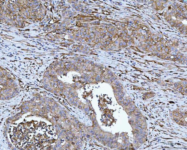
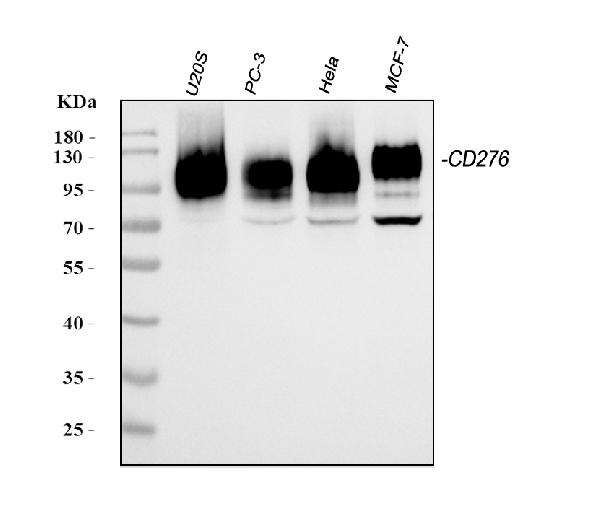
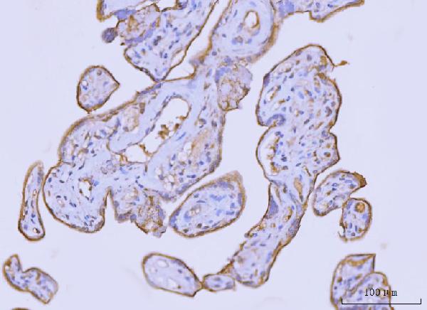
Flow Cytometry, Immunohistochemistry, Western Blot
Purity
Immunogen affinity purified.
Pricing
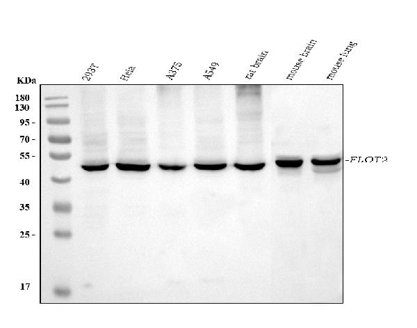
 IF (Immunofluorescence)
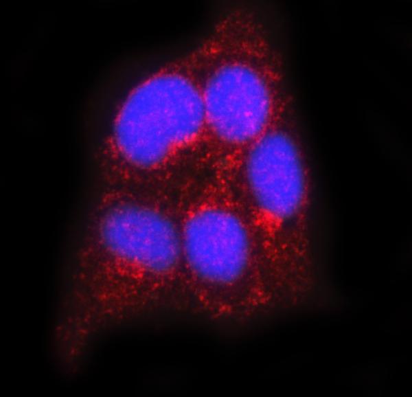
(Figure 3. IF analysis of FLOT2 using anti-FLOT2 antibody (AAA126948).FLOT2 was detected in an immunocytochemical section of SiHa cells. Enzyme antigen retrieval was performed using IHC enzyme antigen retrieval reagent (AR0022) for 15 mins. The cells were blocked with 10% goat serum. And then incubated with 5 ug/mL mouse anti-FLOT2 Antibody (AAA126948) overnight at 4 degree C. Cy3 Conjugated Goat Anti-Mouse IgG (BA1031) was used as secondary antibody at 1:100 dilution and incubated for 30 minutes at 37 degree C. The section was counterstained with DAPI. Visualize using a fluorescence microscope and filter sets appropriate for the label used.)
IF (Immunofluorescence)
(Figure 3. IF analysis of FLOT2 using anti-FLOT2 antibody (AAA126948).FLOT2 was detected in an immunocytochemical section of SiHa cells. Enzyme antigen retrieval was performed using IHC enzyme antigen retrieval reagent (AR0022) for 15 mins. The cells were blocked with 10% goat serum. And then incubated with 5 ug/mL mouse anti-FLOT2 Antibody (AAA126948) overnight at 4 degree C. Cy3 Conjugated Goat Anti-Mouse IgG (BA1031) was used as secondary antibody at 1:100 dilution and incubated for 30 minutes at 37 degree C. The section was counterstained with DAPI. Visualize using a fluorescence microscope and filter sets appropriate for the label used.)
Flotillin 2, Monoclonal Antibody (Cat# AAA126948)
Full Name
Anti-Flotillin 2 Antibody Picoband (monoclonal, 4D8A3)
Gene Names
FLOT2; ESA; ECS1; ESA1; ECS-1; M17S1
Reactivity
Human, Mouse, Rat
Applications
Immunofluorescence, Immunocytochemistry, Immunohistochemistry, Western Blot
Purity
Immunogen affinity purified.
Pricing
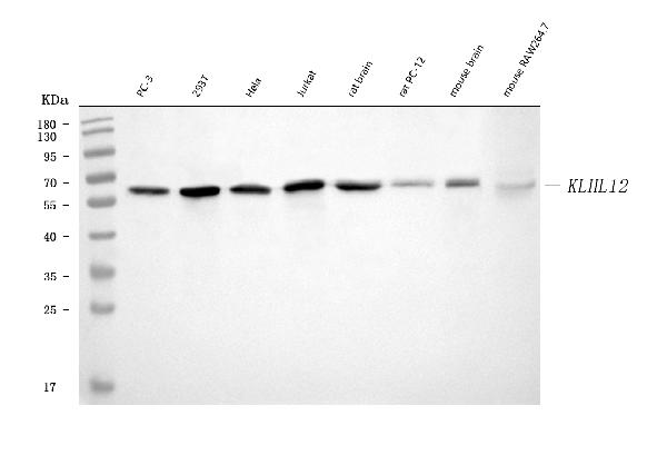
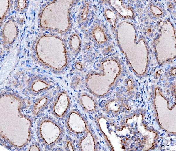 IHC (Immunohistochemisry)
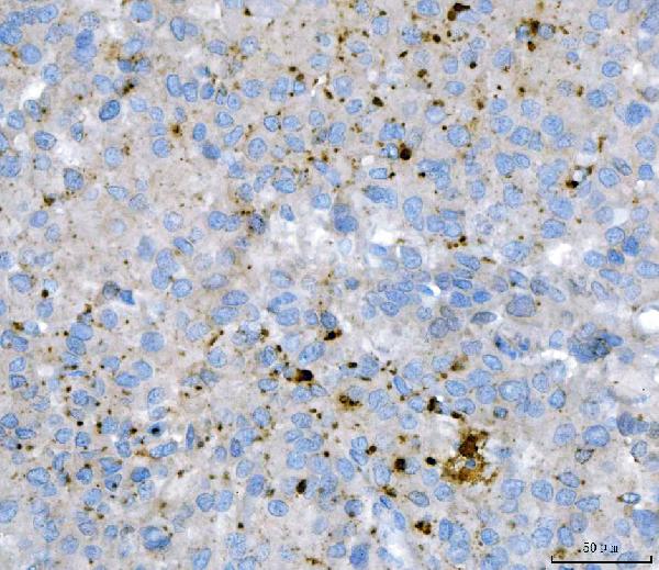
(Figure 3. IHC analysis of KLHL12 using anti-KLHL12 antibody (AAA126956).KLHL12 was detected in a paraffin-embedded section of human thyroid cancer tissue. Heat mediated antigen retrieval was performed in EDTA buffer (pH 8.0, epitope retrieval solution). The tissue section was blocked with 10% goat serum. The tissue section was then incubated with 2 ug/ml mouse anti-KLHL12 Antibody (AAA126956) overnight at 4 degree C. Peroxidase Conjugated Goat Anti-mouse IgG was used as secondary antibody and incubated for 30 minutes at 37 degree C. The tissue section was developed using HRP Conjugated Mouse IgG Super Vision Assay Kit with DAB as the chromogen.)
IHC (Immunohistochemisry)
(Figure 3. IHC analysis of KLHL12 using anti-KLHL12 antibody (AAA126956).KLHL12 was detected in a paraffin-embedded section of human thyroid cancer tissue. Heat mediated antigen retrieval was performed in EDTA buffer (pH 8.0, epitope retrieval solution). The tissue section was blocked with 10% goat serum. The tissue section was then incubated with 2 ug/ml mouse anti-KLHL12 Antibody (AAA126956) overnight at 4 degree C. Peroxidase Conjugated Goat Anti-mouse IgG was used as secondary antibody and incubated for 30 minutes at 37 degree C. The tissue section was developed using HRP Conjugated Mouse IgG Super Vision Assay Kit with DAB as the chromogen.)
KLHL12, Monoclonal Antibody (Cat# AAA126956)
Full Name
Anti-KLHL12 Antibody Picoband (monoclonal, 2G11D1)
Gene Names
KLHL12; DKIR; C3IP1
Reactivity
Human, Mouse, Rat
Applications
Immunohistochemistry, Western Blot
Purity
Immunogen affinity purified.
Pricing
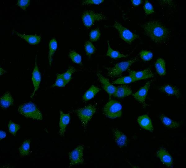
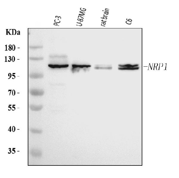
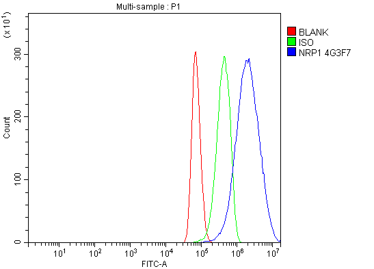 FCM/FACS (Flow Cytometry)
(Figure 3. Flow Cytometry analysis of U87 cells using anti-Neuropilin 1 antibody (AAA126889).Overlay histogram showing U87 cells stained with AAA126889 (Blue line). The cells were blocked with 10% normal goat serum. And then incubated with mouse anti-Neuropilin 1 Antibody (AAA126889, 1 ug/1x10^6 cells) for 30 min at 20 degree C. DyLight488 conjugated goat anti-mouse IgG was used as secondary antibody for 30 minutes at 20 degree C. Isotype control antibody (Green line) was mouse IgG (1 ug/1x10^6) used under the same conditions. Unlabelled sample (Red line) was also used as a control.)
FCM/FACS (Flow Cytometry)
(Figure 3. Flow Cytometry analysis of U87 cells using anti-Neuropilin 1 antibody (AAA126889).Overlay histogram showing U87 cells stained with AAA126889 (Blue line). The cells were blocked with 10% normal goat serum. And then incubated with mouse anti-Neuropilin 1 Antibody (AAA126889, 1 ug/1x10^6 cells) for 30 min at 20 degree C. DyLight488 conjugated goat anti-mouse IgG was used as secondary antibody for 30 minutes at 20 degree C. Isotype control antibody (Green line) was mouse IgG (1 ug/1x10^6) used under the same conditions. Unlabelled sample (Red line) was also used as a control.)
Neuropilin 1, Monoclonal Antibody (Cat# AAA126889)
Full Name
Anti-Neuropilin 1 Antibody Picoband (monoclonal, 4G3F7)
Gene Names
NRP1; NP1; NRP; BDCA4; CD304; VEGF165R
Reactivity
Human, Rat
Applications
Flow Cytometry, Immunofluorescence, Immunocytochemistry, Western Blot
Purity
Immunogen affinity purified.
Pricing
 IHC (Immunohistochemistry)
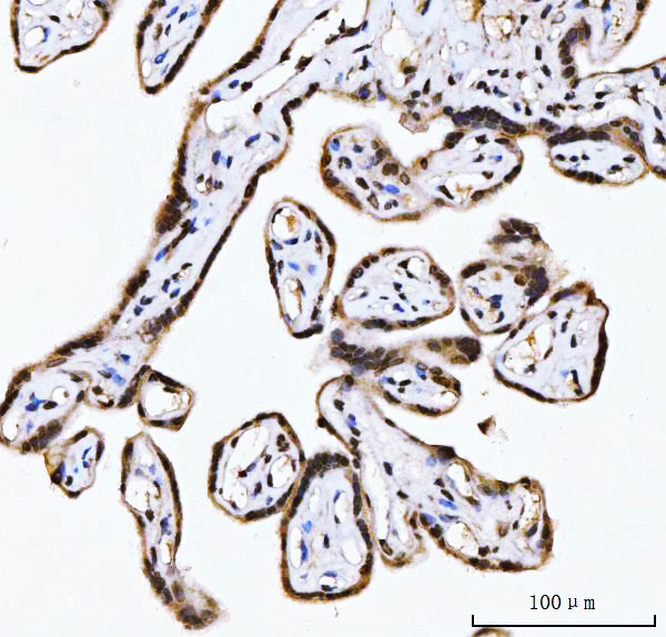
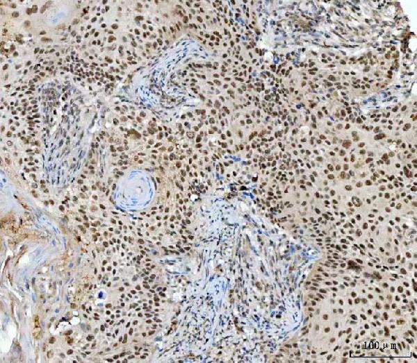
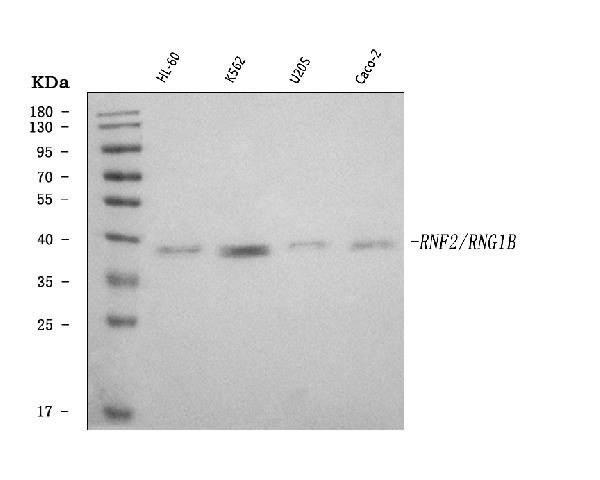
(Figure 4. IHC analysis of RING2/RING1B/RNF2 using anti-RING2/RING1B/RNF2 antibody (AAA126126).RING2/RING1B/RNF2 was detected in a paraffin-embedded section of human thyroid cancer tissue. Heat mediated antigen retrieval was performed in EDTA buffer (pH 8.0, epitope retrieval solution). The tissue section was blocked with 10% goat serum. The tissue section was then incubated with 2 ug/ml rabbit anti-RING2/RING1B/RNF2 Antibody (AAA126126) overnight at 4 degree C. Peroxidase Conjugated Goat Anti-rabbit IgG was used as secondary antibody and incubated for 30 minutes at 37 degree C. The tissue section was developed using HRP Conjugated Rabbit IgG Super Vision Assay Kit with DAB as the chromogen.)
IHC (Immunohistochemistry)
(Figure 4. IHC analysis of RING2/RING1B/RNF2 using anti-RING2/RING1B/RNF2 antibody (AAA126126).RING2/RING1B/RNF2 was detected in a paraffin-embedded section of human thyroid cancer tissue. Heat mediated antigen retrieval was performed in EDTA buffer (pH 8.0, epitope retrieval solution). The tissue section was blocked with 10% goat serum. The tissue section was then incubated with 2 ug/ml rabbit anti-RING2/RING1B/RNF2 Antibody (AAA126126) overnight at 4 degree C. Peroxidase Conjugated Goat Anti-rabbit IgG was used as secondary antibody and incubated for 30 minutes at 37 degree C. The tissue section was developed using HRP Conjugated Rabbit IgG Super Vision Assay Kit with DAB as the chromogen.)
RING2/RING1B/RNF2, Polyclonal Antibody (Cat# AAA126126)
Full Name
Anti-RING2/RING1B/RNF2 Antibody Picoband
Gene Names
RNF2; BAP1; DING; BAP-1; HIPI3; RING2; RING1B
Reactivity
Human
Applications
Western Blot, Immunohistochemistry
Purity
Immunogen affinity purified.
Pricing
 IHC (Immunohistochemistry)
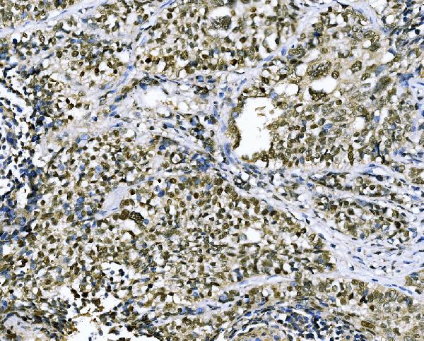
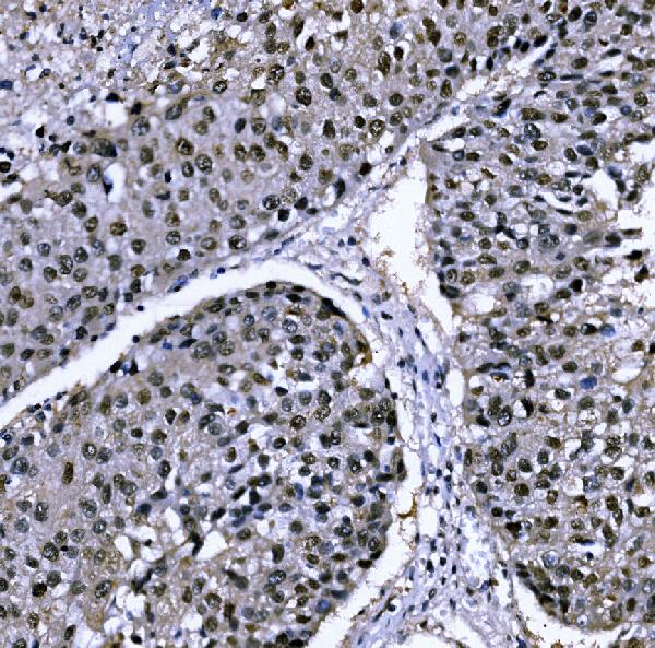
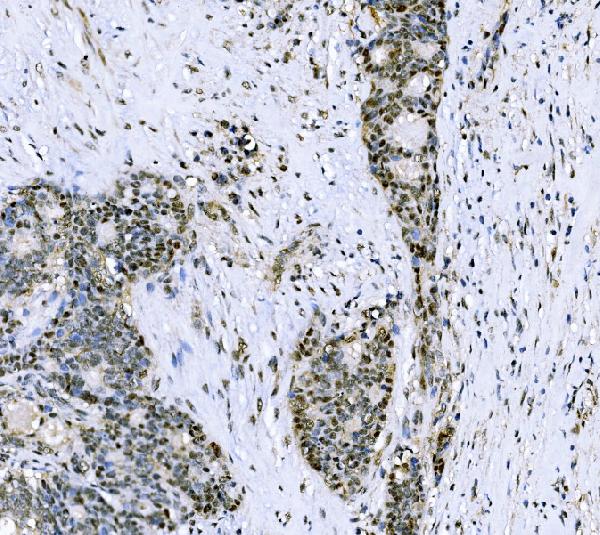
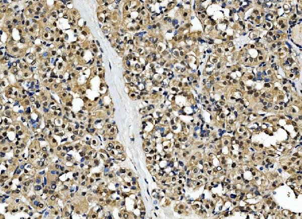
(Figure 5. IHC analysis of CSNK2B using anti-CSNK2B antibody (AAA126311).CSNK2B was detected in a paraffin-embedded section of human thyroid cancer tissue. Heat mediated antigen retrieval was performed in EDTA buffer (pH 8.0, epitope retrieval solution). The tissue section was blocked with 10% goat serum. The tissue section was then incubated with 2 ug/ml rabbit anti-CSNK2B Antibody (AAA126311) overnight at 4 degree C. Biotinylated goat anti-rabbit IgG was used as secondary antibody and incubated for 30 minutes at 37 degree C. The tissue section was developed using Strepavidin-Biotin-Complex (SABC) with DAB as the chromogen.)
IHC (Immunohistochemistry)
(Figure 5. IHC analysis of CSNK2B using anti-CSNK2B antibody (AAA126311).CSNK2B was detected in a paraffin-embedded section of human thyroid cancer tissue. Heat mediated antigen retrieval was performed in EDTA buffer (pH 8.0, epitope retrieval solution). The tissue section was blocked with 10% goat serum. The tissue section was then incubated with 2 ug/ml rabbit anti-CSNK2B Antibody (AAA126311) overnight at 4 degree C. Biotinylated goat anti-rabbit IgG was used as secondary antibody and incubated for 30 minutes at 37 degree C. The tissue section was developed using Strepavidin-Biotin-Complex (SABC) with DAB as the chromogen.)
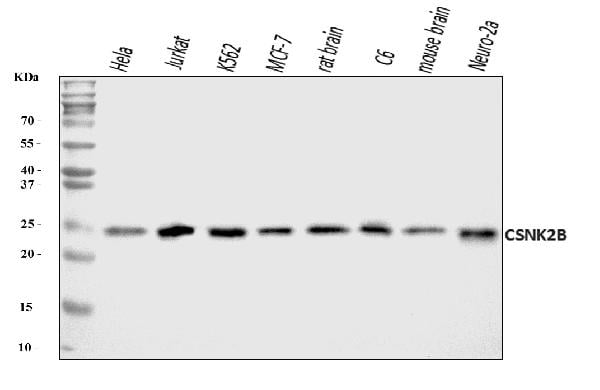
CSNK2B, Polyclonal Antibody (Cat# AAA126311)
Full Name
Anti-CSNK2B Antibody Picoband
Gene Names
CSNK2B; G5A; CK2B; CK2N; CSK2B
Reactivity
Human, Mouse, Rat
Applications
Immunohistochemistry, Western Blot
Purity
Immunogen affinity purified.
Pricing
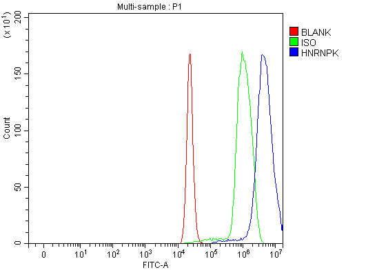 FCM/FACS (Flow Cytometry)
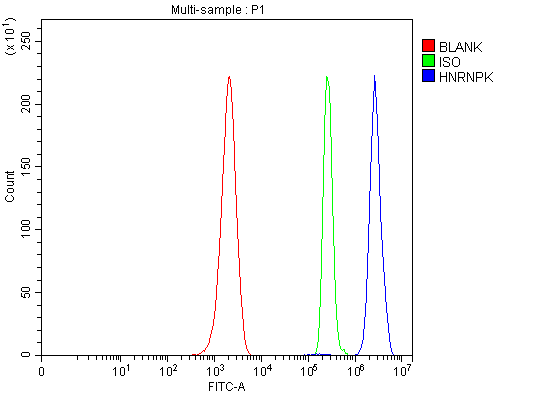
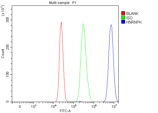
(Figure 5. Flow Cytometry analysis of C6 cells using anti-hnRNP K/HNRNPK antibody (AAA126175).Overlay histogram showing C6 cells stained with AAA126175 (Blue line). The cells were blocked with 10% normal goat serum. And then incubated with rabbit anti-hnRNP K/HNRNPK Antibody (AAA126175, 1 ug/1x10^6 cells) for 30 min at 20 degree C. DyLight488 conjugated goat anti-rabbit IgG was used as secondary antibody for 30 minutes at 20 degree C. Isotype control antibody (Green line) was rabbit IgG (1 ug/1x10^6) used under the same conditions. Unlabelled sample (Red line) was also used as a control.)
FCM/FACS (Flow Cytometry)
(Figure 5. Flow Cytometry analysis of C6 cells using anti-hnRNP K/HNRNPK antibody (AAA126175).Overlay histogram showing C6 cells stained with AAA126175 (Blue line). The cells were blocked with 10% normal goat serum. And then incubated with rabbit anti-hnRNP K/HNRNPK Antibody (AAA126175, 1 ug/1x10^6 cells) for 30 min at 20 degree C. DyLight488 conjugated goat anti-rabbit IgG was used as secondary antibody for 30 minutes at 20 degree C. Isotype control antibody (Green line) was rabbit IgG (1 ug/1x10^6) used under the same conditions. Unlabelled sample (Red line) was also used as a control.)
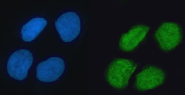
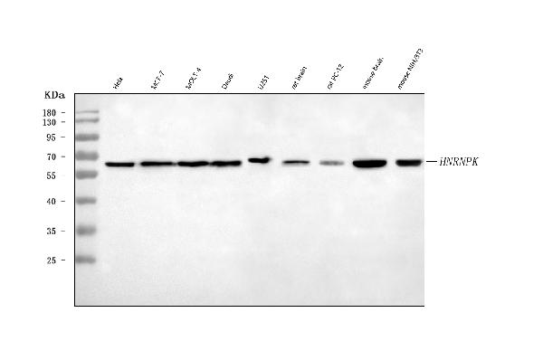
hnRNP K/HNRNPK, Polyclonal Antibody (Cat# AAA126175)
Full Name
Anti-hnRNP K/HNRNPK Antibody Picoband
Gene Names
HNRNPK; CSBP; TUNP; HNRPK
Reactivity
Human, Mouse, Rat
Applications
Flow Cytometry, Immunofluorescence, Immunocytochemistry, Western Blot
Purity
Immunogen affinity purified.
Pricing
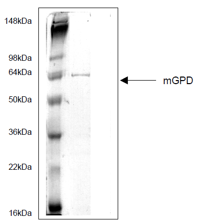 SDS-PAGE
Mitochondrial Glycerol-3-Phosphate Dehydrogenase (mGPD), Recombinant Protein (Cat# AAA58754)
Full Name
Mitochondrial Glycerol-3-Phosphate Dehydrogenase (mGPD)
Purity
>95% by SDS-PAGE
Pricing
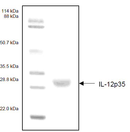 SDS-PAGE
Interleukin-12p35 (IL-12p35), Recombinant Protein (Cat# AAA58748)
Full Name
Interleukin-12p35 (IL-12p35)
Purity
>95% by SDS-PAGE
Pricing
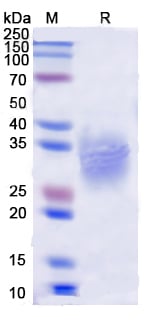
 Bioactivity
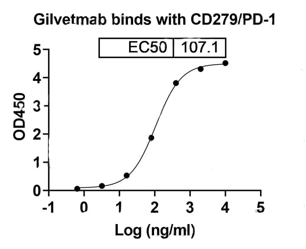
(Detects Gilvetmab in indirect ELISAs.)
Bioactivity
(Detects Gilvetmab in indirect ELISAs.)
CD279/PDCD1/PD1, Recombinant Protein (Cat# AAA120737)
Full Name
Recombinant Dog CD279/PDCD1/PD1 Protein, C-His
Applications
Western Blot
Purity
>90% as determined by SDS-PAGE.
Pricing
 SDS-PAGE
Cytokeratin-18 (CK-18), Recombinant Protein (Cat# AAA58739)
Full Name
Cytokeratin-18 (CK-18)
Gene Names
KRT18; K18; CK-18; CYK18
Purity
>95% by SDS-PAGE
Pricing
 SDS-PAGE
Soluble Tumor Necrosis Factor Receptor 1 (STNF-R1), Recombinant Protein (Cat# AAA58767)
Full Name
Soluble Tumor Necrosis Factor Receptor 1 (STNF-R1)
Purity
>95%
Pricing
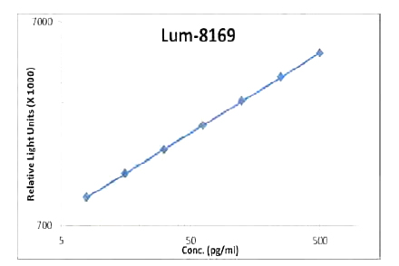
 Standard Curve (Sample)
EGF, CLIA Kit (Cat# AAA318526)
Full Name
EGF (Human) LumiAb ELISA Kit
Gene Names
EGF; URG; HOMG4
Reactivity
Human
Pricing
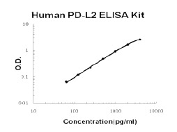 Standard Curve (Sample)
PD-L2/B7-DC, ELISA Kit (Cat# AAA124848)
Full Name
Human PD-L2/B7-DC PicoKine ELISA Kit
Gene Names
PDCD1LG2; B7DC; Btdc; PDL2; CD273; PD-L2; PDCD1L2; bA574F11.2
Reactivity
Human
There is no detectable cross-reactivity with other relevant proteins.
There is no detectable cross-reactivity with other relevant proteins.
Applications
ELISA
Pricing
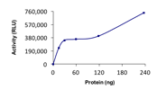
 Standard Curve (Sample)
TWEAK, CLIA Kit (Cat# AAA318564)
Full Name
TWEAK (Human) LumiAb ELISA Kit
Gene Names
TNFSF12; APO3L; DR3LG; TWEAK; TNLG4A
Reactivity
Human
Pricing
 SDS-PAGE
(Purity: Sample Purity Data. For specific information on a given lot, see related technical data sheet.)
SDS-PAGE
(Purity: Sample Purity Data. For specific information on a given lot, see related technical data sheet.)
BIRC3, Recombinant Protein (Cat# AAA73282)
Full Name
BIRC3, Active
Applications
Western Blot
Pricing
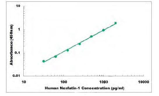 Standard Curve (Sample)
Nesfatin-1, ELISA Kit (Cat# AAA106079)
Full Name
Human Nesfatin-1 ELISA Kit
Gene Names
NUCB2; NEFA; HEL-S-109
Reactivity
Human
Applications
Sandwich ELISA
Pricing

 SDS-PAGE
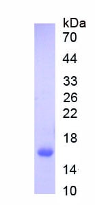
(Figure. SDS-PAGE)
SDS-PAGE
(Figure. SDS-PAGE)
Follicle Stimulating Hormone Beta (FSHb), Recombinant Protein (Cat# AAA150714)
Full Name
Recombinant Follicle Stimulating Hormone Beta (FSHb)
Gene Names
TSHB; TSH-B; TSH-BETA
Applications
Western Blot
Purity
>97%
Pricing
 SDS-PAGE
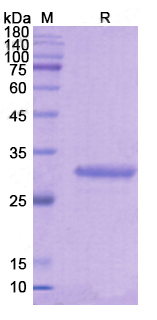
(SDS PAGE for recombinant Human CD61/ITGB3 protein)
SDS-PAGE
(SDS PAGE for recombinant Human CD61/ITGB3 protein)
CD61/ITGB3, Recombinant Protein (Cat# AAA120433)
Full Name
Recombinant Human CD61/ITGB3, N-His
Gene Names
ITGB3; GT; CD61; GP3A; BDPLT2; GPIIIa; BDPLT16
Applications
Western Blot
Purity
>90% as determined by SDS-PAGE
Pricing