Filters
Clonality
Type
Reactivity
Gene Name
Isotype
Host
Application
Clone
666 results for " B Cells" - showing 500-550


 Application Data
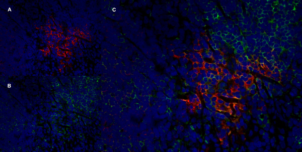
(Published clone specific image Alloimmunity-associated cytotoxicity is mediated through the NKG2D receptor. (A) Liver expression of nkg2d on day ten after liver transplantation. (B) Representative NKG2D expression levels in blood NK cells (left) and monocytes (right) of allogeneic (black) and syngeneic (grey) recipients. Isotype was used as control (dashed lines). (C) Sorted blood NK cell cytotoxicity inhibition with anti-NKG2D antibody or with anti-NKp30 antibody. (D) Levels of NKG2D ligand (rae1l, rrlt and irp94) expression in the liver on day ten after transplantation. (E) Levels of NKG2D ligand (rae1l, rrlt and irp94) expression in rat HCC cell lines. (F) Representative level of recombinant NKG2D-Fc binding to rat HCC cells lines. *p)
Application Data
(Published clone specific image Alloimmunity-associated cytotoxicity is mediated through the NKG2D receptor. (A) Liver expression of nkg2d on day ten after liver transplantation. (B) Representative NKG2D expression levels in blood NK cells (left) and monocytes (right) of allogeneic (black) and syngeneic (grey) recipients. Isotype was used as control (dashed lines). (C) Sorted blood NK cell cytotoxicity inhibition with anti-NKG2D antibody or with anti-NKp30 antibody. (D) Levels of NKG2D ligand (rae1l, rrlt and irp94) expression in the liver on day ten after transplantation. (E) Levels of NKG2D ligand (rae1l, rrlt and irp94) expression in rat HCC cell lines. (F) Representative level of recombinant NKG2D-Fc binding to rat HCC cells lines. *p)
CD172a, Monoclonal Antibody (Cat# AAA11967)
Full Name
MOUSE ANTI RAT CD172a
Gene Names
Sirpa; Bit; Ptpns1; SHPS-1
Applications
FC/FACS, WB
Pricing


 Application Data
(Published clone specific image Alloimmunity-associated cytotoxicity is mediated through the NKG2D receptor. (A) Liver expression of nkg2d on day ten after liver transplantation. (B) Representative NKG2D expression levels in blood NK cells (left) and monocytes (right) of allogeneic (black) and syngeneic (grey) recipients. Isotype was used as control (dashed lines). (C) Sorted blood NK cell cytotoxicity inhibition with anti-NKG2D antibody or with anti-NKp30 antibody. (D) Levels of NKG2D ligand (rae1l, rrlt and irp94) expression in the liver on day ten after transplantation. (E) Levels of NKG2D ligand (rae1l, rrlt and irp94) expression in rat HCC cell lines. (F) Representative level of recombinant NKG2D-Fc binding to rat HCC cells lines. *p)
Application Data
(Published clone specific image Alloimmunity-associated cytotoxicity is mediated through the NKG2D receptor. (A) Liver expression of nkg2d on day ten after liver transplantation. (B) Representative NKG2D expression levels in blood NK cells (left) and monocytes (right) of allogeneic (black) and syngeneic (grey) recipients. Isotype was used as control (dashed lines). (C) Sorted blood NK cell cytotoxicity inhibition with anti-NKG2D antibody or with anti-NKp30 antibody. (D) Levels of NKG2D ligand (rae1l, rrlt and irp94) expression in the liver on day ten after transplantation. (E) Levels of NKG2D ligand (rae1l, rrlt and irp94) expression in rat HCC cell lines. (F) Representative level of recombinant NKG2D-Fc binding to rat HCC cells lines. *p)
CD172a, Monoclonal Antibody (Cat# AAA11968)
Full Name
MOUSE ANTI RAT CD172a
Gene Names
Sirpa; Bit; Ptpns1; SHPS-1
Applications
FC/FACS, WB
Pricing


 ChIP (Chromatin Immunoprecipitation)
(Chromatin immunoprecipitation analysis of extracts of HeLa cells, using Acetyl-Histone H3-K9/K14/K18/K23/K27 antibody and rabbit IgG.The amount of immunoprecipitated DNA was checked by quantitative PCR. Histogram was constructed by the ratios of the immunoprecipitated DNA to the input.)
ChIP (Chromatin Immunoprecipitation)
(Chromatin immunoprecipitation analysis of extracts of HeLa cells, using Acetyl-Histone H3-K9/K14/K18/K23/K27 antibody and rabbit IgG.The amount of immunoprecipitated DNA was checked by quantitative PCR. Histogram was constructed by the ratios of the immunoprecipitated DNA to the input.)
Acetyl-Histone H3-K9/K14/K18/K23/K27, Polyclonal Antibody (Cat# AAA28357)
Full Name
Acetyl-Histone H3-K9/K14/K18/K23/K27 Rabbit pAb
Gene Names
HIST1H3A; H3/A; H3FA
Reactivity
Human, Mouse, Rat, Other (Wide Range)
Applications
WB, IHC, IF, ChIP
Purity
Affinity purification
Pricing


 IHC (Immunohistchemistry)
(Annexin II Antibody for IHC in human colon tissue)
IHC (Immunohistchemistry)
(Annexin II Antibody for IHC in human colon tissue)
Annexin II, Polyclonal Antibody (Cat# AAA31078)
Full Name
Annexin II Antibody
Gene Names
ANXA2; P36; ANX2; LIP2; LPC2; CAL1H; LPC2D; ANX2L4; PAP-IV; HEL-S-270
Reactivity
Human, Mouse, Rat
Applications
WB, IHC, EIA
Purity
The antiserum was purified by peptide affinity chromatography using SulfoLink Coupling Resin.
Pricing


 IF (Immunofluorescence)
(Immunofluorescent analysis of 4% paraformaldehyde-fixed, 0.1% Triton X-100 permeabilized MCF-7 (human breast cancer cell line) cells labeling Pdx1 with AAA14796 at 1:25 dilution, followed by Dylight® 488-conjugated IgG goat anti-rabbit secondary antibody at 1:200 dilution (green). Immunofluorescence image showing cytoplasm staining on MCF-7 cell line. Cytoplasmic actin is detected with Dylight® 554 Phalloidin (PD18466410) at 1:100 dilution (red). The nuclear counter stain is DAPI (blue).)
IF (Immunofluorescence)
(Immunofluorescent analysis of 4% paraformaldehyde-fixed, 0.1% Triton X-100 permeabilized MCF-7 (human breast cancer cell line) cells labeling Pdx1 with AAA14796 at 1:25 dilution, followed by Dylight® 488-conjugated IgG goat anti-rabbit secondary antibody at 1:200 dilution (green). Immunofluorescence image showing cytoplasm staining on MCF-7 cell line. Cytoplasmic actin is detected with Dylight® 554 Phalloidin (PD18466410) at 1:100 dilution (red). The nuclear counter stain is DAPI (blue).)
OPN-a/b, Polyclonal Antibody (Cat# AAA14796)
Full Name
OPN-a/b, NT (SPP1, BNSP, OPN, Osteopontin, Bone sialoprotein 1, Nephropontin, Secreted phosphoprotein 1, Urinary stone protein, Uropontin)
Reactivity
Human
Applications
EL/EIA, WB, IHC, IF
Purity
Affinity Purified
Purified by Protein A affinity chromatography.
Purified by Protein A affinity chromatography.
Pricing


 Application Data
(Staining of human peripheral blood granulocytes with Mouse anti Human CD11b:Biotin)
Application Data
(Staining of human peripheral blood granulocytes with Mouse anti Human CD11b:Biotin)
CD11b, Monoclonal Antibody (Cat# AAA11991)
Full Name
MOUSE ANTI HUMAN CD11b
Gene Names
ITGAM; CR3A; MO1A; CD11B; MAC-1; MAC1A; SLEB6
Reactivity
Baboon, Cynomolgus monkey, Rhesus Monkey
Applications
FC/FACS, IP
Pricing


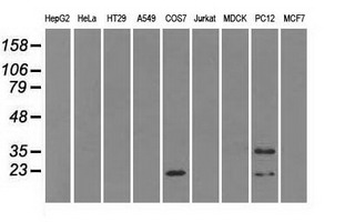
 WB (Western Blot)
(HSPA1B monoclonal antibody Western Blot analysis of HSPA1B expression in Raw 264.7.)
WB (Western Blot)
(HSPA1B monoclonal antibody Western Blot analysis of HSPA1B expression in Raw 264.7.)
HSPA1A, Monoclonal Antibody (Cat# AAA25129)
Full Name
HSPA1A (Heat Shock 70kD Protein 1A/1B, Heat Shock 70kD Protein 1/2, HSP70.1/HSP70.2, HSP70-1/HSP70-2, HSPA1, HSPA1B) (FITC)
Gene Names
HSPA1A; HSP72; HSPA1; HSP70I; HSP70-1; HSP70.1; HSP70-1A; HEL-S-103
Reactivity
Human, Mouse, Rat
Applications
EIA, IF, IHC, WB
Purity
Purified by Protein A Affinity Chromatography.
Pricing


 WB (Western Blot)
(HSPA1B monoclonal antibody Western Blot analysis of HSPA1B expression in Raw 264.7.)
WB (Western Blot)
(HSPA1B monoclonal antibody Western Blot analysis of HSPA1B expression in Raw 264.7.)
HSPA1A, Monoclonal Antibody (Cat# AAA24241)
Full Name
HSPA1A (Heat Shock 70kD Protein 1A/1B, Heat Shock 70kD Protein 1/2, HSP70.1/HSP70.2, HSP70-1/HSP70-2, HSPA1, HSPA1B) (AP)
Gene Names
HSPA1A; HSP72; HSPA1; HSP70I; HSP70-1; HSP70.1; HSP70-1A; HEL-S-103
Reactivity
Human, Mouse, Rat
Applications
EIA, IHC, WB
Purity
Purified by Protein A Affinity Chromatography.
Pricing


 Application Data
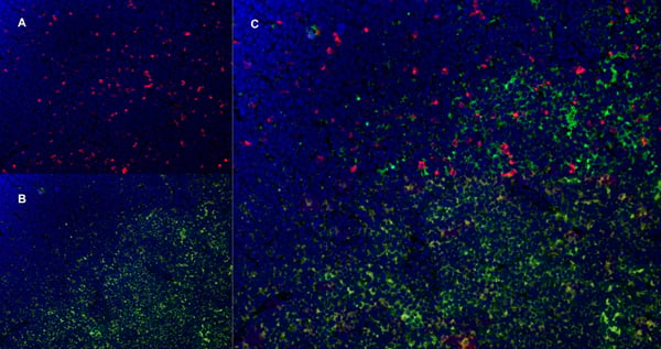
(Staining of mouse spleen with Rat anti Mouse CD4:RPE)
Application Data
(Staining of mouse spleen with Rat anti Mouse CD4:RPE)
CD4, Monoclonal Antibody (Cat# AAA12227)
Full Name
RAT ANTI MOUSE CD4
Gene Names
Cd4; L3T4; Ly-4
Applications
EIA, FC/FACS, IF, IP, WB
Pricing


 Application Data
(Staining of mouse spleen with Rat anti Mouse CD4:RPE)
Application Data
(Staining of mouse spleen with Rat anti Mouse CD4:RPE)
CD4, Monoclonal Antibody (Cat# AAA12230)
Full Name
RAT ANTI MOUSE CD4
Gene Names
Cd4; L3T4; Ly-4
Applications
EIA, FC/FACS, IF, IP, WB
Pricing


 Application Data
(Staining of mouse spleen with Rat anti Mouse CD4:RPE)
Application Data
(Staining of mouse spleen with Rat anti Mouse CD4:RPE)
CD4, Monoclonal Antibody (Cat# AAA12233)
Full Name
RAT ANTI MOUSE CD4
Gene Names
Cd4; L3T4; Ly-4
Applications
EIA, FC/FACS, IF, IP, WB
Pricing


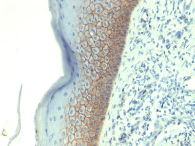

 IHC (Immunohistchemistry)
(Formalin-paraffin human Placenta stained with E-Cadherin MAb (CDH1/1525).)
IHC (Immunohistchemistry)
(Formalin-paraffin human Placenta stained with E-Cadherin MAb (CDH1/1525).)
E-Cadherin/CD324, Monoclonal Antibody (Cat# AAA23896)
Full Name
E-Cadherin/CD324 (Intercellular Junction Marker)
Gene Names
CDH1; UVO; CDHE; ECAD; LCAM; Arc-1; BCDS1; CD324
Reactivity
Human.
Does not react with Mouse and Rat. Others not known.
Does not react with Mouse and Rat. Others not known.
Applications
FC/FACS, IF, WB, IHC
Pricing


 FCM (Flow Cytometry)
(Figure 7. Flow Cytometry analysis of 293T cells using anti-OTULIN antibody (AAA19319).Overlay histogram showing 293T cells stained with AAA19319 (Blue line). The cells were blocked with 10% normal goat serum. And then incubated with rabbit anti-OTULIN Antibody (AAA19319, 1μg/1x106 cells) for 30 min at 20 degree C. DyLight®488 conjugated goat anti-rabbit IgG (5-10μg/1x106 cells) was used as secondary antibody for 30 minutes at 20 degree C. Isotype control antibody (Green line) was rabbit IgG (1μg/1x106) used under the same conditions. Unlabelled sample (Red line) was also used as a control.)
FCM (Flow Cytometry)
(Figure 7. Flow Cytometry analysis of 293T cells using anti-OTULIN antibody (AAA19319).Overlay histogram showing 293T cells stained with AAA19319 (Blue line). The cells were blocked with 10% normal goat serum. And then incubated with rabbit anti-OTULIN Antibody (AAA19319, 1μg/1x106 cells) for 30 min at 20 degree C. DyLight®488 conjugated goat anti-rabbit IgG (5-10μg/1x106 cells) was used as secondary antibody for 30 minutes at 20 degree C. Isotype control antibody (Green line) was rabbit IgG (1μg/1x106) used under the same conditions. Unlabelled sample (Red line) was also used as a control.)
OTULIN, Polyclonal Antibody (Cat# AAA19319)
Full Name
Anti-OTULIN Antibody
Gene Names
OTULIN; GUM; FAM105B
Reactivity
Human, Mouse, Rat
Applications
WB, IHC-P, ICC, IF, FC/FACS/FCM, EIA
Purity
Immunogen affinity purified.
Pricing


 FCM (Flow Cytometry)
(Figure 9. Flow Cytometry analysis of THP-1 cells using anti-CHCHD10 antibody (AAA19310).Overlay histogram showing THP-1 cells stained with AAA19310 (Blue line). The cells were blocked with 10% normal goat serum. And then incubated with rabbit anti-CHCHD10 Antibody (AAA19310, 1μg/1x106 cells) for 30 min at 20 degree C. DyLight®488 conjugated goat anti-rabbit IgG (5-10μg/1x106 cells) was used as secondary antibody for 30 minutes at 20 degree C. Isotype control antibody (Green line) was rabbit IgG (1μg/1x106) used under the same conditions. Unlabelled sample (Red line) was also used as a control.)
FCM (Flow Cytometry)
(Figure 9. Flow Cytometry analysis of THP-1 cells using anti-CHCHD10 antibody (AAA19310).Overlay histogram showing THP-1 cells stained with AAA19310 (Blue line). The cells were blocked with 10% normal goat serum. And then incubated with rabbit anti-CHCHD10 Antibody (AAA19310, 1μg/1x106 cells) for 30 min at 20 degree C. DyLight®488 conjugated goat anti-rabbit IgG (5-10μg/1x106 cells) was used as secondary antibody for 30 minutes at 20 degree C. Isotype control antibody (Green line) was rabbit IgG (1μg/1x106) used under the same conditions. Unlabelled sample (Red line) was also used as a control.)
CHCHD10, Polyclonal Antibody (Cat# AAA19310)
Full Name
Anti-CHCHD10 Antibody
Gene Names
CHCHD10; FTDALS2; N27C7-4; C22orf16
Reactivity
Human, Mouse, Rat, Monkey
Applications
WB, IHC-P, ICC, IF, FC/FACS/FCM, EIA
Purity
Immunogen affinity purified.
Pricing


 FCM (Flow Cytometry)
(Figure 8. Flow Cytometry analysis of HL-60 cells using anti-REA/PHB2 antibody (AAA19273).Overlay histogram showing HL-60 cells stained with AAA19273 (Blue line). The cells were blocked with 10% normal goat serum. And then incubated with rabbit anti-REA/PHB2 Antibody (AAA19273,1μg/1x106 cells) for 30 min at 20 degree C. DyLight®488 conjugated goat anti-rabbit IgG (5-10μg/1x106 cells) was used as secondary antibody for 30 minutes at 20 degree C. Isotype control antibody (Green line) was rabbit IgG (1μg/1x106) used under the same conditions. Unlabelled sample (Red line) was also used as a control.)
FCM (Flow Cytometry)
(Figure 8. Flow Cytometry analysis of HL-60 cells using anti-REA/PHB2 antibody (AAA19273).Overlay histogram showing HL-60 cells stained with AAA19273 (Blue line). The cells were blocked with 10% normal goat serum. And then incubated with rabbit anti-REA/PHB2 Antibody (AAA19273,1μg/1x106 cells) for 30 min at 20 degree C. DyLight®488 conjugated goat anti-rabbit IgG (5-10μg/1x106 cells) was used as secondary antibody for 30 minutes at 20 degree C. Isotype control antibody (Green line) was rabbit IgG (1μg/1x106) used under the same conditions. Unlabelled sample (Red line) was also used as a control.)
REA/PHB2, Polyclonal Antibody (Cat# AAA19273)
Full Name
Anti-REA/PHB2 Antibody
Gene Names
PHB2; BAP; REA; p22; Bap37; BCAP37; PNAS-141
Reactivity
Human, Mouse, Rat
Applications
WB, IHC-P, ICC, IF, FC/FACS/FCM, EIA
Purity
Immunogen affinity purified.
Pricing


 WB (Western Blot)
(HSPA1B monoclonal antibody Western Blot analysis of HSPA1B expression in Raw 264.7.)
WB (Western Blot)
(HSPA1B monoclonal antibody Western Blot analysis of HSPA1B expression in Raw 264.7.)
HSPA1A, Monoclonal Antibody (Cat# AAA25423)
Full Name
HSPA1A (Heat Shock 70kD Protein 1A/1B, Heat Shock 70kD Protein 1/2, HSP70.1/HSP70.2, HSP70-1/HSP70-2, HSPA1, HSPA1B) (HRP)
Gene Names
HSPA1A; HSP72; HSPA1; HSP70I; HSP70-1; HSP70.1; HSP70-1A; HEL-S-103
Reactivity
Human, Mouse, Rat
Applications
EIA, IHC, WB
Purity
Purified by Protein A Affinity Chromatography.
Pricing


 IHC (Immunohistchemistry)
(At 1/100 staining Human kidney cancer and adjacent normal tissues by IHC-P. The sample was formaldehyde fixed and a heat mediated antigen retrieval step in citrate buffer was performed. The sample was then blocked and incubated with the primary antibody at 4 degree C overnight. An HRP conjugated anti-Rabbit antibody was used as the secondary antibody.)
IHC (Immunohistchemistry)
(At 1/100 staining Human kidney cancer and adjacent normal tissues by IHC-P. The sample was formaldehyde fixed and a heat mediated antigen retrieval step in citrate buffer was performed. The sample was then blocked and incubated with the primary antibody at 4 degree C overnight. An HRP conjugated anti-Rabbit antibody was used as the secondary antibody.)
RSK1 p90, Polyclonal Antibody (Cat# AAA31316)
Full Name
Phospho-RSK1 p90 (Thr348) Antibody
Gene Names
RPS6KA1; RSK; HU-1; RSK1; MAPKAPK1A
Reactivity
Human, Mouse, Rat
Applications
IHC, EIA
Purity
The antibody is from purified rabbit serum by affinity purification via sequential chromatography on phospho-peptide and non-phospho-peptide affinity columns.
Pricing


 WB (Western Blot)
(HSPA1B monoclonal antibody Western Blot analysis of HSPA1B expression in Raw 264.7.)
WB (Western Blot)
(HSPA1B monoclonal antibody Western Blot analysis of HSPA1B expression in Raw 264.7.)
HSPA1A, Monoclonal Antibody (Cat# AAA25719)
Full Name
HSPA1A (Heat Shock 70kD Protein 1A/1B, Heat Shock 70kD Protein 1/2, HSP70.1/HSP70.2, HSP70-1/HSP70-2, HSPA1, HSPA1B) (PE)
Gene Names
HSPA1A; HSP72; HSPA1; HSP70I; HSP70-1; HSP70.1; HSP70-1A; HEL-S-103
Reactivity
Human, Mouse, Rat
Applications
EIA, IF, IHC, WB
Purity
Purified by Protein A Affinity Chromatography.
Pricing


 WB (Western Blot)
(Western blot analysis of p70 S6 Kinase expression in mouse muscle tissue lysates, The lane on the right is treated with the antigen-specific peptide.)
WB (Western Blot)
(Western blot analysis of p70 S6 Kinase expression in mouse muscle tissue lysates, The lane on the right is treated with the antigen-specific peptide.)
p70 S6 Kinase, Polyclonal Antibody (Cat# AAA31088)
Full Name
p70 S6 Kinase Antibody
Gene Names
RPS6KB1; S6K; PS6K; S6K1; STK14A; p70-S6K; p70 S6KA; p70-alpha; S6K-beta-1; p70(S6K)-alpha
Reactivity
Human, Mouse, Rat
Applications
WB, IHC, IF, ICC, EIA
Purity
The antiserum was purified by peptide affinity chromatography using SulfoLink Coupling Resin.
Pricing


 ICC (Immunocytochemistry)
(Figure 10 Immunocytochemistry Validation of PD-L1Immunocytochemical analysis of 4% paraformaldehyde-fixed PD-L1 transfected 293 cells labeling PD-L1 with at 1 μg/ml, followed by Goat anti-mouse IgG secondary antibody at 1/250 dilution (red). Lower left: Use mouse IgG antibody at 1 μg/ml as control.)
ICC (Immunocytochemistry)
(Figure 10 Immunocytochemistry Validation of PD-L1Immunocytochemical analysis of 4% paraformaldehyde-fixed PD-L1 transfected 293 cells labeling PD-L1 with at 1 μg/ml, followed by Goat anti-mouse IgG secondary antibody at 1/250 dilution (red). Lower left: Use mouse IgG antibody at 1 μg/ml as control.)
PDL1, Monoclonal Antibody (Cat# AAA10992)
Full Name
PDL1 Antibody [6H10]
Gene Names
CD274; B7-H; B7H1; PDL1; PD-L1; PDCD1L1; PDCD1LG1
Reactivity
Human
Applications
EIA, WB, IHC, ICC, IF
Purity
Protein A purified
Pricing


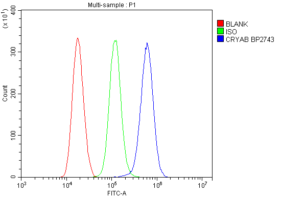 FCM (Flow Cytometry)
(Figure 13. Flow Cytometry analysis of THP-1 cells using anti-Alpha B Crystallin/CRYAB antibody (AAA19277).Overlay histogram showing THP-1 cells stained with AAA19277 (Blue line). The cells were blocked with 10% normal goat serum. And then incubated with rabbit anti-Alpha B Crystallin/CRYAB Antibody (AAA19277, 1μg/1x106 cells) for 30 min at 20 degree C. DyLight®488 conjugated goat anti-rabbit IgG (5-10μg/1x106 cells) was used as secondary antibody for 30 minutes at 20 degree C. Isotype control antibody (Green line) was rabbit IgG (1μg/1x106) used under the same conditions. Unlabelled sample (Red line) was also used as a control.)
FCM (Flow Cytometry)
(Figure 13. Flow Cytometry analysis of THP-1 cells using anti-Alpha B Crystallin/CRYAB antibody (AAA19277).Overlay histogram showing THP-1 cells stained with AAA19277 (Blue line). The cells were blocked with 10% normal goat serum. And then incubated with rabbit anti-Alpha B Crystallin/CRYAB Antibody (AAA19277, 1μg/1x106 cells) for 30 min at 20 degree C. DyLight®488 conjugated goat anti-rabbit IgG (5-10μg/1x106 cells) was used as secondary antibody for 30 minutes at 20 degree C. Isotype control antibody (Green line) was rabbit IgG (1μg/1x106) used under the same conditions. Unlabelled sample (Red line) was also used as a control.)
Alpha B Crystallin/CRYAB, Polyclonal Antibody (Cat# AAA19277)
Full Name
Anti-Alpha B Crystallin/CRYAB Antibody
Gene Names
CRYAB; CRYA2; CTPP2; HSPB5; CMD1II
Reactivity
Human, Mouse, Rat, Monkey
Applications
WB, IHC-P, ICC, IF, FC/FACS/FCM, EIA
Purity
Immunogen affinity purified.
Pricing


 WB (Western Blot)
(Western blot analysis of Human Lysates showing detection of Hsp90 protein using Mouse Anti-Hsp90 Monoclonal Antibody, Clone H9010 . Primary Antibody: Mouse Anti-Hsp90 Monoclonal Antibody at 1:1000. Comparison of clone H9010 behavior with Hsp90 human beta (1) and Hsp90 human alpha (2). Courtesy of: David Toft, Mayo Clinic.)
WB (Western Blot)
(Western blot analysis of Human Lysates showing detection of Hsp90 protein using Mouse Anti-Hsp90 Monoclonal Antibody, Clone H9010 . Primary Antibody: Mouse Anti-Hsp90 Monoclonal Antibody at 1:1000. Comparison of clone H9010 behavior with Hsp90 human beta (1) and Hsp90 human alpha (2). Courtesy of: David Toft, Mayo Clinic.)
HSP90, Monoclonal Antibody (Cat# AAA27660)
Full Name
HSP90 Antibody
Gene Names
HSP90AB1; HSP84; HSPC2; HSPCB; D6S182; HSP90B
Reactivity
Human, Mouse, Rat, Rabbit, Chicken, Dog, Fish, Shark, Hamster
Applications
WB, IHC, ICC, IF, IP, EIA, AM
Purity
Protein G Purified
Pricing


 SDS-PAGE
(Purified ER-beta Mouse Monoclonal Antibody (ESR2/686). Confirmation of Integrity and Purity of Antibody.)
SDS-PAGE
(Purified ER-beta Mouse Monoclonal Antibody (ESR2/686). Confirmation of Integrity and Purity of Antibody.)
ER-beta1 (Estrogen Receptor beta-1), Monoclonal Antibody (Cat# AAA13846)
Full Name
ER-beta1 (Estrogen Receptor beta-1) Mouse Monoclonal Antibody
Gene Names
ESR2; Erb; ESRB; ESTRB; NR3A2; ER-BETA; ESR-BETA
Reactivity
Human
Applications
FC/FACS, IF, WB, IHC
Pricing


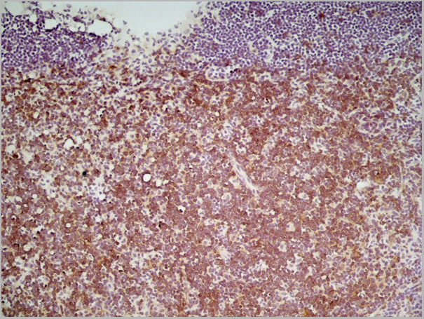
 IP (Immunoprecipitation)
(Immunoprecipitation(IP) of CRYAB by using monoclonal anti-CRYAB antibodies (Negative control: IP without adding anti-CRYAB antibody.). For each experiment, 500ul of DDK tagged CRYAB overexpression lysates (at 1:5 dilution with HEK293T lysate), 2 ug of anti-CRYAB antibody and 20ul (0.1 mg) of goat anti-mouse conjugated magnetic beads were mixed and incubated overnight. After extensive wash to remove any non-specific binding, the immuno-precipitated products were analyzed with rabbit anti-DDK polyclonal antibody.)
IP (Immunoprecipitation)
(Immunoprecipitation(IP) of CRYAB by using monoclonal anti-CRYAB antibodies (Negative control: IP without adding anti-CRYAB antibody.). For each experiment, 500ul of DDK tagged CRYAB overexpression lysates (at 1:5 dilution with HEK293T lysate), 2 ug of anti-CRYAB antibody and 20ul (0.1 mg) of goat anti-mouse conjugated magnetic beads were mixed and incubated overnight. After extensive wash to remove any non-specific binding, the immuno-precipitated products were analyzed with rabbit anti-DDK polyclonal antibody.)
CRYAB / Alpha B Crystallin, Monoclonal Antibody (Cat# AAA12373)
Full Name
Mouse Monoclonal [clone 6D11] (IgG1) to Human CRYAB / Alpha B Crystallin
Gene Names
CRYAB; MFM2; CRYA2; CTPP2; HSPB5; CMD1II; CTRCT16; HEL-S-101
Reactivity
Human, Monkey, Rat
Applications
IHC - Paraffin, IF, WB, IP, FC/FACS
Purity
Protein A/G Purified
Pricing


 WB (Western Blot)
(HSPA1B monoclonal antibody Western Blot analysis of HSPA1B expression in Raw 264.7.)
WB (Western Blot)
(HSPA1B monoclonal antibody Western Blot analysis of HSPA1B expression in Raw 264.7.)
HSPA1A, Monoclonal Antibody (Cat# AAA24537)
Full Name
HSPA1A (Heat Shock 70kD Protein 1A/1B, Heat Shock 70kD Protein 1/2, HSP70.1/HSP70.2, HSP70-1/HSP70-2, HSPA1, HSPA1B) APC
Gene Names
HSPA1A; HSP72; HSPA1; HSP70I; HSP70-1; HSP70.1; HSP70-1A; HEL-S-103
Reactivity
Human, Mouse, Rat
Applications
EIA, IF, IHC, WB
Purity
Purified by Protein A Affinity Chromatography.
Pricing


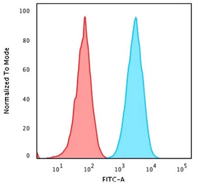
 FCM (Flow Cytometry)
(Flow cytometric analysis of Hela cells with Hsp70 antibody at 1/50 dilution (blue) compared with an unlabelled control (cells without incubation with primary antibody; red). Alexa Fluor 488-conjugated goat anti rabbit IgG was used as the secondary antibody)
FCM (Flow Cytometry)
(Flow cytometric analysis of Hela cells with Hsp70 antibody at 1/50 dilution (blue) compared with an unlabelled control (cells without incubation with primary antibody; red). Alexa Fluor 488-conjugated goat anti rabbit IgG was used as the secondary antibody)
Hsp70, Monoclonal Antibody (Cat# AAA29972)
Full Name
Hsp70 Antibody
Gene Names
HSPA1B; HSP72; HSPA1; HSX70; HSP70-1; HSP70-2; HSP70.1; HSP70.2; HSP70-1B
Reactivity
Human, Mouse, Rat
Applications
WB, ICC, IF, IHC, FC/FACS
Purity
ProA affinity purified
Pricing


 IHC (Immunohistchemistry)
(Immunohistochemical analysis of paraffin-embedded bladder cancer tissues using UBE2I mouse mAb with DAB staining.)
IHC (Immunohistchemistry)
(Immunohistochemical analysis of paraffin-embedded bladder cancer tissues using UBE2I mouse mAb with DAB staining.)
Ubc9, Monoclonal Antibody (Cat# AAA14109)
Full Name
Anti-Ubc9 Mouse mAb
Gene Names
UBE2I; P18; UBC9; C358B7.1
Reactivity
Human, Mouse, Rat
Applications
WB
Pricing


 WB (Western Blot)
(Figure 9 KD Validation of PUMA (Han et al., 2010)Immunoblot analyses of Tet-induced p53 cells treated with NOXA, Puma, Bim or non-targeting siRNAs that were utilized in this experiment. PUMA protein levels were markedly reduced in PUMA KD cells detected by anti-PUMA antibodies (3041).)
WB (Western Blot)
(Figure 9 KD Validation of PUMA (Han et al., 2010)Immunoblot analyses of Tet-induced p53 cells treated with NOXA, Puma, Bim or non-targeting siRNAs that were utilized in this experiment. PUMA protein levels were markedly reduced in PUMA KD cells detected by anti-PUMA antibodies (3041).)
PUMA, Polyclonal Antibody (Cat# AAA10943)
Full Name
PUMA Antibody
Gene Names
BBC3; JFY1; PUMA; JFY-1
Reactivity
Human, Mouse
Applications
EIA, WB, ICC, IF
Purity
PUMA Antibody is affinity chromatography purified via peptide column.
Pricing


 Application Data
(Staining of J774 cells with Rat anti Mouse F4/80 antigen Biotin)
Application Data
(Staining of J774 cells with Rat anti Mouse F4/80 antigen Biotin)
F4/80, Monoclonal Antibody (Cat# AAA12167)
Full Name
RAT ANTI MOUSE F4/80:FITC
Gene Names
Emr1; Ly71; F4/80; Gpf480; TM7LN3; DD7A5-7; EGF-TM7
Applications
FC/FACS, IF
Pricing


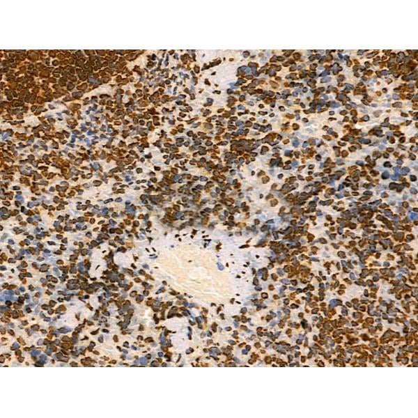
 IHC (Immunohistchemistry)
(AAA31084 at 1/200 staining human liver cancer tissue sections by IHC-P. The tissue was formaldehyde fixed and a heat mediated antigen retrieval step in citrate buffer was performed. The tissue was then blocked and incubated with the antibody for 1.5 hours at 22 degree C. An HRP conjugated goat anti-rabbit antibody was used as the secondary.)
IHC (Immunohistchemistry)
(AAA31084 at 1/200 staining human liver cancer tissue sections by IHC-P. The tissue was formaldehyde fixed and a heat mediated antigen retrieval step in citrate buffer was performed. The tissue was then blocked and incubated with the antibody for 1.5 hours at 22 degree C. An HRP conjugated goat anti-rabbit antibody was used as the secondary.)
HSP90B, Polyclonal Antibody (Cat# AAA31084)
Full Name
HSP90B Antibody
Gene Names
HSP90AB1; HSP84; HSPC2; HSPCB; D6S182; HSP90B
Reactivity
Human, Mouse, Rat
Applications
WB, IHC, IF, ICC, IP, EIA
Purity
The antiserum was purified by peptide affinity chromatography using SulfoLink Coupling Resin.
Pricing


 WB (Western Blot)
(Sample: Recombinant PRF1, Human;Antibody: Rabbit Anti-Human PRF1 Ab)
WB (Western Blot)
(Sample: Recombinant PRF1, Human;Antibody: Rabbit Anti-Human PRF1 Ab)
Perforin 1 (PRF1), Active Protein (Cat# AAA21142)
Full Name
Active Perforin 1 (PRF1)
Gene Names
PRF1; P1; PFP; HPLH2
Reactivity
Homo sapiens (Human)
Applications
Cell culture; Activity Assays.
Purity
>80%
Pricing


 IF (Immunofluorescence)
(Immunofluorescent analysis of 4% paraformaldehyde-fixed, 0.1% Triton X-100 permeabilized MCF-7 (human breast cancer cell line) cells labeling Pdx1 with at 1:25 dilution, followed by DyLight 488-conjugated IgG goat anti-rabbit secondary antibody at 1:200 dilution (green). Immunofluorescence image showing cytoplasm staining on MCF-7 cell line. Cytoplasmic actin is detected with DyLight 554 Phalloidin (PD18466410) at 1:100 dilution (red). The nuclear counter stain is DAPI (blue).)
IF (Immunofluorescence)
(Immunofluorescent analysis of 4% paraformaldehyde-fixed, 0.1% Triton X-100 permeabilized MCF-7 (human breast cancer cell line) cells labeling Pdx1 with at 1:25 dilution, followed by DyLight 488-conjugated IgG goat anti-rabbit secondary antibody at 1:200 dilution (green). Immunofluorescence image showing cytoplasm staining on MCF-7 cell line. Cytoplasmic actin is detected with DyLight 554 Phalloidin (PD18466410) at 1:100 dilution (red). The nuclear counter stain is DAPI (blue).)
OPN-a/b, Polyclonal Antibody (Cat# AAA26861)
Full Name
OPN-a/b, NT (SPP1, BNSP, OPN, Osteopontin, Bone sialoprotein 1, Nephropontin, Secreted phosphoprotein 1, Urinary stone protein, Uropontin) (MaxLight 490)
Gene Names
SPP1; OPN; BNSP; BSPI; ETA-1
Reactivity
Human
Applications
WB, IHC, IF
Purity
Purified by Protein A and Peptide Affinity Chromatography.
Pricing


 IF (Immunofluorescence)
(Immunofluorescent analysis of 4% paraformaldehyde-fixed, 0.1% Triton X-100 permeabilized MCF-7 (human breast cancer cell line) cells labeling Pdx1 with at 1:25 dilution, followed by DyLight 488-conjugated IgG goat anti-rabbit secondary antibody at 1:200 dilution (green). Immunofluorescence image showing cytoplasm staining on MCF-7 cell line. Cytoplasmic actin is detected with DyLight 554 Phalloidin (PD18466410) at 1:100 dilution (red). The nuclear counter stain is DAPI (blue).)
IF (Immunofluorescence)
(Immunofluorescent analysis of 4% paraformaldehyde-fixed, 0.1% Triton X-100 permeabilized MCF-7 (human breast cancer cell line) cells labeling Pdx1 with at 1:25 dilution, followed by DyLight 488-conjugated IgG goat anti-rabbit secondary antibody at 1:200 dilution (green). Immunofluorescence image showing cytoplasm staining on MCF-7 cell line. Cytoplasmic actin is detected with DyLight 554 Phalloidin (PD18466410) at 1:100 dilution (red). The nuclear counter stain is DAPI (blue).)
OPN-a/b, Polyclonal Antibody (Cat# AAA26865)
Full Name
OPN-a/b, NT (SPP1, BNSP, OPN, Osteopontin, Bone sialoprotein 1, Nephropontin, Secreted phosphoprotein 1, Urinary stone protein, Uropontin) (PE)
Gene Names
SPP1; OPN; BNSP; BSPI; ETA-1
Reactivity
Human
Applications
WB, IHC, IF
Purity
Purified by Protein A and Peptide Affinity Chromatography.
Pricing


 IHC (Immunohistochemistry)
(At 1/100 staining Mouse lung tissue by IHC-P. The sample was formaldehyde fixed and a heat mediated antigen retrieval step in citrate buffer was performed. The sample was then blocked and incubated with the primary antibody at 4 degree C overnight. An HRP conjugated anti-Rabbit antibody was used as the secondary antibody.)
IHC (Immunohistochemistry)
(At 1/100 staining Mouse lung tissue by IHC-P. The sample was formaldehyde fixed and a heat mediated antigen retrieval step in citrate buffer was performed. The sample was then blocked and incubated with the primary antibody at 4 degree C overnight. An HRP conjugated anti-Rabbit antibody was used as the secondary antibody.)
Histone H2A, Polyclonal Antibody (Cat# AAA31347)
Full Name
Acetyl-Histone H2A (Lys5) Antibody
Gene Names
HIST1H2AB; H2A/m; H2AFM
Reactivity
Human, Mouse, Rat
Applications
WB, IHC, EIA
Purity
The antiserum was purified by peptide affinity chromatography using SulfoLink Coupling Resin
Pricing


 Application Data
(Staining of canine peripheral blood lymphocytes with Rat anti Canine CD4: Alexa Fluor 488)
Application Data
(Staining of canine peripheral blood lymphocytes with Rat anti Canine CD4: Alexa Fluor 488)
CD4, Monoclonal Antibody (Cat# AAA12064)
Full Name
RAT ANTI DOG CD4:FITC
Applications
FC/FACS
Pricing


 WB (Western Blot)
(Western Blot Analysis of Human Spleen cell lysate using HLA-Pan Mouse Monoclonal Antibody (CR3/43).)
WB (Western Blot)
(Western Blot Analysis of Human Spleen cell lysate using HLA-Pan Mouse Monoclonal Antibody (CR3/43).)
HLA-DP/-DQ/-DR, Monoclonal Antibody (Cat# AAA23889)
Full Name
HLA-DP/-DQ/-DR (MHC II)
Gene Names
HLA-DPB1; DPB1; HLA-DP; HLA-DPB; HLA-DP1B
Reactivity
Human
Applications
IF, IHC
Pricing


 Application Data
(Staining of mouse spleen with Rat anti Mouse CD4:RPE)
Application Data
(Staining of mouse spleen with Rat anti Mouse CD4:RPE)
CD4, Monoclonal Antibody (Cat# AAA12228)
Full Name
RAT ANTI MOUSE CD4:FITC
Gene Names
Cd4; L3T4; Ly-4
Applications
FC/FACS
Pricing


 IF (Immunofluorescence)
(Immunofluorescent analysis of 4% paraformaldehyde-fixed, 0.1% Triton X-100 permeabilized MCF-7 (human breast cancer cell line) cells labeling Pdx1 with at 1:25 dilution, followed by DyLight 488-conjugated IgG goat anti-rabbit secondary antibody at 1:200 dilution (green). Immunofluorescence image showing cytoplasm staining on MCF-7 cell line. Cytoplasmic actin is detected with DyLight 554 Phalloidin (PD18466410) at 1:100 dilution (red). The nuclear counter stain is DAPI (blue).)
IF (Immunofluorescence)
(Immunofluorescent analysis of 4% paraformaldehyde-fixed, 0.1% Triton X-100 permeabilized MCF-7 (human breast cancer cell line) cells labeling Pdx1 with at 1:25 dilution, followed by DyLight 488-conjugated IgG goat anti-rabbit secondary antibody at 1:200 dilution (green). Immunofluorescence image showing cytoplasm staining on MCF-7 cell line. Cytoplasmic actin is detected with DyLight 554 Phalloidin (PD18466410) at 1:100 dilution (red). The nuclear counter stain is DAPI (blue).)
OPN-a/b, Polyclonal Antibody (Cat# AAA26858)
Full Name
OPN-a/b, NT (SPP1, BNSP, OPN, Osteopontin, Bone sialoprotein 1, Nephropontin, Secreted phosphoprotein 1, Urinary stone protein, Uropontin) (Biotin)
Gene Names
SPP1; OPN; BNSP; BSPI; ETA-1
Reactivity
Human
Applications
WB, IHC, IF, EIA
Purity
Purified by Protein A and Peptide Affinity Chromatography.
Pricing


 FCM (Flow Cytometry)
(Figure 8. Flow Cytometry analysis of A431 cells using anti-EGFR antibody (AAA19214).Overlay histogram showing A431 cells stained with AAA19214 (Blue line). The cells were blocked with 10% normal goat serum. And then incubated with rabbit anti-EGFR Antibody (AAA19214,1μg/1x106 cells) for 30 min at 20 degree C. DyLight®488 conjugated goat anti-rabbit IgG (5-10μg/1x106 cells) was used as secondary antibody for 30 minutes at 20 degree C. Isotype control antibody (Green line) was rabbit IgG (1μg/1x106) used under the same conditions. Unlabelled sample (Red line) was also used as a control.)
FCM (Flow Cytometry)
(Figure 8. Flow Cytometry analysis of A431 cells using anti-EGFR antibody (AAA19214).Overlay histogram showing A431 cells stained with AAA19214 (Blue line). The cells were blocked with 10% normal goat serum. And then incubated with rabbit anti-EGFR Antibody (AAA19214,1μg/1x106 cells) for 30 min at 20 degree C. DyLight®488 conjugated goat anti-rabbit IgG (5-10μg/1x106 cells) was used as secondary antibody for 30 minutes at 20 degree C. Isotype control antibody (Green line) was rabbit IgG (1μg/1x106) used under the same conditions. Unlabelled sample (Red line) was also used as a control.)
EGFR, Polyclonal Antibody (Cat# AAA19214)
Full Name
Anti-EGFR Antibody
Gene Names
EGFR; ERBB; HER1; mENA; ERBB1; PIG61
Reactivity
Human, Mouse, Rat
Applications
WB, IHC-P, ICC, IF, FC/FACS/FCM, EIA
Purity
Immunogen affinity purified.
Pricing


 Application Data
(Staining of canine peripheral blood lymphocytes with Rat anti Canine CD8:RPE)
Application Data
(Staining of canine peripheral blood lymphocytes with Rat anti Canine CD8:RPE)
CD8, Monoclonal Antibody (Cat# AAA12066)
Full Name
RAT ANTI DOG CD8:FITC
Applications
FC/FACS
Pricing


 Application Data
(Staining of J774 cells with Rat anti Mouse F4/80 antigen Biotin)
Application Data
(Staining of J774 cells with Rat anti Mouse F4/80 antigen Biotin)
F4/80, Monoclonal Antibody (Cat# AAA12162)
Full Name
RAT ANTI MOUSE F4/80:Biotin
Gene Names
Emr1; Ly71; F4/80; Gpf480; TM7LN3; DD7A5-7; EGF-TM7
Applications
FC/FACS
Pricing


 IF (Immunofluorescence)
(Immunofluorescent analysis of 4% paraformaldehyde-fixed, 0.1% Triton X-100 permeabilized MCF-7 (human breast cancer cell line) cells labeling Pdx1 with at 1:25 dilution, followed by DyLight 488-conjugated IgG goat anti-rabbit secondary antibody at 1:200 dilution (green). Immunofluorescence image showing cytoplasm staining on MCF-7 cell line. Cytoplasmic actin is detected with DyLight 554 Phalloidin (PD18466410) at 1:100 dilution (red). The nuclear counter stain is DAPI (blue).)
IF (Immunofluorescence)
(Immunofluorescent analysis of 4% paraformaldehyde-fixed, 0.1% Triton X-100 permeabilized MCF-7 (human breast cancer cell line) cells labeling Pdx1 with at 1:25 dilution, followed by DyLight 488-conjugated IgG goat anti-rabbit secondary antibody at 1:200 dilution (green). Immunofluorescence image showing cytoplasm staining on MCF-7 cell line. Cytoplasmic actin is detected with DyLight 554 Phalloidin (PD18466410) at 1:100 dilution (red). The nuclear counter stain is DAPI (blue).)
OPN-a/b, Polyclonal Antibody (Cat# AAA26857)
Full Name
OPN-a/b, NT (SPP1, BNSP, OPN, Osteopontin, Bone sialoprotein 1, Nephropontin, Secreted phosphoprotein 1, Urinary stone protein, Uropontin) (APC)
Gene Names
SPP1; OPN; BNSP; BSPI; ETA-1
Reactivity
Human
Applications
WB, IHC, IF
Purity
Purified by Protein A and Peptide Affinity Chromatography.
Pricing


 IF (Immunofluorescence)
(Immunofluorescent analysis of 4% paraformaldehyde-fixed, 0.1% Triton X-100 permeabilized MCF-7 (human breast cancer cell line) cells labeling Pdx1 with at 1:25 dilution, followed by DyLight 488-conjugated IgG goat anti-rabbit secondary antibody at 1:200 dilution (green). Immunofluorescence image showing cytoplasm staining on MCF-7 cell line. Cytoplasmic actin is detected with DyLight 554 Phalloidin (PD18466410) at 1:100 dilution (red). The nuclear counter stain is DAPI (blue).)
IF (Immunofluorescence)
(Immunofluorescent analysis of 4% paraformaldehyde-fixed, 0.1% Triton X-100 permeabilized MCF-7 (human breast cancer cell line) cells labeling Pdx1 with at 1:25 dilution, followed by DyLight 488-conjugated IgG goat anti-rabbit secondary antibody at 1:200 dilution (green). Immunofluorescence image showing cytoplasm staining on MCF-7 cell line. Cytoplasmic actin is detected with DyLight 554 Phalloidin (PD18466410) at 1:100 dilution (red). The nuclear counter stain is DAPI (blue).)
OPN-a/b, Polyclonal Antibody (Cat# AAA26860)
Full Name
OPN-a/b, NT (SPP1, BNSP, OPN, Osteopontin, Bone sialoprotein 1, Nephropontin, Secreted phosphoprotein 1, Urinary stone protein, Uropontin) (MaxLight 405)
Gene Names
SPP1; OPN; BNSP; BSPI; ETA-1
Reactivity
Human
Applications
WB, IHC, IF
Purity
Purified by Protein A and Peptide Affinity Chromatography.
Pricing


 IF (Immunofluorescence)
(Immunofluorescent analysis of 4% paraformaldehyde-fixed, 0.1% Triton X-100 permeabilized MCF-7 (human breast cancer cell line) cells labeling Pdx1 with at 1:25 dilution, followed by DyLight 488-conjugated IgG goat anti-rabbit secondary antibody at 1:200 dilution (green). Immunofluorescence image showing cytoplasm staining on MCF-7 cell line. Cytoplasmic actin is detected with DyLight 554 Phalloidin (PD18466410) at 1:100 dilution (red). The nuclear counter stain is DAPI (blue).)
IF (Immunofluorescence)
(Immunofluorescent analysis of 4% paraformaldehyde-fixed, 0.1% Triton X-100 permeabilized MCF-7 (human breast cancer cell line) cells labeling Pdx1 with at 1:25 dilution, followed by DyLight 488-conjugated IgG goat anti-rabbit secondary antibody at 1:200 dilution (green). Immunofluorescence image showing cytoplasm staining on MCF-7 cell line. Cytoplasmic actin is detected with DyLight 554 Phalloidin (PD18466410) at 1:100 dilution (red). The nuclear counter stain is DAPI (blue).)
OPN-a/b, Polyclonal Antibody (Cat# AAA26859)
Full Name
OPN-a/b, NT (SPP1, BNSP, OPN, Osteopontin, Bone sialoprotein 1, Nephropontin, Secreted phosphoprotein 1, Urinary stone protein, Uropontin) (FITC)
Gene Names
SPP1; OPN; BNSP; BSPI; ETA-1
Reactivity
Human
Applications
WB, IHC, IF
Purity
Purified by Protein A and Peptide Affinity Chromatography.
Pricing


 IF (Immunofluorescence)
(Immunofluorescent analysis of 4% paraformaldehyde-fixed, 0.1% Triton X-100 permeabilized MCF-7 (human breast cancer cell line) cells labeling Pdx1 with at 1:25 dilution, followed by DyLight 488-conjugated IgG goat anti-rabbit secondary antibody at 1:200 dilution (green). Immunofluorescence image showing cytoplasm staining on MCF-7 cell line. Cytoplasmic actin is detected with DyLight 554 Phalloidin (PD18466410) at 1:100 dilution (red). The nuclear counter stain is DAPI (blue).)
IF (Immunofluorescence)
(Immunofluorescent analysis of 4% paraformaldehyde-fixed, 0.1% Triton X-100 permeabilized MCF-7 (human breast cancer cell line) cells labeling Pdx1 with at 1:25 dilution, followed by DyLight 488-conjugated IgG goat anti-rabbit secondary antibody at 1:200 dilution (green). Immunofluorescence image showing cytoplasm staining on MCF-7 cell line. Cytoplasmic actin is detected with DyLight 554 Phalloidin (PD18466410) at 1:100 dilution (red). The nuclear counter stain is DAPI (blue).)
OPN-a/b, Polyclonal Antibody (Cat# AAA26864)
Full Name
OPN-a/b, NT (SPP1, BNSP, OPN, Osteopontin, Bone sialoprotein 1, Nephropontin, Secreted phosphoprotein 1, Urinary stone protein, Uropontin) (MaxLight 750)
Gene Names
SPP1; OPN; BNSP; BSPI; ETA-1
Reactivity
Human
Applications
WB, IHC, IF
Purity
Purified by Protein A and Peptide Affinity Chromatography.
Pricing


 IF (Immunofluorescence)
(Immunofluorescent analysis of 4% paraformaldehyde-fixed, 0.1% Triton X-100 permeabilized MCF-7 (human breast cancer cell line) cells labeling Pdx1 with at 1:25 dilution, followed by DyLight 488-conjugated IgG goat anti-rabbit secondary antibody at 1:200 dilution (green). Immunofluorescence image showing cytoplasm staining on MCF-7 cell line. Cytoplasmic actin is detected with DyLight 554 Phalloidin (PD18466410) at 1:100 dilution (red). The nuclear counter stain is DAPI (blue).)
IF (Immunofluorescence)
(Immunofluorescent analysis of 4% paraformaldehyde-fixed, 0.1% Triton X-100 permeabilized MCF-7 (human breast cancer cell line) cells labeling Pdx1 with at 1:25 dilution, followed by DyLight 488-conjugated IgG goat anti-rabbit secondary antibody at 1:200 dilution (green). Immunofluorescence image showing cytoplasm staining on MCF-7 cell line. Cytoplasmic actin is detected with DyLight 554 Phalloidin (PD18466410) at 1:100 dilution (red). The nuclear counter stain is DAPI (blue).)
OPN-a/b, Polyclonal Antibody (Cat# AAA26863)
Full Name
OPN-a/b, NT (SPP1, BNSP, OPN, Osteopontin, Bone sialoprotein 1, Nephropontin, Secreted phosphoprotein 1, Urinary stone protein, Uropontin) (MaxLight 650)
Gene Names
SPP1; OPN; BNSP; BSPI; ETA-1
Reactivity
Human
Applications
WB, IHC, IF
Purity
Purified by Protein A and Peptide Affinity Chromatography.
Pricing


 IF (Immunofluorescence)
(Immunofluorescent analysis of 4% paraformaldehyde-fixed, 0.1% Triton X-100 permeabilized MCF-7 (human breast cancer cell line) cells labeling Pdx1 with at 1:25 dilution, followed by DyLight 488-conjugated IgG goat anti-rabbit secondary antibody at 1:200 dilution (green). Immunofluorescence image showing cytoplasm staining on MCF-7 cell line. Cytoplasmic actin is detected with DyLight 554 Phalloidin (PD18466410) at 1:100 dilution (red). The nuclear counter stain is DAPI (blue).)
IF (Immunofluorescence)
(Immunofluorescent analysis of 4% paraformaldehyde-fixed, 0.1% Triton X-100 permeabilized MCF-7 (human breast cancer cell line) cells labeling Pdx1 with at 1:25 dilution, followed by DyLight 488-conjugated IgG goat anti-rabbit secondary antibody at 1:200 dilution (green). Immunofluorescence image showing cytoplasm staining on MCF-7 cell line. Cytoplasmic actin is detected with DyLight 554 Phalloidin (PD18466410) at 1:100 dilution (red). The nuclear counter stain is DAPI (blue).)
OPN-a/b, Polyclonal Antibody (Cat# AAA26862)
Full Name
OPN-a/b, NT (SPP1, BNSP, OPN, Osteopontin, Bone sialoprotein 1, Nephropontin, Secreted phosphoprotein 1, Urinary stone protein, Uropontin) (MaxLight 550)
Gene Names
SPP1; OPN; BNSP; BSPI; ETA-1
Reactivity
Human
Applications
WB, IHC, IF
Purity
Purified by Protein A and Peptide Affinity Chromatography.
Pricing


 Application Data
(Published customer image:Phycoerythrin conjugated Mouse anti Human CD83 antibody, clone HB15e used for the evaluation of CD83 expression on monocyte derived dendritic cells by flow cytometry.Phenotypic characterization of immunogenic and tolerogenic moDC populations by flow cytometry. Monocytes were negatively selected from PBMC using magnetic beads. Immature moDC were generated with IL-4 and GM-CSF for 6 days. 15d-PGJ2 (PGJ2 DC) and dexamethasone plus 1alpha,25-dihydroxyvitamin were added to generate tolerogenic moDC, respectively (PGJ2 DC and Dex/VD3 DC). To generate immunogenic moDC, immature moDC were stimulated for 24 h with LPS, polyI:C and a cytokine cocktail containing TNF-alpha, IL-1beta, IL-6 and PGE2, respectively. The phenotypes of the cells were analyzed by flow cytometry. Live cells were gated according to FSC/SSC. One representative experiment out of three is shown.From: Sprater F, Hovden A-O, Appel S (2012)Expression of ESE-3 Isoforms in Immunogenic and Tolerogenic Human Monocyte-Derived Dendritic Cells.PLoS ONE 7(11): e49577.)
Application Data
(Published customer image:Phycoerythrin conjugated Mouse anti Human CD83 antibody, clone HB15e used for the evaluation of CD83 expression on monocyte derived dendritic cells by flow cytometry.Phenotypic characterization of immunogenic and tolerogenic moDC populations by flow cytometry. Monocytes were negatively selected from PBMC using magnetic beads. Immature moDC were generated with IL-4 and GM-CSF for 6 days. 15d-PGJ2 (PGJ2 DC) and dexamethasone plus 1alpha,25-dihydroxyvitamin were added to generate tolerogenic moDC, respectively (PGJ2 DC and Dex/VD3 DC). To generate immunogenic moDC, immature moDC were stimulated for 24 h with LPS, polyI:C and a cytokine cocktail containing TNF-alpha, IL-1beta, IL-6 and PGE2, respectively. The phenotypes of the cells were analyzed by flow cytometry. Live cells were gated according to FSC/SSC. One representative experiment out of three is shown.From: Sprater F, Hovden A-O, Appel S (2012)Expression of ESE-3 Isoforms in Immunogenic and Tolerogenic Human Monocyte-Derived Dendritic Cells.PLoS ONE 7(11): e49577.)
CD83, Monoclonal Antibody (Cat# AAA12281)
Full Name
MOUSE ANTI HUMAN CD83: FITC
Reactivity
Human
Applications
FC/FACS
Pricing

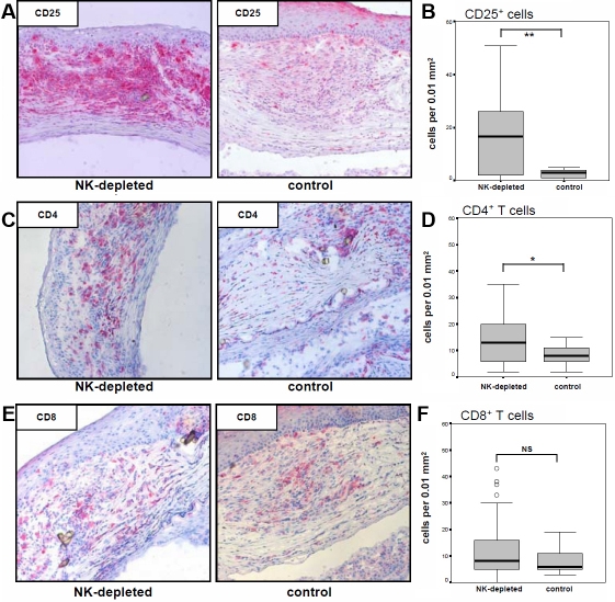


 Application Data
(Immunoperoxidase staining of rat lymph node cryosection with Mouse anti Rat CD25 followed by horseradish peroxidase conjugated Goat anti Mouse IgG1 as a detection reagent. High power)
Application Data
(Immunoperoxidase staining of rat lymph node cryosection with Mouse anti Rat CD25 followed by horseradish peroxidase conjugated Goat anti Mouse IgG1 as a detection reagent. High power)
CD25, Monoclonal Antibody (Cat# AAA12044)
Full Name
MOUSE ANTI RAT CD25:RPE
Gene Names
Il2ra; IL2RAC
Applications
FC/FACS
Pricing


 Application Data
(Staining of J774 cells with Rat anti Mouse F4/80 antigen Biotin)
Application Data
(Staining of J774 cells with Rat anti Mouse F4/80 antigen Biotin)
F4/80, Monoclonal Antibody (Cat# AAA12164)
Full Name
RAT ANTI MOUSE F4/80:Biotin
Gene Names
Emr1; Ly71; F4/80; Gpf480; TM7LN3; DD7A5-7; EGF-TM7
Applications
FC/FACS
Pricing