Filters
Clonality
Type
Reactivity
Gene Name
Isotype
Host
Application
Clone
666 results for " B Cells" - showing 450-500


 Application Data
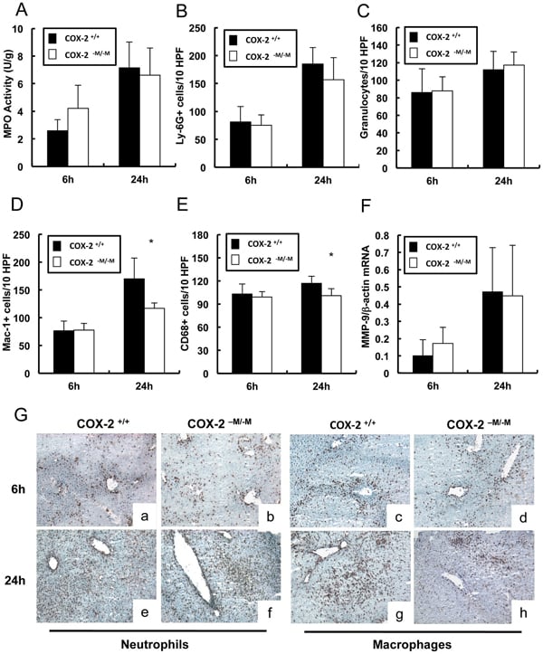
(Published customer image: Leukocyte infiltration in COX-2-M/-M and COX-2+/+ mice. MPO enzymatic activity (panel A) was statistically similar in COX-2-M/-M and COX-2+/+ livers at 6 h and 24 h post-IRI. Ly-6G+ neutrophil (panel B) and granulocyte (panel C) infiltration were also comparable in COX-2-M/-M and COX-2+/+ livers after IRI. Mac-1+ (panel D) and CD68 (panel E) infiltrating macrophages were significantly reduced in COX-2-M/-M livers at 24 h post-reperfusion, but were statistically indistinguishable in COX-2-M/-M and COX-2+/+ livers at 6 h after IRI. No statistical differences in MMP-9 expression (panel F) could be demonstrated in livers of COX-2-M/-M and COX-2+/+ mice post-IRI. Representative immunostaining (panel G) of infiltrating Ly-6G+ (a,b,e,f) and Mac-1+ (c,d,g,h) leukocytes in livers of COX-2+/+ (a,c,e,g) and COX-2-M/-M (b,d,f,h) mice at 6 h (a to d) and 24 h (e to h) post IRI; (n = 5 -6/group; * indicates p)
Application Data
(Published customer image: Leukocyte infiltration in COX-2-M/-M and COX-2+/+ mice. MPO enzymatic activity (panel A) was statistically similar in COX-2-M/-M and COX-2+/+ livers at 6 h and 24 h post-IRI. Ly-6G+ neutrophil (panel B) and granulocyte (panel C) infiltration were also comparable in COX-2-M/-M and COX-2+/+ livers after IRI. Mac-1+ (panel D) and CD68 (panel E) infiltrating macrophages were significantly reduced in COX-2-M/-M livers at 24 h post-reperfusion, but were statistically indistinguishable in COX-2-M/-M and COX-2+/+ livers at 6 h after IRI. No statistical differences in MMP-9 expression (panel F) could be demonstrated in livers of COX-2-M/-M and COX-2+/+ mice post-IRI. Representative immunostaining (panel G) of infiltrating Ly-6G+ (a,b,e,f) and Mac-1+ (c,d,g,h) leukocytes in livers of COX-2+/+ (a,c,e,g) and COX-2-M/-M (b,d,f,h) mice at 6 h (a to d) and 24 h (e to h) post IRI; (n = 5 -6/group; * indicates p)
CD68, Monoclonal Antibody (Cat# AAA12108)
Full Name
RAT ANTI MOUSE CD68:RPE
Gene Names
Cd68; Lamp4; gp110; Scard1
Applications
FC/FACS
Pricing


 Application Data
(Published Customer Image:Mouse CD31 antibody, clone ER-MP12 used for the demonstration of vasculature in mouse brain by immunofluorescence.Image caption:Inhibition of 2-AG hydrolysis reduces LPS-induced BBB permeability. a, b Fibrinogen levels in b plasma and the a ratio of brain to plasma fibrinogen were assessed by ELISA. n?=?5/7 mice per group. c, d Fluorescent immunostaining in the striatum for fibrinogen (red) and vascular marker (CD31; green) demonstrated leakage of fibrinogen into the brain with vehicle treatment, whereas vascular integrity was preserved when (e, f) MAGL was inhibited. g Extravascular fibrinogen was semi-quantitated in fluorescently labeled sections of the striatum. Bar graphs were plotted with mean?+/-SEM and data analyzed using one-way analysis of variance (ANOVA) with Tukey post-hoc comparisons. n = 5/7 mice per group. Significance is shown as *p?)
Application Data
(Published Customer Image:Mouse CD31 antibody, clone ER-MP12 used for the demonstration of vasculature in mouse brain by immunofluorescence.Image caption:Inhibition of 2-AG hydrolysis reduces LPS-induced BBB permeability. a, b Fibrinogen levels in b plasma and the a ratio of brain to plasma fibrinogen were assessed by ELISA. n?=?5/7 mice per group. c, d Fluorescent immunostaining in the striatum for fibrinogen (red) and vascular marker (CD31; green) demonstrated leakage of fibrinogen into the brain with vehicle treatment, whereas vascular integrity was preserved when (e, f) MAGL was inhibited. g Extravascular fibrinogen was semi-quantitated in fluorescently labeled sections of the striatum. Bar graphs were plotted with mean?+/-SEM and data analyzed using one-way analysis of variance (ANOVA) with Tukey post-hoc comparisons. n = 5/7 mice per group. Significance is shown as *p?)
CD31, Monoclonal Antibody (Cat# AAA12258)
Full Name
Rat Anti Mouse CD31: FITC
Gene Names
Pecam1; Cd31; Pecam; C85791; PECAM-1
Reactivity
Mouse
Applications
FC/FACS
Purity
Purified IgG prepared by affinity chromatography on Protein G from tissue culture supernatant
Pricing
super leptin antagonist, Active Protein (Cat# AAA13623)
Full Name
Mouse super leptin antagonist (mutant D23/L39A/D40A/F41A)
Purity
The purity of mouse super-active leptin antagonist is greater than 98.0% as determined by:
(a) Gel filtration analysis.
(b) Analysis by reducing and non-reducing SDS-PAGE gel.
(a) Gel filtration analysis.
(b) Analysis by reducing and non-reducing SDS-PAGE gel.


 WB (Western Blot)
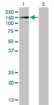
(Western blot analysis of LIN28B Antibody (N-term) in HL60 cell line lysates (35 ug/lane). LIN28B (arrow) was detected using the purified Pab.)
WB (Western Blot)
(Western blot analysis of LIN28B Antibody (N-term) in HL60 cell line lysates (35 ug/lane). LIN28B (arrow) was detected using the purified Pab.)
LIN28B, Polyclonal Antibody (Cat# AAA12320)
Full Name
Rabbit Polyclonal to Human LIN28B
Gene Names
LIN28B; CSDD2
Reactivity
Human
Applications
IHC - Paraffin, IF, WB, FC/FACS
Purity
Ammonium sulfate precipitation
Pricing


 FCM (Flow Cytometry)
(Flow cytometric analysis of A549 cells with UQCRFS1 antibody at 1/100 dilution (yellow) compared with an unlabelled control (cells without incubation with primary antibody; purple).Alexa Fluor 488-conjugated goat anti-rabbit IgG was used as the secondary antibody.)
FCM (Flow Cytometry)
(Flow cytometric analysis of A549 cells with UQCRFS1 antibody at 1/100 dilution (yellow) compared with an unlabelled control (cells without incubation with primary antibody; purple).Alexa Fluor 488-conjugated goat anti-rabbit IgG was used as the secondary antibody.)
UQCRFS1, Monoclonal Antibody (Cat# AAA30490)
Full Name
UQCRFS1 Antibody
Gene Names
UQCRFS1; RIP1; RIS1; RISP; UQCR5
Reactivity
Human, Mouse, Rat
Applications
WB, ICC, IF, IHC, FC/FACS, IP
Purity
ProA affinity purified
Pricing


 WB (Western Blot)
(BUB1B monoclonal antibody Western Blot analysis of BUB1B expression in Hela NE.)
WB (Western Blot)
(BUB1B monoclonal antibody Western Blot analysis of BUB1B expression in Hela NE.)
BUBR1, Monoclonal Antibody (Cat# AAA25625)
Full Name
BUBR1 (Mitotic Checkpoint Serine/Threonine-protein Kinase BUB1 beta, MAD3/BUB1-related Protein Kinase, hBUBR1, Mitotic Checkpoint Kinase MAD3L, Protein SSK1, BUB1B, MAD3L, SSK1) (PE)
Gene Names
BUB1B; MVA1; SSK1; BUBR1; Bub1A; MAD3L; hBUBR1; BUB1beta
Reactivity
Human
Applications
EIA, IHC, WB
Purity
Purified by Protein A Affinity Chromatography.
Pricing


 FCM (Flow Cytometry)
(Figure-6: Epitope binding study by flow cytometric analysis. MCF-7 cells expressing HER2 antigen were treated with Herceptin and Samceptin (2 ug/10^6 Cells). Surface staining was done using FITC conjugated antibodies.)
FCM (Flow Cytometry)
(Figure-6: Epitope binding study by flow cytometric analysis. MCF-7 cells expressing HER2 antigen were treated with Herceptin and Samceptin (2 ug/10^6 Cells). Surface staining was done using FITC conjugated antibodies.)
ErbB2/HER2, Monoclonal Recombinant Antibody (Cat# AAA14887)
Full Name
Recombinant anti-human ErbB2/HER2 Antibody (Trastuzumab) FITC Conjugated
Gene Names
ERBB2; NEU; NGL; HER2; TKR1; CD340; HER-2; MLN 19; HER-2/neu
Reactivity
Human
Applications
FA, FC/FACS, ADCC
Purity
Purity: >98.0% as determined by SEC-HPLC & SDS-PAGE
Pricing


 FCM (Flow Cytometry)
(Flow cytometric analysis of PC-3M cells with Bub3 antibody at 1/100 dilution (purple) compared with an unlabelled control (cells without incubation with primary antibody; yellow). Alexa Fluor 488-conjugated goat anti-rabbit IgG was used as the secondary antibody.)
FCM (Flow Cytometry)
(Flow cytometric analysis of PC-3M cells with Bub3 antibody at 1/100 dilution (purple) compared with an unlabelled control (cells without incubation with primary antibody; yellow). Alexa Fluor 488-conjugated goat anti-rabbit IgG was used as the secondary antibody.)
Bub3, Monoclonal Antibody (Cat# AAA30510)
Full Name
Bub3 Antibody
Gene Names
BUB3; BUB3L; hBUB3
Reactivity
Human, Mouse, Rat
Applications
WB, ICC, IF, IP, IHC, FC/FACS
Purity
ProA affinity purified
Pricing


 WB (Western Blot)
(BUB1B monoclonal antibody Western Blot analysis of BUB1B expression in Hela NE.)
WB (Western Blot)
(BUB1B monoclonal antibody Western Blot analysis of BUB1B expression in Hela NE.)
BUBR1, Monoclonal Antibody (Cat# AAA24149)
Full Name
BUBR1 (Mitotic Checkpoint Serine/Threonine-protein Kinase BUB1 beta, MAD3/BUB1-related Protein Kinase, hBUBR1, Mitotic Checkpoint Kinase MAD3L, Protein SSK1, BUB1B, MAD3L, SSK1) (AP)
Gene Names
BUB1B; MVA1; SSK1; BUBR1; Bub1A; MAD3L; hBUBR1; BUB1beta
Reactivity
Human
Applications
EIA, IHC, WB
Purity
Purified by Protein A Affinity Chromatography.
Pricing


 IP (Immunoprecipitation)
(HDAC2 was immunoprecipitated using:Lane A:0.5 mg Jurkat Whole Cell LysateLane B:0.5 mg NIH-3T3 Whole Cell LysateLane C:0.5 mg Hela Whole Cell Lysate4 uL anti-HDAC2 rabbit polyclonal antibody and 15 ul of 50 % Protein G agarose.Primary antibody:Anti-HDAC2 rabbit polyclonal antibody,at 1:100 dilution Secondary antibody:Dylight 800-labeled antibody to rabbit IgG (H+L), at 1:5000 dilution Developed using the odssey technique.Performed under reducing conditions.Predicted band size: 60 kDaObserved band size: 60 kDa)
IP (Immunoprecipitation)
(HDAC2 was immunoprecipitated using:Lane A:0.5 mg Jurkat Whole Cell LysateLane B:0.5 mg NIH-3T3 Whole Cell LysateLane C:0.5 mg Hela Whole Cell Lysate4 uL anti-HDAC2 rabbit polyclonal antibody and 15 ul of 50 % Protein G agarose.Primary antibody:Anti-HDAC2 rabbit polyclonal antibody,at 1:100 dilution Secondary antibody:Dylight 800-labeled antibody to rabbit IgG (H+L), at 1:5000 dilution Developed using the odssey technique.Performed under reducing conditions.Predicted band size: 60 kDaObserved band size: 60 kDa)
HDAC2, Polyclonal Antibody (Cat# AAA27718)
Full Name
Anti-HDAC2 Antibody, Rabbit Polyclonal
Gene Names
HDAC2; HD2; RPD3; YAF1
Reactivity
Human
Applications
WB, IHC-P, ICC, IF, IP
Purity
Protein A & Antigen Affinity
Pricing


 IP (Immunoprecipitation)
(CBX4 was immunoprecipitated using: Lane A:0.5 mg K562 Whole Cell Lysate 4 uL anti-CBX4 rabbit polyclonal antibody and 60 ug of Immunomagnetic beads Protein A/G. Primary antibody: Anti-CBX4 rabbit polyclonal antibody,at 1:100 dilution Secondary antibody: Clean-Blot IP Detection Reagent (HRP) at 1:1000dilution Developed using the ECL technique. Performed under reducing conditions. Predicted band size: 44 kDa Observed band size :45 kDa)
IP (Immunoprecipitation)
(CBX4 was immunoprecipitated using: Lane A:0.5 mg K562 Whole Cell Lysate 4 uL anti-CBX4 rabbit polyclonal antibody and 60 ug of Immunomagnetic beads Protein A/G. Primary antibody: Anti-CBX4 rabbit polyclonal antibody,at 1:100 dilution Secondary antibody: Clean-Blot IP Detection Reagent (HRP) at 1:1000dilution Developed using the ECL technique. Performed under reducing conditions. Predicted band size: 44 kDa Observed band size :45 kDa)
PAX2, Polyclonal Antibody (Cat# AAA27725)
Full Name
Anti-PAX2 Antibody, Rabbit Polyclonal
Gene Names
PAX2; FSGS7; PAPRS
Reactivity
Human, Mouse
Applications
WB, IHC-P, ICC, IF, IP
Purity
Protein A & Antigen Affinity
Pricing

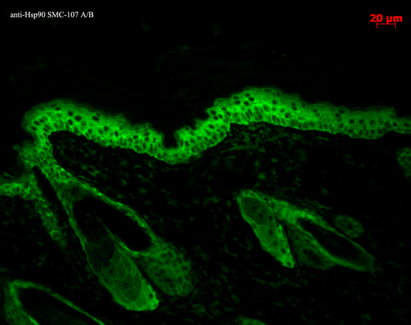


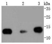
 WB (Western Blot)
(Western blot analysis of Human Lysates showing detection of Hsp90 protein using Mouse Anti-Hsp90 Monoclonal Antibody, Clone H9010. Primary Antibody: Mouse Anti-Hsp90 Monoclonal Antibody at 1:1000. Comparison of clone H9010 behavior with Hsp90 human beta (1) and Hsp90 human alpha (2). Courtesy of: David Toft, Mayo Clinic.)
WB (Western Blot)
(Western blot analysis of Human Lysates showing detection of Hsp90 protein using Mouse Anti-Hsp90 Monoclonal Antibody, Clone H9010. Primary Antibody: Mouse Anti-Hsp90 Monoclonal Antibody at 1:1000. Comparison of clone H9010 behavior with Hsp90 human beta (1) and Hsp90 human alpha (2). Courtesy of: David Toft, Mayo Clinic.)
Hsp90, Monoclonal Antibody (Cat# AAA17789)
Full Name
Hsp90 Antibody: ATTO 488
Gene Names
HSP90AB1; HSP84; HSPC2; HSPCB; D6S182; HSP90B
Reactivity
Human (beta-specific), Rabbit (Beta Specific), Chicken (Alpha/Beta), Rat, Canine
Applications
WB, IP, EIA, IHC, IF
Pricing
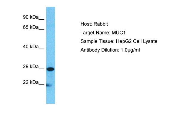


 WB (Western Blot)
(Host: RabbitTarget Name: MUC1Sample Type: HepG2Lane A: Primary AntibodyLane B: Primary Antibody + Blocking PeptidePrimary Antibody Concentration: 1ug/mlPeptide Concentration: 5ug/mlLysate Quantity: 25ug/lane/laneGel Concentration: 0.12)
WB (Western Blot)
(Host: RabbitTarget Name: MUC1Sample Type: HepG2Lane A: Primary AntibodyLane B: Primary Antibody + Blocking PeptidePrimary Antibody Concentration: 1ug/mlPeptide Concentration: 5ug/mlLysate Quantity: 25ug/lane/laneGel Concentration: 0.12)
MUC1, Polyclonal Antibody (Cat# AAA23492)
Full Name
MUC1 antibody - C-terminal region
Gene Names
MUC1; EMA; MCD; PEM; PUM; KL-6; MAM6; MCKD; PEMT; CD227; H23AG; MCKD1; MUC-1; ADMCKD; ADMCKD1; CA 15-3; MUC-1/X; MUC1/ZD; MUC-1/SEC
Reactivity
Cow, Dog, Guinea Pig, Horse, Human, Mouse, Rabbit, Rat, Pig
Applications
IHC, WB
Purity
Affinity Purified
Pricing


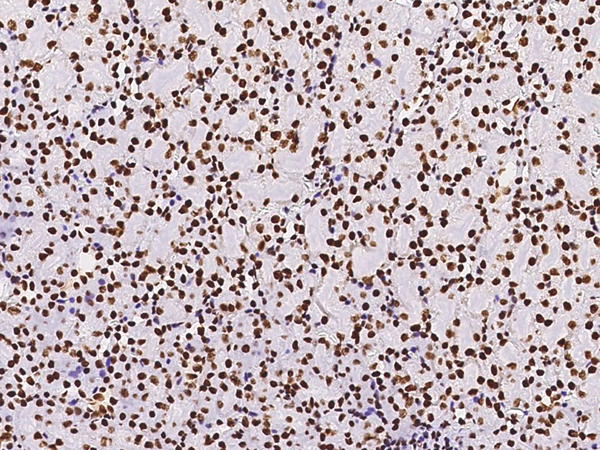
 IF (Immunofluorescence)
(Immunofluorescence analysis of U-2 OS cells using Phospho-Histone H3-S10/T11 Rabbit pAb at dilution of 100 (40x lens). Blue: DAPI for nuclear staining.)
IF (Immunofluorescence)
(Immunofluorescence analysis of U-2 OS cells using Phospho-Histone H3-S10/T11 Rabbit pAb at dilution of 100 (40x lens). Blue: DAPI for nuclear staining.)
Phospho-Histone H3-S10/T11, Polyclonal Antibody (Cat# AAA28391)
Full Name
Phospho-Histone H3-S10/T11 Rabbit pAb
Gene Names
HIST1H3A; H3/A; H3FA
Reactivity
Human, Mouse, Rat, Other (Wide Range)
Applications
WB, IHC, IF
Purity
Affinity purification
Pricing


 WB (Western Blot)
(BUB1B monoclonal antibody Western Blot analysis of BUB1B expression in Hela NE.)
WB (Western Blot)
(BUB1B monoclonal antibody Western Blot analysis of BUB1B expression in Hela NE.)
BUBR1, Monoclonal Antibody (Cat# AAA25332)
Full Name
BUBR1 (Mitotic Checkpoint Serine/Threonine-protein Kinase BUB1 beta, MAD3/BUB1-related Protein Kinase, hBUBR1, Mitotic Checkpoint Kinase MAD3L, Protein SSK1, BUB1B, MAD3L, SSK1) (HRP)
Gene Names
BUB1B; MVA1; SSK1; BUBR1; Bub1A; MAD3L; hBUBR1; BUB1beta
Reactivity
Human
Applications
EIA, IHC, WB
Purity
Purified by Protein A Affinity Chromatography.
Pricing


 FCM (Flow Cytometry)
(Flow cytometric analysis of Hela cells with Histone H2B antibody at 1/50 dilution (red) compared with an unlabelled control (cells without incubation with primary antibody; black). Alexa Fluor 488-conjugated goat anti rabbit IgG was used as the secondary antibody.)
FCM (Flow Cytometry)
(Flow cytometric analysis of Hela cells with Histone H2B antibody at 1/50 dilution (red) compared with an unlabelled control (cells without incubation with primary antibody; black). Alexa Fluor 488-conjugated goat anti rabbit IgG was used as the secondary antibody.)
Histone H2B, Monoclonal Antibody (Cat# AAA30192)
Full Name
Histone H2B Antibody
Gene Names
HIST1H2BK; H2BK; H2B/S; H2BFT; H2BFAiii
Reactivity
Human, Mouse, Rat
Applications
WB, ICC, IF, IHC, IP, FC/FACS
Purity
ProA affinity purified
Pricing


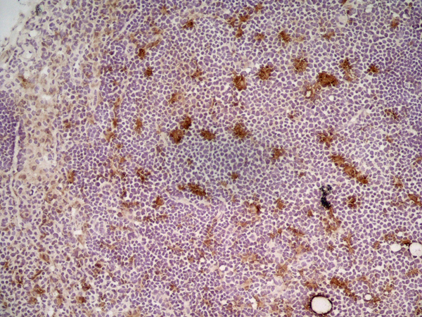
 Application Data
(Immunoperoxidase staining of rat lymph node cryosection with Mouse anti Rat CD11 antibody, clone ED8 followed by horseradish peroxidase conjugated Goat anti Mouse IgG1 as a detection reagent. High power)
Application Data
(Immunoperoxidase staining of rat lymph node cryosection with Mouse anti Rat CD11 antibody, clone ED8 followed by horseradish peroxidase conjugated Goat anti Mouse IgG1 as a detection reagent. High power)
CD11b, Monoclonal Antibody (Cat# AAA12238)
Full Name
MOUSE ANTI RAT CD11b
Gene Names
ITGAM; CD11B
Applications
Immunohistology Frozen*
Pricing


 Application Data
(Staining of human peripheral blood granulocytes with Mouse anti Human CD11b:Biotin)
Application Data
(Staining of human peripheral blood granulocytes with Mouse anti Human CD11b:Biotin)
CD11b, Monoclonal Antibody (Cat# AAA11992)
Full Name
MOUSE ANTI HUMAN CD11b
Gene Names
ITGAM; CR3A; MO1A; CD11B; MAC-1; MAC1A; SLEB6
Applications
FC/FACS, IP
Pricing


 DB (Dot Blot)
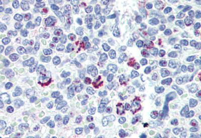
(Formalin-fixed and paraffin-embedded human cancer tissue reacted with the primary antibody, which was peroxidase-conjugated to the secondary antibody, followed by AEC staining. This data demonstrates the use of this antibody for immunohistochemistry; clinical relevance has not been evaluated. BC = breast carcinoma; HC = hepatocarcinoma.)
DB (Dot Blot)
(Formalin-fixed and paraffin-embedded human cancer tissue reacted with the primary antibody, which was peroxidase-conjugated to the secondary antibody, followed by AEC staining. This data demonstrates the use of this antibody for immunohistochemistry; clinical relevance has not been evaluated. BC = breast carcinoma; HC = hepatocarcinoma.)
Phospho-HIST1H3B3 (S10), Polyclonal Antibody (Cat# AAA28670)
Full Name
Phospho-HIST1H3B3 (S10) Antibody
Gene Names
HIST1H3A; H3/A; H3FA
Reactivity
Human
Applications
DB, EIA, IHC, WB
Purity
Peptide Affinity Purified Rabbit Polyclonal Antibody (Pab)
Pricing


 Application Data
(Published customer image: Leukocyte infiltration in COX-2-M/-M and COX-2+/+ mice. MPO enzymatic activity (panel A) was statistically similar in COX-2-M/-M and COX-2+/+ livers at 6 h and 24 h post-IRI. Ly-6G+ neutrophil (panel B) and granulocyte (panel C) infiltration were also comparable in COX-2-M/-M and COX-2+/+ livers after IRI. Mac-1+ (panel D) and CD68 (panel E) infiltrating macrophages were significantly reduced in COX-2-M/-M livers at 24 h post-reperfusion, but were statistically indistinguishable in COX-2-M/-M and COX-2+/+ livers at 6 h after IRI. No statistical differences in MMP-9 expression (panel F) could be demonstrated in livers of COX-2-M/-M and COX-2+/+ mice post-IRI. Representative immunostaining (panel G) of infiltrating Ly-6G+ (a,b,e,f) and Mac-1+ (c,d,g,h) leukocytes in livers of COX-2+/+ (a,c,e,g) and COX-2-M/-M (b,d,f,h) mice at 6 h (a to d) and 24 h (e to h) post IRI; (n = 5 -6/group; * indicates p)
Application Data
(Published customer image: Leukocyte infiltration in COX-2-M/-M and COX-2+/+ mice. MPO enzymatic activity (panel A) was statistically similar in COX-2-M/-M and COX-2+/+ livers at 6 h and 24 h post-IRI. Ly-6G+ neutrophil (panel B) and granulocyte (panel C) infiltration were also comparable in COX-2-M/-M and COX-2+/+ livers after IRI. Mac-1+ (panel D) and CD68 (panel E) infiltrating macrophages were significantly reduced in COX-2-M/-M livers at 24 h post-reperfusion, but were statistically indistinguishable in COX-2-M/-M and COX-2+/+ livers at 6 h after IRI. No statistical differences in MMP-9 expression (panel F) could be demonstrated in livers of COX-2-M/-M and COX-2+/+ mice post-IRI. Representative immunostaining (panel G) of infiltrating Ly-6G+ (a,b,e,f) and Mac-1+ (c,d,g,h) leukocytes in livers of COX-2+/+ (a,c,e,g) and COX-2-M/-M (b,d,f,h) mice at 6 h (a to d) and 24 h (e to h) post IRI; (n = 5 -6/group; * indicates p)
CD68, Monoclonal Antibody (Cat# AAA12109)
Full Name
RAT ANTI MOUSE CD68:RPE
Gene Names
Cd68; Lamp4; gp110; Scard1
Applications
FC/FACS
Pricing


 Application Data
(Published customer image: Leukocyte infiltration in COX-2-M/-M and COX-2+/+ mice. MPO enzymatic activity (panel A) was statistically similar in COX-2-M/-M and COX-2+/+ livers at 6 h and 24 h post-IRI. Ly-6G+ neutrophil (panel B) and granulocyte (panel C) infiltration were also comparable in COX-2-M/-M and COX-2+/+ livers after IRI. Mac-1+ (panel D) and CD68 (panel E) infiltrating macrophages were significantly reduced in COX-2-M/-M livers at 24 h post-reperfusion, but were statistically indistinguishable in COX-2-M/-M and COX-2+/+ livers at 6 h after IRI. No statistical differences in MMP-9 expression (panel F) could be demonstrated in livers of COX-2-M/-M and COX-2+/+ mice post-IRI. Representative immunostaining (panel G) of infiltrating Ly-6G+ (a,b,e,f) and Mac-1+ (c,d,g,h) leukocytes in livers of COX-2+/+ (a,c,e,g) and COX-2-M/-M (b,d,f,h) mice at 6 h (a to d) and 24 h (e to h) post IRI; (n = 5 -6/group; * indicates p)
Application Data
(Published customer image: Leukocyte infiltration in COX-2-M/-M and COX-2+/+ mice. MPO enzymatic activity (panel A) was statistically similar in COX-2-M/-M and COX-2+/+ livers at 6 h and 24 h post-IRI. Ly-6G+ neutrophil (panel B) and granulocyte (panel C) infiltration were also comparable in COX-2-M/-M and COX-2+/+ livers after IRI. Mac-1+ (panel D) and CD68 (panel E) infiltrating macrophages were significantly reduced in COX-2-M/-M livers at 24 h post-reperfusion, but were statistically indistinguishable in COX-2-M/-M and COX-2+/+ livers at 6 h after IRI. No statistical differences in MMP-9 expression (panel F) could be demonstrated in livers of COX-2-M/-M and COX-2+/+ mice post-IRI. Representative immunostaining (panel G) of infiltrating Ly-6G+ (a,b,e,f) and Mac-1+ (c,d,g,h) leukocytes in livers of COX-2+/+ (a,c,e,g) and COX-2-M/-M (b,d,f,h) mice at 6 h (a to d) and 24 h (e to h) post IRI; (n = 5 -6/group; * indicates p)
CD68, Monoclonal Antibody (Cat# AAA12102)
Full Name
RAT ANTI MOUSE CD68
Gene Names
Cd68; Lamp4; gp110; Scard1
Applications
FC/FACS, IF, IP, WB
Pricing


 Application Data
(Published customer image: Leukocyte infiltration in COX-2-M/-M and COX-2+/+ mice. MPO enzymatic activity (panel A) was statistically similar in COX-2-M/-M and COX-2+/+ livers at 6 h and 24 h post-IRI. Ly-6G+ neutrophil (panel B) and granulocyte (panel C) infiltration were also comparable in COX-2-M/-M and COX-2+/+ livers after IRI. Mac-1+ (panel D) and CD68 (panel E) infiltrating macrophages were significantly reduced in COX-2-M/-M livers at 24 h post-reperfusion, but were statistically indistinguishable in COX-2-M/-M and COX-2+/+ livers at 6 h after IRI. No statistical differences in MMP-9 expression (panel F) could be demonstrated in livers of COX-2-M/-M and COX-2+/+ mice post-IRI. Representative immunostaining (panel G) of infiltrating Ly-6G+ (a,b,e,f) and Mac-1+ (c,d,g,h) leukocytes in livers of COX-2+/+ (a,c,e,g) and COX-2-M/-M (b,d,f,h) mice at 6 h (a to d) and 24 h (e to h) post IRI; (n = 5 -6/group; * indicates p)
Application Data
(Published customer image: Leukocyte infiltration in COX-2-M/-M and COX-2+/+ mice. MPO enzymatic activity (panel A) was statistically similar in COX-2-M/-M and COX-2+/+ livers at 6 h and 24 h post-IRI. Ly-6G+ neutrophil (panel B) and granulocyte (panel C) infiltration were also comparable in COX-2-M/-M and COX-2+/+ livers after IRI. Mac-1+ (panel D) and CD68 (panel E) infiltrating macrophages were significantly reduced in COX-2-M/-M livers at 24 h post-reperfusion, but were statistically indistinguishable in COX-2-M/-M and COX-2+/+ livers at 6 h after IRI. No statistical differences in MMP-9 expression (panel F) could be demonstrated in livers of COX-2-M/-M and COX-2+/+ mice post-IRI. Representative immunostaining (panel G) of infiltrating Ly-6G+ (a,b,e,f) and Mac-1+ (c,d,g,h) leukocytes in livers of COX-2+/+ (a,c,e,g) and COX-2-M/-M (b,d,f,h) mice at 6 h (a to d) and 24 h (e to h) post IRI; (n = 5 -6/group; * indicates p)
CD68, Monoclonal Antibody (Cat# AAA12106)
Full Name
RAT ANTI MOUSE CD68:FITC
Gene Names
Cd68; Lamp4; gp110; Scard1
Applications
FC/FACS
Pricing


 Application Data
(Published customer image: Leukocyte infiltration in COX-2-M/-M and COX-2+/+ mice. MPO enzymatic activity (panel A) was statistically similar in COX-2-M/-M and COX-2+/+ livers at 6 h and 24 h post-IRI. Ly-6G+ neutrophil (panel B) and granulocyte (panel C) infiltration were also comparable in COX-2-M/-M and COX-2+/+ livers after IRI. Mac-1+ (panel D) and CD68 (panel E) infiltrating macrophages were significantly reduced in COX-2-M/-M livers at 24 h post-reperfusion, but were statistically indistinguishable in COX-2-M/-M and COX-2+/+ livers at 6 h after IRI. No statistical differences in MMP-9 expression (panel F) could be demonstrated in livers of COX-2-M/-M and COX-2+/+ mice post-IRI. Representative immunostaining (panel G) of infiltrating Ly-6G+ (a,b,e,f) and Mac-1+ (c,d,g,h) leukocytes in livers of COX-2+/+ (a,c,e,g) and COX-2-M/-M (b,d,f,h) mice at 6 h (a to d) and 24 h (e to h) post IRI; (n = 5 -6/group; * indicates p)
Application Data
(Published customer image: Leukocyte infiltration in COX-2-M/-M and COX-2+/+ mice. MPO enzymatic activity (panel A) was statistically similar in COX-2-M/-M and COX-2+/+ livers at 6 h and 24 h post-IRI. Ly-6G+ neutrophil (panel B) and granulocyte (panel C) infiltration were also comparable in COX-2-M/-M and COX-2+/+ livers after IRI. Mac-1+ (panel D) and CD68 (panel E) infiltrating macrophages were significantly reduced in COX-2-M/-M livers at 24 h post-reperfusion, but were statistically indistinguishable in COX-2-M/-M and COX-2+/+ livers at 6 h after IRI. No statistical differences in MMP-9 expression (panel F) could be demonstrated in livers of COX-2-M/-M and COX-2+/+ mice post-IRI. Representative immunostaining (panel G) of infiltrating Ly-6G+ (a,b,e,f) and Mac-1+ (c,d,g,h) leukocytes in livers of COX-2+/+ (a,c,e,g) and COX-2-M/-M (b,d,f,h) mice at 6 h (a to d) and 24 h (e to h) post IRI; (n = 5 -6/group; * indicates p)
CD68, Monoclonal Antibody (Cat# AAA12103)
Full Name
RAT ANTI MOUSE CD68:Biotin
Gene Names
Cd68; Lamp4; gp110; Scard1
Applications
FC/FACS
Pricing


 Application Data
(Published customer image: Leukocyte infiltration in COX-2-M/-M and COX-2+/+ mice. MPO enzymatic activity (panel A) was statistically similar in COX-2-M/-M and COX-2+/+ livers at 6 h and 24 h post-IRI. Ly-6G+ neutrophil (panel B) and granulocyte (panel C) infiltration were also comparable in COX-2-M/-M and COX-2+/+ livers after IRI. Mac-1+ (panel D) and CD68 (panel E) infiltrating macrophages were significantly reduced in COX-2-M/-M livers at 24 h post-reperfusion, but were statistically indistinguishable in COX-2-M/-M and COX-2+/+ livers at 6 h after IRI. No statistical differences in MMP-9 expression (panel F) could be demonstrated in livers of COX-2-M/-M and COX-2+/+ mice post-IRI. Representative immunostaining (panel G) of infiltrating Ly-6G+ (a,b,e,f) and Mac-1+ (c,d,g,h) leukocytes in livers of COX-2+/+ (a,c,e,g) and COX-2-M/-M (b,d,f,h) mice at 6 h (a to d) and 24 h (e to h) post IRI; (n = 5 -6/group; * indicates p)
Application Data
(Published customer image: Leukocyte infiltration in COX-2-M/-M and COX-2+/+ mice. MPO enzymatic activity (panel A) was statistically similar in COX-2-M/-M and COX-2+/+ livers at 6 h and 24 h post-IRI. Ly-6G+ neutrophil (panel B) and granulocyte (panel C) infiltration were also comparable in COX-2-M/-M and COX-2+/+ livers after IRI. Mac-1+ (panel D) and CD68 (panel E) infiltrating macrophages were significantly reduced in COX-2-M/-M livers at 24 h post-reperfusion, but were statistically indistinguishable in COX-2-M/-M and COX-2+/+ livers at 6 h after IRI. No statistical differences in MMP-9 expression (panel F) could be demonstrated in livers of COX-2-M/-M and COX-2+/+ mice post-IRI. Representative immunostaining (panel G) of infiltrating Ly-6G+ (a,b,e,f) and Mac-1+ (c,d,g,h) leukocytes in livers of COX-2+/+ (a,c,e,g) and COX-2-M/-M (b,d,f,h) mice at 6 h (a to d) and 24 h (e to h) post IRI; (n = 5 -6/group; * indicates p)
CD68, Monoclonal Antibody (Cat# AAA12107)
Full Name
RAT ANTI MOUSE CD68
Gene Names
Cd68; Lamp4; gp110; Scard1
Applications
FC/FACS, IF, IP, WB
Purity
Purified
Purified IgG - liquid
Purified IgG - liquid
Pricing


 IP (Immunoprecipitation)
(Figure 13 Immunoprecipitation Validation in HEK293 cells (Kawai et al., 2004)HEK293 cells were transiently transfected with DYKDDDDK-IRF7. Ccell lysates were immunoprecipitated with control rabbit anti-mouse immunoglobulin serum (IgG) or anti-MyD88 (Ab1 and Ab2), followed by immunoblotting with anti-DYKDDDDK.)
IP (Immunoprecipitation)
(Figure 13 Immunoprecipitation Validation in HEK293 cells (Kawai et al., 2004)HEK293 cells were transiently transfected with DYKDDDDK-IRF7. Ccell lysates were immunoprecipitated with control rabbit anti-mouse immunoglobulin serum (IgG) or anti-MyD88 (Ab1 and Ab2), followed by immunoblotting with anti-DYKDDDDK.)
MYD88, Polyclonal Antibody (Cat# AAA10930)
Full Name
MYD88 Antibody
Gene Names
MYD88; MYD88D
Reactivity
Human, Mouse
Applications
EIA, WB, IHC, IF
Purity
MYD88 Antibody is affinity chromatography purified via peptide column.
Pricing


 WB (Western Blot)
(Figure 6 WB Validation of Delta Variant Spike RBD Protein with SARS-CoV-2 Spike RBD Antibodies Loading: 20 ng of SARS-CoV-2 spike RBD proteins, including WT and Delta variant (B.1.671.2).Detection Antibodies: SARS-CoV-2 Spike RBD antibody, 1 μg/mL, incubate at RT for 1 hr.Secondary Antibodies: Goat anti-rabbit HRP at 1:20,000, incubate at RT for 1 hr.)
WB (Western Blot)
(Figure 6 WB Validation of Delta Variant Spike RBD Protein with SARS-CoV-2 Spike RBD Antibodies Loading: 20 ng of SARS-CoV-2 spike RBD proteins, including WT and Delta variant (B.1.671.2).Detection Antibodies: SARS-CoV-2 Spike RBD antibody, 1 μg/mL, incubate at RT for 1 hr.Secondary Antibodies: Goat anti-rabbit HRP at 1:20,000, incubate at RT for 1 hr.)
COVID 19 Delta Variant Spike RBD (His-Avi Tag) Coronavirus, Recombinant Protein (Cat# AAA11039)
Full Name
SARS-CoV-2 (COVID-19) Delta Variant Spike RBD (His-Avi Tag) Recombinant Protein
Applications
EIA, WB
Purity
> 95% as determined by Bis-Tris PAGE
Pricing


 WB (Western Blot)
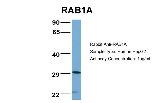
(WB Suggested Anti-RAB1A Antibody Titration: 0.2-1 ug/mlELISA Titer: 1:312500Positive Control: Human Muscle)
WB (Western Blot)
(WB Suggested Anti-RAB1A Antibody Titration: 0.2-1 ug/mlELISA Titer: 1:312500Positive Control: Human Muscle)
RAB1A, Polyclonal Antibody (Cat# AAA23570)
Full Name
RAB1A antibody - middle region
Gene Names
RAB1A; RAB1; YPT1
Reactivity
Cow, Dog, Goat, Guinea Pig, Horse, Human, Mouse, Rabbit, Rat, Zebrafish
Applications
IHC, WB
Purity
Affinity Purified
Pricing


 FCM (Flow Cytometry)
(Figure-6: Epitope binding study by flow cytometric analysis. MCF-7 cells expressing HER2 antigen were treated with Either Herceptin or Samceptin (1 & 2 ug/10^6 Cells). Surface staining was done using FITC conjugated antibodies.)
FCM (Flow Cytometry)
(Figure-6: Epitope binding study by flow cytometric analysis. MCF-7 cells expressing HER2 antigen were treated with Either Herceptin or Samceptin (1 & 2 ug/10^6 Cells). Surface staining was done using FITC conjugated antibodies.)
ErbB2/HER2, Monoclonal Recombinant Antibody (Cat# AAA14881)
Full Name
Recombinant anti- human ErbB2/HER2 Antibody (Trastuzumab)
Gene Names
ERBB2; NEU; NGL; HER2; TKR1; CD340; HER-2; MLN 19; HER-2/neu
Reactivity
Human
Applications
FC/FACS
Purity
>98.0% as determined by SEC-HPLC & SDS-PAGE.
Pricing


 Application Data
(Ref. 3: Isolation, culture and immunocytochemical characterization of HUCPC. The dissection under sterile condition of foetal and full-term cords was performed to expose the WJ, the vein and arteries (A). After the in vitro expansion of foetal HUCPC (B) the cells ..)
Application Data
(Ref. 3: Isolation, culture and immunocytochemical characterization of HUCPC. The dissection under sterile condition of foetal and full-term cords was performed to expose the WJ, the vein and arteries (A). After the in vitro expansion of foetal HUCPC (B) the cells ..)
Chick Embryo Extract, Ultrafiltrate, Reagent (Cat# AAA14749)
Full Name
Chick Embryo Extract, Ultrafiltrate (CEE)
Purity
Molecular Biology Grade
Pricing


 Application Data
(Detection limit for recombinant GST tagged BUB1B is ~0.03ng/ml as a capture antibody.)
Application Data
(Detection limit for recombinant GST tagged BUB1B is ~0.03ng/ml as a capture antibody.)
BUBR1, Monoclonal Antibody (Cat# AAA24444)
Full Name
BUBR1 (Mitotic Checkpoint Serine/Threonine-protein Kinase BUB1 beta, MAD3/BUB1-related Protein Kinase, hBUBR1, Mitotic Checkpoint Kinase MAD3L, Protein SSK1, BUB1B, MAD3L, SSK1) APC
Gene Names
BUB1B; MVA1; SSK1; BUBR1; Bub1A; MAD3L; hBUBR1; BUB1beta
Reactivity
Human
Applications
EIA, IHC, WB
Purity
Purified by Protein A Affinity Chromatography.
Pricing


 WB (Western Blot)
(Western blot analysis of Human Lysates showing detection of Hsp90 protein using Mouse Anti-Hsp90 Monoclonal Antibody, Clone H9010. Primary Antibody: Mouse Anti-Hsp90 Monoclonal Antibody at 1:1000. Comparison of clone H9010 behavior with Hsp90 human beta (1) and Hsp90 human alpha (2). Courtesy of: David Toft, Mayo Clinic.)
WB (Western Blot)
(Western blot analysis of Human Lysates showing detection of Hsp90 protein using Mouse Anti-Hsp90 Monoclonal Antibody, Clone H9010. Primary Antibody: Mouse Anti-Hsp90 Monoclonal Antibody at 1:1000. Comparison of clone H9010 behavior with Hsp90 human beta (1) and Hsp90 human alpha (2). Courtesy of: David Toft, Mayo Clinic.)
Hsp90, Monoclonal Antibody (Cat# AAA17792)
Full Name
Hsp90 Antibody: ATTO 594
Gene Names
HSP90AB1; HSP84; HSPC2; HSPCB; D6S182; HSP90B
Reactivity
Human (beta-specific), Rabbit (Beta Specific), Chicken (Alpha/Beta), Rat, Canine
Applications
WB, IP, EIA, IHC, IF
Pricing


 FCM (Flow Cytometry)
(Flow cytometric analysis of A431 cells with EGFR antibody at 1/50 dilution (red) compared with an unlabelled control (cells without incubation with primary antibody; black). Alexa Fluor 488-conjugated goat anti rabbit IgG was used as the secondary antibody.)
FCM (Flow Cytometry)
(Flow cytometric analysis of A431 cells with EGFR antibody at 1/50 dilution (red) compared with an unlabelled control (cells without incubation with primary antibody; black). Alexa Fluor 488-conjugated goat anti rabbit IgG was used as the secondary antibody.)
EGFR, Monoclonal Antibody (Cat# AAA30021)
Full Name
EGFR Antibody
Gene Names
EGFR; ERBB; HER1; mENA; ERBB1; PIG61
Reactivity
Human, Mouse, Rat
Applications
WB, ICC, IF, IHC, IP, FC/FACS
Purity
ProA affinity purified
Pricing


 FCM (Flow Cytometry)
(Flow cytometric analysis of Hela cells with Histone H4 (acetyl K16) antibody at 1/100 dilution (red) compared with an unlabelled control (cells without incubation with primary antibody; black). Alexa Fluor 488-conjugated goat anti-rabbit IgG was used as the secondary antibody.)
FCM (Flow Cytometry)
(Flow cytometric analysis of Hela cells with Histone H4 (acetyl K16) antibody at 1/100 dilution (red) compared with an unlabelled control (cells without incubation with primary antibody; black). Alexa Fluor 488-conjugated goat anti-rabbit IgG was used as the secondary antibody.)
Histone H4, Monoclonal Antibody (Cat# AAA30539)
Full Name
Histone H4 (Acetyl K16) Antibody
Gene Names
HIST2H4B; H4/o
Reactivity
Human, Mouse, Rat
Applications
WB, ICC, IF, IHC, FC/FACS
Purity
ProA affinity purified
Pricing


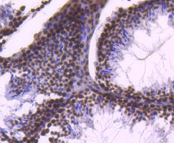
 Application Data
(Published clone specific image Alloimmunity-associated cytotoxicity is mediated through the NKG2D receptor. (A) Liver expression of nkg2d on day ten after liver transplantation. (B) Representative NKG2D expression levels in blood NK cells (left) and monocytes (right) of allogeneic (black) and syngeneic (grey) recipients. Isotype was used as control (dashed lines). (C) Sorted blood NK cell cytotoxicity inhibition with anti-NKG2D antibody or with anti-NKp30 antibody. (D) Levels of NKG2D ligand (rae1l, rrlt and irp94) expression in the liver on day ten after transplantation. (E) Levels of NKG2D ligand (rae1l, rrlt and irp94) expression in rat HCC cell lines. (F) Representative level of recombinant NKG2D-Fc binding to rat HCC cells lines. *p)
Application Data
(Published clone specific image Alloimmunity-associated cytotoxicity is mediated through the NKG2D receptor. (A) Liver expression of nkg2d on day ten after liver transplantation. (B) Representative NKG2D expression levels in blood NK cells (left) and monocytes (right) of allogeneic (black) and syngeneic (grey) recipients. Isotype was used as control (dashed lines). (C) Sorted blood NK cell cytotoxicity inhibition with anti-NKG2D antibody or with anti-NKp30 antibody. (D) Levels of NKG2D ligand (rae1l, rrlt and irp94) expression in the liver on day ten after transplantation. (E) Levels of NKG2D ligand (rae1l, rrlt and irp94) expression in rat HCC cell lines. (F) Representative level of recombinant NKG2D-Fc binding to rat HCC cells lines. *p)
CD172a, Monoclonal Antibody (Cat# AAA11852)
Full Name
MOUSE ANTI RAT CD172a:Biotin
Gene Names
Sirpa; Bit; Ptpns1; SHPS-1
Applications
FC/FACS
Pricing


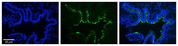
 IHC (Immunohistchemistry)
((3.75ug/ml) staining of paraffin embedded Human Liver. Steamed antigen retrieval with citrate buffer pH 6, AP-staining.)
IHC (Immunohistchemistry)
((3.75ug/ml) staining of paraffin embedded Human Liver. Steamed antigen retrieval with citrate buffer pH 6, AP-staining.)
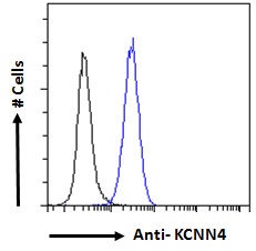
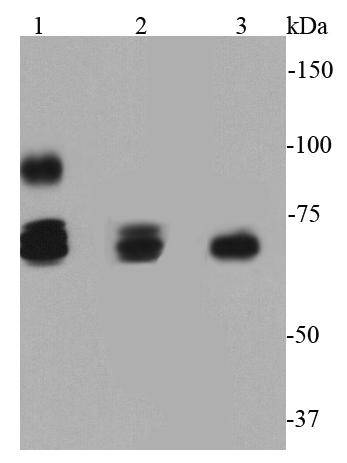
KCNN4/KCa3.1, Antibody (Cat# AAA13678)
Full Name
Goat Anti-KCNN4/KCa3.1 Antibody
Gene Names
KCNN4; IK; IK1; SK4; DHS2; KCA4; hSK4; IKCA1; hKCa4; KCa3.1; hIKCa1
Reactivity
Human, Mouse
Expected from sequence similarity: Human, Mouse, Rat, Dog
Expected from sequence similarity: Human, Mouse, Rat, Dog
Applications
EIA, IF, FC/FACS, IHC
Purity
Purified from goat serum by ammonium sulphate precipitation followed by antigen affinity chromatography using the immunizing peptide.
Pricing


 WB (Western Blot)
(Anti-HA Tag mouse monoclonal antibody at 1:1000 dilutionLane A: HA-GST (Recombinant protein)(30ng)Lane B: GST-HA (Recombinant protein)(10ng)Lane C: HA-ARG1-myc transfected 293 cell lysate (2ug)Lane D: HA-mFABP4-myc transfected 293 cell lysate (2ug)Lane E: myc-mFABP4-HA transfected 293 cell lysate (0.5ug)Lane F: myc-ARG1-HA transfected 293 cell lysate (2ug)SecondaryGoat Anti-Mouse IgG H&L (Dylight800) at 1/15000 dilution.Developed using the Odyssey technique. Performed under reducing conditions.)
WB (Western Blot)
(Anti-HA Tag mouse monoclonal antibody at 1:1000 dilutionLane A: HA-GST (Recombinant protein)(30ng)Lane B: GST-HA (Recombinant protein)(10ng)Lane C: HA-ARG1-myc transfected 293 cell lysate (2ug)Lane D: HA-mFABP4-myc transfected 293 cell lysate (2ug)Lane E: myc-mFABP4-HA transfected 293 cell lysate (0.5ug)Lane F: myc-ARG1-HA transfected 293 cell lysate (2ug)SecondaryGoat Anti-Mouse IgG H&L (Dylight800) at 1/15000 dilution.Developed using the Odyssey technique. Performed under reducing conditions.)
HA, Monoclonal Antibody (Cat# AAA27712)
Full Name
Anti-HA tag Antibody, Mouse Monoclonal
Reactivity
HA tag
Applications
WB, EIA, FC/FACS/FCM, ICC, IF, IP
Purity
Protein A
Pricing


 WB (Western Blot)
(Western blot analysis of Human Lysates showing detection of Hsp90 protein using Mouse Anti-Hsp90 Monoclonal Antibody, Clone H9010. Primary Antibody: Mouse Anti-Hsp90 Monoclonal Antibody at 1:1000. Comparison of clone H9010 behavior with Hsp90 human beta (1) and Hsp90 human alpha (2). Courtesy of: David Toft, Mayo Clinic.)
WB (Western Blot)
(Western blot analysis of Human Lysates showing detection of Hsp90 protein using Mouse Anti-Hsp90 Monoclonal Antibody, Clone H9010. Primary Antibody: Mouse Anti-Hsp90 Monoclonal Antibody at 1:1000. Comparison of clone H9010 behavior with Hsp90 human beta (1) and Hsp90 human alpha (2). Courtesy of: David Toft, Mayo Clinic.)
Hsp90, Monoclonal Antibody (Cat# AAA17781)
Full Name
Hsp90 Antibody: ATTO 390
Gene Names
HSP90AB1; HSP84; HSPC2; HSPCB; D6S182; HSP90B
Reactivity
Human (beta-specific), Rabbit (Beta Specific), Chicken (Alpha/Beta), Rat, Canine
Applications
WB, IP, EIA, IHC, IF
Pricing


 WB (Western Blot)
(BUB1B monoclonal antibody Western Blot analysis of BUB1B expression in Hela NE.)
WB (Western Blot)
(BUB1B monoclonal antibody Western Blot analysis of BUB1B expression in Hela NE.)
BUBR1, Monoclonal Antibody (Cat# AAA25036)
Full Name
BUBR1 (Mitotic Checkpoint Serine/Threonine-protein Kinase BUB1 beta, MAD3/BUB1-related Protein Kinase, hBUBR1, Mitotic Checkpoint Kinase MAD3L, Protein SSK1, BUB1B, MAD3L, SSK1) (FITC)
Gene Names
BUB1B; MVA1; SSK1; BUBR1; Bub1A; MAD3L; hBUBR1; BUB1beta
Reactivity
Human
Applications
EIA, IHC, WB
Purity
Purified by Protein A Affinity Chromatography.
Pricing


 WB (Western Blot)
(WB Suggested Anti-LDHB AntibodyTitration: 1 ug/mlPositive Control: 721_B Whole CellLDHB is strongly supported by BioGPS gene expression data to be expressed in Human 721_B cells)
WB (Western Blot)
(WB Suggested Anti-LDHB AntibodyTitration: 1 ug/mlPositive Control: 721_B Whole CellLDHB is strongly supported by BioGPS gene expression data to be expressed in Human 721_B cells)
LDHB, Polyclonal Antibody (Cat# AAA23530)
Full Name
LDHB Antibody - C-terminal region
Gene Names
LDHB; LDH-B; LDH-H; LDHBD; TRG-5; HEL-S-281
Reactivity
Cow, Dog, Guinea Pig, Horse, Human, Mouse, Rabbit, Rat, Zebrafish
Applications
IHC, WB
Purity
Affinity Purified
Pricing


 Application Data
(Published customer image: Leukocyte infiltration in COX-2-M/-M and COX-2+/+ mice. MPO enzymatic activity (panel A) was statistically similar in COX-2-M/-M and COX-2+/+ livers at 6 h and 24 h post-IRI. Ly-6G+ neutrophil (panel B) and granulocyte (panel C) infiltration were also comparable in COX-2-M/-M and COX-2+/+ livers after IRI. Mac-1+ (panel D) and CD68 (panel E) infiltrating macrophages were significantly reduced in COX-2-M/-M livers at 24 h post-reperfusion, but were statistically indistinguishable in COX-2-M/-M and COX-2+/+ livers at 6 h after IRI. No statistical differences in MMP-9 expression (panel F) could be demonstrated in livers of COX-2-M/-M and COX-2+/+ mice post-IRI. Representative immunostaining (panel G) of infiltrating Ly-6G+ (a,b,e,f) and Mac-1+ (c,d,g,h) leukocytes in livers of COX-2+/+ (a,c,e,g) and COX-2-M/-M (b,d,f,h) mice at 6 h (a to d) and 24 h (e to h) post IRI; (n = 5 -6/group; * indicates p)
Application Data
(Published customer image: Leukocyte infiltration in COX-2-M/-M and COX-2+/+ mice. MPO enzymatic activity (panel A) was statistically similar in COX-2-M/-M and COX-2+/+ livers at 6 h and 24 h post-IRI. Ly-6G+ neutrophil (panel B) and granulocyte (panel C) infiltration were also comparable in COX-2-M/-M and COX-2+/+ livers after IRI. Mac-1+ (panel D) and CD68 (panel E) infiltrating macrophages were significantly reduced in COX-2-M/-M livers at 24 h post-reperfusion, but were statistically indistinguishable in COX-2-M/-M and COX-2+/+ livers at 6 h after IRI. No statistical differences in MMP-9 expression (panel F) could be demonstrated in livers of COX-2-M/-M and COX-2+/+ mice post-IRI. Representative immunostaining (panel G) of infiltrating Ly-6G+ (a,b,e,f) and Mac-1+ (c,d,g,h) leukocytes in livers of COX-2+/+ (a,c,e,g) and COX-2-M/-M (b,d,f,h) mice at 6 h (a to d) and 24 h (e to h) post IRI; (n = 5 -6/group; * indicates p)
CD68, Monoclonal Antibody (Cat# AAA12105)
Full Name
RAT ANTI MOUSE CD68:FITC
Gene Names
Cd68; Lamp4; gp110; Scard1
Applications
FC/FACS
Pricing


 WB (Western Blot)
(Western blot analysis of extracts from various samples, using Phospho-ICAM-1 (Tyr512) Antibody. Lane 1: VERO cells, treated with blocking peptideLane 2: VERO cellsLane 3: A2780 cells.)
WB (Western Blot)
(Western blot analysis of extracts from various samples, using Phospho-ICAM-1 (Tyr512) Antibody. Lane 1: VERO cells, treated with blocking peptideLane 2: VERO cellsLane 3: A2780 cells.)
ICAM-1, Polyclonal Antibody (Cat# AAA30979)
Full Name
Phospho-ICAM-1 (Tyr512) Antibody
Gene Names
ICAM1; BB2; CD54; P3.58
Reactivity
Human, Mouse, Monkey
Applications
WB, IHC, IF, ICC, EIA
Purity
Peptide affinity purification
Pricing


 WB (Western Blot)
(Anti-Beta-Tubulin mouse monoclonal antibody at 1:2000 dilution Lane A: HepG2 Whole Cell Lysate Lane B: Daudi Whole Cell Lysate Lane C: MOLT-4 Whole Cell Lysate Lane D: A549 Whole Cell Lysate Lane E: 293T Whole Cell Lysate Lane F: HelaS3 Whole Cell Lysate Lysates/proteins at 30 ug per lane. Secondary Goat Anti-Mouse IgG H&L (Dylight800) at 1/15000 dilution. Developed using the Odyssey technique. Performed under reducing conditions. Predicted band size:50 kDa Observed band size:54 kDa)
WB (Western Blot)
(Anti-Beta-Tubulin mouse monoclonal antibody at 1:2000 dilution Lane A: HepG2 Whole Cell Lysate Lane B: Daudi Whole Cell Lysate Lane C: MOLT-4 Whole Cell Lysate Lane D: A549 Whole Cell Lysate Lane E: 293T Whole Cell Lysate Lane F: HelaS3 Whole Cell Lysate Lysates/proteins at 30 ug per lane. Secondary Goat Anti-Mouse IgG H&L (Dylight800) at 1/15000 dilution. Developed using the Odyssey technique. Performed under reducing conditions. Predicted band size:50 kDa Observed band size:54 kDa)
Beta-Tubulin, Monoclonal Antibody (Cat# AAA27713)
Full Name
Beta-Tubulin Loading Control Antibody, Mouse MAb
Reactivity
Human
Applications
WB, IHC-P, ICC, IF, IP
Purity
Protein A
Pricing


 WB (Western Blot)
(Anti-GP73 rabbit polyclonal antibody at 1:500 dilution Lane A: GOLM1 konckout Hela Whole Cell Lysate Lane B: Hela Whole Cell Lysate Lysates/proteins at 10 ug per lane. Secondary Goat Anti-Rabbit IgG (H+L)/HRP at 1/10000 dilution. Developed using the ECL technique. Performed under reducing conditions. Predicted band size:45 kDa Observed band size: kDa)
WB (Western Blot)
(Anti-GP73 rabbit polyclonal antibody at 1:500 dilution Lane A: GOLM1 konckout Hela Whole Cell Lysate Lane B: Hela Whole Cell Lysate Lysates/proteins at 10 ug per lane. Secondary Goat Anti-Rabbit IgG (H+L)/HRP at 1/10000 dilution. Developed using the ECL technique. Performed under reducing conditions. Predicted band size:45 kDa Observed band size: kDa)
GOLPH2/GOLM1, Polyclonal Antibody (Cat# AAA27751)
Full Name
Anti-GOLPH2/GOLM1 Antibody, Rabbit Polyclonal
Gene Names
GOLM1; GP73; HEL46; GOLPH2; C9orf155; PSEC0257; bA379P1.3
Reactivity
Human
Applications
WB, EIA, IHC-P, ICC, IF, IP
Purity
Protein A & Antigen Affinity
Pricing


 FCM (Flow Cytometry)
(Figure 8. Flow Cytometry analysis of C6 cells using anti-non-muscle Myosin IIB/MYH10 antibody (AAA19262).Overlay histogram showing C6 cells stained with AAA19262 (Blue line). The cells were blocked with 10% normal goat serum. And then incubated with rabbit anti-non-muscle Myosin IIB/MYH10 Antibody (AAA19262,1μg/1x106 cells) for 30 min at 20 degree C. DyLight®488 conjugated goat anti-rabbit IgG (5-10μg/1x106 cells) was used as secondary antibody for 30 minutes at 20 degree C. Isotype control antibody (Green line) was rabbit IgG (1μg/1x106) used under the same conditions. Unlabelled sample (Red line) was also used as a control.)
FCM (Flow Cytometry)
(Figure 8. Flow Cytometry analysis of C6 cells using anti-non-muscle Myosin IIB/MYH10 antibody (AAA19262).Overlay histogram showing C6 cells stained with AAA19262 (Blue line). The cells were blocked with 10% normal goat serum. And then incubated with rabbit anti-non-muscle Myosin IIB/MYH10 Antibody (AAA19262,1μg/1x106 cells) for 30 min at 20 degree C. DyLight®488 conjugated goat anti-rabbit IgG (5-10μg/1x106 cells) was used as secondary antibody for 30 minutes at 20 degree C. Isotype control antibody (Green line) was rabbit IgG (1μg/1x106) used under the same conditions. Unlabelled sample (Red line) was also used as a control.)
non-muscle Myosin IIB/MYH10, Polyclonal Antibody (Cat# AAA19262)
Full Name
Anti-non-muscle Myosin IIB/MYH10 Antibody
Gene Names
MYH10; NMMHCB; NMMHC-IIB
Reactivity
Human, Mouse, Rat
Applications
WB, IHC-P, ICC, IF, FC/FACS/FCM
Purity
Immunogen affinity purified.
Pricing


 IF (Immunofluorescence)
(Figure 11. IF analysis of Tubulin alpha using anti- Tubulin alpha antibody (AAA19381).Tubulin alpha was detected in immunocytochemical section of CACO-2 cells. Enzyme antigen retrieval was performed using IHC enzyme antigen retrieval reagent for 15 mins. The cells were blocked with 10% goat serum. And then incubated with 5μg/mL mouse anti- Tubulin alpha Antibody (AAA19381) overnight at 4 degree C. DyLight®594 Conjugated Goat Anti-Mouse IgG (BA1141) was used as secondary antibody at 1:100 dilution and incubated for 30 minutes at 37 degree C. The section was counterstained with DAPI. Visualize using a fluorescence microscope and filter sets appropriate for the label used.)
IF (Immunofluorescence)
(Figure 11. IF analysis of Tubulin alpha using anti- Tubulin alpha antibody (AAA19381).Tubulin alpha was detected in immunocytochemical section of CACO-2 cells. Enzyme antigen retrieval was performed using IHC enzyme antigen retrieval reagent for 15 mins. The cells were blocked with 10% goat serum. And then incubated with 5μg/mL mouse anti- Tubulin alpha Antibody (AAA19381) overnight at 4 degree C. DyLight®594 Conjugated Goat Anti-Mouse IgG (BA1141) was used as secondary antibody at 1:100 dilution and incubated for 30 minutes at 37 degree C. The section was counterstained with DAPI. Visualize using a fluorescence microscope and filter sets appropriate for the label used.)
Tubulin alpha, Monoclonal Antibody (Cat# AAA19381)
Full Name
Anti-Tubulin alpha Antibody (monoclonal, 7B12)
Gene Names
TUBA1A; LIS3; TUBA3; B-ALPHA-1
Reactivity
Human, Mouse, Rat
Applications
WB, IHC-P, ICC, IF, FC/FACS/FCM
Purity
Immunogen affinity purified.
Pricing


 FCM (Flow Cytometry)
(Overlay histogram showing Raji cells stained with AAA27027 (red line) at 1:100. The cells were incubated in 1x PBS /10% normal goat serum to block non-specific protein-protein interactions followed by primary antibody for 1 h at 4 degree C. The secondary antibody used was FITC goat anti-mouse IgG(H+L) at 1/200 dilution for 1 h at 4 degree C. Isotype control antibody (green line) was used under the same conditions. Acquisition of >10,000 events was performed.)
FCM (Flow Cytometry)
(Overlay histogram showing Raji cells stained with AAA27027 (red line) at 1:100. The cells were incubated in 1x PBS /10% normal goat serum to block non-specific protein-protein interactions followed by primary antibody for 1 h at 4 degree C. The secondary antibody used was FITC goat anti-mouse IgG(H+L) at 1/200 dilution for 1 h at 4 degree C. Isotype control antibody (green line) was used under the same conditions. Acquisition of >10,000 events was performed.)
CD19, Monoclonal Antibody (Cat# AAA27027)
Full Name
CD19 Monoclonal Antibody
Gene Names
CD19; B4; CVID3
Reactivity
Human
Applications
EIA, WB, IHC, IF, FC/FACS
Purity
>95%, Protein A Purified
Pricing


 Application Data
(Published customer image: Leukocyte infiltration in COX-2-M/-M and COX-2+/+ mice. MPO enzymatic activity (panel A) was statistically similar in COX-2-M/-M and COX-2+/+ livers at 6 h and 24 h post-IRI. Ly-6G+ neutrophil (panel B) and granulocyte (panel C) infiltration were also comparable in COX-2-M/-M and COX-2+/+ livers after IRI. Mac-1+ (panel D) and CD68 (panel E) infiltrating macrophages were significantly reduced in COX-2-M/-M livers at 24 h post-reperfusion, but were statistically indistinguishable in COX-2-M/-M and COX-2+/+ livers at 6 h after IRI. No statistical differences in MMP-9 expression (panel F) could be demonstrated in livers of COX-2-M/-M and COX-2+/+ mice post-IRI. Representative immunostaining (panel G) of infiltrating Ly-6G+ (a,b,e,f) and Mac-1+ (c,d,g,h) leukocytes in livers of COX-2+/+ (a,c,e,g) and COX-2-M/-M (b,d,f,h) mice at 6 h (a to d) and 24 h (e to h) post IRI; (n = 5 -6/group; * indicates p)
Application Data
(Published customer image: Leukocyte infiltration in COX-2-M/-M and COX-2+/+ mice. MPO enzymatic activity (panel A) was statistically similar in COX-2-M/-M and COX-2+/+ livers at 6 h and 24 h post-IRI. Ly-6G+ neutrophil (panel B) and granulocyte (panel C) infiltration were also comparable in COX-2-M/-M and COX-2+/+ livers after IRI. Mac-1+ (panel D) and CD68 (panel E) infiltrating macrophages were significantly reduced in COX-2-M/-M livers at 24 h post-reperfusion, but were statistically indistinguishable in COX-2-M/-M and COX-2+/+ livers at 6 h after IRI. No statistical differences in MMP-9 expression (panel F) could be demonstrated in livers of COX-2-M/-M and COX-2+/+ mice post-IRI. Representative immunostaining (panel G) of infiltrating Ly-6G+ (a,b,e,f) and Mac-1+ (c,d,g,h) leukocytes in livers of COX-2+/+ (a,c,e,g) and COX-2-M/-M (b,d,f,h) mice at 6 h (a to d) and 24 h (e to h) post IRI; (n = 5 -6/group; * indicates p)
CD68, Monoclonal Antibody (Cat# AAA12110)
Full Name
RAT ANTI MOUSE CD68
Gene Names
Cd68; Lamp4; gp110; Scard1
Applications
FC, IA, IF, IP, WB
Pricing


 FCM (Flow Cytometry)
(Figure 8. Flow Cytometry analysis of A549 cells using anti-Caveolin-2/CAV2 antibody (AAA19243).Overlay histogram showing A549 cells stained with AAA19243 (Blue line). The cells were blocked with 10% normal goat serum. And then incubated with rabbit anti-Caveolin-2/CAV2 Antibody (AAA19243,1μg/1x106 cells) for 30 min at 20 degree C. DyLight®488 conjugated goat anti-rabbit IgG (5-10μg/1x106 cells) was used as secondary antibody for 30 minutes at 20 degree C. Isotype control antibody (Green line) was rabbit IgG (1μg/1x106) used under the same conditions. Unlabelled sample (Red line) was also used as a control.)
FCM (Flow Cytometry)
(Figure 8. Flow Cytometry analysis of A549 cells using anti-Caveolin-2/CAV2 antibody (AAA19243).Overlay histogram showing A549 cells stained with AAA19243 (Blue line). The cells were blocked with 10% normal goat serum. And then incubated with rabbit anti-Caveolin-2/CAV2 Antibody (AAA19243,1μg/1x106 cells) for 30 min at 20 degree C. DyLight®488 conjugated goat anti-rabbit IgG (5-10μg/1x106 cells) was used as secondary antibody for 30 minutes at 20 degree C. Isotype control antibody (Green line) was rabbit IgG (1μg/1x106) used under the same conditions. Unlabelled sample (Red line) was also used as a control.)
Caveolin-2/CAV2, Polyclonal Antibody (Cat# AAA19243)
Full Name
Anti-Caveolin-2/CAV2 Antibody
Gene Names
CAV2; CAV
Reactivity
Human, Mouse, Rat, Monkey
Applications
WB, IHC-P, ICC, IF, FC/FACS/FCM, EIA
Purity
Immunogen affinity purified.
Pricing


 ELISA
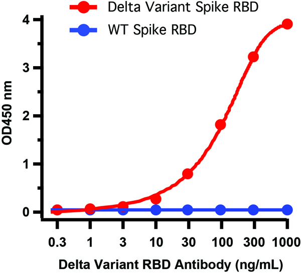
(Figure 6 ELISA Validation of Delta Variant Spike S1 Protein with SARS-CoV-2 Spike S1 Antibodies Coating Antigen: SARS-CoV-2 spike S1 proteins, including WT and Delta variant (B.1.671.2), 5 μg/mL, incubated at 4 ˚C overnight.Detection Antibodies: SARS-CoV-2 Spike S1 antibody, dilution: 0.3-1000 ng/mL, incubate at RT for 1 hr.Secondary Antibodies: Goat anti-rabbit HRP at 1:20,000, incubate at RT for 1 hr.Antibody 9083 does not distinguish between WT and Delta.)
ELISA
(Figure 6 ELISA Validation of Delta Variant Spike S1 Protein with SARS-CoV-2 Spike S1 Antibodies Coating Antigen: SARS-CoV-2 spike S1 proteins, including WT and Delta variant (B.1.671.2), 5 μg/mL, incubated at 4 ˚C overnight.Detection Antibodies: SARS-CoV-2 Spike S1 antibody, dilution: 0.3-1000 ng/mL, incubate at RT for 1 hr.Secondary Antibodies: Goat anti-rabbit HRP at 1:20,000, incubate at RT for 1 hr.Antibody 9083 does not distinguish between WT and Delta.)
COVID 19 Delta Variant Spike S1 (His-Avi Tag) Coronavirus, Recombinant Protein (Cat# AAA11040)
Full Name
SARS-CoV-2 (COVID-19) Delta Variant Spike S1 (His-Avi Tag) Recombinant Protein
Applications
EIA, WB
Purity
> 90% as determined by Bis-Tris PAGE
Pricing


 ICC (Immunocytochemistry)
(ICC staining HSP70 in SKBR-3 cells (green). Cells were fixed in paraformaldehyde, permeabilised with 0.25% Triton X100/PBS.)
ICC (Immunocytochemistry)
(ICC staining HSP70 in SKBR-3 cells (green). Cells were fixed in paraformaldehyde, permeabilised with 0.25% Triton X100/PBS.)
HSP70, Polyclonal Antibody (Cat# AAA29967)
Full Name
HSP70 Antibody
Gene Names
HSPA1A; HSP72; HSPA1; HSP70I; HSP70-1; HSP70-1A
Reactivity
Human, Mouse, Rat
Applications
WB, ICC, IHC
Purity
Peptide affinity purified
Pricing