Filters
Clonality
Type
Reactivity
Gene Name
Isotype
Host
Application
Clone
666 results for " B Cells" - showing 550-600


 Application Data
(Analysis of Protein Array containing more than 19, 000 full-length human proteins using PU.1 Mouse Monoclonal Antibody (PU1/2446). Z- and S- Score: The Z-score represents the strength of a signal that a monoclonal antibody (MAb) (in combination with a fluorescently-tagged anti-IgG secondary antibody) produces when binding to a particular protein on the HuProtTM array. Z-scores are described in units of standard deviations (SD's) above the mean value of all signals generated on that array. If targets on HuProtTM are arranged in descending order of the Z-score, the S-score is the difference (also in units of SD's) between the Z-score. S-score therefore represents the relative target specificity of a MAb to its intended target. A MAb is considered to specific to its intended target, if the MAb has an S-score of at least 2.5. For example, if a MAb binds to protein X with a Z-score of 43 and to protein Y with a Z-score of 14, then the S-score for the binding of that MAb to protein X is equal to 29.)
Application Data
(Analysis of Protein Array containing more than 19, 000 full-length human proteins using PU.1 Mouse Monoclonal Antibody (PU1/2446). Z- and S- Score: The Z-score represents the strength of a signal that a monoclonal antibody (MAb) (in combination with a fluorescently-tagged anti-IgG secondary antibody) produces when binding to a particular protein on the HuProtTM array. Z-scores are described in units of standard deviations (SD's) above the mean value of all signals generated on that array. If targets on HuProtTM are arranged in descending order of the Z-score, the S-score is the difference (also in units of SD's) between the Z-score. S-score therefore represents the relative target specificity of a MAb to its intended target. A MAb is considered to specific to its intended target, if the MAb has an S-score of at least 2.5. For example, if a MAb binds to protein X with a Z-score of 43 and to protein Y with a Z-score of 14, then the S-score for the binding of that MAb to protein X is equal to 29.)
PU.1 (SPI-1), Monoclonal Antibody (Cat# AAA23903)
Full Name
PU.1 (SPI-1) (B-Cell Marker)
Gene Names
SPI1; OF; PU.1; SFPI1; SPI-1; SPI-A
Reactivity
Human. Others not known.
Applications
WB, IHC
Pricing


 Application Data
(Analysis of Protein Array containing more than 19,000 full-length human proteins using HER-2 Mouse Monoclonal Antibody (ERBB2/3080). Z- and S- Score: The Z-score represents the strength of a signal that a monoclonal antibody (MAb) (in combination with a fluorescently-tagged anti-IgG secondary antibody) produces when binding to a particular protein on the HuProtTM array. Z-scores are described in units of standard deviations (SD's) above the mean value of all signals generated on that array. If targets on HuProtTM are arranged in descending order of the Z-score, the S-score is the difference (also in units of SD's) between the Z-score. S-score therefore represents the relative target specificity of a MAb to its intended target. A MAb is considered to specific to its intended target, if the MAb has an S-score of at least 2.5. For example, if a MAb binds to protein X with a Z-score of 43 and to protein Y with a Z-score of 14, then the S-score for the binding of that MAb to protein X is equal to 29.)
Application Data
(Analysis of Protein Array containing more than 19,000 full-length human proteins using HER-2 Mouse Monoclonal Antibody (ERBB2/3080). Z- and S- Score: The Z-score represents the strength of a signal that a monoclonal antibody (MAb) (in combination with a fluorescently-tagged anti-IgG secondary antibody) produces when binding to a particular protein on the HuProtTM array. Z-scores are described in units of standard deviations (SD's) above the mean value of all signals generated on that array. If targets on HuProtTM are arranged in descending order of the Z-score, the S-score is the difference (also in units of SD's) between the Z-score. S-score therefore represents the relative target specificity of a MAb to its intended target. A MAb is considered to specific to its intended target, if the MAb has an S-score of at least 2.5. For example, if a MAb binds to protein X with a Z-score of 43 and to protein Y with a Z-score of 14, then the S-score for the binding of that MAb to protein X is equal to 29.)
HER-2/c-erbB-2/neu/CD340, Monoclonal Antibody (Cat# AAA23912)
Full Name
HER-2/c-erbB-2/neu/CD340
Gene Names
ERBB2; NEU; NGL; HER2; TKR1; CD340; HER-2; MLN 19; HER-2/neu
Reactivity
Human
Applications
IHC
Purity
Purified Ab with BSA and Azide at 200ug/ml OR Purified Ab WITHOUT BSA and Azide at 1.0mg/ml
Pricing


 Application Data
(Published clone specific image: Flow cytometric analysis of AM from NO2-exposed and control rats. Rats were exposed to NO2 for the indicated times and BAL cells were stained with antibodies to ED7, ED9, RM-4, and OX-6. To overcome autofluorescence signals, primary antibodies were detected using a biotin-PE/streptavidin-anti-streptavidin enhancing system and labeling of AM was analyzed by flow cytometry following gating by help of forward and sideward scatter properties. Shown are representative results of at least six animals per group.From: Garn H, Siese A, Stumpf S, Wensing A, Renz H, Gemsa D. Phenotypical and functional characterization of alveolar macrophage subpopulations in the lungs of NO2-exposed rats. Respir Res. 2006 Jan 6;7:4.)
Application Data
(Published clone specific image: Flow cytometric analysis of AM from NO2-exposed and control rats. Rats were exposed to NO2 for the indicated times and BAL cells were stained with antibodies to ED7, ED9, RM-4, and OX-6. To overcome autofluorescence signals, primary antibodies were detected using a biotin-PE/streptavidin-anti-streptavidin enhancing system and labeling of AM was analyzed by flow cytometry following gating by help of forward and sideward scatter properties. Shown are representative results of at least six animals per group.From: Garn H, Siese A, Stumpf S, Wensing A, Renz H, Gemsa D. Phenotypical and functional characterization of alveolar macrophage subpopulations in the lungs of NO2-exposed rats. Respir Res. 2006 Jan 6;7:4.)
CD172a, Monoclonal Antibody (Cat# AAA11888)
Full Name
MOUSE ANTI RAT CD172a:FITC
Gene Names
Sirpa; Bit; Ptpns1; SHPS-1
Applications
FC/FACS
Pricing


 Application Data
(Staining of J774 cells with Rat anti Mouse F4/80 antigen Biotin)
Application Data
(Staining of J774 cells with Rat anti Mouse F4/80 antigen Biotin)
F4/80, Monoclonal Antibody (Cat# AAA12168)
Full Name
RAT ANTI MOUSE F4/80:FITC
Gene Names
Emr1; Ly71; F4/80; Gpf480; TM7LN3; DD7A5-7; EGF-TM7
Applications
FC/FACS, IF
Pricing


 Application Data
(Staining of J774 cells with Rat anti Mouse F4/80 antigen Biotin)
Application Data
(Staining of J774 cells with Rat anti Mouse F4/80 antigen Biotin)
F4/80, Monoclonal Antibody (Cat# AAA12164)
Full Name
RAT ANTI MOUSE F4/80:Biotin
Gene Names
Emr1; Ly71; F4/80; Gpf480; TM7LN3; DD7A5-7; EGF-TM7
Applications
FC/FACS
Pricing


 Application Data
(Staining of J774 cells with Rat anti Mouse F4/80 antigen Biotin)
Application Data
(Staining of J774 cells with Rat anti Mouse F4/80 antigen Biotin)
F4/80, Monoclonal Antibody (Cat# AAA12166)
Full Name
RAT ANTI MOUSE F4/80:FITC
Gene Names
Emr1; Ly71; F4/80; Gpf480; TM7LN3; DD7A5-7; EGF-TM7
Applications
FC/FACS, IF
Pricing


 Application Data
(Published customer image:Phycoerythrin conjugated Mouse anti Human CD83 antibody, clone HB15e used for the evaluation of CD83 expression on monocyte derived dendritic cells by flow cytometry.Phenotypic characterization of immunogenic and tolerogenic moDC populations by flow cytometry. Monocytes were negatively selected from PBMC using magnetic beads. Immature moDC were generated with IL-4 and GM-CSF for 6 days. 15d-PGJ2 (PGJ2 DC) and dexamethasone plus 1alpha,25-dihydroxyvitamin were added to generate tolerogenic moDC, respectively (PGJ2 DC and Dex/VD3 DC). To generate immunogenic moDC, immature moDC were stimulated for 24 h with LPS, polyI:C and a cytokine cocktail containing TNF-alpha, IL-1beta, IL-6 and PGE2, respectively. The phenotypes of the cells were analyzed by flow cytometry. Live cells were gated according to FSC/SSC. One representative experiment out of three is shown.From: Sprater F, Hovden A-O, Appel S (2012)Expression of ESE-3 Isoforms in Immunogenic and Tolerogenic Human Monocyte-Derived Dendritic Cells.PLoS ONE 7(11): e49577.)
Application Data
(Published customer image:Phycoerythrin conjugated Mouse anti Human CD83 antibody, clone HB15e used for the evaluation of CD83 expression on monocyte derived dendritic cells by flow cytometry.Phenotypic characterization of immunogenic and tolerogenic moDC populations by flow cytometry. Monocytes were negatively selected from PBMC using magnetic beads. Immature moDC were generated with IL-4 and GM-CSF for 6 days. 15d-PGJ2 (PGJ2 DC) and dexamethasone plus 1alpha,25-dihydroxyvitamin were added to generate tolerogenic moDC, respectively (PGJ2 DC and Dex/VD3 DC). To generate immunogenic moDC, immature moDC were stimulated for 24 h with LPS, polyI:C and a cytokine cocktail containing TNF-alpha, IL-1beta, IL-6 and PGE2, respectively. The phenotypes of the cells were analyzed by flow cytometry. Live cells were gated according to FSC/SSC. One representative experiment out of three is shown.From: Sprater F, Hovden A-O, Appel S (2012)Expression of ESE-3 Isoforms in Immunogenic and Tolerogenic Human Monocyte-Derived Dendritic Cells.PLoS ONE 7(11): e49577.)
CD83, Monoclonal Antibody (Cat# AAA12282)
Full Name
MOUSE ANTI HUMAN CD83
Reactivity
Human
Applications
FC/FACS, IHC, IHC, IP
Pricing


 Application Data
(Published clone specific image: Flow cytometric analysis of AM from NO2-exposed and control rats. Rats were exposed to NO2 for the indicated times and BAL cells were stained with antibodies to ED7, ED9, RM-4, and OX-6. To overcome autofluorescence signals, primary antibodies were detected using a biotin-PE/streptavidin-anti-streptavidin enhancing system and labeling of AM was analyzed by flow cytometry following gating by help of forward and sideward scatter properties. Shown are representative results of at least six animals per group.From: Garn H, Siese A, Stumpf S, Wensing A, Renz H, Gemsa D. Phenotypical and functional characterization of alveolar macrophage subpopulations in the lungs of NO2-exposed rats. Respir Res. 2006 Jan 6;7:4.)
Application Data
(Published clone specific image: Flow cytometric analysis of AM from NO2-exposed and control rats. Rats were exposed to NO2 for the indicated times and BAL cells were stained with antibodies to ED7, ED9, RM-4, and OX-6. To overcome autofluorescence signals, primary antibodies were detected using a biotin-PE/streptavidin-anti-streptavidin enhancing system and labeling of AM was analyzed by flow cytometry following gating by help of forward and sideward scatter properties. Shown are representative results of at least six animals per group.From: Garn H, Siese A, Stumpf S, Wensing A, Renz H, Gemsa D. Phenotypical and functional characterization of alveolar macrophage subpopulations in the lungs of NO2-exposed rats. Respir Res. 2006 Jan 6;7:4.)
CD172a, Monoclonal Antibody (Cat# AAA12056)
Full Name
MOUSE ANTI RAT CD172a:RPE
Gene Names
Sirpa; Bit; Ptpns1; SHPS-1
Applications
FC/FACS
Pricing


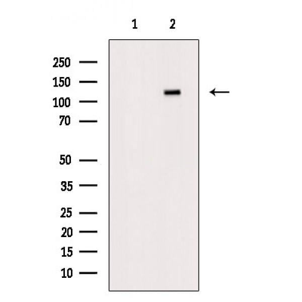
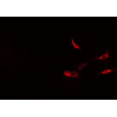
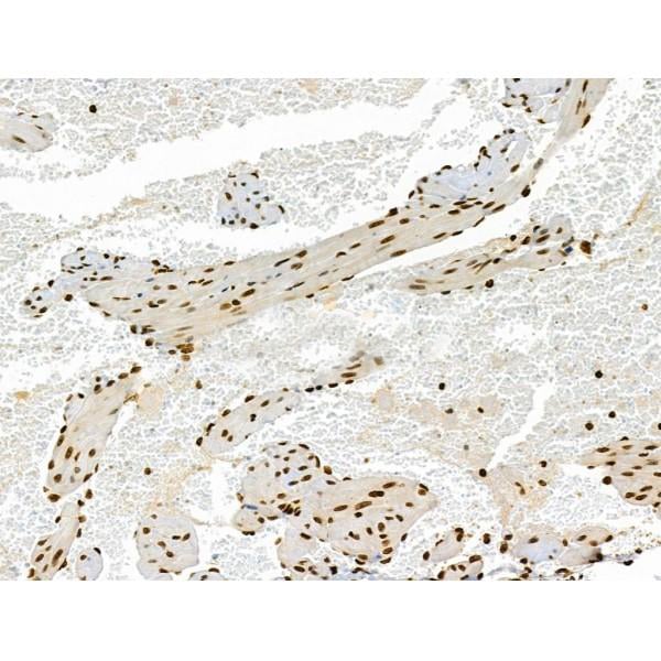
 IHC (Immunohistochemistry)
(At 1/100 staining Human mammary cancer and adjacent normal tissues by IHC-P. The sample was formaldehyde fixed and a heat mediated antigen retrieval step in citrate buffer was performed. The sample was then blocked and incubated with the primary antibody at 4 degree C overnight. An HRP conjugated anti-Rabbit antibody was used as the secondary antibody.)
IHC (Immunohistochemistry)
(At 1/100 staining Human mammary cancer and adjacent normal tissues by IHC-P. The sample was formaldehyde fixed and a heat mediated antigen retrieval step in citrate buffer was performed. The sample was then blocked and incubated with the primary antibody at 4 degree C overnight. An HRP conjugated anti-Rabbit antibody was used as the secondary antibody.)
Jak2, Polyclonal Antibody (Cat# AAA31311)
Full Name
Phospho-Jak2 (Tyr1008) Antibody
Gene Names
JAK2; JTK10; THCYT3
Reactivity
Human, Mouse, Rat
Applications
WB, IHC, EIA
Purity
The antibody is from purified rabbit serum by affinity purification via sequential chromatography on phospho-peptide and non-phospho-peptide affinity columns.
Pricing


 Application Data
(Staining of J774 cells with Rat anti Mouse F4/80 antigen Biotin)
Application Data
(Staining of J774 cells with Rat anti Mouse F4/80 antigen Biotin)
F4/80, Monoclonal Antibody (Cat# AAA12163)
Full Name
RAT ANTI MOUSE F4/80:Biotin
Gene Names
Emr1; Ly71; F4/80; Gpf480; TM7LN3; DD7A5-7; EGF-TM7
Applications
FC/FACS
Pricing


 Application Data
(Staining of J774 cells with Rat anti Mouse F4/80 antigen Biotin)
Application Data
(Staining of J774 cells with Rat anti Mouse F4/80 antigen Biotin)
F4/80, Monoclonal Antibody (Cat# AAA12161)
Full Name
RAT ANTI MOUSE F4/80:APC
Gene Names
Emr1; Ly71; F4/80; Gpf480; TM7LN3; DD7A5-7; EGF-TM7
Applications
FC/FACS
Pricing


 Application Data
(Staining of J774 cells with Rat anti Mouse F4/80 antigen Biotin)
Application Data
(Staining of J774 cells with Rat anti Mouse F4/80 antigen Biotin)
F4/80, Monoclonal Antibody (Cat# AAA12172)
Full Name
RAT ANTI MOUSE F4/80:RPE
Gene Names
Emr1; Ly71; F4/80; Gpf480; TM7LN3; DD7A5-7; EGF-TM7
Applications
FC/FACS, IF
Pricing


 Application Data
(Published customer image:Phycoerythrin conjugated Mouse anti Human CD83 antibody, clone HB15e used for the evaluation of CD83 expression on monocyte derived dendritic cells by flow cytometry.Phenotypic characterization of immunogenic and tolerogenic moDC populations by flow cytometry. Monocytes were negatively selected from PBMC using magnetic beads. Immature moDC were generated with IL-4 and GM-CSF for 6 days. 15d-PGJ2 (PGJ2 DC) and dexamethasone plus 1alpha,25-dihydroxyvitamin were added to generate tolerogenic moDC, respectively (PGJ2 DC and Dex/VD3 DC). To generate immunogenic moDC, immature moDC were stimulated for 24 h with LPS, polyI:C and a cytokine cocktail containing TNF-alpha, IL-1beta, IL-6 and PGE2, respectively. The phenotypes of the cells were analyzed by flow cytometry. Live cells were gated according to FSC/SSC. One representative experiment out of three is shown.From: Sprater F, Hovden A-O, Appel S (2012)Expression of ESE-3 Isoforms in Immunogenic and Tolerogenic Human Monocyte-Derived Dendritic Cells.PLoS ONE 7(11): e49577.)
Application Data
(Published customer image:Phycoerythrin conjugated Mouse anti Human CD83 antibody, clone HB15e used for the evaluation of CD83 expression on monocyte derived dendritic cells by flow cytometry.Phenotypic characterization of immunogenic and tolerogenic moDC populations by flow cytometry. Monocytes were negatively selected from PBMC using magnetic beads. Immature moDC were generated with IL-4 and GM-CSF for 6 days. 15d-PGJ2 (PGJ2 DC) and dexamethasone plus 1alpha,25-dihydroxyvitamin were added to generate tolerogenic moDC, respectively (PGJ2 DC and Dex/VD3 DC). To generate immunogenic moDC, immature moDC were stimulated for 24 h with LPS, polyI:C and a cytokine cocktail containing TNF-alpha, IL-1beta, IL-6 and PGE2, respectively. The phenotypes of the cells were analyzed by flow cytometry. Live cells were gated according to FSC/SSC. One representative experiment out of three is shown.From: Sprater F, Hovden A-O, Appel S (2012)Expression of ESE-3 Isoforms in Immunogenic and Tolerogenic Human Monocyte-Derived Dendritic Cells.PLoS ONE 7(11): e49577.)
CD83, Monoclonal Antibody (Cat# AAA12280)
Full Name
MOUSE ANTI HUMAN CD83
Reactivity
Human
Applications
FC/FACS, IHC, IHC, IP
Pricing


 Application Data
(Published customer image:Phycoerythrin conjugated Mouse anti Human CD83 antibody, clone HB15e used for the evaluation of CD83 expression on monocyte derived dendritic cells by flow cytometry.Phenotypic characterization of immunogenic and tolerogenic moDC populations by flow cytometry. Monocytes were negatively selected from PBMC using magnetic beads. Immature moDC were generated with IL-4 and GM-CSF for 6 days. 15d-PGJ2 (PGJ2 DC) and dexamethasone plus 1alpha,25-dihydroxyvitamin were added to generate tolerogenic moDC, respectively (PGJ2 DC and Dex/VD3 DC). To generate immunogenic moDC, immature moDC were stimulated for 24 h with LPS, polyI:C and a cytokine cocktail containing TNF-alpha, IL-1beta, IL-6 and PGE2, respectively. The phenotypes of the cells were analyzed by flow cytometry. Live cells were gated according to FSC/SSC. One representative experiment out of three is shown.From: Sprater F, Hovden A-O, Appel S (2012)Expression of ESE-3 Isoforms in Immunogenic and Tolerogenic Human Monocyte-Derived Dendritic Cells.PLoS ONE 7(11): e49577.)
Application Data
(Published customer image:Phycoerythrin conjugated Mouse anti Human CD83 antibody, clone HB15e used for the evaluation of CD83 expression on monocyte derived dendritic cells by flow cytometry.Phenotypic characterization of immunogenic and tolerogenic moDC populations by flow cytometry. Monocytes were negatively selected from PBMC using magnetic beads. Immature moDC were generated with IL-4 and GM-CSF for 6 days. 15d-PGJ2 (PGJ2 DC) and dexamethasone plus 1alpha,25-dihydroxyvitamin were added to generate tolerogenic moDC, respectively (PGJ2 DC and Dex/VD3 DC). To generate immunogenic moDC, immature moDC were stimulated for 24 h with LPS, polyI:C and a cytokine cocktail containing TNF-alpha, IL-1beta, IL-6 and PGE2, respectively. The phenotypes of the cells were analyzed by flow cytometry. Live cells were gated according to FSC/SSC. One representative experiment out of three is shown.From: Sprater F, Hovden A-O, Appel S (2012)Expression of ESE-3 Isoforms in Immunogenic and Tolerogenic Human Monocyte-Derived Dendritic Cells.PLoS ONE 7(11): e49577.)
CD83, Monoclonal Antibody (Cat# AAA12283)
Full Name
MOUSE ANTI HUMAN CD83
Reactivity
Human
Applications
FC/FACS, IHC, IHC
Pricing


 WB (Western Blot)
(Western Blot Analysis of Raji cell lysate using CD20 Mouse Monoclonal Antibody (L26).)
WB (Western Blot)
(Western Blot Analysis of Raji cell lysate using CD20 Mouse Monoclonal Antibody (L26).)
CD20/MS4A1, Monoclonal Antibody (Cat# AAA23946)
Full Name
CD20/MS4A1 (B-Cell Marker)
Gene Names
MS4A1; B1; S7; Bp35; CD20; CVID5; MS4A2; LEU-16
Reactivity
Human
Applications
FC/FACS, IF, WB, IHC
Purity
Purified Ab with BSA and Azide at 200ug/ml OR Purified Ab WITHOUT BSA and Azide at 1.0mg/ml
Pricing


 Application Data
(Staining of J774 cells with Rat anti Mouse F4/80 antigen Biotin)
Application Data
(Staining of J774 cells with Rat anti Mouse F4/80 antigen Biotin)
F4/80, Monoclonal Antibody (Cat# AAA12171)
Full Name
RAT ANTI MOUSE F4/80:RPE
Gene Names
Emr1; Ly71; F4/80; Gpf480; TM7LN3; DD7A5-7; EGF-TM7
Applications
FC/FACS
Pricing


 Application Data
(Staining of J774 cells with Rat anti Mouse F4/80 antigen Biotin)
Application Data
(Staining of J774 cells with Rat anti Mouse F4/80 antigen Biotin)
F4/80, Monoclonal Antibody (Cat# AAA12217)
Full Name
RAT ANTI MOUSE F4/80:APC
Gene Names
Emr1; Ly71; F4/80; Gpf480; TM7LN3; DD7A5-7; EGF-TM7
Applications
FC/FACS
Pricing


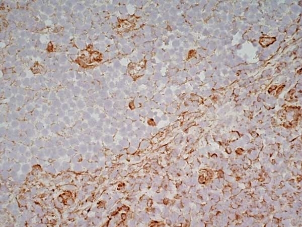
 IHC (Immunohistochemistry)
(At 1/100 staining Human gastric cancer and adjacent normal tissues by IHC-P. The sample was formaldehyde fixed and a heat mediated antigen retrieval step in citrate buffer was performed. The sample was then blocked and incubated with the primary antibody at 4 degree C overnight. An HRP conjugated anti-Rabbit antibody was used as the secondary antibody.)
IHC (Immunohistochemistry)
(At 1/100 staining Human gastric cancer and adjacent normal tissues by IHC-P. The sample was formaldehyde fixed and a heat mediated antigen retrieval step in citrate buffer was performed. The sample was then blocked and incubated with the primary antibody at 4 degree C overnight. An HRP conjugated anti-Rabbit antibody was used as the secondary antibody.)
Moesin/Ezrin/Radixin, Polyclonal Antibody (Cat# AAA31305)
Full Name
Phospho-Moesin/Ezrin/Radixin (Thr558) Antibody
Gene Names
EZR; CVL; CVIL; VIL2
Reactivity
Human, Mouse, Rat
Applications
IHC, EIA
Purity
The antibody is from purified rabbit serum by affinity purification via sequential chromatography on phospho-peptide and non-phospho-peptide affinity columns.
Pricing


 WB (Western Blot)
(Western BlotSample: Recombinant GAL9, Human;Antibody: Rabbit Anti-Human GAL9 Ab)
WB (Western Blot)
(Western BlotSample: Recombinant GAL9, Human;Antibody: Rabbit Anti-Human GAL9 Ab)
Galectin 9 (GAL9), Active Protein (Cat# AAA21103)
Full Name
Active Galectin 9 (GAL9)
Gene Names
LGALS9; HUAT; LGALS9A
Reactivity
Homo sapiens (Human)
Applications
Cell culture; Activity Assays.
Purity
>90%
Pricing


 Application Data
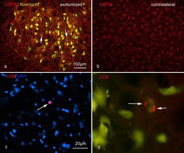
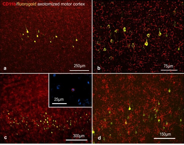
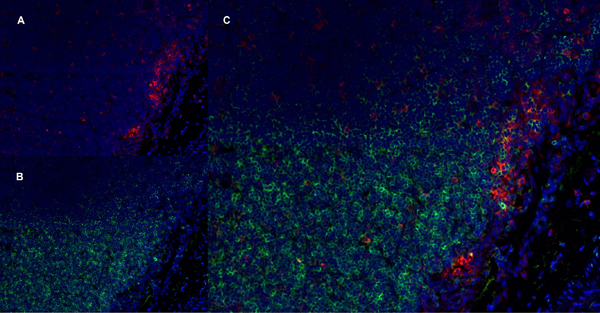
(Published customer image: Representative images of the inflammatory changes in the facial nucleus during axonal regeneration, one week following facial nerve transaction. a, b: CD11b immunoreactivity for microglia is increased in the axotomized facial nucleus, and microglia enwrap the facial motor neurons, e.g. at arrows. The regenerating neurons were retrogradely labelled with fluorogold. c, d: CD6- positive T-cells accumulated in the injured motor nucleus (arrows). They had little cytoplasm but dense nuclei (c) and were sometimes clustered around neurons retrogradely labelled with fluorogold (d). The scale bar in (a) also applies to (b) and that in (c) also applies to (d).From: Shokouhi et al. BMC Neuroscience 2010 11:13.)
Application Data
(Published customer image: Representative images of the inflammatory changes in the facial nucleus during axonal regeneration, one week following facial nerve transaction. a, b: CD11b immunoreactivity for microglia is increased in the axotomized facial nucleus, and microglia enwrap the facial motor neurons, e.g. at arrows. The regenerating neurons were retrogradely labelled with fluorogold. c, d: CD6- positive T-cells accumulated in the injured motor nucleus (arrows). They had little cytoplasm but dense nuclei (c) and were sometimes clustered around neurons retrogradely labelled with fluorogold (d). The scale bar in (a) also applies to (b) and that in (c) also applies to (d).From: Shokouhi et al. BMC Neuroscience 2010 11:13.)
CD11b, Monoclonal Antibody (Cat# AAA12146)
Full Name
MOUSE ANTI RAT CD11b:FITC
Gene Names
ITGAM; CD11B
Applications
FC/FACS
Pricing


 Application Data
(Staining of J774 cells with Rat anti Mouse F4/80 antigen Biotin)
Application Data
(Staining of J774 cells with Rat anti Mouse F4/80 antigen Biotin)
F4/80, Monoclonal Antibody (Cat# AAA12170)
Full Name
RAT ANTI MOUSE F4/80
Gene Names
Emr1; Ly71; F4/80; Gpf480; TM7LN3; DD7A5-7; EGF-TM7
Applications
EM, FC/FACS, IF, IP, RIA, RE, WB
Pricing


 Application Data
(Staining of J774 cells with Rat anti Mouse F4/80 antigen Biotin)
Application Data
(Staining of J774 cells with Rat anti Mouse F4/80 antigen Biotin)
F4/80, Monoclonal Antibody (Cat# AAA12165)
Full Name
RAT ANTI MOUSE F4/80:Low Endotoxin
Gene Names
Emr1; Ly71; F4/80; Gpf480; TM7LN3; DD7A5-7; EGF-TM7
Applications
FC/FACS, FN, IP, RIA, RE, WB
Pricing


 Application Data
(At 25 degree C. Samples were then incubated with primary Ab(At 37 degree C. An AlexaFluor594 conjugated goat anti-rabbit IgG(H+L) Ab(Red) and an AlexaFluor488 conjugated goat anti-mouse IgG(H+L) Ab(Green) were used as the secondary antibody.The nuclear counter stain is DAPI(blue).)
Application Data
(At 25 degree C. Samples were then incubated with primary Ab(At 37 degree C. An AlexaFluor594 conjugated goat anti-rabbit IgG(H+L) Ab(Red) and an AlexaFluor488 conjugated goat anti-mouse IgG(H+L) Ab(Green) were used as the secondary antibody.The nuclear counter stain is DAPI(blue).)
Plastin L, Polyclonal Antibody (Cat# AAA31395)
Full Name
Phospho-Plastin L (Ser5) Antibody
Gene Names
LCP1; LPL; CP64; PLS2; LC64P; HEL-S-37; L-PLASTIN
Reactivity
Human, Mouse, Rat
Predicted Reactivity: Pig (100%), Bovine (100%), Horse (100%), Sheep (100%), Rabbit (100%), Dog (100%)
Predicted Reactivity: Pig (100%), Bovine (100%), Horse (100%), Sheep (100%), Rabbit (100%), Dog (100%)
Applications
WB, IHC, IF, ICC, EIA
Purity
The antibody is from purified rabbit serum by affinity purification via sequential chromatography on phospho-peptide and non-phospho-peptide affinity columns.
Pricing


 Application Data
(At 25 degree C. The primary antibody was diluted at 1/200 and incubated with the sample for 1 hour at 37 degree C. An Alexa Fluor 594 conjugated goat anti-rabbit IgG (H+L) antibody(Red), diluted at 1/600, was used as secondary antibody.)
Application Data
(At 25 degree C. The primary antibody was diluted at 1/200 and incubated with the sample for 1 hour at 37 degree C. An Alexa Fluor 594 conjugated goat anti-rabbit IgG (H+L) antibody(Red), diluted at 1/600, was used as secondary antibody.)
JAK3, Polyclonal Antibody (Cat# AAA31404)
Full Name
Phospho-JAK3 (Tyr904) Antibody
Gene Names
JAK3; JAKL; LJAK; JAK-3; L-JAK; JAK3_HUMAN
Reactivity
Human, Mouse, Rat
Predicted Reactivity: Bovine (100%), Sheep (100%), Dog (100%)
Predicted Reactivity: Bovine (100%), Sheep (100%), Dog (100%)
Applications
WB, IHC, IF, ICC, EIA
Purity
The antibody is from purified rabbit serum by affinity purification via sequential chromatography on phospho-peptide and non-phospho-peptide affinity columns.
Pricing


 Application Data
(At 25 degree C. The primary antibody was diluted at 1/200 and incubated with the sample for 1 hour at 37 degree C. An Alexa Fluor 594 conjugated goat anti-rabbit IgG (H+L) antibody(Red), diluted at 1/600, was used as secondary antibody.)
Application Data
(At 25 degree C. The primary antibody was diluted at 1/200 and incubated with the sample for 1 hour at 37 degree C. An Alexa Fluor 594 conjugated goat anti-rabbit IgG (H+L) antibody(Red), diluted at 1/600, was used as secondary antibody.)
JAK3, Polyclonal Antibody (Cat# AAA31405)
Full Name
Phospho-JAK3 (Tyr981) Antibody
Gene Names
JAK3; JAKL; LJAK; JAK-3; L-JAK; JAK3_HUMAN
Reactivity
Human, Mouse, Rat, Monkey
Predicted Reactivity: Pig (100%), Bovine (91%), Horse (100%), Sheep (100%), Dog (100%)
Predicted Reactivity: Pig (100%), Bovine (91%), Horse (100%), Sheep (100%), Dog (100%)
Applications
WB, IHC, IF, ICC, EIA
Purity
The antibody is from purified rabbit serum by affinity purification via sequential chromatography on phospho-peptide and non-phospho-peptide affinity columns.
Pricing


 Application Data
(At 25 degree C. The primary antibody was diluted at 1/200 and incubated with the sample for 1 hour at 37 degree C. An Alexa Fluor 594 conjugated goat anti-rabbit IgG (H+L) Ab, diluted at 1/600, was used as the secondary antibody.)
Application Data
(At 25 degree C. The primary antibody was diluted at 1/200 and incubated with the sample for 1 hour at 37 degree C. An Alexa Fluor 594 conjugated goat anti-rabbit IgG (H+L) Ab, diluted at 1/600, was used as the secondary antibody.)
CD22, Polyclonal Antibody (Cat# AAA31418)
Full Name
Phospho-CD22 (Tyr842) Antibody
Gene Names
CD22; SIGLEC2; SIGLEC-2
Reactivity
Human, Mouse, Rat
Predicted Reactivity: Horse (83%)
Predicted Reactivity: Horse (83%)
Applications
WB, IHC, IF, ICC, EIA
Purity
The antibody is from purified rabbit serum by affinity purification via sequential chromatography on phospho-peptide and non-phospho-peptide affinity columns.
Pricing


 Application Data
(Immunoperoxidase staining of rat lymph node cryosection with Mouse anti Rat CD25 followed by horseradish peroxidase conjugated Goat anti Mouse IgG1 as a detection reagent. High power)
Application Data
(Immunoperoxidase staining of rat lymph node cryosection with Mouse anti Rat CD25 followed by horseradish peroxidase conjugated Goat anti Mouse IgG1 as a detection reagent. High power)
CD25, Monoclonal Antibody (Cat# AAA11966)
Full Name
MOUSE ANTI RAT CD25
Gene Names
Il2ra; IL2RAC
Applications
EIA, FC/FACS, IP
Pricing


 Application Data
(Published customer image: Representative images of the inflammatory changes in the facial nucleus during axonal regeneration, one week following facial nerve transaction. a, b: CD11b immunoreactivity for microglia is increased in the axotomized facial nucleus, and microglia enwrap the facial motor neurons, e.g. at arrows. The regenerating neurons were retrogradely labelled with fluorogold. c, d: CD6- positive T-cells accumulated in the injured motor nucleus (arrows). They had little cytoplasm but dense nuclei (c) and were sometimes clustered around neurons retrogradely labelled with fluorogold (d). The scale bar in (a) also applies to (b) and that in (c) also applies to (d).From: Shokouhi et al. BMC Neuroscience 2010 11:13.)
Application Data
(Published customer image: Representative images of the inflammatory changes in the facial nucleus during axonal regeneration, one week following facial nerve transaction. a, b: CD11b immunoreactivity for microglia is increased in the axotomized facial nucleus, and microglia enwrap the facial motor neurons, e.g. at arrows. The regenerating neurons were retrogradely labelled with fluorogold. c, d: CD6- positive T-cells accumulated in the injured motor nucleus (arrows). They had little cytoplasm but dense nuclei (c) and were sometimes clustered around neurons retrogradely labelled with fluorogold (d). The scale bar in (a) also applies to (b) and that in (c) also applies to (d).From: Shokouhi et al. BMC Neuroscience 2010 11:13.)
CD11b, Monoclonal Antibody (Cat# AAA11971)
Full Name
MOUSE ANTI RAT CD11b
Gene Names
ITGAM; CD11B
Applications
FC/FACS, IF, IP
Pricing


 Application Data
(Published customer image:RPE conjugated Mouse anti Canine CD4 antibody, clone YKIC302.9used for the assessment of CD4 levels on canine cells by flow cytometry.Image caption:Immunophenotypic profile of tumor infiltrating lymphocyte in canine mammary carcinomas. Analysis of tumor infiltrating T-cells, B-lymphocytes and T-cell subsets from MC-BMT or MC (A), further subcategorized according to the absence (-) or presence (+) of lymph node metastasis (-) (B). Lymphocyte populations and subsets were identified by flow cytometric immunostaining as described in Material and Methods. Data were expressed as percentage of positive cells within gated lymphocytes and CD4+/CD8+ T-cell ratio. Significant differences at p < 0.05 are highlighted by asterisk.)
Application Data
(Published customer image:RPE conjugated Mouse anti Canine CD4 antibody, clone YKIC302.9used for the assessment of CD4 levels on canine cells by flow cytometry.Image caption:Immunophenotypic profile of tumor infiltrating lymphocyte in canine mammary carcinomas. Analysis of tumor infiltrating T-cells, B-lymphocytes and T-cell subsets from MC-BMT or MC (A), further subcategorized according to the absence (-) or presence (+) of lymph node metastasis (-) (B). Lymphocyte populations and subsets were identified by flow cytometric immunostaining as described in Material and Methods. Data were expressed as percentage of positive cells within gated lymphocytes and CD4+/CD8+ T-cell ratio. Significant differences at p < 0.05 are highlighted by asterisk.)
CD4, Monoclonal Antibody (Cat# AAA12278)
Full Name
RAT ANTI DOG CD4: APC
Reactivity
Dog
Applications
FC/FACS
Pricing


 SDS-PAGE
(SDS Page analysis of purified S100B Mouse Monoclonal Antibody (S100B/1012).)
SDS-PAGE
(SDS Page analysis of purified S100B Mouse Monoclonal Antibody (S100B/1012).)
S100B, Monoclonal Antibody (Cat# AAA23893)
Full Name
S100B (Astrocyte and Melanoma Marker)
Gene Names
S100B; NEF; S100; S100-B; S100beta
Reactivity
Human, Mouse, Rat, Cow. Others not known.
Applications
FC/FACS, IF, WB, IHC
Pricing


 Application Data
(Analysis of Protein Array containing more than 19, 000 full-length human proteins using Oct-2 Mouse Monoclonal Antibody (OCT2/2137) Z- and S- Score: The Z-score represents the strength of a signal that a monoclonal antibody (MAb) (in combination with a fluorescently-tagged anti-IgG secondary antibody) produces when binding to a particular protein on the HuProtTM array. Z-scores are described in units of standard deviations (SD's) above the mean value of all signals generated on that array. If targets on HuProtTM are arranged in descending order of the Z-score, the S-score is the difference (also in units of SD's) between the Z-score. S-score therefore represents the relative target specificity of a MAb to its intended target. A MAb is considered to specific to its intended target, if the MAb has an S-score of at least 2.5. For example, if a MAb binds to protein X with a Z-score of 43 and to protein Y with a Z-score of 14, then the S-score for the binding of that MAb to protein X is equal to 29.)
Application Data
(Analysis of Protein Array containing more than 19, 000 full-length human proteins using Oct-2 Mouse Monoclonal Antibody (OCT2/2137) Z- and S- Score: The Z-score represents the strength of a signal that a monoclonal antibody (MAb) (in combination with a fluorescently-tagged anti-IgG secondary antibody) produces when binding to a particular protein on the HuProtTM array. Z-scores are described in units of standard deviations (SD's) above the mean value of all signals generated on that array. If targets on HuProtTM are arranged in descending order of the Z-score, the S-score is the difference (also in units of SD's) between the Z-score. S-score therefore represents the relative target specificity of a MAb to its intended target. A MAb is considered to specific to its intended target, if the MAb has an S-score of at least 2.5. For example, if a MAb binds to protein X with a Z-score of 43 and to protein Y with a Z-score of 14, then the S-score for the binding of that MAb to protein X is equal to 29.)
OCT-2 (POU2F2), Monoclonal Antibody (Cat# AAA23902)
Full Name
OCT-2 (POU2F2) (B-Cell Marker)
Gene Names
POU2F2; OCT2; OTF2; Oct-2
Reactivity
Human. Others not known.
Applications
EIA, WB, IHC
Pricing


 Application Data
(Published customer image: Representative images of the inflammatory changes in the facial nucleus during axonal regeneration, one week following facial nerve transaction. a, b: CD11b immunoreactivity for microglia is increased in the axotomized facial nucleus, and microglia enwrap the facial motor neurons, e.g. at arrows. The regenerating neurons were retrogradely labelled with fluorogold. c, d: CD6- positive T-cells accumulated in the injured motor nucleus (arrows). They had little cytoplasm but dense nuclei (c) and were sometimes clustered around neurons retrogradely labelled with fluorogold (d). The scale bar in (a) also applies to (b) and that in (c) also applies to (d).From: Shokouhi et al. BMC Neuroscience 2010 11:13.)
Application Data
(Published customer image: Representative images of the inflammatory changes in the facial nucleus during axonal regeneration, one week following facial nerve transaction. a, b: CD11b immunoreactivity for microglia is increased in the axotomized facial nucleus, and microglia enwrap the facial motor neurons, e.g. at arrows. The regenerating neurons were retrogradely labelled with fluorogold. c, d: CD6- positive T-cells accumulated in the injured motor nucleus (arrows). They had little cytoplasm but dense nuclei (c) and were sometimes clustered around neurons retrogradely labelled with fluorogold (d). The scale bar in (a) also applies to (b) and that in (c) also applies to (d).From: Shokouhi et al. BMC Neuroscience 2010 11:13.)
CD11b, Monoclonal Antibody (Cat# AAA11876)
Full Name
MOUSE ANTI RAT CD11b:FITC
Gene Names
ITGAM; CD11B
Applications
FC/FACS
Pricing


 FCM (Flow Cytometry)
(Figure 6. Flow Cytometry analysis of U-87 cells using anti-SSH3BP1 antibody (AAA11656).Overlay histogram showing U-87 cells stained with AAA11656 (Blue line).The cells were blocked with 10% normal goat serum. And then incubated with rabbit anti-SSH3BP1 Antibody (AAA11656,1ug/1x10^6 cells) for 30 min at 20 degree C. DyLight®488 conjugated goat anti-rabbit IgG (5-10ug/1x10^6 cells) was used as secondary antibody for 30 minutes at 20 degree C. Isotype control antibody (Green line) was rabbit IgG (1ug/1x106) used under the same conditions. Unlabelled sample (Red line) was also used as a control.)
FCM (Flow Cytometry)
(Figure 6. Flow Cytometry analysis of U-87 cells using anti-SSH3BP1 antibody (AAA11656).Overlay histogram showing U-87 cells stained with AAA11656 (Blue line).The cells were blocked with 10% normal goat serum. And then incubated with rabbit anti-SSH3BP1 Antibody (AAA11656,1ug/1x10^6 cells) for 30 min at 20 degree C. DyLight®488 conjugated goat anti-rabbit IgG (5-10ug/1x10^6 cells) was used as secondary antibody for 30 minutes at 20 degree C. Isotype control antibody (Green line) was rabbit IgG (1ug/1x106) used under the same conditions. Unlabelled sample (Red line) was also used as a control.)
SSH3BP1, Polyclonal Antibody (Cat# AAA11656)
Full Name
Anti-SSH3BP1 Antibody
Gene Names
ABI1; E3B1; ABI-1; ABLBP4; NAP1BP; SSH3BP; SSH3BP1
Reactivity
Human, Mouse, Rat
Applications
WB, IHC
Purity
Immunogen Affinity Purified
Pricing


 FCM (Flow Cytometry)
(Overlay histogram showing Hela cells stained with CSB-MA878942A1m (red line) at 1:300. The cells were incubated in 1x PBS /10% normal goat serum to block non-specific protein-protein interactions followed by primary antibody for 1 h at 4 degree C. The secondary antibody used was FITC goat anti-mouse IgG(H+L) at 1/200 dilution for 1 h at 4 degree C. Isotype control antibody (green line) was used under the same conditions. Acquisition of >10,000 events was performed.)
FCM (Flow Cytometry)
(Overlay histogram showing Hela cells stained with CSB-MA878942A1m (red line) at 1:300. The cells were incubated in 1x PBS /10% normal goat serum to block non-specific protein-protein interactions followed by primary antibody for 1 h at 4 degree C. The secondary antibody used was FITC goat anti-mouse IgG(H+L) at 1/200 dilution for 1 h at 4 degree C. Isotype control antibody (green line) was used under the same conditions. Acquisition of >10,000 events was performed.)
PD-L1, Monoclonal Antibody (Cat# AAA27017)
Full Name
PD-L1 Monoclonal Antibody
Gene Names
CD274; B7-H; B7H1; PDL1; PD-L1; PDCD1L1; PDCD1LG1
Reactivity
Human
Applications
EIA, WB, IHC, IF, FC/FACS
Purity
>95%
Protein G Purified
Protein G Purified
Pricing


 Application Data
(Staining of J774 cells with Rat anti Mouse F4/80 antigen Biotin)
Application Data
(Staining of J774 cells with Rat anti Mouse F4/80 antigen Biotin)
F4/80, Monoclonal Antibody (Cat# AAA12174)
Full Name
RAT ANTI MOUSE F4/80
Gene Names
Emr1; Ly71; F4/80; Gpf480; TM7LN3; DD7A5-7; EGF-TM7
Applications
EM, FC/FACS, IF, IP, RIA, RE, WB
Pricing


 Application Data
(Analysis of Protein Array containing more than 19,000 full-length human proteins using ROR-gamma / RORC Mouse Monoclonal Antibody (RORC/2941). Z- and S- Score: The Z-score represents the strength of a signal that a monoclonal antibody (MAb) (in combination with a fluorescently-tagged anti-IgG secondary antibody) produces when binding to a particular protein on the HuProtTM array. Z-scores are described in units of standard deviations (SD's) above the mean value of all signals generated on that array. If targets on HuProtTM are arranged in descending order of the Z-score, the S-score is the difference (also in units of SD's) between the Z-score. S-score therefore represents the relative target specificity of a MAb to its intended target. A MAb is considered to specific to its intended target, if the MAb has an S-score of at least 2.5. For example, if a MAb binds to protein X with a Z-score of 43 and to protein Y with a Z-score of 14, then the S-score for the binding of that MAb to protein X is equal to 29.)
Application Data
(Analysis of Protein Array containing more than 19,000 full-length human proteins using ROR-gamma / RORC Mouse Monoclonal Antibody (RORC/2941). Z- and S- Score: The Z-score represents the strength of a signal that a monoclonal antibody (MAb) (in combination with a fluorescently-tagged anti-IgG secondary antibody) produces when binding to a particular protein on the HuProtTM array. Z-scores are described in units of standard deviations (SD's) above the mean value of all signals generated on that array. If targets on HuProtTM are arranged in descending order of the Z-score, the S-score is the difference (also in units of SD's) between the Z-score. S-score therefore represents the relative target specificity of a MAb to its intended target. A MAb is considered to specific to its intended target, if the MAb has an S-score of at least 2.5. For example, if a MAb binds to protein X with a Z-score of 43 and to protein Y with a Z-score of 14, then the S-score for the binding of that MAb to protein X is equal to 29.)
ROR-gamma/RORC, Monoclonal Antibody (Cat# AAA23951)
Full Name
ROR-gamma/RORC (RAR-related Orphan Receptor C)
Gene Names
RORC; TOR; RORG; RZRG; IMD42; NR1F3; RZR-GAMMA
Reactivity
Human
Applications
FC, IHC
Purity
Purified Ab with BSA and Azide at 200ug/ml or Purified Ab with BSA and Azide at 200ug/ml or Purified Ab WITHOUT BSA and Azide at 1.0mg/ml
Pricing


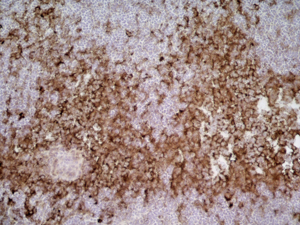
 IHC (Immunohistochemistry)
(At 1/200 staining Human lung cancer tissue sections by IHC-P. The tissue was formaldehyde fixed and a heat mediated antigen retrieval step in citrate buffer was performed. The tissue was then blocked and incubated with the antibody for 1.5 hours at 22 degree C. An HRP conjugated goat anti-rabbit antibody was used as the secondary antibody.)
IHC (Immunohistochemistry)
(At 1/200 staining Human lung cancer tissue sections by IHC-P. The tissue was formaldehyde fixed and a heat mediated antigen retrieval step in citrate buffer was performed. The tissue was then blocked and incubated with the antibody for 1.5 hours at 22 degree C. An HRP conjugated goat anti-rabbit antibody was used as the secondary antibody.)
Caveolin 2, Polyclonal Antibody (Cat# AAA31397)
Full Name
Phospho-Caveolin 2 (Ser36) Antibody
Gene Names
CAV2; CAV
Reactivity
Human, Mouse, Rat
Applications
WB, IHC, EIA
Purity
The antibody is from purified rabbit serum by affinity purification via sequential chromatography on phospho-peptide and non-phospho-peptide affinity columns.
Pricing


 Application Data
(At 25 degree C. The primary antibody was diluted at 1/200 and incubated with the sample for 1 hour at 37 degree C. An Alexa Fluor 594 conjugated goat anti-rabbit IgG (H+L) Ab, diluted at 1/600, was used as the secondary antibody.)
Application Data
(At 25 degree C. The primary antibody was diluted at 1/200 and incubated with the sample for 1 hour at 37 degree C. An Alexa Fluor 594 conjugated goat anti-rabbit IgG (H+L) Ab, diluted at 1/600, was used as the secondary antibody.)
LAT, Polyclonal Antibody (Cat# AAA31413)
Full Name
Phospho-LAT (Tyr161) Antibody
Gene Names
LAT; LAT1; pp36
Reactivity
Human, Mouse, Rat
Predicted Reactivity: Pig (91%), Zebrafish (83%), Bovine (100%), Horse (91%), Sheep (100%), Rabbit (80%), Dog (82%)
Predicted Reactivity: Pig (91%), Zebrafish (83%), Bovine (100%), Horse (91%), Sheep (100%), Rabbit (80%), Dog (82%)
Applications
WB, IHC, IF, ICC, EIA
Purity
The antibody is from purified rabbit serum by affinity purification via sequential chromatography on phospho-peptide and non-phospho-peptide affinity columns.
Pricing


 Application Data
(Immunoperoxidase staining of rat lymph node cryosection with Mouse anti Rat CD25 followed by horseradish peroxidase conjugated Goat anti Mouse IgG1 as a detection reagent. High power)
Application Data
(Immunoperoxidase staining of rat lymph node cryosection with Mouse anti Rat CD25 followed by horseradish peroxidase conjugated Goat anti Mouse IgG1 as a detection reagent. High power)
CD25, Monoclonal Antibody (Cat# AAA11965)
Full Name
MOUSE ANTI RAT CD25
Gene Names
Il2ra; IL2RAC
Applications
EIA, FC/FACS, IP
Pricing


 Application Data
(Published customer image: Representative images of the inflammatory changes in the facial nucleus during axonal regeneration, one week following facial nerve transaction. a, b: CD11b immunoreactivity for microglia is increased in the axotomized facial nucleus, and microglia enwrap the facial motor neurons, e.g. at arrows. The regenerating neurons were retrogradely labelled with fluorogold. c, d: CD6- positive T-cells accumulated in the injured motor nucleus (arrows). They had little cytoplasm but dense nuclei (c) and were sometimes clustered around neurons retrogradely labelled with fluorogold (d). The scale bar in (a) also applies to (b) and that in (c) also applies to (d).From: Shokouhi et al. BMC Neuroscience 2010 11:13.)
Application Data
(Published customer image: Representative images of the inflammatory changes in the facial nucleus during axonal regeneration, one week following facial nerve transaction. a, b: CD11b immunoreactivity for microglia is increased in the axotomized facial nucleus, and microglia enwrap the facial motor neurons, e.g. at arrows. The regenerating neurons were retrogradely labelled with fluorogold. c, d: CD6- positive T-cells accumulated in the injured motor nucleus (arrows). They had little cytoplasm but dense nuclei (c) and were sometimes clustered around neurons retrogradely labelled with fluorogold (d). The scale bar in (a) also applies to (b) and that in (c) also applies to (d).From: Shokouhi et al. BMC Neuroscience 2010 11:13.)
CD11b, Monoclonal Antibody (Cat# AAA11970)
Full Name
MOUSE ANTI RAT CD11b
Gene Names
ITGAM; CD11B
Applications
FC/FACS, IF, IP
Pricing


 Application Data
(At 25 degree C. The primary antibody was diluted at 1/200 and incubated with the sample for 1 hour at 37 degree C. An Alexa Fluor 594 conjugated goat anti-rabbit IgG (H+L) Ab, diluted at 1/600, was used as the secondary antibody.)
Application Data
(At 25 degree C. The primary antibody was diluted at 1/200 and incubated with the sample for 1 hour at 37 degree C. An Alexa Fluor 594 conjugated goat anti-rabbit IgG (H+L) Ab, diluted at 1/600, was used as the secondary antibody.)
Caspase 3, Polyclonal Antibody (Cat# AAA31443)
Full Name
Phospho-Caspase 3 (Ser26) Antibody
Gene Names
CASP3; CPP32; SCA-1; CPP32B
Reactivity
Human, Mouse, Rat
Predicted Reactivity: Rabbit (88%)
Predicted Reactivity: Rabbit (88%)
Applications
WB, IHC, IF, ICC, EIA
Purity
The antibody is from purified rabbit serum by affinity purification via sequential chromatography on phospho-peptide and non-phospho-peptide affinity columns.
Pricing


 Application Data
(Publised customer image:Mouse anti Human CD163 antibody, clone EDHu-1 used for the identification of perivascular macrophages in human brain by immunofluorescence.Image caption:Images demonstrating immunohistological stainings of amylin and double immunofluorescence staining against NG2/amylin, laminin/amylin and CD163/amylin in the hippocampus of the patient with AD and T2D.Amylin cell inclusions are indicated with arrows in (a) and shown in a higher magnification in (b). Pericytes with round cell bodies and NG2-positive coverage of the microvessel surface (green in c), without amylin cell inclusions (red in d), displayed round DAPI-positive cell nuclei (blue in e). The images in (c), (d) and (e) are merged in (f). Cells with more diffuse and weak NG2 staining (indicated by the arrowhead, green in g) and cytosolic amylin cell inclusions (red in h) showed altered cell nuclei (indicated with arrow, blue in i).The adjacent unaffected NG2-positive cell is indicated with an arrowhead in (i). The images in (g), (h) and (i) are merged in (j). Cells enclosed by laminin (green in K) contained amylin grains (red in l) and fragmented DAPI-positive cell nuclei (indicated with an arrow blue in m). The images in (k), (l) and (m) are merged in (n).Loss of NG2 coverage (green in o) was associated with polarized amylin cell inclusion (red in p) and fragmented DAPI-positive cell nuclei (indicated with an arrow, blue in q). The images in (o), (p) and (q) are merged in (r). Staining against macrophage marker CD163 (green in s) did not co-localize with amylin cell inclusions (red in t). The cell nucleus was stained with DAPI (blue in U). The images in (s), (t) and (u) are merged in (v). Scale bars: (a) 50 mum, (b), (c) to (v) 5 mum.From: Schultz, N. et al. (2016).Amylin alters human brain pericyte viability and NG2 expression.J Cereb Blood Flow & Metab. Jun 28 [Epub ahead of print]This is from an open access article distributed under the terms of the Creative Commons Attribution License.)
Application Data
(Publised customer image:Mouse anti Human CD163 antibody, clone EDHu-1 used for the identification of perivascular macrophages in human brain by immunofluorescence.Image caption:Images demonstrating immunohistological stainings of amylin and double immunofluorescence staining against NG2/amylin, laminin/amylin and CD163/amylin in the hippocampus of the patient with AD and T2D.Amylin cell inclusions are indicated with arrows in (a) and shown in a higher magnification in (b). Pericytes with round cell bodies and NG2-positive coverage of the microvessel surface (green in c), without amylin cell inclusions (red in d), displayed round DAPI-positive cell nuclei (blue in e). The images in (c), (d) and (e) are merged in (f). Cells with more diffuse and weak NG2 staining (indicated by the arrowhead, green in g) and cytosolic amylin cell inclusions (red in h) showed altered cell nuclei (indicated with arrow, blue in i).The adjacent unaffected NG2-positive cell is indicated with an arrowhead in (i). The images in (g), (h) and (i) are merged in (j). Cells enclosed by laminin (green in K) contained amylin grains (red in l) and fragmented DAPI-positive cell nuclei (indicated with an arrow blue in m). The images in (k), (l) and (m) are merged in (n).Loss of NG2 coverage (green in o) was associated with polarized amylin cell inclusion (red in p) and fragmented DAPI-positive cell nuclei (indicated with an arrow, blue in q). The images in (o), (p) and (q) are merged in (r). Staining against macrophage marker CD163 (green in s) did not co-localize with amylin cell inclusions (red in t). The cell nucleus was stained with DAPI (blue in U). The images in (s), (t) and (u) are merged in (v). Scale bars: (a) 50 mum, (b), (c) to (v) 5 mum.From: Schultz, N. et al. (2016).Amylin alters human brain pericyte viability and NG2 expression.J Cereb Blood Flow & Metab. Jun 28 [Epub ahead of print]This is from an open access article distributed under the terms of the Creative Commons Attribution License.)
CD163, Monoclonal Antibody (Cat# AAA12256)
Full Name
Mouse Anti Human CD163: RPE
Gene Names
CD163; M130; MM130; SCARI1
Reactivity
Human
Applications
FC/FACS
Pricing


 Application Data
(Published customer image: Representative images of the inflammatory changes in the facial nucleus during axonal regeneration, one week following facial nerve transaction. a, b: CD11b immunoreactivity for microglia is increased in the axotomized facial nucleus, and microglia enwrap the facial motor neurons, e.g. at arrows. The regenerating neurons were retrogradely labelled with fluorogold. c, d: CD6- positive T-cells accumulated in the injured motor nucleus (arrows). They had little cytoplasm but dense nuclei (c) and were sometimes clustered around neurons retrogradely labelled with fluorogold (d). The scale bar in (a) also applies to (b) and that in (c) also applies to (d).From: Shokouhi et al. BMC Neuroscience 2010 11:13.)
Application Data
(Published customer image: Representative images of the inflammatory changes in the facial nucleus during axonal regeneration, one week following facial nerve transaction. a, b: CD11b immunoreactivity for microglia is increased in the axotomized facial nucleus, and microglia enwrap the facial motor neurons, e.g. at arrows. The regenerating neurons were retrogradely labelled with fluorogold. c, d: CD6- positive T-cells accumulated in the injured motor nucleus (arrows). They had little cytoplasm but dense nuclei (c) and were sometimes clustered around neurons retrogradely labelled with fluorogold (d). The scale bar in (a) also applies to (b) and that in (c) also applies to (d).From: Shokouhi et al. BMC Neuroscience 2010 11:13.)
CD11b, Monoclonal Antibody (Cat# AAA12045)
Full Name
MOUSE ANTI RAT CD11b:RPE
Gene Names
ITGAM; CD11B
Applications
FC/FACS
Pricing


 Application Data
(Staining of canine peripheral blood lymphocytes with Rat anti Canine CD8:RPE)
Application Data
(Staining of canine peripheral blood lymphocytes with Rat anti Canine CD8:RPE)


 Application Data
(Published customer image: Representative images of the inflammatory changes in the facial nucleus during axonal regeneration, one week following facial nerve transaction. a, b: CD11b immunoreactivity for microglia is increased in the axotomized facial nucleus, and microglia enwrap the facial motor neurons, e.g. at arrows. The regenerating neurons were retrogradely labelled with fluorogold. c, d: CD6- positive T-cells accumulated in the injured motor nucleus (arrows). They had little cytoplasm but dense nuclei (c) and were sometimes clustered around neurons retrogradely labelled with fluorogold (d). The scale bar in (a) also applies to (b) and that in (c) also applies to (d).From: Shokouhi et al. BMC Neuroscience 2010 11:13.)
Application Data
(Published customer image: Representative images of the inflammatory changes in the facial nucleus during axonal regeneration, one week following facial nerve transaction. a, b: CD11b immunoreactivity for microglia is increased in the axotomized facial nucleus, and microglia enwrap the facial motor neurons, e.g. at arrows. The regenerating neurons were retrogradely labelled with fluorogold. c, d: CD6- positive T-cells accumulated in the injured motor nucleus (arrows). They had little cytoplasm but dense nuclei (c) and were sometimes clustered around neurons retrogradely labelled with fluorogold (d). The scale bar in (a) also applies to (b) and that in (c) also applies to (d).From: Shokouhi et al. BMC Neuroscience 2010 11:13.)
CD11b, Monoclonal Antibody (Cat# AAA11853)
Full Name
MOUSE ANTI RAT CD11b:Biotin
Gene Names
ITGAM; CD11B
Applications
FC/FACS
Pricing


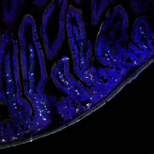
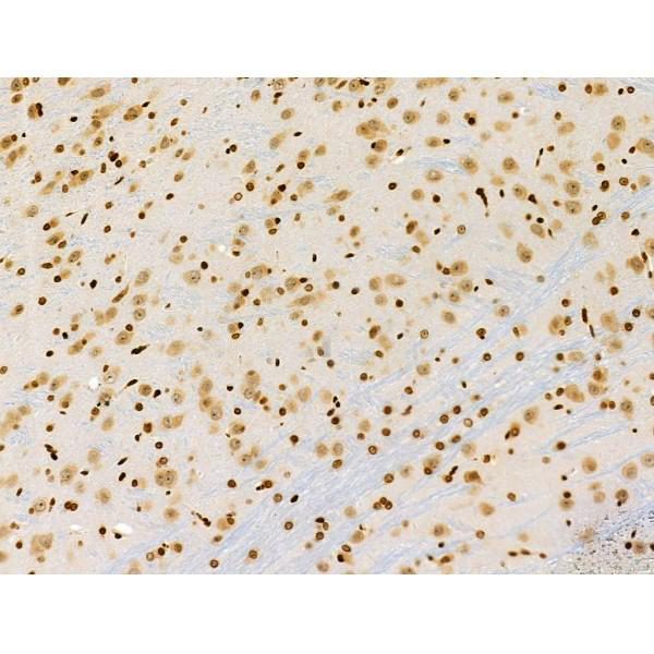
 IHC (Immunohistochemistry)
(Immunohistochemical analysis of paraffin-embedded Human brain section using Pink 1 (AAA28759). AAA28759 was diluted at 1:1000 dilution. A undiluted binotinylated goat polyvalent antibody was used as the secondary, followed by DAB staining)
IHC (Immunohistochemistry)
(Immunohistochemical analysis of paraffin-embedded Human brain section using Pink 1 (AAA28759). AAA28759 was diluted at 1:1000 dilution. A undiluted binotinylated goat polyvalent antibody was used as the secondary, followed by DAB staining)
S100B, Polyclonal Antibody (Cat# AAA28759)
Full Name
S100B Antibody
Gene Names
S100B; NEF; S100; S100-B; S100beta
Reactivity
Human, Mouse, Rat (Predicted: Rabbit)
Applications
WB, IHC-P-Leica, FC/FACS, IHC, EIA
Purity
Peptide Affinity Purified Rabbit Polyclonal Antibody (Pab)
Pricing


 IHC (Immunohistochemistry)
(At 1/100 staining Human ovarian cancer and adjacent normal tissues by IHC-P. The sample was formaldehyde fixed and a heat mediated antigen retrieval step in citrate buffer was performed. The sample was then blocked and incubated with the primary antibody at 4 degree C overnight. An HRP conjugated anti-Rabbit antibody was used as the secondary antibody.)
IHC (Immunohistochemistry)
(At 1/100 staining Human ovarian cancer and adjacent normal tissues by IHC-P. The sample was formaldehyde fixed and a heat mediated antigen retrieval step in citrate buffer was performed. The sample was then blocked and incubated with the primary antibody at 4 degree C overnight. An HRP conjugated anti-Rabbit antibody was used as the secondary antibody.)
RSK1/2/3/4, Polyclonal Antibody (Cat# AAA31315)
Full Name
Phospho-RSK1/2/3/4 (Ser221/Ser227/Ser218/Ser232) Antibody
Gene Names
RPS6KA1; RSK; HU-1; RSK1; MAPKAPK1A
Reactivity
Human, Mouse, Rat, Monkey
Applications
WB, IHC, EIA
Purity
The antibody is from purified rabbit serum by affinity purification via sequential chromatography on phospho-peptide and non-phospho-peptide affinity columns.
Pricing


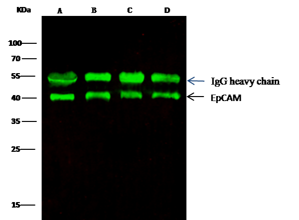 IP (Immunoprecipitation)
(EpCAM was immunoprecipitated using:Lane A:0.5 mg MCF-7 Whole Cell LysateLane B:0.5 mg A431 Whole Cell LysateLane C:0.5 mg HepG2 Whole Cell LysateLane D:0.5 mg Caco-2 Whole Cell Lysate0.5 uL anti-EpCAM rabbit monoclonal antibody and 15 ul of 50 % Protein G agarose.Primary antibody:Anti-EpCAM rabbit monoclonal antibody,at 1:5000 dilution Secondary antibody:Dylight 800-labeled antibody to rabbit IgG (H+L), at 1:5000 dilution Developed using the odssey technique.Performed under reducing conditions.Predicted band size: 40 kDaObserved band size: 40 kDa)
IP (Immunoprecipitation)
(EpCAM was immunoprecipitated using:Lane A:0.5 mg MCF-7 Whole Cell LysateLane B:0.5 mg A431 Whole Cell LysateLane C:0.5 mg HepG2 Whole Cell LysateLane D:0.5 mg Caco-2 Whole Cell Lysate0.5 uL anti-EpCAM rabbit monoclonal antibody and 15 ul of 50 % Protein G agarose.Primary antibody:Anti-EpCAM rabbit monoclonal antibody,at 1:5000 dilution Secondary antibody:Dylight 800-labeled antibody to rabbit IgG (H+L), at 1:5000 dilution Developed using the odssey technique.Performed under reducing conditions.Predicted band size: 40 kDaObserved band size: 40 kDa)
EpCAM, Monoclonal Antibody (Cat# AAA27736)
Full Name
Recombinant Anti-EpCAM Antibody, Rabbit Monoclonal
Gene Names
EPCAM; ESA; KSA; M4S1; MK-1; DIAR5; EGP-2; EGP40; KS1/4; MIC18; TROP1; EGP314; HNPCC8; TACSTD1
Reactivity
Human
Applications
WB, EIA, EIA, Det, IHC-P, FC/FACS/FCM, ICC, IF, IP
Purity
Protein A
Pricing


 Application Data
(Staining of J774 cells with Rat anti Mouse F4/80 antigen Biotin)
Application Data
(Staining of J774 cells with Rat anti Mouse F4/80 antigen Biotin)
F4/80, Monoclonal Antibody (Cat# AAA12169)
Full Name
RAT ANTI MOUSE F4/80
Gene Names
Emr1; Ly71; F4/80; Gpf480; TM7LN3; DD7A5-7; EGF-TM7
Applications
EM, FC/FACS, IF, IP, RIA, RE, WB
Pricing


 Application Data
(At 25 degree C. Samples were then incubated with primary Ab(At 37 degree C. An AlexaFluor594 conjugated goat anti-rabbit IgG(H+L) Ab(Red) and an AlexaFluor488 conjugated goat anti-mouse IgG(H+L) Ab(Green) were used as the secondary antibody.The nuclear counter stain is DAPI(blue).)
Application Data
(At 25 degree C. Samples were then incubated with primary Ab(At 37 degree C. An AlexaFluor594 conjugated goat anti-rabbit IgG(H+L) Ab(Red) and an AlexaFluor488 conjugated goat anti-mouse IgG(H+L) Ab(Green) were used as the secondary antibody.The nuclear counter stain is DAPI(blue).)
RSK1 p90, Polyclonal Antibody (Cat# AAA31374)
Full Name
Phospho-RSK1 p90 (Thr573) Antibody
Gene Names
RPS6KA1; RSK; HU-1; RSK1; MAPKAPK1A
Reactivity
Human, Mouse, Rat
Predicted Reactivity: Pig (100%), Zebrafish (100%), Bovine (100%), Horse (100%), Sheep (100%), Rabbit (100%), Dog (100%), Xenopus (100%)
Predicted Reactivity: Pig (100%), Zebrafish (100%), Bovine (100%), Horse (100%), Sheep (100%), Rabbit (100%), Dog (100%), Xenopus (100%)
Applications
WB, IHC, IF, ICC, EIA
Purity
The antibody is from purified rabbit serum by affinity purification via sequential chromatography on phospho-peptide and non-phospho-peptide affinity columns.
Pricing