Filters
Clonality
Type
Reactivity
Gene Name
Isotype
Host
Application
Clone
666 results for " B Cells" - showing 600-650


 Application Data
(Staining of J774 cells with Rat anti Mouse F4/80 antigen Biotin)
Application Data
(Staining of J774 cells with Rat anti Mouse F4/80 antigen Biotin)
F4/80, Monoclonal Antibody (Cat# AAA12173)
Full Name
RAT ANTI MOUSE F4/80
Gene Names
Emr1; Ly71; F4/80; Gpf480; TM7LN3; DD7A5-7; EGF-TM7
Applications
EM, FC/FACS, IF, IP, RIA, RE, WB
Pricing


 IF (Immunofluorescence)
(Figure 10 Immunofluorescence images of PUMA in the P14 rat retina (Wakabayashi et al., 2012)PUMA expression in the rat retina detected by anti-PUMA antibodies (3043). The specimens were counterstained with Hoechst 33258 to visualize nuclei (+DNA). GCL, ganglion cell layer; INL, inner nuclear layer; IPL, inner plexiform layer; ONL, outer nuclear layer; OPL, outer plexiform layer; P, postnatal day.)
IF (Immunofluorescence)
(Figure 10 Immunofluorescence images of PUMA in the P14 rat retina (Wakabayashi et al., 2012)PUMA expression in the rat retina detected by anti-PUMA antibodies (3043). The specimens were counterstained with Hoechst 33258 to visualize nuclei (+DNA). GCL, ganglion cell layer; INL, inner nuclear layer; IPL, inner plexiform layer; ONL, outer nuclear layer; OPL, outer plexiform layer; P, postnatal day.)
PUMA, Polyclonal Antibody (Cat# AAA10934)
Full Name
PUMA Antibody
Gene Names
BBC3; JFY1; PUMA; JFY-1
Reactivity
Human
Applications
EIA, WB, IHC, IF
Purity
PUMA Antibody is affinity chromatography purified via peptide column.
Pricing


 WB (Western Blot)
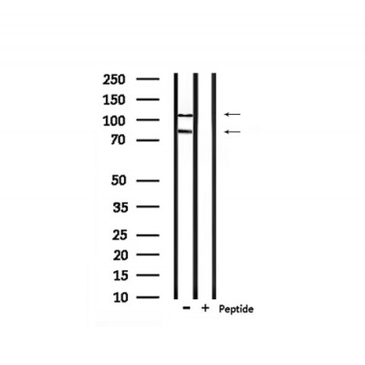
(Figure 9 KD Validation in HeLa Cells (Horinaka et al., 2005)HeLa cells were transfected with DR4siRNA or LacZ control siRNA. At 24 h after transfection, the cells were treated with or without 20 μM luteolin for 24 h. Western blot analysis was carried out with anti-DR4 antibodies. DR4 expression was markedly reduced after DR4 knockdown.)
WB (Western Blot)
(Figure 9 KD Validation in HeLa Cells (Horinaka et al., 2005)HeLa cells were transfected with DR4siRNA or LacZ control siRNA. At 24 h after transfection, the cells were treated with or without 20 μM luteolin for 24 h. Western blot analysis was carried out with anti-DR4 antibodies. DR4 expression was markedly reduced after DR4 knockdown.)
DR4, Polyclonal Antibody (Cat# AAA10950)
Full Name
DR4 Antibody
Gene Names
TNFRSF10A; DR4; APO2; CD261; TRAILR1; TRAILR-1
Reactivity
Human
Applications
EIA, WB
Purity
DR4 Antibody is affinity chromatography purified via peptide column.
Pricing


 Application Data
(Published customer image: Representative images of the inflammatory changes in the facial nucleus during axonal regeneration, one week following facial nerve transaction. a, b: CD11b immunoreactivity for microglia is increased in the axotomized facial nucleus, and microglia enwrap the facial motor neurons, e.g. at arrows. The regenerating neurons were retrogradely labelled with fluorogold. c, d: CD6- positive T-cells accumulated in the injured motor nucleus (arrows). They had little cytoplasm but dense nuclei (c) and were sometimes clustered around neurons retrogradely labelled with fluorogold (d). The scale bar in (a) also applies to (b) and that in (c) also applies to (d).From: Shokouhi et al. BMC Neuroscience 2010 11:13.)
Application Data
(Published customer image: Representative images of the inflammatory changes in the facial nucleus during axonal regeneration, one week following facial nerve transaction. a, b: CD11b immunoreactivity for microglia is increased in the axotomized facial nucleus, and microglia enwrap the facial motor neurons, e.g. at arrows. The regenerating neurons were retrogradely labelled with fluorogold. c, d: CD6- positive T-cells accumulated in the injured motor nucleus (arrows). They had little cytoplasm but dense nuclei (c) and were sometimes clustered around neurons retrogradely labelled with fluorogold (d). The scale bar in (a) also applies to (b) and that in (c) also applies to (d).From: Shokouhi et al. BMC Neuroscience 2010 11:13.)
CD11b, Monoclonal Antibody (Cat# AAA11969)
Full Name
MOUSE ANTI RAT CD11b
Gene Names
ITGAM; CD11B
Applications
FC/FACS, IF, IP
Pricing


 WB (Western Blot)
(Figure 1. Western blot analysis of Lyn using anti-Lyn antibody (AAA19150). Electrophoresis was performed on a 5-20% SDS-PAGE gel at 70V (Stacking gel) / 90V (Resolving gel) for 2-3 hours. The sample well of each lane was loaded with 50ug of sample under reducing conditions. Lane 1: human 293T whole cell lysate. After Electrophoresis, proteins were transferred to a Nitrocellulose membrane at 150mA for 50-90 minutes. Blocked the membrane with 5% Non-fat Milk/ TBS for 1.5 hour at RT. The membrane was incubated with rabbit anti-Lyn antigen affinity purified polyclonal antibody at 0.5ug/mL overnight at 4 degree C, then washed with TBS-0.1%Tween 3 times with 5 minutes each and probed with a goat anti-rabbit IgG-HRP secondary antibody at a dilution of 1:10000 for 1.5 hour at RT. The signal is developed using an Enhanced Chemiluminescent detection (ECL) kit with Tanon 5200 system. A specific band was detected for Lyn at approximately 59KD. The expected band size for Lyn is at 59KD.)
WB (Western Blot)
(Figure 1. Western blot analysis of Lyn using anti-Lyn antibody (AAA19150). Electrophoresis was performed on a 5-20% SDS-PAGE gel at 70V (Stacking gel) / 90V (Resolving gel) for 2-3 hours. The sample well of each lane was loaded with 50ug of sample under reducing conditions. Lane 1: human 293T whole cell lysate. After Electrophoresis, proteins were transferred to a Nitrocellulose membrane at 150mA for 50-90 minutes. Blocked the membrane with 5% Non-fat Milk/ TBS for 1.5 hour at RT. The membrane was incubated with rabbit anti-Lyn antigen affinity purified polyclonal antibody at 0.5ug/mL overnight at 4 degree C, then washed with TBS-0.1%Tween 3 times with 5 minutes each and probed with a goat anti-rabbit IgG-HRP secondary antibody at a dilution of 1:10000 for 1.5 hour at RT. The signal is developed using an Enhanced Chemiluminescent detection (ECL) kit with Tanon 5200 system. A specific band was detected for Lyn at approximately 59KD. The expected band size for Lyn is at 59KD.)
Lyn, Polyclonal Antibody (Cat# AAA19150)
Full Name
Anti-Lyn Picoband Antibody
Gene Names
LYN; JTK8; p53Lyn; p56Lyn
Reactivity
Human, Mouse, Rat
No cross reactivity with other proteins.
No cross reactivity with other proteins.
Applications
IHC, WB
Purity
Immunogen affinity purified
Pricing


 Application Data
(At 25 degree C. Samples were then incubated with primary Ab(At 37 degree C. An AlexaFluor594 conjugated goat anti-rabbit IgG(H+L) Ab(Red) and an AlexaFluor488 conjugated goat anti-mouse IgG(H+L) Ab(Green) were used as the secondary antibody.The nuclear counter stain is DAPI(blue).)
Application Data
(At 25 degree C. Samples were then incubated with primary Ab(At 37 degree C. An AlexaFluor594 conjugated goat anti-rabbit IgG(H+L) Ab(Red) and an AlexaFluor488 conjugated goat anti-mouse IgG(H+L) Ab(Green) were used as the secondary antibody.The nuclear counter stain is DAPI(blue).)
RBL2, Polyclonal Antibody (Cat# AAA31451)
Full Name
Phospho-RBL2 (Thr642) Antibody
Gene Names
RBL2; Rb2; P130
Reactivity
Human, Mouse, Rat
Predicted Reactivity: Pig (100%), Bovine (100%), Horse (89%), Sheep (100%), Rabbit (89%), Dog (100%)
Predicted Reactivity: Pig (100%), Bovine (100%), Horse (89%), Sheep (100%), Rabbit (89%), Dog (100%)
Applications
WB, IHC, IF, ICC, EIA
Purity
The antibody is from purified rabbit serum by affinity purification via sequential chromatography on phospho-peptide and non-phospho-peptide affinity columns.
Pricing


 Application Data
(Published clone specific image: Flow cytometric analysis of AM from NO2-exposed and control rats. Rats were exposed to NO2 for the indicated times and BAL cells were stained with antibodies to ED7, ED9, RM-4, and OX-6. To overcome autofluorescence signals, primary antibodies were detected using a biotin-PE/streptavidin-anti-streptavidin enhancing system and labeling of AM was analyzed by flow cytometry following gating by help of forward and sideward scatter properties. Shown are representative results of at least six animals per group.From: Garn H, Siese A, Stumpf S, Wensing A, Renz H, Gemsa D. Phenotypical and functional characterization of alveolar macrophage subpopulations in the lungs of NO2-exposed rats. Respir Res. 2006 Jan 6;7:4.)
Application Data
(Published clone specific image: Flow cytometric analysis of AM from NO2-exposed and control rats. Rats were exposed to NO2 for the indicated times and BAL cells were stained with antibodies to ED7, ED9, RM-4, and OX-6. To overcome autofluorescence signals, primary antibodies were detected using a biotin-PE/streptavidin-anti-streptavidin enhancing system and labeling of AM was analyzed by flow cytometry following gating by help of forward and sideward scatter properties. Shown are representative results of at least six animals per group.From: Garn H, Siese A, Stumpf S, Wensing A, Renz H, Gemsa D. Phenotypical and functional characterization of alveolar macrophage subpopulations in the lungs of NO2-exposed rats. Respir Res. 2006 Jan 6;7:4.)
CD172a, Monoclonal Antibody (Cat# AAA12001)
Full Name
MOUSE ANTI RAT CD172a
Gene Names
Sirpa; Bit; Ptpns1; SHPS-1
Applications
FC/FACS, IP, WB
Pricing


 Application Data
(Source:Arthrobacter luteus)
Application Data
(Source:Arthrobacter luteus)
Zymolyase 20T, Enzyme (Cat# AAA14844)
Full Name
Zymolyase 20T (Lyticase, Yeast Lytic Enzyme)
Pricing


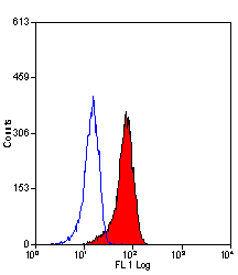
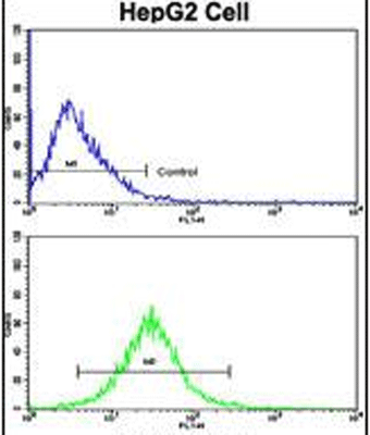
 FCM (Flow Cytometry)
(Figure 6. Flow Cytometry analysis of PC-3 cells using anti-APP antibody (AAA11597).Overlay histogram showing PC-3 cells stained with AAA11597 (Blue line).The cells were blocked with 10% normal goat serum. And then incubated with rabbit anti-APP Antibody (AAA11597,1ug/1x10^6 cells) for 30 min at 20 degree C. DyLight®488 conjugated goat anti-rabbit IgG (5-10ug/1x10^6 cells) was used as secondary antibody for 30 minutes at 20 degree C. Isotype control antibody (Green line) was rabbit IgG (1ug/1x106) used under the same conditions. Unlabelled sample (Red line) was also used as a control.)
FCM (Flow Cytometry)
(Figure 6. Flow Cytometry analysis of PC-3 cells using anti-APP antibody (AAA11597).Overlay histogram showing PC-3 cells stained with AAA11597 (Blue line).The cells were blocked with 10% normal goat serum. And then incubated with rabbit anti-APP Antibody (AAA11597,1ug/1x10^6 cells) for 30 min at 20 degree C. DyLight®488 conjugated goat anti-rabbit IgG (5-10ug/1x10^6 cells) was used as secondary antibody for 30 minutes at 20 degree C. Isotype control antibody (Green line) was rabbit IgG (1ug/1x106) used under the same conditions. Unlabelled sample (Red line) was also used as a control.)
beta Amyloid, Polyclonal Antibody (Cat# AAA11597)
Full Name
Anti-beta Amyloid Antibody
Gene Names
APP; AAA; AD1; PN2; ABPP; APPI; CVAP; ABETA; PN-II; CTFgamma
Reactivity
Human, Mouse, Rat
Applications
WB, IHC
Purity
Immunogen affinity purified.
Pricing


 IF (Immunofluorescence)
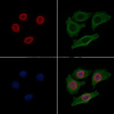
(Fluorescent confocal image of MCF-7 cell stained with BHLH3 Antibody (N-term). MCF-7 cells were fixed with 4% PFA (20 min), permeabilized with Triton X-100 (0.1%, 10 min), then incubated with BHLH3 primary antibody (1:25, 1 h at 37 degree). For secondary antibody, Alexa Fluor 488 conjugated donkey anti-rabbit antibody (green) was used (1:400, 50 min at 37 degree).Cytoplasmic actin was counterstained with Alexa Fluor 555 (red) conjugated Phalloidin (7units/ml, 1 h at 37 degree). Nuclei were counterstained with DAPI (blue) (10 ug/ml, 10 min).BHLH3 immunoreactivity is localized to nucleus significantly.)
IF (Immunofluorescence)
(Fluorescent confocal image of MCF-7 cell stained with BHLH3 Antibody (N-term). MCF-7 cells were fixed with 4% PFA (20 min), permeabilized with Triton X-100 (0.1%, 10 min), then incubated with BHLH3 primary antibody (1:25, 1 h at 37 degree). For secondary antibody, Alexa Fluor 488 conjugated donkey anti-rabbit antibody (green) was used (1:400, 50 min at 37 degree).Cytoplasmic actin was counterstained with Alexa Fluor 555 (red) conjugated Phalloidin (7units/ml, 1 h at 37 degree). Nuclei were counterstained with DAPI (blue) (10 ug/ml, 10 min).BHLH3 immunoreactivity is localized to nucleus significantly.)
BHLH3, Polyclonal Antibody (Cat# AAA28734)
Full Name
BHLH3 Antibody (N-term)
Gene Names
BHLHE41; DEC2; hDEC2; BHLHB3; SHARP1
Reactivity
Human, Mouse
Applications
FC, IF, WB, EIA
Purity
This antibody is purified through a protein A column, followed by peptide affinity purification.
Pricing


 IF (Immunofluorescence)
(AAA31089 staining 293 by IF/ICC. The sample were fixed with PFA and permeabilized in 0.1% Triton X-100, then blocked in 10% serum for 45 minutes at 25 degree C. The primary antibody was diluted at 1/200 and incubated with the sample for 1 hour at 37 degree C. An Alexa Fluor 594 conjugated goat anti-rabbit IgG (H+L) Ab, diluted at 1/600, was used as the secondary antibody.)
IF (Immunofluorescence)
(AAA31089 staining 293 by IF/ICC. The sample were fixed with PFA and permeabilized in 0.1% Triton X-100, then blocked in 10% serum for 45 minutes at 25 degree C. The primary antibody was diluted at 1/200 and incubated with the sample for 1 hour at 37 degree C. An Alexa Fluor 594 conjugated goat anti-rabbit IgG (H+L) Ab, diluted at 1/600, was used as the secondary antibody.)
AKT, Polyclonal Antibody (Cat# AAA31089)
Full Name
AKT Antibody
Gene Names
AKT1; AKT; PKB; RAC; CWS6; PRKBA; PKB-ALPHA; RAC-ALPHA
Reactivity
Human, Mouse, Rat
Applications
WB, IHC, IF, ICC, IP, EIA
Purity
The antiserum was purified by peptide affinity chromatography using SulfoLink Coupling Resin.
Pricing


 Application Data
(Published customer image: Increased accumulation of repair-associated macrophages surrounding collaterals in ischemic hind limbs is PAR2-dependent. (A) Stainings of CD206-positive macrophages (green) and SMA-positive vessels (red) in non-ischemic (control) and ischemic (ligated) hind limbs of WT, PAR1-/- and PAR2-/- mice are shown. Nuclei were visualized with DAPI (blue). Arrows indicate single macrophages in the non-ischemic adductor. Quantification of the average number of repair-associated macrophages per vessel is indicated on the right. (B) Correlation between the number of CD206-positive macrophages in the ischemic tissues and the expression of CD11b and (C) CD115 on monocytes. ** p)
Application Data
(Published customer image: Increased accumulation of repair-associated macrophages surrounding collaterals in ischemic hind limbs is PAR2-dependent. (A) Stainings of CD206-positive macrophages (green) and SMA-positive vessels (red) in non-ischemic (control) and ischemic (ligated) hind limbs of WT, PAR1-/- and PAR2-/- mice are shown. Nuclei were visualized with DAPI (blue). Arrows indicate single macrophages in the non-ischemic adductor. Quantification of the average number of repair-associated macrophages per vessel is indicated on the right. (B) Correlation between the number of CD206-positive macrophages in the ischemic tissues and the expression of CD11b and (C) CD115 on monocytes. ** p)
CD206, Monoclonal Antibody (Cat# AAA12123)
Full Name
RAT ANTI MOUSE CD206
Gene Names
Mrc1; MR; CD206; AW259686
Applications
FC/FACS, IF, IP
Pricing


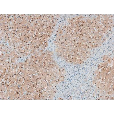
 IHC (Immunohistochemistry)
(At 1/100 staining Mouse kidney tissue by IHC-P. The sample was formaldehyde fixed and a heat mediated antigen retrieval step in citrate buffer was performed. The sample was then blocked and incubated with the primary antibody at 4 degree C overnight. An HRP conjugated anti-Rabbit antibody was used as the secondary antibody.)
IHC (Immunohistochemistry)
(At 1/100 staining Mouse kidney tissue by IHC-P. The sample was formaldehyde fixed and a heat mediated antigen retrieval step in citrate buffer was performed. The sample was then blocked and incubated with the primary antibody at 4 degree C overnight. An HRP conjugated anti-Rabbit antibody was used as the secondary antibody.)
YB1, Polyclonal Antibody (Cat# AAA31294)
Full Name
Phospho-YB1 (Ser102) Antibody
Gene Names
YBX1; YB1; BP-8; CSDB; DBPB; YB-1; CSDA2; NSEP1; NSEP-1; MDR-NF1
Reactivity
Human, Mouse, Rat
Applications
IHC, EIA
Purity
The antibody is from purified rabbit serum by affinity purification via sequential chromatography on phospho-peptide and non-phospho-peptide affinity columns.
Pricing


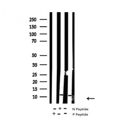
 IP (Immunoprecipitation)
(HSPA1A was immunoprecipitated using:Lane A:0.5 mg Hela Whole Cell Lysate2 uL anti-HSPA1A rabbit monoclonal antibody and 15 ul of 50 % Protein G agarose.Primary antibody:Anti-HSPA1A rabbit monoclonal antibody,at 1:200 dilution Secondary antibody:Dylight 800-labeled antibody to rabbit IgG (H+L), at 1:5000 dilution Developed using the odssey technique.Performed under reducing conditions.Predicted band size: 70 kDaObserved band size: 70 kDa)
IP (Immunoprecipitation)
(HSPA1A was immunoprecipitated using:Lane A:0.5 mg Hela Whole Cell Lysate2 uL anti-HSPA1A rabbit monoclonal antibody and 15 ul of 50 % Protein G agarose.Primary antibody:Anti-HSPA1A rabbit monoclonal antibody,at 1:200 dilution Secondary antibody:Dylight 800-labeled antibody to rabbit IgG (H+L), at 1:5000 dilution Developed using the odssey technique.Performed under reducing conditions.Predicted band size: 70 kDaObserved band size: 70 kDa)
HSP70, Monoclonal Antibody (Cat# AAA27747)
Full Name
Recombinant Anti-HSP70 Antibody, Rabbit Monoclonal
Gene Names
HSPA1B; HSP72; HSPA1; HSX70; HSP70-1; HSP70-2; HSP70.1; HSP70.2; HSP70-1B
Reactivity
Human
Applications
WB, EIA, IHC-P, FC/FACS/FCM, ICC, IF, IP
Purity
Protein A
Pricing


 Application Data
(Figure 10 KD Validation in HeLa cells (Horinaka et al., 2005)HeLa cells were transfected with DR4siRNA or LacZ control siRNA. At 24 h after transfection, the cells were treated with or without 20 ?M luteolin for 24 h. Western blot analysis was carried out with anti-DR4 antibodies (1139). DR4 expression was markedly reduced after DR4 knockdown.)
Application Data
(Figure 10 KD Validation in HeLa cells (Horinaka et al., 2005)HeLa cells were transfected with DR4siRNA or LacZ control siRNA. At 24 h after transfection, the cells were treated with or without 20 ?M luteolin for 24 h. Western blot analysis was carried out with anti-DR4 antibodies (1139). DR4 expression was markedly reduced after DR4 knockdown.)
DR4, Polyclonal Antibody (Cat# AAA10952)
Full Name
DR4 Antibody
Gene Names
TNFRSF10A; DR4; APO2; CD261; TRAILR1; TRAILR-1
Reactivity
Human
Applications
EIA, WB, ICC
Purity
DR4 Antibody is Antibody is affinity chromatography purified via peptide column.
Pricing


 Application Data
(Published Customer Image:Rat anti Mouse Gr-1 antibody, clone RB6-8C5 used for the identification of neutrophils by immunofluorescence.Image caption:S. typhimurium-Infected Macrophages Containing Phagocytosed Neutrophils and T Cells Confocal fluorescence microscopy of 50-mum-thick liver sections from 1-wk-infected Slc11a1 wild-type mice. (A-C) S.Typhimurium (O-antigen, arrows) are red, macrophages (F4-80 and MOMA-2) are blue, DNA (DAPI) is gray, phalloidin is green, and neutrophils (Gr-1/Ly-6G/RB6-8C5) are pink (arrowheads). (A) Collapsed image from a 40-mum Z-stack. Scale bar is 20 mum. (B and C) Sections from (A) that are 4 mum apart. The video from which (A-C) were derived (Video S2) is available online. (D-G) T cells within multinucleate macrophages.Macrophages (F4-80 and MOMA-2) are blue (D, G, and H), T cells (CD3zeta) are red (D, G, arrowheads), DAPI is gray (E, G), actin-bound phalloidin is green (F, G). (G) Is a composite of (D, E, and F). Scale bars are 16 mum. (H) An image from a different mouse stained and labeled as described for (D-G). Scale bar is 8 mum. A video showing a T cell inside of a macrophage is available online (Video S3).From: Nix RN, Altschuler SE, Henson PM, Detweiler CS (2007) Hemophagocytic Macrophages Harbor Salmonella enterica during Persistent Infection.PLoS Pathog 3(12): e193.)
Application Data
(Published Customer Image:Rat anti Mouse Gr-1 antibody, clone RB6-8C5 used for the identification of neutrophils by immunofluorescence.Image caption:S. typhimurium-Infected Macrophages Containing Phagocytosed Neutrophils and T Cells Confocal fluorescence microscopy of 50-mum-thick liver sections from 1-wk-infected Slc11a1 wild-type mice. (A-C) S.Typhimurium (O-antigen, arrows) are red, macrophages (F4-80 and MOMA-2) are blue, DNA (DAPI) is gray, phalloidin is green, and neutrophils (Gr-1/Ly-6G/RB6-8C5) are pink (arrowheads). (A) Collapsed image from a 40-mum Z-stack. Scale bar is 20 mum. (B and C) Sections from (A) that are 4 mum apart. The video from which (A-C) were derived (Video S2) is available online. (D-G) T cells within multinucleate macrophages.Macrophages (F4-80 and MOMA-2) are blue (D, G, and H), T cells (CD3zeta) are red (D, G, arrowheads), DAPI is gray (E, G), actin-bound phalloidin is green (F, G). (G) Is a composite of (D, E, and F). Scale bars are 16 mum. (H) An image from a different mouse stained and labeled as described for (D-G). Scale bar is 8 mum. A video showing a T cell inside of a macrophage is available online (Video S3).From: Nix RN, Altschuler SE, Henson PM, Detweiler CS (2007) Hemophagocytic Macrophages Harbor Salmonella enterica during Persistent Infection.PLoS Pathog 3(12): e193.)
Gr-1, Monoclonal Antibody (Cat# AAA12257)
Full Name
Rat Anti Mouse Gr-1: FITC
Reactivity
Mouse
Applications
FC/FACS
Purity
Purified IgG prepared by affinity chromatography on Protein G from tissue culture supernatant
Pricing


 Application Data
(At 25 degree C. Samples were then incubated with primary Ab(At 37 degree C. An AlexaFluor594 conjugated goat anti-rabbit IgG(H+L) Ab(Red) and an AlexaFluor488 conjugated goat anti-mouse IgG(H+L) Ab(Green) were used as the secondary antibody.The nuclear counter stain is DAPI(blue).)
Application Data
(At 25 degree C. Samples were then incubated with primary Ab(At 37 degree C. An AlexaFluor594 conjugated goat anti-rabbit IgG(H+L) Ab(Red) and an AlexaFluor488 conjugated goat anti-mouse IgG(H+L) Ab(Green) were used as the secondary antibody.The nuclear counter stain is DAPI(blue).)
Histone H3, Polyclonal Antibody (Cat# AAA31345)
Full Name
Acetyl-Histone H3 (Lys14) Antibody
Gene Names
HIST1H3A; H3/A; H3FA
Reactivity
Human, Mouse, Rat
Predicted Reactivity: Bovine (100%)
Predicted Reactivity: Bovine (100%)
Applications
WB, IHC, IF, ICC, EIA
Purity
The antiserum was purified by peptide affinity chromatography using SulfoLink Coupling Resin
Pricing


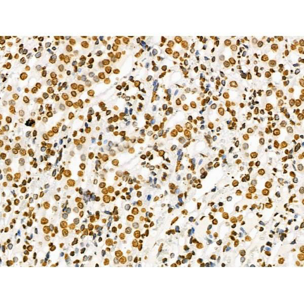
 IHC (Immunohistochemistry)
(At 1/100 staining Human colorectal cancer and adjacent normal tissues by IHC-P. The sample was formaldehyde fixed and a heat mediated antigen retrieval step in citrate buffer was performed. The sample was then blocked and incubated with the primary antibody at 4 degree C overnight. An HRP conjugated anti-Rabbit antibody was used as the secondary antibody.)
IHC (Immunohistochemistry)
(At 1/100 staining Human colorectal cancer and adjacent normal tissues by IHC-P. The sample was formaldehyde fixed and a heat mediated antigen retrieval step in citrate buffer was performed. The sample was then blocked and incubated with the primary antibody at 4 degree C overnight. An HRP conjugated anti-Rabbit antibody was used as the secondary antibody.)
Histone H3, Polyclonal Antibody (Cat# AAA31356)
Full Name
Acetyl-Histone H3 (Lys9/14/18/23/27) Antibody
Gene Names
HIST1H3A; H3/A; H3FA
Reactivity
Human, Mouse, Rat
Predicted Reactivity: Bovine (100%)
Predicted Reactivity: Bovine (100%)
Applications
WB, IHC, IF, ICC, EIA
Purity
The antiserum was purified by peptide affinity chromatography using SulfoLink Coupling Resin
Pricing


 IF (Immunofluorescence)
(Confocal immunofluorescent analysis of GAPDH Antibody (C-term R248) with Hela cell followed by Alexa Fluor 488-conjugated goat anti-rabbit lgG (green). Actin filaments have been labeled with Alexa Fluor 555 phalloidin (red).DAPI was used to stain the cell nuclear (blue).)
IF (Immunofluorescence)
(Confocal immunofluorescent analysis of GAPDH Antibody (C-term R248) with Hela cell followed by Alexa Fluor 488-conjugated goat anti-rabbit lgG (green). Actin filaments have been labeled with Alexa Fluor 555 phalloidin (red).DAPI was used to stain the cell nuclear (blue).)
GAPDH, Polyclonal Antibody (Cat# AAA28693)
Full Name
GAPDH Antibody (C-term R248)
Gene Names
GAPDH; G3PD; GAPD; HEL-S-162eP
Reactivity
Human (Predicted Reactivity: Chicken, Mouse, Pig, Rat)
Applications
WB, EIA, IHC, FC/FACS, IF
Purity
Purified Rabbit Polyclonal Antibody (Pab)
Pricing


 Application Data
(At 25 degree C. Samples were then incubated with primary Ab(At 37 degree C. An AlexaFluor594 conjugated goat anti-rabbit IgG(H+L) Ab(Red) and an AlexaFluor488 conjugated goat anti-mouse IgG(H+L) Ab(Green) were used as the secondary antibody.The nuclear counter stain is DAPI(blue).)
Application Data
(At 25 degree C. Samples were then incubated with primary Ab(At 37 degree C. An AlexaFluor594 conjugated goat anti-rabbit IgG(H+L) Ab(Red) and an AlexaFluor488 conjugated goat anti-mouse IgG(H+L) Ab(Green) were used as the secondary antibody.The nuclear counter stain is DAPI(blue).)
MARK2, Polyclonal Antibody (Cat# AAA31392)
Full Name
Phospho-MARK2 (Thr596) Antibody
Gene Names
MARK2; EMK1; EMK-1; PAR-1; Par1b; Par-1b
Reactivity
Human, Mouse, Rat
Predicted Reactivity: Pig (100%), Bovine (100%), Horse (100%), Sheep (100%), Rabbit (100%), Dog (100%), Xenopus (100%)
Predicted Reactivity: Pig (100%), Bovine (100%), Horse (100%), Sheep (100%), Rabbit (100%), Dog (100%), Xenopus (100%)
Applications
WB, IHC, IF, ICC, EIA
Purity
The antibody is from purified rabbit serum by affinity purification via sequential chromatography on phospho-peptide and non-phospho-peptide affinity columns.
Pricing


 Application Data
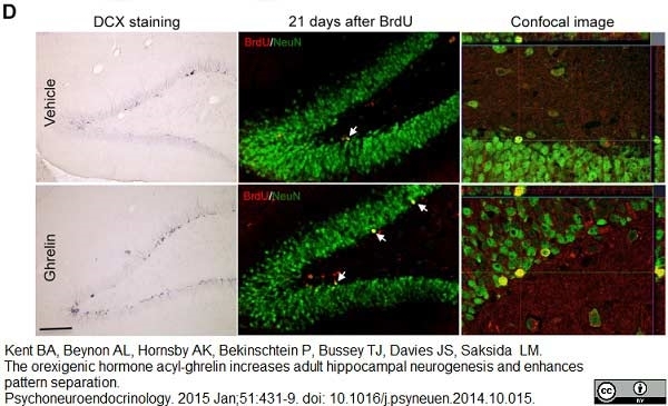
BrdU, Monoclonal Antibody (Cat# AAA12259)
Full Name
Mouse Anti BrdU
Applications
FC/FACS, ICC, IF, IHC
Purity
Purified IgG prepared by affinity chromatography on Protein A from tissue culture supernatant
Pricing


 Application Data
(Published customer image: Increased accumulation of repair-associated macrophages surrounding collaterals in ischemic hind limbs is PAR2-dependent. (A) Stainings of CD206-positive macrophages (green) and SMA-positive vessels (red) in non-ischemic (control) and ischemic (ligated) hind limbs of WT, PAR1-/- and PAR2-/- mice are shown. Nuclei were visualized with DAPI (blue). Arrows indicate single macrophages in the non-ischemic adductor. Quantification of the average number of repair-associated macrophages per vessel is indicated on the right. (B) Correlation between the number of CD206-positive macrophages in the ischemic tissues and the expression of CD11b and (C) CD115 on monocytes. ** p)
Application Data
(Published customer image: Increased accumulation of repair-associated macrophages surrounding collaterals in ischemic hind limbs is PAR2-dependent. (A) Stainings of CD206-positive macrophages (green) and SMA-positive vessels (red) in non-ischemic (control) and ischemic (ligated) hind limbs of WT, PAR1-/- and PAR2-/- mice are shown. Nuclei were visualized with DAPI (blue). Arrows indicate single macrophages in the non-ischemic adductor. Quantification of the average number of repair-associated macrophages per vessel is indicated on the right. (B) Correlation between the number of CD206-positive macrophages in the ischemic tissues and the expression of CD11b and (C) CD115 on monocytes. ** p)
CD206, Monoclonal Antibody (Cat# AAA12122)
Full Name
RAT ANTI MOUSE CD206:FITC
Gene Names
Mrc1; MR; CD206; AW259686
Applications
FC/FACS
Pricing


 Application Data
(Published customer image: Increased accumulation of repair-associated macrophages surrounding collaterals in ischemic hind limbs is PAR2-dependent. (A) Stainings of CD206-positive macrophages (green) and SMA-positive vessels (red) in non-ischemic (control) and ischemic (ligated) hind limbs of WT, PAR1-/- and PAR2-/- mice are shown. Nuclei were visualized with DAPI (blue). Arrows indicate single macrophages in the non-ischemic adductor. Quantification of the average number of repair-associated macrophages per vessel is indicated on the right. (B) Correlation between the number of CD206-positive macrophages in the ischemic tissues and the expression of CD11b and (C) CD115 on monocytes. ** p)
Application Data
(Published customer image: Increased accumulation of repair-associated macrophages surrounding collaterals in ischemic hind limbs is PAR2-dependent. (A) Stainings of CD206-positive macrophages (green) and SMA-positive vessels (red) in non-ischemic (control) and ischemic (ligated) hind limbs of WT, PAR1-/- and PAR2-/- mice are shown. Nuclei were visualized with DAPI (blue). Arrows indicate single macrophages in the non-ischemic adductor. Quantification of the average number of repair-associated macrophages per vessel is indicated on the right. (B) Correlation between the number of CD206-positive macrophages in the ischemic tissues and the expression of CD11b and (C) CD115 on monocytes. ** p)
CD206, Monoclonal Antibody (Cat# AAA12125)
Full Name
RAT ANTI MOUSE CD206:RPE
Gene Names
Mrc1; MR; CD206; AW259686
Applications
FC/FACS
Pricing


 Application Data
(Published customer image: Increased accumulation of repair-associated macrophages surrounding collaterals in ischemic hind limbs is PAR2-dependent. (A) Stainings of CD206-positive macrophages (green) and SMA-positive vessels (red) in non-ischemic (control) and ischemic (ligated) hind limbs of WT, PAR1-/- and PAR2-/- mice are shown. Nuclei were visualized with DAPI (blue). Arrows indicate single macrophages in the non-ischemic adductor. Quantification of the average number of repair-associated macrophages per vessel is indicated on the right. (B) Correlation between the number of CD206-positive macrophages in the ischemic tissues and the expression of CD11b and (C) CD115 on monocytes. ** p)
Application Data
(Published customer image: Increased accumulation of repair-associated macrophages surrounding collaterals in ischemic hind limbs is PAR2-dependent. (A) Stainings of CD206-positive macrophages (green) and SMA-positive vessels (red) in non-ischemic (control) and ischemic (ligated) hind limbs of WT, PAR1-/- and PAR2-/- mice are shown. Nuclei were visualized with DAPI (blue). Arrows indicate single macrophages in the non-ischemic adductor. Quantification of the average number of repair-associated macrophages per vessel is indicated on the right. (B) Correlation between the number of CD206-positive macrophages in the ischemic tissues and the expression of CD11b and (C) CD115 on monocytes. ** p)
CD206, Monoclonal Antibody (Cat# AAA12118)
Full Name
RAT ANTI MOUSE CD206:Biotin
Gene Names
Mrc1; MR; CD206; AW259686
Applications
FC/FACS
Pricing


 Application Data
(Published customer image: Histological staining of control and HBOT wounds at post-wounding days 7 and 29. A -D) H&E staining. E -H) CD34 immunohistochemistry. I+J) CD68 immunohistochemistry.From: uk B, Tong M, Fijneman EMG, van Neck JW (2014) Hyperbaric Oxygen Therapy to Treat Diabetes Impaired Wound Healing in Rats. PLoS ONE 9(10): e108533.)
Application Data
(Published customer image: Histological staining of control and HBOT wounds at post-wounding days 7 and 29. A -D) H&E staining. E -H) CD34 immunohistochemistry. I+J) CD68 immunohistochemistry.From: uk B, Tong M, Fijneman EMG, van Neck JW (2014) Hyperbaric Oxygen Therapy to Treat Diabetes Impaired Wound Healing in Rats. PLoS ONE 9(10): e108533.)
CD68, Monoclonal Antibody (Cat# AAA12151)
Full Name
MOUSE ANTI RAT CD68
Applications
FC/FACS, IF, IP, RIA, WB
Pricing


 Application Data
(Published customer image: Histological staining of control and HBOT wounds at post-wounding days 7 and 29. A -D) H&E staining. E -H) CD34 immunohistochemistry. I+J) CD68 immunohistochemistry.From: uk B, Tong M, Fijneman EMG, van Neck JW (2014) Hyperbaric Oxygen Therapy to Treat Diabetes Impaired Wound Healing in Rats. PLoS ONE 9(10): e108533.)
Application Data
(Published customer image: Histological staining of control and HBOT wounds at post-wounding days 7 and 29. A -D) H&E staining. E -H) CD34 immunohistochemistry. I+J) CD68 immunohistochemistry.From: uk B, Tong M, Fijneman EMG, van Neck JW (2014) Hyperbaric Oxygen Therapy to Treat Diabetes Impaired Wound Healing in Rats. PLoS ONE 9(10): e108533.)
CD68, Monoclonal Antibody (Cat# AAA12148)
Full Name
MOUSE ANTI RAT CD68:FITC
Applications
FC/FACS
Pricing


 Application Data
(Published customer image: Increased accumulation of repair-associated macrophages surrounding collaterals in ischemic hind limbs is PAR2-dependent. (A) Stainings of CD206-positive macrophages (green) and SMA-positive vessels (red) in non-ischemic (control) and ischemic (ligated) hind limbs of WT, PAR1-/- and PAR2-/- mice are shown. Nuclei were visualized with DAPI (blue). Arrows indicate single macrophages in the non-ischemic adductor. Quantification of the average number of repair-associated macrophages per vessel is indicated on the right. (B) Correlation between the number of CD206-positive macrophages in the ischemic tissues and the expression of CD11b and (C) CD115 on monocytes. ** p)
Application Data
(Published customer image: Increased accumulation of repair-associated macrophages surrounding collaterals in ischemic hind limbs is PAR2-dependent. (A) Stainings of CD206-positive macrophages (green) and SMA-positive vessels (red) in non-ischemic (control) and ischemic (ligated) hind limbs of WT, PAR1-/- and PAR2-/- mice are shown. Nuclei were visualized with DAPI (blue). Arrows indicate single macrophages in the non-ischemic adductor. Quantification of the average number of repair-associated macrophages per vessel is indicated on the right. (B) Correlation between the number of CD206-positive macrophages in the ischemic tissues and the expression of CD11b and (C) CD115 on monocytes. ** p)
CD206, Monoclonal Antibody (Cat# AAA12120)
Full Name
RAT ANTI MOUSE CD206:FITC
Gene Names
Mrc1; MR; CD206; AW259686
Applications
FC/FACS
Pricing


 Application Data
(Published customer image: Increased accumulation of repair-associated macrophages surrounding collaterals in ischemic hind limbs is PAR2-dependent. (A) Stainings of CD206-positive macrophages (green) and SMA-positive vessels (red) in non-ischemic (control) and ischemic (ligated) hind limbs of WT, PAR1-/- and PAR2-/- mice are shown. Nuclei were visualized with DAPI (blue). Arrows indicate single macrophages in the non-ischemic adductor. Quantification of the average number of repair-associated macrophages per vessel is indicated on the right. (B) Correlation between the number of CD206-positive macrophages in the ischemic tissues and the expression of CD11b and (C) CD115 on monocytes. ** p)
Application Data
(Published customer image: Increased accumulation of repair-associated macrophages surrounding collaterals in ischemic hind limbs is PAR2-dependent. (A) Stainings of CD206-positive macrophages (green) and SMA-positive vessels (red) in non-ischemic (control) and ischemic (ligated) hind limbs of WT, PAR1-/- and PAR2-/- mice are shown. Nuclei were visualized with DAPI (blue). Arrows indicate single macrophages in the non-ischemic adductor. Quantification of the average number of repair-associated macrophages per vessel is indicated on the right. (B) Correlation between the number of CD206-positive macrophages in the ischemic tissues and the expression of CD11b and (C) CD115 on monocytes. ** p)
CD206, Monoclonal Antibody (Cat# AAA12117)
Full Name
RAT ANTI MOUSE CD206:Biotin
Gene Names
Mrc1; MR; CD206; AW259686
Applications
FC/FACS
Pricing


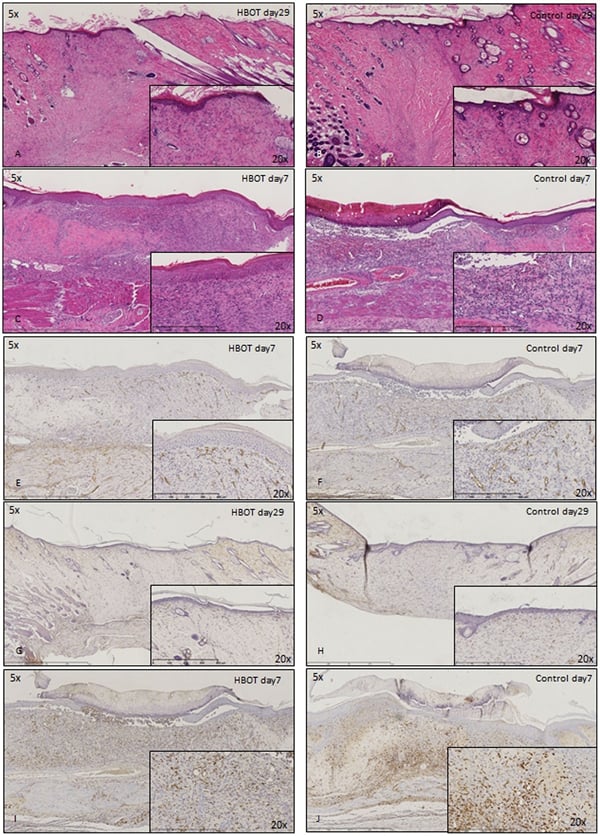 Application Data
(Published customer image: Histological staining of control and HBOT wounds at post-wounding days 7 and 29. A -D) H&E staining. E -H) CD34 immunohistochemistry. I+J) CD68 immunohistochemistry.From: uk B, Tong M, Fijneman EMG, van Neck JW (2014) Hyperbaric Oxygen Therapy to Treat Diabetes Impaired Wound Healing in Rats. PLoS ONE 9(10): e108533.)
Application Data
(Published customer image: Histological staining of control and HBOT wounds at post-wounding days 7 and 29. A -D) H&E staining. E -H) CD34 immunohistochemistry. I+J) CD68 immunohistochemistry.From: uk B, Tong M, Fijneman EMG, van Neck JW (2014) Hyperbaric Oxygen Therapy to Treat Diabetes Impaired Wound Healing in Rats. PLoS ONE 9(10): e108533.)
CD68, Monoclonal Antibody (Cat# AAA12150)
Full Name
MOUSE ANTI RAT CD68:RPE
Applications
FC/FACS
Pricing


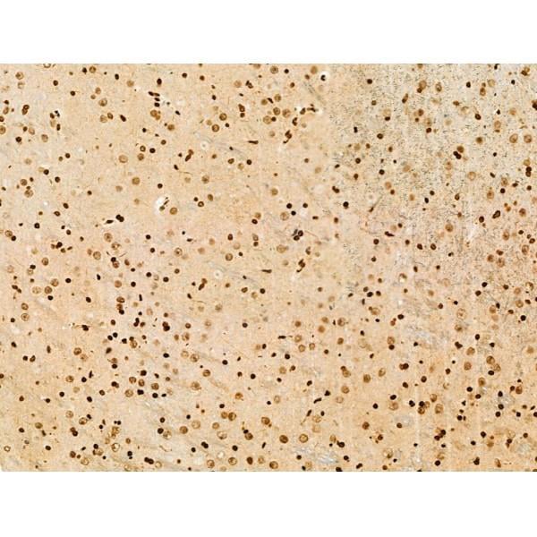
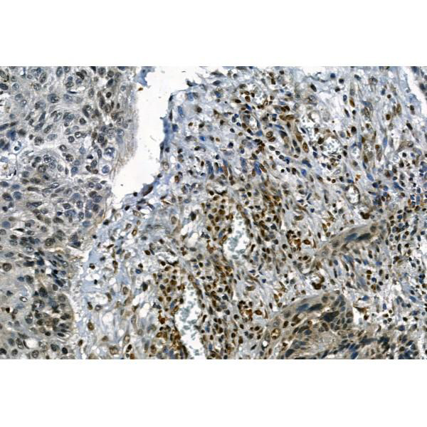
 IHC (Immunohistochemistry)
(At 1/100 staining Human pancreatic cancer by IHC-P. The sample was formaldehyde fixed and a heat mediated antigen retrieval step in citrate buffer was performed. The sample was then blocked and incubated with the primary antibody at 4 degree C overnight. An HRP conjugated anti-Rabbit antibody was used as the secondary antibody.)
IHC (Immunohistochemistry)
(At 1/100 staining Human pancreatic cancer by IHC-P. The sample was formaldehyde fixed and a heat mediated antigen retrieval step in citrate buffer was performed. The sample was then blocked and incubated with the primary antibody at 4 degree C overnight. An HRP conjugated anti-Rabbit antibody was used as the secondary antibody.)
Histone H2B, Polyclonal Antibody (Cat# AAA31351)
Full Name
Phospho-Histone H2B (Tyr43) Antibody
Reactivity
Human, Mouse
Applications
WB, IHC, EIA
Purity
The antibody is from purified rabbit serum by affinity purification via sequential chromatography on phospho-peptide and non-phospho-peptide affinity columns.
Pricing


 IHC (Immunohistochemistry)
(At 1/100 staining Human colorectal cancer by IHC-P. The sample was formaldehyde fixed and a heat mediated antigen retrieval step in citrate buffer was performed. The sample was then blocked and incubated with the primary antibody at 4 degree C overnight. An HRP conjugated anti-Rabbit antibody was used as the secondary antibody.)
IHC (Immunohistochemistry)
(At 1/100 staining Human colorectal cancer by IHC-P. The sample was formaldehyde fixed and a heat mediated antigen retrieval step in citrate buffer was performed. The sample was then blocked and incubated with the primary antibody at 4 degree C overnight. An HRP conjugated anti-Rabbit antibody was used as the secondary antibody.)
NFAT2, Polyclonal Antibody (Cat# AAA31381)
Full Name
Phospho-NFAT2 (Ser294) Antibody
Gene Names
NFATC1; NFAT2; NFATc; NF-ATC
Reactivity
Human, Mouse, Rat
Predicted Reactivity: Bovine (85%), Rabbit (100%)
Predicted Reactivity: Bovine (85%), Rabbit (100%)
Applications
WB, IHC, EIA
Purity
The antibody is from purified rabbit serum by affinity purification via sequential chromatography on phospho-peptide and non-phospho-peptide affinity columns.
Pricing


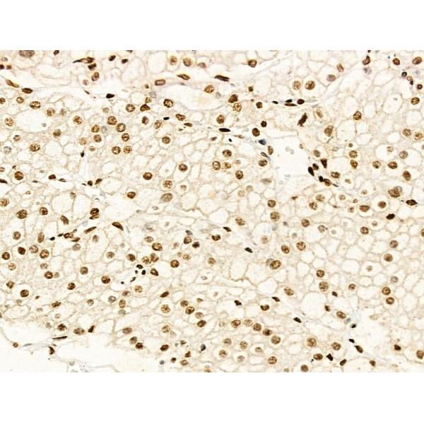
 IHC (Immunohistochemistry)
(At 1/100 staining Human prostate cancer and adjacent normal tissues by IHC-P. The sample was formaldehyde fixed and a heat mediated antigen retrieval step in citrate buffer was performed. The sample was then blocked and incubated with the primary antibody at 4 degree C overnight. An HRP conjugated anti-Rabbit antibody was used as the secondary antibody.)
IHC (Immunohistochemistry)
(At 1/100 staining Human prostate cancer and adjacent normal tissues by IHC-P. The sample was formaldehyde fixed and a heat mediated antigen retrieval step in citrate buffer was performed. The sample was then blocked and incubated with the primary antibody at 4 degree C overnight. An HRP conjugated anti-Rabbit antibody was used as the secondary antibody.)
S100A9, Polyclonal Antibody (Cat# AAA31466)
Full Name
Phospho-S100A9 (Thr113) Antibody
Reactivity
Human
Applications
WB, IHC, EIA
Purity
The antibody is from purified rabbit serum by affinity purification via sequential chromatography on phospho-peptide and non-phospho-peptide affinity columns.
Pricing


 Application Data
(Published customer image: Increased accumulation of repair-associated macrophages surrounding collaterals in ischemic hind limbs is PAR2-dependent. (A) Stainings of CD206-positive macrophages (green) and SMA-positive vessels (red) in non-ischemic (control) and ischemic (ligated) hind limbs of WT, PAR1-/- and PAR2-/- mice are shown. Nuclei were visualized with DAPI (blue). Arrows indicate single macrophages in the non-ischemic adductor. Quantification of the average number of repair-associated macrophages per vessel is indicated on the right. (B) Correlation between the number of CD206-positive macrophages in the ischemic tissues and the expression of CD11b and (C) CD115 on monocytes. ** p)
Application Data
(Published customer image: Increased accumulation of repair-associated macrophages surrounding collaterals in ischemic hind limbs is PAR2-dependent. (A) Stainings of CD206-positive macrophages (green) and SMA-positive vessels (red) in non-ischemic (control) and ischemic (ligated) hind limbs of WT, PAR1-/- and PAR2-/- mice are shown. Nuclei were visualized with DAPI (blue). Arrows indicate single macrophages in the non-ischemic adductor. Quantification of the average number of repair-associated macrophages per vessel is indicated on the right. (B) Correlation between the number of CD206-positive macrophages in the ischemic tissues and the expression of CD11b and (C) CD115 on monocytes. ** p)
CD206, Monoclonal Antibody (Cat# AAA12121)
Full Name
RAT ANTI MOUSE CD206:FITC
Gene Names
Mrc1; MR; CD206; AW259686
Applications
FC/FACS
Pricing


 Application Data
(Published customer image: Increased accumulation of repair-associated macrophages surrounding collaterals in ischemic hind limbs is PAR2-dependent. (A) Stainings of CD206-positive macrophages (green) and SMA-positive vessels (red) in non-ischemic (control) and ischemic (ligated) hind limbs of WT, PAR1-/- and PAR2-/- mice are shown. Nuclei were visualized with DAPI (blue). Arrows indicate single macrophages in the non-ischemic adductor. Quantification of the average number of repair-associated macrophages per vessel is indicated on the right. (B) Correlation between the number of CD206-positive macrophages in the ischemic tissues and the expression of CD11b and (C) CD115 on monocytes. ** p)
Application Data
(Published customer image: Increased accumulation of repair-associated macrophages surrounding collaterals in ischemic hind limbs is PAR2-dependent. (A) Stainings of CD206-positive macrophages (green) and SMA-positive vessels (red) in non-ischemic (control) and ischemic (ligated) hind limbs of WT, PAR1-/- and PAR2-/- mice are shown. Nuclei were visualized with DAPI (blue). Arrows indicate single macrophages in the non-ischemic adductor. Quantification of the average number of repair-associated macrophages per vessel is indicated on the right. (B) Correlation between the number of CD206-positive macrophages in the ischemic tissues and the expression of CD11b and (C) CD115 on monocytes. ** p)
CD206, Monoclonal Antibody (Cat# AAA12119)
Full Name
RAT ANTI MOUSE CD206:FITC
Gene Names
Mrc1; MR; CD206; AW259686
Applications
FC/FACS
Pricing


 Application Data
(Published customer image: Increased accumulation of repair-associated macrophages surrounding collaterals in ischemic hind limbs is PAR2-dependent. (A) Stainings of CD206-positive macrophages (green) and SMA-positive vessels (red) in non-ischemic (control) and ischemic (ligated) hind limbs of WT, PAR1-/- and PAR2-/- mice are shown. Nuclei were visualized with DAPI (blue). Arrows indicate single macrophages in the non-ischemic adductor. Quantification of the average number of repair-associated macrophages per vessel is indicated on the right. (B) Correlation between the number of CD206-positive macrophages in the ischemic tissues and the expression of CD11b and (C) CD115 on monocytes. ** p)
Application Data
(Published customer image: Increased accumulation of repair-associated macrophages surrounding collaterals in ischemic hind limbs is PAR2-dependent. (A) Stainings of CD206-positive macrophages (green) and SMA-positive vessels (red) in non-ischemic (control) and ischemic (ligated) hind limbs of WT, PAR1-/- and PAR2-/- mice are shown. Nuclei were visualized with DAPI (blue). Arrows indicate single macrophages in the non-ischemic adductor. Quantification of the average number of repair-associated macrophages per vessel is indicated on the right. (B) Correlation between the number of CD206-positive macrophages in the ischemic tissues and the expression of CD11b and (C) CD115 on monocytes. ** p)
CD206, Monoclonal Antibody (Cat# AAA12124)
Full Name
RAT ANTI MOUSE CD206:RPE
Gene Names
Mrc1; MR; CD206; AW259686
Applications
FC/FACS
Pricing


 Application Data
(Published customer image: Histological staining of control and HBOT wounds at post-wounding days 7 and 29. A -D) H&E staining. E -H) CD34 immunohistochemistry. I+J) CD68 immunohistochemistry.From: uk B, Tong M, Fijneman EMG, van Neck JW (2014) Hyperbaric Oxygen Therapy to Treat Diabetes Impaired Wound Healing in Rats. PLoS ONE 9(10): e108533.)
Application Data
(Published customer image: Histological staining of control and HBOT wounds at post-wounding days 7 and 29. A -D) H&E staining. E -H) CD34 immunohistochemistry. I+J) CD68 immunohistochemistry.From: uk B, Tong M, Fijneman EMG, van Neck JW (2014) Hyperbaric Oxygen Therapy to Treat Diabetes Impaired Wound Healing in Rats. PLoS ONE 9(10): e108533.)
CD68, Monoclonal Antibody (Cat# AAA12149)
Full Name
MOUSE ANTI RAT CD68
Applications
FC/FACS, IF, IP
Pricing


 Application Data
(Published customer image: Histological staining of control and HBOT wounds at post-wounding days 7 and 29. A -D) H&E staining. E -H) CD34 immunohistochemistry. I+J) CD68 immunohistochemistry.From: uk B, Tong M, Fijneman EMG, van Neck JW (2014) Hyperbaric Oxygen Therapy to Treat Diabetes Impaired Wound Healing in Rats. PLoS ONE 9(10): e108533.)
Application Data
(Published customer image: Histological staining of control and HBOT wounds at post-wounding days 7 and 29. A -D) H&E staining. E -H) CD34 immunohistochemistry. I+J) CD68 immunohistochemistry.From: uk B, Tong M, Fijneman EMG, van Neck JW (2014) Hyperbaric Oxygen Therapy to Treat Diabetes Impaired Wound Healing in Rats. PLoS ONE 9(10): e108533.)
CD68, Monoclonal Antibody (Cat# AAA12147)
Full Name
MOUSE ANTI RAT CD68:Biotin
Applications
FC/FACS
Pricing


 Application Data
(At 25 degree C. Samples were then incubated with primary Ab(At 37 degree C. An AlexaFluor594 conjugated goat anti-rabbit IgG(H+L) Ab(Red) and an AlexaFluor488 conjugated goat anti-mouse IgG(H+L) Ab(Green) were used as the secondary antibody.The nuclear counter stain is DAPI (blue).)
Application Data
(At 25 degree C. Samples were then incubated with primary Ab(At 37 degree C. An AlexaFluor594 conjugated goat anti-rabbit IgG(H+L) Ab(Red) and an AlexaFluor488 conjugated goat anti-mouse IgG(H+L) Ab(Green) were used as the secondary antibody.The nuclear counter stain is DAPI (blue).)
NFAT2, Polyclonal Antibody (Cat# AAA31428)
Full Name
Phospho-NFAT2 (Ser172) Antibody
Gene Names
NFATC1; NFAT2; NFATc; NF-ATC
Reactivity
Human, Mouse, Rat
Applications
WB, IHC, IF, ICC, EIA
Purity
The antibody is from purified rabbit serum by affinity purification via sequential chromatography on phospho-peptide and non-phospho-peptide affinity columns.
Pricing


 Application Data
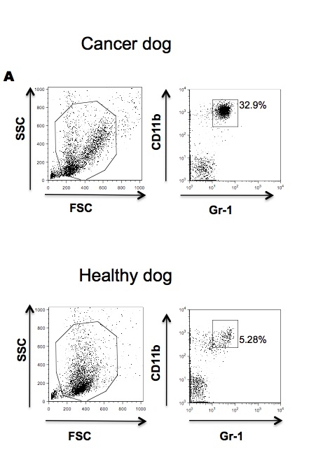
(Published customer image: Effects of EDTA versus media storage of patient blood samples on flow cytometric results. Peripheral blood cells were (a) stained immediate following collection with CD11b and CADO48A or maintained in EDTA collection tubes for (b) 24 or (c) 48 hours or in media for (d) 24 or (e) 48 hours prior to staining with CD11b and CADO48A. Both EDTA and media samples were kept refrigerated. These findings show a decrease over time of population distinction in EDTA with minimal changes when cells are kept in media up to 48 hours. All cells were gated on P1.From: Sherger et al. BMC Veterinary Research 2012 8:209)
Application Data
(Published customer image: Effects of EDTA versus media storage of patient blood samples on flow cytometric results. Peripheral blood cells were (a) stained immediate following collection with CD11b and CADO48A or maintained in EDTA collection tubes for (b) 24 or (c) 48 hours or in media for (d) 24 or (e) 48 hours prior to staining with CD11b and CADO48A. Both EDTA and media samples were kept refrigerated. These findings show a decrease over time of population distinction in EDTA with minimal changes when cells are kept in media up to 48 hours. All cells were gated on P1.From: Sherger et al. BMC Veterinary Research 2012 8:209)
CD11b, Monoclonal Antibody (Cat# AAA12092)
Full Name
MOUSE ANTI DOG CD11b
Gene Names
ITGAM; CD11B
Applications
FC/FACS, IP
Pricing


 Application Data
(At 25 degree C. Samples were then incubated with primary Ab(At 37 degree C. An AlexaFluor594 conjugated goat anti-rabbit IgG(H+L) Ab(Red) and an AlexaFluor488 conjugated goat anti-mouse IgG(H+L) Ab(Green) were used as the secondary antibody.The nuclear counter stain is DAPI(blue).)
Application Data
(At 25 degree C. Samples were then incubated with primary Ab(At 37 degree C. An AlexaFluor594 conjugated goat anti-rabbit IgG(H+L) Ab(Red) and an AlexaFluor488 conjugated goat anti-mouse IgG(H+L) Ab(Green) were used as the secondary antibody.The nuclear counter stain is DAPI(blue).)
H2B, Polyclonal Antibody (Cat# AAA31349)
Full Name
Acetyl-H2B (Lys16) Antibody
Reactivity
Human, Mouse, Rat
Applications
WB, IHC, IF, ICC, EIA
Purity
The antiserum was purified by peptide affinity chromatography using SulfoLink Coupling Resin
Pricing


 Application Data
(At 25 degree C. Samples were then incubated with primary Ab(At 37 degree C. An AlexaFluor594 conjugated goat anti-rabbit IgG(H+L) Ab(Red) and an AlexaFluor488 conjugated goat anti-mouse IgG(H+L) Ab(Green) were used as the secondary antibody.The nuclear counter stain is DAPI(blue).)
Application Data
(At 25 degree C. Samples were then incubated with primary Ab(At 37 degree C. An AlexaFluor594 conjugated goat anti-rabbit IgG(H+L) Ab(Red) and an AlexaFluor488 conjugated goat anti-mouse IgG(H+L) Ab(Green) were used as the secondary antibody.The nuclear counter stain is DAPI(blue).)
H2B, Polyclonal Antibody (Cat# AAA31348)
Full Name
Acetyl-H2B (Lys5) Antibody
Reactivity
Human, Mouse, Rat
Applications
WB, IHC, IF, ICC, EIA
Purity
The antiserum was purified by peptide affinity chromatography using SulfoLink Coupling Resin
Pricing


 Application Data
Quantitative, For Hepatitis B Virus Specific T Cells, Detection Kit (Cat# AAA27976)
Full Name
Quantitative Detection Kit, For Hepatitis B Virus Specific T Cells
Pricing


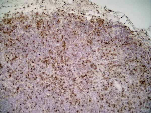
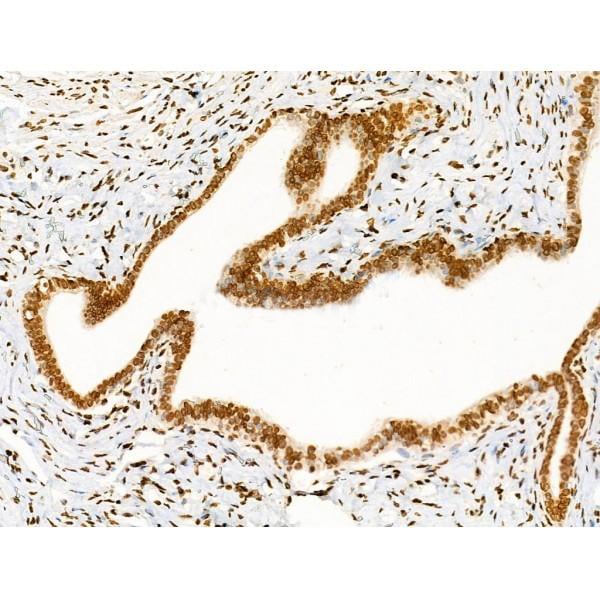
 IHC (Immunohistochemistry)
(At 1/200 staining Human tonsil tissue sections by IHC-P. The tissue was formaldehyde fixed and a heat mediated antigen retrieval step in citrate buffer was performed. The tissue was then blocked and incubated with the antibody for 1.5 hours at 22 degree C. An HRP conjugated goat anti-rabbit antibody was used as the secondary antibody.)
IHC (Immunohistochemistry)
(At 1/200 staining Human tonsil tissue sections by IHC-P. The tissue was formaldehyde fixed and a heat mediated antigen retrieval step in citrate buffer was performed. The tissue was then blocked and incubated with the antibody for 1.5 hours at 22 degree C. An HRP conjugated goat anti-rabbit antibody was used as the secondary antibody.)
TBK1, Polyclonal Antibody (Cat# AAA31411)
Full Name
Phospho-TBK1 (Ser172) Antibody
Gene Names
TBK1; NAK; T2K
Reactivity
Human, Mouse, Rat
Predicted Reactivity: Pig (100%), Zebrafish (100%), Bovine (100%), Horse (100%), Sheep (100%), Rabbit (100%), Dog (100%), Chicken (100%), Xenopus (100%)
Predicted Reactivity: Pig (100%), Zebrafish (100%), Bovine (100%), Horse (100%), Sheep (100%), Rabbit (100%), Dog (100%), Chicken (100%), Xenopus (100%)
Applications
WB, IHC, EIA
Purity
The antibody is from purified rabbit serum by affinity purification via sequential chromatography on phospho-peptide and non-phospho-peptide affinity columns.
Pricing


 FCM (Flow Cytometry)
(Dual staining of pig peripheral blood lymphocytes with Mouse anti Pig CD335 detected with Goat anti Mouse IgG (H/L):FITC (STAR117F), and Mouse anti Pig wCD8a:RPE)
FCM (Flow Cytometry)
(Dual staining of pig peripheral blood lymphocytes with Mouse anti Pig CD335 detected with Goat anti Mouse IgG (H/L):FITC (STAR117F), and Mouse anti Pig wCD8a:RPE)
CD335, Monoclonal Antibody (Cat# AAA12251)
Full Name
MOUSE ANTI PIG CD335: APC
Reactivity
Pig
Applications
FC/FACS
Pricing


 Application Data
(Published customer image: Mouse anti Human CD49d antibody, clone HP2/1 used for binding efficiency determinationImage caption:Binding efficiencies (BE) of different a4beta7 molecules composed of distinct a4 mutants to monoclonal antibodies against a4, beta7 or the a4beta7 heterodimer. Binding efficiency is determined by the ratio between the mean fluorescence of antibody binding to each a4 molecule and of the binding in a mock-transfected cell culture (see Materials and Methods for details). Dark gray bars represent binding to the human (wild type) a4 clone, whereas light gray bars are those of binding to the different a4 mutants (as shown in the x-axis). a4 mutants which included substitutions at codon 201 are boxed. A, binding of anti-a4 2b4 antibody. B, binding of anti-a4 HP2/1 antibody. C, BE of different anti-a4 and beta7 antibodies to the human a4 and the quintuple a4 mutant (5 aa mut). Bars represent the range of standard errors deduced from triplicate experiments. p-values of Student's t tests are shown above each comparison. NS, non-significant (> 0.05).From:Darc M, Hait SH, Soares EA, Cicala C, Seuanez HN, et al. (2011) Polymorphisms in the a4 Integrin of Neotropical Primates: Insights for Binding of Natural Ligands and HIV-1 gp120 to the Human a4beta7. PLoS ONE 6(9): e24461.)
Application Data
(Published customer image: Mouse anti Human CD49d antibody, clone HP2/1 used for binding efficiency determinationImage caption:Binding efficiencies (BE) of different a4beta7 molecules composed of distinct a4 mutants to monoclonal antibodies against a4, beta7 or the a4beta7 heterodimer. Binding efficiency is determined by the ratio between the mean fluorescence of antibody binding to each a4 molecule and of the binding in a mock-transfected cell culture (see Materials and Methods for details). Dark gray bars represent binding to the human (wild type) a4 clone, whereas light gray bars are those of binding to the different a4 mutants (as shown in the x-axis). a4 mutants which included substitutions at codon 201 are boxed. A, binding of anti-a4 2b4 antibody. B, binding of anti-a4 HP2/1 antibody. C, BE of different anti-a4 and beta7 antibodies to the human a4 and the quintuple a4 mutant (5 aa mut). Bars represent the range of standard errors deduced from triplicate experiments. p-values of Student's t tests are shown above each comparison. NS, non-significant (> 0.05).From:Darc M, Hait SH, Soares EA, Cicala C, Seuanez HN, et al. (2011) Polymorphisms in the a4 Integrin of Neotropical Primates: Insights for Binding of Natural Ligands and HIV-1 gp120 to the Human a4beta7. PLoS ONE 6(9): e24461.)
CD49d, Monoclonal Antibody (Cat# AAA12003)
Full Name
MOUSE ANTI HUMAN CD49d
Gene Names
ITGA4; IA4; CD49D
Applications
FC/FACS, FN, IP
Pricing


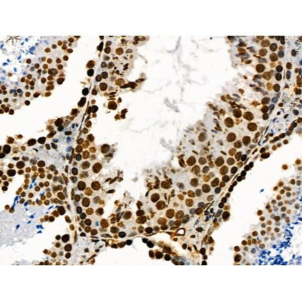
 IHC (Immunohistochemistry)
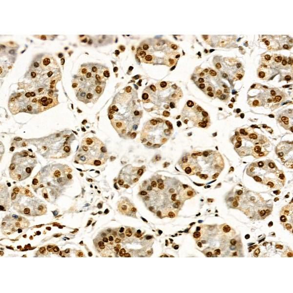
(At 1/100 staining Human pancreatic cancer by IHC-P. The sample was formaldehyde fixed and a heat mediated antigen retrieval step in citrate buffer was performed. The sample was then blocked and incubated with the primary antibody at 4 degree C overnight. An HRP conjugated anti-Rabbit antibody was used as the secondary antibody.)
IHC (Immunohistochemistry)
(At 1/100 staining Human pancreatic cancer by IHC-P. The sample was formaldehyde fixed and a heat mediated antigen retrieval step in citrate buffer was performed. The sample was then blocked and incubated with the primary antibody at 4 degree C overnight. An HRP conjugated anti-Rabbit antibody was used as the secondary antibody.)
Histone H4, Polyclonal Antibody (Cat# AAA31350)
Full Name
Acetyl-Histone H4 (Lys91) Antibody
Gene Names
HIST2H4B; H4/o
Reactivity
Human, Mouse, Rat, Pig, Bovine
Predicted Reactivity: Chicken (100%), Xenopus (100%)
Predicted Reactivity: Chicken (100%), Xenopus (100%)
Applications
WB, IHC, EIA
Purity
The antiserum was purified by peptide affinity chromatography using SulfoLink Coupling Resin
Pricing


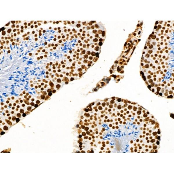
 IHC (Immunohistochemistry)
(At 1/100 staining Human ovarian cancer and adjacent normal tissues by IHC-P. The sample was formaldehyde fixed and a heat mediated antigen retrieval step in citrate buffer was performed. The sample was then blocked and incubated with the primary antibody at 4 degree C overnight. An HRP conjugated anti-Rabbit antibody was used as the secondary antibody.)
IHC (Immunohistochemistry)
(At 1/100 staining Human ovarian cancer and adjacent normal tissues by IHC-P. The sample was formaldehyde fixed and a heat mediated antigen retrieval step in citrate buffer was performed. The sample was then blocked and incubated with the primary antibody at 4 degree C overnight. An HRP conjugated anti-Rabbit antibody was used as the secondary antibody.)
histone H4, Polyclonal Antibody (Cat# AAA31295)
Full Name
Acetyl-histone H4 (Lys16) Antibody
Gene Names
HIST2H4B; H4/o
Reactivity
Human, Mouse, Rat
Applications
WB, IHC, IF, ICC, EIA
Purity
The antiserum was purified by peptide affinity chromatography using SulfoLink Coupling Resin
Pricing


 IHC (Immunohistochemistry)
(At 1/100 staining Human ovarian cancer and adjacent normal tissues by IHC-P. The sample was formaldehyde fixed and a heat mediated antigen retrieval step in citrate buffer was performed. The sample was then blocked and incubated with the primary antibody at 4 degree C overnight. An HRP conjugated anti-Rabbit antibody was used as the secondary antibody.)
IHC (Immunohistochemistry)
(At 1/100 staining Human ovarian cancer and adjacent normal tissues by IHC-P. The sample was formaldehyde fixed and a heat mediated antigen retrieval step in citrate buffer was performed. The sample was then blocked and incubated with the primary antibody at 4 degree C overnight. An HRP conjugated anti-Rabbit antibody was used as the secondary antibody.)
Histone H4, Polyclonal Antibody (Cat# AAA31346)
Full Name
Acetyl-Histone H4 (Lys8) Antibody
Gene Names
HIST2H4B; H4/o
Reactivity
Human, Mouse, Rat, Pig, Bovine
Predicted Reactivity: Chicken (100%), Xenopus (100%)
Predicted Reactivity: Chicken (100%), Xenopus (100%)
Applications
WB, IHC, IF, ICC, EIA
Purity
The antiserum was purified by peptide affinity chromatography using SulfoLink Coupling Resin
Pricing


 Application Data
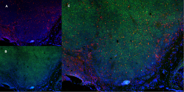
(Published customer imageSLAMF3 blockade in human hepatocytes is associated with lower susceptibility to HCV. (A) SLAMF3 was stained in primary human hepatocytes (PHHs) and cells from the Huh-7 human hepatoma cell line with a specific antibody (HLy9.1.25 clone; grey) and an isotype-matched control (empty). One of four independent experiments is shown. Huh-7 cells were transfected with scrambled control (sc) siRNA or three specific siRNAs (#1, #2 and #3) targeting SLAMF3, prior to infection with HCVcc; siRNA efficiency was checked by quantifying SLAMF3 mRNA (B) and the CD81 expression level (C) by flow cytometry analysis at 48 h post-transfection. Results are presented as the mean +/-SD (n = 3). Intracellular viral RNA was quantified at 72 h p.i. (D) and the infection was measured at 72 h p.i. by focus-forming units FFUs counting (E) (as inhibition percent; mean of three independent experiments; error bars: SD. **p)
Application Data
(Published customer imageSLAMF3 blockade in human hepatocytes is associated with lower susceptibility to HCV. (A) SLAMF3 was stained in primary human hepatocytes (PHHs) and cells from the Huh-7 human hepatoma cell line with a specific antibody (HLy9.1.25 clone; grey) and an isotype-matched control (empty). One of four independent experiments is shown. Huh-7 cells were transfected with scrambled control (sc) siRNA or three specific siRNAs (#1, #2 and #3) targeting SLAMF3, prior to infection with HCVcc; siRNA efficiency was checked by quantifying SLAMF3 mRNA (B) and the CD81 expression level (C) by flow cytometry analysis at 48 h post-transfection. Results are presented as the mean +/-SD (n = 3). Intracellular viral RNA was quantified at 72 h p.i. (D) and the infection was measured at 72 h p.i. by focus-forming units FFUs counting (E) (as inhibition percent; mean of three independent experiments; error bars: SD. **p)
CD229, Monoclonal Antibody (Cat# AAA11951)
Full Name
MOUSE ANTI HUMAN CD229
Gene Names
LY9; hly9; mLY9; CD229; SLAMF3
Applications
FC/FACS, IP
Pricing


 Application Data
(Published customer image: Mouse anti Human CD49d antibody, clone HP2/1 used for binding efficiency determinationImage caption:Binding efficiencies (BE) of different a4beta7 molecules composed of distinct a4 mutants to monoclonal antibodies against a4, beta7 or the a4beta7 heterodimer. Binding efficiency is determined by the ratio between the mean fluorescence of antibody binding to each a4 molecule and of the binding in a mock-transfected cell culture (see Materials and Methods for details). Dark gray bars represent binding to the human (wild type) a4 clone, whereas light gray bars are those of binding to the different a4 mutants (as shown in the x-axis). a4 mutants which included substitutions at codon 201 are boxed. A, binding of anti-a4 2b4 antibody. B, binding of anti-a4 HP2/1 antibody. C, BE of different anti-a4 and beta7 antibodies to the human a4 and the quintuple a4 mutant (5 aa mut). Bars represent the range of standard errors deduced from triplicate experiments. p-values of Student's t tests are shown above each comparison. NS, non-significant (> 0.05).From:Darc M, Hait SH, Soares EA, Cicala C, Seuanez HN, et al. (2011) Polymorphisms in the a4 Integrin of Neotropical Primates: Insights for Binding of Natural Ligands and HIV-1 gp120 to the Human a4beta7. PLoS ONE 6(9): e24461.)
Application Data
(Published customer image: Mouse anti Human CD49d antibody, clone HP2/1 used for binding efficiency determinationImage caption:Binding efficiencies (BE) of different a4beta7 molecules composed of distinct a4 mutants to monoclonal antibodies against a4, beta7 or the a4beta7 heterodimer. Binding efficiency is determined by the ratio between the mean fluorescence of antibody binding to each a4 molecule and of the binding in a mock-transfected cell culture (see Materials and Methods for details). Dark gray bars represent binding to the human (wild type) a4 clone, whereas light gray bars are those of binding to the different a4 mutants (as shown in the x-axis). a4 mutants which included substitutions at codon 201 are boxed. A, binding of anti-a4 2b4 antibody. B, binding of anti-a4 HP2/1 antibody. C, BE of different anti-a4 and beta7 antibodies to the human a4 and the quintuple a4 mutant (5 aa mut). Bars represent the range of standard errors deduced from triplicate experiments. p-values of Student's t tests are shown above each comparison. NS, non-significant (> 0.05).From:Darc M, Hait SH, Soares EA, Cicala C, Seuanez HN, et al. (2011) Polymorphisms in the a4 Integrin of Neotropical Primates: Insights for Binding of Natural Ligands and HIV-1 gp120 to the Human a4beta7. PLoS ONE 6(9): e24461.)
CD49d, Monoclonal Antibody (Cat# AAA12002)
Full Name
MOUSE ANTI HUMAN CD49d
Gene Names
ITGA4; IA4; CD49D
Reactivity
Bovine, Cat, Cynomolgus monkey, Goat, Horse, Llama, Mink, Mustelid, Pig, Rabbit, Rat, Rhesus Monkey
Applications
FC/FACS, FN, IP
Pricing