
Filters
Clonality
Type
Reactivity
Gene Name
Isotype
Host
Application
Clone
10000 results for "Recombinant Proteins" - showing 7850-7900
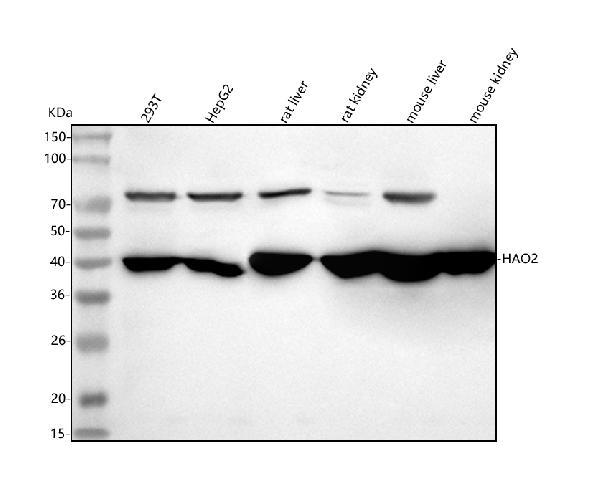
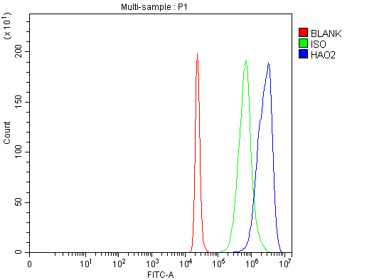 FCM/FACS (Flow Cytometry)
(Figure 2. Flow Cytometry analysis of 293T cells using anti-HAO2 antibody (AAA127740).Overlay histogram showing 293T cells stained with AAA127740 (Blue line). To facilitate intracellular staining, cells were fixed with 4% paraformaldehyde and permeabilized with permeabilization buffer. The cells were blocked with 10% normal goat serum. And then incubated with rabbit anti-HAO2 Antibody (AAA127740, 1ug/1x106 cells) for 30 min at 20 degree C. DyLight488 conjugated goat anti-rabbit IgG was used as secondary antibody for 30 minutes at 20 degree C. Isotype control antibody (Green line) was rabbit IgG (1ug/1x106) used under the same conditions. Unlabelled sample (Red line) was also used as a control.)
FCM/FACS (Flow Cytometry)
(Figure 2. Flow Cytometry analysis of 293T cells using anti-HAO2 antibody (AAA127740).Overlay histogram showing 293T cells stained with AAA127740 (Blue line). To facilitate intracellular staining, cells were fixed with 4% paraformaldehyde and permeabilized with permeabilization buffer. The cells were blocked with 10% normal goat serum. And then incubated with rabbit anti-HAO2 Antibody (AAA127740, 1ug/1x106 cells) for 30 min at 20 degree C. DyLight488 conjugated goat anti-rabbit IgG was used as secondary antibody for 30 minutes at 20 degree C. Isotype control antibody (Green line) was rabbit IgG (1ug/1x106) used under the same conditions. Unlabelled sample (Red line) was also used as a control.)
HAO2, Polyclonal Antibody (Cat# AAA127740)
Full Name
Anti-HAO2 Antibody Picoband
Gene Names
HAO2; GIG16; HAOX2
Reactivity
Human, Mouse, Rat
Applications
Flow Cytometry, Western Blot
Purity
Immunogen affinity purified.
Pricing
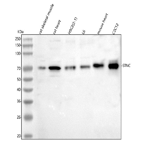
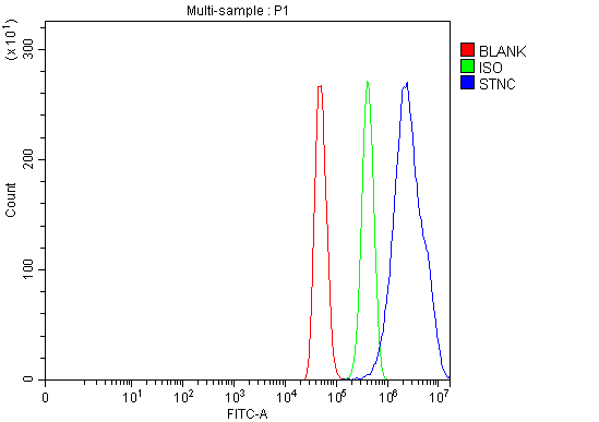 FCM/FACS (Flow Cytometry)
(Figure 2. Flow Cytometry analysis of C2C12 cells using anti-Syncoilin/Sync antibody (AAA127347).Overlay histogram showing C2C12 cells stained with AAA127347 (Blue line). To facilitate intracellular staining, cells were fixed with 4% paraformaldehyde and permeabilized with permeabilization buffer. The cells were blocked with 10% normal goat serum. And then incubated with rabbit anti-Syncoilin/Sync Antibody (AAA127347, 1ug/1x106 cells) for 30 min at 20 degree C. DyLight488 conjugated goat anti-rabbit IgG was used as secondary antibody for 30 minutes at 20 degree C. Isotype control antibody (Green line) was rabbit IgG (1ug/1x106) used under the same conditions. Unlabelled sample (Red line) was also used as a control.)
FCM/FACS (Flow Cytometry)
(Figure 2. Flow Cytometry analysis of C2C12 cells using anti-Syncoilin/Sync antibody (AAA127347).Overlay histogram showing C2C12 cells stained with AAA127347 (Blue line). To facilitate intracellular staining, cells were fixed with 4% paraformaldehyde and permeabilized with permeabilization buffer. The cells were blocked with 10% normal goat serum. And then incubated with rabbit anti-Syncoilin/Sync Antibody (AAA127347, 1ug/1x106 cells) for 30 min at 20 degree C. DyLight488 conjugated goat anti-rabbit IgG was used as secondary antibody for 30 minutes at 20 degree C. Isotype control antibody (Green line) was rabbit IgG (1ug/1x106) used under the same conditions. Unlabelled sample (Red line) was also used as a control.)
Syncoilin/Sync, Polyclonal Antibody (Cat# AAA127347)
Full Name
Anti-Syncoilin/Sync Antibody Picoband
Gene Names
Sync; SNIP4; 1110057H03Rik
Reactivity
Mouse, Rat
Applications
Flow Cytometry, Western Blot
Purity
Immunogen affinity purified.
Pricing
 IHC (Immunohiostchemistry)
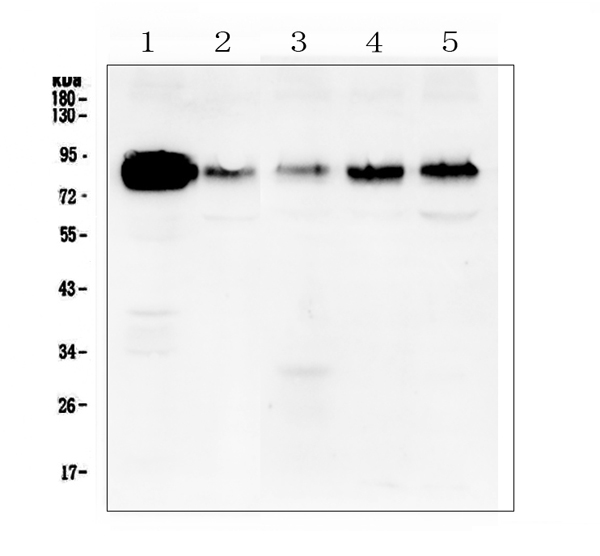
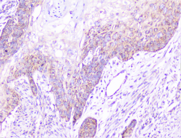
(Figure 3. IHC analysis of cortactin using anti-cortactin antibody.cortactin was detected in paraffin-embedded section of human gastric cancer tissue. Heat mediated antigen retrieval was performed in citrate buffer (pH6, epitope retrieval solution) for 20 mins. The tissue section was blocked with 10% goat serum. The tissue section was then incubated with 1ug/ml rabbit anti-cortactin antibody.cortactin was detected in paraffin-embedded section of human gastric cancer tissue. Heat mediated antigen retrieval was performed in citrate buffer (pH6, epitope retrieval solution) for 20 mins. The tissue section was blocked with 10% goat serum. The tissue section was then incubated with 1ug/ml rabbit anti-cortactin antibody.cortactin was detected in paraffin-embedded section of human glioma tissue. Heat mediated antigen retrieval was performed in citrate buffer (pH6, epitope retrieval solution) for 20 mins. The tissue section was blocked with 10% goat serum. The tissue section was then incubated with 1ug/ml rabbit anti-cortactin antibody.cortactin was detected in paraffin-embedded section of human ovary cancer tissue. Heat mediated antigen retrieval was performed in citrate buffer (pH6, epitope retrieval solution) for 20 mins. The tissue section was blocked with 10% goat serum. The tissue section was then incubated with 1ug/ml rabbit anti-cortactin antibody.cortactin was detected in paraffin-embedded section of human sarcoma tissue. Heat mediated antigen retrieval was performed in citrate buffer (pH6, epitope retrieval solution) for 20 mins. The tissue section was blocked with 10% goat serum. The tissue section was then incubated with 1ug/ml rabbit anti-cortactin antibody.cortactin was detected in paraffin-embedded section of human appendicitis tissue. Heat mediated antigen retrieval was performed in citrate buffer (pH6, epitope retrieval solution) for 20 mins. The tissue section was blocked with 10% goat serum. The tissue section was then incubated with 1ug/ml rabbit anti-cortactin antibody.cortactin was detected in paraffin-embedded section of human liver cancer tissue. Heat mediated antigen retrieval was performed in citrate buffer (pH6, epitope retrieval solution) for 20 mins. The tissue section was blocked with 10% goat serum. The tissue section was then incubated with 1ug/ml rabbit anti-cortactin antibody.cortactin was detected in paraffin-embedded section of human lung cancer tissue. Heat mediated antigen retrieval was performed in citrate buffer (pH6, epitope retrieval solution) for 20 mins. The tissue section was blocked with 10% goat serum. The tissue section was then incubated with 1ug/ml rabbit anti-cortactin antibody.cortactin was detected in paraffin-embedded section of human oesophagus squama cancer tissue. Heat mediated antigen retrieval was performed in citrate buffer (pH6, epitope retrieval solution) for 20 mins. The tissue section was blocked with 10% goat serum. The tissue section was then incubated with 1ug/ml rabbit anti-cortactin Antibody overnight at 4 degree C. Biotinylated goat anti-rabbit IgG was used as secondary antibody and incubated for 30 minutes at 37 degree C. The tissue section was developed using Strepavidin-Biotin-Complex (SABC) with DAB as the chromogen.)
IHC (Immunohiostchemistry)
(Figure 3. IHC analysis of cortactin using anti-cortactin antibody.cortactin was detected in paraffin-embedded section of human gastric cancer tissue. Heat mediated antigen retrieval was performed in citrate buffer (pH6, epitope retrieval solution) for 20 mins. The tissue section was blocked with 10% goat serum. The tissue section was then incubated with 1ug/ml rabbit anti-cortactin antibody.cortactin was detected in paraffin-embedded section of human gastric cancer tissue. Heat mediated antigen retrieval was performed in citrate buffer (pH6, epitope retrieval solution) for 20 mins. The tissue section was blocked with 10% goat serum. The tissue section was then incubated with 1ug/ml rabbit anti-cortactin antibody.cortactin was detected in paraffin-embedded section of human glioma tissue. Heat mediated antigen retrieval was performed in citrate buffer (pH6, epitope retrieval solution) for 20 mins. The tissue section was blocked with 10% goat serum. The tissue section was then incubated with 1ug/ml rabbit anti-cortactin antibody.cortactin was detected in paraffin-embedded section of human ovary cancer tissue. Heat mediated antigen retrieval was performed in citrate buffer (pH6, epitope retrieval solution) for 20 mins. The tissue section was blocked with 10% goat serum. The tissue section was then incubated with 1ug/ml rabbit anti-cortactin antibody.cortactin was detected in paraffin-embedded section of human sarcoma tissue. Heat mediated antigen retrieval was performed in citrate buffer (pH6, epitope retrieval solution) for 20 mins. The tissue section was blocked with 10% goat serum. The tissue section was then incubated with 1ug/ml rabbit anti-cortactin antibody.cortactin was detected in paraffin-embedded section of human appendicitis tissue. Heat mediated antigen retrieval was performed in citrate buffer (pH6, epitope retrieval solution) for 20 mins. The tissue section was blocked with 10% goat serum. The tissue section was then incubated with 1ug/ml rabbit anti-cortactin antibody.cortactin was detected in paraffin-embedded section of human liver cancer tissue. Heat mediated antigen retrieval was performed in citrate buffer (pH6, epitope retrieval solution) for 20 mins. The tissue section was blocked with 10% goat serum. The tissue section was then incubated with 1ug/ml rabbit anti-cortactin antibody.cortactin was detected in paraffin-embedded section of human lung cancer tissue. Heat mediated antigen retrieval was performed in citrate buffer (pH6, epitope retrieval solution) for 20 mins. The tissue section was blocked with 10% goat serum. The tissue section was then incubated with 1ug/ml rabbit anti-cortactin antibody.cortactin was detected in paraffin-embedded section of human oesophagus squama cancer tissue. Heat mediated antigen retrieval was performed in citrate buffer (pH6, epitope retrieval solution) for 20 mins. The tissue section was blocked with 10% goat serum. The tissue section was then incubated with 1ug/ml rabbit anti-cortactin Antibody overnight at 4 degree C. Biotinylated goat anti-rabbit IgG was used as secondary antibody and incubated for 30 minutes at 37 degree C. The tissue section was developed using Strepavidin-Biotin-Complex (SABC) with DAB as the chromogen.)
Cortactin/CTTN, Polyclonal Antibody (Cat# AAA124920)
Full Name
Anti-Cortactin/CTTN Antibody
Gene Names
CTTN; EMS1
Reactivity
Human, Mouse, Rat
Applications
Direct ELISA, Immunofluorescence, Flow Cytometry, Immunocytochemistry, Immunohistochemistry, Western Blot
Purity
Immunogen Affinity Purified
Pricing
 FCM/FACS (Flow Cytometry)
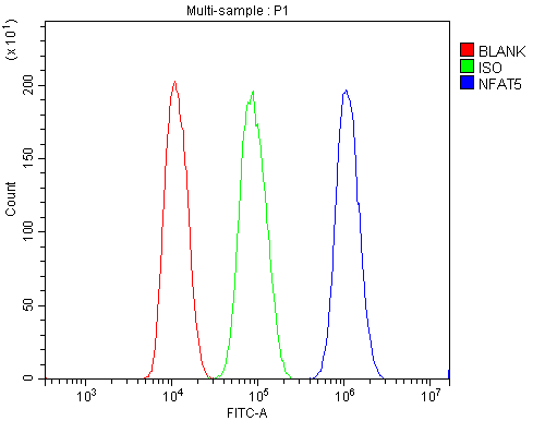
(Figure 2. Flow Cytometry analysis of HepG2 cells using anti-NFAT5 antibody (AAA125623).Overlay histogram showing HepG2 cells stained with AAA125623 (Blue line). The cells were blocked with 10% normal goat serum. And then incubated with rabbit anti-NFAT5 Antibody (AAA125623, 1μg/1x106 cells) for 30 min at 20 degree C. DyLight®488 conjugated goat anti-rabbit IgG (5-10μg/1x106 cells) was used as secondary antibody for 30 minutes at 20 degree C. Isotype control antibody (Green line) was rabbit IgG (1μg/1x106) used under the same conditions. Unlabelled sample (Red line) was also used as a control.)
FCM/FACS (Flow Cytometry)
(Figure 2. Flow Cytometry analysis of HepG2 cells using anti-NFAT5 antibody (AAA125623).Overlay histogram showing HepG2 cells stained with AAA125623 (Blue line). The cells were blocked with 10% normal goat serum. And then incubated with rabbit anti-NFAT5 Antibody (AAA125623, 1μg/1x106 cells) for 30 min at 20 degree C. DyLight®488 conjugated goat anti-rabbit IgG (5-10μg/1x106 cells) was used as secondary antibody for 30 minutes at 20 degree C. Isotype control antibody (Green line) was rabbit IgG (1μg/1x106) used under the same conditions. Unlabelled sample (Red line) was also used as a control.)
NFAT5, Polyclonal Antibody (Cat# AAA125623)
Full Name
Anti-NFAT5 Antibody
Gene Names
NFAT5; NFATZ; OREBP; NF-AT5; NFATL1; TONEBP
Reactivity
Human
Applications
Direct ELISA, Flow Cytometry, Western Blot
Purity
Immunogen affinity purified.
Pricing
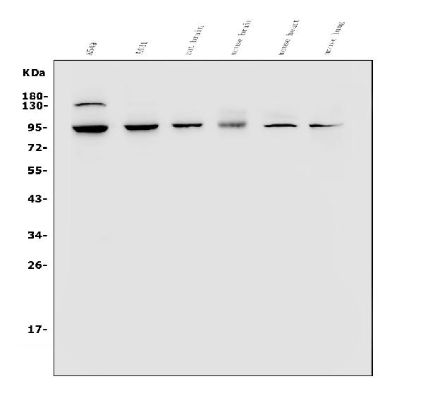
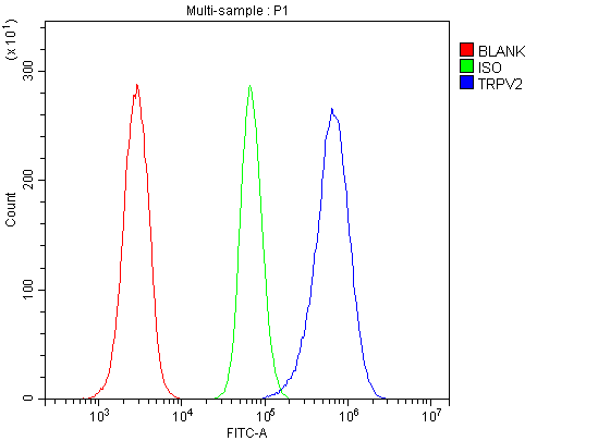 FCM/FACS (Flow Cytometry)
(Figure 2. Flow Cytometry analysis of HL-60 cells using anti-VRL1/TRPV2 antibody (AAA125670).Overlay histogram showing HL-60 cells stained with AAA125670 (Blue line). The cells were blocked with 10% normal goat serum. And then incubated with rabbit anti-VRL1/TRPV2 Antibody (AAA125670, 1μg/1x106 cells) for 30 min at 20 degree C. DyLight®488 conjugated goat anti-rabbit IgG (5-10μg/1x106 cells) was used as secondary antibody for 30 minutes at 20 degree C. Isotype control antibody (Green line) was rabbit IgG (1μg/1x106) used under the same conditions. Unlabelled sample (Red line) was also used as a control.)
FCM/FACS (Flow Cytometry)
(Figure 2. Flow Cytometry analysis of HL-60 cells using anti-VRL1/TRPV2 antibody (AAA125670).Overlay histogram showing HL-60 cells stained with AAA125670 (Blue line). The cells were blocked with 10% normal goat serum. And then incubated with rabbit anti-VRL1/TRPV2 Antibody (AAA125670, 1μg/1x106 cells) for 30 min at 20 degree C. DyLight®488 conjugated goat anti-rabbit IgG (5-10μg/1x106 cells) was used as secondary antibody for 30 minutes at 20 degree C. Isotype control antibody (Green line) was rabbit IgG (1μg/1x106) used under the same conditions. Unlabelled sample (Red line) was also used as a control.)
VRL1/TRPV2, Polyclonal Antibody (Cat# AAA125670)
Full Name
Anti-VRL1/TRPV2 Antibody
Gene Names
TRPV2; VRL; VRL1; VRL-1
Reactivity
Human, Mouse, Rat
Applications
Direct ELISA, Flow Cytometry, Western Blot
Purity
Immunogen affinity purified.
Pricing
 Application Data
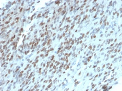
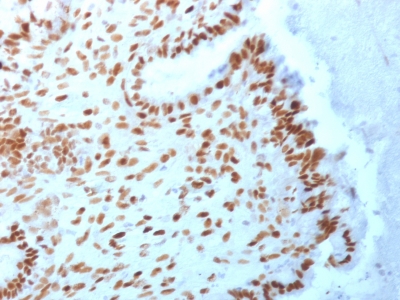
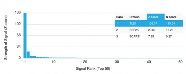
(Analysis of Protein Array containing more than 19, 000 full-length human proteins using TLE1 Mouse Monoclonal Antibody (TLE1/2062).Z- and S- Score: The Z-score represents the strength of a signal that a monoclonal antibody (MAb) (in combination with a fluorescently-tagged anti-IgG secondary antibody) produces when binding to a particular protein on the HuProtTM array. Z-scores are described in units of standard deviations (SD's) above the mean value of all signals generated on that array. If targets on HuProtTM are arranged in descending order of the Z-score, the S-score is the difference (also in units of SD's) between the Z-score. S-score therefore represents the relative target specificity of a MAb to its intended target. A MAb is considered to specific to its intended target, if the MAb has an S-score of at least 2.5. For example, if a MAb binds to protein X with a Z-score of 43 and to protein Y with a Z-score of 14, then the S-score for the binding of that MAb to protein X is equal to 29.)
Application Data
(Analysis of Protein Array containing more than 19, 000 full-length human proteins using TLE1 Mouse Monoclonal Antibody (TLE1/2062).Z- and S- Score: The Z-score represents the strength of a signal that a monoclonal antibody (MAb) (in combination with a fluorescently-tagged anti-IgG secondary antibody) produces when binding to a particular protein on the HuProtTM array. Z-scores are described in units of standard deviations (SD's) above the mean value of all signals generated on that array. If targets on HuProtTM are arranged in descending order of the Z-score, the S-score is the difference (also in units of SD's) between the Z-score. S-score therefore represents the relative target specificity of a MAb to its intended target. A MAb is considered to specific to its intended target, if the MAb has an S-score of at least 2.5. For example, if a MAb binds to protein X with a Z-score of 43 and to protein Y with a Z-score of 14, then the S-score for the binding of that MAb to protein X is equal to 29.)
TLE1, Monoclonal Antibody (Cat# AAA214700)
Full Name
TLE1 (Synovial Sarcoma Marker)
Gene Names
TLE1; ESG; ESG1; GRG1
Reactivity
Human. Others not known.
Applications
Immunohistochemistry, Western Blot
Pricing

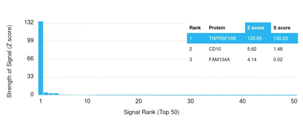 Application Data
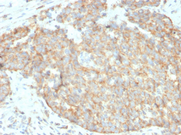
(Analysis of Protein Array containing >19,000 full-length human proteins using DR5 Mouse Monoclonal Antibody (DR5/3381) Z- and S- Score: The Z-score represents the strength of a signal that a monoclonal antibody (Monoclonal Antibody) (in combination with a fluorescently-tagged anti-IgG secondary antibody) produces when binding to a particular protein on the HuProtTM array. Z-scores are described in units of standard deviations (SD’s) above the mean value of all signals generated on that array. If targets on HuProtTM are arranged in descending order of the Z-score, the S-score is the difference (also in units of SD’s) between the Z-score. S-score therefore represents the relative target specificity of a Monoclonal Antibody to its intended target. A Monoclonal Antibody is considered to specific to its intended target, if the Monoclonal Antibody has an S-score of at least 2.5. For example, if a Monoclonal Antibody binds to protein X with a Z-score of 43 and to protein Y with a Z-score of 14, then the S-score for the binding of that Monoclonal Antibody to protein X is equal to 29.)
Application Data
(Analysis of Protein Array containing >19,000 full-length human proteins using DR5 Mouse Monoclonal Antibody (DR5/3381) Z- and S- Score: The Z-score represents the strength of a signal that a monoclonal antibody (Monoclonal Antibody) (in combination with a fluorescently-tagged anti-IgG secondary antibody) produces when binding to a particular protein on the HuProtTM array. Z-scores are described in units of standard deviations (SD’s) above the mean value of all signals generated on that array. If targets on HuProtTM are arranged in descending order of the Z-score, the S-score is the difference (also in units of SD’s) between the Z-score. S-score therefore represents the relative target specificity of a Monoclonal Antibody to its intended target. A Monoclonal Antibody is considered to specific to its intended target, if the Monoclonal Antibody has an S-score of at least 2.5. For example, if a Monoclonal Antibody binds to protein X with a Z-score of 43 and to protein Y with a Z-score of 14, then the S-score for the binding of that Monoclonal Antibody to protein X is equal to 29.)
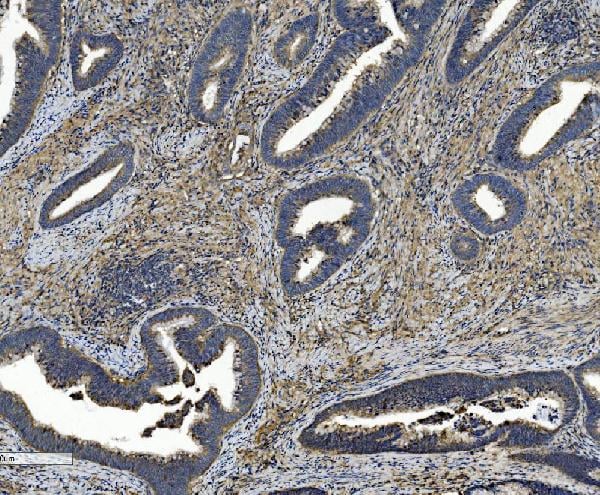
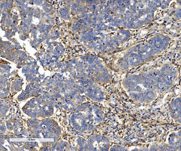
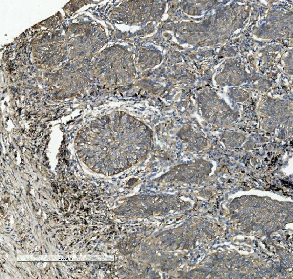
CD262/DR5, Monoclonal Antibody (Cat# AAA215274)
Full Name
CD262/DR5
Reactivity
Human
Applications
Immunohistochemistry
Purity
Purified Ab with BSA and Azide at 200ug/ml OR Purified Ab WITHOUT BSA and Azide at 1.0mg/ml
Pricing
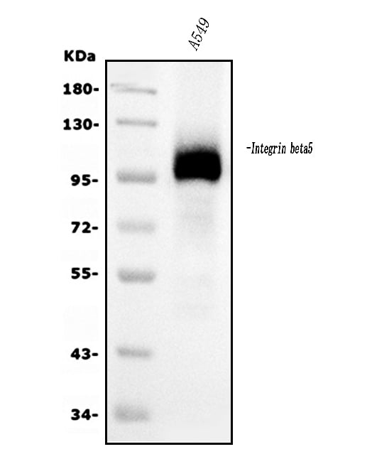
 FCM/FACS (Flow Cytometry)
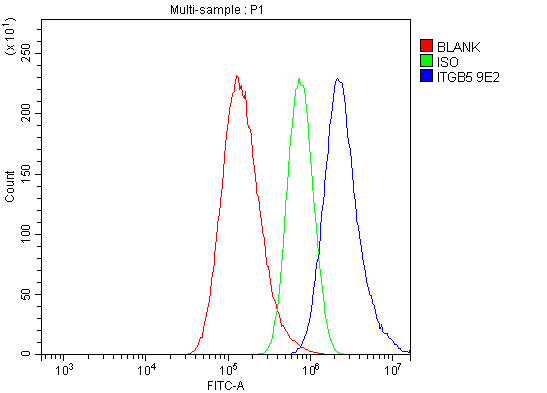
(Figure 5. Flow Cytometry analysis of A549 cells using anti-Integrin beta 5/ITGB5 antibody (AAA125933).Overlay histogram showing A549 cells stained with AAA125933 (Blue line). The cells were blocked with 10% normal goat serum. And then incubated with mouse anti- Integrin beta 5/ITGB5 Antibody (AAA125933, 1μg/1x106 cells) for 30 min at 20 degree C. DyLight®488 conjugated goat anti-mouse IgG (BA1126, 5-10μg/1x106 cells) was used as secondary antibody for 30 minutes at 20 degree C. Isotype control antibody (Green line) was mouse IgG (1μg/1x106) used under the same conditions. Unlabelled sample (Red line) was also used as a control.)
FCM/FACS (Flow Cytometry)
(Figure 5. Flow Cytometry analysis of A549 cells using anti-Integrin beta 5/ITGB5 antibody (AAA125933).Overlay histogram showing A549 cells stained with AAA125933 (Blue line). The cells were blocked with 10% normal goat serum. And then incubated with mouse anti- Integrin beta 5/ITGB5 Antibody (AAA125933, 1μg/1x106 cells) for 30 min at 20 degree C. DyLight®488 conjugated goat anti-mouse IgG (BA1126, 5-10μg/1x106 cells) was used as secondary antibody for 30 minutes at 20 degree C. Isotype control antibody (Green line) was mouse IgG (1μg/1x106) used under the same conditions. Unlabelled sample (Red line) was also used as a control.)
Integrin beta 5/ITGB5, Monoclonal Antibody (Cat# AAA125933)
Full Name
Anti-Integrin beta 5/ITGB5 Antibody (monoclonal, 9E2)
Reactivity
Human
Applications
Flow Cytometry, Immunohistochemistry, Western Blot
Purity
Immunogen affinity purified.
Pricing
 IHC (Immunohistochemisry)
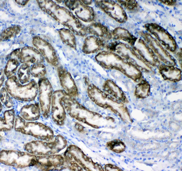
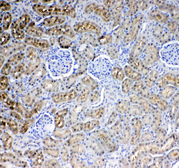
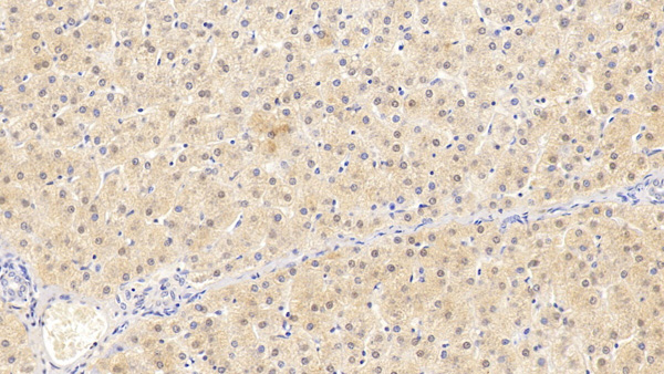
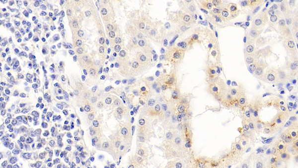
(Figure 3. IHC analysis of Glycerol kinase using anti-Glycerol kinase antibody (AAA124696).Glycerol kinase was detected in paraffin-embedded section of rat kidney tissue. Heat mediated antigen retrieval was performed in citrate buffer (pH6, epitope retrieval solution) for 20 mins. The tissue section was blocked with 10% goat serum. The tissue section was then incubated with 1ug/ml rabbit anti-Glycerol kinase Antibody (AAA124696) overnight at 4 degree C. Biotinylated goat anti-rabbit IgG was used as secondary antibody and incubated for 30 minutes at 37 degree C. The tissue section was developed using Strepavidin-Biotin-Complex (SABC) with DAB as the chromogen.)
IHC (Immunohistochemisry)
(Figure 3. IHC analysis of Glycerol kinase using anti-Glycerol kinase antibody (AAA124696).Glycerol kinase was detected in paraffin-embedded section of rat kidney tissue. Heat mediated antigen retrieval was performed in citrate buffer (pH6, epitope retrieval solution) for 20 mins. The tissue section was blocked with 10% goat serum. The tissue section was then incubated with 1ug/ml rabbit anti-Glycerol kinase Antibody (AAA124696) overnight at 4 degree C. Biotinylated goat anti-rabbit IgG was used as secondary antibody and incubated for 30 minutes at 37 degree C. The tissue section was developed using Strepavidin-Biotin-Complex (SABC) with DAB as the chromogen.)
Glycerol kinase, Polyclonal Antibody (Cat# AAA124696)
Full Name
Anti-Glycerol kinase Picoband antibody
Gene Names
GK; GK1; GKD
Reactivity
Human, Mouse, Rat
No cross reactivity with other proteins.
No cross reactivity with other proteins.
Applications
Western Blot, Immunohistochemistry
Purity
Immunogen affinity purified
Pricing
 IF (Immunofluorescence)
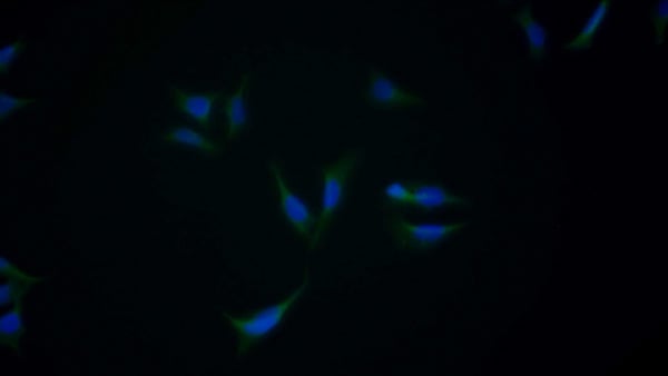
(FITC staining on IF; Samples: Human HepG2 cell; Primary Ab: 20ug/ml Mouse AntiHuman a2PI Antibody Second Ab: 3ug/ml FITCLinked Caprine AntiMouse IgG Polyclonal Antibody (Catalog: SAA544Mu18))
IF (Immunofluorescence)
(FITC staining on IF; Samples: Human HepG2 cell; Primary Ab: 20ug/ml Mouse AntiHuman a2PI Antibody Second Ab: 3ug/ml FITCLinked Caprine AntiMouse IgG Polyclonal Antibody (Catalog: SAA544Mu18))
Alpha 2Antiplasmin (a2PI), Monoclonal Antibody (Cat# AAA151498)
Full Name
Monoclonal Antibody to Alpha 2Antiplasmin (a2PI)
Gene Names
SERPINF2; AAP; API; PLI; A2AP; ALPHA-2-PI
Reactivity
Human
Applications
Immunoprecipitation, Immunocytochemistry, Immunohistochemistry, Western Blot
Purity
Protein A + Protein G affinity chromatography
Pricing
 Application Data
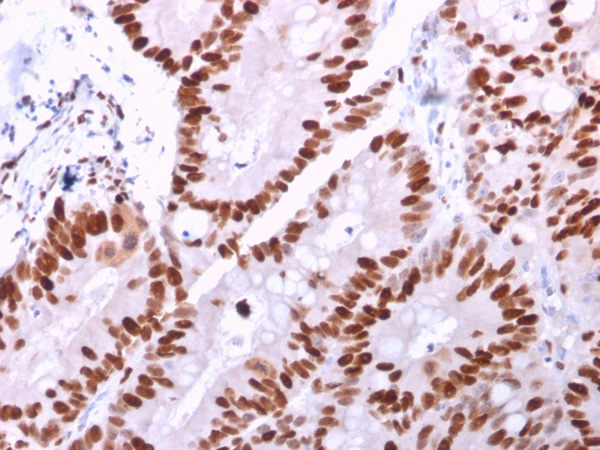
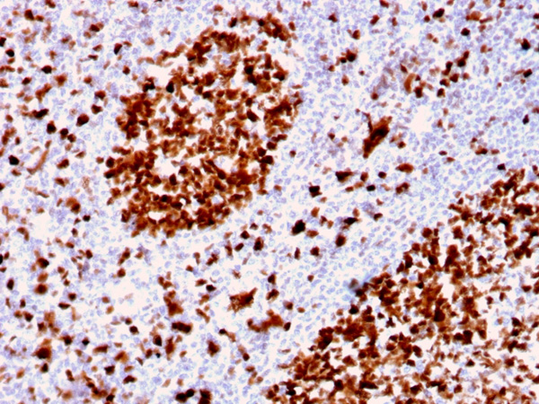
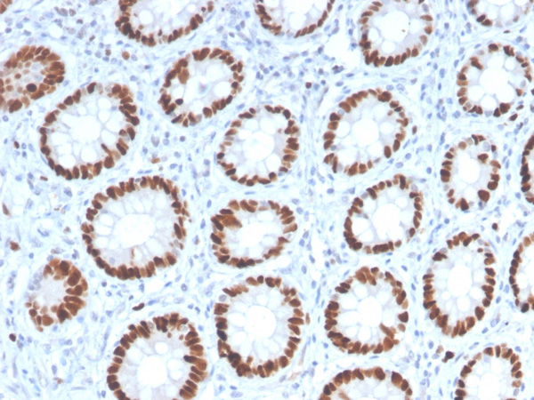
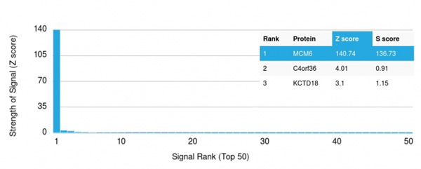
(Analysis of Protein Array containing >19,000 full-length human proteins using MCM6 Mouse Monoclonal Antibody (MCM6/2999) Z- and S- Score: The Z-score represents the strength of a signal that a monoclonal antibody (MAb) (in combination with a fluorescently-tagged anti-IgG secondary antibody) produces when binding to a particular protein on the HuProtTM array. Z-scores are described in units of standard deviations (SD’s) above the mean value of all signals generated on that array. If targets on HuProtTM are arranged in descending order of the Z-score, the S-score is the difference (also in units of SD’s) between the Z-score. S-score therefore represents the relative target specificity of a MAb to its intended target. A MAb is considered to specific to its intended target, if the MAb has an S-score of at least 2.5. For example, if a MAb binds to protein X with a Z-score of 43 and to protein Y with a Z-score of 14, then the S-score for the binding of that MAb to protein X is equal to 29.)
Application Data
(Analysis of Protein Array containing >19,000 full-length human proteins using MCM6 Mouse Monoclonal Antibody (MCM6/2999) Z- and S- Score: The Z-score represents the strength of a signal that a monoclonal antibody (MAb) (in combination with a fluorescently-tagged anti-IgG secondary antibody) produces when binding to a particular protein on the HuProtTM array. Z-scores are described in units of standard deviations (SD’s) above the mean value of all signals generated on that array. If targets on HuProtTM are arranged in descending order of the Z-score, the S-score is the difference (also in units of SD’s) between the Z-score. S-score therefore represents the relative target specificity of a MAb to its intended target. A MAb is considered to specific to its intended target, if the MAb has an S-score of at least 2.5. For example, if a MAb binds to protein X with a Z-score of 43 and to protein Y with a Z-score of 14, then the S-score for the binding of that MAb to protein X is equal to 29.)
MCM6, Monoclonal Antibody (Cat# AAA215080)
Full Name
MCM6 (Proliferation Marker)
Gene Names
MCM6; Mis5; P105MCM; MCG40308
Reactivity
Human
Applications
Immunohistochemistry
Purity
Purified Ab with BSA and Azide at 200ug/ml OR Purified Ab WITHOUT BSA and Azide at 1.0mg/ml
Pricing
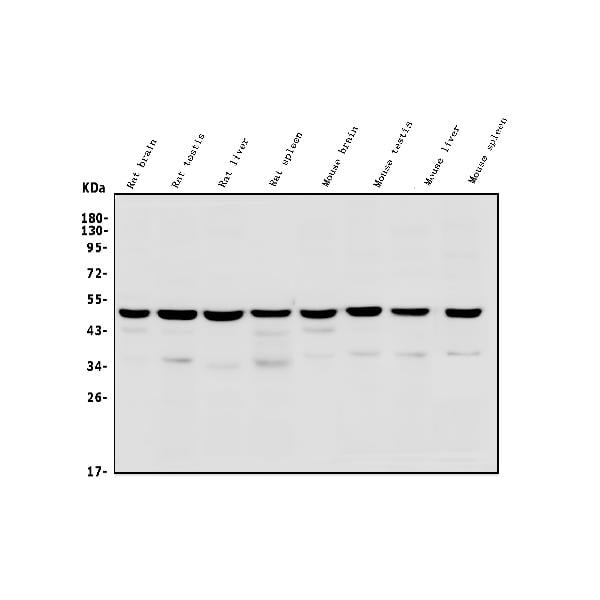
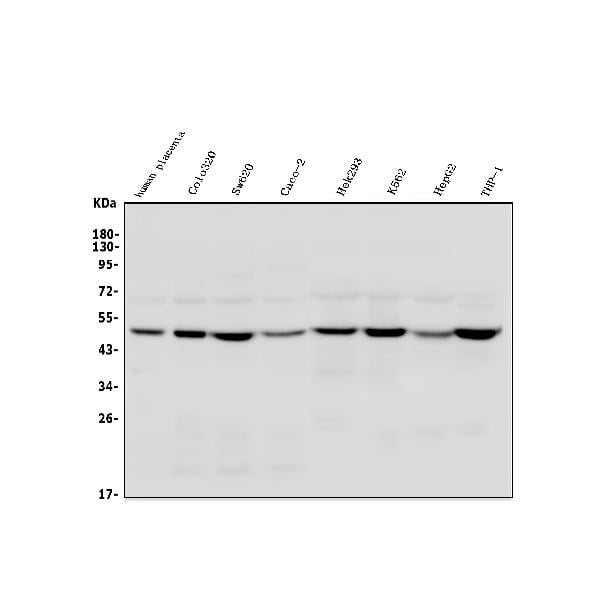
 FCM/FACS (Flow Cytometry)
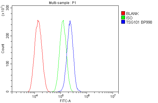
(Figure 3. Flow Cytometry analysis of SiHa cells using anti-TSG101 antibody (AAA125596).Overlay histogram showing SiHa cells stained with AAA125596 (Blue line). The cells were blocked with 10% normal goat serum. And then incubated with rabbit anti-TSG101 Antibody (AAA125596, 1μg/1x106 cells) for 30 min at 20 degree C. DyLight®488 conjugated goat anti-rabbit IgG (5-10μg/1x106 cells) was used as secondary antibody for 30 minutes at 20 degree C. Isotype control antibody (Green line) was rabbit IgG (1μg/1x106) used under the same conditions. Unlabelled sample (Red line) was also used as a control.)
FCM/FACS (Flow Cytometry)
(Figure 3. Flow Cytometry analysis of SiHa cells using anti-TSG101 antibody (AAA125596).Overlay histogram showing SiHa cells stained with AAA125596 (Blue line). The cells were blocked with 10% normal goat serum. And then incubated with rabbit anti-TSG101 Antibody (AAA125596, 1μg/1x106 cells) for 30 min at 20 degree C. DyLight®488 conjugated goat anti-rabbit IgG (5-10μg/1x106 cells) was used as secondary antibody for 30 minutes at 20 degree C. Isotype control antibody (Green line) was rabbit IgG (1μg/1x106) used under the same conditions. Unlabelled sample (Red line) was also used as a control.)
TSG101, Polyclonal Antibody (Cat# AAA125596)
Full Name
Anti-TSG101 Antibody
Gene Names
TSG101; TSG10; VPS23
Reactivity
Human, Mouse, Rat
Applications
Direct ELISA, Flow Cytometry, Western Blot
Purity
Immunogen affinity purified.
Pricing
 FCM/FACS (Flow Cytometry)
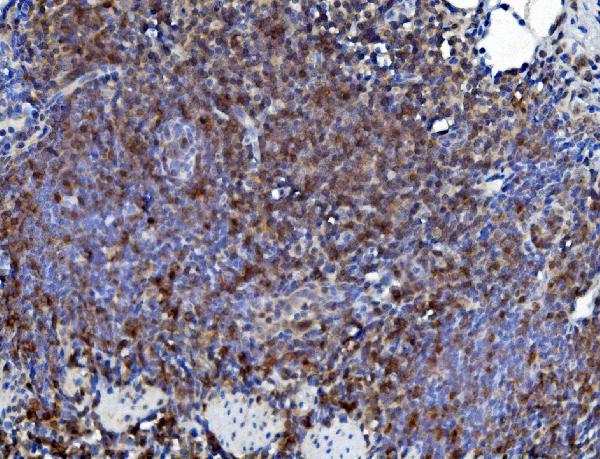
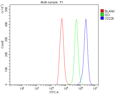
(Figure 4. Flow Cytometry analysis of NRK cells using anti-CD226 antibody (AAA125589).Overlay histogram showing NRK cells stained with AAA125589 (Blue line). The cells were blocked with 10% normal goat serum. And then incubated with rabbit anti-CD226 Antibody (AAA125589, 1μg/1x106 cells) for 30 min at 20 degree C. DyLight®488 conjugated goat anti-rabbit IgG (5-10μg/1x106 cells) was used as secondary antibody for 30 minutes at 20 degree C. Isotype control antibody (Green line) was rabbit IgG (1μg/1x106) used under the same conditions. Unlabelled sample (Red line) was also used as a control.)
FCM/FACS (Flow Cytometry)
(Figure 4. Flow Cytometry analysis of NRK cells using anti-CD226 antibody (AAA125589).Overlay histogram showing NRK cells stained with AAA125589 (Blue line). The cells were blocked with 10% normal goat serum. And then incubated with rabbit anti-CD226 Antibody (AAA125589, 1μg/1x106 cells) for 30 min at 20 degree C. DyLight®488 conjugated goat anti-rabbit IgG (5-10μg/1x106 cells) was used as secondary antibody for 30 minutes at 20 degree C. Isotype control antibody (Green line) was rabbit IgG (1μg/1x106) used under the same conditions. Unlabelled sample (Red line) was also used as a control.)
CD226, Polyclonal Antibody (Cat# AAA125589)
Full Name
Anti-CD226 Antibody
Gene Names
Cd226; Pta1; DNAM1; DNAM-1; TLiSA1; BC051526
Reactivity
Mouse, Rat
Applications
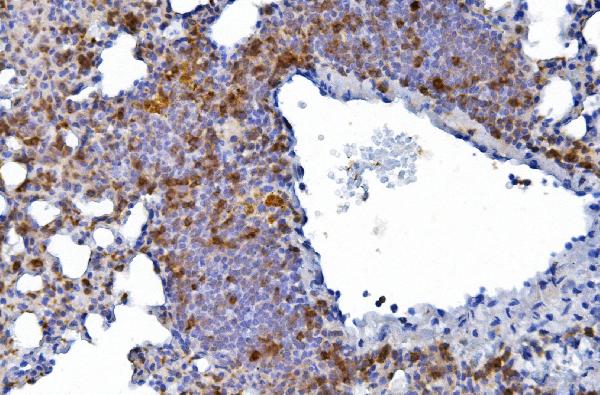
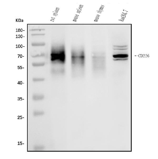
Direct ELISA, Flow Cytometry, Immunohistochemistry, Western Blot
Purity
Immunogen affinity purified.
Pricing
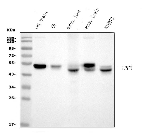
 FCM/FACS (Flow Cytometry)
(Figure 3. Flow Cytometry analysis of HEPA1-6 cells using anti-IRF3 antibody (AAA125901).Overlay histogram showing HEPA1-6 cells stained with AAA125901 (Blue line). The cells were blocked with 10% normal goat serum. And then incubated with mouse anti- IRF3 Antibody (AAA125901, 1μg/1x106 cells) for 30 min at 20 degree C. DyLight®488 conjugated goat anti-mouse IgG (BA1126, 5-10μg/1x106 cells) was used as secondary antibody for 30 minutes at 20 degree C. Isotype control antibody (Green line) was mouse IgG (1μg/1x106) used under the same conditions. Unlabelled sample (Red line) was also used as a control.)
FCM/FACS (Flow Cytometry)
(Figure 3. Flow Cytometry analysis of HEPA1-6 cells using anti-IRF3 antibody (AAA125901).Overlay histogram showing HEPA1-6 cells stained with AAA125901 (Blue line). The cells were blocked with 10% normal goat serum. And then incubated with mouse anti- IRF3 Antibody (AAA125901, 1μg/1x106 cells) for 30 min at 20 degree C. DyLight®488 conjugated goat anti-mouse IgG (BA1126, 5-10μg/1x106 cells) was used as secondary antibody for 30 minutes at 20 degree C. Isotype control antibody (Green line) was mouse IgG (1μg/1x106) used under the same conditions. Unlabelled sample (Red line) was also used as a control.)
IRF3, Monoclonal Antibody (Cat# AAA125901)
Full Name
Anti-IRF3 Antibody (monoclonal, 3B4)
Gene Names
Irf3; IRF-3; C920001K05Rik
Reactivity
Mouse, Rat
Applications
Flow Cytometry, Immunofluorescence, Immunocytochemistry, Western Blot
Purity
Immunogen affinity purified.
Pricing
 WB (Western Blot)
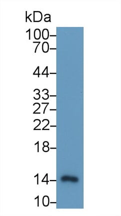
(Western Blot; Sample: Human Leukocyte lysate Primary Ab: 2ug/ml Mouse AntiHuman S100A9 Antibody Second Ab: 0.2ug/mL HRPLinked Caprine AntiMouse IgG Polyclonal Antibody (Catalog: SAA544Mu19))
WB (Western Blot)
(Western Blot; Sample: Human Leukocyte lysate Primary Ab: 2ug/ml Mouse AntiHuman S100A9 Antibody Second Ab: 0.2ug/mL HRPLinked Caprine AntiMouse IgG Polyclonal Antibody (Catalog: SAA544Mu19))
S100 Calcium Binding Protein A9 (S100A9), Monoclonal Antibody (Cat# AAA151747)
Full Name
Monoclonal Antibody to S100 Calcium Binding Protein A9 (S100A9)
Reactivity
Human, Pig
Applications
Immunoprecipitation, Immunocytochemistry, Immunohistochemistry, Western Blot
Purity
Protein A + Protein G affinity chromatography
Pricing
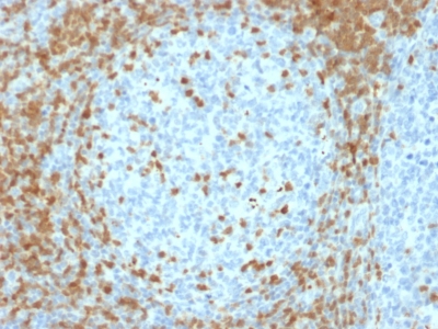
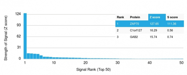 Application Data
(Analysis of Protein Array containing more than 19, 000 full-length human proteins using ZAP70 Mouse Monoclonal Antibody (ZAP70/2035).Z- and S- Score: The Z-score represents the strength of a signal that a monoclonal antibody (MAb) (in combination with a fluorescently-tagged anti-IgG secondary antibody) produces when binding to a particular protein on the HuProtTM array. Z-scores are described in units of standard deviations (SD's) above the mean value of all signals generated on that array. If targets on HuProtTM are arranged in descending order of the Z-score, the S-score is the difference (also in units of SD's) between the Z-score. S-score therefore represents the relative target specificity of a MAb to its intended target. A MAb is considered to specific to its intended target, if the MAb has an S-score of at least 2.5. For example, if a MAb binds to protein X with a Z-score of 43 and to protein Y with a Z-score of 14, then the S-score for the binding of that MAb to protein X is equal to 29.)
Application Data
(Analysis of Protein Array containing more than 19, 000 full-length human proteins using ZAP70 Mouse Monoclonal Antibody (ZAP70/2035).Z- and S- Score: The Z-score represents the strength of a signal that a monoclonal antibody (MAb) (in combination with a fluorescently-tagged anti-IgG secondary antibody) produces when binding to a particular protein on the HuProtTM array. Z-scores are described in units of standard deviations (SD's) above the mean value of all signals generated on that array. If targets on HuProtTM are arranged in descending order of the Z-score, the S-score is the difference (also in units of SD's) between the Z-score. S-score therefore represents the relative target specificity of a MAb to its intended target. A MAb is considered to specific to its intended target, if the MAb has an S-score of at least 2.5. For example, if a MAb binds to protein X with a Z-score of 43 and to protein Y with a Z-score of 14, then the S-score for the binding of that MAb to protein X is equal to 29.)
ZAP70, Monoclonal Antibody (Cat# AAA214708)
Full Name
ZAP70 (Chronic Lymphocytic Leukemia Marker)
Gene Names
ZAP70; SRK; STD; TZK; STCD; IMD48; ADMIO2; ZAP-70
Reactivity
Human. Others not known
Applications
Immunohistochemistry
Pricing
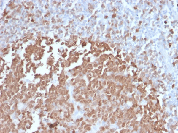
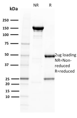
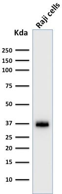
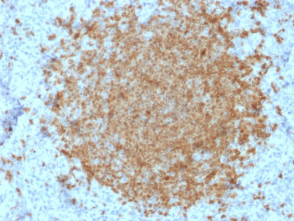
 Application Data
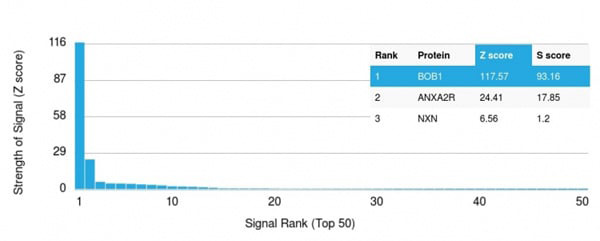
(Analysis of Protein Array containing more than 19,000 full-length human proteins using BOB1 Mouse Monoclonal Antibody (BOB1/2422). Z- and S- Score: The Z-score represents the strength of a signal that a monoclonal antibody (MAb) (in combination with a fluorescently-tagged anti-IgG secondary antibody) produces when binding to a particular protein on the HuProtTM array. Z-scores are described in units of standard deviations (SD's) above the mean value of all signals generated on that array. If targets on HuProtTM are arranged in descending order of the Z-score, the S-score is the difference (also in units of SD's) between the Z-score. S-score therefore represents the relative target specificity of a MAb to its intended target. A MAb is considered to specific to its intended target, if the MAb has an S-score of at least 2.5. For example, if a MAb binds to protein X with a Z-score of 43 and to protein Y with a Z-score of 14, then the S-score for the binding of that MAb to protein X is equal to 29.)
Application Data
(Analysis of Protein Array containing more than 19,000 full-length human proteins using BOB1 Mouse Monoclonal Antibody (BOB1/2422). Z- and S- Score: The Z-score represents the strength of a signal that a monoclonal antibody (MAb) (in combination with a fluorescently-tagged anti-IgG secondary antibody) produces when binding to a particular protein on the HuProtTM array. Z-scores are described in units of standard deviations (SD's) above the mean value of all signals generated on that array. If targets on HuProtTM are arranged in descending order of the Z-score, the S-score is the difference (also in units of SD's) between the Z-score. S-score therefore represents the relative target specificity of a MAb to its intended target. A MAb is considered to specific to its intended target, if the MAb has an S-score of at least 2.5. For example, if a MAb binds to protein X with a Z-score of 43 and to protein Y with a Z-score of 14, then the S-score for the binding of that MAb to protein X is equal to 29.)
BOB.1, Monoclonal Antibody (Cat# AAA215156)
Full Name
BOB.1 (B-Cell Marker)
Gene Names
POU2AF1; BOB1; OBF1; OCAB; OBF-1
Reactivity
Human
Applications
Immunohistochemistry, Western Blot
Purity
Purified Ab with BSA and Azide at 200ug/ml OR Purified Ab WITHOUT BSA and Azide at 1.0mg/ml
Pricing
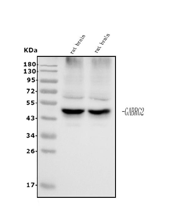
 FCM/FACS (Flow Cytometry)
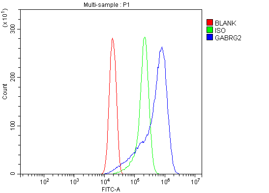
(Figure 3. Flow Cytometry analysis of THP-1 cells using anti-GABA A Receptor gamma 2/GABRG2 antibody (AAA125651).Overlay histogram showing THP-1 cells stained with AAA125651 (Blue line). The cells were blocked with 10% normal goat serum. And then incubated with rabbit anti-GABA A Receptor gamma 2/GABRG2 Antibody (AAA125651, 1μg/1x106 cells) for 30 min at 20 degree C. DyLight®488 conjugated goat anti-rabbit IgG (5-10μg/1x106 cells) was used as secondary antibody for 30 minutes at 20 degree C. Isotype control antibody (Green line) was rabbit IgG (1μg/1x106) used under the same conditions. Unlabelled sample (Red line) was also used as a control.)
FCM/FACS (Flow Cytometry)
(Figure 3. Flow Cytometry analysis of THP-1 cells using anti-GABA A Receptor gamma 2/GABRG2 antibody (AAA125651).Overlay histogram showing THP-1 cells stained with AAA125651 (Blue line). The cells were blocked with 10% normal goat serum. And then incubated with rabbit anti-GABA A Receptor gamma 2/GABRG2 Antibody (AAA125651, 1μg/1x106 cells) for 30 min at 20 degree C. DyLight®488 conjugated goat anti-rabbit IgG (5-10μg/1x106 cells) was used as secondary antibody for 30 minutes at 20 degree C. Isotype control antibody (Green line) was rabbit IgG (1μg/1x106) used under the same conditions. Unlabelled sample (Red line) was also used as a control.)
GABA A Receptor gamma 2/GABRG2, Polyclonal Antibody (Cat# AAA125651)
Full Name
Anti-GABA A Receptor gamma 2/GABRG2 Antibody
Gene Names
GABRG2; CAE2; ECA2; GEFSP3
Reactivity
Human, Mouse, Rat
Applications
Direct ELISA, Flow Cytometry, Immunohistochemistry, Western Blot
Purity
Immunogen affinity purified.
Pricing
 WB (Western Blot)
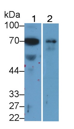
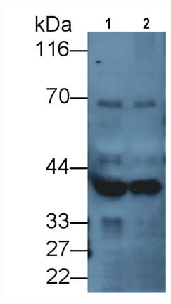
(Western Blot; Sample: Lane1: Porcine Cerebrum lysate; Lane2: Rat Cerebrum lysate Primary Ab: 2ug/ml Mouse AntiHuman DCLK1 Antibody Second Ab: 0.2ug/mL HRPLinked Caprine AntiMouse IgG Polyclonal Antibody (Catalog: SAA544Mu19))
WB (Western Blot)
(Western Blot; Sample: Lane1: Porcine Cerebrum lysate; Lane2: Rat Cerebrum lysate Primary Ab: 2ug/ml Mouse AntiHuman DCLK1 Antibody Second Ab: 0.2ug/mL HRPLinked Caprine AntiMouse IgG Polyclonal Antibody (Catalog: SAA544Mu19))
Doublecortin Like Kinase 1 (DCLK1), Monoclonal Antibody (Cat# AAA151869)
Full Name
Monoclonal Antibody to Doublecortin Like Kinase 1 (DCLK1)
Gene Names
DCLK1; CL1; DCLK; CLICK1; DCDC3A; DCAMKL1
Reactivity
Human, Rat, Pig
Applications
Immunoprecipitation, Immunocytochemistry, Immunohistochemistry, Western Blot
Purity
Protein A + Protein G affinity chromatography
Pricing
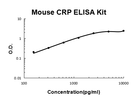 Application Data
CRP/C Reactive Protein/PTX1, ELISA Kit (Cat# AAA125887)
Full Name
Mouse CRP/C Reactive Protein/PTX1 Quick ELISA Kit
Gene Names
Crp; AI255847
Reactivity
Mouse
Pricing
 IHC (Immunohiostchemistry)
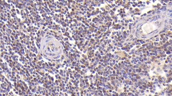
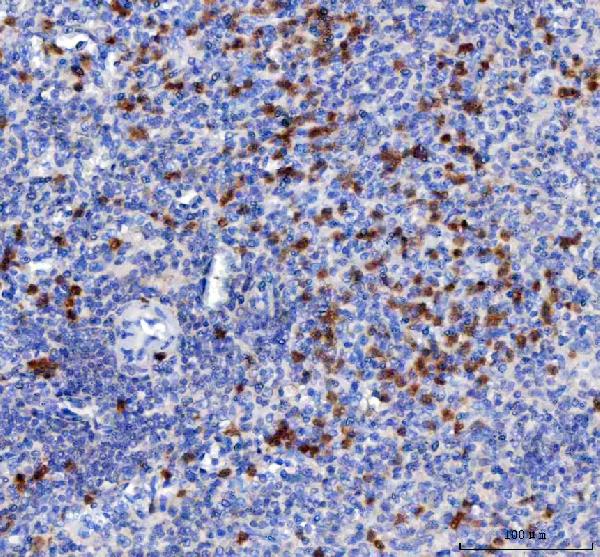
(DAB staining on IHCP;Sample: Human Spleen Tissue; Primary Ab: 30ug/ml Mouse AntiHuman PDCD1LG1 AntibodySecond Ab: 2ug/mL HRPLinked Caprine AntiMouse IgG Polyclonal Antibody(Catalog: SAA544Mu19))
IHC (Immunohiostchemistry)
(DAB staining on IHCP;Sample: Human Spleen Tissue; Primary Ab: 30ug/ml Mouse AntiHuman PDCD1LG1 AntibodySecond Ab: 2ug/mL HRPLinked Caprine AntiMouse IgG Polyclonal Antibody(Catalog: SAA544Mu19))
Programmed Cell Death Protein 1 Ligand 1 (PDL1), Monoclonal Antibody (Cat# AAA151623)
Full Name
Monoclonal Antibody to Programmed Cell Death Protein 1 Ligand 1 (PDL1)
Gene Names
CD274; B7-H; B7H1; PDL1; PD-L1; PDCD1L1; PDCD1LG1
Reactivity
Human, Pig
Applications
Immunoprecipitation, Immunocytochemistry, Immunohistochemistry, Western Blot
Purity
Protein A + Protein G affinity chromatography
Pricing
 FCM/FACS (Flow Cytometry)
(Figure 4. Flow Cytometry analysis of PC-3 cells using anti-ULBP1 antibody (AAA125721).Overlay histogram showing PC-3 cells stained with AAA125721 (Blue line). The cells were blocked with 10% normal goat serum. And then incubated with rabbit anti-ULBP1 Antibody (AAA125721,1μg/1x106 cells) for 30 min at 20 degree C. DyLight®488 conjugated goat anti-rabbit IgG (5-10μg/1x106 cells) was used as secondary antibody for 30 minutes at 20 degree C. Isotype control antibody (Green line) was rabbit IgG (1μg/1x106) used under the same conditions. Unlabelled sample (Red line) was also used as a control.)
FCM/FACS (Flow Cytometry)
(Figure 4. Flow Cytometry analysis of PC-3 cells using anti-ULBP1 antibody (AAA125721).Overlay histogram showing PC-3 cells stained with AAA125721 (Blue line). The cells were blocked with 10% normal goat serum. And then incubated with rabbit anti-ULBP1 Antibody (AAA125721,1μg/1x106 cells) for 30 min at 20 degree C. DyLight®488 conjugated goat anti-rabbit IgG (5-10μg/1x106 cells) was used as secondary antibody for 30 minutes at 20 degree C. Isotype control antibody (Green line) was rabbit IgG (1μg/1x106) used under the same conditions. Unlabelled sample (Red line) was also used as a control.)
ULBP1, Polyclonal Antibody (Cat# AAA125721)
Full Name
Anti-ULBP1 Antibody
Gene Names
ULBP1; N2DL-1; RAET1I; NKG2DL1
Reactivity
Human, Mouse, Rat
Applications
Direct ELISA, Flow Cytometry, Immunohistochemistry, Western Blot
Purity
Immunogen affinity purified.
Pricing
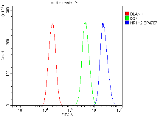 FCM/FACS (Flow Cytometry)
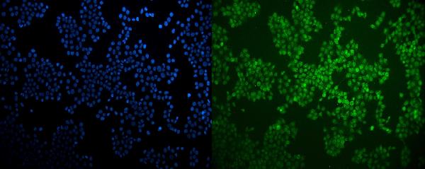
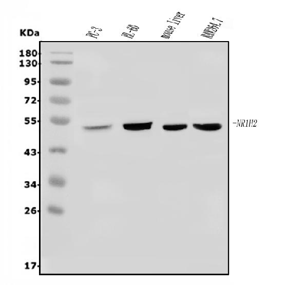
(Figure 3. Flow Cytometry analysis of U20S cells using anti-LXR beta/NER/NR1H2 antibody (AAA125726).Overlay histogram showing U20S cells stained with AAA125726 (Blue line). The cells were blocked with 10% normal goat serum. And then incubated with rabbit anti-LXR beta/NER/NR1H2 Antibody (AAA125726, 1μg/1x106 cells) for 30 min at 20 degree C. DyLight®488 conjugated goat anti-rabbit IgG (5-10μg/1x106 cells) was used as secondary antibody for 30 minutes at 20 degree C. Isotype control antibody (Green line) was rabbit IgG (1μg/1x106) used under the same conditions. Unlabelled sample (Red line) was also used as a control.)
FCM/FACS (Flow Cytometry)
(Figure 3. Flow Cytometry analysis of U20S cells using anti-LXR beta/NER/NR1H2 antibody (AAA125726).Overlay histogram showing U20S cells stained with AAA125726 (Blue line). The cells were blocked with 10% normal goat serum. And then incubated with rabbit anti-LXR beta/NER/NR1H2 Antibody (AAA125726, 1μg/1x106 cells) for 30 min at 20 degree C. DyLight®488 conjugated goat anti-rabbit IgG (5-10μg/1x106 cells) was used as secondary antibody for 30 minutes at 20 degree C. Isotype control antibody (Green line) was rabbit IgG (1μg/1x106) used under the same conditions. Unlabelled sample (Red line) was also used as a control.)
LXR beta/NER/NR1H2, Polyclonal Antibody (Cat# AAA125726)
Full Name
Anti-LXR beta/NER/NR1H2 Antibody
Gene Names
NR1H2; NER; UNR; LXRB; LXR-b; NER-I; RIP15
Reactivity
Human, Mouse
Applications
Direct ELISA, Flow Cytometry, Immunofluorescence, Immunocytochemistry, Western Blot
Purity
Immunogen affinity purified.
Pricing
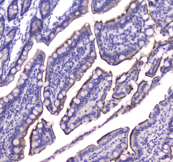
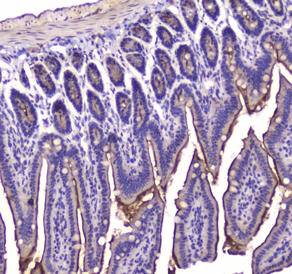
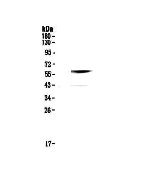
 FCM/FACS (Flow Cytometry)
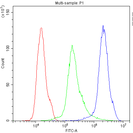
(Figure 4. Flow Cytometry analysis of Hela cells using anti-CNR1 antibody (AAA124638).Overlay histogram showing Hela cells stained with AAA124638 (Blue line).The cells were blocked with 10% normal goat serum. And then incubated with rabbit anti-CNR1 Antibody (AAA124638,1ug/1x10^6 cells) for 30 min at 20 degree C. DyLight®488 conjugated goat anti-rabbit IgG (5-10ug/1x10^6 cells) was used as secondary antibody for 30 minutes at 20 degree C. Isotype control antibody (Green line) was rabbit IgG (1ug/1x106) used under the same conditions. Unlabelled sample (Red line) was also used as a control.)
FCM/FACS (Flow Cytometry)
(Figure 4. Flow Cytometry analysis of Hela cells using anti-CNR1 antibody (AAA124638).Overlay histogram showing Hela cells stained with AAA124638 (Blue line).The cells were blocked with 10% normal goat serum. And then incubated with rabbit anti-CNR1 Antibody (AAA124638,1ug/1x10^6 cells) for 30 min at 20 degree C. DyLight®488 conjugated goat anti-rabbit IgG (5-10ug/1x10^6 cells) was used as secondary antibody for 30 minutes at 20 degree C. Isotype control antibody (Green line) was rabbit IgG (1ug/1x106) used under the same conditions. Unlabelled sample (Red line) was also used as a control.)
Cannabinoid Receptor I, Polyclonal Antibody (Cat# AAA124638)
Full Name
Anti-Cannabinoid Receptor I antibody
Gene Names
CNR1; CB1; CNR; CB-R; CB1A; CB1R; CANN6; CB1K5
Reactivity
Human, Mouse, Rat
Applications
Western Blot, Immunocytochemistry, Immunohistochemistry, Flow Cytometry, Direct ELISA
Pricing
 Application Data
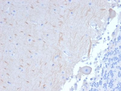
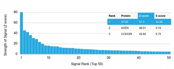
(Analysis of Protein Array containing more than 19, 000 full-length human proteins using ATG5 Mouse Monoclonal Antibody (ATG5/2101). Z- and S- Score: The Z-score represents the strength of a signal that a monoclonal antibody (MAb) (in combination with a fluorescently-tagged anti-IgG secondary antibody) produces when binding to a particular protein on the HuProtTM array. Z-scores are described in units of standard deviations (SD's) above the mean value of all signals generated on that array. If targets on HuProtTM are arranged in descending order of the Z-score, the S-score is the difference (also in units of SD's) between the Z-score. S-score therefore represents the relative target specificity of a MAb to its intended target. A MAb is considered to specific to its intended target, if the MAb has an S-score of at least 2.5. For example, if a MAb binds to protein X with a Z-score of 43 and to protein Y with a Z-score of 14, then the S-score for the binding of that MAb to protein X is equal to 29.)
Application Data
(Analysis of Protein Array containing more than 19, 000 full-length human proteins using ATG5 Mouse Monoclonal Antibody (ATG5/2101). Z- and S- Score: The Z-score represents the strength of a signal that a monoclonal antibody (MAb) (in combination with a fluorescently-tagged anti-IgG secondary antibody) produces when binding to a particular protein on the HuProtTM array. Z-scores are described in units of standard deviations (SD's) above the mean value of all signals generated on that array. If targets on HuProtTM are arranged in descending order of the Z-score, the S-score is the difference (also in units of SD's) between the Z-score. S-score therefore represents the relative target specificity of a MAb to its intended target. A MAb is considered to specific to its intended target, if the MAb has an S-score of at least 2.5. For example, if a MAb binds to protein X with a Z-score of 43 and to protein Y with a Z-score of 14, then the S-score for the binding of that MAb to protein X is equal to 29.)
ATG5, Monoclonal Antibody (Cat# AAA214715)
Full Name
ATG5 (Autophagy Marker)
Gene Names
ATG5; ASP; APG5; APG5L; hAPG5; SCAR25; APG5-LIKE
Reactivity
Human. Others not known.
Applications
Immunohistochemistry, Western Blot
Pricing
 FCM/FACS (Flow Cytometry)
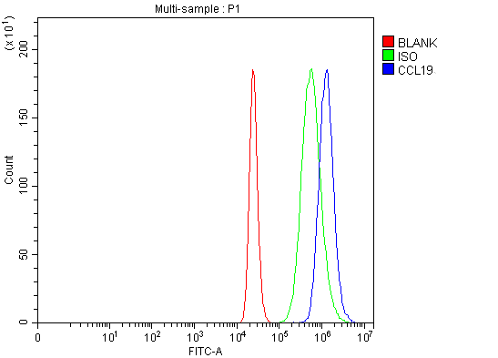
(Figure 2. Flow Cytometry analysis of THP-1 cells using anti-MIP-3 Beta/CCL19 antibody (AAA127069).Overlay histogram showing THP-1 cells stained with AAA127069 (Blue line). The cells were fixed with 4% paraformaldehyde and blocked with 10% normal goat serum. And then incubated with rabbit anti-MIP-3 Beta/CCL19 Antibody (AAA127069, 1ug/1x106 cells) for 30 min at 20 degree C. DyLight488 conjugated goat anti-rabbit IgG was used as secondary antibody for 30 minutes at 20 degree C. Isotype control antibody (Green line) was rabbit IgG (1ug/1x106) used under the same conditions. Unlabelled sample (Red line) was also used as a control.)
FCM/FACS (Flow Cytometry)
(Figure 2. Flow Cytometry analysis of THP-1 cells using anti-MIP-3 Beta/CCL19 antibody (AAA127069).Overlay histogram showing THP-1 cells stained with AAA127069 (Blue line). The cells were fixed with 4% paraformaldehyde and blocked with 10% normal goat serum. And then incubated with rabbit anti-MIP-3 Beta/CCL19 Antibody (AAA127069, 1ug/1x106 cells) for 30 min at 20 degree C. DyLight488 conjugated goat anti-rabbit IgG was used as secondary antibody for 30 minutes at 20 degree C. Isotype control antibody (Green line) was rabbit IgG (1ug/1x106) used under the same conditions. Unlabelled sample (Red line) was also used as a control.)
MIP-3 Beta/CCL19, Polyclonal Antibody (Cat# AAA127069)
Full Name
Anti-MIP-3 Beta/CCL19 Antibody
Gene Names
CCL19; ELC; CKb11; MIP3B; MIP-3b; SCYA19
Reactivity
Human
Applications
Flow Cytometry, Immunohistochemistry
Purity
Immunogen affinity purified.
Pricing
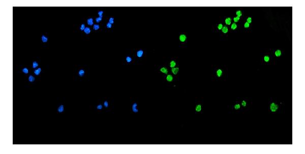
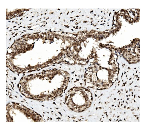
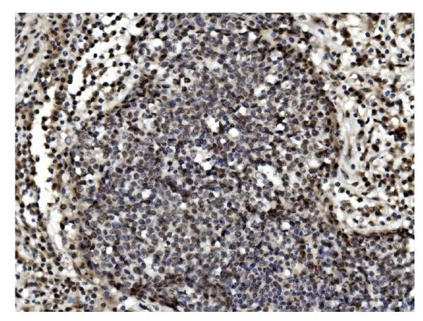
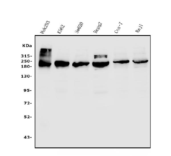
 FCM/FACS (Flow Cytometry)
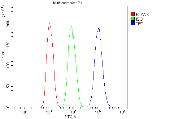
(Figure 5. Flow Cytometry analysis of HepG2 cells using anti-TET1 antibody (AAA125564).Overlay histogram showing HepG2 cells stained with AAA125564 (Blue line). The cells were blocked with 10% normal goat serum. And then incubated with rabbit anti-TET1 Antibody (AAA125564,1μg/1x106 cells) for 30 min at 20 degree C. DyLight®488 conjugated goat anti-rabbit IgG (5-10μg/1x106 cells) was used as secondary antibody for 30 minutes at 20 degree C. Isotype control antibody (Green line) was rabbit IgG (1μg/1x106) used under the same conditions. Unlabelled sample (Red line) was also used as a control.)
FCM/FACS (Flow Cytometry)
(Figure 5. Flow Cytometry analysis of HepG2 cells using anti-TET1 antibody (AAA125564).Overlay histogram showing HepG2 cells stained with AAA125564 (Blue line). The cells were blocked with 10% normal goat serum. And then incubated with rabbit anti-TET1 Antibody (AAA125564,1μg/1x106 cells) for 30 min at 20 degree C. DyLight®488 conjugated goat anti-rabbit IgG (5-10μg/1x106 cells) was used as secondary antibody for 30 minutes at 20 degree C. Isotype control antibody (Green line) was rabbit IgG (1μg/1x106) used under the same conditions. Unlabelled sample (Red line) was also used as a control.)
TET1, Polyclonal Antibody (Cat# AAA125564)
Full Name
Anti-TET1 Antibody
Gene Names
TET1; LCX; CXXC6; bA119F7.1
Reactivity
Human, Monkey
Applications
Direct ELISA, Flow Cytometry, Immunofluorescence, Immunocytochemistry, Immunohistochemistry, Western Blot
Purity
Immunogen affinity purified.
Pricing
 WB (Western Blot)
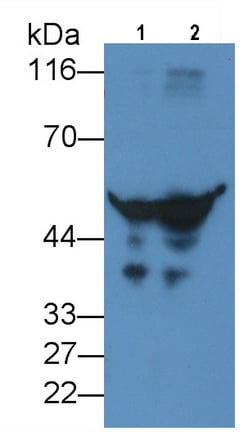
(Western Blot; Sample: Lane1: Human Serum; Lane2: Human Plasma Primary Ab: 2ug/ml Mouse AntiHuman a1AT Antibody Second Ab: 0.2ug/mL HRPLinked Caprine AntiMouse IgG Polyclonal Antibody (Catalog: SAA544Mu19))
WB (Western Blot)
(Western Blot; Sample: Lane1: Human Serum; Lane2: Human Plasma Primary Ab: 2ug/ml Mouse AntiHuman a1AT Antibody Second Ab: 0.2ug/mL HRPLinked Caprine AntiMouse IgG Polyclonal Antibody (Catalog: SAA544Mu19))
Alpha1Antitrypsin (a1AT), Monoclonal Antibody (Cat# AAA151739)
Full Name
Monoclonal Antibody to Alpha1Antitrypsin (a1AT)
Gene Names
SERPINA1; PI; A1A; AAT; PI1; A1AT; PRO2275; alpha1AT
Reactivity
Human, Pig
Applications
Immunoprecipitation, Immunocytochemistry, Immunohistochemistry, Western Blot
Purity
Protein A + Protein G affinity chromatography
Pricing
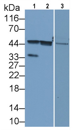 WB (Western Blot)
(Western Blot Sample: Lane1: Human Liver lysate; Lane2: HepG2 cell lysate; Lane3: Porcine Liver lysate Primary Ab: 2ug/ml Mouse AntiHuman Arg Antibody Second Ab: 0.2ug/mL HRPLinked Caprine AntiMouse IgG Polyclonal Antibody (Catalog: SAA544Mu19))
WB (Western Blot)
(Western Blot Sample: Lane1: Human Liver lysate; Lane2: HepG2 cell lysate; Lane3: Porcine Liver lysate Primary Ab: 2ug/ml Mouse AntiHuman Arg Antibody Second Ab: 0.2ug/mL HRPLinked Caprine AntiMouse IgG Polyclonal Antibody (Catalog: SAA544Mu19))
Arginase (ARG), Monoclonal Antibody (Cat# AAA151681)
Full Name
Monoclonal Antibody to Arginase (ARG)
Reactivity
Human, Pig
Applications
Immunoprecipitation, Immunocytochemistry, Immunohistochemistry, Western Blot
Purity
Protein A + Protein G affinity chromatography
Pricing
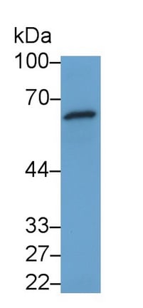
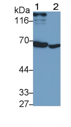 WB (Western Blot)
(Western Blot; Sample: Lane1: Rat Cerebrum lysate; Lane2: Porcine Cerebrum lysatePrimary Ab: 0.6ug/ml Mouse AntiHuman IFNa/bR1 AntibodySecond Ab: 0.2ug/mL HRPLinked Caprine AntiMouse IgG Polyclonal Antibody(Catalog: SAA544Mu19))
WB (Western Blot)
(Western Blot; Sample: Lane1: Rat Cerebrum lysate; Lane2: Porcine Cerebrum lysatePrimary Ab: 0.6ug/ml Mouse AntiHuman IFNa/bR1 AntibodySecond Ab: 0.2ug/mL HRPLinked Caprine AntiMouse IgG Polyclonal Antibody(Catalog: SAA544Mu19))
Interferon Alpha/Beta Receptor 1 (IFNa/bR1), Monoclonal Antibody (Cat# AAA151710)
Full Name
Monoclonal Antibody to Interferon Alpha/Beta Receptor 1 (IFNa/bR1)
Gene Names
IFNAR1; AVP; IFRC; IFNAR; IFNBR; IFN-alpha-REC
Reactivity
Human, Rat, Pig
Applications
Immunoprecipitation, Immunocytochemistry, Immunohistochemistry, Western Blot
Purity
Protein A + Protein G affinity chromatography
Pricing
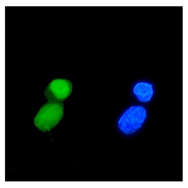
 FCM/FACS (Flow Cytometry)
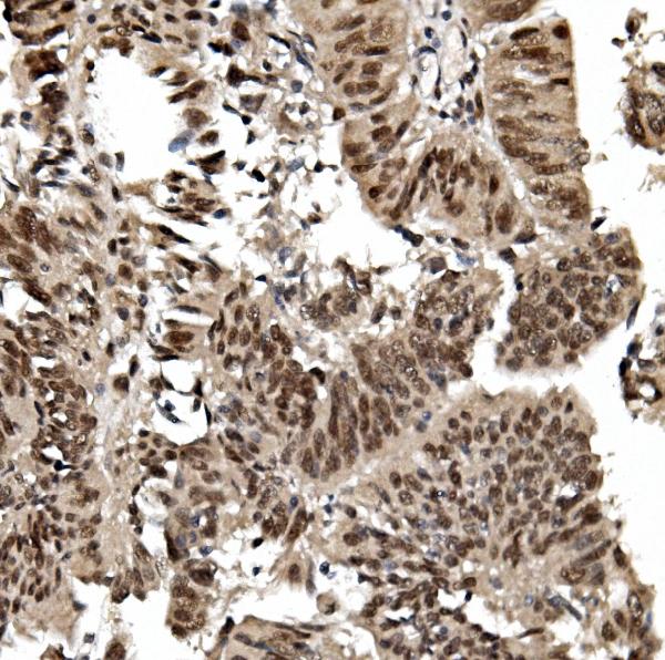
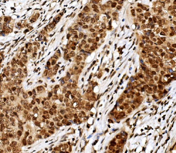
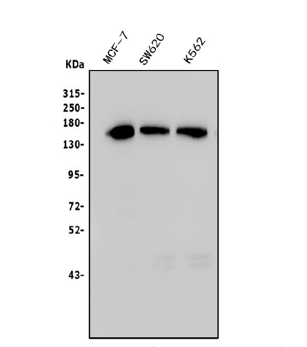
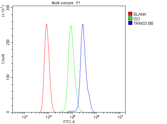
(Figure 5. Flow Cytometry analysis of HL-60 cells using anti- TIF1 gamma/TRIM33 antibody (AAA125926).Overlay histogram showing HL-60 cells stained with AAA125926 (Blue line). The cells were blocked with 10% normal goat serum. And then incubated with mouse anti-TIF1 gamma/TRIM33 Antibody (AAA125926, 1μg/1x106 cells) for 30 min at 20 degree C. DyLight®488 conjugated goat anti-mouse IgG (BA1126, 5-10μg/1x106 cells) was used as secondary antibody for 30 minutes at 20 degree C. Isotype control antibody (Green line) was mouse IgG (1μg/1x106) used under the same conditions. Unlabelled sample (Red line) was also used as a control.)
FCM/FACS (Flow Cytometry)
(Figure 5. Flow Cytometry analysis of HL-60 cells using anti- TIF1 gamma/TRIM33 antibody (AAA125926).Overlay histogram showing HL-60 cells stained with AAA125926 (Blue line). The cells were blocked with 10% normal goat serum. And then incubated with mouse anti-TIF1 gamma/TRIM33 Antibody (AAA125926, 1μg/1x106 cells) for 30 min at 20 degree C. DyLight®488 conjugated goat anti-mouse IgG (BA1126, 5-10μg/1x106 cells) was used as secondary antibody for 30 minutes at 20 degree C. Isotype control antibody (Green line) was mouse IgG (1μg/1x106) used under the same conditions. Unlabelled sample (Red line) was also used as a control.)
TIF1 gamma/TRIM33, Monoclonal Antibody (Cat# AAA125926)
Full Name
Anti-TIF1 gamma/TRIM33 Antibody (monoclonal, 8I8)
Gene Names
TRIM33; ECTO; PTC7; RFG7; TF1G; TIF1G; TIFGAMMA; TIF1GAMMA
Reactivity
Human
Applications
Flow Cytometry, Immunofluorescence, Immunocytochemistry, Immunohistochemistry, Western Blot
Purity
Immunogen affinity purified.
Pricing
 IF (Immunofluorescence)
(Figure 2. IF analysis of SLC8A1 using anti-SLC8A1 antibody (AAA127202).SLC8A1 was detected in an immunocytochemical section of U2OS cells. Enzyme antigen retrieval was performed using IHC enzyme antigen retrieval reagent for 15 mins. The cells were blocked with 10% goat serum. And then incubated with 5ug/mL rabbit anti-SLC8A1 Antibody (AAA127202) overnight at 4 degree C. DyLight488 Conjugated Goat Anti-Rabbit IgG was used as secondary antibody at 1:100 dilution and incubated for 30 minutes at 37 degree C. The tissue section was developed using Phalloidin-iFluor 594 Conjugated. Visualize using a fluorescence microscope and filter sets appropriate for the label used.)
IF (Immunofluorescence)
(Figure 2. IF analysis of SLC8A1 using anti-SLC8A1 antibody (AAA127202).SLC8A1 was detected in an immunocytochemical section of U2OS cells. Enzyme antigen retrieval was performed using IHC enzyme antigen retrieval reagent for 15 mins. The cells were blocked with 10% goat serum. And then incubated with 5ug/mL rabbit anti-SLC8A1 Antibody (AAA127202) overnight at 4 degree C. DyLight488 Conjugated Goat Anti-Rabbit IgG was used as secondary antibody at 1:100 dilution and incubated for 30 minutes at 37 degree C. The tissue section was developed using Phalloidin-iFluor 594 Conjugated. Visualize using a fluorescence microscope and filter sets appropriate for the label used.)
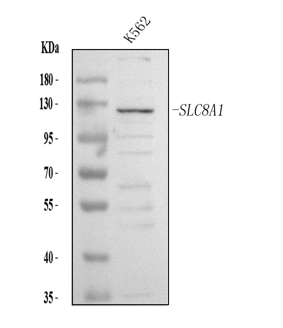
NCX1/SLC8A1, Polyclonal Antibody (Cat# AAA127202)
Full Name
Anti-NCX1/SLC8A1 Antibody Picoband
Gene Names
SLC8A1; NCX1
Reactivity
Human
Applications
Western Blot, Immunofluorescence, Immunocytochemistry
Purity
Immunogen affinity purified.
Pricing
 Application Data
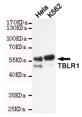
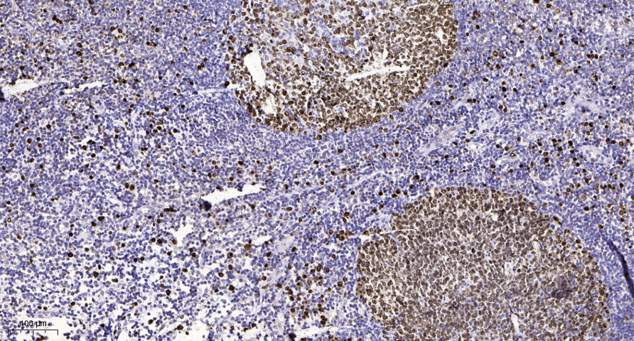
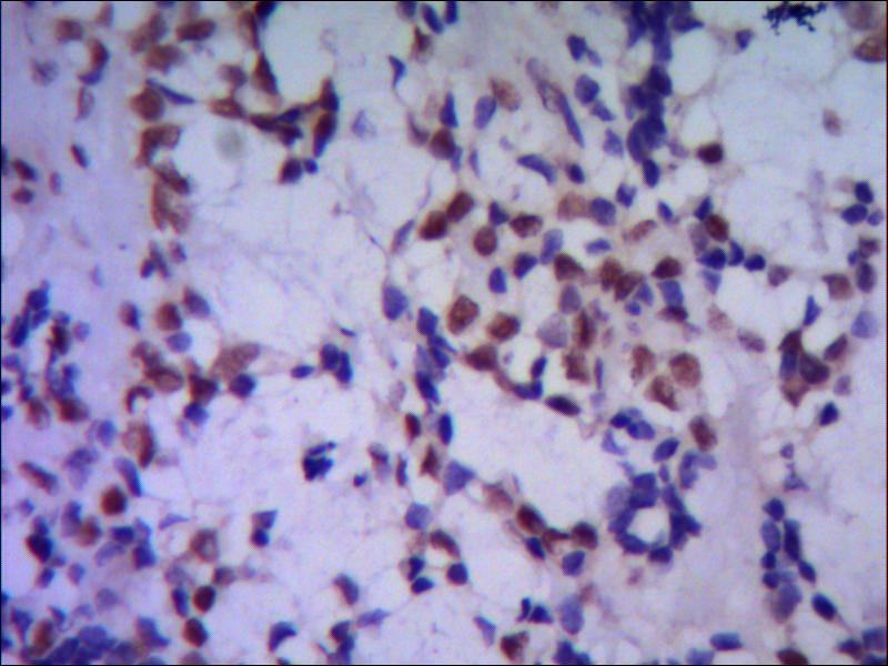
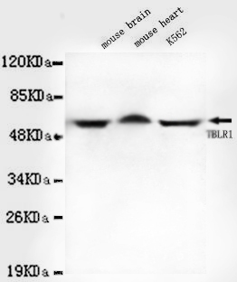
TBLR1, Monoclonal Antibody (Cat# AAA320316)
Full Name
TBLR1 Mouse mAb
Gene Names
TBL1XR1; C21; DC42; IRA1; TBLR1
Reactivity
Human, Mouse
Applications
Immunocytochemistry, Immunohistochemistry, Western Blot
Purity
The antibody was affinity-purified from mouse ascites by affinity-chromatography using epitope-specific immunogen.
Pricing
 IP (Immunoprecipitation)
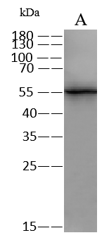
(RUVBL1 was immunoprecipitated using:; Lane A:0.5 mg HeLa Whole Cell Lysate; Lane B:0.5 mg Jurkat Whole Cell Lysate; Lane C:0.5 mg HepG2 Whole Cell Lysate; Lane D:0.5 mg Raji Whole Cell Lysate; 4 uL anti-RUVBL1 rabbit polyclonal antibody and 60 ug of Immunomagnetic beads Protein A/G.; Primary antibody:; Anti-RUVBL1 rabbit polyclonal antibody,at 1:100 dilution; Secondary antibody:; Clean-Blot IP Detection Reagent (HRP) at 1:1000dilution; Developed using the ECL technique.; Performed under reducing conditions.; Predicted band size: 50 kDa; Observed band size :55 kDa)
IP (Immunoprecipitation)
(RUVBL1 was immunoprecipitated using:; Lane A:0.5 mg HeLa Whole Cell Lysate; Lane B:0.5 mg Jurkat Whole Cell Lysate; Lane C:0.5 mg HepG2 Whole Cell Lysate; Lane D:0.5 mg Raji Whole Cell Lysate; 4 uL anti-RUVBL1 rabbit polyclonal antibody and 60 ug of Immunomagnetic beads Protein A/G.; Primary antibody:; Anti-RUVBL1 rabbit polyclonal antibody,at 1:100 dilution; Secondary antibody:; Clean-Blot IP Detection Reagent (HRP) at 1:1000dilution; Developed using the ECL technique.; Performed under reducing conditions.; Predicted band size: 50 kDa; Observed band size :55 kDa)
RUVBL1, Polyclonal Antibody (Cat# AAA258641)
Full Name
RUVBL1 Antibody, Rabbit PAb, Antigen Affinity Purified
Gene Names
RUVBL1; RVB1; TIH1; ECP54; TIP49; INO80H; NMP238; PONTIN; TIP49A; Pontin52
Reactivity
Human
Applications
Immunoprecipitation, Western Blot
Pricing
 FCM/FACS (Flow Cytometry)
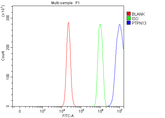
(Figure 2. Flow Cytometry analysis of RT4 cells using anti-PTPN13 antibody (AAA127121).Overlay histogram showing RT4 cells stained with AAA127121 (Blue line). To facilitate intracellular staining, cells were fixed with 4% paraformaldehyde and permeabilized with permeabilization buffer. The cells were blocked with 10% normal goat serum. And then incubated with rabbit anti-PTPN13 Antibody (AAA127121, 1ug/1x106 cells) for 30 min at 20 degree C. DyLight488 conjugated goat anti-rabbit IgG was used as secondary antibody for 30 minutes at 20 degree C. Isotype control antibody (Green line) was rabbit IgG (1ug/1x106) used under the same conditions. Unlabelled sample (Red line) was also used as a control.)
FCM/FACS (Flow Cytometry)
(Figure 2. Flow Cytometry analysis of RT4 cells using anti-PTPN13 antibody (AAA127121).Overlay histogram showing RT4 cells stained with AAA127121 (Blue line). To facilitate intracellular staining, cells were fixed with 4% paraformaldehyde and permeabilized with permeabilization buffer. The cells were blocked with 10% normal goat serum. And then incubated with rabbit anti-PTPN13 Antibody (AAA127121, 1ug/1x106 cells) for 30 min at 20 degree C. DyLight488 conjugated goat anti-rabbit IgG was used as secondary antibody for 30 minutes at 20 degree C. Isotype control antibody (Green line) was rabbit IgG (1ug/1x106) used under the same conditions. Unlabelled sample (Red line) was also used as a control.)
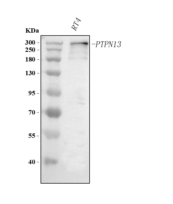
PTPN13, Polyclonal Antibody (Cat# AAA127121)
Full Name
Anti-PTPN13 Antibody Picoband
Gene Names
PTPN13; PNP1; FAP-1; PTP1E; PTPL1; PTPLE; PTP-BL; hPTP1E; PTP-BAS
Reactivity
Human
Applications
Flow Cytometry, Western Blot
Purity
Immunogen affinity purified.
Pricing
 Bioactivity
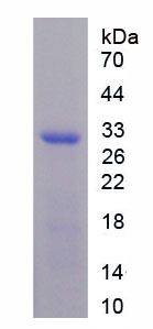
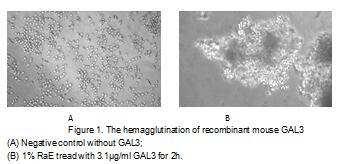
(Galectin 3 (GAL3) is a member of the lectin family, of which 14 mammalian galectins have been identified. It is also a member of the beta-galactoside-binding protein family that plays an important role in cell-cell adhesion, cell-matrix interactions, macrophage activation, angiogenesis, metastasis, apoptosis. The protein also has been demonstrated to be involved in cancer, inflammation and fibrosis, heart disease, and stroke. GAL3 is expressed in the nucleus, cytoplasm, mitochondrion, cell surface, and extracellular space. It also can agglutinate red blood. In this case, we chose rabbit erythrocyte (RaE) to assay its ability of agglutination. A general procedure for hemagglutination assay (or haemagglutination assay; HA) is as follows, two-fold dilute the recombinant mouse GAL3 with 0.9% sodium chloride injection, add 50uL a serial dilution of GAL3 to each well of a U or V-bottom shaped 96-well microtiter plate. The final well serves as a negative control without GAL3, replace with 50uL 0.9% sodium chloride injection. Then add 50uL 1% rabbit erythrocyte to each well and mixed gently. The plate is incubated for 3 hours at room temperature. The results are shown in Figure 1. It was obvious that the minimal effective concentration of GAL3 is 3.1ug/ml.)
Bioactivity
(Galectin 3 (GAL3) is a member of the lectin family, of which 14 mammalian galectins have been identified. It is also a member of the beta-galactoside-binding protein family that plays an important role in cell-cell adhesion, cell-matrix interactions, macrophage activation, angiogenesis, metastasis, apoptosis. The protein also has been demonstrated to be involved in cancer, inflammation and fibrosis, heart disease, and stroke. GAL3 is expressed in the nucleus, cytoplasm, mitochondrion, cell surface, and extracellular space. It also can agglutinate red blood. In this case, we chose rabbit erythrocyte (RaE) to assay its ability of agglutination. A general procedure for hemagglutination assay (or haemagglutination assay; HA) is as follows, two-fold dilute the recombinant mouse GAL3 with 0.9% sodium chloride injection, add 50uL a serial dilution of GAL3 to each well of a U or V-bottom shaped 96-well microtiter plate. The final well serves as a negative control without GAL3, replace with 50uL 0.9% sodium chloride injection. Then add 50uL 1% rabbit erythrocyte to each well and mixed gently. The plate is incubated for 3 hours at room temperature. The results are shown in Figure 1. It was obvious that the minimal effective concentration of GAL3 is 3.1ug/ml.)
Galectin 3 (GAL3), Active Protein (Cat# AAA152172)
Full Name
Active Galectin 3 (GAL3)
Gene Names
Lgals3; GBP; L-34; gal3; Mac-2
Applications
Activity Assay, Cell Culture
Purity
Greater than 97% by SDS-PAGE
Pricing
 FCM/FACS (Flow Cytometry)
(Figure 2. Flow Cytometry analysis of Hela cells using anti-MYO6 antibody (AAA127132).Overlay histogram showing Hela cells stained with AAA127132 (Blue line). To facilitate intracellular staining, cells were fixed with 4% paraformaldehyde and permeabilized with permeabilization buffer. The cells were blocked with 10% normal goat serum. And then incubated with rabbit anti-MYO6 Antibody (AAA127132, 1ug/1x106 cells) for 30 min at 20 degree C. DyLight488 conjugated goat anti-rabbit IgG was used as secondary antibody for 30 minutes at 20 degree C. Isotype control antibody (Green line) was rabbit IgG (1ug/1x106) used under the same conditions. Unlabelled sample (Red line) was also used as a control.)
FCM/FACS (Flow Cytometry)
(Figure 2. Flow Cytometry analysis of Hela cells using anti-MYO6 antibody (AAA127132).Overlay histogram showing Hela cells stained with AAA127132 (Blue line). To facilitate intracellular staining, cells were fixed with 4% paraformaldehyde and permeabilized with permeabilization buffer. The cells were blocked with 10% normal goat serum. And then incubated with rabbit anti-MYO6 Antibody (AAA127132, 1ug/1x106 cells) for 30 min at 20 degree C. DyLight488 conjugated goat anti-rabbit IgG was used as secondary antibody for 30 minutes at 20 degree C. Isotype control antibody (Green line) was rabbit IgG (1ug/1x106) used under the same conditions. Unlabelled sample (Red line) was also used as a control.)
MYO6, Polyclonal Antibody (Cat# AAA127132)
Full Name
Anti-MYO6 Antibody Picoband
Gene Names
MYO6; DFNA22; DFNB37
Reactivity
Human, Mouse
Applications
Flow Cytometry, Western Blot
Purity
Immunogen affinity purified.
Pricing
 FCM/FACS (Flow Cytometry)
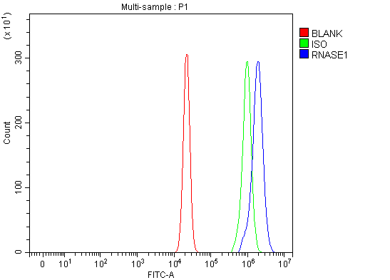
(Figure 2. Flow Cytometry analysis of U20S cells using anti-RNASE1 antibody (AAA127187).Overlay histogram showing U20S cells stained with AAA127187 (Blue line). The cells were fixed with 4% paraformaldehyde and blocked with 10% normal goat serum. And then incubated with rabbit anti-RNASE1 Antibody (AAA127187, 1ug/1x106 cells) for 30 min at 20 degree C. DyLight488 conjugated goat anti-rabbit IgG was used as secondary antibody for 30 minutes at 20 degree C. Isotype control antibody (Green line) was rabbit IgG (1ug/1x106) used under the same conditions. Unlabelled sample (Red line) was also used as a control.)
FCM/FACS (Flow Cytometry)
(Figure 2. Flow Cytometry analysis of U20S cells using anti-RNASE1 antibody (AAA127187).Overlay histogram showing U20S cells stained with AAA127187 (Blue line). The cells were fixed with 4% paraformaldehyde and blocked with 10% normal goat serum. And then incubated with rabbit anti-RNASE1 Antibody (AAA127187, 1ug/1x106 cells) for 30 min at 20 degree C. DyLight488 conjugated goat anti-rabbit IgG was used as secondary antibody for 30 minutes at 20 degree C. Isotype control antibody (Green line) was rabbit IgG (1ug/1x106) used under the same conditions. Unlabelled sample (Red line) was also used as a control.)
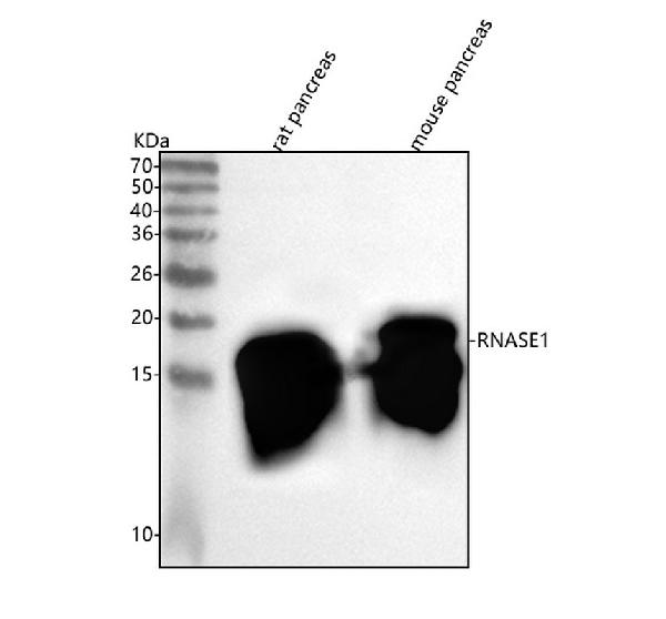
RNASE1, Polyclonal Antibody (Cat# AAA127187)
Full Name
Anti-RNASE1 Antibody Picoband
Gene Names
RNASE1; RAC1; RIB1; RNS1
Reactivity
Human, Mouse, Rat
Applications
Western Blot, Flow Cytometry
Purity
Immunogen affinity purified.
Pricing
 ICC (Immunocytochemistry)
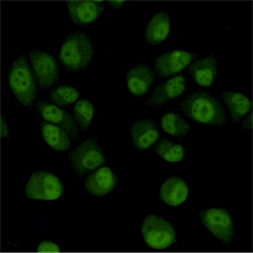
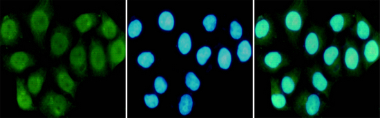
(Immunocytochemistry staining of HeLa cells fixed in 1% Paraformaldehyde and using RPA70 mouse mAb (dilution 1:100).)
ICC (Immunocytochemistry)
(Immunocytochemistry staining of HeLa cells fixed in 1% Paraformaldehyde and using RPA70 mouse mAb (dilution 1:100).)
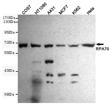
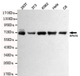
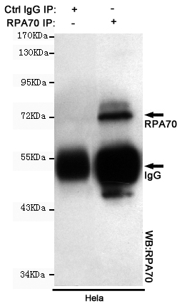
RPA70, Monoclonal Antibody (Cat# AAA320372)
Full Name
RPA70 Mouse mAb
Gene Names
RPA1; HSSB; RF-A; RP-A; REPA1; RPA70; MST075
Reactivity
Human, Monkey, Mouse, Rat
Applications
Immunoprecipitation, Immunocytochemistry, Western Blot
Purity
The antibody was affinity-purified from mouse ascites by affinity-chromatography using epitope-specific immunogen.
Pricing
 FCM/FACS (Flow Cytometry)
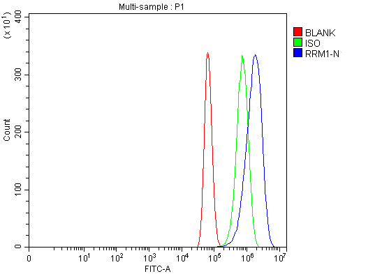
(Figure 2. Flow Cytometry analysis of U87 cells using anti-RRM1 antibody (AAA126170).Overlay histogram showing U87 cells stained with AAA126170 (Blue line). The cells were blocked with 10% normal goat serum. And then incubated with rabbit anti-RRM1 Antibody (AAA126170, 1 ug/1x10^6 cells) for 30 min at 20 degree C. DyLight488 conjugated goat anti-rabbit IgG was used as secondary antibody for 30 minutes at 20 degree C. Isotype control antibody (Green line) was rabbit IgG (1 ug/1x10^6) used under the same conditions. Unlabelled sample (Red line) was also used as a control.)
FCM/FACS (Flow Cytometry)
(Figure 2. Flow Cytometry analysis of U87 cells using anti-RRM1 antibody (AAA126170).Overlay histogram showing U87 cells stained with AAA126170 (Blue line). The cells were blocked with 10% normal goat serum. And then incubated with rabbit anti-RRM1 Antibody (AAA126170, 1 ug/1x10^6 cells) for 30 min at 20 degree C. DyLight488 conjugated goat anti-rabbit IgG was used as secondary antibody for 30 minutes at 20 degree C. Isotype control antibody (Green line) was rabbit IgG (1 ug/1x10^6) used under the same conditions. Unlabelled sample (Red line) was also used as a control.)
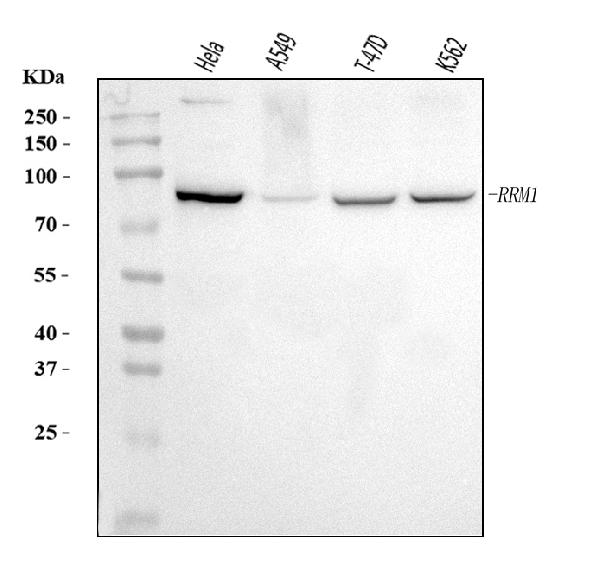
RRM1, Polyclonal Antibody (Cat# AAA126170)
Full Name
Anti-RRM1 Antibody Picoband
Gene Names
RRM1; R1; RR1; RIR1
Reactivity
Human
Applications
Flow Cytometry, Western Blot
Purity
Immunogen affinity purified.
Pricing
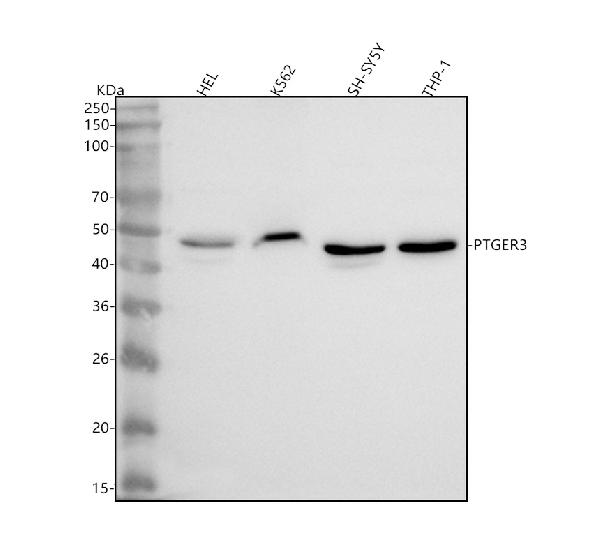
 FCM/FACS (Flow Cytometry)
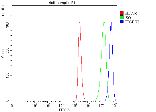
(Figure 2. Flow Cytometry analysis of THP-1 cells using anti-PTGER3 antibody (AAA127126).Overlay histogram showing THP-1 cells stained with AAA127126 (Blue line). The cells were fixed with 4% paraformaldehyde and blocked with 10% normal goat serum. And then incubated with rabbit anti-PTGER3 Antibody (AAA127126, 1ug/1x106 cells) for 30 min at 20 degree C. DyLight488 conjugated goat anti-rabbit IgG was used as secondary antibody for 30 minutes at 20 degree C. Isotype control antibody (Green line) was rabbit IgG (1ug/1x106) used under the same conditions. Unlabelled sample (Red line) was also used as a control.)
FCM/FACS (Flow Cytometry)
(Figure 2. Flow Cytometry analysis of THP-1 cells using anti-PTGER3 antibody (AAA127126).Overlay histogram showing THP-1 cells stained with AAA127126 (Blue line). The cells were fixed with 4% paraformaldehyde and blocked with 10% normal goat serum. And then incubated with rabbit anti-PTGER3 Antibody (AAA127126, 1ug/1x106 cells) for 30 min at 20 degree C. DyLight488 conjugated goat anti-rabbit IgG was used as secondary antibody for 30 minutes at 20 degree C. Isotype control antibody (Green line) was rabbit IgG (1ug/1x106) used under the same conditions. Unlabelled sample (Red line) was also used as a control.)
PTGER3, Polyclonal Antibody (Cat# AAA127126)
Full Name
Anti-PTGER3 Antibody Picoband
Gene Names
PTGER3; EP3; EP3e; EP3-I; EP3-II; EP3-IV; PGE2-R; EP3-III
Reactivity
Human
Applications
Flow Cytometry, Western Blot
Purity
Immunogen affinity purified.
Pricing
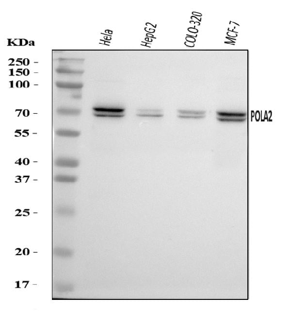
 FCM/FACS (Flow Cytometry)
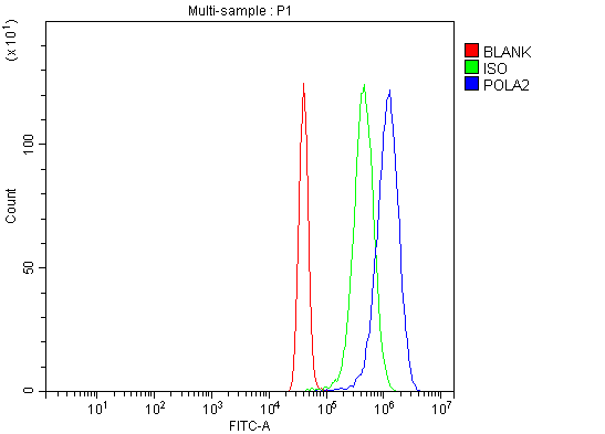
(Figure 2. Flow Cytometry analysis of U251 cells using anti-POLA2 antibody (AAA126519).Overlay histogram showing U251 cells stained with AAA126519 (Blue line). The cells were blocked with 10% normal goat serum. And then incubated with rabbit anti-POLA2 Antibody (AAA126519, 1 ug/1x10^6 cells) for 30 min at 20 degree C. DyLight488 conjugated goat anti-rabbit IgG was used as secondary antibody for 30 minutes at 20 degree C. Isotype control antibody (Green line) was rabbit IgG (1 ug/1x10^6) used under the same conditions. Unlabelled sample (Red line) was also used as a control.)
FCM/FACS (Flow Cytometry)
(Figure 2. Flow Cytometry analysis of U251 cells using anti-POLA2 antibody (AAA126519).Overlay histogram showing U251 cells stained with AAA126519 (Blue line). The cells were blocked with 10% normal goat serum. And then incubated with rabbit anti-POLA2 Antibody (AAA126519, 1 ug/1x10^6 cells) for 30 min at 20 degree C. DyLight488 conjugated goat anti-rabbit IgG was used as secondary antibody for 30 minutes at 20 degree C. Isotype control antibody (Green line) was rabbit IgG (1 ug/1x10^6) used under the same conditions. Unlabelled sample (Red line) was also used as a control.)
POLA2, Polyclonal Antibody (Cat# AAA126519)
Full Name
Anti-POLA2 Antibody Picoband
Reactivity
Human
Applications
Western Blot, Flow Cytometry
Purity
Immunogen affinity purified.
Pricing
 FCM/FACS (Flow Cytometry)
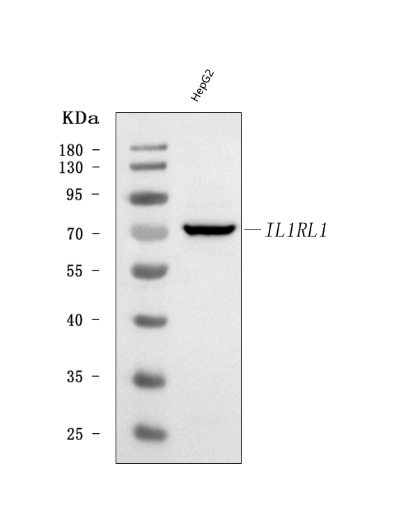
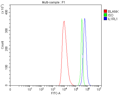
(Figure 2. Flow Cytometry analysis of HEL cells using anti-ST2/IL1RL1 antibody (AAA126030).Overlay histogram showing HEL cells stained with AAA126030 (Blue line). The cells were blocked with 10% normal goat serum. And then incubated with rabbit anti-ST2/IL1RL1 Antibody (AAA126030, 1 ug/1x10^6 cells) for 30 min at 20 degree C. DyLight488 conjugated goat anti-rabbit IgG was used as secondary antibody for 30 minutes at 20 degree C. Isotype control antibody (Green line) was rabbit IgG (1 ug/1x10^6) used under the same conditions. Unlabelled sample (Red line) was also used as a control.)
FCM/FACS (Flow Cytometry)
(Figure 2. Flow Cytometry analysis of HEL cells using anti-ST2/IL1RL1 antibody (AAA126030).Overlay histogram showing HEL cells stained with AAA126030 (Blue line). The cells were blocked with 10% normal goat serum. And then incubated with rabbit anti-ST2/IL1RL1 Antibody (AAA126030, 1 ug/1x10^6 cells) for 30 min at 20 degree C. DyLight488 conjugated goat anti-rabbit IgG was used as secondary antibody for 30 minutes at 20 degree C. Isotype control antibody (Green line) was rabbit IgG (1 ug/1x10^6) used under the same conditions. Unlabelled sample (Red line) was also used as a control.)
ST2/IL1RL1, Polyclonal Antibody (Cat# AAA126030)
Full Name
Anti-ST2/IL1RL1 Antibody Picoband
Gene Names
IL1RL1; T1; ST2; DER4; ST2L; ST2V; FIT-1; IL33R
Reactivity
Human
Applications
Flow Cytometry, Western Blot
Purity
Immunogen affinity purified.
Pricing
 IHC (Immunohistochemistry)
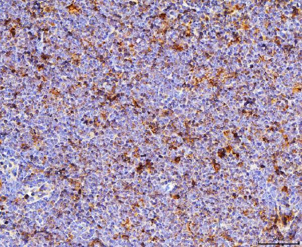
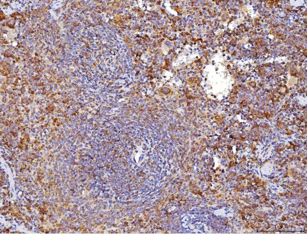
(Figure 4. IHC analysis of Cd70 using anti-Cd70 antibody (AAA126264).Cd70 was detected in a paraffin-embedded section of mouse spleen tissue. Heat mediated antigen retrieval was performed in EDTA buffer (pH 8.0, epitope retrieval solution). The tissue section was blocked with 10% goat serum. The tissue section was then incubated with 2 ug/ml rabbit anti-Cd70 Antibody (AAA126264) overnight at 4 degree C. Peroxidase Conjugated Goat Anti-rabbit IgG was used as secondary antibody and incubated for 30 minutes at 37 degree C. The tissue section was developed using HRP Conjugated Rabbit IgG Super Vision Assay Kit with DAB as the chromogen.)
IHC (Immunohistochemistry)
(Figure 4. IHC analysis of Cd70 using anti-Cd70 antibody (AAA126264).Cd70 was detected in a paraffin-embedded section of mouse spleen tissue. Heat mediated antigen retrieval was performed in EDTA buffer (pH 8.0, epitope retrieval solution). The tissue section was blocked with 10% goat serum. The tissue section was then incubated with 2 ug/ml rabbit anti-Cd70 Antibody (AAA126264) overnight at 4 degree C. Peroxidase Conjugated Goat Anti-rabbit IgG was used as secondary antibody and incubated for 30 minutes at 37 degree C. The tissue section was developed using HRP Conjugated Rabbit IgG Super Vision Assay Kit with DAB as the chromogen.)
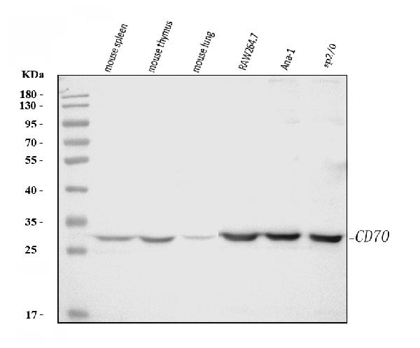
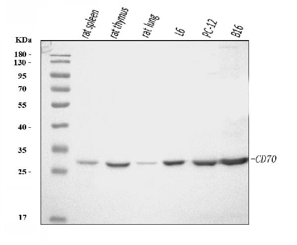
Cd70, Polyclonal Antibody (Cat# AAA126264)
Full Name
Anti-Cd70 Antibody Picoband
Gene Names
Cd70; Cd27l; CD27LG; Tnfsf7
Reactivity
Mouse, Rat
Applications
Immunohistochemistry, Western Blot
Purity
Immunogen affinity purified.
Pricing
 FCM/FACS (Flow Cytometry)
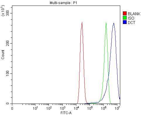
(Figure 2. Flow Cytometry analysis of MCF-7 cells using anti-TRP2/DCT antibody (AAA126180).Overlay histogram showing MCF-7 cells stained with AAA126180 (Blue line). The cells were blocked with 10% normal goat serum. And then incubated with rabbit anti-TRP2/DCT Antibody (AAA126180, 1 ug/1x10^6 cells) for 30 min at 20 degree C. DyLight488 conjugated goat anti-rabbit IgG was used as secondary antibody for 30 minutes at 20 degree C. Isotype control antibody (Green line) was rabbit IgG (1 ug/1x10^6) used under the same conditions. Unlabelled sample (Red line) was also used as a control.)
FCM/FACS (Flow Cytometry)
(Figure 2. Flow Cytometry analysis of MCF-7 cells using anti-TRP2/DCT antibody (AAA126180).Overlay histogram showing MCF-7 cells stained with AAA126180 (Blue line). The cells were blocked with 10% normal goat serum. And then incubated with rabbit anti-TRP2/DCT Antibody (AAA126180, 1 ug/1x10^6 cells) for 30 min at 20 degree C. DyLight488 conjugated goat anti-rabbit IgG was used as secondary antibody for 30 minutes at 20 degree C. Isotype control antibody (Green line) was rabbit IgG (1 ug/1x10^6) used under the same conditions. Unlabelled sample (Red line) was also used as a control.)
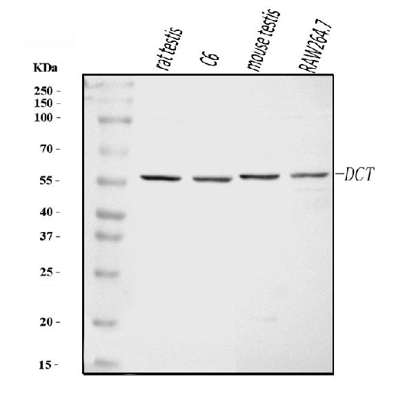
TRP2/DCT, Polyclonal Antibody (Cat# AAA126180)
Full Name
Anti-TRP2/DCT Antibody Picoband
Gene Names
DCT; TRP-2; TYRP2
Reactivity
Human, Mouse, Rat
Applications
Western Blot, Flow Cytometry
Purity
Immunogen affinity purified.
Pricing
 FCM/FACS (Flow Cytometry)
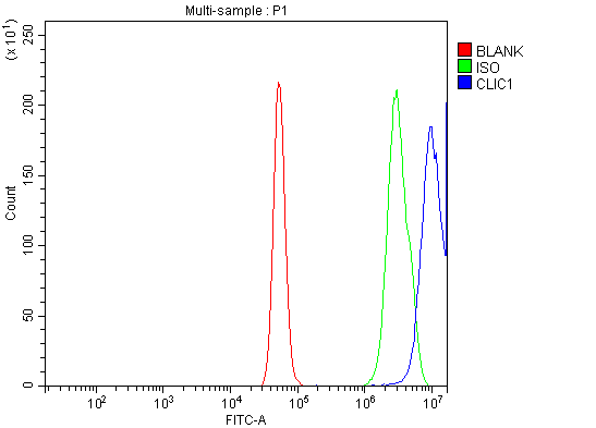
(Figure 2. Flow Cytometry analysis of A431 cells using anti-CLIC1 antibody (AAA126224).Overlay histogram showing A431 cells stained with AAA126224 (Blue line). The cells were blocked with 10% normal goat serum. And then incubated with rabbit anti-CLIC1 Antibody (AAA126224, 1 ug/1x10^6 cells) for 30 min at 20 degree C. DyLight488 conjugated goat anti-rabbit IgG was used as secondary antibody for 30 minutes at 20 degree C. Isotype control antibody (Green line) was rabbit IgG (1 ug/1x10^6) used under the same conditions. Unlabelled sample (Red line) was also used as a control.)
FCM/FACS (Flow Cytometry)
(Figure 2. Flow Cytometry analysis of A431 cells using anti-CLIC1 antibody (AAA126224).Overlay histogram showing A431 cells stained with AAA126224 (Blue line). The cells were blocked with 10% normal goat serum. And then incubated with rabbit anti-CLIC1 Antibody (AAA126224, 1 ug/1x10^6 cells) for 30 min at 20 degree C. DyLight488 conjugated goat anti-rabbit IgG was used as secondary antibody for 30 minutes at 20 degree C. Isotype control antibody (Green line) was rabbit IgG (1 ug/1x10^6) used under the same conditions. Unlabelled sample (Red line) was also used as a control.)
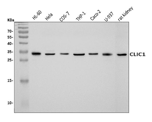
CLIC1, Polyclonal Antibody (Cat# AAA126224)
Full Name
Anti-CLIC1 Antibody Picoband
Gene Names
CLIC1; G6; NCC27
Reactivity
Human, Rat, Monkey
Applications
Flow Cytometry, Western Blot
Purity
Immunogen affinity purified.
Pricing
 IF (Immunofluorescence)
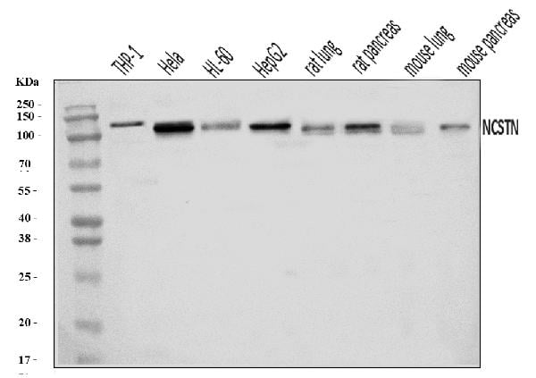
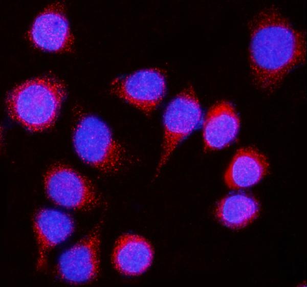
(Figure 2. IF analysis of Nicastrin/NCSTN using anti-Nicastrin/NCSTN antibody (AAA126288).Nicastrin/NCSTN was detected in an immunocytochemical section of SiHa cells. Enzyme antigen retrieval was performed using IHC enzyme antigen retrieval reagent (AR0022) for 15 mins. The cells were blocked with 10% goat serum. And then incubated with 5 ug/mL rabbit anti-Nicastrin/NCSTN Antibody (AAA126288) overnight at 4 degree C. DyLight594 Conjugated Goat Anti-Rabbit IgG (BA1142) was used as secondary antibody at 1:100 dilution and incubated for 30 minutes at 37 degree C. The section was counterstained with DAPI. Visualize using a fluorescence microscope and filter sets appropriate for the label used.)
IF (Immunofluorescence)
(Figure 2. IF analysis of Nicastrin/NCSTN using anti-Nicastrin/NCSTN antibody (AAA126288).Nicastrin/NCSTN was detected in an immunocytochemical section of SiHa cells. Enzyme antigen retrieval was performed using IHC enzyme antigen retrieval reagent (AR0022) for 15 mins. The cells were blocked with 10% goat serum. And then incubated with 5 ug/mL rabbit anti-Nicastrin/NCSTN Antibody (AAA126288) overnight at 4 degree C. DyLight594 Conjugated Goat Anti-Rabbit IgG (BA1142) was used as secondary antibody at 1:100 dilution and incubated for 30 minutes at 37 degree C. The section was counterstained with DAPI. Visualize using a fluorescence microscope and filter sets appropriate for the label used.)
Nicastrin/NCSTN, Polyclonal Antibody (Cat# AAA126288)
Full Name
Anti-Nicastrin/NCSTN Antibody Picoband
Gene Names
NCSTN; ATAG1874
Reactivity
Human, Mouse, Rat
Applications
Immunofluorescence, Immunocytochemistry, Western Blot
Purity
Immunogen affinity purified.
Pricing
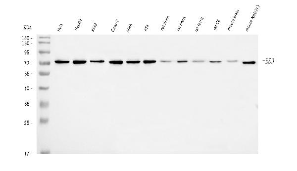
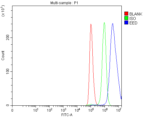 FCM/FACS (Flow Cytometry)
(Figure 2. Flow Cytometry analysis of A549 cells using anti-EED antibody (AAA126135).Overlay histogram showing A549 cells stained with AAA126135 (Blue line). The cells were blocked with 10% normal goat serum. And then incubated with rabbit anti-EED Antibody (AAA126135, 1 ug/1x10^6 cells) for 30 min at 20 degree C. DyLight488 conjugated goat anti-rabbit IgG was used as secondary antibody for 30 minutes at 20 degree C. Isotype control antibody (Green line) was rabbit IgG (1 ug/1x10^6) used under the same conditions. Unlabelled sample (Red line) was also used as a control.)
FCM/FACS (Flow Cytometry)
(Figure 2. Flow Cytometry analysis of A549 cells using anti-EED antibody (AAA126135).Overlay histogram showing A549 cells stained with AAA126135 (Blue line). The cells were blocked with 10% normal goat serum. And then incubated with rabbit anti-EED Antibody (AAA126135, 1 ug/1x10^6 cells) for 30 min at 20 degree C. DyLight488 conjugated goat anti-rabbit IgG was used as secondary antibody for 30 minutes at 20 degree C. Isotype control antibody (Green line) was rabbit IgG (1 ug/1x10^6) used under the same conditions. Unlabelled sample (Red line) was also used as a control.)
EED, Polyclonal Antibody (Cat# AAA126135)
Full Name
Anti-EED Antibody Picoband
Gene Names
EED; HEED; WAIT1
Reactivity
Human, Mouse, Rat
Applications
Flow Cytometry, Western Blot
Purity
Immunogen affinity purified.
Pricing
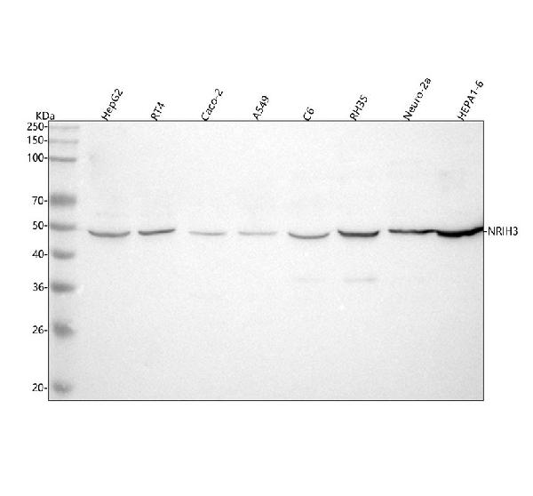
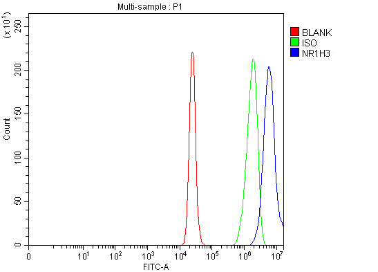 FCM/FACS (Flow Cytometry)
(Figure 2. Flow Cytometry analysis of RT4 cells using anti-NR1H3 antibody (AAA127171).Overlay histogram showing RT4 cells stained with AAA127171 (Blue line). To facilitate intracellular staining, cells were fixed with 4% paraformaldehyde and permeabilized with permeabilization buffer. The cells were blocked with 10% normal goat serum. And then incubated with rabbit anti-NR1H3 Antibody (AAA127171, 1ug/1x106 cells) for 30 min at 20 degree C. DyLight488 conjugated goat anti-rabbit IgG was used as secondary antibody for 30 minutes at 20 degree C. Isotype control antibody (Green line) was rabbit IgG (1ug/1x106) used under the same conditions. Unlabelled sample (Red line) was also used as a control.)
FCM/FACS (Flow Cytometry)
(Figure 2. Flow Cytometry analysis of RT4 cells using anti-NR1H3 antibody (AAA127171).Overlay histogram showing RT4 cells stained with AAA127171 (Blue line). To facilitate intracellular staining, cells were fixed with 4% paraformaldehyde and permeabilized with permeabilization buffer. The cells were blocked with 10% normal goat serum. And then incubated with rabbit anti-NR1H3 Antibody (AAA127171, 1ug/1x106 cells) for 30 min at 20 degree C. DyLight488 conjugated goat anti-rabbit IgG was used as secondary antibody for 30 minutes at 20 degree C. Isotype control antibody (Green line) was rabbit IgG (1ug/1x106) used under the same conditions. Unlabelled sample (Red line) was also used as a control.)
NR1H3, Polyclonal Antibody (Cat# AAA127171)
Full Name
Anti-NR1H3 Antibody Picoband
Gene Names
NR1H3; LXRA; LXR-a; RLD-1
Reactivity
Human, Mouse, Rat
Applications
Flow Cytometry, Western Blot
Purity
Immunogen affinity purified.
Pricing
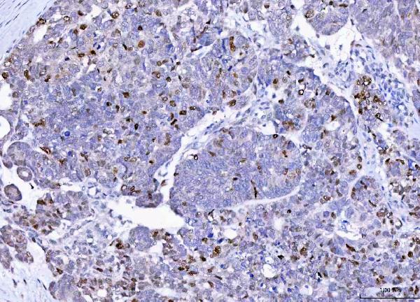
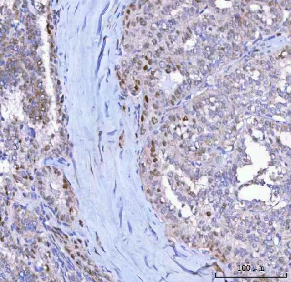
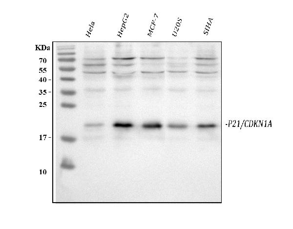
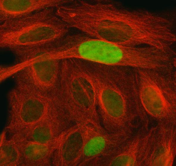 IF (Immunofluorescence)
(Figure 4. IF analysis of P21/CDKN1A using anti-P21/CDKN1A antibody (AAA126971) and anti-Beta Tubulin antibody (M01857-3).P21/CDKN1A was detected in immunocytochemical section of Hela cell. Enzyme antigen retrieval was performed using IHC enzyme antigen retrieval reagent for 15 mins. The cells were blocked with 10% goat serum. And then incubated with 5ug/mL rabbit anti-P21/CDKN1A Antibody (AAA126971) and mouse anti-Beta Tubulin antibody (M01857-3) overnight at 4 degree C. DyLight488 Goat Anti-Rabbit IgG and DyLight550 Conjugated Goat Anti-Mouse IgG (BA1133) were used as secondary antibody at 1:500 dilution and incubated for 30 minutes at 37 degree C. Visualize using a fluorescence microscope and filter sets appropriate for the label used.)
IF (Immunofluorescence)
(Figure 4. IF analysis of P21/CDKN1A using anti-P21/CDKN1A antibody (AAA126971) and anti-Beta Tubulin antibody (M01857-3).P21/CDKN1A was detected in immunocytochemical section of Hela cell. Enzyme antigen retrieval was performed using IHC enzyme antigen retrieval reagent for 15 mins. The cells were blocked with 10% goat serum. And then incubated with 5ug/mL rabbit anti-P21/CDKN1A Antibody (AAA126971) and mouse anti-Beta Tubulin antibody (M01857-3) overnight at 4 degree C. DyLight488 Goat Anti-Rabbit IgG and DyLight550 Conjugated Goat Anti-Mouse IgG (BA1133) were used as secondary antibody at 1:500 dilution and incubated for 30 minutes at 37 degree C. Visualize using a fluorescence microscope and filter sets appropriate for the label used.)
P21/CDKN1A, Polyclonal Antibody (Cat# AAA126971)
Full Name
Anti-P21/CDKN1A Antibody Picoband
Gene Names
CDKN1A; P21; CIP1; SDI1; WAF1; CAP20; CDKN1; MDA-6; p21CIP1
Reactivity
Human
Applications
Western Blot, Immunocytochemistry, Immunohistochemistry, Immunofluorescence
Purity
Immunogen affinity purified.
Pricing