
Filters
Clonality
Type
Reactivity
Gene Name
Isotype
Host
Application
Clone
10000 results for "Recombinant Proteins" - showing 8900-8950
 ELISA
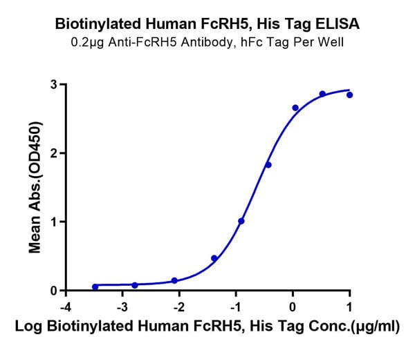
(Biotinylated Human FcRH5/FcRL5 Protein ELISA DataImmobilized Anti-FcRH5 Antibody at 2?g/ml (100?l/well) on the plate. Dose response curve for Biotinylated Human FcRH5, His Tag with the EC50 of 0.23?g/ml determined by ELISA.)
ELISA
(Biotinylated Human FcRH5/FcRL5 Protein ELISA DataImmobilized Anti-FcRH5 Antibody at 2?g/ml (100?l/well) on the plate. Dose response curve for Biotinylated Human FcRH5, His Tag with the EC50 of 0.23?g/ml determined by ELISA.)
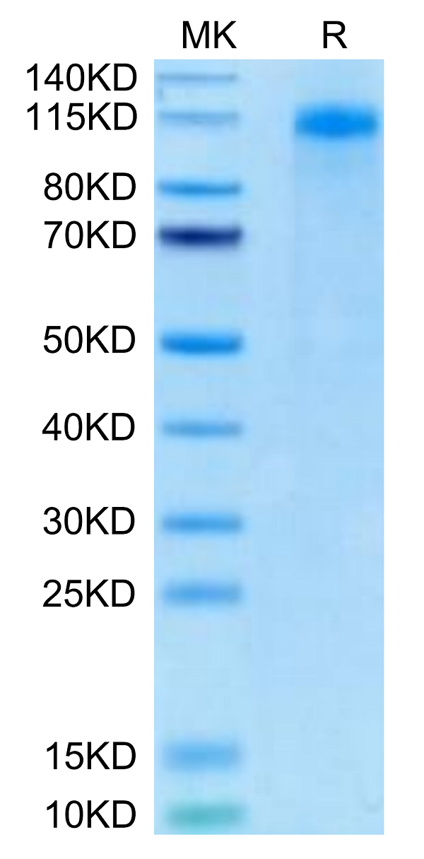
FcRH5/FcRL5, Recombinant Protein (Cat# AAA76174)
Full Name
Biotinylated Human FcRH5/FcRL5 Protein Recombinant
Gene Names
FCRL5; CD307; FCRH5; IRTA2; BXMAS1; CD307e; PRO820; RP11-217A12.1
Purity
>95%, as determined by SDS-PAGE and HPLC
Pricing
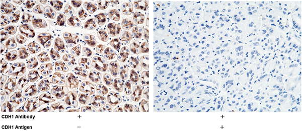
 WB (Western Blot)
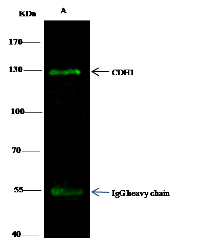
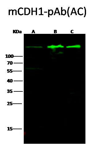
(Anti-CDH1 rabbit polyclonal antibody at 1:500 dilutionLane A: A431 Whole Cell LysateLane B: MCF7 Whole Cell LysateLane C: MDA-MB-468 Whole Cell LysateLysates/proteins at 30 ug per lane.SecondaryGoat Anti-Rabbit IgG H&L (Dylight800) at 1/10000 dilution.Developed using the Odyssey technique.Performed under reducing conditions.Predicted band size:97 kDaObserved band size:150 kDa)
WB (Western Blot)
(Anti-CDH1 rabbit polyclonal antibody at 1:500 dilutionLane A: A431 Whole Cell LysateLane B: MCF7 Whole Cell LysateLane C: MDA-MB-468 Whole Cell LysateLysates/proteins at 30 ug per lane.SecondaryGoat Anti-Rabbit IgG H&L (Dylight800) at 1/10000 dilution.Developed using the Odyssey technique.Performed under reducing conditions.Predicted band size:97 kDaObserved band size:150 kDa)
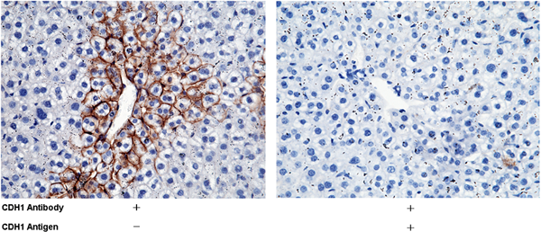
E-Cadherin, Polyclonal Antibody (Cat# AAA257205)
Full Name
Anti-E-Cadherin Antibody, Rabbit Polyclonal
Gene Names
Cdh1; Um; UVO; Ecad; E-cad; L-CAM; AA960649
Reactivity
Mouse
Applications
Immunoprecipitation, Immunohistochemistry, Western Blot
Purity
Protein A & Antigen Affinity
Pricing
 SDS-PAGE
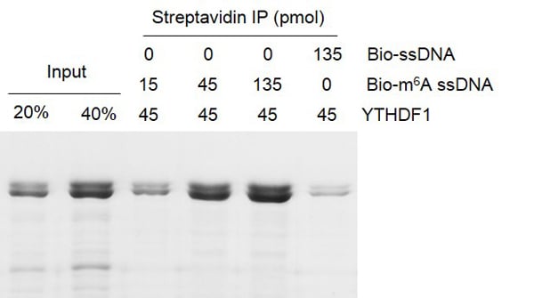
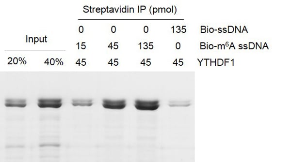
(Binding Assay for Recombinant YTHDF1 protein. 45 pmol biotinylated m6A ssDNA oligos (5’-GTTGCCTGTTCGTGTTGG/m6A/CTTGCCTGT-3’) were incubated with YTHDF1 protein in binding buffer for 3 hr at 4C. DNA-YTHDF1 compounds were incubated with streptavidin beads for 1 hour at 4C, and pull-down products were were run on a 10% SDS-PAGE gel, stained with Coomassie Blue. (Biotinylated,unmethylated ssDNA oligos used as negative control). YTHDF1 preferentially binds to ssDNA with m6A modifications compared with unmodified ssDNA.)
SDS-PAGE
(Binding Assay for Recombinant YTHDF1 protein. 45 pmol biotinylated m6A ssDNA oligos (5’-GTTGCCTGTTCGTGTTGG/m6A/CTTGCCTGT-3’) were incubated with YTHDF1 protein in binding buffer for 3 hr at 4C. DNA-YTHDF1 compounds were incubated with streptavidin beads for 1 hour at 4C, and pull-down products were were run on a 10% SDS-PAGE gel, stained with Coomassie Blue. (Biotinylated,unmethylated ssDNA oligos used as negative control). YTHDF1 preferentially binds to ssDNA with m6A modifications compared with unmodified ssDNA.)
YTHDF1, Recombinant Protein (Cat# AAA59780)
Full Name
Recombinant YTHDF1 protein
Gene Names
YTHDF1; C20orf21
Pricing
 SDS-PAGE
Killer Cell Immunoglobulin Like Receptor 2DL2 (KIR2DL2), Recombinant Protein (Cat# AAA134658)
Full Name
Recombinant Killer Cell Immunoglobulin Like Receptor 2DL2 (KIR2DL2)
Gene Names
KIR2DL2; NKAT6; p58.2; CD158b; CD158B1
Reactivity
Homo sapiens (Human)
Applications
Western Blot
Purity
> 90%
Pricing
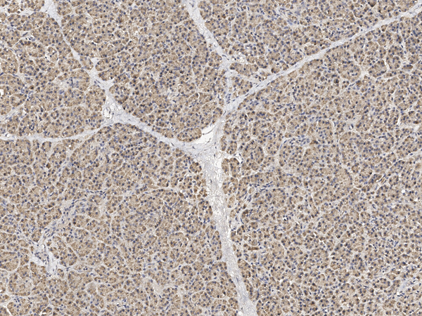
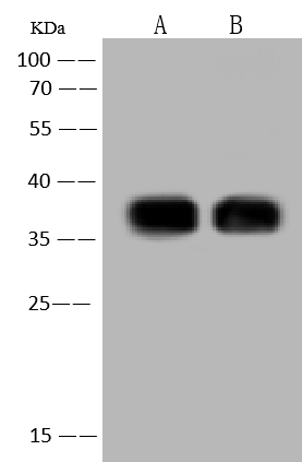
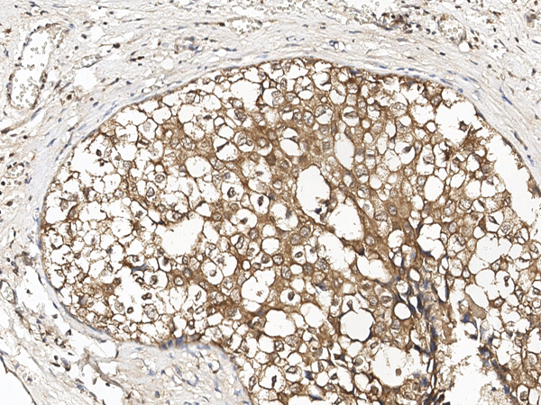 IHC (Immunohistochemisry)
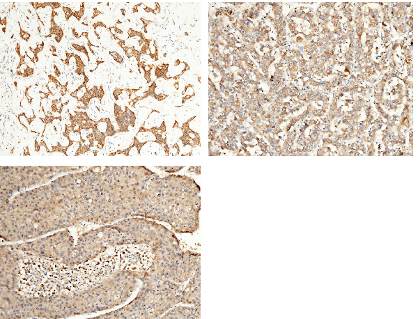
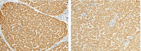
(Immunochemical staining of human EBAG9 in human breast carcinoma with rabbit polyclonal antibody at 1:100 dilution, formalin-fixed paraffin embedded sections.)
IHC (Immunohistochemisry)
(Immunochemical staining of human EBAG9 in human breast carcinoma with rabbit polyclonal antibody at 1:100 dilution, formalin-fixed paraffin embedded sections.)
EBAG9, Polyclonal Antibody (Cat# AAA258915)
Full Name
Anti-EBAG9 Antibody, Rabbit Polyclonal
Reactivity
Human
Applications
Immunohistochemistry, Western Blot
Purity
Protein A & Antigen Affinity
Pricing
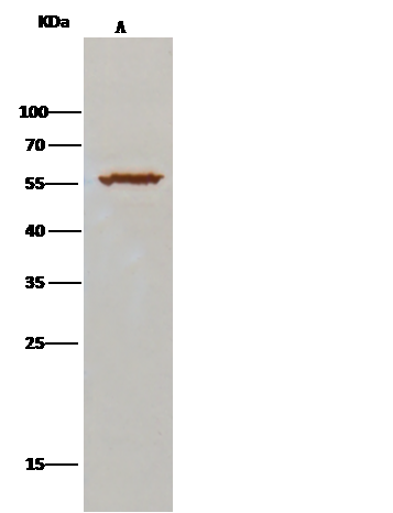
 WB (Western Blot)
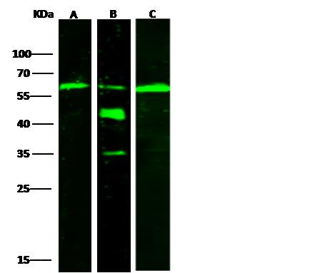
(Anti-ALDH7A1 rabbit polyclonal antibody at 1:500 dilutionLane A: HepG2 Whole Cell LysateLane B: SH-SY5Y Whole Cell LysateLane C: 293T Whole Cell LysateLysates/proteins at 30 ug per lane.SecondaryGoat Anti-Rabbit IgG H&L (Dylight800) at 1/10000 dilution.Developed using the Odyssey technique.Performed under reducing conditions.Predicted band size:58 kDaObserved band size:58 kDa(We are unsure as to the identity of these extra bands.))
WB (Western Blot)
(Anti-ALDH7A1 rabbit polyclonal antibody at 1:500 dilutionLane A: HepG2 Whole Cell LysateLane B: SH-SY5Y Whole Cell LysateLane C: 293T Whole Cell LysateLysates/proteins at 30 ug per lane.SecondaryGoat Anti-Rabbit IgG H&L (Dylight800) at 1/10000 dilution.Developed using the Odyssey technique.Performed under reducing conditions.Predicted band size:58 kDaObserved band size:58 kDa(We are unsure as to the identity of these extra bands.))
ALDH7A1, Polyclonal Antibody (Cat# AAA255438)
Full Name
Anti-ALDH7A1 Antibody, Rabbit Polyclonal
Gene Names
ALDH7A1; EPD; PDE; ATQ1
Reactivity
Human
Applications
Immunoprecipitation, Immunohistochemistry, Western Blot
Purity
Protein A & Antigen Affinity
Pricing
 WB (Western Blot)
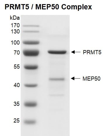
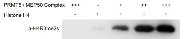
(PRMT / MEP50 complex, Activity Assay 0.5 ug Histone H4 was incubated with 0 ug (-), 0.225 ug (+), 0.45 ug (++), or 0.9 ug (+++) PRMT5 / MEP50 complex in 30 ul reaction system containing 50 mM Tris-HCl pH 8.6, 0.02% Triton X-100, 2 mM MgCl2, 1 mM TCEP, 50 uM SAM for 3 hours at room temperature. 6 ul reaction products were loaded and run on a 12.5% SDS-PAGE gel. Western Blot was used to detect the generation of reaction products (H4R3me2s antibody). PRMT5 / MEP50 Complex only was used as negative control.)
WB (Western Blot)
(PRMT / MEP50 complex, Activity Assay 0.5 ug Histone H4 was incubated with 0 ug (-), 0.225 ug (+), 0.45 ug (++), or 0.9 ug (+++) PRMT5 / MEP50 complex in 30 ul reaction system containing 50 mM Tris-HCl pH 8.6, 0.02% Triton X-100, 2 mM MgCl2, 1 mM TCEP, 50 uM SAM for 3 hours at room temperature. 6 ul reaction products were loaded and run on a 12.5% SDS-PAGE gel. Western Blot was used to detect the generation of reaction products (H4R3me2s antibody). PRMT5 / MEP50 Complex only was used as negative control.)
PRMT5/MEP50, Recombinant Protein (Cat# AAA59732)
Full Name
Recombinant PRMT5/MEP50 complex
Gene Names
PRMT5; JBP1; SKB1; IBP72; SKB1Hs; HRMT1L5
Pricing
 WB (Western Blot)
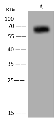
(Anti-Albumin rabbit polyclonal antibody at 1:500 dilutionLane A: HepG2 Whole Cell LysateLysates/proteins at 30 ug per lane.SecondaryGoat Anti-Rabbit IgG (H+L)/HRP at 1/10000 dilutionDeveloped using the ECL technique.Performed under reducing conditions.Predicted band size:69 kDa)
WB (Western Blot)
(Anti-Albumin rabbit polyclonal antibody at 1:500 dilutionLane A: HepG2 Whole Cell LysateLysates/proteins at 30 ug per lane.SecondaryGoat Anti-Rabbit IgG (H+L)/HRP at 1/10000 dilutionDeveloped using the ECL technique.Performed under reducing conditions.Predicted band size:69 kDa)
Albumin, Polyclonal Antibody (Cat# AAA258864)
Full Name
Anti-Albumin Antibody, Rabbit Polyclonal
Reactivity
Human
Applications
Immunoprecipitation, Western Blot
Purity
Protein A & Antigen Affinity
Pricing
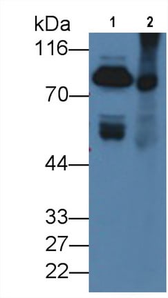 WB (Western Blot)
(Western Blot; Sample: Lane1: Rat Serum; Lane2: Rat Liver lysate Primary Ab: 1ug/ml Mouse AntiRat F2 Antibody Second Ab: 0.2ug/mL HRPLinked Caprine AntiMouse IgG Polyclonal Antibody (Catalog: SAA544Mu19))
WB (Western Blot)
(Western Blot; Sample: Lane1: Rat Serum; Lane2: Rat Liver lysate Primary Ab: 1ug/ml Mouse AntiRat F2 Antibody Second Ab: 0.2ug/mL HRPLinked Caprine AntiMouse IgG Polyclonal Antibody (Catalog: SAA544Mu19))
Coagulation Factor II (F2), Monoclonal Antibody (Cat# AAA151636)
Full Name
Monoclonal Antibody to Coagulation Factor II (F2)
Reactivity
Rat
Applications
Immunoprecipitation, Immunocytochemistry, Immunohistochemistry, Western Blot
Purity
Protein A + Protein G affinity chromatography
Pricing
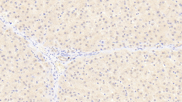 IHC (Immunohistochemistry)
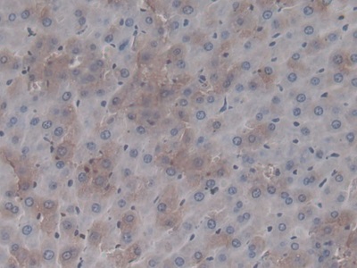
(DAB staining on IHC-P;Sample: Human Liver Tissue;Primary Ab: 20ug/ml Mouse Anti-Human FIS1 AntibodySecond Ab: 2ug/mL HRP-Linked Caprine Anti-Mouse IgG Polyclonal Antibody)
IHC (Immunohistochemistry)
(DAB staining on IHC-P;Sample: Human Liver Tissue;Primary Ab: 20ug/ml Mouse Anti-Human FIS1 AntibodySecond Ab: 2ug/mL HRP-Linked Caprine Anti-Mouse IgG Polyclonal Antibody)
Fission 1 (FIS1), Monoclonal Antibody (Cat# AAA151892)
Full Name
Monoclonal Antibody to Fission 1 (FIS1)
Gene Names
FIS1; TTC11; CGI-135
Reactivity
Human, Pig
Applications
Immunoprecipitation, Immunocytochemistry, Immunohistochemistry, Western Blot
Purity
Protein A + Protein G affinity chromatography
Pricing
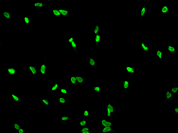
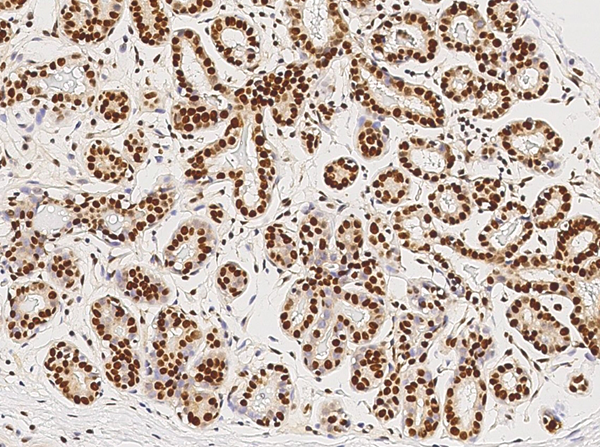
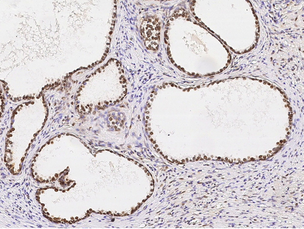
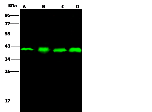
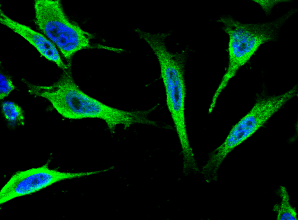
 IP (Immunoprecipitation)
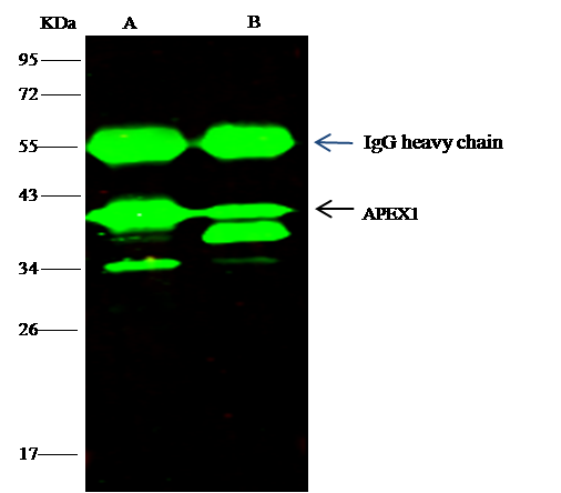
(APEX1 was immunoprecipitated using:Lane A:0.5 mg Hela Whole Cell LysateLane B:0.5 mg HepG2 Whole Cell Lysate4 uL anti-APEX1 rabbit polyclonal antibody and 15 ul of 50 % Protein G agarose.Primary antibody:Anti-APEX1 rabbit polyclonal antibody,at 1:100 dilutionSecondary antibody:Dylight 800-labeled antibody to rabbit IgG (H+L), at 1:5000 dilutionDeveloped using the odssey technique.Performed under reducing conditions.Predicted band size: 36 kDaObserved band size: 36 kDa)
IP (Immunoprecipitation)
(APEX1 was immunoprecipitated using:Lane A:0.5 mg Hela Whole Cell LysateLane B:0.5 mg HepG2 Whole Cell Lysate4 uL anti-APEX1 rabbit polyclonal antibody and 15 ul of 50 % Protein G agarose.Primary antibody:Anti-APEX1 rabbit polyclonal antibody,at 1:100 dilutionSecondary antibody:Dylight 800-labeled antibody to rabbit IgG (H+L), at 1:5000 dilutionDeveloped using the odssey technique.Performed under reducing conditions.Predicted band size: 36 kDaObserved band size: 36 kDa)
APE1/APEX1, Polyclonal Antibody (Cat# AAA255562)
Full Name
Anti-APE1/APEX1 Antibody, Rabbit Polyclonal
Gene Names
APEX1; APE; APX; APE1; APEN; APEX; HAP1; REF1
Reactivity
Human
Applications
Immunoprecipitation, Immunofluorescence, Immunocytochemistry, Immunohistochemistry, Western Blot
Purity
Protein A & Antigen Affinity
Pricing
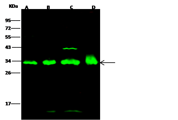
 IP (Immunoprecipitation)
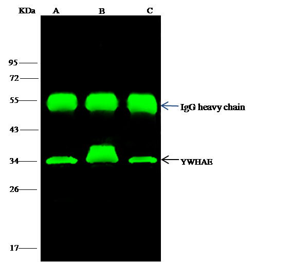
(YWHAE was immunoprecipitated using:Lane A:0.5 mg Jurkat Whole Cell LysateLane B:0.5 mg HepG2 Whole Cell LysateLane C:0.5 mg SH-SY5Y Whole Cell Lysate4 uL anti-YWHAE rabbit polyclonal antibody and 15 ul of 50 % Protein G agarose.Primary antibody:Anti-YWHAE rabbit polyclonal antibody,at 1:100 dilutionSecondary antibody:Dylight 800-labeled antibody to rabbit IgG (H+L), at 1:5000 dilutionDeveloped using the odssey technique.Performed under reducing conditions.Predicted band size: 29 kDaObserved band size: 34 kDa)
IP (Immunoprecipitation)
(YWHAE was immunoprecipitated using:Lane A:0.5 mg Jurkat Whole Cell LysateLane B:0.5 mg HepG2 Whole Cell LysateLane C:0.5 mg SH-SY5Y Whole Cell Lysate4 uL anti-YWHAE rabbit polyclonal antibody and 15 ul of 50 % Protein G agarose.Primary antibody:Anti-YWHAE rabbit polyclonal antibody,at 1:100 dilutionSecondary antibody:Dylight 800-labeled antibody to rabbit IgG (H+L), at 1:5000 dilutionDeveloped using the odssey technique.Performed under reducing conditions.Predicted band size: 29 kDaObserved band size: 34 kDa)
14-3-3 epsilon, Polyclonal Antibody (Cat# AAA255219)
Full Name
Anti-14-3-3 epsilon Antibody, Rabbit Polyclonal
Gene Names
YWHAE; MDS; MDCR; KCIP-1; 14-3-3E
Reactivity
Human
Applications
Immunoprecipitation, Immunofluorescence, Immunocytochemistry, Western Blot
Purity
Protein A & Antigen Affinity
Pricing
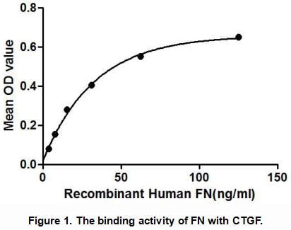
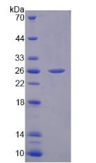
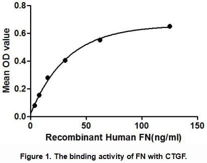
 Application Data
Fibronectin (FN), Active Protein (Cat# AAA148157)
Full Name
Active Fibronectin (FN)
Gene Names
FN1; FN; CIG; FNZ; MSF; ED-B; FINC; GFND; LETS; GFND2; SMDCF
Reactivity
Human
Applications
Activity Assay, Cell Culture
Purity
> 90%
Pricing
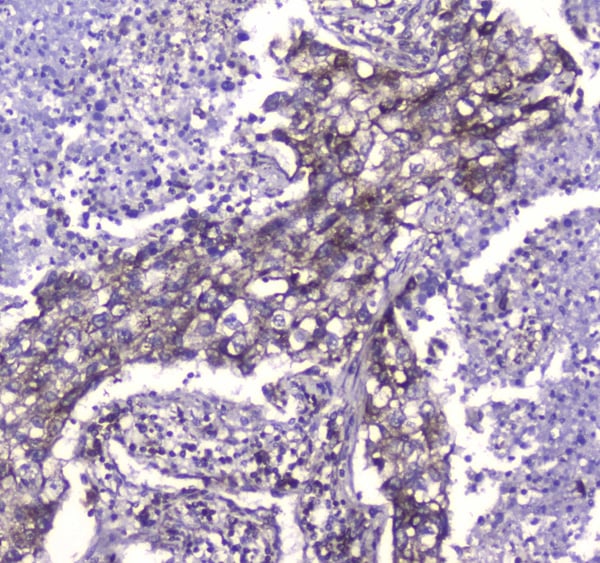 IHC (Immunohistochemisry)
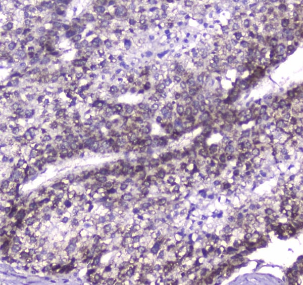
(Figure 3. IHC analysis of CLPP using anti-CLPP antibody (AAA124622).CLPP was detected in paraffin-embedded section of human lung cancer tissue. Heat mediated antigen retrieval was performed in citrate buffer (pH6, epitope retrieval solution) for 20 mins. The tissue section was blocked with 10% goat serum. The tissue section was then incubated with 1ug/ml rabbit anti-CLPP Antibody (AAA124622) overnight at 4 degree C. Biotinylated goat anti-rabbit IgG was used as secondary antibody and incubated for 30 minutes at 37 degree C. The tissue section was developed using Strepavidin-Biotin-Complex (SABC) with DAB as the chromogen.)
IHC (Immunohistochemisry)
(Figure 3. IHC analysis of CLPP using anti-CLPP antibody (AAA124622).CLPP was detected in paraffin-embedded section of human lung cancer tissue. Heat mediated antigen retrieval was performed in citrate buffer (pH6, epitope retrieval solution) for 20 mins. The tissue section was blocked with 10% goat serum. The tissue section was then incubated with 1ug/ml rabbit anti-CLPP Antibody (AAA124622) overnight at 4 degree C. Biotinylated goat anti-rabbit IgG was used as secondary antibody and incubated for 30 minutes at 37 degree C. The tissue section was developed using Strepavidin-Biotin-Complex (SABC) with DAB as the chromogen.)
CLPP, Polyclonal Antibody (Cat# AAA124622)
Full Name
Anti-CLPP Picoband antibody
Gene Names
CLPP; DFNB81; PRLTS3
Reactivity
Human, Mouse, Rat
No cross reactivity with other proteins.
No cross reactivity with other proteins.
Applications
Western Blot, Immunohistochemistry
Pricing
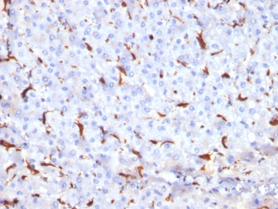
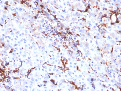
 Application Data
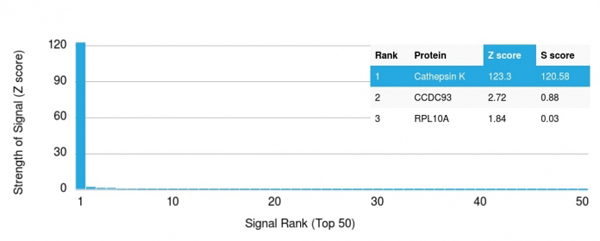
(Analysis of Protein Array containing more than 19,000 full-length human proteins using Cathepsin K Mouse Monoclonal Antibody (CTSK/2791). Z- and S- Score: The Z-score represents the strength of a signal that a monoclonal antibody (MAb) (in combination with a fluorescently-tagged anti-IgG secondary antibody) produces when binding to a particular protein on the HuProtTM array. Z-scores are described in units of standard deviations (SD's) above the mean value of all signals generated on that array. If targets on HuProtTM are arranged in descending order of the Z-score, the S-score is the difference (also in units of SD's) between the Z-score. S-score therefore represents the relative target specificity of a MAb to its intended target. A MAb is considered to specific to its intended target, if the MAb has an S-score of at least 2.5. For example, if a MAb binds to protein X with a Z-score of 43 and to protein Y with a Z-score of 14, then the S-score for the binding of that MAb to protein X is equal to 29.)
Application Data
(Analysis of Protein Array containing more than 19,000 full-length human proteins using Cathepsin K Mouse Monoclonal Antibody (CTSK/2791). Z- and S- Score: The Z-score represents the strength of a signal that a monoclonal antibody (MAb) (in combination with a fluorescently-tagged anti-IgG secondary antibody) produces when binding to a particular protein on the HuProtTM array. Z-scores are described in units of standard deviations (SD's) above the mean value of all signals generated on that array. If targets on HuProtTM are arranged in descending order of the Z-score, the S-score is the difference (also in units of SD's) between the Z-score. S-score therefore represents the relative target specificity of a MAb to its intended target. A MAb is considered to specific to its intended target, if the MAb has an S-score of at least 2.5. For example, if a MAb binds to protein X with a Z-score of 43 and to protein Y with a Z-score of 14, then the S-score for the binding of that MAb to protein X is equal to 29.)
Cathepsin K, Monoclonal Antibody (Cat# AAA214731)
Full Name
Cathepsin K (Marker)
Gene Names
CTSK; CTSO; PKND; PYCD; CTS02; CTSO1; CTSO2
Reactivity
Human. Others not known
Applications
Immunohistochemistry
Purity
Purified Ab with BSA and Azide at 200ug/ml OR Purified Ab WITHOUT BSA and Azide at 1.0mg/ml
Pricing
 FCM/FACS (Flow Cytometry)
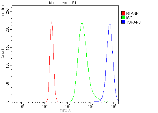
(Figure 2. Flow Cytometry analysis of RAW264. 7 cells using anti-Tspan-8/Tspan8 antibody (AAA125783).Overlay histogram showing RAW264. 7 cells stained with AAA125783 (Blue line). The cells were blocked with 10% normal goat serum. And then incubated with rabbit anti-Tspan-8/Tspan8 Antibody (AAA125783, 1μg/1x106 cells) for 30 min at 20 degree C. DyLight®488 conjugated goat anti-rabbit IgG (5-10μg/1x106 cells) was used as secondary antibody for 30 minutes at 20 degree C. Isotype control antibody (Green line) was rabbit IgG (1μg/1x106) used under the same conditions. Unlabelled sample (Red line) was also used as a control.)
FCM/FACS (Flow Cytometry)
(Figure 2. Flow Cytometry analysis of RAW264. 7 cells using anti-Tspan-8/Tspan8 antibody (AAA125783).Overlay histogram showing RAW264. 7 cells stained with AAA125783 (Blue line). The cells were blocked with 10% normal goat serum. And then incubated with rabbit anti-Tspan-8/Tspan8 Antibody (AAA125783, 1μg/1x106 cells) for 30 min at 20 degree C. DyLight®488 conjugated goat anti-rabbit IgG (5-10μg/1x106 cells) was used as secondary antibody for 30 minutes at 20 degree C. Isotype control antibody (Green line) was rabbit IgG (1μg/1x106) used under the same conditions. Unlabelled sample (Red line) was also used as a control.)
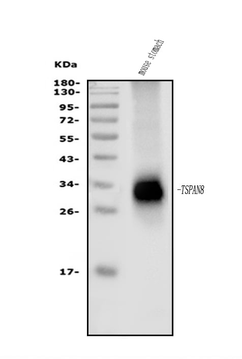
Tspan-8/Tspan8, Polyclonal Antibody (Cat# AAA125783)
Full Name
Anti-Tspan-8/Tspan8 Antibody
Gene Names
Tspan8; C76990; Tm4sf3; E330007O21Rik
Reactivity
Mouse
Applications
Direct ELISA, Flow Cytometry, Western Blot
Purity
Immunogen affinity purified.
Pricing
 IF (Immunofluorescence)
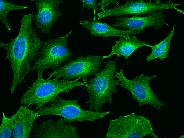
(Immunofluorescence staining of TFRC in Hela cells. Cells were fixed with 4% PFA, permeabilzed with 0.3% Triton X-100 in PBS,blocked with 10% serum, and incubated with rabbit anti-Human TFRC polyclonal antibody (dilution ratio 1:200) at 4? overnight. Then cells were stained with the Alexa Fluor488-conjugated Goat Anti-rabbit IgG secondary antibody (green) and counterstained with DAPI (blue).Positive staining was localized to Cytoplasm and Cell membrane.)
IF (Immunofluorescence)
(Immunofluorescence staining of TFRC in Hela cells. Cells were fixed with 4% PFA, permeabilzed with 0.3% Triton X-100 in PBS,blocked with 10% serum, and incubated with rabbit anti-Human TFRC polyclonal antibody (dilution ratio 1:200) at 4? overnight. Then cells were stained with the Alexa Fluor488-conjugated Goat Anti-rabbit IgG secondary antibody (green) and counterstained with DAPI (blue).Positive staining was localized to Cytoplasm and Cell membrane.)
Transferrin Receptor/TFR1/CD71, Polyclonal Antibody (Cat# AAA258872)
Full Name
Anti-Transferrin Receptor/TFR1/CD71 Antibody, Rabbit Polyclonal
Reactivity
Human
Applications
Immunofluorescence, Immunocytochemistry, Western Blot
Purity
Protein A & Antigen Affinity
Pricing
 FCM/FACS (Flow Cytometry)
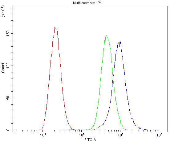
(Figure 2. Flow Cytometry analysis of U-87 cells using anti-CaV1.3 antibody (AAA46315).Overlay histogram showing U-87 cells stained with AAA46315 (Blue line).The cells were blocked with 10% normal goat serum. And then incubated with rabbit anti-CaV1.3 Antibody (AAA46315,1ug/1x10^6 cells) for 30 min at 20 degree C. DyLight®488 conjugated goat anti-rabbit IgG (5-10ug/1x10^6 cells) was used as secondary antibody for 30 minutes at 20 degree C. Isotype control antibody (Green line) was rabbit IgG (1ug/1x106) used under the same conditions. Unlabelled sample (Red line) was also used as a control.)
FCM/FACS (Flow Cytometry)
(Figure 2. Flow Cytometry analysis of U-87 cells using anti-CaV1.3 antibody (AAA46315).Overlay histogram showing U-87 cells stained with AAA46315 (Blue line).The cells were blocked with 10% normal goat serum. And then incubated with rabbit anti-CaV1.3 Antibody (AAA46315,1ug/1x10^6 cells) for 30 min at 20 degree C. DyLight®488 conjugated goat anti-rabbit IgG (5-10ug/1x10^6 cells) was used as secondary antibody for 30 minutes at 20 degree C. Isotype control antibody (Green line) was rabbit IgG (1ug/1x106) used under the same conditions. Unlabelled sample (Red line) was also used as a control.)
CaV1.3, Polyclonal Antibody (Cat# AAA46315)
Full Name
Anti-CaV1.3 Antibody
Gene Names
CACNA1D; CACH3; CACN4; PASNA; SANDD; Cav1.3; CCHL1A2; CACNL1A2
Reactivity
Mouse, Rat
Predicted to work with: Human
Predicted to work with: Human
Applications
Western Blot
Purity
Immunogen Affinity Purified
Pricing
 Application Data
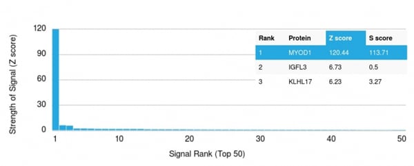
(Analysis of Protein Array containing more than 19, 000 full-length human proteins using MyoD1 Mouse Recombinant Monoclonal Antibody (rMYD712).Z- and S- Score: The Z-score represents the strength of a signal that a monoclonal antibody (MAb) (in combination with a fluorescently-tagged anti-IgG secondary antibody) produces when binding to a particular protein on the HuProtTM array. Z-scores are described in units of standard deviations (SD's) above the mean value of all signals generated on that array. If targets on HuProtTM are arranged in descending order of the Z-score, the S-score is the difference (also in units of SD's) between the Z-score. S-score therefore represents the relative target specificity of a MAb to its intended target. A MAb is considered to specific to its intended target, if the MAb has an S-score of at least 2.5. For example, if a MAb binds to protein X with a Z-score of 43 and to protein Y with a Z-score of 14, then the S-score for the binding of that MAb to protein X is equal to 29.)
Application Data
(Analysis of Protein Array containing more than 19, 000 full-length human proteins using MyoD1 Mouse Recombinant Monoclonal Antibody (rMYD712).Z- and S- Score: The Z-score represents the strength of a signal that a monoclonal antibody (MAb) (in combination with a fluorescently-tagged anti-IgG secondary antibody) produces when binding to a particular protein on the HuProtTM array. Z-scores are described in units of standard deviations (SD's) above the mean value of all signals generated on that array. If targets on HuProtTM are arranged in descending order of the Z-score, the S-score is the difference (also in units of SD's) between the Z-score. S-score therefore represents the relative target specificity of a MAb to its intended target. A MAb is considered to specific to its intended target, if the MAb has an S-score of at least 2.5. For example, if a MAb binds to protein X with a Z-score of 43 and to protein Y with a Z-score of 14, then the S-score for the binding of that MAb to protein X is equal to 29.)
MyoD1, Monoclonal Antibody (Cat# AAA214461)
Full Name
MyoD1 (Rhabdomyosarcoma Marker)
Gene Names
MYOD1; PUM; MYF3; MYOD; bHLHc1
Reactivity
Human. Others not known.
Applications
Immunohistochemistry
Pricing
 FCM/FACS (Flow Cytometry)
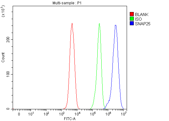
(Figure 2. Flow Cytometry analysis of SH-SY5Y cells using anti-SNAP25 antibody (AAA127072).Overlay histogram showing SH-SY5Y cells stained with AAA127072 (Blue line). To facilitate intracellular staining, cells were fixed with 4% paraformaldehyde and permeabilized with permeabilization buffer. The cells were blocked with 10% normal goat serum. And then incubated with rabbit anti-SNAP25 Antibody (AAA127072, 1ug/1x106 cells) for 30 min at 20 degree C. DyLight488 conjugated goat anti-rabbit IgG was used as secondary antibody for 30 minutes at 20 degree C. Isotype control antibody (Green line) was rabbit IgG (1ug/1x106) used under the same conditions. Unlabelled sample (Red line) was also used as a control.)
FCM/FACS (Flow Cytometry)
(Figure 2. Flow Cytometry analysis of SH-SY5Y cells using anti-SNAP25 antibody (AAA127072).Overlay histogram showing SH-SY5Y cells stained with AAA127072 (Blue line). To facilitate intracellular staining, cells were fixed with 4% paraformaldehyde and permeabilized with permeabilization buffer. The cells were blocked with 10% normal goat serum. And then incubated with rabbit anti-SNAP25 Antibody (AAA127072, 1ug/1x106 cells) for 30 min at 20 degree C. DyLight488 conjugated goat anti-rabbit IgG was used as secondary antibody for 30 minutes at 20 degree C. Isotype control antibody (Green line) was rabbit IgG (1ug/1x106) used under the same conditions. Unlabelled sample (Red line) was also used as a control.)
SNAP25, Polyclonal Antibody (Cat# AAA127072)
Full Name
Anti-SNAP25 Antibody Picoband
Gene Names
SNAP25; RIC4; SEC9; SNAP; RIC-4; SNAP-25; bA416N4.2; dJ1068F16.2
Reactivity
Human, Mouse, Rat
Applications
Flow Cytometry, Western Blot
Purity
Immunogen affinity purified.
Pricing
 IHC (Immunohistochemisry)
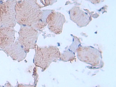
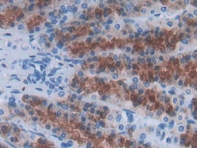
(DAB staining on IHCP;Sample: Rat Stomach Tissue; Primary Ab: 30ug/ml Mouse AntiRat IL24 AntibodySecond Ab: 2ug/mL HRPLinked Caprine AntiMouse IgG Polyclonal Antibody(Catalog: SAA544Mu19))
IHC (Immunohistochemisry)
(DAB staining on IHCP;Sample: Rat Stomach Tissue; Primary Ab: 30ug/ml Mouse AntiRat IL24 AntibodySecond Ab: 2ug/mL HRPLinked Caprine AntiMouse IgG Polyclonal Antibody(Catalog: SAA544Mu19))
Interleukin 24 (IL24), Monoclonal Antibody (Cat# AAA151786)
Full Name
Monoclonal Antibody to Interleukin 24 (IL24)
Gene Names
Il24; Mda7; Mda-7
Reactivity
Rat
Applications
Immunoprecipitation, Immunocytochemistry, Immunohistochemistry, Western Blot
Purity
Protein A + Protein G affinity chromatography
Pricing
 Application Data
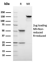
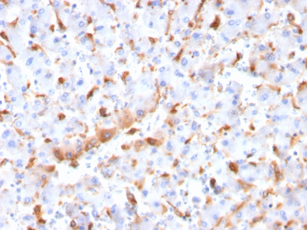
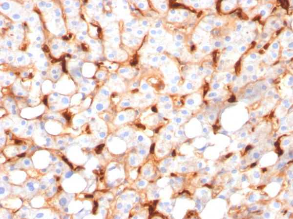
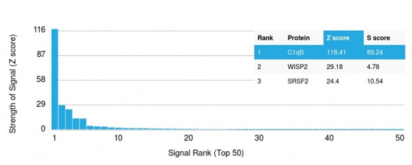
(Analysis of Protein Array containing more than 19,000 full-length human proteins using C1QB Mouse Monoclonal Antibody (C1QB/2966).Z- and S- Score: The Z-score represents the strength of a signal that a monoclonal antibody (MAb) (in combination with a fluorescently-tagged anti-IgG secondary antibody) produces when binding to a particular protein on the HuProtTM array. Z-scores are described in units of standard deviations (SD's) above the mean value of all signals generated on that array. If targets on HuProtTM are arranged in descending order of the Z-score, the S-score is the difference (also in units of SD's) between the Z-score. S-score therefore represents the relative target specificity of a MAb to its intended target. A MAb is considered to specific to its intended target, if the MAb has an S-score of at least 2.5. For example, if a MAb binds to protein X with a Z-score of 43 and to protein Y with a Z-score of 14, then the S-score for the binding of that MAb to protein X is equal to 29.)
Application Data
(Analysis of Protein Array containing more than 19,000 full-length human proteins using C1QB Mouse Monoclonal Antibody (C1QB/2966).Z- and S- Score: The Z-score represents the strength of a signal that a monoclonal antibody (MAb) (in combination with a fluorescently-tagged anti-IgG secondary antibody) produces when binding to a particular protein on the HuProtTM array. Z-scores are described in units of standard deviations (SD's) above the mean value of all signals generated on that array. If targets on HuProtTM are arranged in descending order of the Z-score, the S-score is the difference (also in units of SD's) between the Z-score. S-score therefore represents the relative target specificity of a MAb to its intended target. A MAb is considered to specific to its intended target, if the MAb has an S-score of at least 2.5. For example, if a MAb binds to protein X with a Z-score of 43 and to protein Y with a Z-score of 14, then the S-score for the binding of that MAb to protein X is equal to 29.)
C1QB/Complement C1q B-Chain, Monoclonal Antibody (Cat# AAA215227)
Full Name
C1QB/Complement C1q B-Chain
Reactivity
Human
Applications
Immunohistochemistry
Purity
Purified Ab with BSA and Azide at 200ug/ml OR Purified Ab WITHOUT BSA and Azide at 1.0mg/ml
Pricing
 Application Data
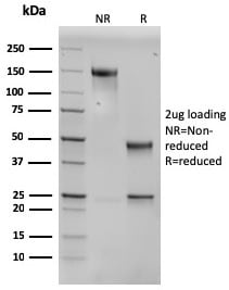
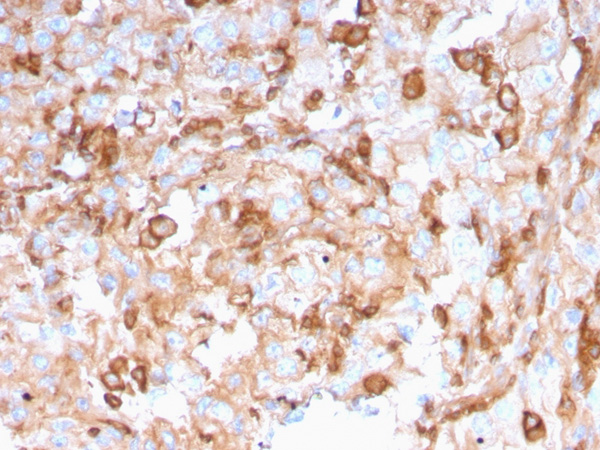
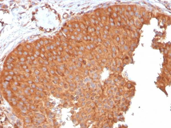
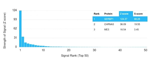
(Analysis of Protein Array containing more than 19,000 full-length human proteins using PAI-RBP1 Mouse Monoclonal Antibody (SERBP1/3496). Z- and S- Score: The Z-score represents the strength of a signal that a monoclonal antibody (MAb) (in combination with a fluorescently-tagged anti-IgG secondary antibody) produces when binding to a particular protein on the HuProtTM array. Z-scores are described in units of standard deviations (SD’s) above the mean value of all signals generated on that array. If targets on HuProtTM are arranged in descending order of the Z-score, the S-score is the difference (also in units of SD’s) between the Z-score. S-score therefore represents the relative target specificity of a MAb to its intended target. A MAb is considered to specific to its intended target, if the MAb has an S-score of at least 2.5. For example, if a MAb binds to protein X with a Z-score of 43 and to protein Y with a Z-score of 14, then the S-score for the binding of that MAb to protein X is equal to 29.)
Application Data
(Analysis of Protein Array containing more than 19,000 full-length human proteins using PAI-RBP1 Mouse Monoclonal Antibody (SERBP1/3496). Z- and S- Score: The Z-score represents the strength of a signal that a monoclonal antibody (MAb) (in combination with a fluorescently-tagged anti-IgG secondary antibody) produces when binding to a particular protein on the HuProtTM array. Z-scores are described in units of standard deviations (SD’s) above the mean value of all signals generated on that array. If targets on HuProtTM are arranged in descending order of the Z-score, the S-score is the difference (also in units of SD’s) between the Z-score. S-score therefore represents the relative target specificity of a MAb to its intended target. A MAb is considered to specific to its intended target, if the MAb has an S-score of at least 2.5. For example, if a MAb binds to protein X with a Z-score of 43 and to protein Y with a Z-score of 14, then the S-score for the binding of that MAb to protein X is equal to 29.)
PAI-RBP1/SERBP1/SERPINE1, Monoclonal Antibody (Cat# AAA214986)
Full Name
PAI-RBP1/SERBP1/SERPINE1
Gene Names
SERBP1; CGI-55; CHD3IP; HABP4L; PAIRBP1; PAI-RBP1
Reactivity
Human
Applications
Immunohistochemistry
Purity
Purified Ab with BSA and Azide at 200ug/ml OR Purified Ab WITHOUT BSA and Azide at 1.0mg/ml
Pricing
ICAM-1/CD54, Active Protein (Cat# AAA48983)
Full Name
Recombinant human ICAM-1/CD54 protein
Gene Names
ICAM1; BB2; CD54; P3.58
Applications
Bioactivity, SDS-PAGE
Purity
> 90% by SDS-PAGE
Pricing
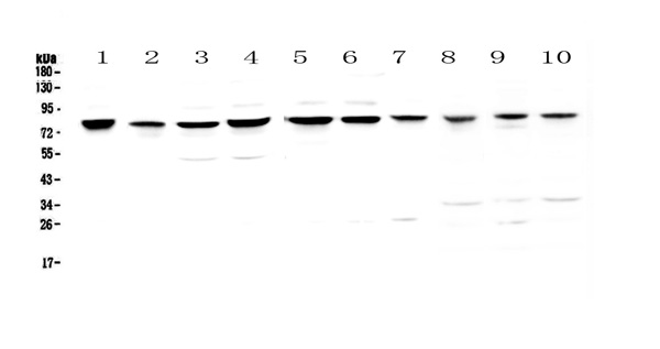
 IHC (Immunohistochemistry)
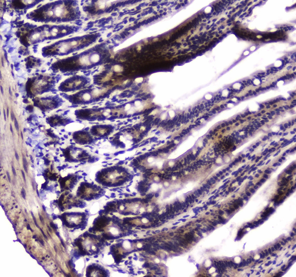
(Figure 4. IHC analysis of PARN using anti-PARN antibody (AAA124651).PARN was detected in paraffin-embedded section of rat small intestine tissue. Heat mediated antigen retrieval was performed in citrate buffer (pH6, epitope retrieval solution) for 20 mins. The tissue section was blocked with 10% goat serum. The tissue section was then incubated with 1ug/ml rabbit anti-PARN Antibody (AAA124651) overnight at 4 degree C. Biotinylated goat anti-rabbit IgG was used as secondary antibody and incubated for 30 minutes at 37 degree C. The tissue section was developed using Strepavidin-Biotin-Complex (SABC) with DAB as the chromogen.)
IHC (Immunohistochemistry)
(Figure 4. IHC analysis of PARN using anti-PARN antibody (AAA124651).PARN was detected in paraffin-embedded section of rat small intestine tissue. Heat mediated antigen retrieval was performed in citrate buffer (pH6, epitope retrieval solution) for 20 mins. The tissue section was blocked with 10% goat serum. The tissue section was then incubated with 1ug/ml rabbit anti-PARN Antibody (AAA124651) overnight at 4 degree C. Biotinylated goat anti-rabbit IgG was used as secondary antibody and incubated for 30 minutes at 37 degree C. The tissue section was developed using Strepavidin-Biotin-Complex (SABC) with DAB as the chromogen.)
PARN, Polyclonal Antibody (Cat# AAA124651)
Full Name
Anti-PARN Picoband antibody
Gene Names
PARN; DAN; DKCB6; PFBMFT4
Reactivity
Human, Mouse, Rat
No cross reactivity with other proteins.
No cross reactivity with other proteins.
Applications
Western Blot, Immunohistochemistry
Pricing
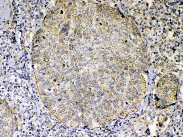
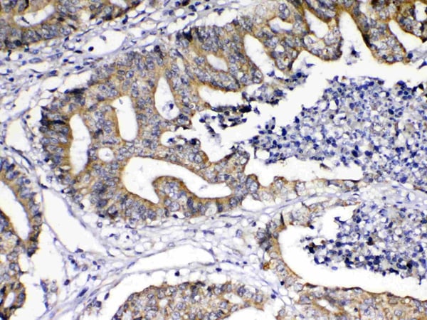
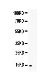
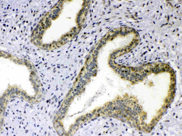 IHC (Immunohistochemistry)
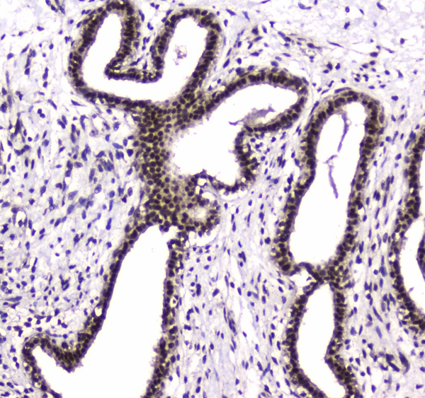
(Figure 4. IHC analysis of TNFSF18 using anti-TNFSF18 antibody (AAA124721).TNFSF18 was detected in paraffin-embedded section of human mammary cancer tissue. Heat mediated antigen retrieval was performed in citrate buffer (pH6, epitope retrieval solution) for 20 mins. The tissue section was blocked with 10% goat serum. The tissue section was then incubated with 1ug/ml rabbit anti-TNFSF18 Antibody (AAA124721) overnight at 4 degree C. Biotinylated goat anti-rabbit IgG was used as secondary antibody and incubated for 30 minutes at 37 degree C. The tissue section was developed using Strepavidin-Biotin-Complex (SABC) with DAB as the chromogen.)
IHC (Immunohistochemistry)
(Figure 4. IHC analysis of TNFSF18 using anti-TNFSF18 antibody (AAA124721).TNFSF18 was detected in paraffin-embedded section of human mammary cancer tissue. Heat mediated antigen retrieval was performed in citrate buffer (pH6, epitope retrieval solution) for 20 mins. The tissue section was blocked with 10% goat serum. The tissue section was then incubated with 1ug/ml rabbit anti-TNFSF18 Antibody (AAA124721) overnight at 4 degree C. Biotinylated goat anti-rabbit IgG was used as secondary antibody and incubated for 30 minutes at 37 degree C. The tissue section was developed using Strepavidin-Biotin-Complex (SABC) with DAB as the chromogen.)
TNFSF18/Gitrl, Polyclonal Antibody (Cat# AAA124721)
Full Name
Anti-TNFSF18/Gitrl Picoband Antibody
Gene Names
TNFSF18; TL6; AITRL; GITRL; TNLG2A; hGITRL
Reactivity
Human
No cross reactivity with other proteins.
No cross reactivity with other proteins.
Applications
Western Blot, Immunohistochemistry
Purity
Immunogen affinity purified
Pricing
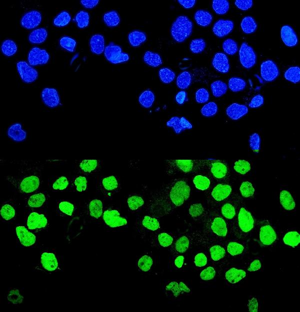
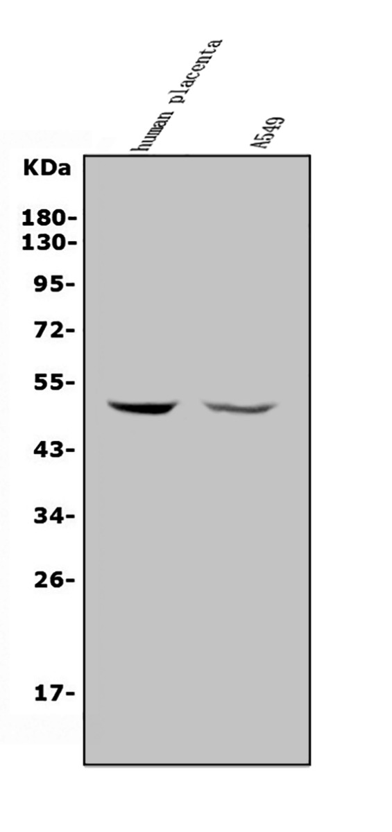
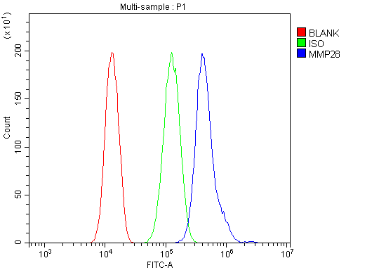 FCM/FACS (Flow Cytometry)
(Figure 3. Flow Cytometry analysis of A549 cells using anti-MMP28 antibody (AAA125788).Overlay histogram showing A549 cells stained with AAA125788 (Blue line). The cells were blocked with 10% normal goat serum. And then incubated with rabbit anti-MMP28 Antibody (AAA125788, 1μg/1x106 cells) for 30 min at 20 degree C. DyLight®488 conjugated goat anti-rabbit IgG (5-10μg/1x106 cells) was used as secondary antibody for 30 minutes at 20 degree C. Isotype control antibody (Green line) was rabbit IgG (1μg/1x106) used under the same conditions. Unlabelled sample (Red line) was also used as a control.)
FCM/FACS (Flow Cytometry)
(Figure 3. Flow Cytometry analysis of A549 cells using anti-MMP28 antibody (AAA125788).Overlay histogram showing A549 cells stained with AAA125788 (Blue line). The cells were blocked with 10% normal goat serum. And then incubated with rabbit anti-MMP28 Antibody (AAA125788, 1μg/1x106 cells) for 30 min at 20 degree C. DyLight®488 conjugated goat anti-rabbit IgG (5-10μg/1x106 cells) was used as secondary antibody for 30 minutes at 20 degree C. Isotype control antibody (Green line) was rabbit IgG (1μg/1x106) used under the same conditions. Unlabelled sample (Red line) was also used as a control.)
MMP28, Polyclonal Antibody (Cat# AAA125788)
Full Name
Anti-MMP28 Antibody
Gene Names
MMP28; MM28; MMP25; MMP-28; EPILYSIN
Reactivity
Human
Applications
Direct ELISA, Flow Cytometry, Immunofluorescence, Immunocytochemistry, Western Blot
Purity
Immunogen affinity purified.
Pricing
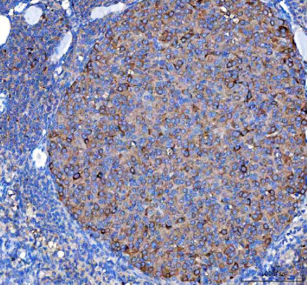
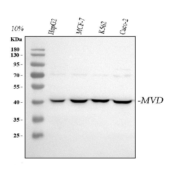
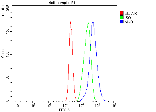 FCM/FACS (Flow Cytometry)
(Figure 3. Flow Cytometry analysis of MCF-7 cells using anti-MVD antibody (AAA127073).Overlay histogram showing MCF-7 cells stained with AAA127073 (Blue line). To facilitate intracellular staining, cells were fixed with 4% paraformaldehyde and permeabilized with permeabilization buffer. The cells were blocked with 10% normal goat serum. And then incubated with rabbit anti-MVD Antibody (AAA127073, 1ug/1x106 cells) for 30 min at 20 degree C. DyLight488 conjugated goat anti-rabbit IgG was used as secondary antibody for 30 minutes at 20 degree C. Isotype control antibody (Green line) was rabbit IgG (1ug/1x106) used under the same conditions. Unlabelled sample (Red line) was also used as a control.)
FCM/FACS (Flow Cytometry)
(Figure 3. Flow Cytometry analysis of MCF-7 cells using anti-MVD antibody (AAA127073).Overlay histogram showing MCF-7 cells stained with AAA127073 (Blue line). To facilitate intracellular staining, cells were fixed with 4% paraformaldehyde and permeabilized with permeabilization buffer. The cells were blocked with 10% normal goat serum. And then incubated with rabbit anti-MVD Antibody (AAA127073, 1ug/1x106 cells) for 30 min at 20 degree C. DyLight488 conjugated goat anti-rabbit IgG was used as secondary antibody for 30 minutes at 20 degree C. Isotype control antibody (Green line) was rabbit IgG (1ug/1x106) used under the same conditions. Unlabelled sample (Red line) was also used as a control.)
MVD, Polyclonal Antibody (Cat# AAA127073)
Full Name
Anti-MVD Antibody Picoband
Gene Names
MVD; MPD; MDDase; FP17780
Reactivity
Human, Mouse
Applications
Flow Cytometry, Immunohistochemistry, Western Blot
Purity
Immunogen affinity purified.
Pricing
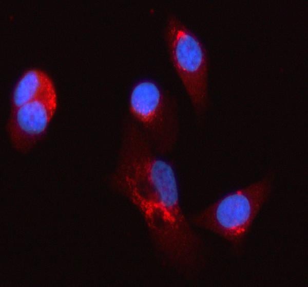
 FCM/FACS (Flow Cytometry)
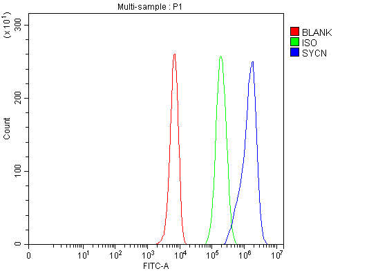
(Figure 3. Flow Cytometry analysis of K562 cells using anti-SYCN antibody (AAA127914).Overlay histogram showing K562 cells stained with AAA127914 (Blue line). To facilitate intracellular staining, cells were fixed with 4% paraformaldehyde and permeabilized with permeabilization buffer. The cells were blocked with 10% normal goat serum. And then incubated with rabbit anti-SYCN Antibody (AAA127914, 1ug/1x106 cells) for 30 min at 20 degree C. DyLight488 conjugated goat anti-rabbit IgG was used as secondary antibody for 30 minutes at 20 degree C. Isotype control antibody (Green line) was rabbit IgG (1ug/1x106) used under the same conditions. Unlabelled sample (Red line) was also used as a control.)
FCM/FACS (Flow Cytometry)
(Figure 3. Flow Cytometry analysis of K562 cells using anti-SYCN antibody (AAA127914).Overlay histogram showing K562 cells stained with AAA127914 (Blue line). To facilitate intracellular staining, cells were fixed with 4% paraformaldehyde and permeabilized with permeabilization buffer. The cells were blocked with 10% normal goat serum. And then incubated with rabbit anti-SYCN Antibody (AAA127914, 1ug/1x106 cells) for 30 min at 20 degree C. DyLight488 conjugated goat anti-rabbit IgG was used as secondary antibody for 30 minutes at 20 degree C. Isotype control antibody (Green line) was rabbit IgG (1ug/1x106) used under the same conditions. Unlabelled sample (Red line) was also used as a control.)
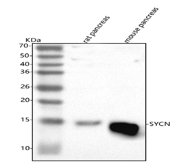
SYCN, Polyclonal Antibody (Cat# AAA127914)
Full Name
Anti-SYCN Antibody Picoband
Gene Names
SYCN; SYL; INSSA1
Reactivity
Human, Mouse, Rat
Applications
Flow Cytometry, Immunofluorescence, Immunocytochemistry, Western Blot
Purity
Immunogen affinity purified.
Pricing

 Amino Acid Info
(Amino Acid info)
Amino Acid Info
(Amino Acid info)
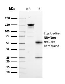
Eukaryotic Growth Differentiation Factor 15 (GDF15), Protein (Cat# AAA153215)
Full Name
Eukaryotic Growth Differentiation Factor 15 (GDF15)
Gene Names
GDF15; PDF; MIC1; PLAB; MIC-1; NAG-1; PTGFB; GDF-15
Applications
Western Blot
Purity
Greater than 95% by SDS-PAGE
Pricing
 Application Data
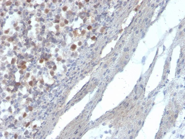
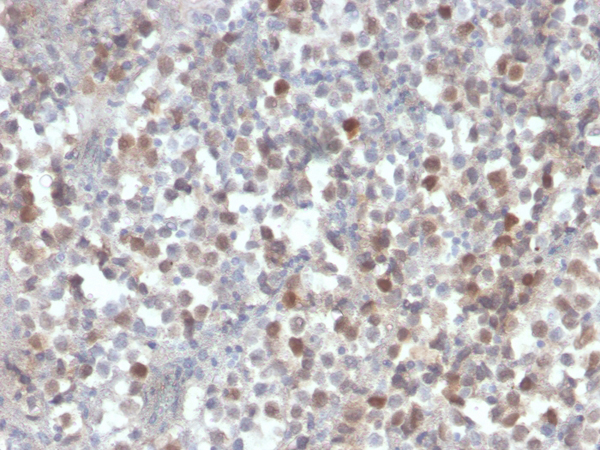
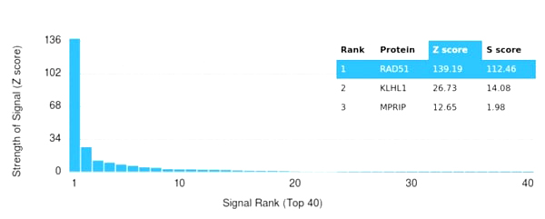
(Analysis of Protein Array containing more than 19,000 full-length human proteins using RAD51 Mouse Monoclonal Antibody (RAD51/2856) Z- and S- Score: The Z-score represents the strength of a signal that a monoclonal antibody (MAb) (in combination with a fluorescently-tagged anti-IgG secondary antibody) produces when binding to a particular protein on the HuProtTM array. Z-scores are described in units of standard deviations (SD's) above the mean value of all signals generated on that array. If targets on HuProtTM are arranged in descending order of the Z-score, the S-score is the difference (also in units of SD's) between the Z-score. S-score therefore represents the relative target specificity of a MAb to its intended target. A MAb is considered to specific to its intended target, if the MAb has an S-score of at least 2.5. For example, if a MAb binds to protein X with a Z-score of 43 and to protein Y with a Z-score of 14, then the S-score for the binding of that MAb to protein X is equal to 29.)
Application Data
(Analysis of Protein Array containing more than 19,000 full-length human proteins using RAD51 Mouse Monoclonal Antibody (RAD51/2856) Z- and S- Score: The Z-score represents the strength of a signal that a monoclonal antibody (MAb) (in combination with a fluorescently-tagged anti-IgG secondary antibody) produces when binding to a particular protein on the HuProtTM array. Z-scores are described in units of standard deviations (SD's) above the mean value of all signals generated on that array. If targets on HuProtTM are arranged in descending order of the Z-score, the S-score is the difference (also in units of SD's) between the Z-score. S-score therefore represents the relative target specificity of a MAb to its intended target. A MAb is considered to specific to its intended target, if the MAb has an S-score of at least 2.5. For example, if a MAb binds to protein X with a Z-score of 43 and to protein Y with a Z-score of 14, then the S-score for the binding of that MAb to protein X is equal to 29.)
RAD51, Monoclonal Antibody (Cat# AAA215404)
Full Name
RAD51 (Prognostic and Response to Chemotherapy Marker)
Gene Names
RAD51; RECA; BRCC5; FANCR; MRMV2; HRAD51; RAD51A; HsRad51; HsT16930
Reactivity
Human
Applications
Immunohistochemistry
Purity
Purified Ab with BSA and Azide at 200ug/ml or Purified Ab with BSA and Azide at 200ug/ml or Purified Ab WITHOUT BSA and Azide at 1.0mg/ml
Pricing

 WB (Western Blot)
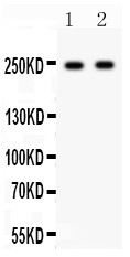
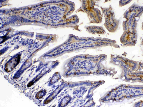
(Figure 1. Western blot analysis of Xanthine Oxidase using anti-Xanthine Oxidase antibody (AAA124674).Electrophoresis was performed on a 5-20% SDS-PAGE gel at 70V (Stacking gel) / 90V (Resolving gel) for 2-3 hours. The sample well of each lane was loaded with 50ug of sample under reducing conditions.Lane 1: rat liver tissue lysate,Lane 2: mouse liver tissue lysate.After Electrophoresis, proteins were transferred to a Nitrocellulose membrane at 150mA for 50-90 minutes. Blocked the membrane with 5% Non-fat Milk/ TBS for 1.5 hour at RT. The membrane was incubated with rabbit anti-Xanthine Oxidase antigen affinity purified polyclonal antibody at 0.5ug/mL overnight at 4 degree C, then washed with TBS-0.1%Tween 3 times with 5 minutes each and probed with a goat anti-rabbit IgG-HRP secondary antibody at a dilution of 1:10000 for 1.5 hour at RT. The signal is developed using an Enhanced Chemiluminescent detection (ECL) kit with Tanon 5200 system. A specific band was detected for Xanthine Oxidase at approximately 146KD. The expected band size for Xanthine Oxidase is at 146KD.)
WB (Western Blot)
(Figure 1. Western blot analysis of Xanthine Oxidase using anti-Xanthine Oxidase antibody (AAA124674).Electrophoresis was performed on a 5-20% SDS-PAGE gel at 70V (Stacking gel) / 90V (Resolving gel) for 2-3 hours. The sample well of each lane was loaded with 50ug of sample under reducing conditions.Lane 1: rat liver tissue lysate,Lane 2: mouse liver tissue lysate.After Electrophoresis, proteins were transferred to a Nitrocellulose membrane at 150mA for 50-90 minutes. Blocked the membrane with 5% Non-fat Milk/ TBS for 1.5 hour at RT. The membrane was incubated with rabbit anti-Xanthine Oxidase antigen affinity purified polyclonal antibody at 0.5ug/mL overnight at 4 degree C, then washed with TBS-0.1%Tween 3 times with 5 minutes each and probed with a goat anti-rabbit IgG-HRP secondary antibody at a dilution of 1:10000 for 1.5 hour at RT. The signal is developed using an Enhanced Chemiluminescent detection (ECL) kit with Tanon 5200 system. A specific band was detected for Xanthine Oxidase at approximately 146KD. The expected band size for Xanthine Oxidase is at 146KD.)
Xanthine Oxidase, Polyclonal Antibody (Cat# AAA124674)
Full Name
Anti-Xanthine Oxidase Picoband Antibody
Gene Names
XDH; XO; XOR; XAN1
Reactivity
Reacts with: Mouse, Rat
Predicted to work with: Human
Predicted to work with: Human
Applications
Western Blot, Immunohistochemistry
Purity
Immunogen affinity purified
Pricing
CD23/FCER2, Active Protein (Cat# AAA49009)
Full Name
Recombinant human CD23/FCER2 protein
Gene Names
FCER2; CD23; FCE2; CD23A; IGEBF; CLEC4J
Applications
Bioactivity, SDS-PAGE
Purity
> 90% by SDS-PAGE
Pricing
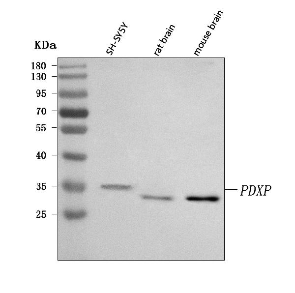
 FCM/FACS (Flow Cytometry)
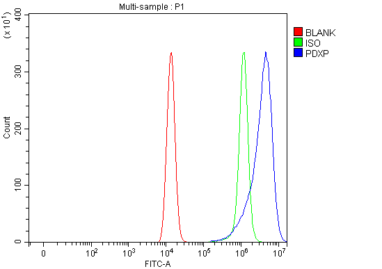
(Figure 2. Flow Cytometry analysis of HEL cells using anti-PDXP antibody (AAA127600).Overlay histogram showing HEL cells stained with AAA127600 (Blue line). To facilitate intracellular staining, cells were fixed with 4% paraformaldehyde and permeabilized with permeabilization buffer. The cells were blocked with 10% normal goat serum. And then incubated with rabbit anti-PDXP Antibody (AAA127600, 1ug/1x106 cells) for 30 min at 20 degree C. DyLight488 conjugated goat anti-rabbit IgG was used as secondary antibody for 30 minutes at 20 degree C. Isotype control antibody (Green line) was rabbit IgG (1ug/1x106) used under the same conditions. Unlabelled sample (Red line) was also used as a control.)
FCM/FACS (Flow Cytometry)
(Figure 2. Flow Cytometry analysis of HEL cells using anti-PDXP antibody (AAA127600).Overlay histogram showing HEL cells stained with AAA127600 (Blue line). To facilitate intracellular staining, cells were fixed with 4% paraformaldehyde and permeabilized with permeabilization buffer. The cells were blocked with 10% normal goat serum. And then incubated with rabbit anti-PDXP Antibody (AAA127600, 1ug/1x106 cells) for 30 min at 20 degree C. DyLight488 conjugated goat anti-rabbit IgG was used as secondary antibody for 30 minutes at 20 degree C. Isotype control antibody (Green line) was rabbit IgG (1ug/1x106) used under the same conditions. Unlabelled sample (Red line) was also used as a control.)
PDXP, Polyclonal Antibody (Cat# AAA127600)
Full Name
Anti-PDXP Antibody Picoband
Gene Names
PDXP; CIN; PLP; dJ37E16.5
Reactivity
Human, Mouse, Rat
Applications
Flow Cytometry, Western Blot
Purity
Immunogen affinity purified.
Pricing
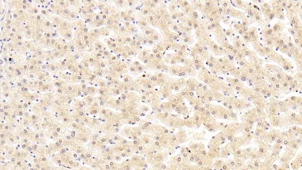
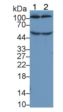 WB (Western Blot)
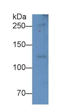
(Western Blot; Sample: Lane1: Human Serum; Lane2: Human PlasmaPrimary Ab: 5ug/ml Mouse AntiHuman C5a AntibodySecond Ab: 0.2ug/mL HRPLinked Caprine AntiMouse IgG Polyclonal Antibody(Catalog: SAA544Mu19))
WB (Western Blot)
(Western Blot; Sample: Lane1: Human Serum; Lane2: Human PlasmaPrimary Ab: 5ug/ml Mouse AntiHuman C5a AntibodySecond Ab: 0.2ug/mL HRPLinked Caprine AntiMouse IgG Polyclonal Antibody(Catalog: SAA544Mu19))
Complement Component 5a (C5a), Monoclonal Antibody (Cat# AAA151525)
Full Name
Monoclonal Antibody to Complement Component 5a (C5a)
Gene Names
C5; C5a; C5b; CPAMD4
Reactivity
Human, Rat, Pig
Applications
Immunoprecipitation, Immunocytochemistry, Immunohistochemistry, Western Blot
Purity
Protein A + Protein G affinity chromatography
Pricing
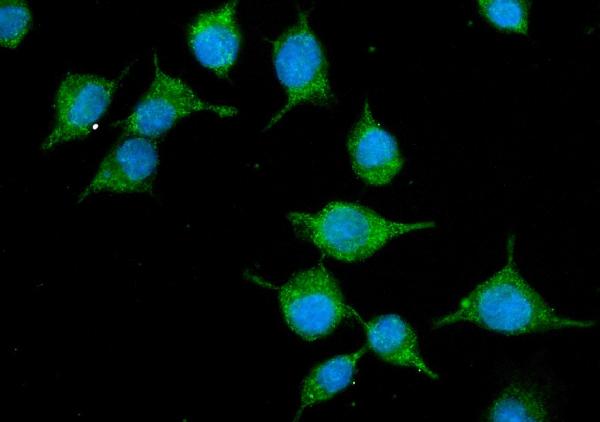
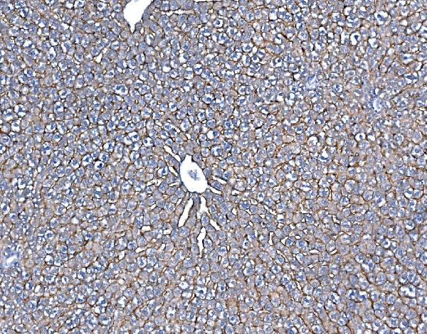
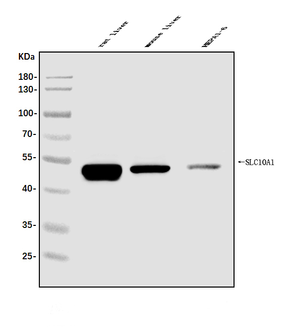
 FCM/FACS (Flow Cytometry)
(Figure 5. Flow Cytometry analysis of HEPA1-6 cells using anti-SLC10A1/NTCP1 antibody (AAA125779).Overlay histogram showing HEPA1-6 cells stained with AAA125779 (Blue line). The cells were blocked with 10% normal goat serum. And then incubated with rabbit anti-SLC10A1/NTCP1 Antibody (AAA125779, 1μg/1x106 cells) for 30 min at 20 degree C. DyLight®488 conjugated goat anti-rabbit IgG (5-10μg/1x106 cells) was used as secondary antibody for 30 minutes at 20 degree C. Isotype control antibody (Green line) was rabbit IgG (1μg/1x106) used under the same conditions. Unlabelled sample (Red line) was also used as a control.)
FCM/FACS (Flow Cytometry)
(Figure 5. Flow Cytometry analysis of HEPA1-6 cells using anti-SLC10A1/NTCP1 antibody (AAA125779).Overlay histogram showing HEPA1-6 cells stained with AAA125779 (Blue line). The cells were blocked with 10% normal goat serum. And then incubated with rabbit anti-SLC10A1/NTCP1 Antibody (AAA125779, 1μg/1x106 cells) for 30 min at 20 degree C. DyLight®488 conjugated goat anti-rabbit IgG (5-10μg/1x106 cells) was used as secondary antibody for 30 minutes at 20 degree C. Isotype control antibody (Green line) was rabbit IgG (1μg/1x106) used under the same conditions. Unlabelled sample (Red line) was also used as a control.)
SLC10A1/NTCP1, Polyclonal Antibody (Cat# AAA125779)
Full Name
Anti-SLC10A1/NTCP1 Antibody
Gene Names
Slc10a1; Ntcp
Reactivity
Mouse, Rat
Applications
Direct ELISA, Flow Cytometry, Immunofluorescence, Immunocytochemistry, Immunohistochemistry, Western Blot
Purity
Immunogen affinity purified.
Pricing
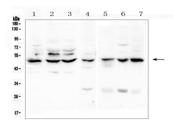
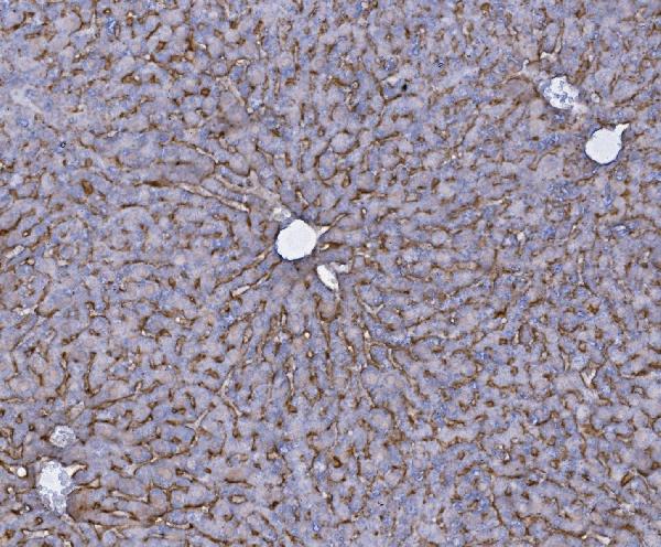
 IHC (Immunohiostchemistry)
(IHC analysis of TNF Receptor I using anti-TNF Receptor I antibody.TNF Receptor I was detected in paraffin-embedded section of mouse liver tissue. Heat mediated antigen retrieval was performed in citrate buffer (pH6, epitope retrieval solution) for 20 mins. The tissue section was blocked with 10% goat serum. The tissue section was then incubated with 1ug/ml rabbit anti-TNF Receptor I antibody.TNF Receptor I was detected in paraffin-embedded section of mouse liver tissue. Heat mediated antigen retrieval was performed in citrate buffer (pH6, epitope retrieval solution) for 20 mins. The tissue section was blocked with 10% goat serum. The tissue section was then incubated with 1ug/ml rabbit anti-TNF Receptor I antibody.TNF Receptor I was detected in paraffin-embedded section of mouse lung tissue. Heat mediated antigen retrieval was performed in citrate buffer (pH6, epitope retrieval solution) for 20 mins. The tissue section was blocked with 10% goat serum. The tissue section was then incubated with 1ug/ml rabbit anti-TNF Receptor I antibody.Overlay histogram showing U937 cells stained with AAA124897 (Blue line).The cells were blocked with 10% normal goat serum. And then incubated with rabbit anti-TNF Receptor I Antibody for 30 min at 20 degree C. DyLight488 conjugated goat anti-rabbit IgG (5-10ug/1x106 cells) was used as secondary antibody for 30 minutes at 20 degree C. Isotype control antibody (Green line) was rabbit IgG (1ug/1x106) used under the same conditions. Unlabelled sample (Red line) was also used as a control.)
IHC (Immunohiostchemistry)
(IHC analysis of TNF Receptor I using anti-TNF Receptor I antibody.TNF Receptor I was detected in paraffin-embedded section of mouse liver tissue. Heat mediated antigen retrieval was performed in citrate buffer (pH6, epitope retrieval solution) for 20 mins. The tissue section was blocked with 10% goat serum. The tissue section was then incubated with 1ug/ml rabbit anti-TNF Receptor I antibody.TNF Receptor I was detected in paraffin-embedded section of mouse liver tissue. Heat mediated antigen retrieval was performed in citrate buffer (pH6, epitope retrieval solution) for 20 mins. The tissue section was blocked with 10% goat serum. The tissue section was then incubated with 1ug/ml rabbit anti-TNF Receptor I antibody.TNF Receptor I was detected in paraffin-embedded section of mouse lung tissue. Heat mediated antigen retrieval was performed in citrate buffer (pH6, epitope retrieval solution) for 20 mins. The tissue section was blocked with 10% goat serum. The tissue section was then incubated with 1ug/ml rabbit anti-TNF Receptor I antibody.Overlay histogram showing U937 cells stained with AAA124897 (Blue line).The cells were blocked with 10% normal goat serum. And then incubated with rabbit anti-TNF Receptor I Antibody for 30 min at 20 degree C. DyLight488 conjugated goat anti-rabbit IgG (5-10ug/1x106 cells) was used as secondary antibody for 30 minutes at 20 degree C. Isotype control antibody (Green line) was rabbit IgG (1ug/1x106) used under the same conditions. Unlabelled sample (Red line) was also used as a control.)
TNF Receptor I/TNFRSF1A, Polyclonal Antibody (Cat# AAA124897)
Full Name
Anti-TNF Receptor I/TNFRSF1A Antibody
Gene Names
TNFRSF1A; FPF; p55; p60; TBP1; TNF-R; TNFAR; TNFR1; p55-R; CD120a; TNFR55; TNFR60; TNF-R-I; TNF-R55
Reactivity
Human, Mouse, Rat
Applications
Flow Cytometry, Immunocytochemistry, Immunohistochemistry, Western Blot
Purity
Immunogen Affinity Purified
Pricing
 FCM/FACS (Flow Cytometry)
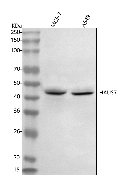
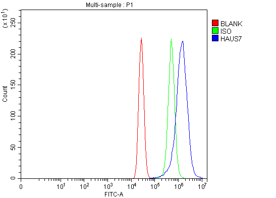
(Figure 3. Flow Cytometry analysis of MCF-7 cells using anti-UCHL5IP/HAUS7 antibody (AAA127752).Overlay histogram showing MCF-7 cells stained with AAA127752 (Blue line). To facilitate intracellular staining, cells were fixed with 4% paraformaldehyde and permeabilized with permeabilization buffer. The cells were blocked with 10% normal goat serum. And then incubated with rabbit anti-UCHL5IP/HAUS7 Antibody (AAA127752, 1ug/1x106 cells) for 30 min at 20 degree C. DyLight488 conjugated goat anti-rabbit IgG was used as secondary antibody for 30 minutes at 20 degree C. Isotype control antibody (Green line) was rabbit IgG (1ug/1x106) used under the same conditions. Unlabelled sample (Red line) was also used as a control.)
FCM/FACS (Flow Cytometry)
(Figure 3. Flow Cytometry analysis of MCF-7 cells using anti-UCHL5IP/HAUS7 antibody (AAA127752).Overlay histogram showing MCF-7 cells stained with AAA127752 (Blue line). To facilitate intracellular staining, cells were fixed with 4% paraformaldehyde and permeabilized with permeabilization buffer. The cells were blocked with 10% normal goat serum. And then incubated with rabbit anti-UCHL5IP/HAUS7 Antibody (AAA127752, 1ug/1x106 cells) for 30 min at 20 degree C. DyLight488 conjugated goat anti-rabbit IgG was used as secondary antibody for 30 minutes at 20 degree C. Isotype control antibody (Green line) was rabbit IgG (1ug/1x106) used under the same conditions. Unlabelled sample (Red line) was also used as a control.)
UCHL5IP/HAUS7, Polyclonal Antibody (Cat# AAA127752)
Full Name
Anti-UCHL5IP/HAUS7 Antibody Picoband
Gene Names
HAUS7; UIP1; UCHL5IP
Reactivity
Human
Applications
Flow Cytometry, Immunofluorescence, Immunocytochemistry, Western Blot
Purity
Immunogen affinity purified.
Pricing
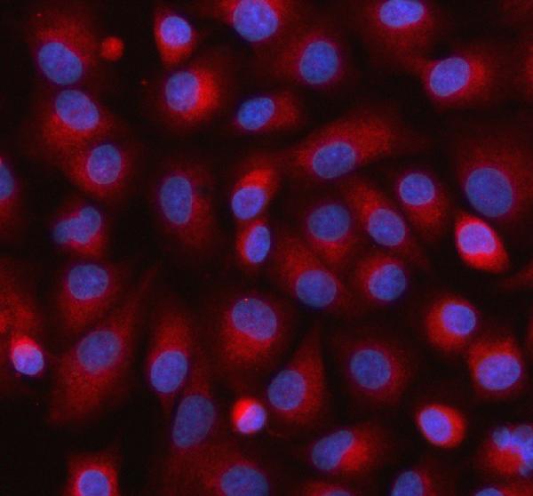
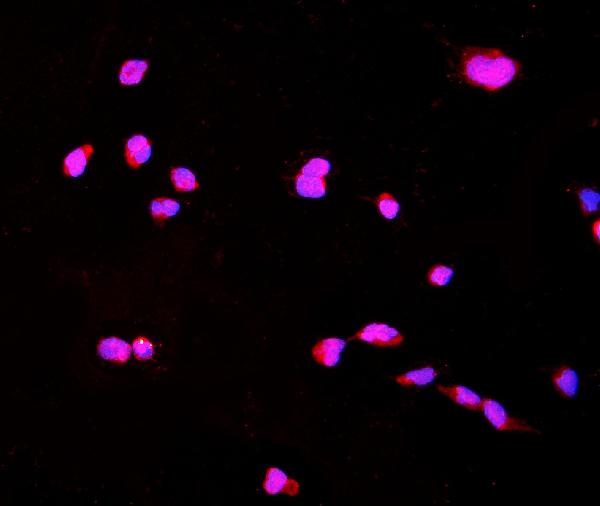 IF (Immunofluorescence)
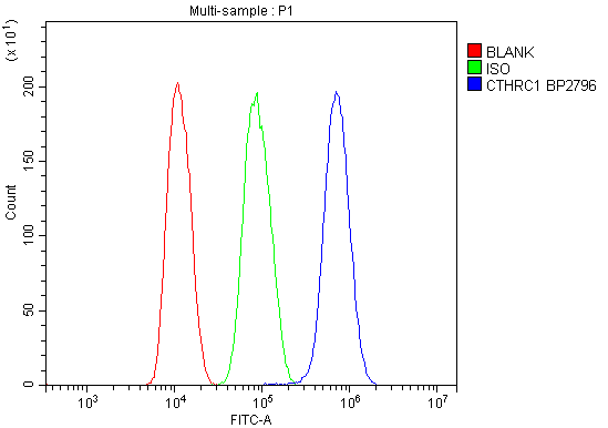
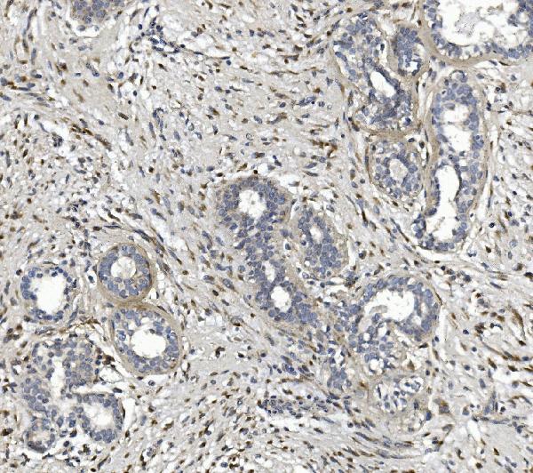
(Figure 4. IF analysis of CTHRC1 using anti-CTHRC1 antibody (AAA125741).CTHRC1 was detected in immunocytochemical section of HPEPA1-6 cells. Enzyme antigen retrieval was performed using IHC enzyme antigen retrieval reagent for 15 mins. The cells were blocked with 10% goat serum. And then incubated with 4μg/mL rabbit anti-CTHRC1 Antibody (AAA125741) overnight at 4 degree C. DyLight®550 Conjugated Goat Anti-Rabbit IgG (BA1135) was used as secondary antibody at 1:100 dilution and incubated for 30 minutes at 37 degree C. The section was counterstained with DAPI. Visualize using a fluorescence microscope and filter sets appropriate for the label used.)
IF (Immunofluorescence)
(Figure 4. IF analysis of CTHRC1 using anti-CTHRC1 antibody (AAA125741).CTHRC1 was detected in immunocytochemical section of HPEPA1-6 cells. Enzyme antigen retrieval was performed using IHC enzyme antigen retrieval reagent for 15 mins. The cells were blocked with 10% goat serum. And then incubated with 4μg/mL rabbit anti-CTHRC1 Antibody (AAA125741) overnight at 4 degree C. DyLight®550 Conjugated Goat Anti-Rabbit IgG (BA1135) was used as secondary antibody at 1:100 dilution and incubated for 30 minutes at 37 degree C. The section was counterstained with DAPI. Visualize using a fluorescence microscope and filter sets appropriate for the label used.)
CTHRC1, Polyclonal Antibody (Cat# AAA125741)
Full Name
Anti-CTHRC1 Antibody
Reactivity
Human, Mouse, Rat
Applications
Direct ELISA, Flow Cytometry, Immunofluorescence, Immunocytochemistry, Immunohistochemistry, Western Blot
Purity
Immunogen affinity purified.
Pricing
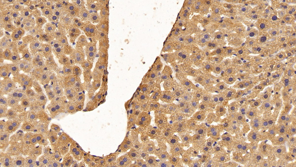
 WB (Western Blot)
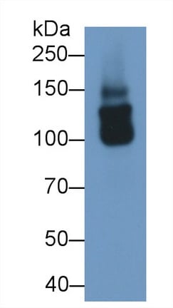
(Western Blot; Sample: 293T cell lysate Primary Ab: 0.1ug/ml Rabbit AntiMouse IGF1 Antibody Second Ab: 0.2ug/mL HRPLinked Caprine AntiRabbit IgG Polyclonal Antibody (Catalog: SAA544Rb19))
WB (Western Blot)
(Western Blot; Sample: 293T cell lysate Primary Ab: 0.1ug/ml Rabbit AntiMouse IGF1 Antibody Second Ab: 0.2ug/mL HRPLinked Caprine AntiRabbit IgG Polyclonal Antibody (Catalog: SAA544Rb19))
Collagen Type XVIII (COL18), Polyclonal Antibody (Cat# AAA151959)
Full Name
Polyclonal Antibody to Collagen Type XVIII (COL18)
Reactivity
Human, Mouse
Applications
Immunoprecipitation, Immunocytochemistry, Immunohistochemistry, Western Blot
Purity
Antigenspecific affinity chromatography followed by Protein A affinity chromatography
Pricing
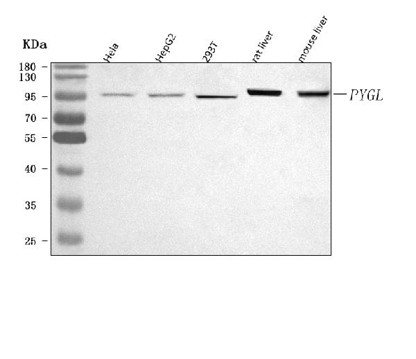
 FCM/FACS (Flow Cytometry)
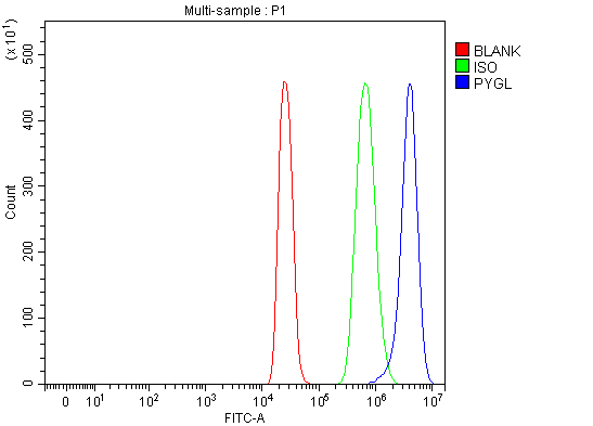
(Figure 2. Flow Cytometry analysis of 293T cells using anti-PYGL antibody (AAA127356).Overlay histogram showing 293T cells stained with AAA127356 (Blue line). To facilitate intracellular staining, cells were fixed with 4% paraformaldehyde and permeabilized with permeabilization buffer. The cells were blocked with 10% normal goat serum. And then incubated with rabbit anti-PYGL Antibody (AAA127356, 1ug/1x106 cells) for 30 min at 20 degree C. DyLight488 conjugated goat anti-rabbit IgG was used as secondary antibody for 30 minutes at 20 degree C. Isotype control antibody (Green line) was rabbit IgG (1ug/1x106) used under the same conditions. Unlabelled sample (Red line) was also used as a control.)
FCM/FACS (Flow Cytometry)
(Figure 2. Flow Cytometry analysis of 293T cells using anti-PYGL antibody (AAA127356).Overlay histogram showing 293T cells stained with AAA127356 (Blue line). To facilitate intracellular staining, cells were fixed with 4% paraformaldehyde and permeabilized with permeabilization buffer. The cells were blocked with 10% normal goat serum. And then incubated with rabbit anti-PYGL Antibody (AAA127356, 1ug/1x106 cells) for 30 min at 20 degree C. DyLight488 conjugated goat anti-rabbit IgG was used as secondary antibody for 30 minutes at 20 degree C. Isotype control antibody (Green line) was rabbit IgG (1ug/1x106) used under the same conditions. Unlabelled sample (Red line) was also used as a control.)
PYGL, Polyclonal Antibody (Cat# AAA127356)
Full Name
Anti-PYGL Antibody Picoband
Gene Names
PYGL; GSD6
Reactivity
Human, Mouse, Rat
Applications
Flow Cytometry, Western Blot
Purity
Immunogen affinity purified.
Pricing
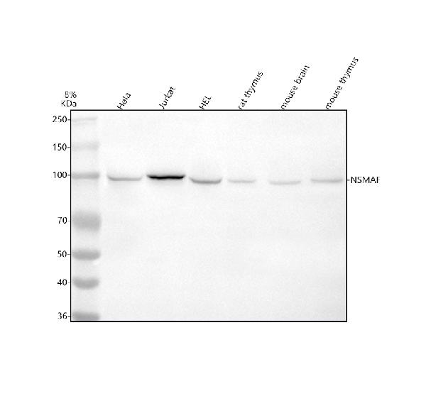
 FCM/FACS (Flow Cytometry)
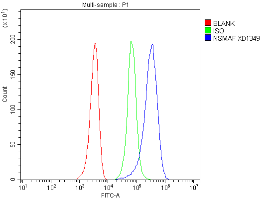
(Figure 2. Flow Cytometry analysis of HEL cells using anti-NSMAF antibody (AAA128213).Overlay histogram showing HEL cells stained with AAA128213 (Blue line). The cells were fixed with 4% paraformaldehyde and blocked with 10% normal goat serum. And then incubated with rabbit anti-NSMAF Antibody (AAA128213, 1ug/1x106 cells) for 30 min at 20 degree C. DyLight®488 conjugated goat anti-rabbit IgG was used as secondary antibody for 30 minutes at 20 degree C. Isotype control antibody (Green line) was rabbit IgG (1ug/1x106) used under the same conditions. Unlabelled sample without incubation with primary antibody and secondary antibody (Red line) was used as a blank control.)
FCM/FACS (Flow Cytometry)
(Figure 2. Flow Cytometry analysis of HEL cells using anti-NSMAF antibody (AAA128213).Overlay histogram showing HEL cells stained with AAA128213 (Blue line). The cells were fixed with 4% paraformaldehyde and blocked with 10% normal goat serum. And then incubated with rabbit anti-NSMAF Antibody (AAA128213, 1ug/1x106 cells) for 30 min at 20 degree C. DyLight®488 conjugated goat anti-rabbit IgG was used as secondary antibody for 30 minutes at 20 degree C. Isotype control antibody (Green line) was rabbit IgG (1ug/1x106) used under the same conditions. Unlabelled sample without incubation with primary antibody and secondary antibody (Red line) was used as a blank control.)
NSMAF, Polyclonal Antibody (Cat# AAA128213)
Full Name
Anti-NSMAF Antibody Picoband
Gene Names
NSMAF; FAN
Reactivity
Human, Mouse, Rat
Applications
Flow Cytometry, Western Blot
Purity
Immunogen affinity purified.
Pricing
 FCM/FACS (Flow Cytometry)
(Figure 2. Flow Cytometry analysis of HEL cells using anti-PLVAP antibody (AAA127418).Overlay histogram showing HEL cells stained with AAA127418 (Blue line). To facilitate intracellular staining, cells were fixed with 4% paraformaldehyde and permeabilized with permeabilization buffer. The cells were blocked with 10% normal goat serum. And then incubated with rabbit anti-PLVAP Antibody (AAA127418, 1ug/1x106 cells) for 30 min at 20 degree C. DyLight488 conjugated goat anti-rabbit IgG was used as secondary antibody for 30 minutes at 20 degree C. Isotype control antibody (Green line) was rabbit IgG (1ug/1x106) used under the same conditions. Unlabelled sample (Red line) was also used as a control.)
FCM/FACS (Flow Cytometry)
(Figure 2. Flow Cytometry analysis of HEL cells using anti-PLVAP antibody (AAA127418).Overlay histogram showing HEL cells stained with AAA127418 (Blue line). To facilitate intracellular staining, cells were fixed with 4% paraformaldehyde and permeabilized with permeabilization buffer. The cells were blocked with 10% normal goat serum. And then incubated with rabbit anti-PLVAP Antibody (AAA127418, 1ug/1x106 cells) for 30 min at 20 degree C. DyLight488 conjugated goat anti-rabbit IgG was used as secondary antibody for 30 minutes at 20 degree C. Isotype control antibody (Green line) was rabbit IgG (1ug/1x106) used under the same conditions. Unlabelled sample (Red line) was also used as a control.)
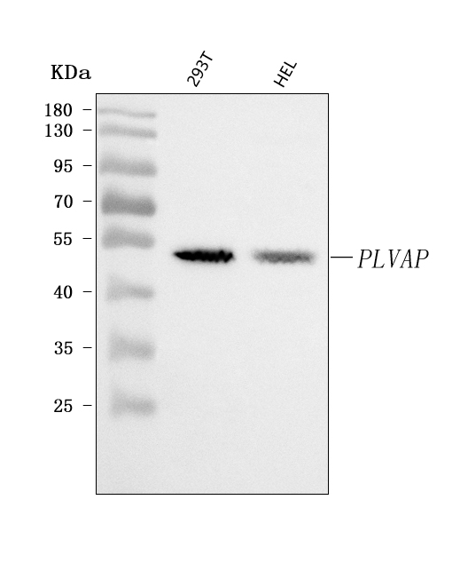
PLVAP, Polyclonal Antibody (Cat# AAA127418)
Full Name
Anti-PLVAP Antibody Picoband
Gene Names
PLVAP; PV1; FELS; PV-1; gp68
Reactivity
Human
Applications
Flow Cytometry, Western Blot
Purity
Immunogen affinity purified.
Pricing
 IP (Immunoprecipitation)
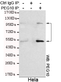
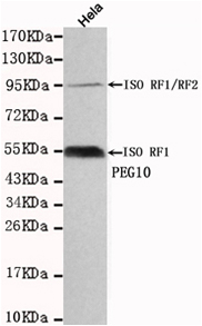
(Immunoprecipitation analysis of Hela cell lysates using PEG10 mouse mAb.)
IP (Immunoprecipitation)
(Immunoprecipitation analysis of Hela cell lysates using PEG10 mouse mAb.)
PEG10, Monoclonal Antibody (Cat# AAA320361)
Full Name
PEG10 Mouse mAb
Gene Names
PEG10; EDR; HB-1; Mar2; MEF3L; Mart2; RGAG3
Reactivity
Human
Applications
Immunoprecipitation, Western Blot
Purity
The antibody was affinity-purified from mouse ascites by affinity-chromatography using epitope-specific immunogen.
Pricing
 Bioactivity
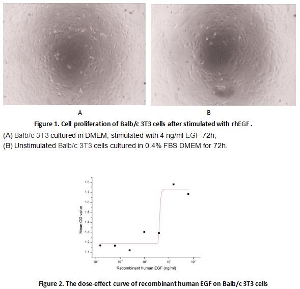
(Epidermal growth factor (EGF) is a growth factor that stimulates cell growth, proliferation, and differentiation by binding to its receptor EGFR. To test the effect of EGF on cell proliferation of 3T3 fibroblasts, Balb/c 3T3 cells were seeded into triplicate wells of 96-well plates at a density of 8, 000 cells/well and allowed to attach overnight, then the medium was replaced with 0.4% FBS standard DMEM prior to the addition of various concentrations of recombinant human EGF. After incubated for 48h, cells were observed by inverted microscope and cell proliferation was measured by Cell Counting Kit-8 (CCK-8). Briefly, 10 ul of CCK-8 solution was added to each well of the plate, then measure the absorbance at 450 nm using a microplate reader after incubating the plate for 1-4 hours at 37 degree C. Cell proliferation of Balb/c 3T3 cells after incubation with rhEGF for 48h observed by inverted microscope was shown in Figure 1. The dose-effect curve of rhEGF was shown in Figure2. It was obvious that rhEGF significantly promoted cell proliferation of 3T3 cells. The ED50 for this effect is typically 4.2 ng/ml.)
Bioactivity
(Epidermal growth factor (EGF) is a growth factor that stimulates cell growth, proliferation, and differentiation by binding to its receptor EGFR. To test the effect of EGF on cell proliferation of 3T3 fibroblasts, Balb/c 3T3 cells were seeded into triplicate wells of 96-well plates at a density of 8, 000 cells/well and allowed to attach overnight, then the medium was replaced with 0.4% FBS standard DMEM prior to the addition of various concentrations of recombinant human EGF. After incubated for 48h, cells were observed by inverted microscope and cell proliferation was measured by Cell Counting Kit-8 (CCK-8). Briefly, 10 ul of CCK-8 solution was added to each well of the plate, then measure the absorbance at 450 nm using a microplate reader after incubating the plate for 1-4 hours at 37 degree C. Cell proliferation of Balb/c 3T3 cells after incubation with rhEGF for 48h observed by inverted microscope was shown in Figure 1. The dose-effect curve of rhEGF was shown in Figure2. It was obvious that rhEGF significantly promoted cell proliferation of 3T3 cells. The ED50 for this effect is typically 4.2 ng/ml.)
Epidermal Growth Factor (EGF), Active Protein (Cat# AAA161748)
Full Name
Active Epidermal Growth Factor (EGF)
Gene Names
EGF; URG; HOMG4
Applications
Activity Assay, Cell Culture
Purity
Greater than 90% by SDS-PAGE
Pricing
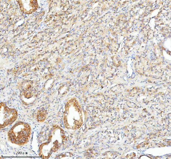
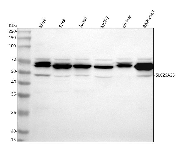
 FCM/FACS (Flow Cytometry)
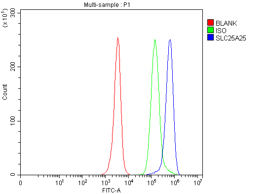
(Figure 4. Flow Cytometry analysis of JK cells using anti-SLC25A25 antibody (AAA127565).Overlay histogram showing JK cells stained with AAA127565 (Blue line). To facilitate intracellular staining, cells were fixed with 4% paraformaldehyde and permeabilized with permeabilization buffer. The cells were blocked with 10% normal goat serum. And then incubated with rabbit anti-SLC25A25 Antibody (AAA127565, 1ug/1x106 cells) for 30 min at 20 degree C. DyLight488 conjugated goat anti-rabbit IgG was used as secondary antibody for 30 minutes at 20 degree C. Isotype control antibody (Green line) was rabbit IgG (1ug/1x106) used under the same conditions. Unlabelled sample (Red line) was also used as a control.)
FCM/FACS (Flow Cytometry)
(Figure 4. Flow Cytometry analysis of JK cells using anti-SLC25A25 antibody (AAA127565).Overlay histogram showing JK cells stained with AAA127565 (Blue line). To facilitate intracellular staining, cells were fixed with 4% paraformaldehyde and permeabilized with permeabilization buffer. The cells were blocked with 10% normal goat serum. And then incubated with rabbit anti-SLC25A25 Antibody (AAA127565, 1ug/1x106 cells) for 30 min at 20 degree C. DyLight488 conjugated goat anti-rabbit IgG was used as secondary antibody for 30 minutes at 20 degree C. Isotype control antibody (Green line) was rabbit IgG (1ug/1x106) used under the same conditions. Unlabelled sample (Red line) was also used as a control.)
SLC25A25, Polyclonal Antibody (Cat# AAA127565)
Full Name
Anti-SLC25A25 Antibody Picoband
Gene Names
SLC25A25; MCSC; PCSCL; SCAMC-2; RP11-395P17.4
Reactivity
Human, Mouse, Rat
Applications
Flow Cytometry, Immunohistochemistry, Western Blot
Purity
Immunogen affinity purified.
Pricing
 WB (Western Blot)
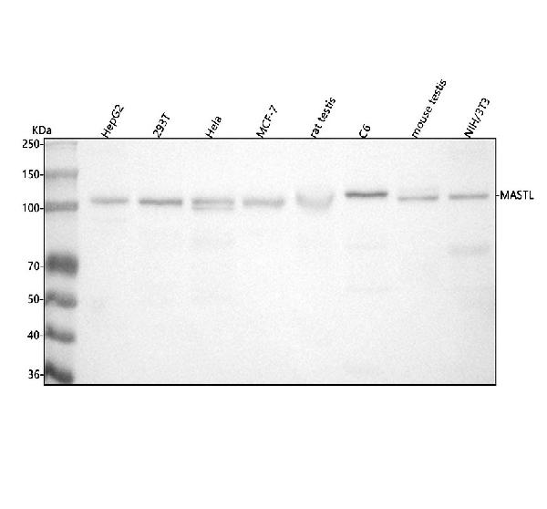
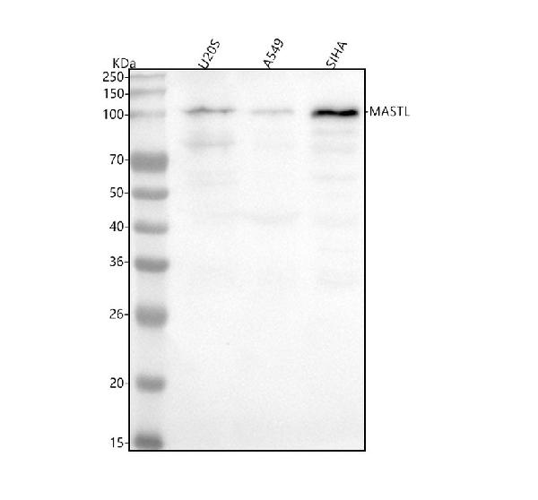
(Figure 2. Western blot analysis of MASTL using anti-MASTL antibody (AAA127239).Electrophoresis was performed on a 5-20% SDS-PAGE gel at 70V (Stacking gel)/90V (Resolving gel) for 2-3 hours. The sample well of each lane was loaded with 30 ug of sample under reducing conditions.Lane 1: human U2OS whole cell lysates,Lane 2: human A549 whole cell lysates,Lane 3: human SIHA whole cell lysates.After electrophoresis, proteins were transferred to a nitrocellulose membrane at 150 mA for 50-90 minutes. Blocked the membrane with 5% non-fat milk/TBS for 1.5 hour at RT. The membrane was incubated with rabbit anti-MASTL antigen affinity purified polyclonal antibody (#AAA127239) at 0.5ug/mL overnight at 4 degree C, then washed with TBS-0.1%Tween 3 times with 5 minutes each and probed with a goat anti-rabbit IgG-HRP secondary antibody at a dilution of 1:5000 for 1.5 hour at RT. The signal is developed using an Enhanced Chemiluminescent detection (ECL) kit with Tanon 5200 system. A specific band was detected for MASTL at approximately 110 kDa. The expected band size for MASTL is at 96 kDa.)
WB (Western Blot)
(Figure 2. Western blot analysis of MASTL using anti-MASTL antibody (AAA127239).Electrophoresis was performed on a 5-20% SDS-PAGE gel at 70V (Stacking gel)/90V (Resolving gel) for 2-3 hours. The sample well of each lane was loaded with 30 ug of sample under reducing conditions.Lane 1: human U2OS whole cell lysates,Lane 2: human A549 whole cell lysates,Lane 3: human SIHA whole cell lysates.After electrophoresis, proteins were transferred to a nitrocellulose membrane at 150 mA for 50-90 minutes. Blocked the membrane with 5% non-fat milk/TBS for 1.5 hour at RT. The membrane was incubated with rabbit anti-MASTL antigen affinity purified polyclonal antibody (#AAA127239) at 0.5ug/mL overnight at 4 degree C, then washed with TBS-0.1%Tween 3 times with 5 minutes each and probed with a goat anti-rabbit IgG-HRP secondary antibody at a dilution of 1:5000 for 1.5 hour at RT. The signal is developed using an Enhanced Chemiluminescent detection (ECL) kit with Tanon 5200 system. A specific band was detected for MASTL at approximately 110 kDa. The expected band size for MASTL is at 96 kDa.)
MASTL, Polyclonal Antibody (Cat# AAA127239)
Full Name
Anti-MASTL Antibody Picoband
Gene Names
MASTL; GW; GWL; THC2; hGWL; MAST-L; GREATWALL; RP11-85G18.2
Reactivity
Human, Mouse, Rat
Applications
Western Blot
Purity
Immunogen affinity purified.
Pricing
 FCM/FACS (Flow Cytometry)
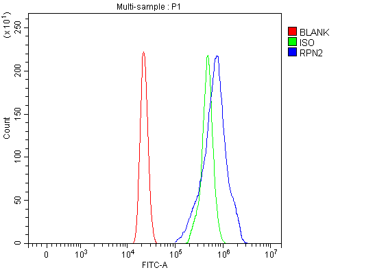
(Figure 3. Flow Cytometry analysis of HL-60 cells using anti-RPN2 antibody (AAA127272).Overlay histogram showing HL-60 cells stained with AAA127272 (Blue line). To facilitate intracellular staining, cells were fixed with 4% paraformaldehyde and permeabilized with permeabilization buffer. The cells were blocked with 10% normal goat serum. And then incubated with rabbit anti-RPN2 Antibody (AAA127272, 1ug/1x106 cells) for 30 min at 20 degree C. DyLight488 conjugated goat anti-rabbit IgG was used as secondary antibody for 30 minutes at 20 degree C. Isotype control antibody (Green line) was rabbit IgG (1ug/1x106) used under the same conditions. Unlabelled sample without incubation with primary antibody and secondary antibody (Red line) was used as a blank control.)
FCM/FACS (Flow Cytometry)
(Figure 3. Flow Cytometry analysis of HL-60 cells using anti-RPN2 antibody (AAA127272).Overlay histogram showing HL-60 cells stained with AAA127272 (Blue line). To facilitate intracellular staining, cells were fixed with 4% paraformaldehyde and permeabilized with permeabilization buffer. The cells were blocked with 10% normal goat serum. And then incubated with rabbit anti-RPN2 Antibody (AAA127272, 1ug/1x106 cells) for 30 min at 20 degree C. DyLight488 conjugated goat anti-rabbit IgG was used as secondary antibody for 30 minutes at 20 degree C. Isotype control antibody (Green line) was rabbit IgG (1ug/1x106) used under the same conditions. Unlabelled sample without incubation with primary antibody and secondary antibody (Red line) was used as a blank control.)
RPN2, Polyclonal Antibody (Cat# AAA127272)
Full Name
Anti-RPN2 Antibody Picoband
Gene Names
RPN2; SWP1; RPNII; RIBIIR; RPN-II
Reactivity
Human
Applications
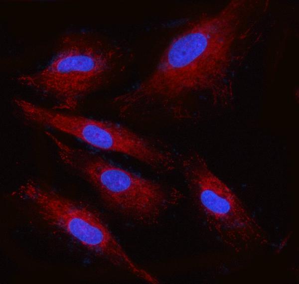
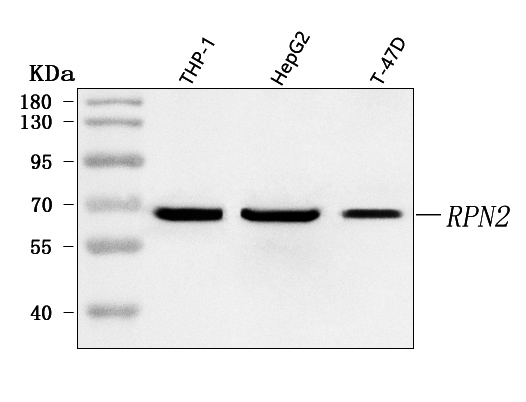
Western Blot, Immunofluorescence, Immunocytochemistry, Flow Cytometry
Purity
Immunogen affinity purified.
Pricing
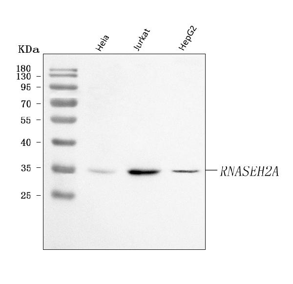
 FCM/FACS (Flow Cytometry)
(Figure 2. Flow Cytometry analysis of K562 cells using anti-RNASEH2A antibody (AAA127442).Overlay histogram showing K562 cells stained with AAA127442 (Blue line). To facilitate intracellular staining, cells were fixed with 4% paraformaldehyde and permeabilized with permeabilization buffer. The cells were blocked with 10% normal goat serum. And then incubated with rabbit anti-RNASEH2A Antibody (AAA127442, 1ug/1x106 cells) for 30 min at 20 degree C. DyLight488 conjugated goat anti-rabbit IgG was used as secondary antibody for 30 minutes at 20 degree C. Isotype control antibody (Green line) was rabbit IgG (1ug/1x106) used under the same conditions. Unlabelled sample without incubation with primary antibody and secondary antibody (Red line) was used as a blank control.)
FCM/FACS (Flow Cytometry)
(Figure 2. Flow Cytometry analysis of K562 cells using anti-RNASEH2A antibody (AAA127442).Overlay histogram showing K562 cells stained with AAA127442 (Blue line). To facilitate intracellular staining, cells were fixed with 4% paraformaldehyde and permeabilized with permeabilization buffer. The cells were blocked with 10% normal goat serum. And then incubated with rabbit anti-RNASEH2A Antibody (AAA127442, 1ug/1x106 cells) for 30 min at 20 degree C. DyLight488 conjugated goat anti-rabbit IgG was used as secondary antibody for 30 minutes at 20 degree C. Isotype control antibody (Green line) was rabbit IgG (1ug/1x106) used under the same conditions. Unlabelled sample without incubation with primary antibody and secondary antibody (Red line) was used as a blank control.)
Ribonuclease H2, subunit A/RNASEH2A, Polyclonal Antibody (Cat# AAA127442)
Full Name
Anti-Ribonuclease H2, subunit A/RNASEH2A Antibody Picoband
Gene Names
RNASEH2A; AGS4; JUNB; RNHL; RNHIA; RNASEHI
Reactivity
Human
Applications
Flow Cytometry, Western Blot
Purity
Immunogen affinity purified.
Pricing
 Bioactivity
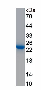
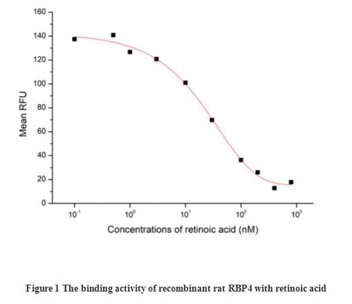
(Retinol-binding protein 4 (RBP4) is the specific carrier for retinol (also known as vitamin A), and is responsible for the conversion of unstable and insoluble retinol in aqueous solution into stable and soluble complex in plasma through their tight interaction. As a member of the lipocalin superfamily, RBP4 containing a beta-barrel structure with a well-defined cavity is secreted from the liver, and in turn delivers retinol from the liver stores to the peripheral tissues. In plasma, the RBP4-retinol complex interacts with transthyretin (TTR), and this binding is crucial for preventing RBP4 excretion through the kidney glomeruli. RBP4 expressed from an ectopic source efficiently delivers retinol to the eyes, and its deficiency affects night vision largely. Recently, RBP4 as an adipokine, is found to be expressed in adipose tissue and correlated with obesity, insulin resistance (IR) and type 2 diabetes (T2DM). The activity of recombinant rat RBP4 was measured by its ability to bind all-trans retinoic acid. The binding of retinoic acid results in the quenching of Trp fluorescence in RBP4. RBP4 was diluted to 50 ug/ml in 50 mM Tris, 10 mM CaCl2, 150 mM NaCl, pH 7.5 (assay buffer) and the retinoic acid was diluted to 800, 400, 200, 100, 30, 10, 3, 1, 0.5 and 0.1 uM in 95% ethanol. Mixing 112.5 uL of 50 ug/mL rrRBP4 and 12.5 uL of retinoic acid serial dilutions in microtubes and a blank containing 112.5 uL of 50 ug/mL rrRBP4 and 12.5 uL of 95% ethanol, then incubate at room temperature for 30 minutes. Loading 100 ul of the reaction mixtures and blank and read at excitation and emission wavelengths of 280 nm and 340 nm (top read), respectively, in endpoint mode. The result was shown in figure 1, the 50% binding concentration (BC50) is >22 uM.)
Bioactivity
(Retinol-binding protein 4 (RBP4) is the specific carrier for retinol (also known as vitamin A), and is responsible for the conversion of unstable and insoluble retinol in aqueous solution into stable and soluble complex in plasma through their tight interaction. As a member of the lipocalin superfamily, RBP4 containing a beta-barrel structure with a well-defined cavity is secreted from the liver, and in turn delivers retinol from the liver stores to the peripheral tissues. In plasma, the RBP4-retinol complex interacts with transthyretin (TTR), and this binding is crucial for preventing RBP4 excretion through the kidney glomeruli. RBP4 expressed from an ectopic source efficiently delivers retinol to the eyes, and its deficiency affects night vision largely. Recently, RBP4 as an adipokine, is found to be expressed in adipose tissue and correlated with obesity, insulin resistance (IR) and type 2 diabetes (T2DM). The activity of recombinant rat RBP4 was measured by its ability to bind all-trans retinoic acid. The binding of retinoic acid results in the quenching of Trp fluorescence in RBP4. RBP4 was diluted to 50 ug/ml in 50 mM Tris, 10 mM CaCl2, 150 mM NaCl, pH 7.5 (assay buffer) and the retinoic acid was diluted to 800, 400, 200, 100, 30, 10, 3, 1, 0.5 and 0.1 uM in 95% ethanol. Mixing 112.5 uL of 50 ug/mL rrRBP4 and 12.5 uL of retinoic acid serial dilutions in microtubes and a blank containing 112.5 uL of 50 ug/mL rrRBP4 and 12.5 uL of 95% ethanol, then incubate at room temperature for 30 minutes. Loading 100 ul of the reaction mixtures and blank and read at excitation and emission wavelengths of 280 nm and 340 nm (top read), respectively, in endpoint mode. The result was shown in figure 1, the 50% binding concentration (BC50) is >22 uM.)
Retinol Binding Protein 4 (RBP4), Active Protein (Cat# AAA161808)
Full Name
Active Retinol Binding Protein 4 (RBP4)
Gene Names
Rbp4; RBPA
Applications
Activity Assay, Cell Culture
Purity
Greater than 95% by SDS-PAGE
Pricing