Filters
▼Clonality
▼Type
▼Reactivity
▼Gene Name
▼Isotype
▼Host
▼Application
▼Clone
▼Viewing 5000-5050 of 265407 product results
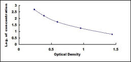
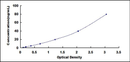
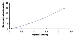
 Standard Curve (Sample)
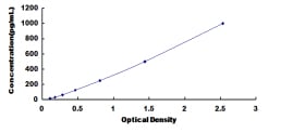
Monocyte Chemotactic Protein 5 (MCP5), ELISA Kit (Cat# AAA137294)
Full Name
Mouse Monocyte Chemotactic Protein 5 (MCP5) ELISA Kit
Reactivity
Mouse
Pricing
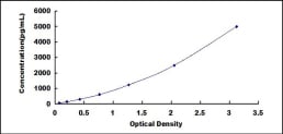
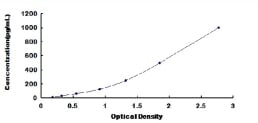
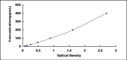
 Standard Curve (Sample)
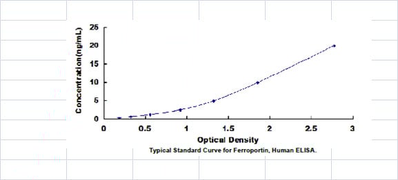
Ferroportin (FPN), ELISA Kit (Cat# AAA137297)
Full Name
Human Ferroportin (FPN) ELISA Kit
Reactivity
Human
Pricing
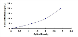
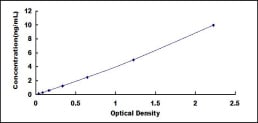
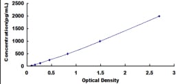
 Standard Curve (Sample)
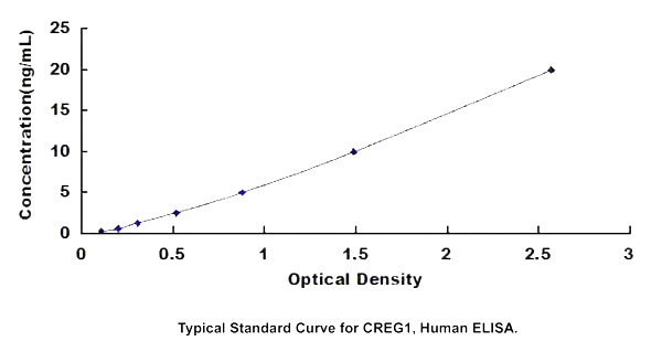
Cellular Repressor Of E1A Stimulated Genes 1 (CREG1), ELISA Kit (Cat# AAA137299)
Full Name
Human Cellular Repressor Of E1A Stimulated Genes 1 (CREG1) ELISA Kit
Gene Names
CREG1; CREG
Reactivity
Human
Pricing
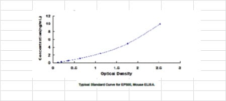
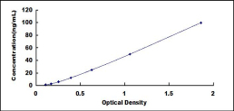
 Standard Curve (Sample)
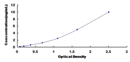
Rhodopsin (RHO), ELISA Kit (Cat# AAA137304)
Full Name
Mouse Rhodopsin (RHO) ELISA Kit
Gene Names
RHO; RP4; OPN2; CSNBAD1
Reactivity
Mouse
Pricing
 Standard Curve (Sample)
Chromogranin B (CHGB), ELISA Kit (Cat# AAA137308)
Full Name
Human Chromogranin B (CHGB) ELISA Kit
Reactivity
Human
Pricing
 Standard Curve (Sample)
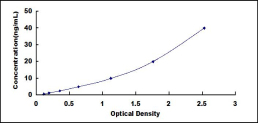
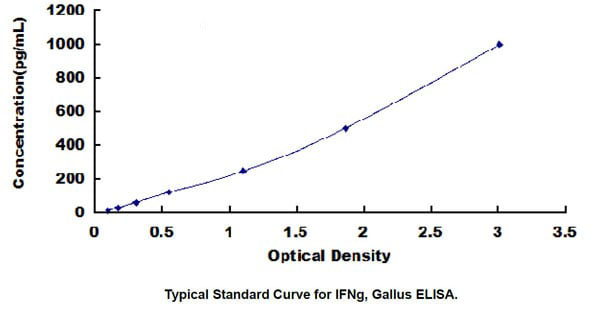
Interferon Gamma (IFNg), ELISA Kit (Cat# AAA137309)
Full Name
Chicken Interferon Gamma (IFNg) ELISA Kit
Gene Names
IFNG; IFG; IFI
Reactivity
Chicken
Pricing
 Standard Curve (Sample)
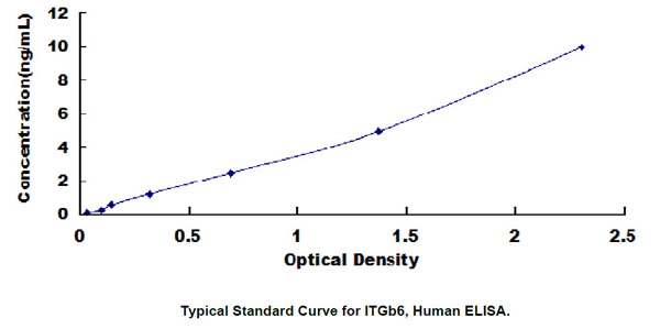
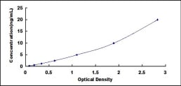
Integrin Beta 6 (ITGb6), ELISA Kit (Cat# AAA137313)
Full Name
Human Integrin Beta 6 (ITGb6) ELISA Kit
Gene Names
ITGB6; AI1H
Reactivity
Human
Pricing
 Standard Curve (Sample)
Glucose Transporter 4 (GLUT4), ELISA Kit (Cat# AAA137314)
Full Name
Human Glucose Transporter 4 (GLUT4) ELISA Kit
Gene Names
SLC2A4; GLUT4
Reactivity
Human
Pricing
 Standard Curve (Sample)
Hypoxia Up Regulated 1 (HYOU1), ELISA Kit (Cat# AAA137320)
Full Name
Human Hypoxia Up Regulated 1 (HYOU1) ELISA Kit
Reactivity
Human
Pricing
 Standard Curve (Sample)
A Disintegrin And Metalloproteinase With Thrombospondin 7 (ADAMTS7), ELISA Kit (Cat# AAA137321)
Full Name
Human A Disintegrin And Metalloproteinase With Thrombospondin 7 (ADAMTS7) ELISA Kit
Gene Names
ADAMTS7; ADAM-TS7; ADAMTS-7; ADAM-TS 7
Reactivity
Human
Pricing
 Standard Curve (Sample)
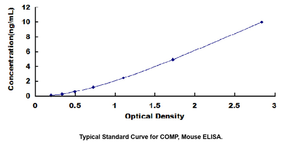
Cartilage Oligomeric Matrix Protein (COMP), ELISA Kit (Cat# AAA137324)
Full Name
Mouse Cartilage Oligomeric Matrix Protein (COMP) ELISA Kit
Gene Names
COMP; MED; EDM1; EPD1; PSACH; THBS5
Reactivity
Mouse
Pricing
 Standard Curve (Sample)
Macrophage Inflammatory Protein 1 Alpha (MIP1a), ELISA Kit (Cat# AAA137327)
Full Name
Mouse Macrophage Inflammatory Protein 1 Alpha (MIP1a) ELISA Kit
Gene Names
CCL3; MIP1A; SCYA3; G0S19-1; LD78ALPHA; MIP-1-alpha
Reactivity
Mouse
Pricing
 Standard Curve (Sample)
Bactericidal/Permeability Increasing Protein (BPI), ELISA Kit (Cat# AAA137333)
Full Name
Human Bactericidal/Permeability Increasing Protein (BPI) ELISA Kit
Reactivity
Human
Pricing
 Standard Curve (Sample)
Collagen Type VIII Alpha 1 (COL8a1), ELISA Kit (Cat# AAA137335)
Full Name
Human Collagen Type VIII Alpha 1 (COL8a1) ELISA Kit
Gene Names
COL8A1; C3orf7
Reactivity
Human
Pricing
 Standard Curve (Sample)
Toll Like Receptor 2 (TLR2), ELISA Kit (Cat# AAA137339)
Full Name
Mouse Toll Like Receptor 2 (TLR2) ELISA Kit
Gene Names
TLR2; TIL4; CD282
Reactivity
Mouse
Pricing
 Standard Curve (Sample)
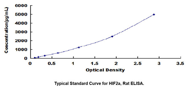
Hypoxia Inducible Factor 2 Alpha (HIF2a), ELISA Kit (Cat# AAA137340)
Full Name
Rat Hypoxia Inducible Factor 2 Alpha (HIF2a) ELISA Kit
Gene Names
Epas1; HLF; Hif2a
Reactivity
Rat
Pricing
 Standard Curve (Sample)
Thyrotropin Releasing Hormone (TRH), ELISA Kit (Cat# AAA137345)
Full Name
Mouse Thyrotropin Releasing Hormone (TRH) ELISA Kit
Gene Names
TRH; TRF; Pro-TRH
Reactivity
Mouse
Pricing
 Standard Curve (Sample)
Platelet Derived Growth Factor AB (PDGFAB), ELISA Kit (Cat# AAA137362)
Full Name
Human Platelet Derived Growth Factor AB (PDGFAB) ELISA Kit
Reactivity
Human
Pricing
 Standard Curve (Sample)
Alpha-Fetoprotein (aFP), ELISA Kit (Cat# AAA137364)
Full Name
Bovine Alpha-Fetoprotein (aFP) ELISA Kit
Gene Names
AFP; AFPD; FETA; HPAFP
Reactivity
Bovine
Pricing
 Standard Curve (Sample)
E1A Binding Protein P300 (EP300), ELISA Kit (Cat# AAA137369)
Full Name
Mouse E1A Binding Protein P300 (EP300) ELISA Kit
Gene Names
EP300; p300; KAT3B; RSTS2
Reactivity
Mouse
Pricing
 Standard Curve (Sample)
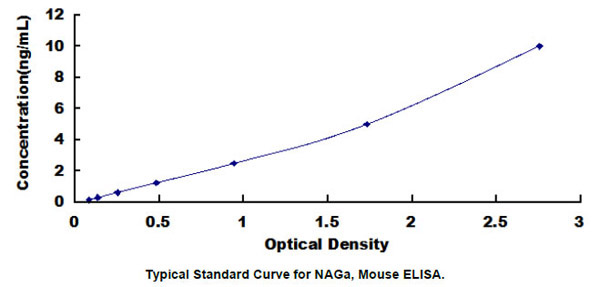
N-Acetylgalactosaminidase Alpha (NAGa), ELISA Kit (Cat# AAA137616)
Full Name
Mouse N-Acetylgalactosaminidase Alpha (NAGa) ELISA Kit
Gene Names
NAGA; GALB; D22S674
Reactivity
Mouse
Pricing
 Standard Curve (Sample)
Fibroblast Growth Factor 3 (FGF3), ELISA Kit (Cat# AAA137621)
Full Name
Human Fibroblast Growth Factor 3 (FGF3) ELISA Kit
Gene Names
FGF3; INT2; HBGF-3
Reactivity
Human
Pricing
 Standard Curve (Sample)
H2A Histone Family, Member V (H2AFV), ELISA Kit (Cat# AAA137622)
Full Name
Human H2A Histone Family, Member V (H2AFV) ELISA Kit
Gene Names
H2AFV; H2AV; H2A.Z-2
Reactivity
Human
Pricing
 Standard Curve (Sample)
Sclerostin (SOST), ELISA Kit (Cat# AAA137631)
Full Name
Mouse Sclerostin (SOST) ELISA Kit
Gene Names
SOST; CDD; VBCH; SOST1
Reactivity
Mouse
Pricing
 Standard Curve (Sample)
Alkaline Phosphatase (ALP), ELISA Kit (Cat# AAA137635)
Full Name
Rat Alkaline Phosphatase (ALP) ELISA Kit
Gene Names
ALPL; HOPS; TNAP; APTNAP; TNSALP; AP-TNAP
Reactivity
Rat
Pricing
 Standard Curve (Sample)
Inhibin A (INHA), ELISA Kit (Cat# AAA137636)
Full Name
Mouse Inhibin A (INHA) ELISA Kit
Reactivity
Mouse
Pricing
 Standard Curve (Sample)
Chemokine C-X-C-Motif Receptor 7 (CXCR7), ELISA Kit (Cat# AAA137641)
Full Name
Human Chemokine C-X-C-Motif Receptor 7 (CXCR7) ELISA Kit
Reactivity
Human
Pricing
 Standard Curve (Sample)
Proline Rich Protein 4, Lacrimal (PRR4), ELISA Kit (Cat# AAA137642)
Full Name
Human Proline Rich Protein 4, Lacrimal (PRR4) ELISA Kit
Gene Names
PRR4; LPRP; PROL4
Reactivity
Human
Pricing
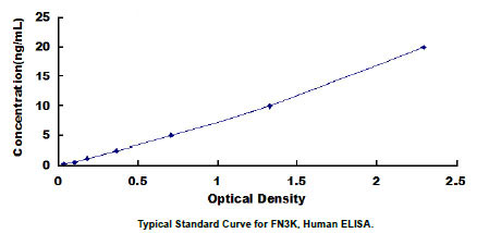 Standard Curve (Sample)
Fructosamine-3-Kinase (FN3K), ELISA Kit (Cat# AAA137643)
Full Name
Human Fructosamine-3-Kinase (FN3K) ELISA Kit
Reactivity
Human
Pricing
 Standard Curve (Sample)
Calponin 2 (CNN2), ELISA Kit (Cat# AAA137646)
Full Name
Human Calponin 2 (CNN2) ELISA Kit
Reactivity
Human
Pricing
 Standard Curve (Sample)
Growth Arrest Specific Protein 6 (GAS6), ELISA Kit (Cat# AAA137647)
Full Name
Human Growth Arrest Specific Protein 6 (GAS6) ELISA Kit
Gene Names
GAS6; AXSF; AXLLG
Reactivity
Human
Pricing
 Standard Curve (Sample)
Regenerating Islet Derived Protein 1 Alpha (REG1a), ELISA Kit (Cat# AAA137648)
Full Name
Rat Regenerating Islet Derived Protein 1 Alpha (REG1a) ELISA Kit
Reactivity
Rat
Pricing
 Standard Curve (Sample)
C4 Binding Protein Alpha (C4BPa), ELISA Kit (Cat# AAA137654)
Full Name
Human C4 Binding Protein Alpha (C4BPa) ELISA Kit
Gene Names
C4bpa; C4BP
Reactivity
Human
Pricing
 Standard Curve (Sample)
S100 Calcium Binding Protein A9 (S100A9), ELISA Kit (Cat# AAA137657)
Full Name
Mouse S100 Calcium Binding Protein A9 (S100A9) ELISA Kit
Gene Names
S100A9; MIF; NIF; P14; CAGB; CFAG; CGLB; L1AG; LIAG; MRP14; 60B8AG; MAC387
Reactivity
Mouse
Pricing
 Standard Curve (Sample)
Cytochrome P450 11B1 (CYP11B1), ELISA Kit (Cat# AAA137662)
Full Name
Mouse Cytochrome P450 11B1 (CYP11B1) ELISA Kit
Gene Names
CYP11B1; FHI; CPN1; CYP11B; P450C11
Reactivity
Mouse
Pricing
 Standard Curve (Sample)
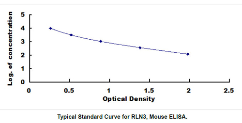
Relaxin 3 (RLN3), ELISA Kit (Cat# AAA137669)
Full Name
Mouse Relaxin 3 (RLN3) ELISA Kit
Gene Names
RLN3; H3; RXN3; ZINS4; insl7
Reactivity
Mouse
Pricing
 Standard Curve (Sample)
Heat Shock 70kDa Protein 5 (HSPA5), ELISA Kit (Cat# AAA137670)
Full Name
Mouse Heat Shock 70kDa Protein 5 (HSPA5) ELISA Kit
Reactivity
Mouse
Pricing
 Standard Curve (Sample)
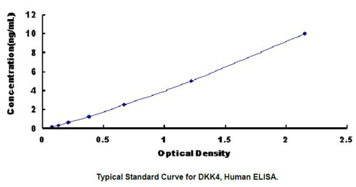
Dickkopf Related Protein 4 (DKK4), ELISA Kit (Cat# AAA137674)
Full Name
Human Dickkopf Related Protein 4 (DKK4) ELISA Kit
Gene Names
DKK4; DKK-4
Reactivity
Human
Pricing
 Standard Curve (Sample)
Proprotein Convertase Subtilisin/Kexin Type 1 (PCSK1), ELISA Kit (Cat# AAA137681)
Full Name
Human Proprotein Convertase Subtilisin/Kexin Type 1 (PCSK1) ELISA Kit
Gene Names
PCSK1; PC1; PC3; NEC1; SPC3; BMIQ12
Reactivity
Human
Pricing
 Standard Curve (Sample)
Galectin 1 (GAL1), ELISA Kit (Cat# AAA137683)
Full Name
Human Galectin 1 (GAL1) ELISA Kit
Gene Names
LGALS1; GBP; GAL1
Reactivity
Human
Pricing
 Standard Curve (Sample)
Inositol Polyphosphate-4-Phosphatase Type I 107kDa (INPP4A), ELISA Kit (Cat# AAA137687)
Full Name
Human Inositol Polyphosphate-4-Phosphatase Type I 107kDa (INPP4A) ELISA Kit
Reactivity
Human
Pricing
 Standard Curve (Sample)
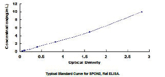
Spondin 2 (SPON2), ELISA Kit (Cat# AAA137689)
Full Name
Rat Spondin 2 (SPON2) ELISA Kit
Gene Names
SPON2; DIL1; DIL-1; MINDIN; M-SPONDIN
Reactivity
Rat
Pricing
 Standard Curve (Sample)
Mucin 1 (MUC1), ELISA Kit (Cat# AAA137690)
Full Name
Human Mucin 1 (MUC1) ELISA Kit
Gene Names
MUC1; EMA; MCD; PEM; PUM; KL-6; MAM6; MCKD; PEMT; CD227; H23AG; MCKD1; MUC-1; ADMCKD; ADMCKD1; CA 15-3; MUC-1/X; MUC1/ZD; MUC-1/SEC
Reactivity
Human
Pricing
 Standard Curve (Sample)
Protein Kinase R (PKR), ELISA Kit (Cat# AAA137692)
Full Name
Human Protein Kinase R (PKR) ELISA Kit
Reactivity
Human
Pricing
 Standard Curve (Sample)
Carbonyl Reductase 3 (CBR3), ELISA Kit (Cat# AAA137382)
Full Name
Human Carbonyl Reductase 3 (CBR3) ELISA Kit
Gene Names
CBR3; hCBR3; SDR21C2; HEL-S-25
Reactivity
Human
Pricing
 Standard Curve (Sample)
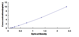
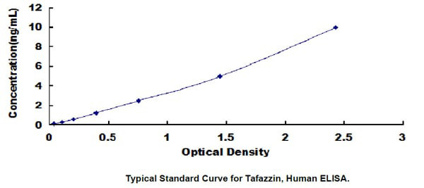
Tafazzin (TAZ), ELISA Kit (Cat# AAA137385)
Full Name
Human Tafazzin (TAZ) ELISA Kit
Gene Names
Taz; CG8766; DmelCG8766; dTAZ; tafazzin; TAZ
Reactivity
Human
Pricing
 Standard Curve (Sample)
Fatty Acid Synthase (FASN), ELISA Kit (Cat# AAA137396)
Full Name
Chicken Fatty Acid Synthase (FASN) ELISA Kit
Gene Names
FASN; FAS; OA-519; SDR27X1
Reactivity
Chicken
Pricing
 Standard Curve (Sample)
Hyaluronan Binding Protein 1 (HABP1), ELISA Kit (Cat# AAA137398)
Full Name
Human Hyaluronan Binding Protein 1 (HABP1) ELISA Kit
Reactivity
Human
Pricing
 Standard Curve (Sample)
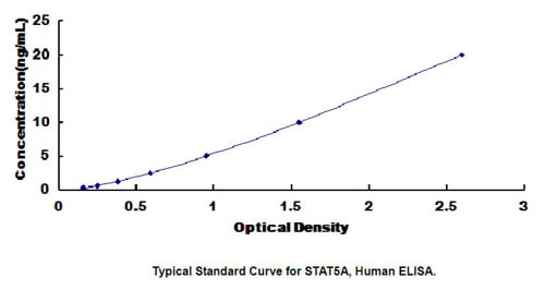
Signal Transducer And Activator Of Transcription 5A (STAT5A), ELISA Kit (Cat# AAA137404)
Full Name
Human Signal Transducer And Activator Of Transcription 5A (STAT5A) ELISA Kit
Gene Names
Stat5a; Stat5
Reactivity
Human
Pricing
 Standard Curve (Sample)
Matrix Metalloproteinase 2 (MMP2), ELISA Kit (Cat# AAA137407)
Full Name
Rat Matrix Metalloproteinase 2 (MMP2) ELISA Kit
Reactivity
Rat
Pricing