Get accurate results in your research with our Monoclonal Antibodies, which are specially made to target exactly what you require for your research, and will produce consistent, reliable performance in lab tests.
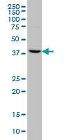
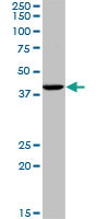
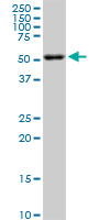
WB (Western Blot) (CDC2 monoclonal antibody, Western Blot analysis of CDC2 expression in Hela.)
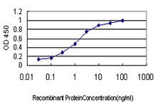
Application Data (Detection limit for recombinant GST tagged CDC2 is ~30ng/ml as a capture antibody.)
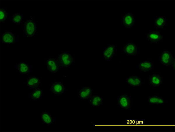
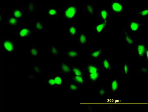
IF (Immunofluorescence) (Immunofluorescence of monoclonal antibody to CDC2 on HeLa cell. [antibody concentration 10ug/ml].)
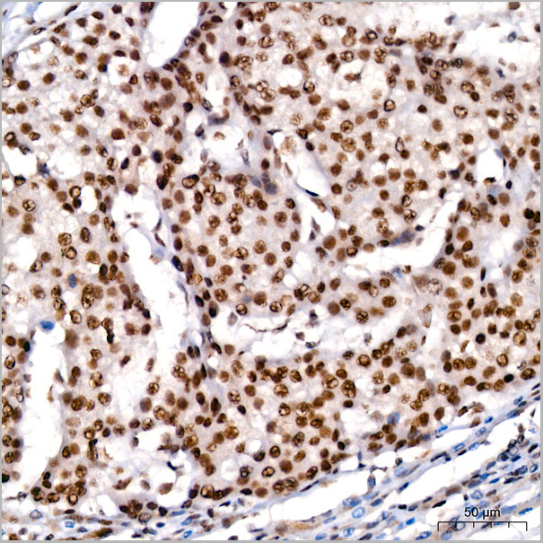
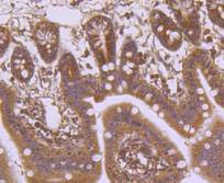
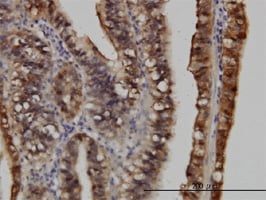
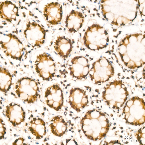
IHC (Immunohistochemistry) (Immunoperoxidase of monoclonal antibody to CDC2 on formalin-fixed paraffin-embedded human small Intestine. [antibody concentration 0.3ug/ml].)
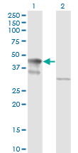
WB (Western Blot) (Western Blot analysis of CDC2 expression in transfected 293T cell line by CDC2 monoclonal antibody. Lane 1: CDC2 transfected lysate (34.1kD). Lane 2: Non-transfected lysate.)
WB (Western Blot) (Western Blot detection against Immunogen (58.41kD).)
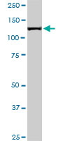
WB (Western Blot) (ACTN4 monoclonal antibody Western Blot analysis of ACTN4 expression in HeLa NE.)
WB (Western Blot) (ACTN4 monoclonal antibody Western Blot analysis of ACTN4 expression in NIH/3T3.)
Application Data (Detection limit for recombinant GST tagged ACTN4 is ~0.1ng/ml as a capture antibody.)
IF (Immunofluorescence) (Immunofluorescence of monoclonal antibody to ACTN4 on HeLa cell. [antibody concentration 20ug/ml].)
IHC (Immunohistochemistry) (Immunoperoxidase of monoclonal antibody to ACTN4 on formalin-fixed paraffin-embedded human testis. [antibody concentration 3ug/ml].)
WB (Western Blot) (Western Blot analysis of ACTN4 expression in transfected 293T cell line by ACTN4 monoclonal antibody. Lane 1: ACTN4 transfected lysate (104.9kD). Lane 2: Non-transfected lysate.)
WB (Western Blot) (ACTN4 monoclonal antibody Western Blot analysis of ACTN4 expression in PC-12.)
WB (Western Blot) (Western Blot detection against Immunogen (85.84kD).)
Application Data (Detection limit for recombinant GST tagged STIP1 is ~0.1ng/ml as a capture antibody.)
IP (Immunoprecipitation) (Immunoprecipitation of STIP1 transfected lysate using STIP1 monoclonal antibody and Protein A Magnetic Bead and immunoblotted with STIP1 rabbit polyclonal antibody.)
IF (Immunofluorescence) (Immunofluorescence of monoclonal antibody to STIP1 on HeLa cell. [antibody concentration 10ug/ml].)
IHC (Immunohistochemistry) (Immunoperoxidase of monoclonal antibody to STIP1 on formalin-fixed paraffin-embedded human testis. [antibody concentration 0.3ug/ml].)
WB (Western Blot) (Western Blot analysis of STIP1 expression in transfected 293T cell line by STIP1 monoclonal antibody Lane 1: STIP1 transfected lysate (62.6kD). Lane 2: Non-transfected lysate.)
WB (Western Blot) (MAP3K7IP1 monoclonal antibody Western Blot analysis of MAP3K7IP1 expression in PC-12.)
Application Data (Proximity Ligation Analysis (PLA) of protein-protein interactions between HSPA1L and MAP3K7IP1 HeLa cells were stained with HSPA1L rabbit purified polyclonal 1:1200 and MAP3K7IP1 mouse monoclonal antibody 1:50. Signals were detected 30 Detection Kit 613 (red), and nuclei were counterstained with DAPI (blue). Each red dot represents the detection of protein-protein interaction complex.)
Application Data (Detection limit for recombinant GST tagged MAP3K7IP1 is ~0.3ng/ml as a capture antibody.)
IF (Immunofluorescence) (Immunofluorescence of monoclonal antibody to MAP3K7IP1 on HeLa cell. [antibody concentration 10ug/ml])
WB (Western Blot) (MAP3K7IP1 monoclonal antibody Western Blot analysis of MAP3K7IP1 expression in HeLa.)
WB (Western Blot) (Western Blot detection against Immunogen (36.89kD).)
TAB1 (TGF-beta-activated Kinase 1 and MAP3K7-binding Protein 1, Mitogen-activated Protein Kinase Kinase Kinase 7-interacting Protein 1, TGF-beta-activated Kinase 1-binding Protein 1, TAK1-binding Protein 1, MAP3K7IP1) (FITC)
Gene Names
TAB1; 3'-Tab1; MAP3K7IP1
Reactivity
Human, Rat
Applications
ELISA (EIA), Immunofluorescence (IF), Western Blot (WB)
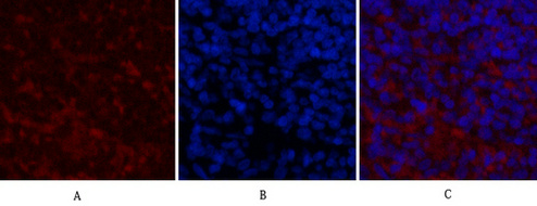
ICC (Immunocytochemistry) (Confocal imaging of paraffin-embedded Rat brain tissue using NeuN Mouse mAb (AAA28598, dilution 1:200) followed by a further incubation with Cy3 Goat Anti-Mouse IgG (H+L) (AS008, dilution 1:500) (Red). DAPI was used for nuclear staining (Blue). High pressure antigen retrieval performed with 0.01M Citrate Buffer (pH 6.0) prior to IF staining. Objective: 40x.)
ICC (Immunocytochemistry) (Confocal imaging of paraffin-embedded Mouse brain tissue using NeuN Mouse mAb (AAA28598, dilution 1:200) followed by a further incubation with Cy3 Goat Anti-Mouse IgG (H+L) (AS008, dilution 1:500) (Red). DAPI was used for nuclear staining (Blue). High pressure antigen retrieval performed with 0.01M Citrate Buffer (pH 6.0) prior to IF staining. Objective: 40x.)
ICC (Immunocytochemistry) (Confocal imaging of paraffin-embedded Human brain tissue using NeuN Mouse mAb (AAA28598, dilution 1:200) followed by a further incubation with Cy3 Goat Anti-Mouse IgG (H+L) (AS008, dilution 1:500) (Red). DAPI was used for nuclear staining (Blue). High pressure antigen retrieval performed with 0.01M Citrate Buffer (pH 6.0) prior to IF staining. Objective: 40x.)
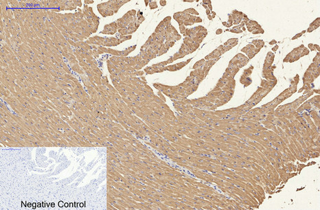
IHC (Immunohistochemistry) (Immunohistochemistry analysis of paraffin-embedded Rat brain tissue using NeuN Mouse mAb (AAA28598) at a dilution of 1:200 (40x lens). High pressure antigen retrieval performed with 0.01M Citrate Buffer (pH 6.0) prior to IHC staining.)
IHC (Immunohistochemistry) (Immunohistochemistry analysis of paraffin-embedded Human brain tissue using NeuN Mouse mAb (AAA28598) at a dilution of 1:200 (40x lens). High pressure antigen retrieval performed with 0.01M Citrate Buffer (pH 6.0) prior to IHC staining.)
IHC (Immunohistochemistry) (Immunohistochemistry analysis of paraffin-embedded Mouse brain tissue using NeuN Mouse mAb (AAA28598) at a dilution of 1:200 (40x lens). High pressure antigen retrieval performed with 0.01M Citrate Buffer (pH 6.0) prior to IHC staining.)
WB (Western Blot) (Western blot analysis of various lysates using NeuN Mouse mAb (AAA28598) at 1:5000 dilution incubated overnight at 4?.Secondary antibody: HRP-conjugated Goat anti-Mouse IgG (H+L) (AS003) at 1:10000 dilution.Lysates/proteins: 25 ug per lane.Blocking buffer: 3% nonfat dry milk in TBST.Detection: ECL Basic Kit (RM00020).Exposure time: 90s.)
SDS-PAGE (Whole cell lysates were separated by 6% SDS-PAGE, and the membrane was blotted with anti-Ki-67(ABT001)antibody. The HRP-conjugated Goat anti-M)
IP (Immunoprecipitation) (Figure 6. Immunoprecipitation analysis. Cellular overexpression lysates (made from HEK293F cells transfected with DYKDDDDK tagged human BCMA full length gene) were pre-incubated with 6 different rabbit DimAb clones and negative control IgG. The immunocomplexes were further pulled down by protein A beads, fractionated, and blotted with mouse anti-DYKDDDDK monoclonal antibody.)
FCM (Flow Cytometry) (Figure 6. Immunoprecipitation analysis. Cellular overexpression lysates (made from HEK293F cells transfected with DYKDDDDK tagged human BCMA full length gene) were pre-incubated with 6 different rabbit DimAb clones and negative control IgG. The immunocomplexes were further pulled down by protein A beads, fractionated, and blotted with mouse anti-DYKDDDDK monoclonal antibody.)
Application Data (Figure 6. Immunoprecipitation analysis. Cellular overexpression lysates (made from HEK293F cells transfected with DYKDDDDK tagged human BCMA full length gene) were pre-incubated with 6 different rabbit DimAb clones and negative control IgG. The immunocomplexes were further pulled down by protein A beads, fractionated, and blotted with mouse anti-DYKDDDDK monoclonal antibody.)
WB (Western Blot) (TUBB2A monoclonal antibody (M04), clone 3B2. Western Blot analysis of TUBB2A expression in NIH/3T3.)
WB (Western Blot) (TUBB2A monoclonal antibody (M04), clone 3B2. Western Blot analysis of TUBB2A expression in Jurkat.)
IHC (Immunohistochemistry) (Immunoperoxidase of monoclonal antibody to TUBB2A on formalin-fixed paraffin-embedded human colon. [antibody concentration 1.5 ug/ml])
IF (Immunofluorescence) (Immunofluorescence of monoclonal antibody to TUBB2A on HeLa cell. [antibody concentration 10 ug/ml])
Application Data (Published customer imageSLAMF3 blockade in human hepatocytes is associated with lower susceptibility to HCV. (A) SLAMF3 was stained in primary human hepatocytes (PHHs) and cells from the Huh-7 human hepatoma cell line with a specific antibody (HLy9.1.25 clone; grey) and an isotype-matched control (empty). One of four independent experiments is shown. Huh-7 cells were transfected with scrambled control (sc) siRNA or three specific siRNAs (#1, #2 and #3) targeting SLAMF3, prior to infection with HCVcc; siRNA efficiency was checked by quantifying SLAMF3 mRNA (B) and the CD81 expression level (C) by flow cytometry analysis at 48 h post-transfection. Results are presented as the mean +/-SD (n = 3). Intracellular viral RNA was quantified at 72 h p.i. (D) and the infection was measured at 72 h p.i. by focus-forming units FFUs counting (E) (as inhibition percent; mean of three independent experiments; error bars: SD. **p)
Application Data (Published customer image: Effect of SLAMF3 expression on the organization of the actin cytoskeleton. Cells (Huh-7) were stained with phalloidin (rhodamine, red) and anti-SLAMF3 (FITC, green) and SLAMF3 positive (Huh-7-SLAMF3pos) and SLAMF3-negative (Huh-7-SLAMF3pos) cells were examined under the microscope. One representative of two independent experiments is shown.From: Marcq I, Nyga R, Cartier F, Amrathlal RS, Ossart C, et al. (2013) Identification of SLAMF3 (CD229) as an Inhibitor of Hepatocellular Carcinoma Cell Proliferation and Tumour Progression. PLoS ONE 8(12): e82918.)
Application Data (Published customer image: Expression of SLAM-R family by HHPHs and Huh-7 and HepG2 human HCC cell lines. SLAM-Rs were stained with specific antibodies against SLAMF1 (CD150), SLAMF2 (CD84), SLAMF4 (CD224) and SLAMF6 (NTBA) (grey) or with matched isotype controls (empty). One of four independent experiments is shown.From: Marcq I, Nyga R, Cartier F, Amrathlal RS, Ossart C, et al. (2013) Identification of SLAMF3 (CD229) as an Inhibitor of Hepatocellular Carcinoma Cell Proliferation and Tumour Progression. PLoS ONE 8(12): e82918.)
Application Data (Published customer image: Expression of SLAMF3 assessed by flow cytometry analysis after transfection with vector coding for SLAMF3 or an empty vector (Mock) in Huh-7 and HepG2 cells. SLAMF3 staining (in grey) is overlaid by negative control (in white). One of four independent experiments is shown. doi:10.1371/journal.pone.0082918.s002From: Marcq I, Nyga R, Cartier F, Amrathlal RS, Ossart C, et al. (2013) Identification of SLAMF3 (CD229) as an Inhibitor of Hepatocellular Carcinoma Cell Proliferation and Tumour Progression. PLoS ONE 8(12): e82918.)
Application Data (Published customer image: Restoration of SLAMF3 expression in HCC cells inhibits MAPK ERK1/2, JNK and mTOR pathways and induces caspase-dependent apoptosis.(A) SLAMF3 expression was confirmed by Western blot analysis in SLAMF3-over-expressing Huh-7 and mock cells at 48 h post-transfection. ERK1/2, JNK, p38, AKT and mTOR proteins were detected as controls. The activation levels of MAPK ERK1/2 (Thr 202/Tyr 204), JNK (Thr 183/Tyr 185), p38 (Thr 180/Tyr 183), PI3K (p85/p110 Thr 467/Tyr 199), AKT (Ser 473, Thr 308) and mTOR (Ser 2448, Ser 2481) are represented. One of three representative experiments is shown here. (B) The results of an active caspase assay based on cell-permeable fluorochrome inhibitor of caspases (FLICA). Caspase activity was evaluated in HCC cells (Huh-7 and HepG2 lines) at the indicated times after the ectopic introduction of SLAMF3 vector. Caspase activity is shown in the SLAMF3neg subpopulation (in white) and the SLAMF3pos (overlaid in grey) subpopulation from one representative of two independent experiments.From: Marcq I, Nyga R, Cartier F, Amrathlal RS, Ossart C, et al. (2013) Identification of SLAMF3 (CD229) as an Inhibitor of Hepatocellular Carcinoma Cell Proliferation and Tumour Progression. PLoS ONE 8(12): e82918.)
Application Data (Published customer image: SLAMF3 expression is repressed in tumour cells from ten resected HCC patients. (A) SLAMF3 mRNA expression was analysed in hepatocytes from resected HCC patients. Specific amplification of SLAMF3 mRNAs using PCR (representative patients #2, #7 and #12) and quantitative PCR in tumour tissues (T) and peritumoral tissues (pT) presented separately (B) and as median (**p)
Application Data (Published customer image SLAMF3 over-expression increases hepatocyte permissiveness to HCV. Huh-7 cells were transfected with empty vector pBud (mock) or human SLAMF3 (to yield Huh-7+ cells) and incubated with HCVcc. (A) SLAMF3 expression was analyzed by flow cytometry (HLy9.1.25 clone) and (B) visualized by confocal microscopy in one out of three experiment. Unshaded histograms show the isotype-matched control (Co) and shaded histograms show SLAMF3 expression). (C, D, E) Infection was assessed at 4 and 72 h p.i. as the number of FFUs and (F and G) by flow cytometry analysis; (C) intracellular E2 (stained red) were measured at 4 h and 72 h p.i. Green staining corresponds to the SLAMF3 expression. One of three representatives independent experiments is shown. (D) The HCV permissiveness of Huh-7 mock and Huh-7+ cells was evaluated in terms of the number of FFUs based on intracellular E2 viral protein staining and (E) fluorescent microscopy analysis (mean of five experiments; error bars: SD; *p)
Application Data (Staining of human peripheral blood lymphocytes with Mouse anti Human CD229 : RPE)
Application Data (Published customer image: Correlation between SLAMF3 expression and HCC cell proliferation.(A) Effects of the introduction of specific siRNAs on SLAMF3 expression in Huh-7 cells. Cells were transiently transfected with a scrambled siRNA (Sc) or one of two pre-designed anti-SLAMF3 siRNAs (referred to as #2 and # 3); SLAMF3 expression was measured by Western blot analysis (anti-SLAMF3, K12). One representative of two independent experiments is shown. (B) siRNA-treated Huh-7 cells were cultured and proliferation was assayed in a Trypan blue exclusion test after 48 h. Proliferation is presented as the mean number of viable cells +/- SD (n = 5; statistical significance: ***p)
Application Data (Staining of human peripheral blood lymphocytes with Mouse anti Human CD299: Azide Free)
WB (Western Blot) (EIF2S2 monoclonal antibody. Western Blot analysis of EIF2S2 expression in HeLa.)
WB (Western Blot) (EIF2S2 monoclonal antibody. Western Blot analysis of EIF2S2 expression in PC-12.)
Application Data (Detection limit for recombinant GST tagged EIF2S2 is 0.3ng/ml as a capture antibody.)
IHC (Immunohistochemistry) (Immunoperoxidase of monoclonal antibody to EIF2S2 on formalin-fixed paraffin-embedded human pancreatic cancer. [antibody concentration 3ug/ml].)
WB (Western Blot) (Western Blot analysis of EIF2S2 expression in transfected 293T cell line by EIF2S2 monoclonal antibody. Lane 1: EIF2S2 transfected lysate (38.4kD). Lane 2: Non-transfected lysate.)
WB (Western Blot) (EIF2S2 monoclonal antibody. Western Blot analysis of EIF2S2 expression in NIH/3T3.)
WB (Western Blot) (EIF2S2 monoclonal antibody. Western Blot analysis of EIF2S2 expression in Raw 264.7.)
WB (Western Blot) (CD58 monoclonal antibody, Western Blot analysis of CD58 expression in Jurkat.)
Application Data (Detection limit for recombinant GST tagged CD58 is ~0.3ng/ml as a capture antibody.)
IF (Immunofluorescence) (Immunofluorescence of monoclonal antibody to CD58 on HeLa cell. [antibody concentration 10ug/ml].)
IHC (Immunohistochemistry) (Immunoperoxidase of monoclonal antibody to CD58 on formalin-fixed paraffin-embedded human tonsil. [antibody concentration 2ug/ml].)
IHC (Immunohistochemistry) (Immunoperoxidase of monoclonal antibody to CD58 on formalin-fixed paraffin-embedded human lymphoma tissue [antibody concentration 3ug/ml].)
WB (Western Blot) (Western Blot detection against Immunogen (52.14kD).)
IF (Immunofluorescence) (Immunofluorescence of monoclonal antibody to TIMP2 on HeLa cell. [antibody concentration 10 ug/ml])
Application Data (Detection limit for recombinant GST tagged TIMP2 is approximately 0.1ng/ml as a capture antibody.)
IHC (Immunohistochemistry) (Immunoperoxidase of monoclonal antibody to TIMP2 on formalin-fixed paraffin-embedded human kidney. [antibody concentration 3 ug/ml])
WB (Western Blot) (TIMP2 monoclonal antibody (M03), clone 1C3 Western Blot analysis of TIMP2 expression in HeLa (Cat # L013V1).)
FCM (Flow Cytometry) (Flow cytometric analysis of Hela cells with Sonic Hedgehog Protein antibody at 1/50 dilution (red) compared with an unlabelled control (cells without incubation with primary antibody; black). Alexa Fluor 488-conjugated goat anti rabbit IgG was used as the secondary antibody.)
ICC (Immunocytochemistry) (ICC staining Sonic Hedgehog Protein in 293 cells (green). The nuclear counter stain is DAPI (blue). Cells were fixed in paraformaldehyde, permeabilised with 0.25% Triton X100/PBS.)
ICC (Immunocytochemistry) (ICC staining Sonic Hedgehog Protein in A549 cells (green). The nuclear counter stain is DAPI (blue). Cells were fixed in paraformaldehyde, permeabilised with 0.25% Triton X100/PBS.)
ICC (Immunocytochemistry) (ICC staining Sonic Hedgehog Protein in Hela cells (green). The nuclear counter stain is DAPI (blue). Cells were fixed in paraformaldehyde, permeabilised with 0.25% Triton X100/PBS.)
IHC (Immunohistochemistry) (Immunohistochemical analysis of paraffin-embedded human kidney tissue using anti-Sonic Hedgehog Protein antibody. Counter stained with hematoxylin.)
IHC (Immunohistochemistry) (Immunohistochemical analysis of paraffin-embedded human liver cancer tissue using anti-Sonic Hedgehog Protein antibody. Counter stained with hematoxylin.)
WB (Western Blot) (Western blot analysis of Sonic Hedgehog Protein on different lysates using anti-Sonic Hedgehog Protein antibody at 1/1, 000 dilution. Positive control: Lane 1: Hela Lane 2: HepG2)
WB (Western Blot) (Western Blot detection against Immunogen (37.11kD).)
WB (Western Blot) (FGL2 monoclonal antibody Western Blot analysis of FGL2 expression in PC-12.)
Application Data (Detection limit for recombinant GST tagged FGL2 is ~0.1ng/ml as a capture antibody.)
IP (Immunoprecipitation) (Immunoprecipitation of FGL2 transfected lysate using FGL2 monoclonal antibody and Protein A Magnetic Bead and immunoblotted with FGL2 rabbit polyclonal antibody.)
IHC (Immunohistochemistry) (Immunoperoxidase of monoclonal antibody to FGL2 on formalin-fixed paraffin-embedded human stomach. [antibody concentration 0.5ug/ml].)
WB (Western Blot) (Western Blot analysis of FGL2 expression in transfected 293T cell line by FGL2 monoclonal antibody Lane 1: FGL2 transfected lysate (50kD).Lane 2: Non-transfected lysate.)
WB (Western Blot) (FGL2 monoclonal antibody Western Blot analysis of FGL2 expression in Raw 264.7.)
WB (Western Blot) (CDK6 monoclonal antibody, Western Blot analysis of CDK6 expression in Jurkat.)
WB (Western Blot) (CDK6 monoclonal antibody. Western Blot analysis of CDK6 expression in PC-12.)
Application Data (Proximity Ligation Analysis (PLA) of protein-protein interactions between CDK2 and CDK6 HeLa cells were stained with CDK2 rabbit purified polyclonal 1:1200 and CDK6 mouse monoclonal antibody 1:50. Signals were detected by 30 Detection Kit 613 (red), and nuclei were counterstained with DAPI (blue). Each red dot represents the detection of protein-protein interaction complex.)
IF (Immunofluorescence) (Immunofluorescence of monoclonal antibody to CDK6 on HeLa cell. [antibody concentration 10ug/ml].)
IHC (Immunohistochemistry) (Immunoperoxidase of monoclonal antibody to CDK6 on formalin-fixed paraffin-embedded human colon. [antibody concentration 3ug/ml].)
WB (Western Blot) (Western Blot analysis of CDK6 expression in transfected 293T cell line by CDK6 monoclonal antibody. Lane 1: CDK6 transfected lysate (36.9kD). Lane 2: Non-transfected lysate.)
WB (Western Blot) (Western Blot detection against Immunogen (36.41kD).)
IF (Immunofluorescence) (Immunofluorescence of monoclonal antibody to TIMP2 on HeLa cell. [antibody concentration 10 ug/ml])
Application Data (Detection limit for recombinant GST tagged TIMP2 is approximately 0.3ng/ml as a capture antibody.)
IHC (Immunohistochemistry) (Immunoperoxidase of monoclonal antibody to TIMP2 on formalin-fixed paraffin-embedded human spleen. [antibody concentration 3 ug/ml])
WB (Western Blot) (TIMP2 monoclonal antibody (M04), clone 5B11 Western Blot analysis of TIMP2 expression in HeLa (Cat # L013V1).)
ICC (Immunocytochemistry) (ICC staining CD21 in A549 cells (green). The nuclear counter stain is DAPI (blue). Cells were fixed in paraformaldehyde, permeabilised with 0.25% Triton X100/PBS.)
ICC (Immunocytochemistry) (ICC staining CD21 in HepG2 cells (green). The nuclear counter stain is DAPI (blue). Cells were fixed in paraformaldehyde, permeabilised with 0.25% Triton X100/PBS.)
IHC (Immunohistochemistry) (Immunohistochemical analysis of paraffin-embedded human gastric cancer tissue using anti-CD21 antibody. Counter stained with hematoxylin.)
IHC (Immunohistochemistry) (Immunohistochemical analysis of paraffin-embedded human spleen tissue using anti-CD21 antibody. Counter stained with hematoxylin.)
IHC (Immunohistochemistry) (Immunohistochemical analysis of paraffin-embedded human tonsil tissue using anti-CD21 antibody. Counter stained with hematoxylin.)
WB (Western Blot) (Western blot analysis of CD21 on different lysates using anti-CD21 antibody at 1/1, 000 dilution. Positive control: Lane 1: Daudi Lane 2: Raji)
SDS-PAGE (Whole cell lysates were separated by 10% SDS-PAGE, and the membrane was blotted with anti-Her-2(ABT088) antibody. The HRP-conjugated Goat anti-M)
Application Data (Detection limit for recombinant GST tagged PGGT1B is ~1ng/ml as a capture antibody.)
IF (Immunofluorescence) (Immunofluorescence of monoclonal antibody to PGGT1B on HeLa cell. [antibody concentration 10ug/ml])
IHC (Immunohistochemistry) (Immunoperoxidase of monoclonal antibody to PGGT1B on formalin-fixed paraffin-embedded human kidney. [antibody concentration 1.2ug/ml])
WB (Western Blot) (PGGT1B monoclonal antibody Western Blot analysis of PGGT1B expression in PC-12.)
WB (Western Blot) (PGGT1B monoclonal antibody Western Blot analysis of PGGT1B expression in HeLa.)
WB (Western Blot) (Western Blot detection against Immunogen (37.29kD).)
PGGT1B (Geranylgeranyl Transferase Type-1 Subunit beta, Geranylgeranyl Transferase Type I Subunit beta, GGTase-I-beta, Type I Protein Geranyl-Geranyltransferase Subunit beta)
Application Data (Detection limit for recombinant GST tagged SRPK1 is ~0.03ng/ml as a capture antibody.)
IHC (Immunohistochemistry) (Immunoperoxidase of monoclonal antibody to SRPK1 on formalin-fixed paraffin-embedded human colon. [antibody concentration 1ug/ml].)
WB (Western Blot) (Western Blot analysis of SRPK1 expression in transfected 293T cell line by SRPK1 monoclonal antibody. Lane 1: SRPK1 transfected lysate (74.3kD). Lane 2: Non-transfected lysate.)
WB (Western Blot) (SRPK1 monoclonal antibody, Western Blot analysis of SRPK1 expression in IMR-32.)
WB (Western Blot) (SRPK1 monoclonal antibody. Western Blot analysis of SRPK1 expression in HepG2.)
WB (Western Blot) (Western Blot detection against Immunogen (37kD).)
WB (Western Blot) (Western Blot analysis of FTL expression in transfected 293T cell line by FTL monoclonal antibody (M16), clone X1.Lane 1: FTL transfected lysate (20 KDa).Lane 2: Non-transfected lysate.)
WB (Western Blot) (Western blot analysis of FTL over-expressed 293 cell line, cotransfected with FTL Validated Chimera RNAi (Lane 2) or non-transfected control (Lane 1). Blot probed with FTL monoclonal antibody (M16), clone X1. GAPDH (36.1 kDa) used as specificity and loading control.)
Application Data (Detection limit for recombinant GST tagged FTL is approximately 0.03ng/ml as a capture antibody.)
WB (Western Blot) (FTL monoclonal antibody (M16), clone X1 Western Blot analysis of FTL expression in K-562 (Cat # L009V1).)
IF (Immunofluorescence) (Immunofluorescence of monoclonal antibody to FTL on HeLa cell. [antibody concentration 10 ug/ml])
FCM (Flow Cytometry) (Flow cytometric analysis of Hela cells with Apg7 antibody at 1/50 dilution (red) compared with an unlabelled control (cells without incubation with primary antibody; black). Alexa Fluor 488-conjugated goat anti rabbit IgG was used as the secondary antibody.)
ICC (Immunocytochemistry) (ICC staining Apg7 in HepG2 cells (green). The nuclear counter stain is DAPI (blue). Cells were fixed in paraformaldehyde, permeabilised with 0.25% Triton X100/PBS.)
ICC (Immunocytochemistry) (ICC staining Apg7 in Hela cells (green). The nuclear counter stain is DAPI (blue). Cells were fixed in paraformaldehyde, permeabilised with 0.25% Triton X100/PBS.)
IHC (Immunohistochemistry) (Immunohistochemical analysis of paraffin-embedded mouse brain tissue using anti-Apg7 antibody. Counter stained with hematoxylin.)
IHC (Immunohistochemistry) (Immunohistochemical analysis of paraffin-embedded human kidney tissue using anti-Apg7 antibody. Counter stained with hematoxylin.)
IHC (Immunohistochemistry) (Immunohistochemical analysis of paraffin-embedded human spleen tissue using anti-Apg7 antibody. Counter stained with hematoxylin.)
IHC (Immunohistochemistry) (Immunohistochemical analysis of paraffin-embedded human tonsil tissue using anti-Apg7 antibody. Counter stained with hematoxylin.)
WB (Western Blot) (Western blot analysis of Apg7 on Jurkat cells lysates using anti-Apg7 antibody at 1/1, 000 dilution.)
WB (Western Blot) (ACTB monoclonal antibody Western Blot analysis of ACTB expression in K-562.)
WB (Western Blot) (ACTB monoclonal antibody Western Blot analysis of ACTB expression in NIH/3T3.)
Application Data (Detection limit for recombinant GST tagged ACTB is ~1ng/ml as a capture antibody.)
IHC (Immunohistochemistry) (Immunoperoxidase of monoclonal antibody to ACTB on formalin-fixed paraffin-embedded human smooth muscle. [antibody concentration 3ug/ml].)
WB (Western Blot) (Western Blot analysis of ACTB expression in transfected 293T cell line by ACTB monoclonal antibody Lane 1: ACTB transfected lysate (42kD). Lane 2: Non-transfected lysate.)
WB (Western Blot) (ACTB monoclonal antibody Western Blot analysis of ACTB expression in Raw 264.7.)
WB (Western Blot) (ACTB monoclonal antibody Western Blot analysis of ACTB expression in HeLa.)
WB (Western Blot) (Western Blot detection against Immunogen (36.89kD).)
Application Data (Detection limit for recombinant GST tagged RICTOR is ~0.03ng/ml as a capture antibody.)
IP (Immunoprecipitation) (Immunoprecipitation of RICTOR transfected lysate using RICTOR monoclonal antibody and Protein A Magnetic Bead, and immunoblotted with RICTOR rabbit polyclonal antibody)
IHC (Immunohistochemistry) (Immunoperoxidase of monoclonal antibody to RICTOR on formalin-fixed paraffin-embedded human small Intestine. [antibody concentration 3ug/ml].)
WB (Western Blot) (Western Blot analysis of RICTOR expression in transfected 293T cell line by RICTOR monoclonal antibody. Lane 1: RICTOR transfected lysate (29.8kD). Lane 2: Non-transfected lysate.)
WB (Western Blot) (RICTOR monoclonal antibody Western Blot analysis of RICTOR expression in Hela NE.)
WB (Western Blot) (LIMK1 monoclonal antibody. Western Blot analysis of LIMK1 expression in PC-12.)
WB (Western Blot) (Western Blot detection against Immunogen (36.74kD).)
Application Data (Detection limit for recombinant GST tagged LIMK1 is ~0.03ng/ml as a capture antibody.)
IHC (Immunohistochemistry) (Immunoperoxidase of monoclonal antibody to LIMK1 on formalin-fixed paraffin-embedded human stomach. [antibody concentration 3ug/ml])
WB (Western Blot) (LIMK1 monoclonal antibody. Western Blot analysis of LIMK1 expression in NIH/3T3.)
WB (Western Blot) (LIMK1 monoclonal antibody. Western Blot analysis of LIMK1 expression in Raw 264.7.)
WB (Western Blot) (LIMK1 monoclonal antibody Western Blot analysis of LIMK1 expression in SW-13.)
WB (Western Blot) (Western Blot analysis of CSTF3 expression in transfected 293T cell line by CSTF3 monoclonal antibody. Lane 1: CSTF3 transfected lysate (11.44kD). Lane 2: Non-transfected lysate.)
WB (Western Blot) (Western blot analysis of CSTF3 over-expressed 293 cell line, cotransfected with CSTF3 Validated Chimera RNAi (Lane 2) or non-transfected control (Lane 1). Blot probed with CSTF3 monoclonal antibody. GAPDH (36.1kD) used as specificity and loading control.)
Application Data (Detection limit for recombinant GST tagged CSTF3 is ~0.1ng/ml as a capture antibody.)
IF (Immunofluorescence) (Immunofluorescence of monoclonal antibody to CSTF3 on HeLa cell. [antibody concentration 10ug/ml].)
IHC (Immunohistochemistry) (Immunoperoxidase of monoclonal antibody to CSTF3 on formalin-fixed paraffin-embedded human testis. [antibody concentration 3ug/ml].)
WB (Western Blot) (Western Blot detection against Immunogen (37.07kD).)
WB (Western Blot) (Western blot analysis of ARNT over-expressed 293 cell line, cotransfected with ARNT Validated Chimera RNAi (Lane 2) or non-transfected control (Lane 1). Blot probed with ARNT monoclonal antibody. GAPDH (36.1kD) used as specificity and loading control.)
WB (Western Blot) (Western Blot analysis of ARNT expression in transfected 293T cell line by ARNT monoclonal antibody. Lane 1: ARNT transfected lysate (86.6kD). Lane 2: Non-transfected lysate.)
Application Data (Detection limit for recombinant GST tagged ARNT is ~0.03ng/ml as a capture antibod)
IF (Immunofluorescence) (Immunofluorescence of monoclonal antibody to ARNT on HeLa cell. [antibody concentration 10ug/ml].)
IHC (Immunohistochemistry) (Immunoperoxidase of monoclonal antibody to ARNT on formalin-fixed paraffin-embedded human lung. [antibody concentration 3ug/ml].)
WB (Western Blot) (ARNT monoclonal antibody Western Blot analysis of ARNT expression in Hela NE.)
WB (Western Blot) (Western Blot detection against Immunogen (37.73kD).)
WB (Western Blot) (Western Blot detection against Immunogen (36.41kD).)
Application Data (Detection limit for recombinant GST tagged TSC22D3 is ~0.03ng/ml as a capture antibody.)
IP (Immunoprecipitation) (Immunoprecipitation of TSC22D3 transfected lysate using TSC22D3 monoclonal antibody and Protein A Magnetic Bead and immunoblotted with TSC22D3 rabbit polyclonal antibody.)
IF (Immunofluorescence) (Immunofluorescence of monoclonal antibody to TSC22D3 on HeLa cell. [antibody concentration 10ug/ml].)
IHC (Immunohistochemistry) (Immunoperoxidase of monoclonal antibody to TSC22D3 on formalin-fixed paraffin-embedded human lymph node. [antibody concentration 3ug/ml].)
WB (Western Blot) (Western Blot analysis of TSC22D3 expression in transfected 293T cell line by TSC22D3 monoclonal antibody. Lane 1: TSC22D3 transfected lysate (22.2kD). Lane 2: Non-transfected lysate.)
WB (Western Blot) (Western blot analysis of PIP5K3 over-expressed 293 cell line, cotransfected with PIP5K3 Validated Chimera RNAi (Lane 2) or non-transfected control (Lane 1). Blot probed with PIP5K3 monoclonal antibody. GAPDH (36.1kD) used as specificity and loading control.)
Application Data (Detection limit for recombinant GST tagged PIP5K3 is ~0.3ng/ml as a capture antibody.)
IF (Immunofluorescence) (Immunofluorescence of monoclonal antibody to PIP5K3 on HeLa cell. [antibody concentration 10ug/ml].)
IHC (Immunohistochemistry) (Immunoperoxidase of monoclonal antibody to PIP5K3 on formalin-fixed paraffin-embedded human testis. [antibody concentration 3ug/ml].)
WB (Western Blot) (Western Blot analysis of PIP5K3 expression in transfected 293T cell line by PIP5K3 monoclonal antibody. Lane 1: PIP5K3 transfected lysate (50.2kD). Lane 2: Non-transfected lysate.)
WB (Western Blot) (Western Blot detection against Immunogen (38.21kD).)
FCM (Flow Cytometry) (Flow cytometric analysis of HepG2 cells with ADIPOR1 antibody at 1/50 dilution (red) compared with an unlabelled control (cells without incubation with primary antibody; black). Alexa Fluor 488-conjugated goat anti rabbit IgG was used as the secondary antibody.)
ICC (Immunocytochemistry) (ICC staining ADIPOR1 in LO2 cells (green). The nuclear counter stain is DAPI (blue). Cells were fixed in paraformaldehyde, permeabilised with 0.25% Triton X100/PBS.)
ICC (Immunocytochemistry) (ICC staining ADIPOR1 in HepG2 cells (green). The nuclear counter stain is DAPI (blue). Cells were fixed in paraformaldehyde, permeabilised with 0.25% Triton X100/PBS.)
ICC (Immunocytochemistry) (ICC staining ADIPOR1 in Hela cells (green). The nuclear counter stain is DAPI (blue). Cells were fixed in paraformaldehyde, permeabilised with 0.25% Triton X100/PBS.)
IHC (Immunohistochemistry) (Immunohistochemical analysis of paraffin-embedded mouse liver tissue using anti-ADIPOR1 antibody. Counter stained with hematoxylin.)
IHC (Immunohistochemistry) (Immunohistochemical analysis of paraffin-embedded human kidney tissue using anti-ADIPOR1 antibody. Counter stained with hematoxylin.)
WB (Western Blot) (Western blot analysis of ADIPOR1 on human skeletal muscle lysates using anti-ADIPOR1 antibody at 1/1, 000 dilution.)
ICC (Immunocytochemistry) (ICC staining Histone H1.2 in SK-Br-3 cells (green). The nuclear counter stain is DAPI (blue). Cells were fixed in paraformaldehyde, permeabilised with 0.25% Triton X100/PBS.)
ICC (Immunocytochemistry) (ICC staining Histone H1.2 in PC-3M cells (green). The nuclear counter stain is DAPI (blue). Cells were fixed in paraformaldehyde, permeabilised with 0.25% Triton X100/PBS.)
ICC (Immunocytochemistry) (ICC staining Histone H1.2 in Hela cells (green). The nuclear counter stain is DAPI (blue). Cells were fixed in paraformaldehyde, permeabilised with 0.25% Triton X100/PBS.)
IHC (Immunohistochemistry) (Immunohistochemical analysis of paraffin-embedded mouse colon tissue using anti-Histone H1.2 antibody. The section was pre-treated using heat mediated antigen retrieval with sodium citrate buffer (pH6) for 20 mins. Counter stained with hematoxylin.)
IHC (Immunohistochemistry) (Immunohistochemical analysis of paraffin-embedded human kidney tissue using anti-Histone H1.2 antibody. The section was pre-treated using heat mediated antigen retrieval with sodium citrate buffer (pH6) for 20 mins. Counter stained with hematoxylin.)
IHC (Immunohistochemistry) (Immunohistochemical analysis of paraffin-embedded mouse colon tissue using anti-Histone H1.2 antibody. The section was pre-treated using heat mediated antigen retrieval with sodium citrate buffer (pH6) for 20 mins. Counter stained with hematoxylin.)
WB (Western Blot) (Western blot analysis of Histone H1.2 on Rat liver and Mouse lung tissue lysates using anti-Histone H1.2 antibody at 1/500 dilution.)
WB (Western Blot) (Western blot analysis of Histone H1.2 on different cell lysates using anti-Histone H1.2 antibody at 1/500 dilution. Positive control: Lane 1: Hela Lane 2: 293 Lane 3: MCF-7)
SDS-PAGE (Whole cell lysates were separated by 10% SDS-PAGE, and the membrane was blotted with anti-Actin, smooth muscle (ABT-SMA) antibody. The HRP-conjugated Goat anti-M)
ELISA (EIA), Immunofluorescence (IF), Immunohistochemistry (IHC), Western Blot (WB)
Purity
Purified by Protein A Affinity Chromatography.
Pricing
What are Monoclonal Antibodies?
Monoclonal antibodies are specialized laboratory-produced proteins developed for binding to specific biological antigens or other molecular targets. Since they come from a single cell (or clone), they are especially consistent and accurate in the data they are involved in producing.
This type of antibody material has been shown to be a powerful tool in finding and subsequently destroying harmful cells in an organism, such as those found in cancers or various autoimmune diseases. This makes them excellent aids in medical testing and research, which is why they are so widely used.
AAA Biotech offers a comprehensive range of high-quality monoclonal antibodies that perform effectively in various laboratory tests, including (amongst others) ELISA, western blotting, immunohistochemistry, and flow cytometry. All of the products in our catalog are thoroughly quality tested to make sure that they are reliable and will consistently perform well in your research.
What Are The Uses of Monoclonal Antibodies
Monoclonal antibodies are used in many lab tests, including (amongst others) ELISA, western blotting, immunohistochemistry, and flow cytometry.
ELISA is a test that helps detect a specific substance/analyte in a sample. It uses antibodies (often monoclonal) bound to a solid surface (such as the well of a microplate) to “capture” the substance/analyte in the sample and immobilize it so that the detection antibody component can then bind to it and produce a signal, which can then be measured.
Western blotting identifies specific proteins in a sample. The sample is first separated on a gel, and then antibodies are applied that will typically bind to the target, which will all be localized to a single band in a lane.
Immunohistochemistry helps locate specific proteins in cells or tissue samples using antibodies.
Flow cytometry looks at and sorts cells. It uses antibodies that are conjugated to reporter molecules called “fluorophores”, which, under special lights, emit light themselves, which can then be measured by a detector instrument.
How Monoclonal Antibodies Are Used as Medicine?
Please note that all of the products listed in AAA Biotech’s catalog are strictly for research-use only (RUO).
Monoclonal antibodies can also be used as therapeutic/medical treatments, particularly in the context of cancers. They are designed to find and bind to specific cells or proteins, helping the immune system recognize and attack the cancer. These treatments work in different ways, such as:
Radioimmunotherapy attaches a small amount of radioactive molecule to the antibody, so it delivers the radiation directly to the cancer cells that the antibody is specifically binding to.
Antibody-directed enzyme prodrug therapy uses antibodies that are specifically bound to special enzymes. These enzymes activate a harmless drug in the body and turn it into a cancer-killing drug only near the cancer cells—this helps avoid harming healthy cells.
Immunoliposomes are tiny “bubbles” filled with medicine/drug and coated with antibodies. They carry the drug straight to the cancer cells.
Why Buy Monoclonal Antibodies From Us?
At AAA Biotech, we provide high-performance monoclonal antibodies designed to support a wide range of research needs.
1. Validated for Versatile Applications
The antibodies in our catalog are extensively validated and compatible with multiple techniques, including (but not limited to) ELISA, flow cytometry (FC), immunocytochemistry (ICC), immunofluorescence (IF), immunohistochemistry (IHC), immunoprecipitation (IP), and western blotting (WB).
2. Wide Selection & Specialized Options
We offer antibodies for common and rare species, that are available in various conjugated forms, and also in recombinant formats. Essentially, there is almost anything one might need to meet their experimental model’s requirements.
3. High-Quality Proteins
Our proteins meet high purity standards—90% or more as confirmed by SDS-PAGE. Many are available with tags like His, Flag, GST, or MBP, and we also supply native and biologically active proteins for functional studies.
Frequently Asked Questions
1. Are your monoclonal antibodies validated for specific applications?
Yes, our antibodies are tested and validated for use in methods such as ELISA, western blot, IHC, flow cytometry, and more. Refer to specific product pages or datasheets for individual product information.
2. How do I choose the right monoclonal antibody for my application?
Review the product details directly for application validation, species reactivity, and target information. You may also contact our support team at any time for help.
3. How quickly can I receive my order?
Most orders are processed and shipped within 1–3 business days, depending on product availability and your shipping location.
Submit a Question
Please complete the form below and a representative will contact you as soon as possible.
Request more Information
Please complete the form below and a representative will contact you as soon as possible.
Request a Manual
Please complete the form below and a representative will contact you as soon as possible.
Request a Quote
Please complete the form below and a representative will contact you as soon as possible.