Loading...

Skip to main content

Call us on + 1 (800) 604-9114 for more information about our products

Have a question or looking for a specific datasheet Manual/COA/MSDS?
Submit a Question

Interested to get a quote about our products?
Request a Quote

Monoclonal Antibodies

Viewing 1500-1550 of 4673 product results


Application Data (Published customer image: Histological staining of control and HBOT wounds at post-wounding days 7 and 29. A -D) H&E staining. E -H) CD34 immunohistochemistry. I+J) CD68 immunohistochemistry.From: uk B, Tong M, Fijneman EMG, van Neck JW (2014) Hyperbaric Oxygen Therapy to Treat Diabetes Impaired Wound Healing in Rats. PLoS ONE 9(10): e108533.)

CD68, Monoclonal Antibody (Cat# AAA12147)

Full Name
MOUSE ANTI RAT CD68:Biotin
Applications
Flow cytometry (FC/FACS)*
Pricing
Application Data (Staining of mouse peritoneal macrophages with Rat anti Mouse CD204: Alexa Fluor 488)

CD204, Monoclonal Antibody (Cat# AAA12075)

Full Name
RAT ANTI MOUSE CD204:Low Endotoxin
Gene Names
Msr1; MSR; Scvr; MRS-A; MSR-A; SR-AI; SR-AII; Scara1
Applications
Immunohistology Frozen*, ELISA (EIA), Flow cytometry (FC/FACS), Functional Assays (FN), Immunoprecipitation (IP), Western Blot (WB)*
Pricing
Application Data (Staining of mouse spleen with Hamster anti Mouse CD81: Alexa Fluor 488 (AAA11941A488))

CD81, Monoclonal Antibody (Cat# AAA11941)

Full Name
HAMSTER ANTI MOUSE CD81
Gene Names
Cd81; Tapa1; Tapa-1; Tspan28
Reactivity
Rat
Applications
Immunohistology Frozen*, ELISA (EIA), Flow cytometry (FC/FACS), Immunoprecipitation (IP), Western Blot (WB)*
Pricing
Application Data (Published clone specific image: Flow cytometric analysis of AM from NO2-exposed and control rats. Rats were exposed to NO2 for the indicated times and BAL cells were stained with antibodies to ED7, ED9, RM-4, and OX-6. To overcome autofluorescence signals, primary antibodies were detected using a biotin-PE/streptavidin-anti-streptavidin enhancing system and labeling of AM was analyzed by flow cytometry following gating by help of forward and sideward scatter properties. Shown are representative results of at least six animals per group.From: Garn H, Siese A, Stumpf S, Wensing A, Renz H, Gemsa D. Phenotypical and functional characterization of alveolar macrophage subpopulations in the lungs of NO2-exposed rats. Respir Res. 2006 Jan 6;7:4.)

CD172a, Monoclonal Antibody (Cat# AAA12001)

Full Name
MOUSE ANTI RAT CD172a
Gene Names
Sirpa; Bit; Ptpns1; SHPS-1
Applications
Immunohistology Frozen*, Flow cytometry (FC/FACS), Immunoprecipitation (IP), Western Blot (WB)
Pricing
Application Data (Published customer image: Increased accumulation of repair-associated macrophages surrounding collaterals in ischemic hind limbs is PAR2-dependent. (A) Stainings of CD206-positive macrophages (green) and SMA-positive vessels (red) in non-ischemic (control) and ischemic (ligated) hind limbs of WT, PAR1-/- and PAR2-/- mice are shown. Nuclei were visualized with DAPI (blue). Arrows indicate single macrophages in the non-ischemic adductor. Quantification of the average number of repair-associated macrophages per vessel is indicated on the right. (B) Correlation between the number of CD206-positive macrophages in the ischemic tissues and the expression of CD11b and (C) CD115 on monocytes. ** p)

CD206, Monoclonal Antibody (Cat# AAA12121)

Full Name
RAT ANTI MOUSE CD206:FITC
Gene Names
Mrc1; MR; CD206; AW259686
Applications
Flow cytometry (FC/FACS)*
Pricing
Application Data (Published customer image: Increased accumulation of repair-associated macrophages surrounding collaterals in ischemic hind limbs is PAR2-dependent. (A) Stainings of CD206-positive macrophages (green) and SMA-positive vessels (red) in non-ischemic (control) and ischemic (ligated) hind limbs of WT, PAR1-/- and PAR2-/- mice are shown. Nuclei were visualized with DAPI (blue). Arrows indicate single macrophages in the non-ischemic adductor. Quantification of the average number of repair-associated macrophages per vessel is indicated on the right. (B) Correlation between the number of CD206-positive macrophages in the ischemic tissues and the expression of CD11b and (C) CD115 on monocytes. ** p)

CD206, Monoclonal Antibody (Cat# AAA12119)

Full Name
RAT ANTI MOUSE CD206:FITC
Gene Names
Mrc1; MR; CD206; AW259686
Applications
Flow cytometry (FC/FACS)*
Pricing
Application Data (Published customer image: Increased accumulation of repair-associated macrophages surrounding collaterals in ischemic hind limbs is PAR2-dependent. (A) Stainings of CD206-positive macrophages (green) and SMA-positive vessels (red) in non-ischemic (control) and ischemic (ligated) hind limbs of WT, PAR1-/- and PAR2-/- mice are shown. Nuclei were visualized with DAPI (blue). Arrows indicate single macrophages in the non-ischemic adductor. Quantification of the average number of repair-associated macrophages per vessel is indicated on the right. (B) Correlation between the number of CD206-positive macrophages in the ischemic tissues and the expression of CD11b and (C) CD115 on monocytes. ** p)

CD206, Monoclonal Antibody (Cat# AAA12124)

Full Name
RAT ANTI MOUSE CD206:RPE
Gene Names
Mrc1; MR; CD206; AW259686
Applications
Flow cytometry (FC/FACS)*
Pricing
Application Data (Published customer image: Leukocyte infiltration in COX-2-M/-M and COX-2+/+ mice. MPO enzymatic activity (panel A) was statistically similar in COX-2-M/-M and COX-2+/+ livers at 6 h and 24 h post-IRI. Ly-6G+ neutrophil (panel B) and granulocyte (panel C) infiltration were also comparable in COX-2-M/-M and COX-2+/+ livers after IRI. Mac-1+ (panel D) and CD68 (panel E) infiltrating macrophages were significantly reduced in COX-2-M/-M livers at 24 h post-reperfusion, but were statistically indistinguishable in COX-2-M/-M and COX-2+/+ livers at 6 h after IRI. No statistical differences in MMP-9 expression (panel F) could be demonstrated in livers of COX-2-M/-M and COX-2+/+ mice post-IRI. Representative immunostaining (panel G) of infiltrating Ly-6G+ (a,b,e,f) and Mac-1+ (c,d,g,h) leukocytes in livers of COX-2+/+ (a,c,e,g) and COX-2-M/-M (b,d,f,h) mice at 6 h (a to d) and 24 h (e to h) post IRI; (n = 5 -6/group; * indicates p)

CD68, Monoclonal Antibody (Cat# AAA12105)

Full Name
RAT ANTI MOUSE CD68:FITC
Gene Names
Cd68; Lamp4; gp110; Scard1
Applications
Flow cytometry (FC/FACS)*
Pricing
Application Data (Immunoperoxidase staining of mouse lymph node cryosection stained with Rat antii Mouse CD8alpha antibody, clone KT15 followed by horseradish peroxidase conjugatedGoat anti Rat IgG s a detection reagent. High power)

CD8 ALPHA, Monoclonal Antibody (Cat# AAA12000)

Full Name
RAT ANTI MOUSE CD8 ALPHA
Gene Names
Cd8a; Ly-2; Ly-B; Ly-35; Lyt-2; BB154331
Applications
Immunohistology Frozen*, Flow cytometry (FC/FACS), Immunofluorescence (IF)
Pricing
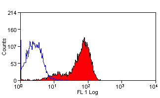
FCM (Flow Cytometry) (Flow cytometric analysis of Hela cells with KAP1 antibody at 1/50 dilution (red) compared with an unlabelled control (cells without incubation with primary antibody; black). Alexa Fluor 488-conjugated goat anti rabbit IgG was used as the secondary antibody.)

KAP1, Monoclonal Antibody (Cat# AAA30205)

Full Name
KAP1 Antibody
Gene Names
TRIM28; KAP1; TF1B; RNF96; TIF1B; PPP1R157
Reactivity
Human, Mouse, Rat
Applications
Western Blot (WB), Immunocytochemistry (ICC), Immunofluorescence (IF), Immunohistochemistry (IHC), Flow Cytometry (FC/FACS)
Purity
ProA affinity purified
Pricing
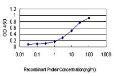
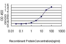
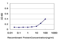
ELISA (Titration curve analysis of TIGIT mAbs to detect recombinant TIGIT in ELISA at decreasing concentrations.)

TIGIT, Monoclonal Antibody (Cat# AAA11006)

Full Name
TIGIT Antibody [4A12]
Gene Names
TIGIT; VSIG9; VSTM3; WUCAM
Reactivity
Human
Applications
ELISA (EIA), Western Blot (WB), Immunohistochemistry (IHC) Paraffin, Immunocytochemistry (ICC), Immunofluorescence (IF), Flow Cytometry (FC/FACS)
Purity
Protein A purified
Pricing
Application Data (Figure A. FITC conjugated Rat anti Mouse CD19 . Figure B. FITC conjugated Rat anti Mouse CD19 and SBV440 conjugated Rat anti Mouse CD45R . All experiments performed on mouse splenocytes in the presence of 10% mouse seru)

CD19, Monoclonal Antibody (Cat# AAA12286)

Full Name
Rat anti Mouse CD19:Amethyst Orange
Gene Names
Cd19; AW495831
Reactivity
Mouse
Applications
Flow Cytometry (FC/FACS)
Purity
Purified IgG prepared by affinity chromatography on Protein G from tissue culture supernatant
Pricing
Application Data (Immunoperoxidase staining of mouse lymph node cryosection stained with Rat antii Mouse CD8alpha antibody, clone KT15 followed by horseradish peroxidase conjugatedGoat anti Rat IgG s a detection reagent. High power)

CD8 ALPHA, Monoclonal Antibody (Cat# AAA12180)

Full Name
RAT ANTI MOUSE CD8 ALPHA
Gene Names
Cd8a; Ly-2; Ly-B; Ly-35; Lyt-2; BB154331
Applications
Immunohistology Frozen*, Flow cytometry (FC/FACS), Immunofluorescence (IF)
Pricing
Application Data (Staining of mouse peritoneal macrophages with Rat anti Mouse CD204: Alexa Fluor 488)

CD204, Monoclonal Antibody (Cat# AAA12080)

Full Name
RAT ANTI MOUSE CD204
Gene Names
Msr1; MSR; Scvr; MRS-A; MSR-A; SR-AI; SR-AII; Scara1
Applications
Immunohistology Frozen*, ELISA (EIA), Flow cytometry (FC/FACS), Immunoprecipitation (IP), Western Blot (WB)*
Pricing
Application Data (Staining of mouse peritoneal macrophages with Rat anti Mouse CD204: Alexa Fluor 488 (AAA12072A488))

CD204, Monoclonal Antibody (Cat# AAA12072)

Full Name
RAT ANTI MOUSE CD204
Gene Names
Msr1; MSR; Scvr; MRS-A; MSR-A; SR-AI; SR-AII; Scara1
Reactivity
Channel catfish, Pig
Applications
Immunohistology Frozen*, ELISA (EIA), Flow cytometry (FC/FACS), Immunoprecipitation (IP), Western Blot (WB)*
Pricing
Application Data (Staining of mouse spleen cells with Rat anti Mouse CD4)

CD4, Monoclonal Antibody (Cat# AAA11939)

Full Name
RAT ANTI MOUSE CD4
Gene Names
Cd4; L3T4; Ly-4
Applications
Immunohistology Frozen*, Flow cytometry (FC/FACS)
Pricing
Application Data (Published customer image: Histological staining of control and HBOT wounds at post-wounding days 7 and 29. A -D) H&E staining. E -H) CD34 immunohistochemistry. I+J) CD68 immunohistochemistry.From: uk B, Tong M, Fijneman EMG, van Neck JW (2014) Hyperbaric Oxygen Therapy to Treat Diabetes Impaired Wound Healing in Rats. PLoS ONE 9(10): e108533.)

CD68, Monoclonal Antibody (Cat# AAA12149)

Full Name
MOUSE ANTI RAT CD68
Applications
Immunohistology Frozen, Flow cytometry (FC/FACS)*, Immunofluorescence (IF), Immunoprecipitation (IP), Immunohistology Paraffin*
Pricing
Application Data (Staining of human peripheral blood lymphocytes with Mouse anti Human HLA ABC:Alexa Fluor 647 (AAA12013A647))

HLA ABC, Monoclonal Antibody (Cat# AAA12013)

Full Name
MOUSE ANTI HUMAN HLA ABC
Reactivity
Baboon, Bovine, Chimpanzee, Cynomolgus monkey, Gorilla, Macaque, Rhesus Monkey, Shrew
Applications
Immunohistology Frozen*, ELISA (EIA), Flow cytometry (FC/FACS), Immunofluorescence (IF), Immunoprecipitation (IP)
Pricing
Application Data (Staining of human peripheral blood granulocytes with Mouse anti Human CD11b:Biotin)

CD11b, Monoclonal Antibody (Cat# AAA11991)

Full Name
MOUSE ANTI HUMAN CD11b
Gene Names
ITGAM; CR3A; MO1A; CD11B; MAC-1; MAC1A; SLEB6
Reactivity
Baboon, Cynomolgus monkey, Rhesus Monkey
Applications
Immunohistology Frozen*, Flow cytometry (FC/FACS), Immunoprecipitation (IP)
Pricing

Malaria, Monoclonal Antibody (Cat# AAA14541)

Full Name
Malaria (pLDH) Antibody
Applications
ELISA (EIA), Lateral Flow
Purity
>90%
Pricing

Chlamydia trachomatis LPS, Monoclonal Antibody (Cat# AAA13394)

Full Name
MAb to C. trachomatis LPS
Applications
Lateral Flow Assay
Purity
Protein A chromatography
Pricing
IF (Immunofluorescence) (Immunofluorescence analysis of 293T cells by overexpressed SARS-CoV Spike Protein (Right) or not (Left) using SARS-CoV Spike Monoclonal Antibody at dilution of 1:50.)

COVID 19 Spike Protein Coronavirus, Monoclonal Antibody (Cat# AAA22149)

Full Name
SARS-COV/SARS-COV-2 Spike Monoclonal Antibody(2019-nCoV)
Reactivity
SARS-COV2
Applications
ELISA (EIA), Immunofluorescence (IF)
Pricing
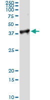
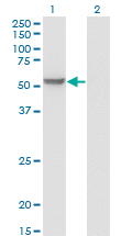
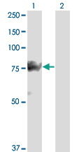
WB (Western Blot) (EHD3 monoclonal antibody. Western Blot analysis of EHD3 expression in COLO 320 HSR.)

EHD3, Monoclonal Antibody (Cat# AAA24062)

Full Name
EHD3 (EH Domain-containing Protein 3, PAST Homolog 3, EHD2, PAST3)
Gene Names
EHD3; PAST3
Reactivity
Human
Applications
ELISA (EL/EIA), Western Blot (WB), Immunohistochemistry (IHC), Immunofluorescence (IF)
Purity
Affinity Purified
Purified by Protein A affinity chromatography.
Pricing
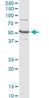
WB (Western Blot) (EIF2S1 monoclonal antibody, Western Blot analysis of EIF2S1 expression in HeLa.)

EIF2S1, Monoclonal Antibody (Cat# AAA25086)

Full Name
EIF2S1 (Eukaryotic Translation Initiation Factor 2, Subunit 1 alpha, eIF2 alpha, eIF-2-alpha, eIF-2A, eIF-2alpha, EIF2A, Eukaryotic Translation Initiation Factor 2 Subunit 1) (FITC)
Gene Names
EIF2S1; EIF2; EIF-2; EIF2A; EIF-2A; EIF-2alpha
Reactivity
Human
Applications
ELISA (EIA), Immunofluorescence (IF), Western Blot (WB)
Purity
Purified by Protein A Affinity Chromatography.
Pricing
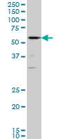
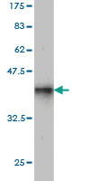
WB (Western Blot) (Western Blot detection against Immunogen (37.44kD).)

CCT7, Monoclonal Antibody (Cat# AAA25342)

Full Name
CCT7 (T-complex Protein 1 Subunit eta, TCP-1-eta, CCT-eta, HIV-1 Nef-interacting Protein, CCTH, NIP7-1) (HRP)
Gene Names
CCT7; CCTH; CCTETA; NIP7-1; TCP1ETA
Reactivity
Human, Mouse, Rat
Applications
ELISA (EIA), Western Blot (WB)
Purity
Purified by Protein A Affinity Chromatography.
Pricing
WB (Western Blot) (ACTN4 monoclonal antibody Western Blot analysis of ACTN4 expression in HeLa NE.)

ACTN4, Monoclonal Antibody (Cat# AAA25598)

Full Name
ACTN4 (Alpha-actinin-4, Non-muscle alpha-actinin 4, F-actin Cross-linking Protein) (PE)
Gene Names
ACTN4; FSGS; FSGS1; ACTININ-4
Reactivity
Human, Mouse, Rat
Applications
ELISA (EIA), Immunofluorescence (IF), Immunohistochemistry (IHC), Western Blot (WB)
Purity
Purified by Protein A Affinity Chromatography.
Pricing
WB (Western Blot) (SSH3 monoclonal antibody, Western Blot analysis of SSH3 expression in A-431.)

SSH3, Monoclonal Antibody (Cat# AAA25854)

Full Name
SSH3 (Protein Phosphatase Slingshot Homolog 3, SSH-like Protein 3, SSH3L, SSH-3L, hSSH-3L) (PE)
Gene Names
SSH3; SSH3L
Reactivity
Human
Applications
ELISA (EIA), Immunofluorescence (IF), Immunohistochemistry (IHC), Western Blot (WB)
Purity
Purified by Protein A Affinity Chromatography.
Pricing
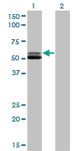
WB (Western Blot) (CAND1 monoclonal antibody (M04), clone 4D10. Western Blot analysis of CAND1 expression in Raw 264.7.)

CAND1, Monoclonal Antibody (Cat# AAA26366)

Full Name
CAND1 (Cullin-Associated and Neddylation-dissociated 1, DKFZp434M1414, FLJ10114, FLJ10929, FLJ38691, FLJ90441, KIAA0829, TIP120, TIP120A) (FITC)
Gene Names
CAND1; TIP120; TIP120A
Applications
ELISA (EIA), Immunofluorescence (IF), Western Blot (WB)
Purity
Purified
Pricing
FCM (Flow Cytometry) (Flow cytometric analysis of Hela cells with MEK2 antibody at 1/50 dilution (red) compared with an unlabelled control (cells without incubation with primary antibody; black). Alexa Fluor 488-conjugated goat anti rabbit IgG was used as the secondary antibody.)

MEK2, Monoclonal Antibody (Cat# AAA30206)

Full Name
MEK2 Antibody
Gene Names
MAP2K2; CFC4; MEK2; MKK2; MAPKK2; PRKMK2
Reactivity
Human, Mouse
Applications
Western Blot (WB), Immunocytochemistry (ICC), Immunofluorescence (IF), Immunohistochemistry (IHC), Immunoprecipitation (IP), Flow Cytometry (FC/FACS)
Purity
ProA affinity purified
Pricing

PITX1, Monoclonal Antibody (Cat# AAA24319)

Full Name
PITX1 (Pituitary Homeobox 1, Paired-like Homeodomain Transcription Factor 1, Homeobox Protein PITX1, Hindlimb-expressed Homeobox Protein Backfoot, PTX1, BFT) (AP)
Gene Names
PITX1; BFT; CCF; POTX; PTX1; LBNBG
Reactivity
Human
Applications
ELISA (EIA), Immunoprecipitation (IP), Western Blot (WB)
Purity
Purified by Protein A Affinity Chromatography.
Pricing
WB (Western Blot) (Western blot analysis of MRPL12 over-expressed 293 cell line, cotransfected with MRPL12 Validated Chimera RNAi (Lane 2) or non-transfected control (Lane 1). Blot probed with MRPL12 monoclonal antibody. GAPDH (36.1kD) used as specificity and loading control.)

MRPL12, Monoclonal Antibody (Cat# AAA24575)

Full Name
MRPL12 (Mitochondrial Ribosomal Protein L12, 39S Ribosomal Protein L12, Mitochondrial, 5c5-2, FLJ60124, L12mt, MGC8610, MRP-L12, MRP-L31/34, MRPL7, MRPL7/L12, RPML12) APC
Gene Names
MRPL12; 5c5-2; L12mt; MRPL7; RPML12; MRPL7/L12; MRP-L31/34
Reactivity
Human
Applications
ELISA (EIA), Immunofluorescence (IF), Immunohistochemistry (IHC), Western Blot (WB)
Purity
Purified by Protein A Affinity Chromatography.
Pricing
Application Data (Figure A. Alexa Fluor 488 conjugated Rat anti Mouse CD4 . Figure B. Alexa Fluor 488 conjugated Rat anti Mouse CD4 and StarBright Violet 610 conjugated Rat anti Mouse CD3 . All experiments performed on red blood lys)

CD4, Monoclonal Antibody (Cat# AAA12287)

Full Name
Rat anti Mouse CD4:Amethyst Orange
Gene Names
Cd4; L3T4; Ly-4
Reactivity
Mouse
Applications
ELISA (EIA)
Purity
Purified IgG prepared by affinity chromatography on Protein G from tissue culture supernatant
Pricing
FCM (Flow Cytometry) (Figure 6. Flow Cytometry analysis of A431 cells using anti-Cytokeratin 5 antibody (AAA19666).Overlay histogram showing A431 cells stained with AAA19666 (Blue line). The cells were blocked with 10% normal goat serum. And then incubated with mouse anti-Cytokeratin 5 Antibody (AAA19666, 1 ug/1x10^6 cells) for 30 min at 20 degree C. DyLight488 conjugated goat anti-mouse IgG was used as secondary antibody for 30 minutes at 20 degree C. Isotype control antibody (Green line) was mouse IgG (1 ug/1x10^6) used under the same conditions. Unlabelled sample (Red line) was also used as a control.)

Cytokeratin 5, Monoclonal Antibody (Cat# AAA19666)

Full Name
Anti-Cytokeratin 5 Antibody Picoband (monoclonal, 9H2F8)
Gene Names
KRT5; K5; CK5; DDD; EBS2; KRT5A
Reactivity
Human
Applications
Western Blot (WB), Immunohistochemistry (IHC), Immunocytochemistry (ICC), Immunofluorescence (IF), Flow Cytometry (FC/FACS)
Purity
Immunogen affinity purified.
Pricing
IF (Immunofluorescence) (Figure 11. IF analysis of AREB6/ZEB1 using anti-AREB6/ZEB1 antibody (AAA19667).AREB6/ZEB1 was detected in a paraffin-embedded section of human glioma tissue. Heat mediated antigen retrieval was performed in EDTA buffer (pH 8.0, epitope retrieval solution). The tissue section was blocked with 10% goat serum. The tissue section was then incubated with 5 ug/mL mouse anti-AREB6/ZEB1 Antibody (AAA19667) overnight at 4 degree C. Biotin conjugated goat anti-mouse IgG (BA1001) was used as secondary antibody and incubated for 30 minutes at 37 degree C. The tissue section was developed using DyLight550 Conjugated Avidin (BA1134). The section was counterstained with DAPI. Visualize using a fluorescence microscope and filter sets appropriate for the label used.)

AREB6/ZEB1, Monoclonal Antibody (Cat# AAA19667)

Full Name
Anti-AREB6/ZEB1 Antibody Picoband (monoclonal, 8B12D7)
Gene Names
ZEB1; BZP; TCF8; AREB6; FECD6; NIL2A; PPCD3; ZFHEP; ZFHX1A; DELTAEF1
Reactivity
Human, Mouse, Rat
Applications
Immunofluorescence (IF), Immunohistochemistry (IHC), Western Blot (WB)
Purity
Immunogen affinity purified.
Pricing
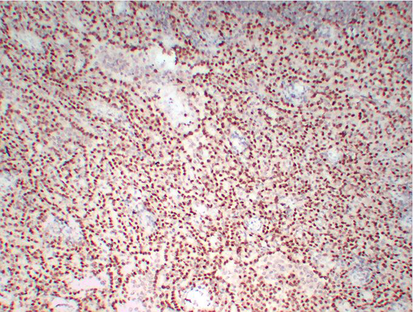
IHC (Immunohistochemistry) (Figure 7. IHC analysis of Poliovirus Receptor/PVR using anti-Poliovirus Receptor/PVR antibody (AAA19668).Poliovirus Receptor/PVR was detected in a paraffin-embedded section of mouse liver tissue. Heat mediated antigen retrieval was performed in EDTA buffer (pH 8.0, epitope retrieval solution). The tissue section was blocked with 10% goat serum. The tissue section was then incubated with 2 ug/ml mouse anti-Poliovirus Receptor/PVR Antibody (AAA19668) overnight at 4 degree C. Peroxidase Conjugated Goat Anti-mouse IgG was used as secondary antibody and incubated for 30 minutes at 37 degree C. The tissue section was developed using HRP Conjugated Mouse IgG Super Vision Assay Kit with DAB as the chromogen.)

Receptor/PVR, Monoclonal Antibody (Cat# AAA19668)

Full Name
Anti-Poliovirus Receptor/PVR Antibody Picoband (monoclonal, 9B9F1)
Gene Names
PVR; PVS; HVED; CD155; NECL5; TAGE4; Necl-5
Reactivity
Human, Mouse
Applications
Western Blot (WB), Immunohistochemistry (IHC)
Purity
Immunogen affinity purified.
Pricing
FCM (Flow Cytometry) (Figure 9. Flow Cytometry analysis of PC-3 cells using anti-Poliovirus Receptor/PVR antibody (AAA19669).Overlay histogram showing PC-3 cells stained with AAA19669 (Blue line). The cells were blocked with 10% normal goat serum. And then incubated with mouse anti-Poliovirus Receptor/PVR Antibody (AAA19669, 1 ug/1x10^6 cells) for 30 min at 20 degree C. DyLight488 conjugated goat anti-mouse IgG was used as secondary antibody for 30 minutes at 20 degree C. Isotype control antibody (Green line) was mouse IgG (1 ug/1x10^6) used under the same conditions. Unlabelled sample (Red line) was also used as a control.)

Receptor/PVR, Monoclonal Antibody (Cat# AAA19669)

Full Name
Anti-Poliovirus Receptor/PVR Antibody Picoband (monoclonal, 5I13D1)
Gene Names
PVR; PVS; HVED; CD155; NECL5; TAGE4; Necl-5
Reactivity
Human, Mouse
Applications
Western Blot (WB), Immunohistochemistry (IHC), Flow Cytometry (FC/FACS)
Purity
Immunogen affinity purified.
Pricing
FCM (Flow Cytometry) (Figure 6. Flow Cytometry analysis of HeLa cells using anti-PARK7/DJ1 antibody (AAA19670).Overlay histogram showing HeLa cells stained with AAA19670 (Blue line). The cells were blocked with 10% normal goat serum. And then incubated with mouse anti-PARK7/DJ1 Antibody (AAA19670, 1 ug/1x10^6 cells) for 30 min at 20 degree C. DyLight488 conjugated goat anti-mouse IgG was used as secondary antibody for 30 minutes at 20 degree C. Isotype control antibody (Green line) was mouse IgG (1 ug/1x10^6) used under the same conditions. Unlabelled sample (Red line) was also used as a control.)

PARK7/DJ1, Monoclonal Antibody (Cat# AAA19670)

Full Name
Anti-PARK7/DJ1 Antibody Picoband (monoclonal, 4B10)
Gene Names
PARK7; DJ1; DJ-1
Reactivity
Human
Applications
Western Blot (WB), Immunohistochemistry (IHC), Immunocytochemistry (ICC), Immunofluorescence (IF), Flow Cytometry (FC/FACS)
Purity
Immunogen affinity purified.
Pricing
FCM (Flow Cytometry) (Figure 6. Flow Cytometry analysis of PC-3 cells using anti-YY1 antibody (AAA19672).Overlay histogram showing PC-3 cells stained with AAA19672 (Blue line). The cells were blocked with 10% normal goat serum. And then incubated with mouse anti-YY1 Antibody (AAA19672, 1 ug/1x10^6 cells) for 30 min at 20 degree C. DyLight488 conjugated goat anti-mouse IgG was used as secondary antibody for 30 minutes at 20 degree C. Isotype control antibody (Green line) was mouse IgG (1 ug/1x10^6) used under the same conditions. Unlabelled sample (Red line) was also used as a control.)

YY1, Monoclonal Antibody (Cat# AAA19672)

Full Name
Anti-YY1 Antibody Picoband (monoclonal, 2C10F9)
Gene Names
YY1; DELTA; NF-E1; UCRBP; INO80S; YIN-YANG-1
Reactivity
Human, Mouse, Rat
Applications
Western Blot (WB), Immunohistochemistry (IHC), Flow Cytometry (FC/FACS)
Purity
Immunogen affinity purified.
Pricing
FCM (Flow Cytometry) (Figure 9. Flow Cytometry analysis of A431 cells using anti-YY1 antibody (AAA19673).Overlay histogram showing A431 cells stained with AAA19673 (Blue line). The cells were blocked with 10% normal goat serum. And then incubated with mouse anti-YY1 Antibody (AAA19673, 1 ug/1x10^6 cells) for 30 min at 20 degree C. DyLight488 conjugated goat anti-mouse IgG was used as secondary antibody for 30 minutes at 20 degree C. Isotype control antibody (Green line) was mouse IgG (1 ug/1x10^6) used under the same conditions. Unlabelled sample (Red line) was also used as a control.)

YY1, Monoclonal Antibody (Cat# AAA19673)

Full Name
Anti-YY1 Antibody Picoband (monoclonal, 3F3E7)
Gene Names
YY1; DELTA; NF-E1; UCRBP; INO80S; YIN-YANG-1
Reactivity
Human, Mouse, Rat
Applications
Western Blot (WB), Immunohistochemistry (IHC), Immunocytochemistry (ICC), Immunofluorescence (IF), Flow Cytometry (FC/FACS)
Purity
Immunogen affinity purified.
Pricing
FCM (Flow Cytometry) (Figure 7. Flow Cytometry analysis of Caco-2 cells using anti-SHP1/PTPN6 antibody (AAA19675).Overlay histogram showing Caco-2 cells stained with AAA19675 (Blue line). The cells were blocked with 10% normal goat serum. And then incubated with mouse anti-SHP1/PTPN6 Antibody (AAA19675, 1 ug/1x10^6 cells) for 30 min at 20 degree C. DyLight488 conjugated goat anti-mouse IgG was used as secondary antibody for 30 minutes at 20 degree C. Isotype control antibody (Green line) was mouse IgG (1 ug/1x10^6) used under the same conditions. Unlabelled sample (Red line) was also used as a control.)

SHP1/PTPN6, Monoclonal Antibody (Cat# AAA19675)

Full Name
Anti-SHP1/PTPN6 Antibody Picoband (monoclonal, 8H11B10)
Gene Names
PTPN6; HCP; HCPH; SHP1; SHP-1; HPTP1C; PTP-1C; SHP-1L; SH-PTP1
Reactivity
Human, Mouse, Rat
Applications
Flow Cytometry (FC/FACS), Immunofluorescence (IF), Immunohistochemistry (IHC), Immunocytochemistry (ICC), Western Blot (WB)
Purity
Immunogen affinity purified.
Pricing
FCM (Flow Cytometry) (Figure 9. Flow Cytometry analysis of Caco-2 cells using anti-HGS antibody (AAA19678).Overlay histogram showing Caco-2 cells stained with AAA19678 (Blue line). The cells were blocked with 10% normal goat serum. And then incubated with mouse anti-HGS Antibody (AAA19678, 1 ug/1x10^6 cells) for 30 min at 20 degree C. DyLight488 conjugated goat anti-mouse IgG was used as secondary antibody for 30 minutes at 20 degree C. Isotype control antibody (Green line) was mouse IgG (1 ug/1x10^6) used under the same conditions. Unlabelled sample (Red line) was also used as a control.)

HGS, Monoclonal Antibody (Cat# AAA19678)

Full Name
Anti-HGS Antibody Picoband (monoclonal, 6C2E1)
Gene Names
HGS; HRS
Reactivity
Human, Mouse, Rat
Applications
Western Blot (WB), Immunohistochemistry (IHC), Flow Cytometry (FC/FACS)
Purity
Immunogen affinity purified.
Pricing

PAI-1, Monoclonal Antibody (Cat# AAA27729)

Full Name
Recombinant Anti-PAI-1 Antibody, Rabbit Monoclonal
Gene Names
SERPINE1; PAI; PAI1; PAI-1; PLANH1
Applications
ELISA (EIA), Sandwich ELISA (Det)
Pricing
FCM (Flow Cytometry) (Figure 10. Flow Cytometry analysis of A431 cells using anti-SP1 antibody (AAA19656).Overlay histogram showing A431 cells stained with AAA19656 (Blue line). The cells were blocked with 10% normal goat serum. And then incubated with mouse anti-SP1 Antibody (AAA19656, 1 ug/1x10^6 cells) for 30 min at 20 degree C. DyLight488 conjugated goat anti-mouse IgG was used as secondary antibody for 30 minutes at 20 degree C. Isotype control antibody (Green line) was mouse IgG (1 ug/1x10^6) used under the same conditions. Unlabelled sample (Red line) was also used as a control.)

SP1, Monoclonal Antibody (Cat# AAA19656)

Full Name
Anti-SP1 Antibody Picoband (monoclonal, 3C4C3)
Reactivity
Human
Applications
Western Blot (WB), Immunohistochemistry (IHC), Immunocytochemistry (ICC), Immunofluorescence (IF), Flow Cytometry (FC/FACS)
Purity
Immunogen affinity purified.
Pricing
FCM (Flow Cytometry) (Figure 6. Flow Cytometry analysis of Caco-2 cells using anti-ICAM1 antibody (AAA19659).Overlay histogram showing Caco-2 cells stained with AAA19659 (Blue line). The cells were blocked with 10% normal goat serum. And then incubated with mouse anti-ICAM1 Antibody (AAA19659, 1 ug/1x10^6 cells) for 30 min at 20 degree C. DyLight488 conjugated goat anti-mouse IgG was used as secondary antibody for 30 minutes at 20 degree C. Isotype control antibody (Green line) was mouse IgG (1 ug/1x10^6) used under the same conditions. Unlabelled sample (Red line) was also used as a control.)

ICAM1, Monoclonal Antibody (Cat# AAA19659)

Full Name
Anti-ICAM1 Antibody Picoband (monoclonal, 6F2C3)
Gene Names
ICAM1; BB2; CD54; P3.58
Reactivity
Human
Applications
Western Blot (WB), Immunohistochemistry (IHC), Flow Cytometry (FC/FACS)
Purity
Immunogen affinity purified.
Pricing
FCM (Flow Cytometry) (Figure 10. Flow Cytometry analysis of SiHa cells using anti-CD147/Emmprin antibody (AAA19661).Overlay histogram showing SiHa cells stained with AAA19661 (Blue line). The cells were blocked with 10% normal goat serum. And then incubated with mouse anti-CD147/Emmprin Antibody (AAA19661, 1 ug/1x10^6 cells) for 30 min at 20 degree C. DyLight488 conjugated goat anti-mouse IgG was used as secondary antibody for 30 minutes at 20 degree C. Isotype control antibody (Green line) was mouse IgG (1 ug/1x10^6) used under the same conditions. Unlabelled sample (Red line) was also used as a control.)

CD147/Emmprin, Monoclonal Antibody (Cat# AAA19661)

Full Name
Anti-CD147/Emmprin Antibody Picoband (monoclonal, 7H5E7)
Gene Names
BSG; M6; OK; 5F7; TCSF; CD147; EMMPRIN
Reactivity
Human, Mouse
Applications
Western Blot (WB), Immunohistochemistry-Paraffin (IHC-P), Immunofluorescence (IF), Flow Cytometry (FC/FACS), Immunocytochemistry (ICC)
Purity
Immunogen affinity purified.
Pricing
IF (Immunofluorescence) (Figure 8. IF analysis of CD147/Emmprin using anti-CD147/Emmprin antibody (AAA19662).CD147/Emmprin was detected in a paraffin-embedded section of human esophageal squamous carcinoma tissue. Heat mediated antigen retrieval was performed in EDTA buffer (pH 8.0, epitope retrieval solution). The tissue section was blocked with 10% goat serum. The tissue section was then incubated with 5 ug/mL mouse anti-CD147/Emmprin Antibody (AAA19662) overnight at 4 degree C. Biotin conjugated goat anti-mouse IgG (BA1001) was used as secondary antibody and incubated for 30 minutes at 37 degree C. The tissue section was developed using DyLight488 Conjugated Avidin (BA1128). The section was counterstained with DAPI. Visualize using a fluorescence microscope and filter sets appropriate for the label used.)

CD147/Emmprin, Monoclonal Antibody (Cat# AAA19662)

Full Name
Anti-CD147/Emmprin Antibody Picoband (monoclonal, 6H2B2)
Gene Names
BSG; M6; OK; 5F7; TCSF; CD147; EMMPRIN
Reactivity
Human, Mouse
Applications
Western Blot (WB), Immunohistochemistry-Paraffin (IHC-P), Immunofluorescence (IF), Immunocytochemistry (ICC)
Purity
Immunogen affinity purified.
Pricing

HBsAg, Monoclonal Antibody (Cat# AAA14288)

Full Name
HBsAg antibody
Applications
ELISA (EIA)
Pricing
FCM (Flow Cytometry) (Figure 6. Flow Cytometry analysis of K562 cells using anti-D2AP antibody (M01756). Overlay histogram showing K562 cells stained with M01756 (Blue line).The cells were blocked with 10% normal goat serum. And then incubated with mouse anti-D2AP Antibody (M01756, 1ug/1x106 cells) for 30 min at 20 degree C. DyLight488 conjugated goat anti-mouse IgG (BA1126, 5-10ug/1x106 cells) was used as secondary antibody for 30 minutes at 20 degree C. Isotype control antibody (Green line) was mouse IgG (1ug/1x106) used under the same conditions. Unlabelled sample (Red line) was also used as a control.)

CD2AP, Monoclonal Antibody (Cat# AAA19185)

Full Name
Anti-CD2AP Antibody (monoclonal, 5F8)
Gene Names
CD2AP; CMS
Reactivity
Human, Mouse, Rat
Applications
Western Blot (WB), Immunohistochemistry (IHC) Paraffin, Flow Cytometry (FC/FACS)
Purity
Immunogen Affinity Purified
Pricing
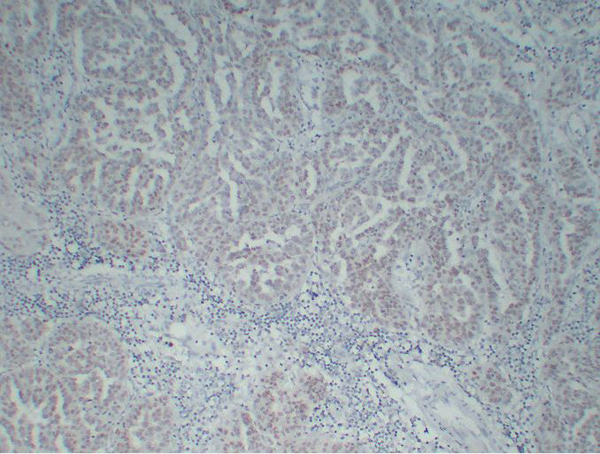
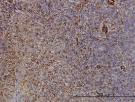
IHC (Immunohistchemistry) (Figure 6. IHC analysis of DHODH using anti-DHODH antibody (AAA19712).DHODH was detected in a paraffin-embedded section of human renal carcinoma tissue. Heat mediated antigen retrieval was performed in EDTA buffer (pH 8.0, epitope retrieval solution). The tissue section was blocked with 10% goat serum. The tissue section was then incubated with 2 ug/ml mouse anti-DHODH Antibody (AAA19712) overnight at 4 degree C. Biotinylated goat anti-mouse IgG was used as secondary antibody and incubated for 30 minutes at 37 degree C. The tissue section was developed using Strepavidin-Biotin-Complex (SABC) (#SA1021) with DAB as the chromogen.)

DHODH, Monoclonal Antibody (Cat# AAA19712)

Full Name
Anti-DHODH Antibody Picoband (monoclonal, 2G7)
Gene Names
DHODH; URA1; POADS; DHOdehase
Reactivity
Human, Mouse, Rat
Applications
Western Blot (WB), Immunohistochemistry (IHC)
Purity
Immunogen affinity purified.
Pricing
FCM (Flow Cytometry) (Figure 7. Flow Cytometry analysis of JK cells using anti-ERLIN2 antibody (AAA19715).Overlay histogram showing JK cells stained with AAA19715 (Blue line). The cells were blocked with 10% normal goat serum. And then incubated with mouse anti-ERLIN2 Antibody (AAA19715, 1 ug/1x10^6 cells) for 30 min at 20 degree C. DyLight488 conjugated goat anti-mouse IgG was used as secondary antibody for 30 minutes at 20 degree C. Isotype control antibody (Green line) was mouse IgG (1 ug/1x10^6) used under the same conditions. Unlabelled sample (Red line) was also used as a control.)

Erlin-2/ERLIN2, Monoclonal Antibody (Cat# AAA19715)

Full Name
Anti-Erlin-2/ERLIN2 Antibody Picoband (monoclonal, 3H9A2)
Gene Names
ERLIN2; NET32; SPFH2; SPG18; C8orf2; Erlin-2
Reactivity
Human
Applications
Western Blot (WB), Immunohistochemistry (IHC), Immunocytochemistry (ICC), Immunofluorescence (IF), Flow Cytometry (FC/FACS)
Purity
Immunogen affinity purified.
Pricing

Submit a Question

Please complete the form below and a representative will contact you as soon as possible.

Request more Information

Please complete the form below and a representative will contact you as soon as possible.

Request a Manual

Please complete the form below and a representative will contact you as soon as possible.

Request a Quote

Please complete the form below and a representative will contact you as soon as possible.