Filters
▼Clonality
▼Type
▼Reactivity
▼Gene Name
▼Isotype
▼Host
▼Application
▼Clone
▼Monoclonal Antibodies
Get accurate results in your research with our Monoclonal Antibodies, which are specially made to target exactly what you require for your research, and will produce consistent, reliable performance in lab tests.
Viewing 1450-1500 of 27645 product results
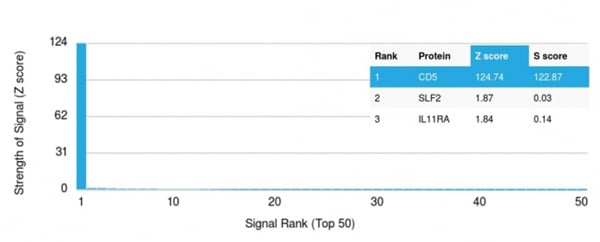 Application Data
(Analysis of Protein Array containing more than 19,000 full-length human proteins using CD5Mouse Monoclonal Antibody (CD5/2418). Z- and S- Score: The Z-score represents the strength of a signal that a monoclonal antibody (MAb) (in combination with a fluorescently-tagged anti-IgG secondary antibody) produces when binding to a particular protein on the HuProtTM array. Z-scores are described in units of standard deviations (SD's) above the mean value of all signals generated on that array. If targets on HuProtTM are arranged in descending order of the Z-score, the S-score is the difference (also in units of SD's) between the Z-score. S-score therefore represents the relative target specificity of a MAb to its intended target. A MAb is considered to specific to its intended target, if the MAb has an S-score of at least 2.5. For example, if a MAb binds to protein X with a Z-score of 43 and to protein Y with a Z-score of 14, then the S-score for the binding of that MAb to protein X is equal to 29.)
Application Data
(Analysis of Protein Array containing more than 19,000 full-length human proteins using CD5Mouse Monoclonal Antibody (CD5/2418). Z- and S- Score: The Z-score represents the strength of a signal that a monoclonal antibody (MAb) (in combination with a fluorescently-tagged anti-IgG secondary antibody) produces when binding to a particular protein on the HuProtTM array. Z-scores are described in units of standard deviations (SD's) above the mean value of all signals generated on that array. If targets on HuProtTM are arranged in descending order of the Z-score, the S-score is the difference (also in units of SD's) between the Z-score. S-score therefore represents the relative target specificity of a MAb to its intended target. A MAb is considered to specific to its intended target, if the MAb has an S-score of at least 2.5. For example, if a MAb binds to protein X with a Z-score of 43 and to protein Y with a Z-score of 14, then the S-score for the binding of that MAb to protein X is equal to 29.)
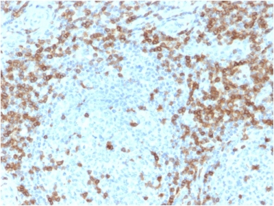
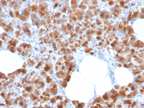
CD5, Monoclonal Antibody (Cat# AAA214846)
 IHC (Immunohistochemisry)
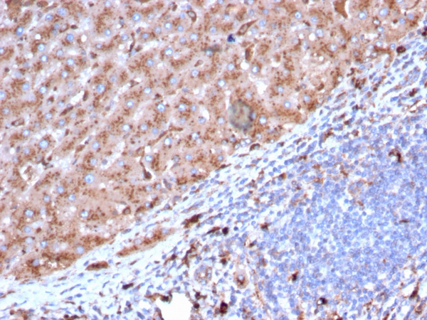
(Formalin-fixed, paraffin-embedded human Tonsil stained withCD163 Mouse Monoclonal Antibody (M130/2164).)
IHC (Immunohistochemisry)
(Formalin-fixed, paraffin-embedded human Tonsil stained withCD163 Mouse Monoclonal Antibody (M130/2164).)
CD163, Monoclonal Antibody (Cat# AAA214849)
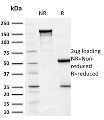
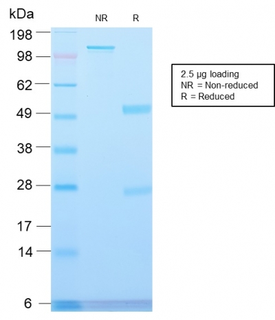
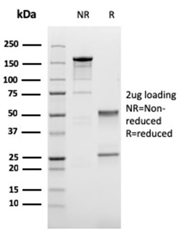
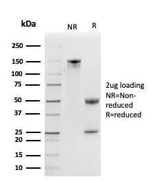
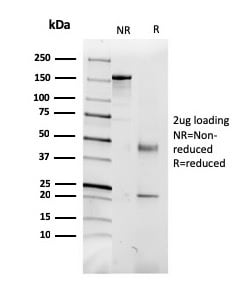
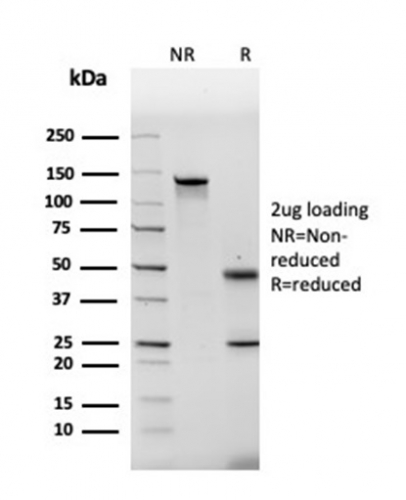
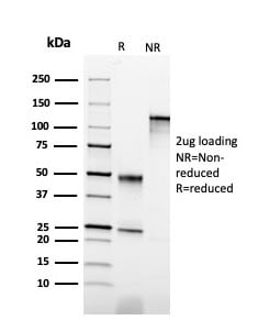
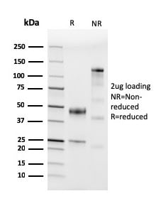
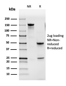
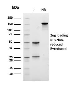
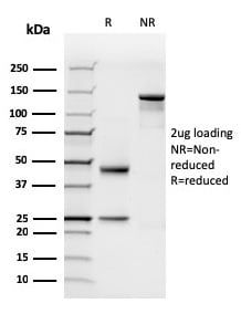
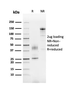
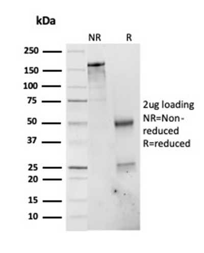
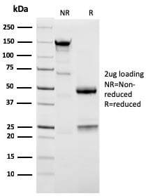
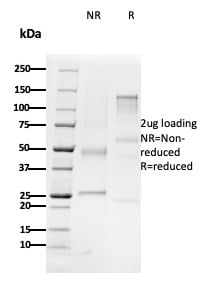
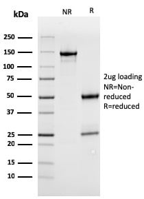
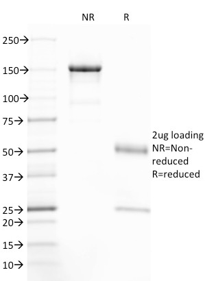
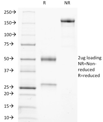
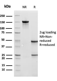
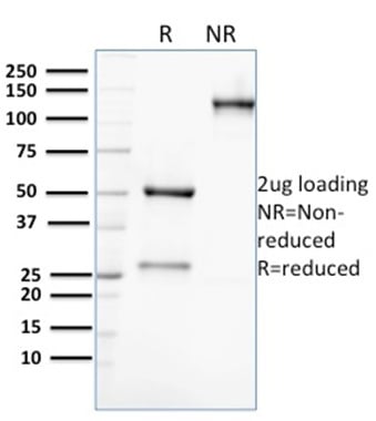
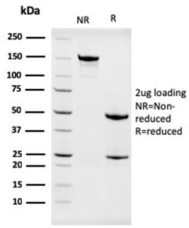
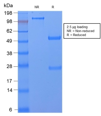 SDS-PAGE
(SDS-PAGE Analysis Purified CD59 Rabbit Recombinant Monoclonal Antibody (MACIF/2867R). Confirmation of Purity and Integrity of Antibody.)
SDS-PAGE
(SDS-PAGE Analysis Purified CD59 Rabbit Recombinant Monoclonal Antibody (MACIF/2867R). Confirmation of Purity and Integrity of Antibody.)
CD59/Complement Regulatory Protein/Protectin, Monoclonal Antibody (Cat# AAA214854)
Does not react with baboon, horse. Others not tested.
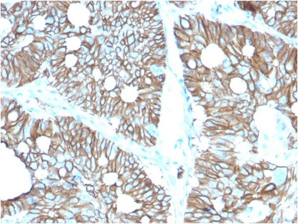
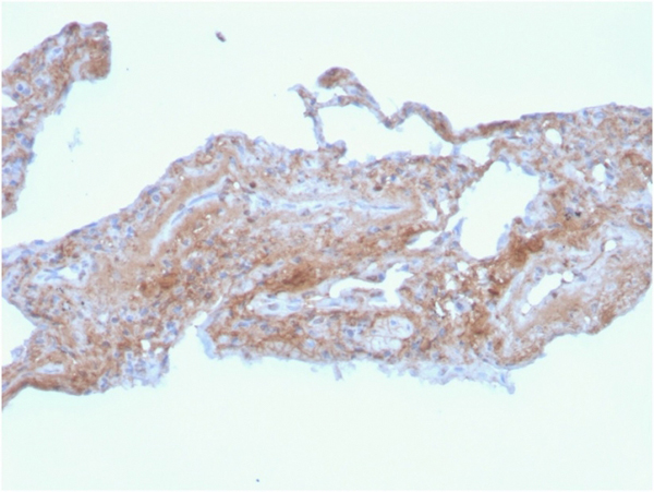
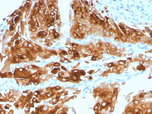
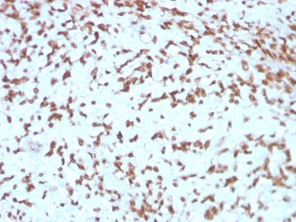
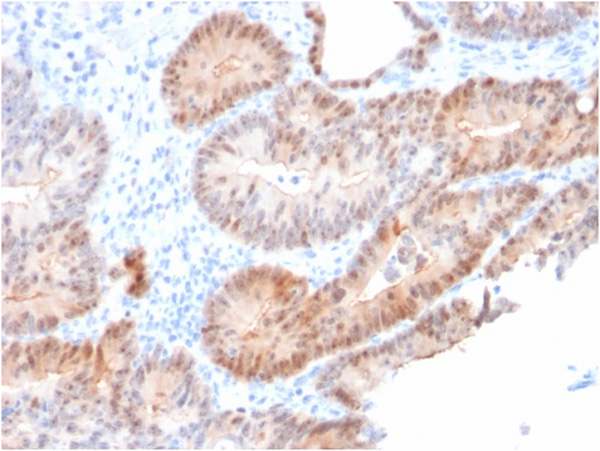
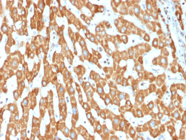
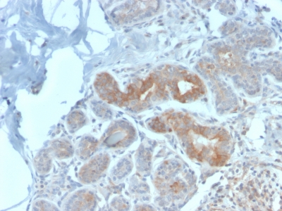 IHC (Immunohiostchemistry)
(Formalin-fixed, paraffin-embedded human Breast Carcinoma stained with Major Vault Protein Rabbit Recombinant Monoclonal Antibody (VP2897R).)
IHC (Immunohiostchemistry)
(Formalin-fixed, paraffin-embedded human Breast Carcinoma stained with Major Vault Protein Rabbit Recombinant Monoclonal Antibody (VP2897R).)
Major Vault Protein (MVP), Monoclonal Antibody (Cat# AAA214860)
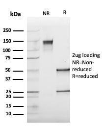
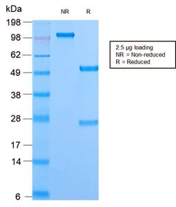 SDS-PAGE
(SDS-PAGE Analysis Purified Mitochondria Rabbit Recombinant Monoclonal (MTC02/2860R). Confirmation of Purity and Integrity of Antibody)
SDS-PAGE
(SDS-PAGE Analysis Purified Mitochondria Rabbit Recombinant Monoclonal (MTC02/2860R). Confirmation of Purity and Integrity of Antibody)
Mitochondria, Monoclonal Antibody (Cat# AAA214866)
Does not react with mouse, rat. Others not known
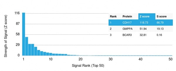 Application Data
(Analysis of Protein Array containing more than 19,000 full-length human proteins using Cadherin 17 (CDH17) Mouse Monoclonal Antibody (CDH17/2616). Z- and S- Score: The Z-score represents the strength of a signal that a monoclonal antibody (Monoclonal Antibody) (in combination with a fluorescently-tagged anti-IgG secondary antibody) produces when binding to a particular protein on the HuProtTM array. Z-scores are described in units of standard deviations (SD's) above the mean value of all signals generated on that array. If targets on HuProtTM are arranged in descending order of the Z-score, the S-score is the difference (also in units of SD's) between the Z-score. S-score therefore represents the relative target specificity of a Monoclonal Antibody to its intended target. A Monoclonal Antibody is considered to specific to its intended target, if the Monoclonal Antibody has an S-score of at least 2.5. For example, if a Monoclonal Antibody binds to protein X with a Z-score of 43 and to protein Y with a Z-score of 14, then the S-score for the binding of that Monoclonal Antibody to protein X is equal to 29.)
Application Data
(Analysis of Protein Array containing more than 19,000 full-length human proteins using Cadherin 17 (CDH17) Mouse Monoclonal Antibody (CDH17/2616). Z- and S- Score: The Z-score represents the strength of a signal that a monoclonal antibody (Monoclonal Antibody) (in combination with a fluorescently-tagged anti-IgG secondary antibody) produces when binding to a particular protein on the HuProtTM array. Z-scores are described in units of standard deviations (SD's) above the mean value of all signals generated on that array. If targets on HuProtTM are arranged in descending order of the Z-score, the S-score is the difference (also in units of SD's) between the Z-score. S-score therefore represents the relative target specificity of a Monoclonal Antibody to its intended target. A Monoclonal Antibody is considered to specific to its intended target, if the Monoclonal Antibody has an S-score of at least 2.5. For example, if a Monoclonal Antibody binds to protein X with a Z-score of 43 and to protein Y with a Z-score of 14, then the S-score for the binding of that Monoclonal Antibody to protein X is equal to 29.)
Cadherin 17/LI Cadherin, Monoclonal Antibody (Cat# AAA214873)
 Application Data
(Analysis of Protein Array containing more than 19,000 full-length human proteins using Kallikrein 5 Mouse Monoclonal Antibody (KLK5/3841) Z- and S- Score: The Z-score represents the strength of a signal that a monoclonal antibody (MAb) (in combination with a fluorescently-tagged anti-IgG secondary antibody) produces when binding to a particular protein on the HuProtTM array. Z-scores are described in units of standard deviations (SD's) above the mean value of all signals generated on that array. If targets on HuProtTM are arranged in descending order of the Z-score, the S-score is the difference (also in units of SD's) between the Z-score. S-score therefore represents the relative target specificity of a MAb to its intended target. A MAb is considered to specific to its intended target, if the MAb has an S-score of at least 2.5. For example, if a MAb binds to protein X with a Z-score of 43 and to protein Y with a Z-score of 14, then the S-score for the binding of that MAb to protein X is equal to 29.)
Application Data
(Analysis of Protein Array containing more than 19,000 full-length human proteins using Kallikrein 5 Mouse Monoclonal Antibody (KLK5/3841) Z- and S- Score: The Z-score represents the strength of a signal that a monoclonal antibody (MAb) (in combination with a fluorescently-tagged anti-IgG secondary antibody) produces when binding to a particular protein on the HuProtTM array. Z-scores are described in units of standard deviations (SD's) above the mean value of all signals generated on that array. If targets on HuProtTM are arranged in descending order of the Z-score, the S-score is the difference (also in units of SD's) between the Z-score. S-score therefore represents the relative target specificity of a MAb to its intended target. A MAb is considered to specific to its intended target, if the MAb has an S-score of at least 2.5. For example, if a MAb binds to protein X with a Z-score of 43 and to protein Y with a Z-score of 14, then the S-score for the binding of that MAb to protein X is equal to 29.)
Kallikrein 5 (KLK5), Monoclonal Antibody (Cat# AAA215354)
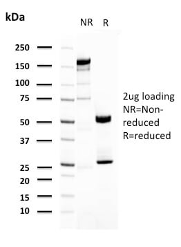
 SDS-PAGE
(SDS-PAGE Analysis Purified CD74 Recombinant Mouse Monoclonal Antibody (rCLIP/813). Confirmation of Purity and Integrity of Antibody.)
SDS-PAGE
(SDS-PAGE Analysis Purified CD74 Recombinant Mouse Monoclonal Antibody (rCLIP/813). Confirmation of Purity and Integrity of Antibody.)
CD74, Monoclonal Antibody (Cat# AAA215359)
Does not react with Rat
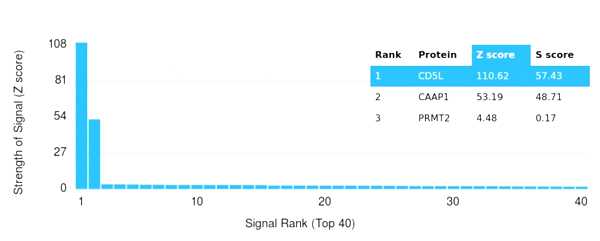 Application Data
(Analysis of Protein Array containing more than 19,000 full-length human proteins using CD5L Mouse Monoclonal Antibody (CD5L/2991) Z- and S- Score: The Z-score represents the strength of a signal that a monoclonal antibody (MAb) (in combination with a fluorescently-tagged anti-IgG secondary antibody) produces when binding to a particular protein on the HuProtTM array. Z-scores are described in units of standard deviations (SD's) above the mean value of all signals generated on that array. If targets on HuProtTM are arranged in descending order of the Z-score, the S-score is the difference (also in units of SD's) between the Z-score. S-score therefore represents the relative target specificity of a MAb to its intended target. A MAb is considered to specific to its intended target, if the MAb has an S-score of at least 2.5. For example, if a MAb binds to protein X with a Z-score of 43 and to protein Y with a Z-score of 14, then the S-score for the binding of that MAb to protein X is equal to 29.)
Application Data
(Analysis of Protein Array containing more than 19,000 full-length human proteins using CD5L Mouse Monoclonal Antibody (CD5L/2991) Z- and S- Score: The Z-score represents the strength of a signal that a monoclonal antibody (MAb) (in combination with a fluorescently-tagged anti-IgG secondary antibody) produces when binding to a particular protein on the HuProtTM array. Z-scores are described in units of standard deviations (SD's) above the mean value of all signals generated on that array. If targets on HuProtTM are arranged in descending order of the Z-score, the S-score is the difference (also in units of SD's) between the Z-score. S-score therefore represents the relative target specificity of a MAb to its intended target. A MAb is considered to specific to its intended target, if the MAb has an S-score of at least 2.5. For example, if a MAb binds to protein X with a Z-score of 43 and to protein Y with a Z-score of 14, then the S-score for the binding of that MAb to protein X is equal to 29.)
CD5L/CD5LG/CD5, Monoclonal Antibody (Cat# AAA215361)
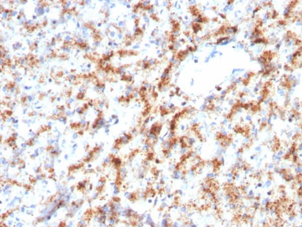
 IHC (Immunohiostchemistry)
(Formalin-fixed, paraffin-embedded human breast carcinoma stained with TNFAIP3 Mouse Monoclonal Antibody (TNFAIP3/2813).)
IHC (Immunohiostchemistry)
(Formalin-fixed, paraffin-embedded human breast carcinoma stained with TNFAIP3 Mouse Monoclonal Antibody (TNFAIP3/2813).)
A20/TNFAIP3, Monoclonal Antibody (Cat# AAA215364)
 Application Data
(Analysis of Protein Array containing more than 19,000 full-length human proteins using Fibronectin Mouse Monoclonal Antibody (FN1/2948). Z- and S- Score: The Z-score represents the strength of a signal that a monoclonal antibody (MAb) (in combination with a fluorescently-tagged anti-IgG secondary antibody) produces when binding to a particular protein on the HuProtTM array. Z-scores are described in units of standard deviations (SD's) above the mean value of all signals generated on that array. If targets on HuProtTM are arranged in descending order of the Z-score, the S-score is the difference (also in units of SD's) between the Z-score. S-score therefore represents the relative target specificity of a MAb to its intended target. A MAb is considered to specific to its intended target, if the MAb has an S-score of at least 2.5. For example, if a MAb binds to protein X with a Z-score of 43 and to protein Y with a Z-score of 14, then the S-score for the binding of that MAb to protein X is equal to 29.)
Application Data
(Analysis of Protein Array containing more than 19,000 full-length human proteins using Fibronectin Mouse Monoclonal Antibody (FN1/2948). Z- and S- Score: The Z-score represents the strength of a signal that a monoclonal antibody (MAb) (in combination with a fluorescently-tagged anti-IgG secondary antibody) produces when binding to a particular protein on the HuProtTM array. Z-scores are described in units of standard deviations (SD's) above the mean value of all signals generated on that array. If targets on HuProtTM are arranged in descending order of the Z-score, the S-score is the difference (also in units of SD's) between the Z-score. S-score therefore represents the relative target specificity of a MAb to its intended target. A MAb is considered to specific to its intended target, if the MAb has an S-score of at least 2.5. For example, if a MAb binds to protein X with a Z-score of 43 and to protein Y with a Z-score of 14, then the S-score for the binding of that MAb to protein X is equal to 29.)
Fibronectin, Monoclonal Antibody (Cat# AAA215365)
 SDS-PAGE
(SDS-PAGE Analysis Purified Apolipoprotein A1 Mouse Monoclonal Antibody (APOA1/3661). Confirmation of Purity and Integrity of Antibody.)
SDS-PAGE
(SDS-PAGE Analysis Purified Apolipoprotein A1 Mouse Monoclonal Antibody (APOA1/3661). Confirmation of Purity and Integrity of Antibody.)
Apolipoprotein A1/APOA1, Monoclonal Antibody (Cat# AAA215366)
 SDS-PAGE
(SDS-PAGE Analysis Purified Cytokeratin 6B (KRT6B) Monoclonal Antibody (KRT6B/2116). Confirmation of Purity and Integrity of Antibody.)
SDS-PAGE
(SDS-PAGE Analysis Purified Cytokeratin 6B (KRT6B) Monoclonal Antibody (KRT6B/2116). Confirmation of Purity and Integrity of Antibody.)
Cytokeratin 6B (KRT6B), Monoclonal Antibody (Cat# AAA215367)
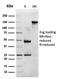
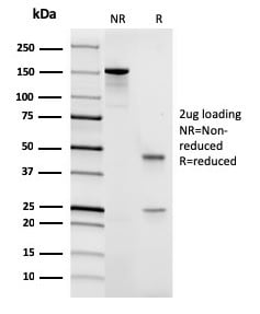
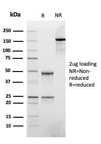
 SDS-PAGE
(SDS-PAGE Analysis Purified CD45 Recombinant Rabbit Monoclonal Antibody (PTPRC/3881R) Confirmation of Purity and Integrity of Antibody.)
SDS-PAGE
(SDS-PAGE Analysis Purified CD45 Recombinant Rabbit Monoclonal Antibody (PTPRC/3881R) Confirmation of Purity and Integrity of Antibody.)
CD45/LCA, Monoclonal Antibody (Cat# AAA215368)
 SDS-PAGE
(SDS-PAGE Analysis Purified CD45 Recombinant Mouse Monoclonal Antibody (rPTPRC/1461) Confirmation of Purity and Integrity of Antibody.)
SDS-PAGE
(SDS-PAGE Analysis Purified CD45 Recombinant Mouse Monoclonal Antibody (rPTPRC/1461) Confirmation of Purity and Integrity of Antibody.)
CD45/LCA, Monoclonal Antibody (Cat# AAA215370)
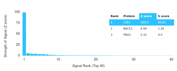 Application Data
(Analysis of Protein Array containing more than 19,000 full-length human proteins using GM-CSF Mouse Monoclonal Antibody (CSF2/3402). Z- and S- Score: The Z-score represents the strength of a signal that a monoclonal antibody (MAb) (in combination with a fluorescently-tagged anti-IgG secondary antibody) produces when binding to a particular protein on the HuProtTM array. Z-scores are described in units of standard deviations (SD's) above the mean value of all signals generated on that array. If targets on HuProtTM are arranged in descending order of the Z-score, the S-score is the difference (also in units of SD's) between the Z-score. S-score therefore represents the relative target specificity of a MAb to its intended target. A MAb is considered to specific to its intended target, if the MAb has an S-score of at least 2.5. For example, if a MAb binds to protein X with a Z-score of 43 and to protein Y with a Z-score of 14, then the S-score for the binding of that MAb to protein X is equal to 29.)
Application Data
(Analysis of Protein Array containing more than 19,000 full-length human proteins using GM-CSF Mouse Monoclonal Antibody (CSF2/3402). Z- and S- Score: The Z-score represents the strength of a signal that a monoclonal antibody (MAb) (in combination with a fluorescently-tagged anti-IgG secondary antibody) produces when binding to a particular protein on the HuProtTM array. Z-scores are described in units of standard deviations (SD's) above the mean value of all signals generated on that array. If targets on HuProtTM are arranged in descending order of the Z-score, the S-score is the difference (also in units of SD's) between the Z-score. S-score therefore represents the relative target specificity of a MAb to its intended target. A MAb is considered to specific to its intended target, if the MAb has an S-score of at least 2.5. For example, if a MAb binds to protein X with a Z-score of 43 and to protein Y with a Z-score of 14, then the S-score for the binding of that MAb to protein X is equal to 29.)
GM-CSF, Monoclonal Antibody (Cat# AAA215377)
 SDS-PAGE
(SDS-PAGE Analysis Purified ATRX Recombinant Mouse Monoclonal Antibody (rATRX/3446). Confirmation of Integrity and Purity of Antibody.)
SDS-PAGE
(SDS-PAGE Analysis Purified ATRX Recombinant Mouse Monoclonal Antibody (rATRX/3446). Confirmation of Integrity and Purity of Antibody.)
ATRX/RAD54, Monoclonal Antibody (Cat# AAA215378)
 SDS-PAGE
(SDS-PAGE Analysis Purified CD29 Mouse Monoclonal Antibody (ITGB1/3613). Confirmation of Purity and Integrity of Antibody.)
SDS-PAGE
(SDS-PAGE Analysis Purified CD29 Mouse Monoclonal Antibody (ITGB1/3613). Confirmation of Purity and Integrity of Antibody.)
CD29, Monoclonal Antibody (Cat# AAA215385)
Does not react with Mouse or Rat
 SDS-PAGE
(SDS-PAGE Analysis Purified N-Cadherin Recombinant Mouse Monoclonal Antibody (rCDH2/1426). Confirmation of Integrity and Purity of Antibody.)
SDS-PAGE
(SDS-PAGE Analysis Purified N-Cadherin Recombinant Mouse Monoclonal Antibody (rCDH2/1426). Confirmation of Integrity and Purity of Antibody.)
N-Cadherin/Cadherin-2/CD325 (NCAD), Monoclonal Antibody (Cat# AAA215387)


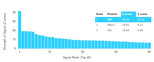 Application Data
(Analysis of Protein Array containing more than 19,000 full-length human proteins using Myelin Basic Protein Recombinant Rabbit Antibody (MBP/4277R). Z- and S- Score: The Z-score represents the strength of a signal that a monoclonal antibody (MAb) (in combination with a fluorescently-tagged anti-IgG secondary antibody) produces when binding to a particular protein on the HuProtTM array. Z-scores are described in units of standard deviations (SD's) above the mean value of all signals generated on that array. If targets on HuProtTM are arranged in descending order of the Z-score, the S-score is the difference (also in units of SD's) between the Z-score. S-score therefore represents the relative target specificity of a MAb to its intended target. A MAb is considered to specific to its intended target, if the MAb has an S-score of at least 2.5. For example, if a MAb binds to protein X with a Z-score of 43 and to protein Y with a Z-score of 14, then the S-score for the binding of that MAb to protein X is equal to 29.)
Application Data
(Analysis of Protein Array containing more than 19,000 full-length human proteins using Myelin Basic Protein Recombinant Rabbit Antibody (MBP/4277R). Z- and S- Score: The Z-score represents the strength of a signal that a monoclonal antibody (MAb) (in combination with a fluorescently-tagged anti-IgG secondary antibody) produces when binding to a particular protein on the HuProtTM array. Z-scores are described in units of standard deviations (SD's) above the mean value of all signals generated on that array. If targets on HuProtTM are arranged in descending order of the Z-score, the S-score is the difference (also in units of SD's) between the Z-score. S-score therefore represents the relative target specificity of a MAb to its intended target. A MAb is considered to specific to its intended target, if the MAb has an S-score of at least 2.5. For example, if a MAb binds to protein X with a Z-score of 43 and to protein Y with a Z-score of 14, then the S-score for the binding of that MAb to protein X is equal to 29.)
Myelin Basic Protein, Monoclonal Antibody (Cat# AAA215394)
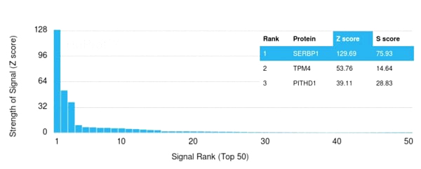
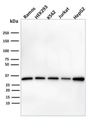
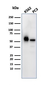 WB (Western Blot)
(Western Blot Analysis of K562 and PC3 cell lysate using SERBP1 Mouse Monoclonal Antibody (SERBP1/3492).)
WB (Western Blot)
(Western Blot Analysis of K562 and PC3 cell lysate using SERBP1 Mouse Monoclonal Antibody (SERBP1/3492).)
SERBP, Monoclonal Antibody (Cat# AAA215401)
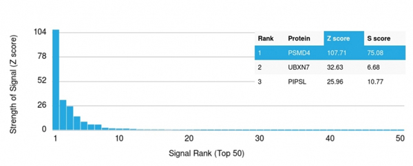 Application Data
(Analysis of Protein Array containing more than 19,000 full-length human proteins using 26S proteasome non-ATPase regulatory subunit 4 Mouse Monoclonal Antibody (CPTC-PSMD4-3). Z- and S- Score: The Z-score represents the strength of a signal that a monoclonal antibody (MAb) (in combination with a fluorescently-tagged anti-IgG secondary antibody) produces when binding to a particular protein on the HuProtTM array. Z-scores are described in units of standard deviations (SD's) above the mean value of all signals generated on that array. If targets on HuProtTM are arranged in descending order of the Z-score, the S-score is the difference (also in units of SD's) between the Z-score. S-score therefore represents the relative target specificity of a MAb to its intended target. A MAb is considered to specific to its intended target, if the MAb has an S-score of at least 2.5. For example, if a MAb binds to protein X with a Z-score of 43 and to protein Y with a Z-score of 14, then the S-score for the binding of that MAb to protein X is equal to 29.)
Application Data
(Analysis of Protein Array containing more than 19,000 full-length human proteins using 26S proteasome non-ATPase regulatory subunit 4 Mouse Monoclonal Antibody (CPTC-PSMD4-3). Z- and S- Score: The Z-score represents the strength of a signal that a monoclonal antibody (MAb) (in combination with a fluorescently-tagged anti-IgG secondary antibody) produces when binding to a particular protein on the HuProtTM array. Z-scores are described in units of standard deviations (SD's) above the mean value of all signals generated on that array. If targets on HuProtTM are arranged in descending order of the Z-score, the S-score is the difference (also in units of SD's) between the Z-score. S-score therefore represents the relative target specificity of a MAb to its intended target. A MAb is considered to specific to its intended target, if the MAb has an S-score of at least 2.5. For example, if a MAb binds to protein X with a Z-score of 43 and to protein Y with a Z-score of 14, then the S-score for the binding of that MAb to protein X is equal to 29.)
26S proteasome non-ATPase4, Monoclonal Antibody (Cat# AAA215405)
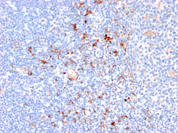
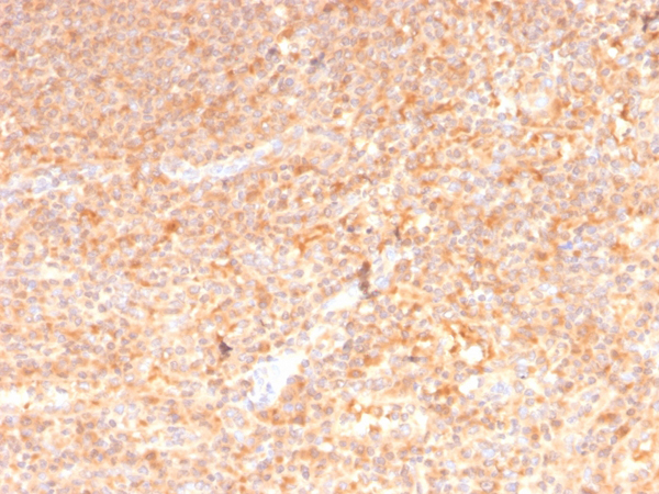
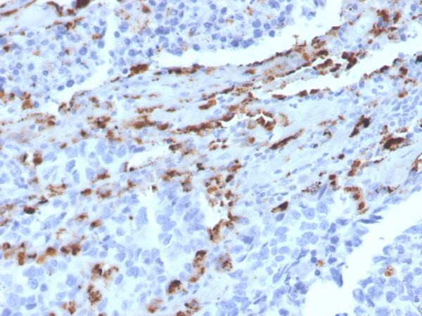
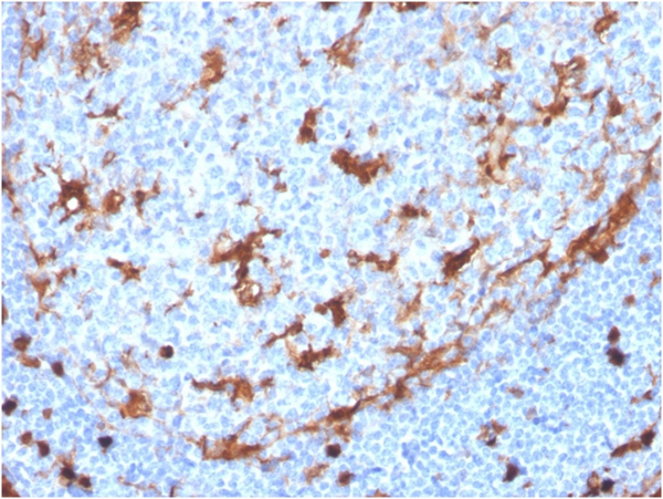 IHC (Immunohiostchemistry)
(Formalin-fixed, paraffin-embedded human tonsil stained with Annexin A1 Recombinant Rabbit Monoclonal Antibody (ANXA1/3869R).)
IHC (Immunohiostchemistry)
(Formalin-fixed, paraffin-embedded human tonsil stained with Annexin A1 Recombinant Rabbit Monoclonal Antibody (ANXA1/3869R).)
Annexin A1, Monoclonal Antibody (Cat# AAA215413)

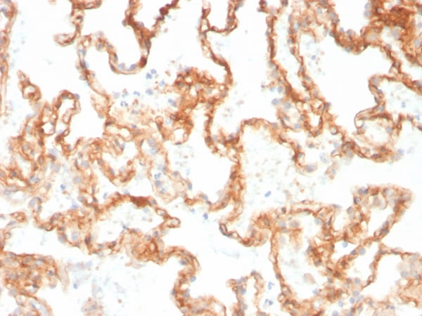
 IHC (Immunohiostchemistry)
(Formalin-fixed, paraffin-embedded human tonsil stained with Collagen IV Recombinant Rabbit Monoclonal Antibody (COL4/4241R).)
IHC (Immunohiostchemistry)
(Formalin-fixed, paraffin-embedded human tonsil stained with Collagen IV Recombinant Rabbit Monoclonal Antibody (COL4/4241R).)
Collagen IV, Monoclonal Antibody (Cat# AAA215420)
 SDS-PAGE
(SDS-PAGE Analysis Purified Aurora B Mouse Monoclonal Antibody (AURKB/1592). Confirmation of Integrity and Purity of Antibody.)
SDS-PAGE
(SDS-PAGE Analysis Purified Aurora B Mouse Monoclonal Antibody (AURKB/1592). Confirmation of Integrity and Purity of Antibody.)
Aurora B, Monoclonal Antibody (Cat# AAA215286)
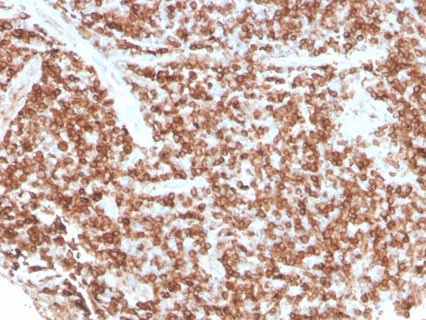
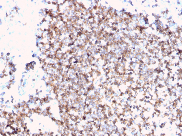
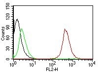
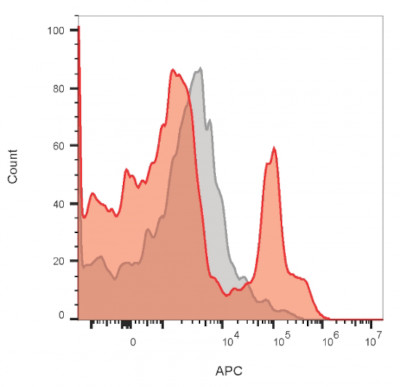 FCM/FACS (Flow Cytometry)
(Flow cytometry of lymphocyte gated PBMCs stained with CD19 monoclonal antibody (PDR134) (red) or isotype control (gray) followed by goat anti-mouse CF640R (red).)
FCM/FACS (Flow Cytometry)
(Flow cytometry of lymphocyte gated PBMCs stained with CD19 monoclonal antibody (PDR134) (red) or isotype control (gray) followed by goat anti-mouse CF640R (red).)
CD19, Monoclonal Antibody (Cat# AAA215291)
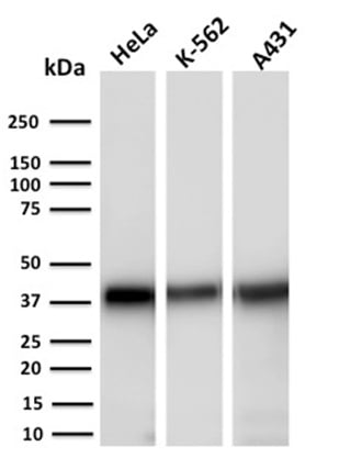
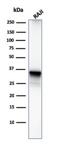 WB (Western Blot)
(Western Blot Analysis of Raji cell lysate using CD20 Mouse Monoclonal Antibody (MS4A1/3411).)
WB (Western Blot)
(Western Blot Analysis of Raji cell lysate using CD20 Mouse Monoclonal Antibody (MS4A1/3411).)
CD20/ MS4A1, Monoclonal Antibody (Cat# AAA215295)
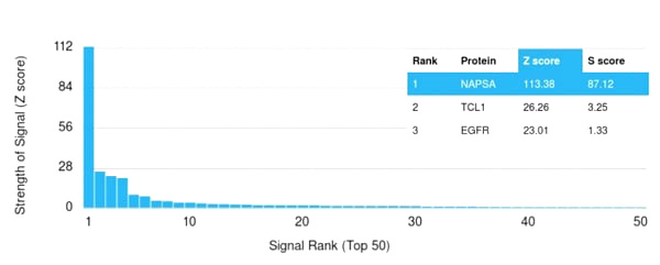 Application Data
(Analysis of Protein Array containing more than 19,000 full-length human proteins using Napsin A Mouse Monoclonal Antibody (NAPSA/3309). Z- and S- Score: The Z-score represents the strength of a signal that a monoclonal antibody (Monoclonal Antibody) (in combination with a fluorescently-tagged anti-IgG secondary antibody) produces when binding to a particular protein on the HuProtTM array. Z-scores are described in units of standard deviations (SD’s) above the mean value of all signals generated on that array. If targets on HuProtTM are arranged in descending order of the Z-score, the S-score is the difference (also in units of SD’s) between the Z-score. S-score therefore represents the relative target specificity of a Monoclonal Antibody to its intended target. A Monoclonal Antibody is considered to specific to its intended target, if the Monoclonal Antibody has an S-score of at least 2.5. For example, if a Monoclonal Antibody binds to protein X with a Z-score of 43 and to protein Y with a Z-score of 14, then the S-score for the binding of that Monoclonal Antibody to protein X is equal to 29.)
Application Data
(Analysis of Protein Array containing more than 19,000 full-length human proteins using Napsin A Mouse Monoclonal Antibody (NAPSA/3309). Z- and S- Score: The Z-score represents the strength of a signal that a monoclonal antibody (Monoclonal Antibody) (in combination with a fluorescently-tagged anti-IgG secondary antibody) produces when binding to a particular protein on the HuProtTM array. Z-scores are described in units of standard deviations (SD’s) above the mean value of all signals generated on that array. If targets on HuProtTM are arranged in descending order of the Z-score, the S-score is the difference (also in units of SD’s) between the Z-score. S-score therefore represents the relative target specificity of a Monoclonal Antibody to its intended target. A Monoclonal Antibody is considered to specific to its intended target, if the Monoclonal Antibody has an S-score of at least 2.5. For example, if a Monoclonal Antibody binds to protein X with a Z-score of 43 and to protein Y with a Z-score of 14, then the S-score for the binding of that Monoclonal Antibody to protein X is equal to 29.)
Napsin A, Monoclonal Antibody (Cat# AAA215302)
 Application Data
(Analysis of Protein Array containing >19,000 full-length human proteins using CD63 Mouse Monoclonal Antibody (LAMP3/2790) Z- and S- Score: The Z-score represents the strength of a signal that a monoclonal antibody (MAb) (in combination with a fluorescently-tagged anti-IgG secondary antibody) produces when binding to a particular protein on the HuProtTM array. Z-scores are described in units of standard deviations (SD’s) above the mean value of all signals generated on that array. If targets on HuProtTM are arranged in descending order of the Z-score, the S-score is the difference (also in units of SD’s) between the Z-score. S-score therefore represents the relative target specificity of a MAb to its intended target. A MAb is considered to specific to its intended target, if the MAb has an S-score of at least 2.5. For example, if a MAb binds to protein X with a Z-score of 43 and to protein Y with a Z-score of 14, then the S-score for the binding of that MAb to protein X is equal to 29.)
Application Data
(Analysis of Protein Array containing >19,000 full-length human proteins using CD63 Mouse Monoclonal Antibody (LAMP3/2790) Z- and S- Score: The Z-score represents the strength of a signal that a monoclonal antibody (MAb) (in combination with a fluorescently-tagged anti-IgG secondary antibody) produces when binding to a particular protein on the HuProtTM array. Z-scores are described in units of standard deviations (SD’s) above the mean value of all signals generated on that array. If targets on HuProtTM are arranged in descending order of the Z-score, the S-score is the difference (also in units of SD’s) between the Z-score. S-score therefore represents the relative target specificity of a MAb to its intended target. A MAb is considered to specific to its intended target, if the MAb has an S-score of at least 2.5. For example, if a MAb binds to protein X with a Z-score of 43 and to protein Y with a Z-score of 14, then the S-score for the binding of that MAb to protein X is equal to 29.)
CD63, Monoclonal Antibody (Cat# AAA215308)
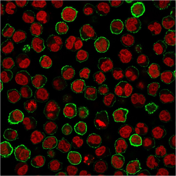
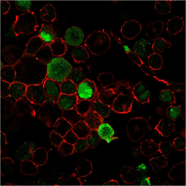
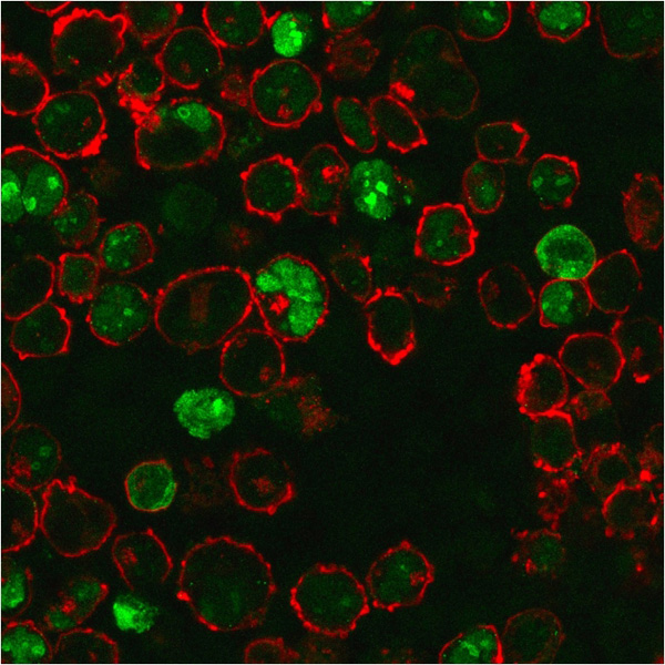
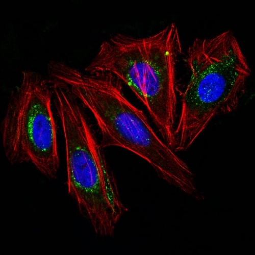 IF (Immunofluorescence)
(IF staining of HeLa cells using CF488 labeled CD63 Monoclonal Antibody (NKI/C3) (Green). F-actin filaments are labeled with Dylight 554 phalloidin (red). DAPI was used to stain the cell nucleus (blue).)
IF (Immunofluorescence)
(IF staining of HeLa cells using CF488 labeled CD63 Monoclonal Antibody (NKI/C3) (Green). F-actin filaments are labeled with Dylight 554 phalloidin (red). DAPI was used to stain the cell nucleus (blue).)
CD63, Monoclonal Antibody (Cat# AAA215310)
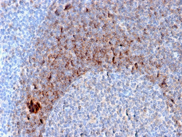
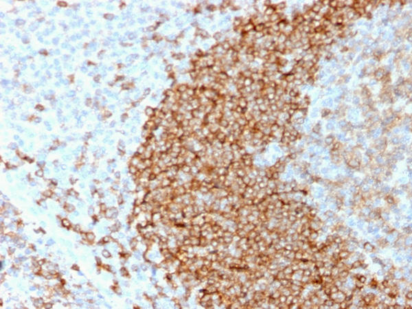
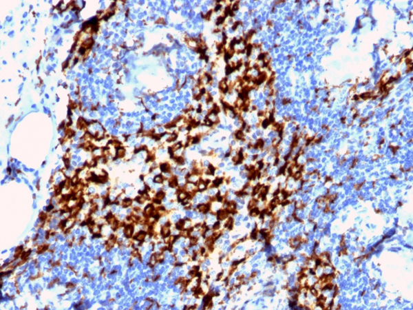
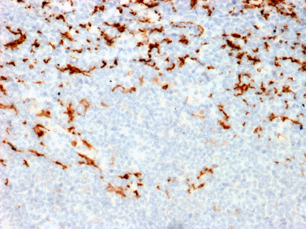
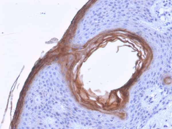
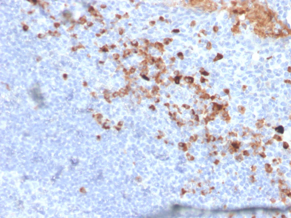
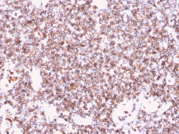
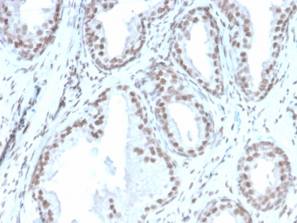
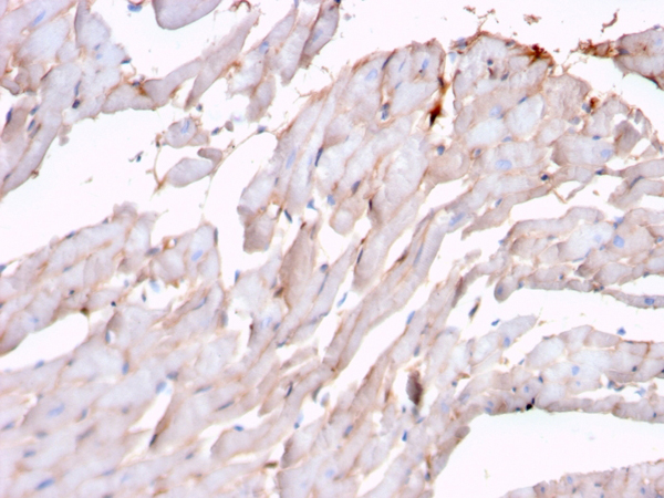
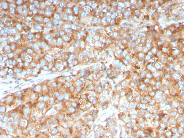
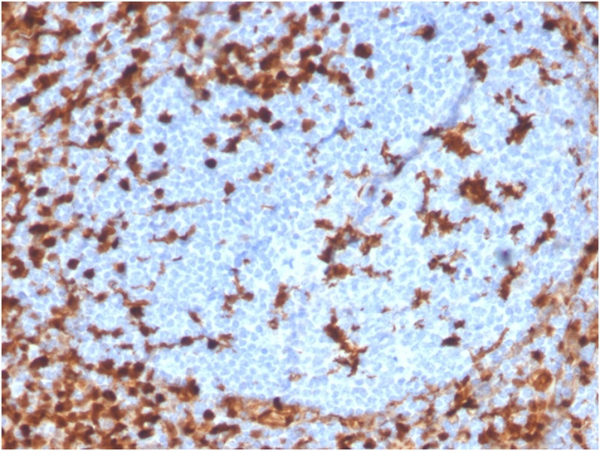
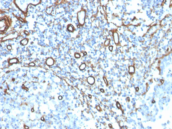
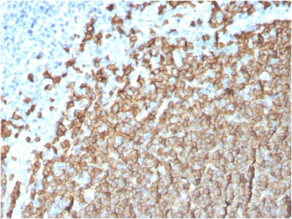
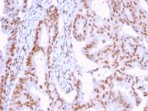
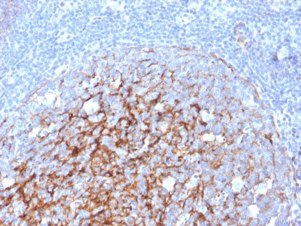
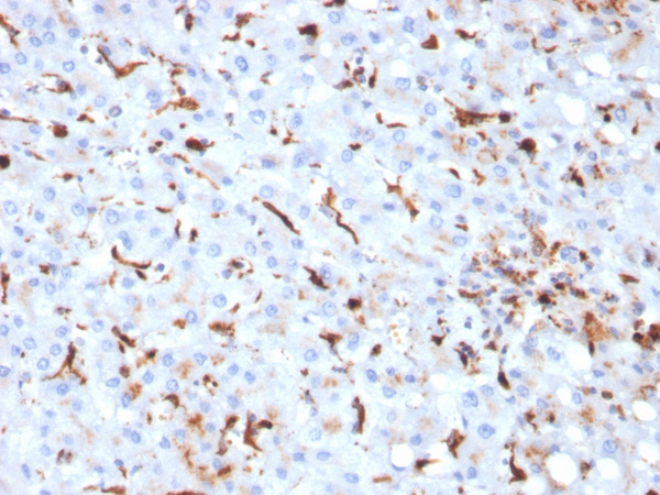
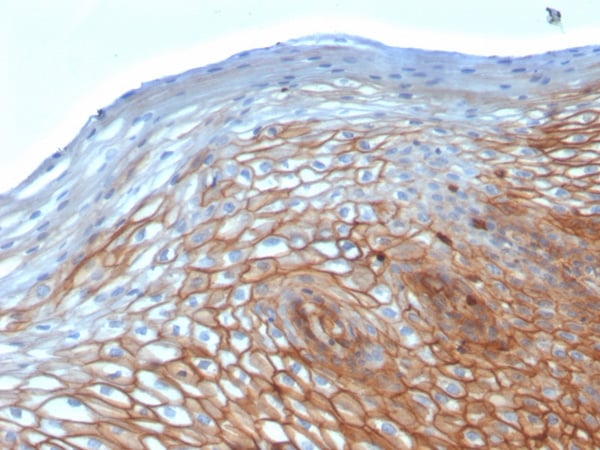
 IHC (Immunohistochemistry)
(Formalin-fixed, paraffin-embedded human Tonsil stained with CD79b Mouse Monoclonal Antibody (B29/123).)
IHC (Immunohistochemistry)
(Formalin-fixed, paraffin-embedded human Tonsil stained with CD79b Mouse Monoclonal Antibody (B29/123).)
CD79b, Monoclonal Antibody (Cat# AAA215319)

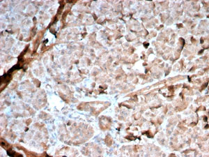
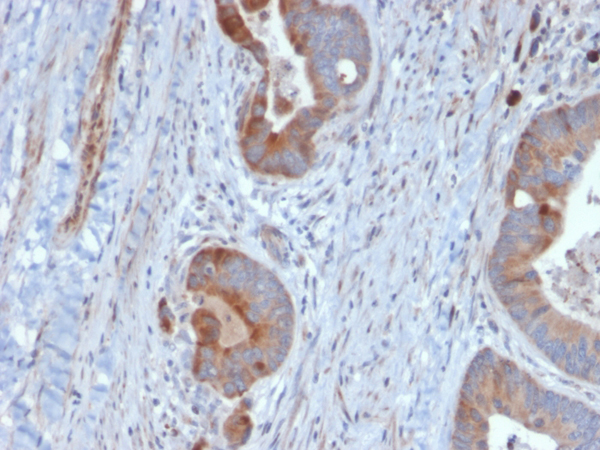
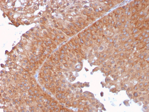
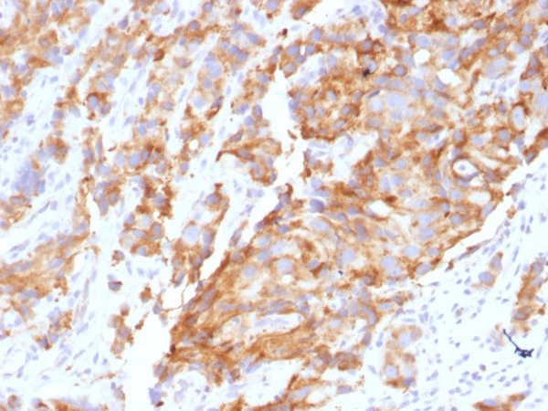
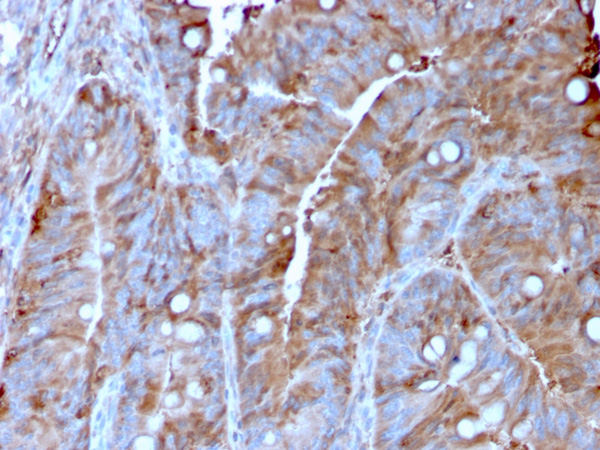
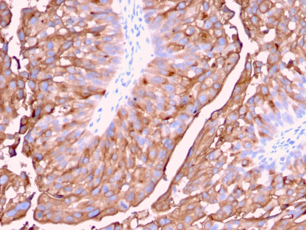
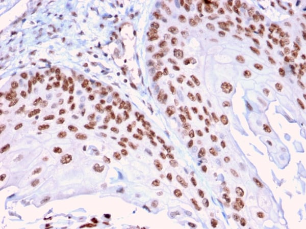
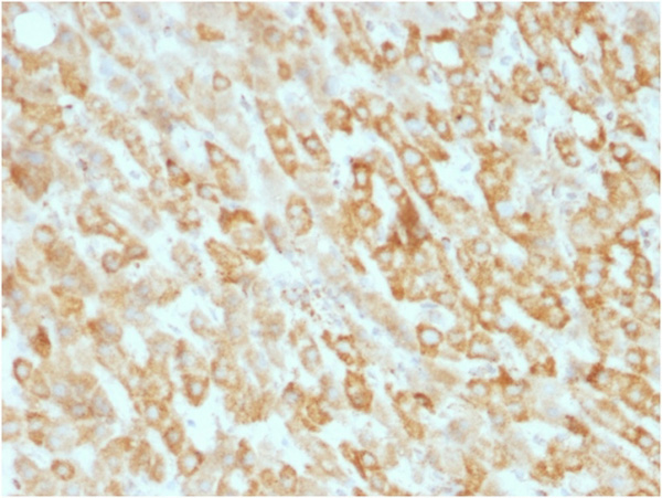
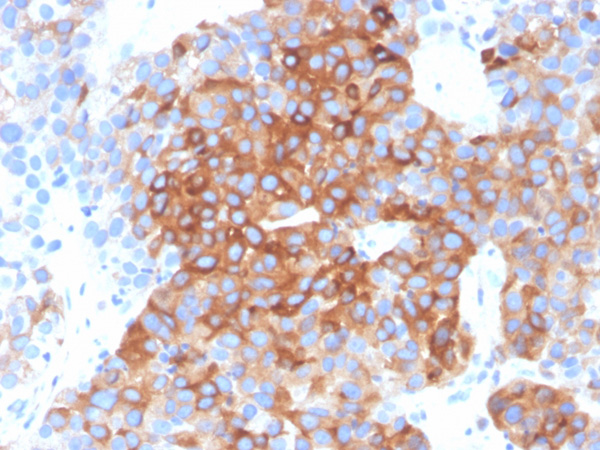 IHC (Immunohiostchemistry)
(Formalin-fixed, paraffin-embedded human bladder carcinoma stained with Tubulin beta 3 Mouse Monoclonal Antibody (TUBB3/3732).)
IHC (Immunohiostchemistry)
(Formalin-fixed, paraffin-embedded human bladder carcinoma stained with Tubulin beta 3 Mouse Monoclonal Antibody (TUBB3/3732).)
Tubulin beta 3/TUBB3, Monoclonal Antibody (Cat# AAA214878)
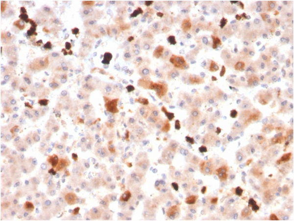
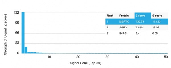 Application Data
(Analysis of Protein Array containing more than 19,000 full-length human proteins using MerTK Mouse Monoclonal Antibody (MERTK/3022). Z- and S- Score: The Z-score represents the strength of a signal that a monoclonal antibody (MAb) (in combination with a fluorescently-tagged anti-IgG secondary antibody) produces when binding to a particular protein on the HuProtTM array. Z-scores are described in units of standard deviations (SD's) above the mean value of all signals generated on that array. If targets on HuProtTM are arranged in descending order of the Z-score, the S-score is the difference (also in units of SD's) between the Z-score. S-score therefore represents the relative target specificity of a MAb to its intended target. A MAb is considered to specific to its intended target, if the MAb has an S-score of at least 2.5. For example, if a MAb binds to protein X with a Z-score of 43 and to protein Y with a Z-score of 14, then the S-score for the binding of that MAb to protein X is equal to 29.)
Application Data
(Analysis of Protein Array containing more than 19,000 full-length human proteins using MerTK Mouse Monoclonal Antibody (MERTK/3022). Z- and S- Score: The Z-score represents the strength of a signal that a monoclonal antibody (MAb) (in combination with a fluorescently-tagged anti-IgG secondary antibody) produces when binding to a particular protein on the HuProtTM array. Z-scores are described in units of standard deviations (SD's) above the mean value of all signals generated on that array. If targets on HuProtTM are arranged in descending order of the Z-score, the S-score is the difference (also in units of SD's) between the Z-score. S-score therefore represents the relative target specificity of a MAb to its intended target. A MAb is considered to specific to its intended target, if the MAb has an S-score of at least 2.5. For example, if a MAb binds to protein X with a Z-score of 43 and to protein Y with a Z-score of 14, then the S-score for the binding of that MAb to protein X is equal to 29.)
MerTK, Monoclonal Antibody (Cat# AAA214882)
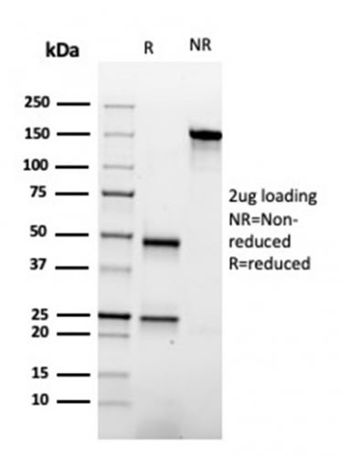
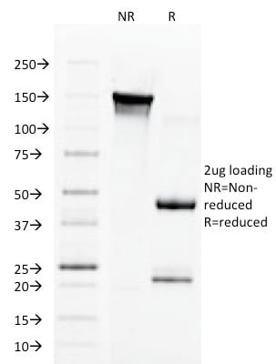
 SDS-PAGE
(SDS-PAGE Analysis Purified CFTR Mouse Monoclonal Antibody (CFTR/1785).)
SDS-PAGE
(SDS-PAGE Analysis Purified CFTR Mouse Monoclonal Antibody (CFTR/1785).)
CFTR (Cystic Fibrosis Transmembrane Conductance Regulator), Monoclonal Antibody (Cat# AAA214884)
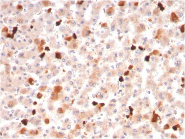
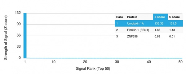 Application Data
(Analysis of Protein Array containing more than 19,000 full-length human proteins using Uroplakin 1A Mouse Monoclonal Antibody (UPK1A/2921) Z- and S- Score: The Z-score represents the strength of a signal that a monoclonal antibody (MAb) (in combination with a fluorescently-tagged anti-IgG secondary antibody) produces when binding to a particular protein on the HuProtTM array. Z-scores are described in units of standard deviations (SD's) above the mean value of all signals generated on that array. If targets on HuProtTM are arranged in descending order of the Z-score, the S-score is the difference (also in units of SD's) between the Z-score. S-score therefore represents the relative target specificity of a MAb to its intended target. A MAb is considered to specific to its intended target, if the MAb has an S-score of at least 2.5. For example, if a MAb binds to protein X with a Z-score of 43 and to protein Y with a Z-score of 14, then the S-score for the binding of that MAb to protein X is equal to 29.)
Application Data
(Analysis of Protein Array containing more than 19,000 full-length human proteins using Uroplakin 1A Mouse Monoclonal Antibody (UPK1A/2921) Z- and S- Score: The Z-score represents the strength of a signal that a monoclonal antibody (MAb) (in combination with a fluorescently-tagged anti-IgG secondary antibody) produces when binding to a particular protein on the HuProtTM array. Z-scores are described in units of standard deviations (SD's) above the mean value of all signals generated on that array. If targets on HuProtTM are arranged in descending order of the Z-score, the S-score is the difference (also in units of SD's) between the Z-score. S-score therefore represents the relative target specificity of a MAb to its intended target. A MAb is considered to specific to its intended target, if the MAb has an S-score of at least 2.5. For example, if a MAb binds to protein X with a Z-score of 43 and to protein Y with a Z-score of 14, then the S-score for the binding of that MAb to protein X is equal to 29.)
Uroplakin 1A, Monoclonal Antibody (Cat# AAA214887)
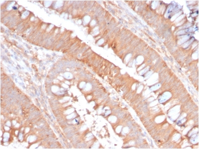
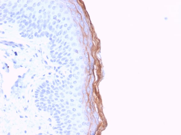
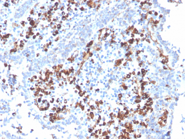
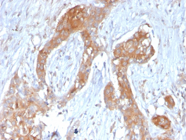
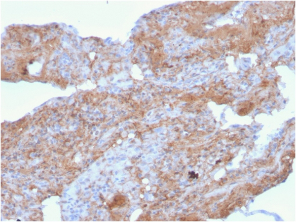
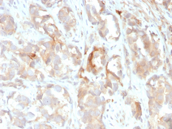
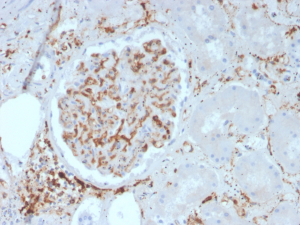
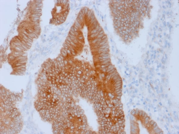
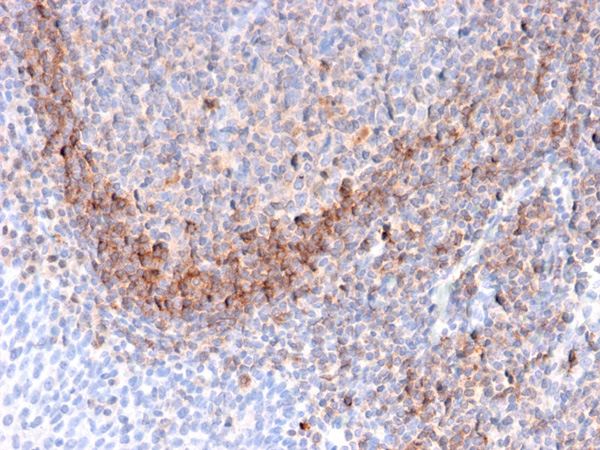
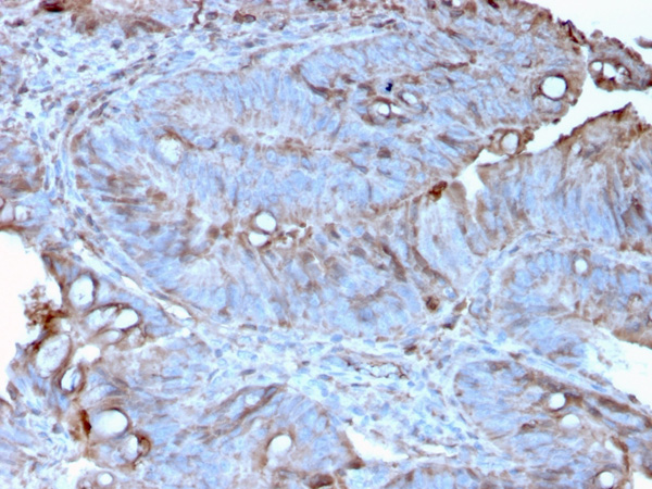
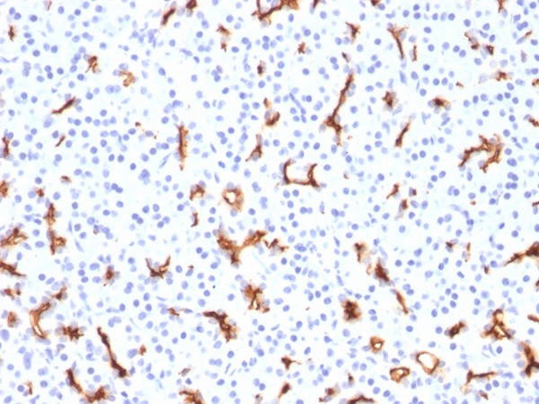
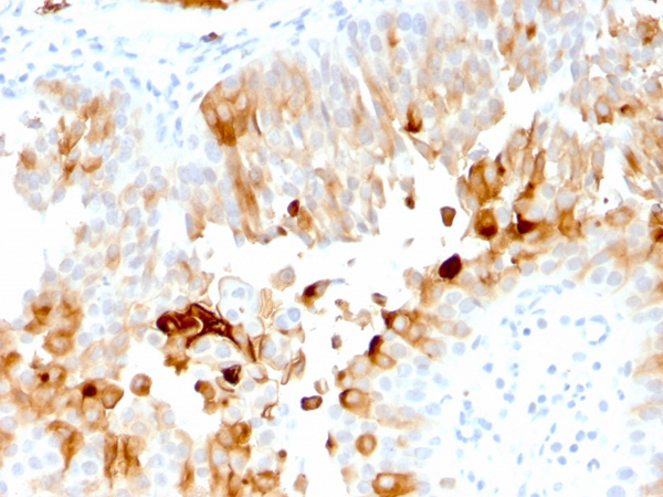
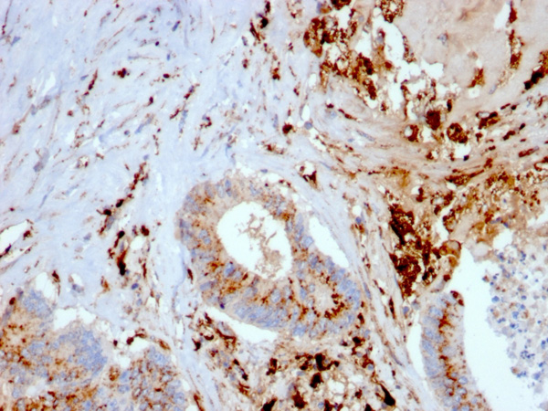
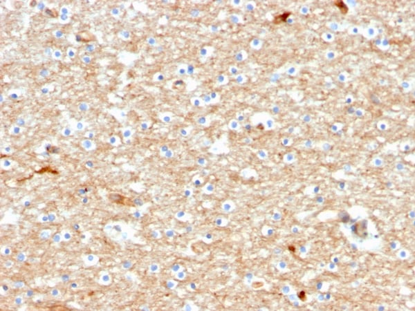
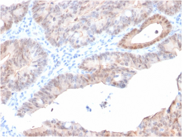
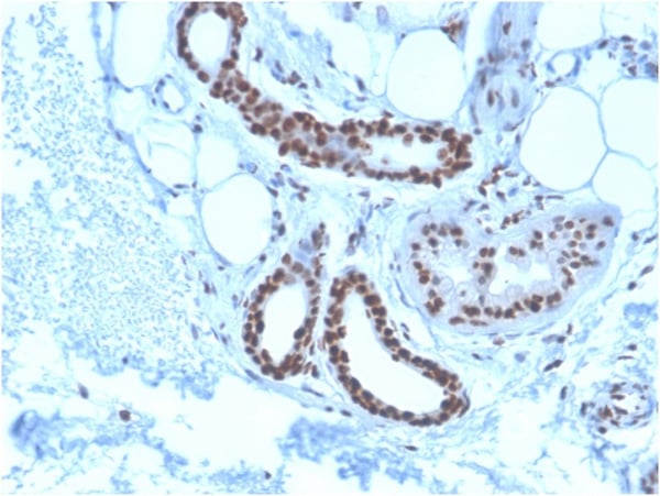
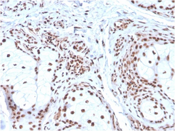
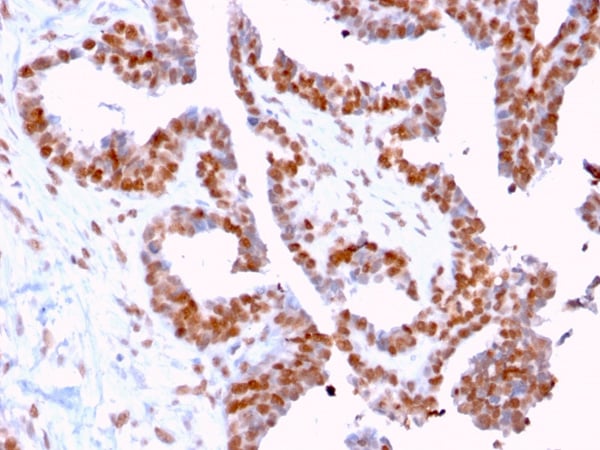 IHC (Immunohistochemisry)
(Formalin-fixed, paraffin-embedded human Prostate Carcinoma stained with CLEC9A Mouse Monoclonal Antibody (2H12/4).)
IHC (Immunohistochemisry)
(Formalin-fixed, paraffin-embedded human Prostate Carcinoma stained with CLEC9A Mouse Monoclonal Antibody (2H12/4).)
DMC1, Monoclonal Antibody (Cat# AAA214893)
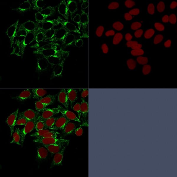 IF (Immunofluorescence)
(Confocal Immunofluorescence image of HeLa cells stained with Clathrin, HC Monoclonal Antibody (CHC/1432) followed by Goat anti-Mouse CF488 (green). Reddot is used to label the nuceli red.)
IF (Immunofluorescence)
(Confocal Immunofluorescence image of HeLa cells stained with Clathrin, HC Monoclonal Antibody (CHC/1432) followed by Goat anti-Mouse CF488 (green). Reddot is used to label the nuceli red.)
Clathrin, Monoclonal Antibody (Cat# AAA214896)
 Application Data
(Analysis of Protein Array containing more than 19,000 full-length human proteins using Carboxypeptidase A1/CPA1 Mouse Monoclonal Antibody (CPA1/2713). Z- and S- Score: The Z-score represents the strength of a signal that a monoclonal antibody (Monoclonal Antibody) (in combination with a fluorescently-tagged anti-IgG secondary antibody) produces when binding to a particular protein on the HuProtTM array. Z-scores are described in units of standard deviations (SD's) above the mean value of all signals generated on that array. If targets on HuProtTM are arranged in descending order of the Z-score, the S-score is the difference (also in units of SD's) between the Z-score. S-score therefore represents the relative target specificity of a Monoclonal Antibody to its intended target. A Monoclonal Antibody is considered to specific to its intended target, if the Monoclonal Antibody has an S-score of at least 2.5. For example, if a Monoclonal Antibody binds to protein X with a Z-score of 43 and to protein Y with a Z-score of 14, then the S-score for the binding of that Monoclonal Antibody to protein X is equal to 29.)
Application Data
(Analysis of Protein Array containing more than 19,000 full-length human proteins using Carboxypeptidase A1/CPA1 Mouse Monoclonal Antibody (CPA1/2713). Z- and S- Score: The Z-score represents the strength of a signal that a monoclonal antibody (Monoclonal Antibody) (in combination with a fluorescently-tagged anti-IgG secondary antibody) produces when binding to a particular protein on the HuProtTM array. Z-scores are described in units of standard deviations (SD's) above the mean value of all signals generated on that array. If targets on HuProtTM are arranged in descending order of the Z-score, the S-score is the difference (also in units of SD's) between the Z-score. S-score therefore represents the relative target specificity of a Monoclonal Antibody to its intended target. A Monoclonal Antibody is considered to specific to its intended target, if the Monoclonal Antibody has an S-score of at least 2.5. For example, if a Monoclonal Antibody binds to protein X with a Z-score of 43 and to protein Y with a Z-score of 14, then the S-score for the binding of that Monoclonal Antibody to protein X is equal to 29.)
Carboxypeptidase A1/CPA1, Monoclonal Antibody (Cat# AAA214900)
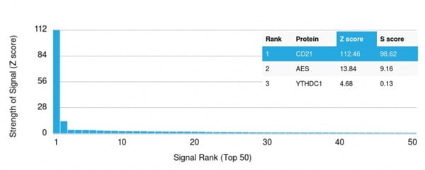 Application Data
(Analysis of Protein Array containing more than 19,000 full-length human proteins using CD21-Monospecific Mouse Monoclonal Antibody (CR2/3247) Z- and S- Score: The Z-score represents the strength of a signal that a monoclonal antibody (Monoclonal Antibody) (in combination with a fluorescently-tagged anti-IgG secondary antibody) produces when binding to a particular protein on the HuProtTM array. Z-scores are described in units of standard deviations (SD’s) above the mean value of all signals generated on that array. If targets on HuProtTM are arranged in descending order of the Z-score, the S-score is the difference (also in units of SD’s) between the Z-score. S-score therefore represents the relative target specificity of a Monoclonal Antibody to its intended target. A Monoclonal Antibody is considered to specific to its intended target, if the Monoclonal Antibody has an S-score of at least 2.5. For example, if a Monoclonal Antibody binds to protein X with a Z-score of 43 and to protein Y with a Z-score of 14, then the S-score for the binding of that Monoclonal Antibody to protein X is equal to 29.)
Application Data
(Analysis of Protein Array containing more than 19,000 full-length human proteins using CD21-Monospecific Mouse Monoclonal Antibody (CR2/3247) Z- and S- Score: The Z-score represents the strength of a signal that a monoclonal antibody (Monoclonal Antibody) (in combination with a fluorescently-tagged anti-IgG secondary antibody) produces when binding to a particular protein on the HuProtTM array. Z-scores are described in units of standard deviations (SD’s) above the mean value of all signals generated on that array. If targets on HuProtTM are arranged in descending order of the Z-score, the S-score is the difference (also in units of SD’s) between the Z-score. S-score therefore represents the relative target specificity of a Monoclonal Antibody to its intended target. A Monoclonal Antibody is considered to specific to its intended target, if the Monoclonal Antibody has an S-score of at least 2.5. For example, if a Monoclonal Antibody binds to protein X with a Z-score of 43 and to protein Y with a Z-score of 14, then the S-score for the binding of that Monoclonal Antibody to protein X is equal to 29.)
CD21, Monoclonal Antibody (Cat# AAA214903)

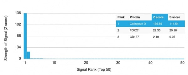
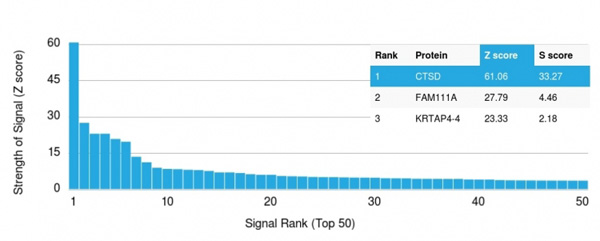 Application Data
(Analysis of Protein Array containing more than 19,000 full-length human proteins using Cathepsin D Mouse Monoclonal Antibody (CTSD/3082) Z- and S- Score: The Z-score represents the strength of a signal that a monoclonal antibody (MAb) (in combination with a fluorescently-tagged anti-IgG secondary antibody) produces when binding to a particular protein on the HuProtTM array. Z-scores are described in units of standard deviations (SD's) above the mean value of all signals generated on that array. If targets on HuProtTM are arranged in descending order of the Z-score, the S-score is the difference (also in units of SD's) between the Z-score. S-score therefore represents the relative target specificity of a MAb to its intended target. A MAb is considered to specific to its intended target, if the MAb has an S-score of at least 2.5. For example, if a MAb binds to protein X with a Z-score of 43 and to protein Y with a Z-score of 14, then the S-score for the binding of that MAb to protein X is equal to 29.)
Application Data
(Analysis of Protein Array containing more than 19,000 full-length human proteins using Cathepsin D Mouse Monoclonal Antibody (CTSD/3082) Z- and S- Score: The Z-score represents the strength of a signal that a monoclonal antibody (MAb) (in combination with a fluorescently-tagged anti-IgG secondary antibody) produces when binding to a particular protein on the HuProtTM array. Z-scores are described in units of standard deviations (SD's) above the mean value of all signals generated on that array. If targets on HuProtTM are arranged in descending order of the Z-score, the S-score is the difference (also in units of SD's) between the Z-score. S-score therefore represents the relative target specificity of a MAb to its intended target. A MAb is considered to specific to its intended target, if the MAb has an S-score of at least 2.5. For example, if a MAb binds to protein X with a Z-score of 43 and to protein Y with a Z-score of 14, then the S-score for the binding of that MAb to protein X is equal to 29.)
Cathepsin D, Monoclonal Antibody (Cat# AAA214910)
 Application Data
(Analysis of Protein Array containing more than 19,000 full-length human proteins using Cathepsin D Mouse Monoclonal Antibody (CTSD/3275) Z- and S- Score: The Z-score represents the strength of a signal that a monoclonal antibody (Monoclonal Antibody) (in combination with a fluorescently-tagged anti-IgG secondary antibody) produces when binding to a particular protein on the HuProtTM array. Z-scores are described in units of standard deviations (SD’s) above the mean value of all signals generated on that array. If targets on HuProtTM are arranged in descending order of the Z-score, the S-score is the difference (also in units of SD’s) between the Z-score. S-score therefore represents the relative target specificity of a Monoclonal Antibody to its intended target. A Monoclonal Antibody is considered to specific to its intended target, if the Monoclonal Antibody has an S-score of at least 2.5. For example, if a Monoclonal Antibody binds to protein X with a Z-score of 43 and to protein Y with a Z-score of 14, then the S-score for the binding of that Monoclonal Antibody to protein X is equal to 29.)
Application Data
(Analysis of Protein Array containing more than 19,000 full-length human proteins using Cathepsin D Mouse Monoclonal Antibody (CTSD/3275) Z- and S- Score: The Z-score represents the strength of a signal that a monoclonal antibody (Monoclonal Antibody) (in combination with a fluorescently-tagged anti-IgG secondary antibody) produces when binding to a particular protein on the HuProtTM array. Z-scores are described in units of standard deviations (SD’s) above the mean value of all signals generated on that array. If targets on HuProtTM are arranged in descending order of the Z-score, the S-score is the difference (also in units of SD’s) between the Z-score. S-score therefore represents the relative target specificity of a Monoclonal Antibody to its intended target. A Monoclonal Antibody is considered to specific to its intended target, if the Monoclonal Antibody has an S-score of at least 2.5. For example, if a Monoclonal Antibody binds to protein X with a Z-score of 43 and to protein Y with a Z-score of 14, then the S-score for the binding of that Monoclonal Antibody to protein X is equal to 29.)
Cathepsin D, Monoclonal Antibody (Cat# AAA214912)
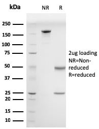
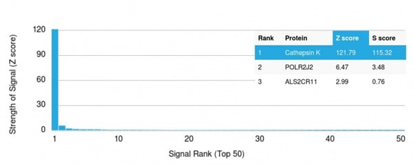 Application Data
(Analysis of Protein Array containing more than 19,000 full-length human proteins using Cathepsin K Mouse Monoclonal Antibody (CTSK/2793) Z- and S- Score: The Z-score represents the strength of a signal that a monoclonal antibody (MAb) (in combination with a fluorescently-tagged anti-IgG secondary antibody) produces when binding to a particular protein on the HuProtTM array. Z-scores are described in units of standard deviations (SD’s) above the mean value of all signals generated on that array. If targets on HuProtTM are arranged in descending order of the Z-score, the S-score is the difference (also in units of SD’s) between the Z-score. S-score therefore represents the relative target specificity of a MAb to its intended target. A MAb is considered to specific to its intended target, if the MAb has an S-score of at least 2.5. For example, if a MAb binds to protein X with a Z-score of 43 and to protein Y with a Z-score of 14, then the S-score for the binding of that MAb to protein X is equal to 29.)
Application Data
(Analysis of Protein Array containing more than 19,000 full-length human proteins using Cathepsin K Mouse Monoclonal Antibody (CTSK/2793) Z- and S- Score: The Z-score represents the strength of a signal that a monoclonal antibody (MAb) (in combination with a fluorescently-tagged anti-IgG secondary antibody) produces when binding to a particular protein on the HuProtTM array. Z-scores are described in units of standard deviations (SD’s) above the mean value of all signals generated on that array. If targets on HuProtTM are arranged in descending order of the Z-score, the S-score is the difference (also in units of SD’s) between the Z-score. S-score therefore represents the relative target specificity of a MAb to its intended target. A MAb is considered to specific to its intended target, if the MAb has an S-score of at least 2.5. For example, if a MAb binds to protein X with a Z-score of 43 and to protein Y with a Z-score of 14, then the S-score for the binding of that MAb to protein X is equal to 29.)
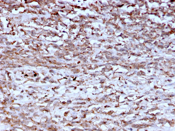
Cathepsin K, Monoclonal Antibody (Cat# AAA214915)
 Application Data
(Analysis of Protein Array containing more than 19,000 full-length human proteins using Drebrin-1 Mouse Monoclonal Antibody (DBN1/2880). Z- and S- Score: The Z-score represents the strength of a signal that a monoclonal antibody (MAb) (in combination with a fluorescently-tagged anti-IgG secondary antibody) produces when binding to a particular protein on the HuProtTM array. Z-scores are described in units of standard deviations (SD's) above the mean value of all signals generated on that array. If targets on HuProtTM are arranged in descending order of the Z-score, the S-score is the difference (also in units of SD's) between the Z-score. S-score therefore represents the relative target specificity of a MAb to its intended target. A MAb is considered to specific to its intended target, if the MAb has an S-score of at least 2.5. For example, if a MAb binds to protein X with a Z-score of 43 and to protein Y with a Z-score of 14, then the S-score for the binding of that MAb to protein X is equal to 29.)
Application Data
(Analysis of Protein Array containing more than 19,000 full-length human proteins using Drebrin-1 Mouse Monoclonal Antibody (DBN1/2880). Z- and S- Score: The Z-score represents the strength of a signal that a monoclonal antibody (MAb) (in combination with a fluorescently-tagged anti-IgG secondary antibody) produces when binding to a particular protein on the HuProtTM array. Z-scores are described in units of standard deviations (SD's) above the mean value of all signals generated on that array. If targets on HuProtTM are arranged in descending order of the Z-score, the S-score is the difference (also in units of SD's) between the Z-score. S-score therefore represents the relative target specificity of a MAb to its intended target. A MAb is considered to specific to its intended target, if the MAb has an S-score of at least 2.5. For example, if a MAb binds to protein X with a Z-score of 43 and to protein Y with a Z-score of 14, then the S-score for the binding of that MAb to protein X is equal to 29.)
Drebrin 1 (DBN1), Monoclonal Antibody (Cat# AAA214917)
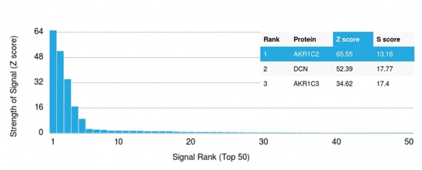 Application Data
(Analysis of Protein Array containing more than 19,000 full-length human proteins using Aldo-keto Reductase Family 1 Member C2/DD2 Mouse Monoclonal Antibody (CPTC- AKR1C2-1). Z- and S- Score: The Z-score represents the strength of a signal that a monoclonal antibody (MAb) (in combination with a fluorescently-tagged anti-IgG secondary antibody) produces when binding to a particular protein on the HuProtTM array. Z-scores are described in units of standard deviations (SD's) above the mean value of all signals generated on that array. If targets on HuProtTM are arranged in descending order of the Z-score, the S-score is the difference (also in units of SD's) between the Z-score. S-score therefore represents the relative target specificity of a MAb to its intended target. A MAb is considered to specific to its intended target, if the MAb has an S-score of at least 2.5. For example, if a MAb binds to protein X with a Z-score of 43 and to protein Y with a Z-score of 14, then the S-score for the binding of that MAb to protein X is equal to 29.)
Application Data
(Analysis of Protein Array containing more than 19,000 full-length human proteins using Aldo-keto Reductase Family 1 Member C2/DD2 Mouse Monoclonal Antibody (CPTC- AKR1C2-1). Z- and S- Score: The Z-score represents the strength of a signal that a monoclonal antibody (MAb) (in combination with a fluorescently-tagged anti-IgG secondary antibody) produces when binding to a particular protein on the HuProtTM array. Z-scores are described in units of standard deviations (SD's) above the mean value of all signals generated on that array. If targets on HuProtTM are arranged in descending order of the Z-score, the S-score is the difference (also in units of SD's) between the Z-score. S-score therefore represents the relative target specificity of a MAb to its intended target. A MAb is considered to specific to its intended target, if the MAb has an S-score of at least 2.5. For example, if a MAb binds to protein X with a Z-score of 43 and to protein Y with a Z-score of 14, then the S-score for the binding of that MAb to protein X is equal to 29.)
Aldo-keto Reductase Family 1 Member C2/DD2, Monoclonal Antibody (Cat# AAA214921)

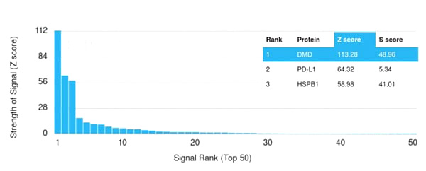 Application Data
(Analysis of Protein Array containing more than 19,000 full-length human proteins using Dystrophin Monospecific Mouse Monoclonal Antibody (DMD/3245). Z- and S- Score: The Z-score represents the strength of a signal that a monoclonal antibody (Monoclonal Antibody) (in combination with a fluorescently-tagged anti-IgG secondary antibody) produces when binding to a particular protein on the HuProtTM array. Z-scores are described in units of standard deviations (SD’s) above the mean value of all signals generated on that array. If targets on HuProtTM are arranged in descending order of the Z-score, the S-score is the difference (also in units of SD’s) between the Z-score. S-score therefore represents the relative target specificity of a Monoclonal Antibody to its intended target. A Monoclonal Antibody is considered to specific to its intended target, if the Monoclonal Antibody has an S-score of at least 2.5. For example, if a Monoclonal Antibody binds to protein X with a Z-score of 43 and to protein Y with a Z-score of 14, then the S-score for the binding of that Monoclonal Antibody to protein X is equal to 29.)
Application Data
(Analysis of Protein Array containing more than 19,000 full-length human proteins using Dystrophin Monospecific Mouse Monoclonal Antibody (DMD/3245). Z- and S- Score: The Z-score represents the strength of a signal that a monoclonal antibody (Monoclonal Antibody) (in combination with a fluorescently-tagged anti-IgG secondary antibody) produces when binding to a particular protein on the HuProtTM array. Z-scores are described in units of standard deviations (SD’s) above the mean value of all signals generated on that array. If targets on HuProtTM are arranged in descending order of the Z-score, the S-score is the difference (also in units of SD’s) between the Z-score. S-score therefore represents the relative target specificity of a Monoclonal Antibody to its intended target. A Monoclonal Antibody is considered to specific to its intended target, if the Monoclonal Antibody has an S-score of at least 2.5. For example, if a Monoclonal Antibody binds to protein X with a Z-score of 43 and to protein Y with a Z-score of 14, then the S-score for the binding of that Monoclonal Antibody to protein X is equal to 29.)
Dystrophin (DMD), Monoclonal Antibody (Cat# AAA214926)
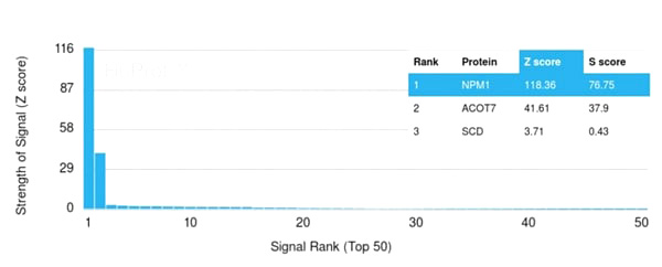 Application Data
(Analysis of Protein Array containing more than 19,000 full-length human proteins using Nucleophosmin-Monospecific Mouse Monoclonal Antibody (NPM1/3285) Z- and S- Score: The Z-score represents the strength of a signal that a monoclonal antibody (Monoclonal Antibody) (in combination with a fluorescently-tagged anti-IgG secondary antibody) produces when binding to a particular protein on the HuProtTM array. Z-scores are described in units of standard deviations (SD’s) above the mean value of all signals generated on that array. If targets on HuProtTM are arranged in descending order of the Z-score, the S-score is the difference (also in units of SD’s) between the Z-score. S-score therefore represents the relative target specificity of a Monoclonal Antibody to its intended target. A Monoclonal Antibody is considered to specific to its intended target, if the Monoclonal Antibody has an S-score of at least 2.5. For example, if a Monoclonal Antibody binds to protein X with a Z-score of 43 and to protein Y with a Z-score of 14, then the S-score for the binding of that Monoclonal Antibody to protein X is equal to 29.)
Application Data
(Analysis of Protein Array containing more than 19,000 full-length human proteins using Nucleophosmin-Monospecific Mouse Monoclonal Antibody (NPM1/3285) Z- and S- Score: The Z-score represents the strength of a signal that a monoclonal antibody (Monoclonal Antibody) (in combination with a fluorescently-tagged anti-IgG secondary antibody) produces when binding to a particular protein on the HuProtTM array. Z-scores are described in units of standard deviations (SD’s) above the mean value of all signals generated on that array. If targets on HuProtTM are arranged in descending order of the Z-score, the S-score is the difference (also in units of SD’s) between the Z-score. S-score therefore represents the relative target specificity of a Monoclonal Antibody to its intended target. A Monoclonal Antibody is considered to specific to its intended target, if the Monoclonal Antibody has an S-score of at least 2.5. For example, if a Monoclonal Antibody binds to protein X with a Z-score of 43 and to protein Y with a Z-score of 14, then the S-score for the binding of that Monoclonal Antibody to protein X is equal to 29.)
Nucleophosmin, Monoclonal Antibody (Cat# AAA215107)
 Application Data
(Analysis of Protein Array containing more than 19,000 full-length human proteins using Nucleophosmin-Monospecific Mouse Monoclonal Antibody (NPM1/3286) Z- and S- Score: The Z-score represents the strength of a signal that a monoclonal antibody (MAb) (in combination with a fluorescently-tagged anti-IgG secondary antibody) produces when binding to a particular protein on the HuProtTM array. Z-scores are described in units of standard deviations (SD’s) above the mean value of all signals generated on that array. If targets on HuProtTM are arranged in descending order of the Z-score, the S-score is the difference (also in units of SD’s) between the Z-score. S-score therefore represents the relative target specificity of a MAb to its intended target. A MAb is considered to specific to its intended target, if the MAb has an S-score of at least 2.5. For example, if a MAb binds to protein X with a Z-score of 43 and to protein Y with a Z-score of 14, then the S-score for the binding of that MAb to protein X is equal to 29.)
Application Data
(Analysis of Protein Array containing more than 19,000 full-length human proteins using Nucleophosmin-Monospecific Mouse Monoclonal Antibody (NPM1/3286) Z- and S- Score: The Z-score represents the strength of a signal that a monoclonal antibody (MAb) (in combination with a fluorescently-tagged anti-IgG secondary antibody) produces when binding to a particular protein on the HuProtTM array. Z-scores are described in units of standard deviations (SD’s) above the mean value of all signals generated on that array. If targets on HuProtTM are arranged in descending order of the Z-score, the S-score is the difference (also in units of SD’s) between the Z-score. S-score therefore represents the relative target specificity of a MAb to its intended target. A MAb is considered to specific to its intended target, if the MAb has an S-score of at least 2.5. For example, if a MAb binds to protein X with a Z-score of 43 and to protein Y with a Z-score of 14, then the S-score for the binding of that MAb to protein X is equal to 29.)
Nucleophosmin, Monoclonal Antibody (Cat# AAA215108)
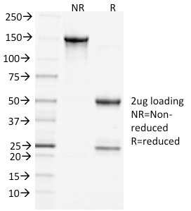
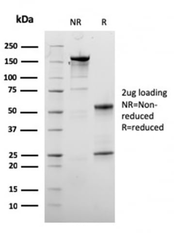
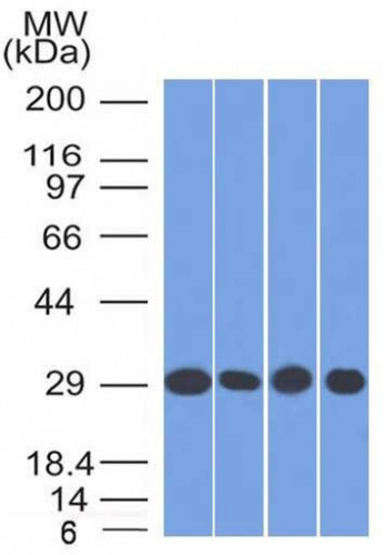
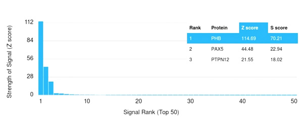
 SDS-PAGE
(SDS-PAGE Analysis Purified Prohibitin Mouse Monoclonal Antibody (SPM311). Confirmation of Purity and Integrity of Antibody.)
SDS-PAGE
(SDS-PAGE Analysis Purified Prohibitin Mouse Monoclonal Antibody (SPM311). Confirmation of Purity and Integrity of Antibody.)
Prohibitin, Monoclonal Antibody (Cat# AAA215140)
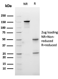
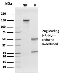 SDS_PAGE
(SDS-PAGE Analysis Purified Prohibitin Mouse Monoclonal Antibody (PHB/1882). Confirmation of Purity and Integrity of Antibody.)
SDS_PAGE
(SDS-PAGE Analysis Purified Prohibitin Mouse Monoclonal Antibody (PHB/1882). Confirmation of Purity and Integrity of Antibody.)
Prohibitin, Monoclonal Antibody (Cat# AAA215141)
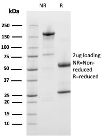
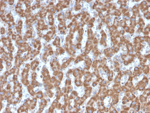
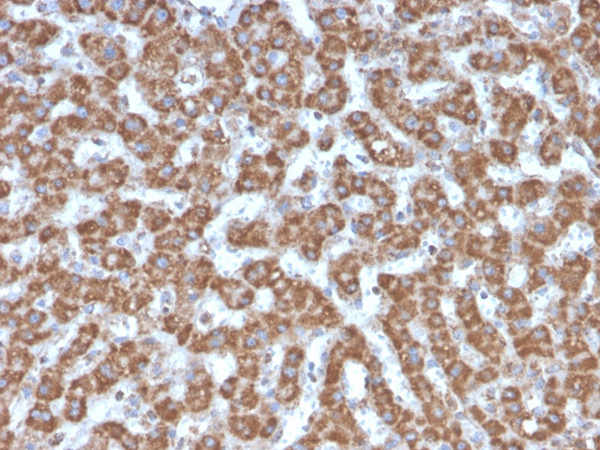
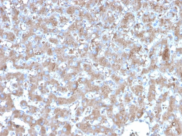
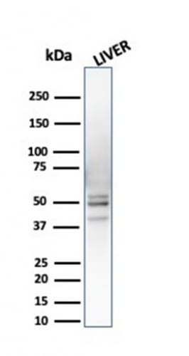
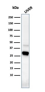 WB (Western Blot)
(Western Blot Analysis of human liver tissue lysate using Prohibitin Mouse Monoclonal Antibody (PHB/3226).)
WB (Western Blot)
(Western Blot Analysis of human liver tissue lysate using Prohibitin Mouse Monoclonal Antibody (PHB/3226).)
Prohibitin, Monoclonal Antibody (Cat# AAA215143)
What are Monoclonal Antibodies?
Monoclonal antibodies are specialized laboratory-produced proteins developed for binding to specific biological antigens or other molecular targets. Since they come from a single cell (or clone), they are especially consistent and accurate in the data they are involved in producing.
This type of antibody material has been shown to be a powerful tool in finding and subsequently destroying harmful cells in an organism, such as those found in cancers or various autoimmune diseases. This makes them excellent aids in medical testing and research, which is why they are so widely used.
AAA Biotech offers a comprehensive range of high-quality monoclonal antibodies that perform effectively in various laboratory tests, including (amongst others) ELISA, western blotting, immunohistochemistry, and flow cytometry. All of the products in our catalog are thoroughly quality tested to make sure that they are reliable and will consistently perform well in your research.
What Are The Uses of Monoclonal Antibodies
Monoclonal antibodies are used in many lab tests, including (amongst others) ELISA, western blotting, immunohistochemistry, and flow cytometry.
ELISA is a test that helps detect a specific substance/analyte in a sample. It uses antibodies (often monoclonal) bound to a solid surface (such as the well of a microplate) to “capture” the substance/analyte in the sample and immobilize it so that the detection antibody component can then bind to it and produce a signal, which can then be measured.
Western blotting identifies specific proteins in a sample. The sample is first separated on a gel, and then antibodies are applied that will typically bind to the target, which will all be localized to a single band in a lane.
Immunohistochemistry helps locate specific proteins in cells or tissue samples using antibodies.
Flow cytometry looks at and sorts cells. It uses antibodies that are conjugated to reporter molecules called “fluorophores”, which, under special lights, emit light themselves, which can then be measured by a detector instrument. For a deeper understanding of these techniques, explore our complete guide to monoclonal antibodies and their benefits.
How Monoclonal Antibodies Are Used as Medicine?
Please note that all of the products listed in AAA Biotech’s also known as AAA Bio or AAABio catalog are strictly for research-use only (RUO).
Monoclonal antibodies can also be used as therapeutic/medical treatments, particularly in the context of cancers. They are designed to find and bind to specific cells or proteins, helping the immune system recognize and attack the cancer. These treatments work in different ways, such as:
- Radioimmunotherapy attaches a small amount of radioactive molecule to the antibody, so it delivers the radiation directly to the cancer cells that the antibody is specifically binding to.
- Antibody-directed enzyme prodrug therapy uses antibodies that are specifically bound to special enzymes. These enzymes activate a harmless drug in the body and turn it into a cancer-killing drug only near the cancer cells—this helps avoid harming healthy cells.
- Immunoliposomes are tiny “bubbles” filled with medicine/drug and coated with antibodies. They carry the drug straight to the cancer cells.
Why Buy Monoclonal Antibodies From Us?
At AAA Biotech, we provide high-performance monoclonal antibodies designed to support a wide range of research needs.
1. Validated for Versatile Applications
The antibodies in our catalog are extensively validated and compatible with multiple techniques, including (but not limited to) ELISA, flow cytometry (FC), immunocytochemistry (ICC), immunofluorescence (IF), immunohistochemistry (IHC), immunoprecipitation (IP), and western blotting (WB).
2. Wide Selection & Specialized Options
We offer antibodies for common and rare species, that are available in various conjugated forms, and also in recombinant formats. Essentially, there is almost anything one might need to meet their experimental model’s requirements.
3. High-Quality Proteins
Our proteins meet high purity standards—90% or more as confirmed by SDS-PAGE. Many are available with tags like His, Flag, GST, or MBP, and we also supply native and biologically active proteins for functional studies.
Frequently Asked Questions
1. Are your monoclonal antibodies validated for specific applications?
Yes, our antibodies are tested and validated for use in methods such as ELISA, western blot, IHC, flow cytometry, and more. Refer to specific product pages or datasheets for individual product information.
2. How do I choose the right monoclonal antibody for my application?
Review the product details directly for application validation, species reactivity, and target information. You may also contact our support team at any time for help.
3. How quickly can I receive my order?
Most orders are processed and shipped within 1–3 business days, depending on product availability and your shipping location.