Filters
▼Clonality
▼Type
▼Reactivity
▼Gene Name
▼Isotype
▼Host
▼Application
▼Clone
▼Monoclonal Antibodies
Get accurate results in your research with our Monoclonal Antibodies, which are specially made to target exactly what you require for your research, and will produce consistent, reliable performance in lab tests.
Viewing 1600-1650 of 27645 product results
 WB (Western Blot)
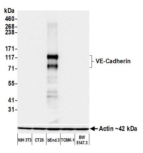
(Detection of mouse VE-Cadherin by western blot. Samples: Whole cell lysate (2 ug) from NIH 3T3, CT26, bEnd.3, TCMK-1, and BW5147.3 cells prepared using NETN lysis buffer. Antibody: Rabbit anti-VE-Cadherin recombinant monoclonal antibody (AAA213655 lot 1) used at 1:1000. Secondary: HRP-conjugated goat anti-rabbit IgG . Detection: Chemiluminescence with an exposure time of 3 seconds. Lower Panel: Rabbit anti-Actin recombinant monoclonal antibody .)
WB (Western Blot)
(Detection of mouse VE-Cadherin by western blot. Samples: Whole cell lysate (2 ug) from NIH 3T3, CT26, bEnd.3, TCMK-1, and BW5147.3 cells prepared using NETN lysis buffer. Antibody: Rabbit anti-VE-Cadherin recombinant monoclonal antibody (AAA213655 lot 1) used at 1:1000. Secondary: HRP-conjugated goat anti-rabbit IgG . Detection: Chemiluminescence with an exposure time of 3 seconds. Lower Panel: Rabbit anti-Actin recombinant monoclonal antibody .)
VE-Cadherin, Monoclonal Recombinant Antibody (Cat# AAA213655)
 WB (Western Blot)
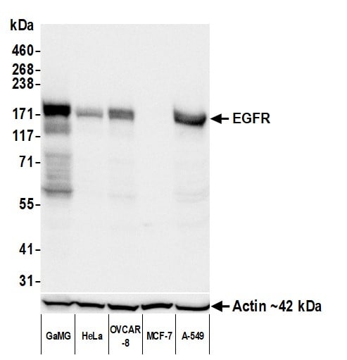
(Detection of human EGFR by western blot. Samples: Whole cell lysate (50 ug) from GaMG, HeLa, OVCAR-8, MCF-7, and A-549 cells prepared using NETN lysis buffer. Antibody: Rabbit anti-EGFR recombinant monoclonal antibody (AAA213664 lot 1) used at 1:1000. Secondary: HRP-conjugated goat anti-rabbit IgG . Detection: Chemiluminescence with an exposure time of 1 second. Lower Panel: Rabbit anti-Actin recombinant monoclonal antibody .)
WB (Western Blot)
(Detection of human EGFR by western blot. Samples: Whole cell lysate (50 ug) from GaMG, HeLa, OVCAR-8, MCF-7, and A-549 cells prepared using NETN lysis buffer. Antibody: Rabbit anti-EGFR recombinant monoclonal antibody (AAA213664 lot 1) used at 1:1000. Secondary: HRP-conjugated goat anti-rabbit IgG . Detection: Chemiluminescence with an exposure time of 1 second. Lower Panel: Rabbit anti-Actin recombinant monoclonal antibody .)
EGFR, Monoclonal Recombinant Antibody (Cat# AAA213664)
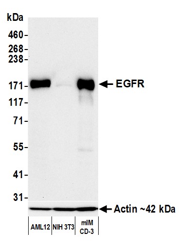 WB (Western Blot)
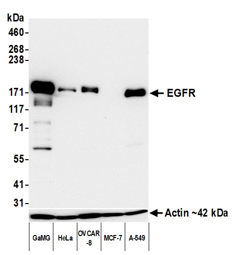
(Detection of mouse EGFR by western blot. Samples: Whole cell lysate (10 ug) from AML12, NIH 3T3, and mIMCD-3 cells prepared using NETN lysis buffer. Antibody: Rabbit anti-EGFR recombinant monoclonal antibody (AAA213665 lot 1) used at 1:1000. Secondary: HRP-conjugated goat anti-rabbit IgG . Detection: Chemiluminescence with an exposure time of 30 seconds. Lower Panel: Rabbit anti-Actin recombinant monoclonal antibody .)
WB (Western Blot)
(Detection of mouse EGFR by western blot. Samples: Whole cell lysate (10 ug) from AML12, NIH 3T3, and mIMCD-3 cells prepared using NETN lysis buffer. Antibody: Rabbit anti-EGFR recombinant monoclonal antibody (AAA213665 lot 1) used at 1:1000. Secondary: HRP-conjugated goat anti-rabbit IgG . Detection: Chemiluminescence with an exposure time of 30 seconds. Lower Panel: Rabbit anti-Actin recombinant monoclonal antibody .)
EGFR, Monoclonal Recombinant Antibody (Cat# AAA213665)
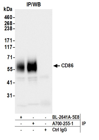
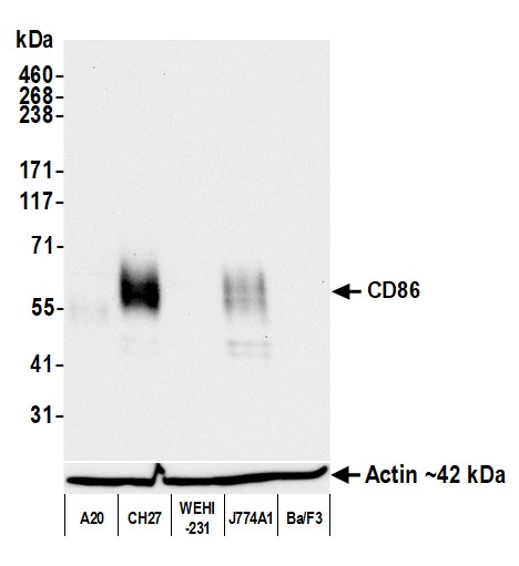 WB (Western Blot)
(Detection of mouse CD86 by western blot. Samples: Whole cell lysate (10 ug) from A20, CH27, WEHI-231, J774A1, and Ba/F3 cells prepared using NETN lysis buffer. Antibody: Rabbit anti-CD86 recombinant monoclonal antibody (AAA213668 lot 1) used at 1:1000. Secondary: HRP-conjugated goat anti-rabbit IgG . Detection: Chemiluminescence with an exposure time of 30 seconds. Lower Panel: Rabbit anti-Actin recombinant monoclonal antibody .)
WB (Western Blot)
(Detection of mouse CD86 by western blot. Samples: Whole cell lysate (10 ug) from A20, CH27, WEHI-231, J774A1, and Ba/F3 cells prepared using NETN lysis buffer. Antibody: Rabbit anti-CD86 recombinant monoclonal antibody (AAA213668 lot 1) used at 1:1000. Secondary: HRP-conjugated goat anti-rabbit IgG . Detection: Chemiluminescence with an exposure time of 30 seconds. Lower Panel: Rabbit anti-Actin recombinant monoclonal antibody .)
CD86, Monoclonal Recombinant Antibody (Cat# AAA213668)
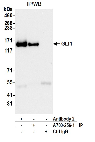
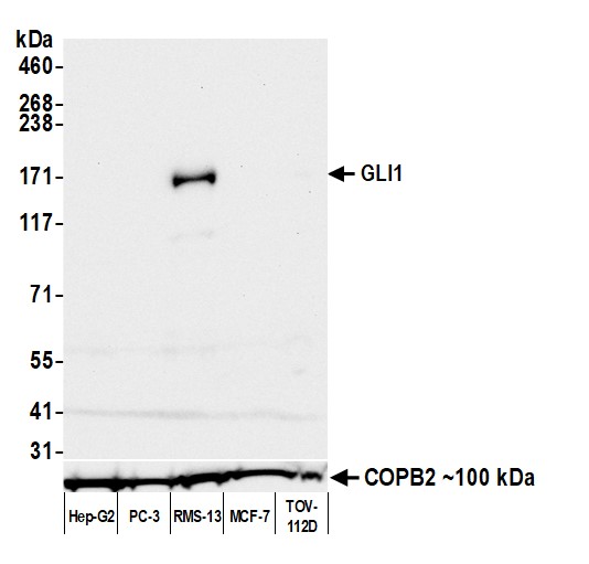 WB (Western Blot)
(Detection of human GLI1 by western blot. Samples: Whole cell lysate (50 ug) from Hep-G2, PC-3, RMS-13, MCF-7, and TOV-112D cells prepared using NETN lysis buffer. Antibody: Rabbit anti-GLI1 recombinant monoclonal antibody (AAA213669 lot 1) used at 1:1000. Secondary: HRP-conjugated goat anti-rabbit IgG . Detection: Chemiluminescence with an exposure time of 30 seconds. Lower Panel: Rabbit anti-COPB2 antibody .)
WB (Western Blot)
(Detection of human GLI1 by western blot. Samples: Whole cell lysate (50 ug) from Hep-G2, PC-3, RMS-13, MCF-7, and TOV-112D cells prepared using NETN lysis buffer. Antibody: Rabbit anti-GLI1 recombinant monoclonal antibody (AAA213669 lot 1) used at 1:1000. Secondary: HRP-conjugated goat anti-rabbit IgG . Detection: Chemiluminescence with an exposure time of 30 seconds. Lower Panel: Rabbit anti-COPB2 antibody .)
GLI1, Monoclonal Recombinant Antibody (Cat# AAA213669)
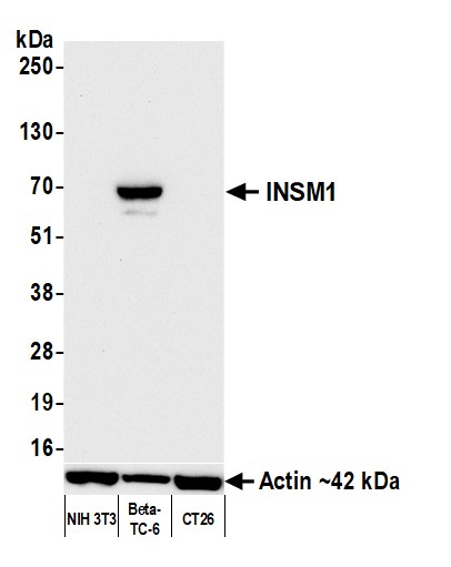
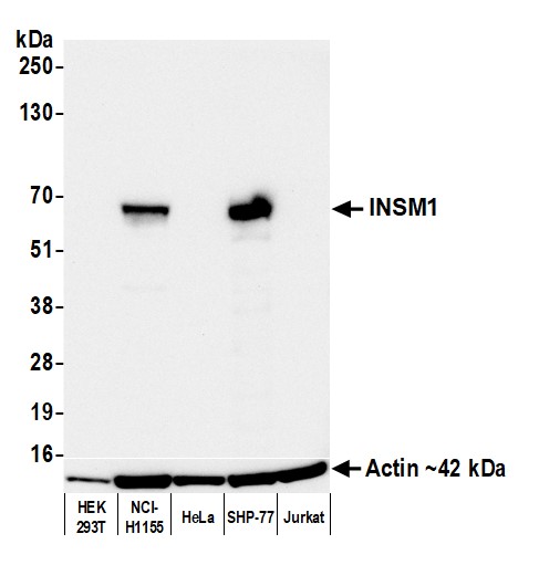 WB (Western Blot)
(Detection of human INSM1 by western blot. Samples: Whole cell lysate (25 ug) from HEK293T, NCI-H1155, HeLa, SHP-77, and Jurkat cells prepared using NETN lysis buffer. Antibody: Rabbit anti-INSM1 recombinant monoclonal antibody (AAA213677 lot 1) used at 1:1000. Secondary: HRP-conjugated goat anti-rabbit IgG . Detection: Chemiluminescence with an exposure time of 10 seconds. Lower Panel: Rabbit anti-Actin recombinant monoclonal antibody .)
WB (Western Blot)
(Detection of human INSM1 by western blot. Samples: Whole cell lysate (25 ug) from HEK293T, NCI-H1155, HeLa, SHP-77, and Jurkat cells prepared using NETN lysis buffer. Antibody: Rabbit anti-INSM1 recombinant monoclonal antibody (AAA213677 lot 1) used at 1:1000. Secondary: HRP-conjugated goat anti-rabbit IgG . Detection: Chemiluminescence with an exposure time of 10 seconds. Lower Panel: Rabbit anti-Actin recombinant monoclonal antibody .)
INSM1, Monoclonal Recombinant Antibody (Cat# AAA213677)
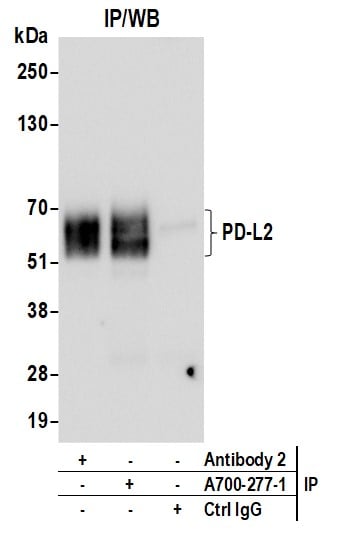
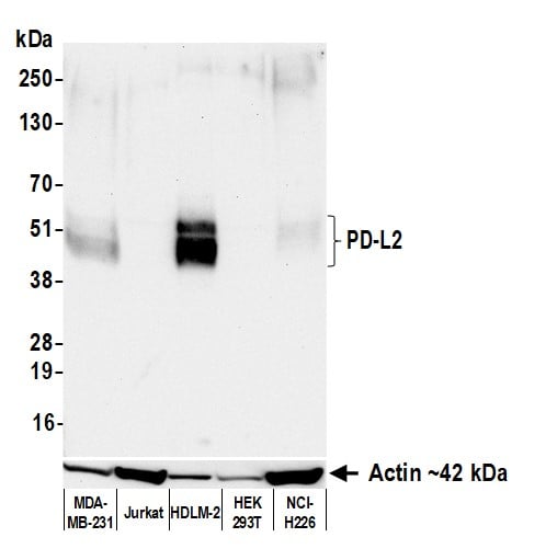 WB (Western Blot)
(Detection of human PD-L2 by western blot. Samples: Whole cell lysate (50 ug) from MDA-MB-231, Jurkat, HDLM-2 (10 ug), HEK293T, and NCI-H226 cells prepared using NETN lysis buffer. Antibody: Rabbit anti-PD-L2 recombinant monoclonal antibody (AAA213679 lot 1) used at 1:1000. Secondary: HRP-conjugated goat anti-rabbit IgG . Detection: Chemiluminescence with an exposure time of 30 seconds.)
WB (Western Blot)
(Detection of human PD-L2 by western blot. Samples: Whole cell lysate (50 ug) from MDA-MB-231, Jurkat, HDLM-2 (10 ug), HEK293T, and NCI-H226 cells prepared using NETN lysis buffer. Antibody: Rabbit anti-PD-L2 recombinant monoclonal antibody (AAA213679 lot 1) used at 1:1000. Secondary: HRP-conjugated goat anti-rabbit IgG . Detection: Chemiluminescence with an exposure time of 30 seconds.)
PD-L2, Monoclonal Recombinant Antibody (Cat# AAA213679)
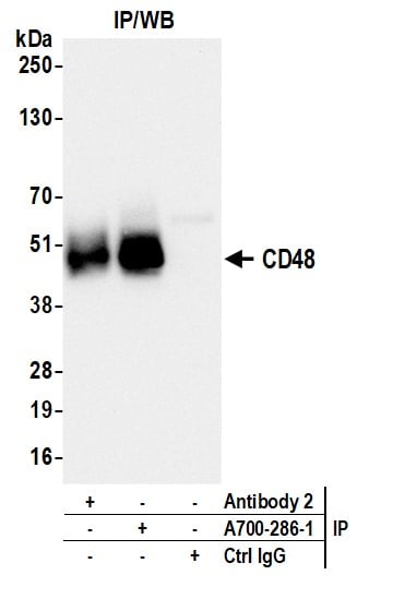
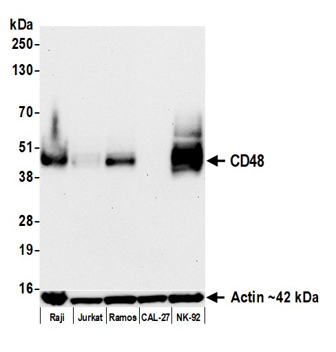 WB (Western Blot)
(Detection of human CD48 by western blot. Samples: Whole cell lysate (10 ug) from Raji, Jurkat, Ramos, CAL-27, and NK-92 cells prepared using NETN lysis buffer. Antibody: Rabbit anti-CD48 recombinant monoclonal antibody (AAA213686 lot 1) used at 1:1000. Secondary: HRP-conjugated goat anti-rabbit IgG . Detection: Chemiluminescence with an exposure time of 75 seconds. Lower Panel: Rabbit anti-Actin recombinant monoclonal antibody .)
WB (Western Blot)
(Detection of human CD48 by western blot. Samples: Whole cell lysate (10 ug) from Raji, Jurkat, Ramos, CAL-27, and NK-92 cells prepared using NETN lysis buffer. Antibody: Rabbit anti-CD48 recombinant monoclonal antibody (AAA213686 lot 1) used at 1:1000. Secondary: HRP-conjugated goat anti-rabbit IgG . Detection: Chemiluminescence with an exposure time of 75 seconds. Lower Panel: Rabbit anti-Actin recombinant monoclonal antibody .)
CD48, Monoclonal Recombinant Antibody (Cat# AAA213686)
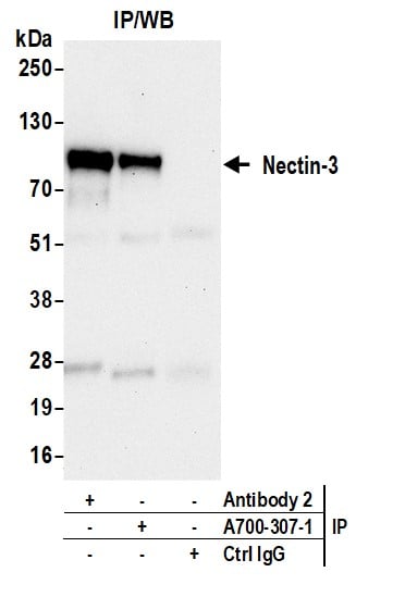
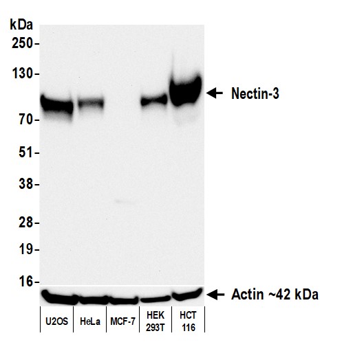 WB (Western Blot)
(Detection of human Nectin-3 by western blot. Samples: Whole cell lysate (50 ug) from U2OS, HeLa, MCF-7, HEK293T, and HCT 116 cells prepared using NETN lysis buffer. Antibody: Rabbit anti-Nectin-3 recombinant monoclonal antibody (AAA213703 lot 1) used at 1:1000. Secondary: HRP-conjugated goat anti-rabbit IgG . Detection: Chemiluminescence with an exposure time of 30 seconds. Lower Panel: Rabbit anti-Actin recombinant monoclonal antibody .)
WB (Western Blot)
(Detection of human Nectin-3 by western blot. Samples: Whole cell lysate (50 ug) from U2OS, HeLa, MCF-7, HEK293T, and HCT 116 cells prepared using NETN lysis buffer. Antibody: Rabbit anti-Nectin-3 recombinant monoclonal antibody (AAA213703 lot 1) used at 1:1000. Secondary: HRP-conjugated goat anti-rabbit IgG . Detection: Chemiluminescence with an exposure time of 30 seconds. Lower Panel: Rabbit anti-Actin recombinant monoclonal antibody .)
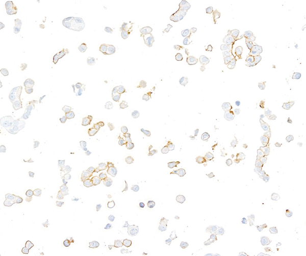
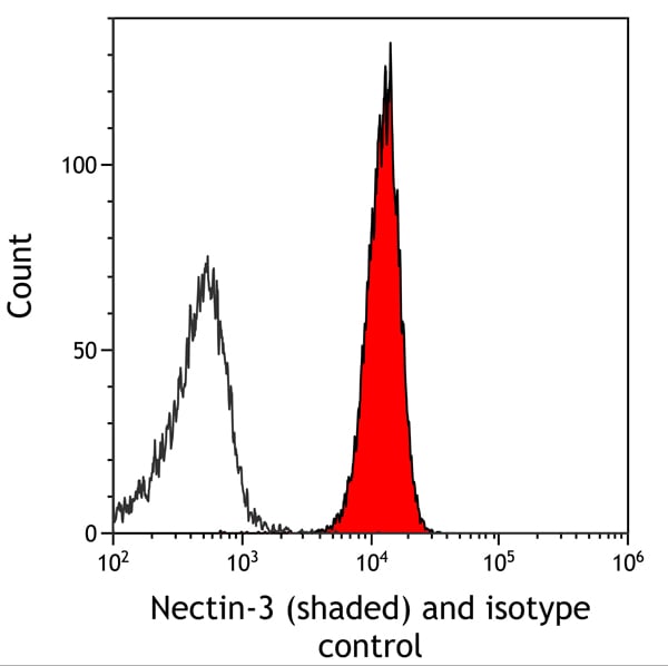
Nectin-3, Monoclonal Recombinant Antibody (Cat# AAA213703)
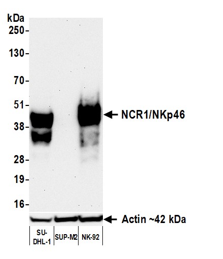 WB (Western Blot)
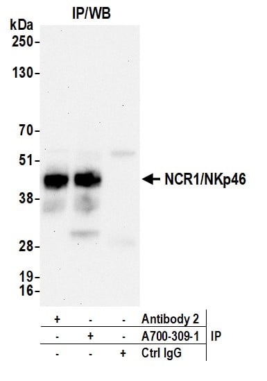
(Detection of human NCR1/NKp46 by western blot. Samples: Whole cell lysate (50 ug) from SU-DHL-1, SUP-M2, and NK-92 cells prepared using NETN lysis buffer. Antibody: Rabbit anti-NCR1/NKp46 recombinant monoclonal antibody (AAA213705 lot 1) used at 1:1000. Secondary: HRP-conjugated goat anti-rabbit IgG . Detection: Chemiluminescence with an exposure time of 3 minutes.)
WB (Western Blot)
(Detection of human NCR1/NKp46 by western blot. Samples: Whole cell lysate (50 ug) from SU-DHL-1, SUP-M2, and NK-92 cells prepared using NETN lysis buffer. Antibody: Rabbit anti-NCR1/NKp46 recombinant monoclonal antibody (AAA213705 lot 1) used at 1:1000. Secondary: HRP-conjugated goat anti-rabbit IgG . Detection: Chemiluminescence with an exposure time of 3 minutes.)
NCR1/NKp46, Monoclonal Recombinant Antibody (Cat# AAA213705)
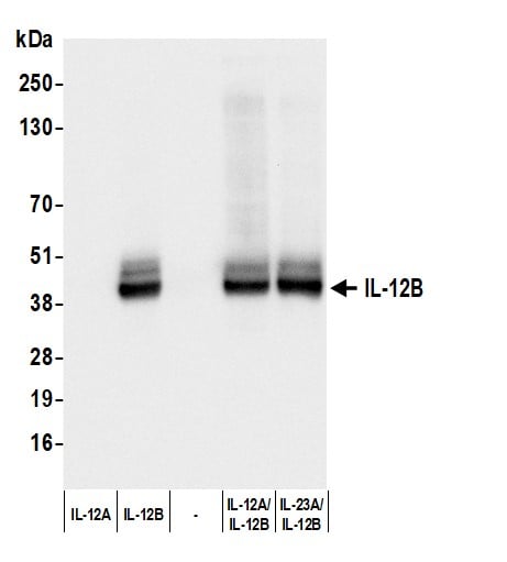
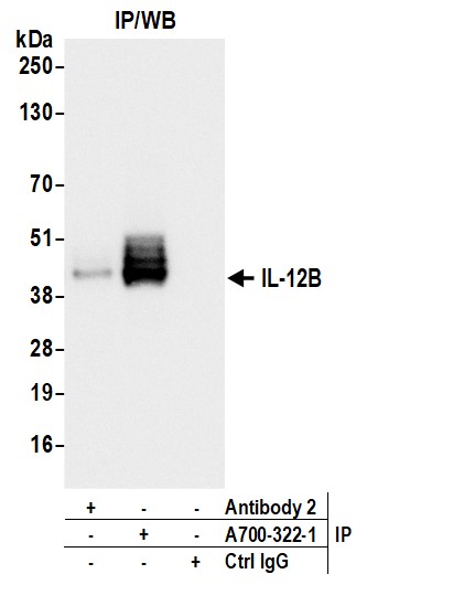
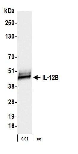 WB (Western Blot)
(Detection of human IL-12B by western blot. Samples: Human recombinant IL-12B protein (0.01 ug). Antibody: Rabbit anti-IL-12B recombinant monoclonal antibody (AAA213709 lot 1) used at 1:1000. Secondary: HRP-conjugated goat anti-rabbit IgG . Detection: Chemiluminescence with an exposure time of 1 second.)
WB (Western Blot)
(Detection of human IL-12B by western blot. Samples: Human recombinant IL-12B protein (0.01 ug). Antibody: Rabbit anti-IL-12B recombinant monoclonal antibody (AAA213709 lot 1) used at 1:1000. Secondary: HRP-conjugated goat anti-rabbit IgG . Detection: Chemiluminescence with an exposure time of 1 second.)
IL-12B, Monoclonal Recombinant Antibody (Cat# AAA213709)
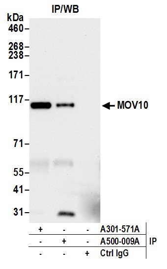
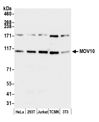 WB (Western Blot)
(Detection of human and mouse MOV10 by western blot. Samples: Whole cell lysate (15 ug) from HeLa, HEK293T, Jurkat, mouse TCMK-1, and mouse NIH 3T3 cells prepared using NETN lysis buffer. Antibody: Mouse monoclonal anti-MOV10 antibody [15C1B8] (AAA213495 lot 3) used at 1:1000. Secondary: HRP-conjugated goat anti-mouse IgG . Detection: Chemiluminescence with an exposure time of 10 seconds.)
WB (Western Blot)
(Detection of human and mouse MOV10 by western blot. Samples: Whole cell lysate (15 ug) from HeLa, HEK293T, Jurkat, mouse TCMK-1, and mouse NIH 3T3 cells prepared using NETN lysis buffer. Antibody: Mouse monoclonal anti-MOV10 antibody [15C1B8] (AAA213495 lot 3) used at 1:1000. Secondary: HRP-conjugated goat anti-mouse IgG . Detection: Chemiluminescence with an exposure time of 10 seconds.)
MOV10, Monoclonal Antibody (Cat# AAA213495)
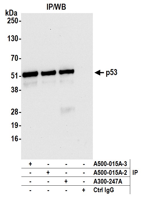
 WB (Western Blot)
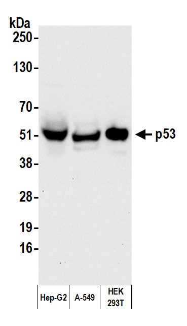
(Detection of human p53 by western blot. Samples: Whole cell lysate from Hep-G2 (50 ug), A-549 (50 ug), and HEK293T (1 ug) cells prepared using NETN lysis buffer. Antibody: Mouse anti-p53 monoclonal antibody [DO-1] (AAA213501 lot 3) used at 1:1000. Secondary: HRP-conjugated goat anti-mouse IgG . Detection: Chemiluminescence with an exposure time of 10 seconds.)
WB (Western Blot)
(Detection of human p53 by western blot. Samples: Whole cell lysate from Hep-G2 (50 ug), A-549 (50 ug), and HEK293T (1 ug) cells prepared using NETN lysis buffer. Antibody: Mouse anti-p53 monoclonal antibody [DO-1] (AAA213501 lot 3) used at 1:1000. Secondary: HRP-conjugated goat anti-mouse IgG . Detection: Chemiluminescence with an exposure time of 10 seconds.)
p53, Monoclonal Antibody (Cat# AAA213501)
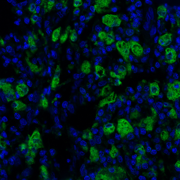
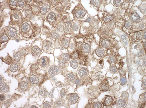
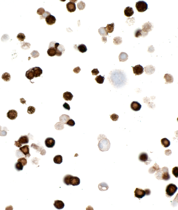
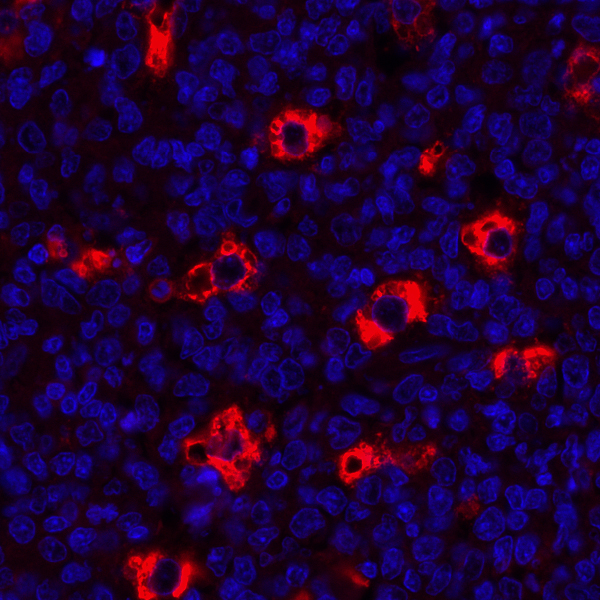 IHC (Immunohistochemistry)
(Detection of human CD68 by immunohistochemistry. Sample: FFPE section of human tonsil. Antibody: Mouse monoclonal anti-CD68 antibody [KP-1] (AAA213502) used at 1:100. Secondary: DyLight 594-conjugated goat anti-mouse IgG .)
IHC (Immunohistochemistry)
(Detection of human CD68 by immunohistochemistry. Sample: FFPE section of human tonsil. Antibody: Mouse monoclonal anti-CD68 antibody [KP-1] (AAA213502) used at 1:100. Secondary: DyLight 594-conjugated goat anti-mouse IgG .)
CD68, Monoclonal Recombinant Antibody (Cat# AAA213502)
 IHC (Immunohistochemisry)
(Detection of human CD57 in FFPE tonsil by IHC. Antibody: Mouse anti-CD57 monoclonal antibody [HNK-1] (AAA213507 Lot 1). Secondary: HRP-conjugated goat anti-mouse IgM . Substrate: DAB.)
IHC (Immunohistochemisry)
(Detection of human CD57 in FFPE tonsil by IHC. Antibody: Mouse anti-CD57 monoclonal antibody [HNK-1] (AAA213507 Lot 1). Secondary: HRP-conjugated goat anti-mouse IgM . Substrate: DAB.)
CD57, Monoclonal Antibody (Cat# AAA213507)
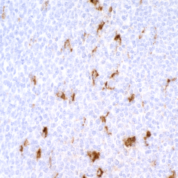
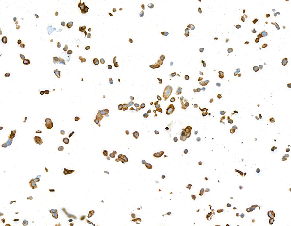
 IHC (Immunohiostchemistry)
(Detection of mouse Ly-6G in mouse spleen by IHC. Antibody: Rat anti-Ly-6G monoclonal antibody [1A8] (AAA213509 lot 1). Secondary: HRP-conjugated goat anti-rat IgG . Substrate: DAB)
IHC (Immunohiostchemistry)
(Detection of mouse Ly-6G in mouse spleen by IHC. Antibody: Rat anti-Ly-6G monoclonal antibody [1A8] (AAA213509 lot 1). Secondary: HRP-conjugated goat anti-rat IgG . Substrate: DAB)
Ly-6G, Monoclonal Antibody (Cat# AAA213509)

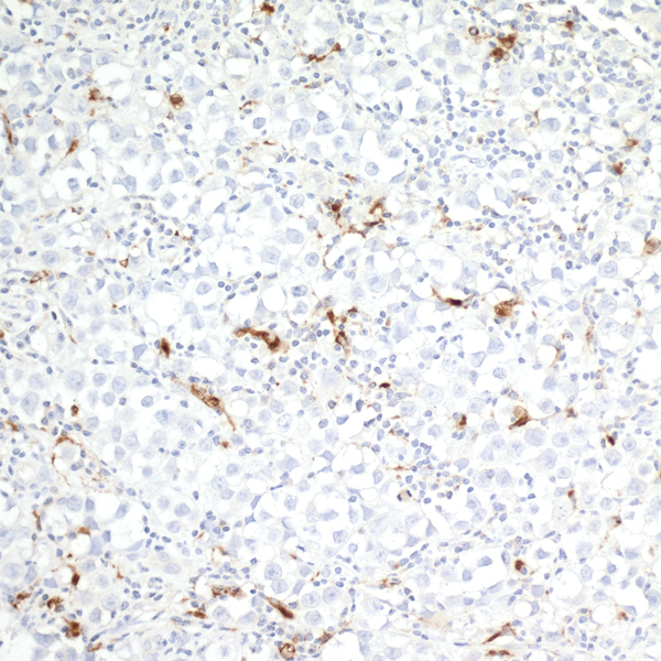
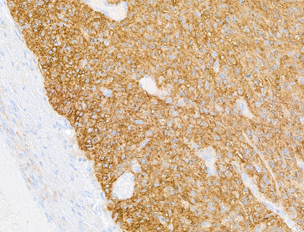
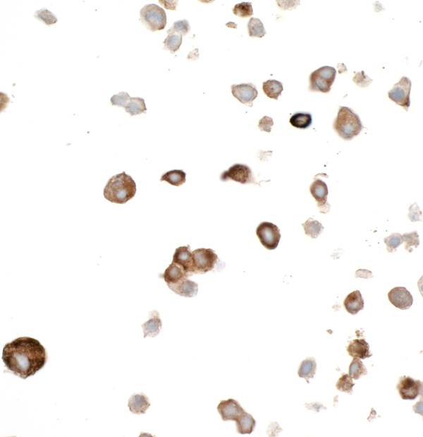
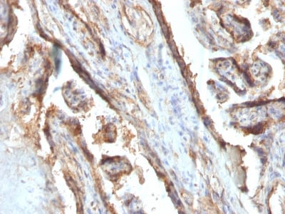
 IHC (Immunohiostchemistry)
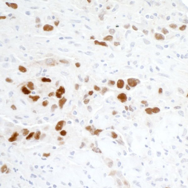
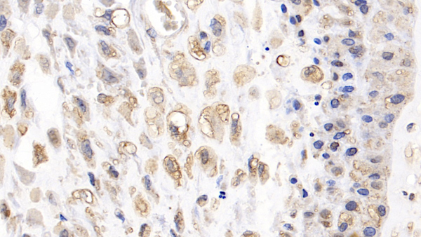
(Detection of human Fibronectin by immunohistochemistry. Sample: FFPE section of small cell lung cancer. Antibody: Mouse anti-Fibronectin monoclonal antibody [TV-1] (AAA213510 lot 1). Secondary: HRP-conjugated goat anti-mouse IgG . Substrate: DAB)
IHC (Immunohiostchemistry)
(Detection of human Fibronectin by immunohistochemistry. Sample: FFPE section of small cell lung cancer. Antibody: Mouse anti-Fibronectin monoclonal antibody [TV-1] (AAA213510 lot 1). Secondary: HRP-conjugated goat anti-mouse IgG . Substrate: DAB)
Fibronectin, Monoclonal Antibody (Cat# AAA213510)
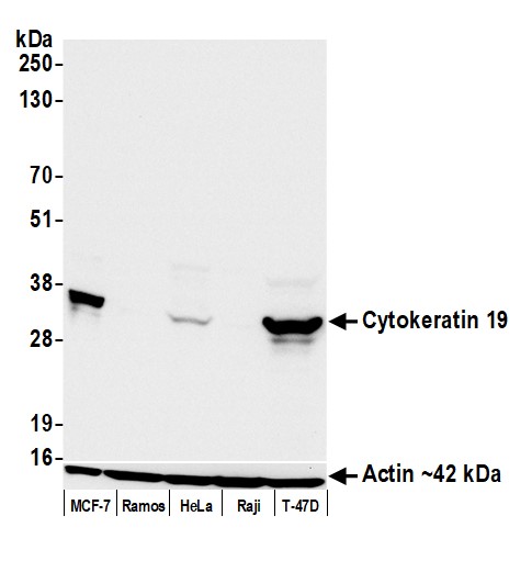 WB (Western Blot)
(Detection of human Cytokeratin 19 by western blot. Samples: Whole cell lysate (50 ug) from MCF-7 (5 ug), Ramos, HeLa, Raji, and T-47D cells prepared using NETN lysis buffer. Antibody: Mouse anti-Cytokeratin 19 monoclonal antibody [BA17] (AAA213515 lot 1) used at 1:1000. Secondary: HRP-conjugated goat anti-mouse IgG . Detection: Chemiluminescence with an exposure time of 10 seconds. Lower Panel: Rabbit anti-Actin recombinant monoclonal antibody .)
WB (Western Blot)
(Detection of human Cytokeratin 19 by western blot. Samples: Whole cell lysate (50 ug) from MCF-7 (5 ug), Ramos, HeLa, Raji, and T-47D cells prepared using NETN lysis buffer. Antibody: Mouse anti-Cytokeratin 19 monoclonal antibody [BA17] (AAA213515 lot 1) used at 1:1000. Secondary: HRP-conjugated goat anti-mouse IgG . Detection: Chemiluminescence with an exposure time of 10 seconds. Lower Panel: Rabbit anti-Actin recombinant monoclonal antibody .)
Cytokeratin 19, Monoclonal Antibody (Cat# AAA213515)
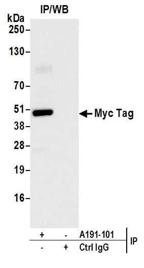
 WB (Western Blot)
(Detection of Myc-tagged protein by western blot of lysate from non-transfected human HEK293 cells, HEK293 transfected with Met-Myc-IDO1, HEK293 transfected with IDO1-Myc (C-terminal Tag), and HEK293 transfected with IDO1-Myc-HIS (Internal Tag). Antibody: Rabbit anti-Myc Tag recombinant monoclonal antibody (AAA210713 lot 1) used at 1:1000. Secondary: HRP-conjugated goat anti-rabbit IgG . Detection: Chemiluminescence with an exposure time of 3 seconds.)
WB (Western Blot)
(Detection of Myc-tagged protein by western blot of lysate from non-transfected human HEK293 cells, HEK293 transfected with Met-Myc-IDO1, HEK293 transfected with IDO1-Myc (C-terminal Tag), and HEK293 transfected with IDO1-Myc-HIS (Internal Tag). Antibody: Rabbit anti-Myc Tag recombinant monoclonal antibody (AAA210713 lot 1) used at 1:1000. Secondary: HRP-conjugated goat anti-rabbit IgG . Detection: Chemiluminescence with an exposure time of 3 seconds.)
Myc Tag, Monoclonal Recombinant Antibody (Cat# AAA210713)
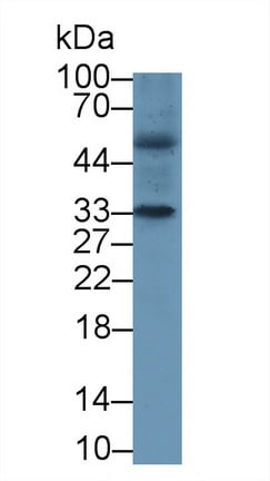
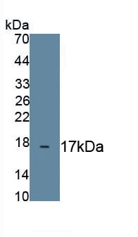
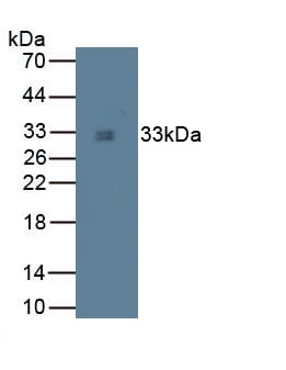
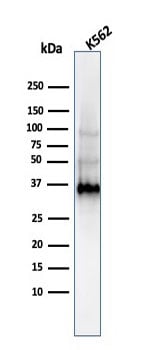 WB (Western Blot)
(Western Blot of K562 Cell Lysate using Galeactin-13 MAb (PP13/1161).)
WB (Western Blot)
(Western Blot of K562 Cell Lysate using Galeactin-13 MAb (PP13/1161).)
Galectin-13 (GAL13)/Placental Protein 13 (PP13), Monoclonal Antibody (Cat# AAA214372)

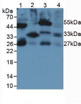
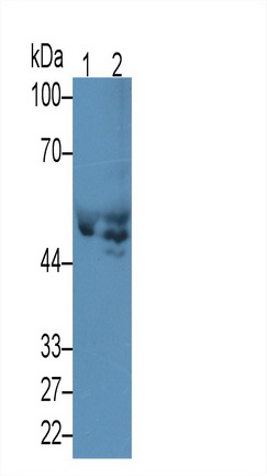
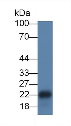
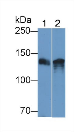
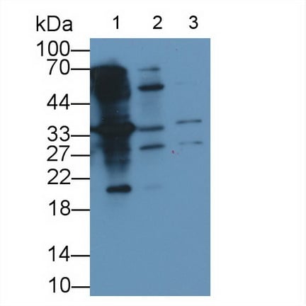
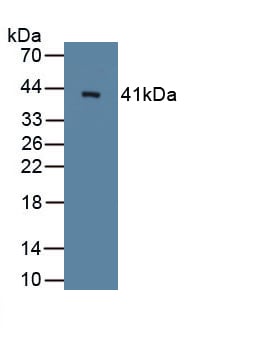
 WB (Western Blot)
(Western Blot Analysis (A) MCF-7 (B) PC3 Cell lysate Using FOXA1 Monoclonal Antibody (FOXA1/1512))
WB (Western Blot)
(Western Blot Analysis (A) MCF-7 (B) PC3 Cell lysate Using FOXA1 Monoclonal Antibody (FOXA1/1512))
FOXA1/HNF3A, Monoclonal Antibody (Cat# AAA214383)
Does not react with rat. Others not known.
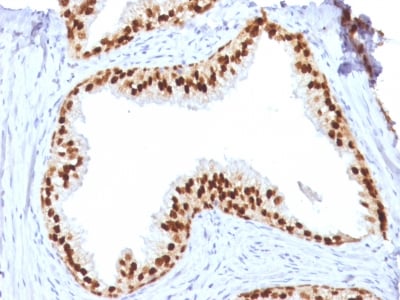
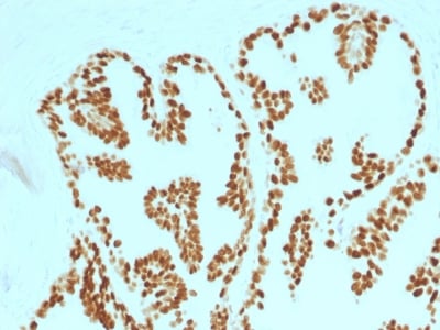
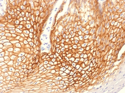
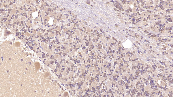
 IHC (Immunohiostchemistry)
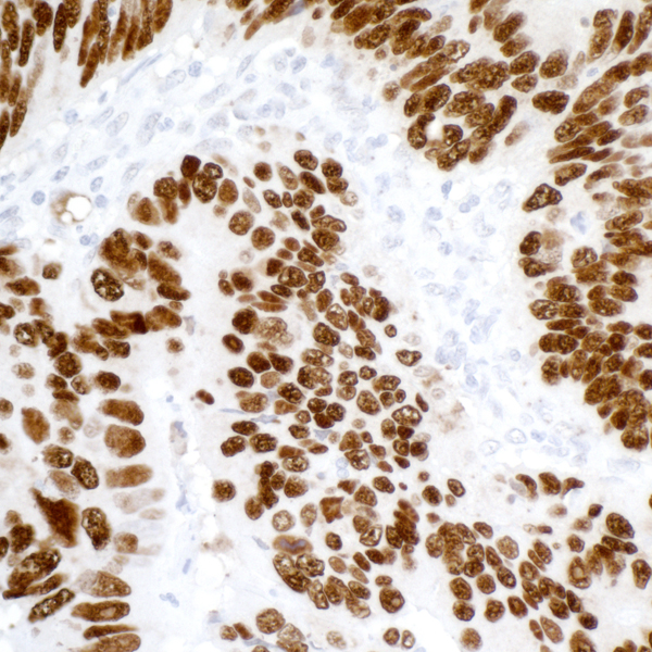
(Formalin-fixed, paraffin-embedded human Prostate Carcinoma stained with FOXA1 Monoclonal Antibody (FOXA1/1514).)
IHC (Immunohiostchemistry)
(Formalin-fixed, paraffin-embedded human Prostate Carcinoma stained with FOXA1 Monoclonal Antibody (FOXA1/1514).)
FOXA1/HNF3A, Monoclonal Antibody (Cat# AAA214384)
Does not react with rat. Others not known.
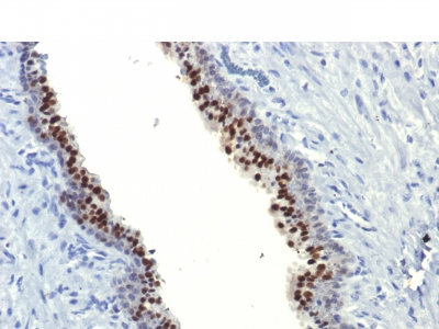
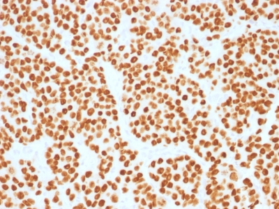 IHC (Immunohiostchemistry)
(Formalin-fixed, paraffin-embedded human Prostate Carcinoma stained with FOXA1 Monoclonal Antibody (FOXA1/1515).)
IHC (Immunohiostchemistry)
(Formalin-fixed, paraffin-embedded human Prostate Carcinoma stained with FOXA1 Monoclonal Antibody (FOXA1/1515).)
FOXA1/HNF3A, Monoclonal Antibody (Cat# AAA214385)
Does not react with rat. Others not known.

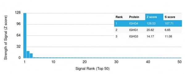 Application Data
(Analysis of Protein Array containing more than 19, 000 full-length human proteins using IgG4 Recombinant Rabbit Monoclonal Antibody (IGHG4/2042R). Z- and S- Score: The Z-score represents the strength of a signal that a monoclonal antibody (MAb) (in combination with a fluorescently-tagged anti-IgG secondary antibody) produces when binding to a particular protein on the HuProtTM array. Z-scores are described in units of standard deviations (SD's) above the mean value of all signals generated on that array. If targets on HuProtTM are arranged in descending order of the Z-score, the S-score is the difference (also in units of SD's) between the Z-score. S-score therefore represents the relative target specificity of a MAb to its intended target. A MAb is considered to specific to its intended target, if the MAb has an S-score of at least 2.5. For example, if a MAb binds to protein X with a Z-score of 43 and to protein Y with a Z-score of 14, then the S-score for the binding of that MAb to protein X is equal to 29.)
Application Data
(Analysis of Protein Array containing more than 19, 000 full-length human proteins using IgG4 Recombinant Rabbit Monoclonal Antibody (IGHG4/2042R). Z- and S- Score: The Z-score represents the strength of a signal that a monoclonal antibody (MAb) (in combination with a fluorescently-tagged anti-IgG secondary antibody) produces when binding to a particular protein on the HuProtTM array. Z-scores are described in units of standard deviations (SD's) above the mean value of all signals generated on that array. If targets on HuProtTM are arranged in descending order of the Z-score, the S-score is the difference (also in units of SD's) between the Z-score. S-score therefore represents the relative target specificity of a MAb to its intended target. A MAb is considered to specific to its intended target, if the MAb has an S-score of at least 2.5. For example, if a MAb binds to protein X with a Z-score of 43 and to protein Y with a Z-score of 14, then the S-score for the binding of that MAb to protein X is equal to 29.)
IgG4 (Ig Heavy Constant Gamma 4), Monoclonal Antibody (Cat# AAA214396)
 Application Data
Insulin Receptor Alpha, Monoclonal Antibody (Cat# AAA214403)
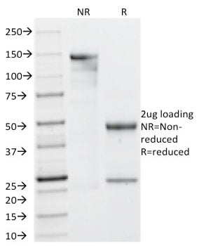
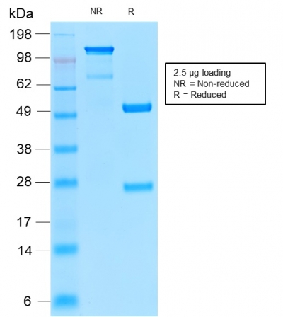
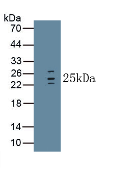
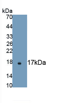
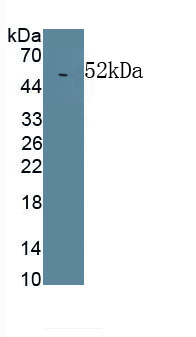
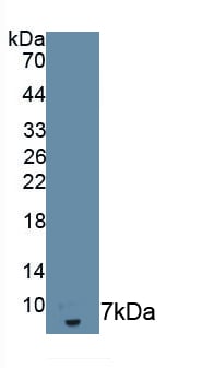
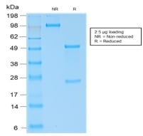 SDS-PAGE
(SDS-PAGE Analysis of Purified Kappa Light Chain Rabbit Recombinant Monoclonal (KLC2289R).)
SDS-PAGE
(SDS-PAGE Analysis of Purified Kappa Light Chain Rabbit Recombinant Monoclonal (KLC2289R).)
CD11c, Monoclonal Antibody (Cat# AAA214404)
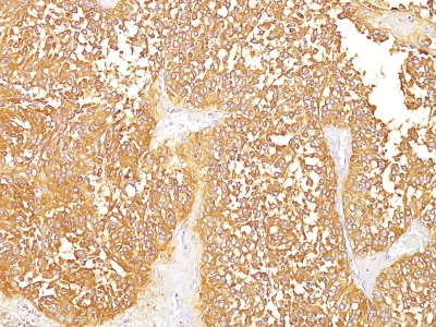
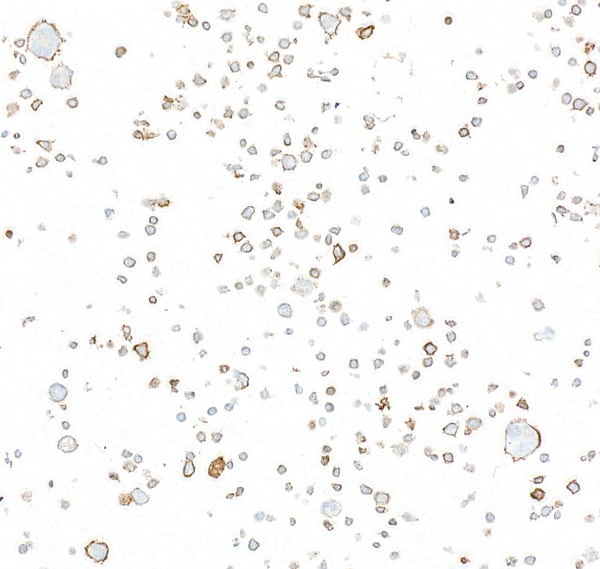
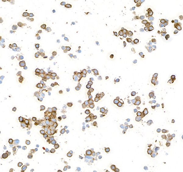
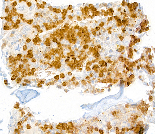
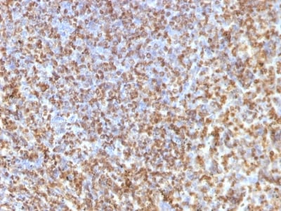
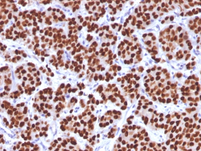
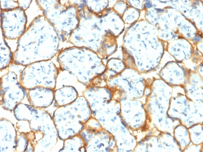
 IHC (Immunohiostchemistry)
(Formalin-fixed, paraffin-embedded human Melanoma stained with gp100 / Melanosome Monoclonal Antibody (HMB45).)
IHC (Immunohiostchemistry)
(Formalin-fixed, paraffin-embedded human Melanoma stained with gp100 / Melanosome Monoclonal Antibody (HMB45).)
gp100 / Melanosome / PMEL17 / SILV, Monoclonal Antibody (Cat# AAA214406)
Does not react with Dog and Rat.
Others not tested
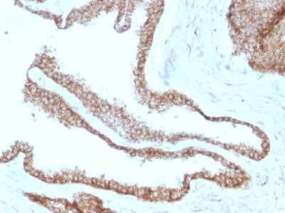
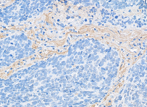
 IHC (Immunohiostchemistry)
(Formalin-fixed, paraffin-embedded human Cervical Carcinoma stained with Catenin, gamma Monoclonal Antibody (CTNG/1664))
IHC (Immunohiostchemistry)
(Formalin-fixed, paraffin-embedded human Cervical Carcinoma stained with Catenin, gamma Monoclonal Antibody (CTNG/1664))
Catenin, gamma, Monoclonal Antibody (Cat# AAA214410)
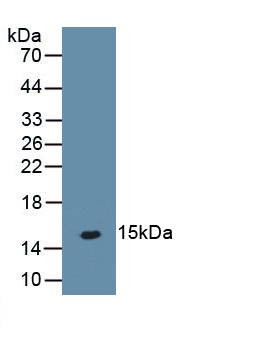
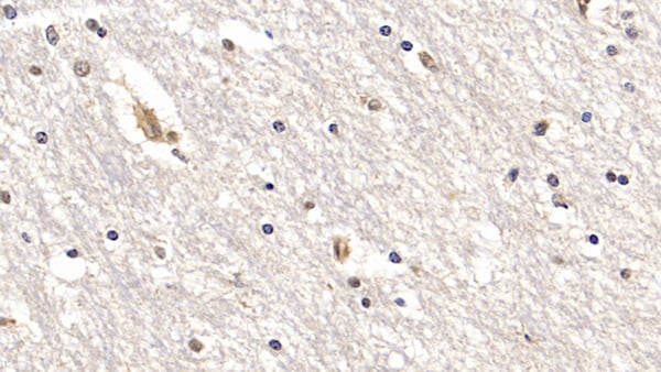
 IHC (Immunohistochemistry)
(DAB staining on IHC-P; Samples: Rat Stomach Tissue.)
IHC (Immunohistochemistry)
(DAB staining on IHC-P; Samples: Rat Stomach Tissue.)
Interleukin 33, Monoclonal Antibody (Cat# AAA141339)
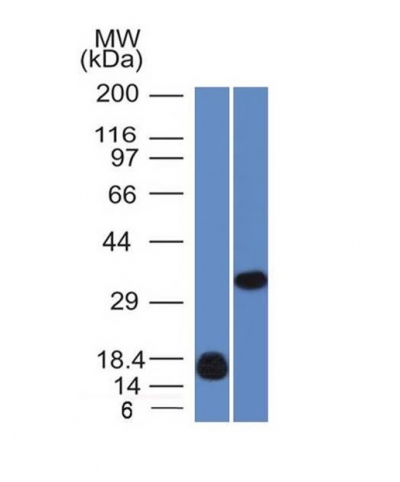
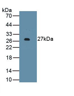
 WB (Western Blot)
(Western Blot: Sample: Recombinant protein.)
WB (Western Blot)
(Western Blot: Sample: Recombinant protein.)
Glutathione S Transferase Alpha 3, Monoclonal Antibody (Cat# AAA141359)
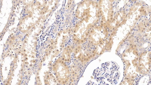
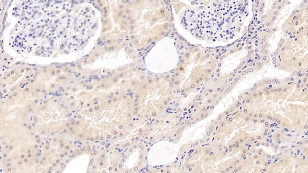
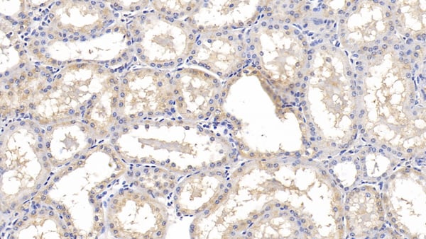
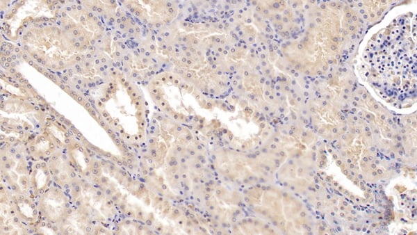 IHC (Immunohiostchemistry)
(DAB staining on IHC-P; Samples: Human Kidney Tissue; Primary Ab: 20ug/ml Mouse Anti-Human Tie1 AntibodySecond Ab: 2ug/mL HRP-Linked Caprine Anti-Mouse IgG Polyclonal Antibody)
IHC (Immunohiostchemistry)
(DAB staining on IHC-P; Samples: Human Kidney Tissue; Primary Ab: 20ug/ml Mouse Anti-Human Tie1 AntibodySecond Ab: 2ug/mL HRP-Linked Caprine Anti-Mouse IgG Polyclonal Antibody)
Tyrosine Kinase With Immunoglobulin Like And EGF Like Domains Protein 1 (Tie1), Monoclonal Antibody (Cat# AAA149510)
Carcinoembryonic Antigen (CEA), Monoclonal Antibody (Cat# AAA149512)
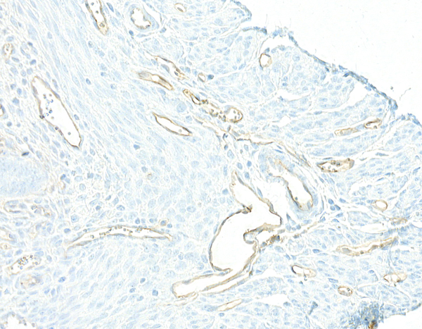
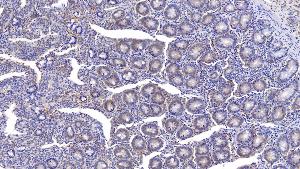
 IHC (Immunohiostchemistry)
(DAB staining on IHC-P; Samples: Human Stomach Tissue; Primary Ab: 30ug/ml Mouse Anti-Human IL1RA Antibody Second Ab: 2ug/mL HRP-Linked Caprine Anti-Mouse IgG Polyclonal Antibody)
IHC (Immunohiostchemistry)
(DAB staining on IHC-P; Samples: Human Stomach Tissue; Primary Ab: 30ug/ml Mouse Anti-Human IL1RA Antibody Second Ab: 2ug/mL HRP-Linked Caprine Anti-Mouse IgG Polyclonal Antibody)
Interleukin 1 Receptor Antagonist (IL1RA), Monoclonal Antibody (Cat# AAA149515)
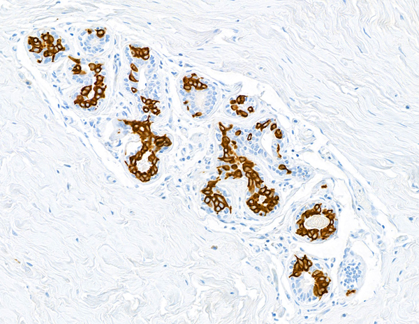
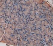
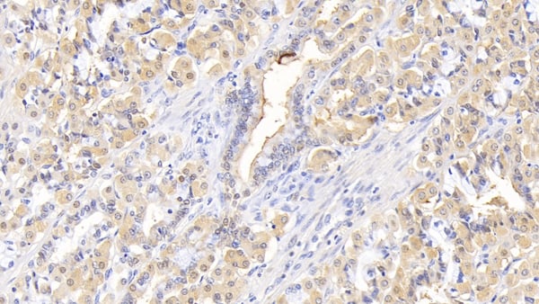
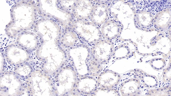
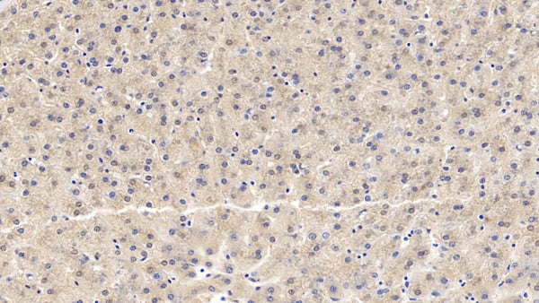
 IHC (Immunohiostchemistry)
(DAB staining on IHC-P; Samples: Human Liver Tissue; Primary Ab: 10ug/ml Mouse Anti-Human SHBG Antibody Second Ab: 2ug/mL HRP-Linked Caprine Anti-Mouse IgG Polyclonal Antibody)
IHC (Immunohiostchemistry)
(DAB staining on IHC-P; Samples: Human Liver Tissue; Primary Ab: 10ug/ml Mouse Anti-Human SHBG Antibody Second Ab: 2ug/mL HRP-Linked Caprine Anti-Mouse IgG Polyclonal Antibody)
Sex Hormone Binding Globulin (SHBG), Monoclonal Antibody (Cat# AAA149520)
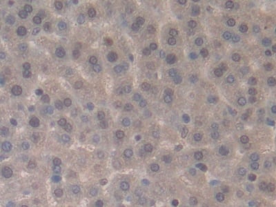
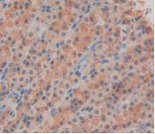
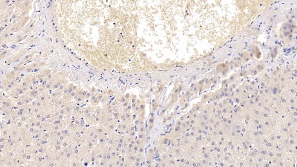
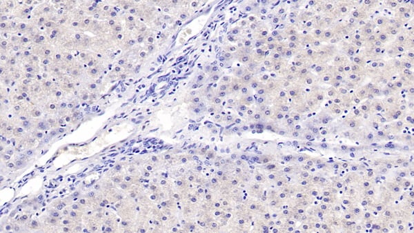 IHC (Immunohistochemisry)
(DAB staining on IHC-P; Samples: Human Liver Tissue; Primary Ab: 40ug/ml Mouse Anti-Human aHSP AntibodySecond Ab: 2ug/mL HRP-Linked Caprine Anti-Mouse IgG Polyclonal Antibody)
IHC (Immunohistochemisry)
(DAB staining on IHC-P; Samples: Human Liver Tissue; Primary Ab: 40ug/ml Mouse Anti-Human aHSP AntibodySecond Ab: 2ug/mL HRP-Linked Caprine Anti-Mouse IgG Polyclonal Antibody)
Alpha-Hemoglobin Stabilizing Protein (aHSP), Monoclonal Antibody (Cat# AAA149521)
 WB (Western Blot)
(Western Blot; Sample: Human MilkPrimary Ab: 2?g/ml Mouse Anti-Human MUC1 AntibodySecond Ab: 0.2ug/mL HRP-Linked Rabbit Anti-Mouse IgG Polyclonal Antibody)
WB (Western Blot)
(Western Blot; Sample: Human MilkPrimary Ab: 2?g/ml Mouse Anti-Human MUC1 AntibodySecond Ab: 0.2ug/mL HRP-Linked Rabbit Anti-Mouse IgG Polyclonal Antibody)
Mucin 1 (MUC1), Monoclonal Antibody (Cat# AAA149522)
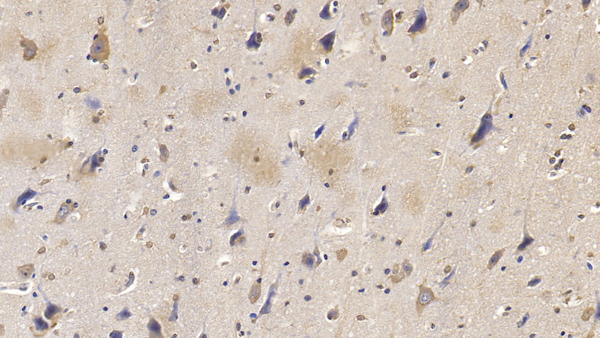
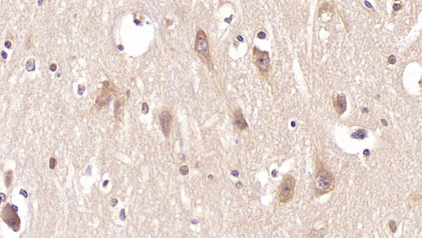 IHC (Immunohistochemistry)
(DAB staining on IHC-P; Samples: Human Cerebrum Tissue; Primary Ab: 40ug/ml Mouse Anti-Human PTHR2 AntibodySecond Ab: 2ug/mL HRP-Linked Caprine Anti-Mouse IgG Polyclonal Antibody)
IHC (Immunohistochemistry)
(DAB staining on IHC-P; Samples: Human Cerebrum Tissue; Primary Ab: 40ug/ml Mouse Anti-Human PTHR2 AntibodySecond Ab: 2ug/mL HRP-Linked Caprine Anti-Mouse IgG Polyclonal Antibody)
Parathyroid Hormone Receptor 2 (PTHR2), Monoclonal Antibody (Cat# AAA149523)
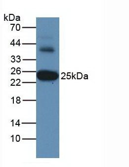
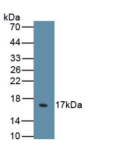
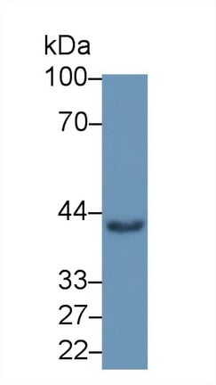
 WB (Western Blot)
(Western Blot; Sample: Recombinant ICAM1, Rabbit.)
WB (Western Blot)
(Western Blot; Sample: Recombinant ICAM1, Rabbit.)
Intercellular Adhesion Molecule 1 (ICAM1), Monoclonal Antibody (Cat# AAA149528)
 IHC (Immunohistochemisry)
(DAB staining on IHC-P; Samples: Human Kidney Tissue; Primary Ab: 20ug/ml Mouse Anti-Human EGF Antibody Second Ab: 2ug/mL HRP-Linked Caprine Anti-Mouse IgG Polyclonal Antibody)
IHC (Immunohistochemisry)
(DAB staining on IHC-P; Samples: Human Kidney Tissue; Primary Ab: 20ug/ml Mouse Anti-Human EGF Antibody Second Ab: 2ug/mL HRP-Linked Caprine Anti-Mouse IgG Polyclonal Antibody)
Epidermal Growth Factor (EGF), Monoclonal Antibody (Cat# AAA149529)
 IHC (Immunohiostchemistry)
(DAB staining on IHC-P; Samples: Bovine Colon Tissue; Primary Ab: 10ug/ml Mouse Anti-Bovine MPO AntibodySecond Ab: 2ug/mL HRP-Linked Caprine Anti-Mouse IgG Polyclonal Antibody)
IHC (Immunohiostchemistry)
(DAB staining on IHC-P; Samples: Bovine Colon Tissue; Primary Ab: 10ug/ml Mouse Anti-Bovine MPO AntibodySecond Ab: 2ug/mL HRP-Linked Caprine Anti-Mouse IgG Polyclonal Antibody)
Myeloperoxidase (MPO), Monoclonal Antibody (Cat# AAA149531)
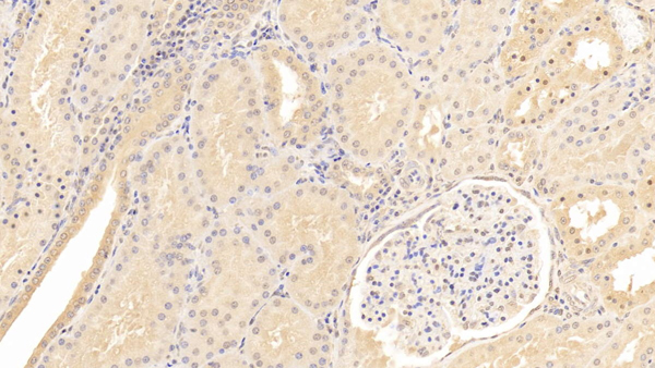
 IHC (Immunohiostchemistry)
(DAB staining on IHC-P; Samples: Human Kidney Tissue; Primary Ab: 30ug/ml Mouse Anti-Human CASP3 Antibody Second Ab: 2ug/mL HRP-Linked Caprine Anti-Mouse IgG Polyclonal Antibody)
IHC (Immunohiostchemistry)
(DAB staining on IHC-P; Samples: Human Kidney Tissue; Primary Ab: 30ug/ml Mouse Anti-Human CASP3 Antibody Second Ab: 2ug/mL HRP-Linked Caprine Anti-Mouse IgG Polyclonal Antibody)
Caspase 3 (CASP3), Monoclonal Antibody (Cat# AAA149533)
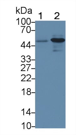
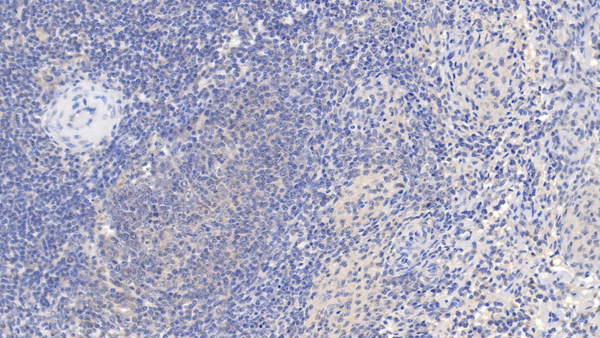
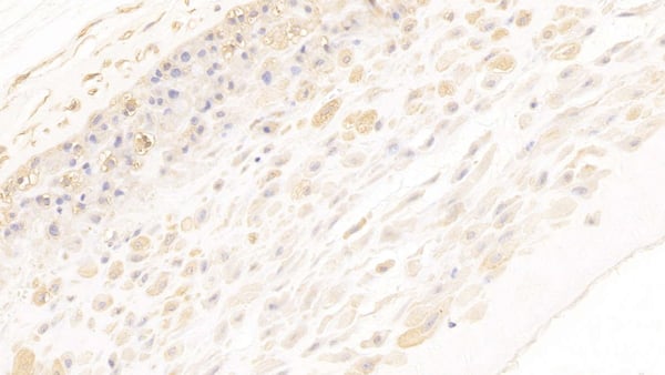
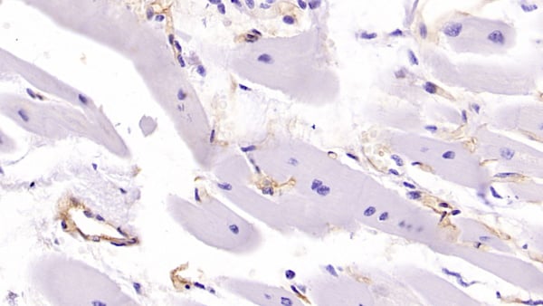
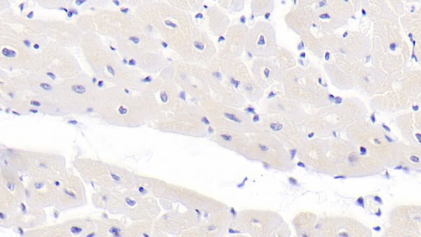 IHC (Immunohiostchemistry)
(DAB staining on IHC-P; Samples: Human Cardiac Muscle Tissue; Primary Ab: 30?g/ml Mouse Anti-Human CASP9 Antibody Second Ab: 2ug/mL HRP-Linked Caprine Anti-Mouse IgG Polyclonal Antibody)
IHC (Immunohiostchemistry)
(DAB staining on IHC-P; Samples: Human Cardiac Muscle Tissue; Primary Ab: 30?g/ml Mouse Anti-Human CASP9 Antibody Second Ab: 2ug/mL HRP-Linked Caprine Anti-Mouse IgG Polyclonal Antibody)
Caspase 9 (CASP9), Monoclonal Antibody (Cat# AAA149534)
 IHC (Immunohiostchemistry)
(DAB staining on IHC-P;Sample: Rat Testis Tissue;Primary Ab: 20ug/ml Mouse Anti-Rat Kim1 AntibodySecond Ab: 2ug/mL HRP-Linked Caprine Anti-Mouse IgG Polyclonal Antibody)
IHC (Immunohiostchemistry)
(DAB staining on IHC-P;Sample: Rat Testis Tissue;Primary Ab: 20ug/ml Mouse Anti-Rat Kim1 AntibodySecond Ab: 2ug/mL HRP-Linked Caprine Anti-Mouse IgG Polyclonal Antibody)
Kidney Injury Molecule 1 (Kim1), Monoclonal Antibody (Cat# AAA149537)
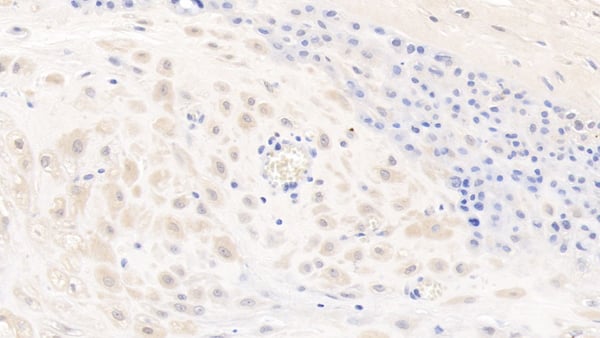
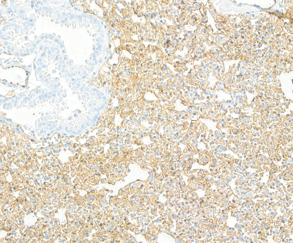
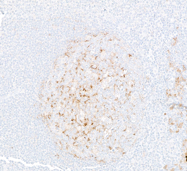
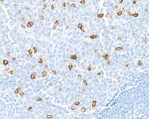
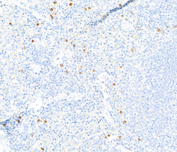
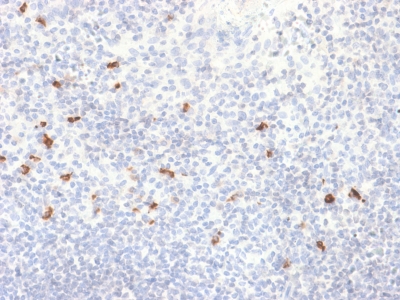
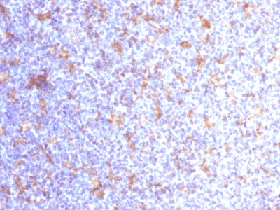
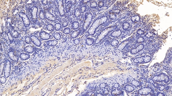
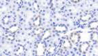
 IHC (Immunohistochemistry)
(DAB staining on IHC-P; Samples: Human Spleen Tissue; Primary Ab: 30ug/ml Mouse Anti-Human PDCD1LG2 AntibodySecond Ab: 2ug/mL HRP-Linked Caprine Anti-Mouse IgG Polyclonal Antibody)
IHC (Immunohistochemistry)
(DAB staining on IHC-P; Samples: Human Spleen Tissue; Primary Ab: 30ug/ml Mouse Anti-Human PDCD1LG2 AntibodySecond Ab: 2ug/mL HRP-Linked Caprine Anti-Mouse IgG Polyclonal Antibody)
Programmed Cell Death Protein 1 Ligand 2 (PDL2), Monoclonal Antibody (Cat# AAA149538)
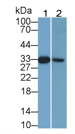
 WB (Western Blot)
(Western Blot; Sample: Canine Liver Tissue.)
WB (Western Blot)
(Western Blot; Sample: Canine Liver Tissue.)
Tryptase (TPS), Monoclonal Antibody (Cat# AAA149542)
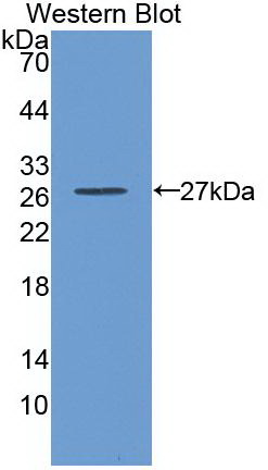
 WB (Western Blot)
(Western Blot; Sample: Recombinant LBP, Human.)
WB (Western Blot)
(Western Blot; Sample: Recombinant LBP, Human.)
Lipopolysaccharide Binding Protein (LBP), Monoclonal Antibody (Cat# AAA149550)
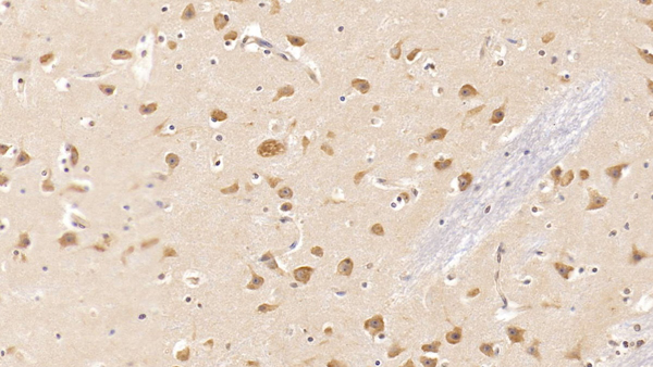
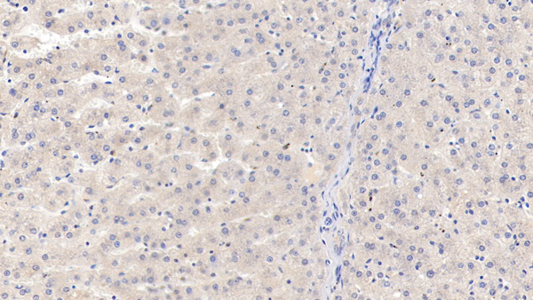
 IHC (Immunohistochemistry)
(DAB staining on IHC-P; Samples: Human Liver Tissue; Primary Ab: 40ug/ml Mouse Anti-Human IL18R1 AntibodySecond Ab: 2ug/mL HRP-Linked Caprine Anti-Mouse IgG Polyclonal Antibody)
IHC (Immunohistochemistry)
(DAB staining on IHC-P; Samples: Human Liver Tissue; Primary Ab: 40ug/ml Mouse Anti-Human IL18R1 AntibodySecond Ab: 2ug/mL HRP-Linked Caprine Anti-Mouse IgG Polyclonal Antibody)
Interleukin 18 Receptor 1 (IL18R1), Monoclonal Antibody (Cat# AAA149557)
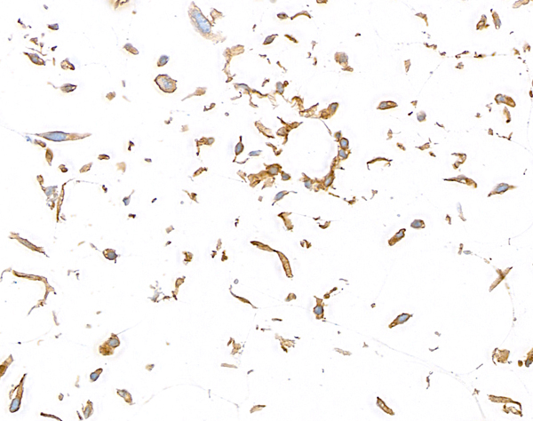
 IHC (Immunohiostchemistry)
(DAB staining on IHC-P; Samples: Human Placenta Tissue; Primary Ab: 10ug/ml Mouse Anti-Human ANXA2 AntibodySecond Ab: 2ug/mL HRP-Linked Caprine Anti-Mouse IgG Polyclonal Antibody)
IHC (Immunohiostchemistry)
(DAB staining on IHC-P; Samples: Human Placenta Tissue; Primary Ab: 10ug/ml Mouse Anti-Human ANXA2 AntibodySecond Ab: 2ug/mL HRP-Linked Caprine Anti-Mouse IgG Polyclonal Antibody)
Annexin A2 (ANXA2), Monoclonal Antibody (Cat# AAA149561)

 IHC (Immunohistochemisry)
(DAB staining on IHC-P; Samples: Human Cardiac Muscle Tissue; Primary Ab: 40ug/ml Mouse Anti-Human MHCC Antibody Second Ab: 2ug/mL HRP-Linked Caprine Anti-Mouse IgG Polyclonal Antibody)
IHC (Immunohistochemisry)
(DAB staining on IHC-P; Samples: Human Cardiac Muscle Tissue; Primary Ab: 40ug/ml Mouse Anti-Human MHCC Antibody Second Ab: 2ug/mL HRP-Linked Caprine Anti-Mouse IgG Polyclonal Antibody)
Major Histocompatibility Complex Class I C (MHCC), Monoclonal Antibody (Cat# AAA149565)


 IHC (Immunohistochemistry)
(DAB staining on IHC-P; Samples: Human Placenta Tissue; Primary Ab: 40ug/ml Mouse Anti-Human Smad3 AntibodySecond Ab: 2ug/mL HRP-Linked Caprine Anti-Mouse IgG Polyclonal Antibody)
IHC (Immunohistochemistry)
(DAB staining on IHC-P; Samples: Human Placenta Tissue; Primary Ab: 40ug/ml Mouse Anti-Human Smad3 AntibodySecond Ab: 2ug/mL HRP-Linked Caprine Anti-Mouse IgG Polyclonal Antibody)
SMAD family member 3 (SMAD3), Monoclonal Antibody (Cat# AAA149567)
What are Monoclonal Antibodies?
Monoclonal antibodies are specialized laboratory-produced proteins developed for binding to specific biological antigens or other molecular targets. Since they come from a single cell (or clone), they are especially consistent and accurate in the data they are involved in producing.
This type of antibody material has been shown to be a powerful tool in finding and subsequently destroying harmful cells in an organism, such as those found in cancers or various autoimmune diseases. This makes them excellent aids in medical testing and research, which is why they are so widely used.
AAA Biotech offers a comprehensive range of high-quality monoclonal antibodies that perform effectively in various laboratory tests, including (amongst others) ELISA, western blotting, immunohistochemistry, and flow cytometry. All of the products in our catalog are thoroughly quality tested to make sure that they are reliable and will consistently perform well in your research.
What Are The Uses of Monoclonal Antibodies
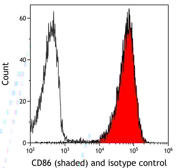
Monoclonal antibodies are used in many lab tests, including (amongst others) ELISA, western blotting, immunohistochemistry, and flow cytometry.
ELISA is a test that helps detect a specific substance/analyte in a sample. It uses antibodies (often monoclonal) bound to a solid surface (such as the well of a microplate) to “capture” the substance/analyte in the sample and immobilize it so that the detection antibody component can then bind to it and produce a signal, which can then be measured.
Western blotting identifies specific proteins in a sample. The sample is first separated on a gel, and then antibodies are applied that will typically bind to the target, which will all be localized to a single band in a lane.
Immunohistochemistry helps locate specific proteins in cells or tissue samples using antibodies.
Flow cytometry looks at and sorts cells. It uses antibodies that are conjugated to reporter molecules called “fluorophores”, which, under special lights, emit light themselves, which can then be measured by a detector instrument. For a deeper understanding of these techniques, explore our complete guide to monoclonal antibodies and their benefits.
How Monoclonal Antibodies Are Used as Medicine?
Please note that all of the products listed in AAA Biotech’s also known as AAA Bio or AAABio catalog are strictly for research-use only (RUO).
Monoclonal antibodies can also be used as therapeutic/medical treatments, particularly in the context of cancers. They are designed to find and bind to specific cells or proteins, helping the immune system recognize and attack the cancer. These treatments work in different ways, such as:
- Radioimmunotherapy attaches a small amount of radioactive molecule to the antibody, so it delivers the radiation directly to the cancer cells that the antibody is specifically binding to.
- Antibody-directed enzyme prodrug therapy uses antibodies that are specifically bound to special enzymes. These enzymes activate a harmless drug in the body and turn it into a cancer-killing drug only near the cancer cells—this helps avoid harming healthy cells.
- Immunoliposomes are tiny “bubbles” filled with medicine/drug and coated with antibodies. They carry the drug straight to the cancer cells.
Why Buy Monoclonal Antibodies From Us?
At AAA Biotech, we provide high-performance monoclonal antibodies designed to support a wide range of research needs.
1. Validated for Versatile Applications
The antibodies in our catalog are extensively validated and compatible with multiple techniques, including (but not limited to) ELISA, flow cytometry (FC), immunocytochemistry (ICC), immunofluorescence (IF), immunohistochemistry (IHC), immunoprecipitation (IP), and western blotting (WB).
2. Wide Selection & Specialized Options
We offer antibodies for common and rare species, that are available in various conjugated forms, and also in recombinant formats. Essentially, there is almost anything one might need to meet their experimental model’s requirements.
3. High-Quality Proteins
Our proteins meet high purity standards—90% or more as confirmed by SDS-PAGE. Many are available with tags like His, Flag, GST, or MBP, and we also supply native and biologically active proteins for functional studies.
Frequently Asked Questions
1. Are your monoclonal antibodies validated for specific applications?
Yes, our antibodies are tested and validated for use in methods such as ELISA, western blot, IHC, flow cytometry, and more. Refer to specific product pages or datasheets for individual product information.
2. How do I choose the right monoclonal antibody for my application?
Review the product details directly for application validation, species reactivity, and target information. You may also contact our support team at any time for help.
3. How quickly can I receive my order?
Most orders are processed and shipped within 1–3 business days, depending on product availability and your shipping location.