Get accurate results in your research with our Monoclonal Antibodies, which are specially made to target exactly what you require for your research, and will produce consistent, reliable performance in lab tests.
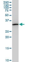
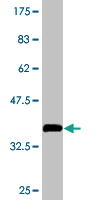
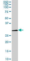
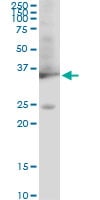
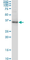
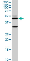
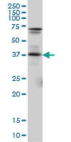
WB (Western Blot) (Western Blot detection against Immunogen (37kD).)
Application Data (Detection limit for recombinant GST tagged GMNN is ~0.03ng/ml as a capture antibody.)
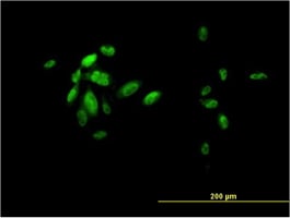
IF (Immunofluorescence) (Immunofluorescence of monoclonal antibody to GMNN on HeLa cell. [antibody concentration 10ug/ml].)
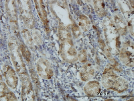
IHC (Immunohistochemistry) (Immunoperoxidase of monoclonal antibody to GMNN on formalin-fixed paraffin-embedded human placenta. [antibody concentration 1ug/ml].)
IHC (Immunohistochemistry) (Immunoperoxidase of monoclonal antibody to GMNN on formalin-fixed paraffin-embedded human lymph node. [antibody concentration 3ug/ml].)
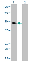
WB (Western Blot) (Western Blot analysis of GMNN expression in transfected 293T cell line by GMNN monoclonal antibody. Lane 1: GMNN transfected lysate (23.6kD). Lane 2: Non-transfected lysate.)
Application Data (Detection limit for recombinant GST tagged CSE1L is approximately 3ng/ml as a capture antibody.)
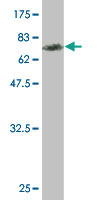
WB (Western Blot) (CSE1L monoclonal antibody (M08), clone 3D8 Western Blot analysis of CSE1L expression in Hela S3 NE.)
IF (Immunofluorescence) (Immunofluorescence of monoclonal antibody to CSE1L on HeLa cell. [antibody concentration 10 ug/ml])
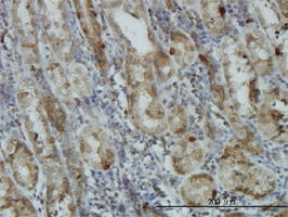
IHC (Immunohistochemistry) (Immunoperoxidase of monoclonal antibody to CSE1L on formalin-fixed paraffin-embedded human prostate. [antibody concentration 3 ug/ml])
Application Data (Immunoperoxidase of monoclonal antibody to CSE1L on formalin-fixed paraffin-embedded human prostate. [antibody concentration 3 ug/ml])
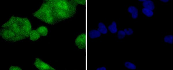
ICC (Immunocytochemistry) (ICC staining NSE in 293 cells (green). The nuclear counter stain is DAPI (blue). Cells were fixed in paraformaldehyde, permeabilised with 0.25% Triton X100/PBS.)
ICC (Immunocytochemistry) (ICC staining NSE in SH-SY-5Y cells (green). The nuclear counter stain is DAPI (blue). Cells were fixed in paraformaldehyde, permeabilised with 0.25% Triton X100/PBS.)
IHC (Immunohistochemistry) (Immunohistochemical analysis of paraffin-embedded human tonsil tissue using anti-NSE antibody. Counter stained with hematoxylin.)
IHC (Immunohistochemistry) (Immunohistochemical analysis of paraffin-embedded rat brain tissue using anti-NSE antibody. Counter stained with hematoxylin.)
WB (Western Blot) (Western blot analysis of NSE on hybrid fish (crucian-carp) brain tissue lysates using anti-NSE antibody at 1/500 dilution.)
WB (Western Blot) (Western blot analysis of NSE on different lysates using anti-NSE antibody at 1/1, 000 dilution. Positive control: Lane 1: HepG2 Lane 2: Hela Lane 3: 293)
FCM (Flow Cytometry) (Flow cytometric analysis of Jurkat cells with USP14 antibody at 1/100 dilution (red) compared with an unlabelled control (cells without incubation with primary antibody; black). Alexa Fluor 488-conjugated goat anti rabbit IgG was used as the secondary antibody.)
ICC (Immunocytochemistry) (ICC staining USP14 in LOVO cells (green). The nuclear counter stain is DAPI (blue). Cells were fixed in paraformaldehyde, permeabilised with 0.25% Triton X100/PBS.)
ICC (Immunocytochemistry) (ICC staining USP14 in HUVEC cells (green). The nuclear counter stain is DAPI (blue). Cells were fixed in paraformaldehyde, permeabilised with 0.25% Triton X100/PBS.)
ICC (Immunocytochemistry) (ICC staining USP14 in A431 cells (green). The nuclear counter stain is DAPI (blue). Cells were fixed in paraformaldehyde, permeabilised with 0.25% Triton X100/PBS.)
IHC (Immunohistchemistry) (Immunohistochemical analysis of paraffin-embedded rat epididymis tissue using anti-USP14 antibody. Counter stained with hematoxylin.)
IHC (Immunohistochemistry) (Immunohistochemical analysis of paraffin-embedded mouse prostate tissue using anti-USP14 antibody. Counter stained with hematoxylin.)
IHC (Immunohistochemistry) (Immunohistochemical analysis of paraffin-embedded human kidney tissue using anti-USP14 antibody. Counter stained with hematoxylin.)
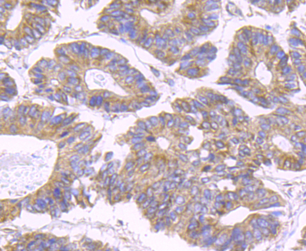
IHC (Immunohistochemistry) (Immunohistochemical analysis of paraffin-embedded human colon cancer tissue using anti-USP14 antibody. Counter stained with hematoxylin.)
WB (Western Blot) (Western blot analysis of USP14 on Zebrafish tissue lysates using anti-USP14 antibody at 1/200 dilution.)
WB (Western Blot) (Western blot analysis of USP14 on K562 (1) and Hela (2) cell lysate using anti-USP14 antibody at 1/1, 000 dilution.)
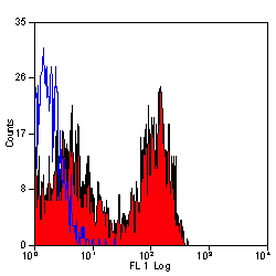
FCM (Flow Cytometry) (Flow cytometric analysis of Hela cells with RAD18 antibody at 1/50 dilution (red) compared with an unlabelled control (cells without incubation with primary antibody; black). Alexa Fluor 488-conjugated goat anti rabbit IgG was used as the secondary antibody)
ICC (Immunocytochemistry) (ICC staining RAD18 in 293 cells (green). The nuclear counter stain is DAPI (blue). Cells were fixed in paraformaldehyde, permeabilised with 0.25% Triton X100/PBS.)
ICC (Immunocytochemistry) (ICC staining RAD18 in NIH/3T3 cells (green). The nuclear counter stain is DAPI (blue). Cells were fixed in paraformaldehyde, permeabilised with 0.25% Triton X100/PBS.)
ICC (Immunocytochemistry) (ICC staining RAD18 in Hela cells (green). The nuclear counter stain is DAPI (blue). Cells were fixed in paraformaldehyde, permeabilised with 0.25% Triton X100/PBS.)
IHC (Immunohistochemistry) (Immunohistochemical analysis of paraffin-embedded rat testis tissue using anti-RAD18 antibody. Counter stained with hematoxylin.)
WB (Western Blot) (Western blot analysis of RAD18 on different lysates using anti-RAD18 antibody at 1/1, 000 dilution. Positive control: Lane 1: NIH/3T3 Lane 2: 293T Lane 3: MCF-7)
WB (Western Blot) (BUB1B monoclonal antibody Western Blot analysis of BUB1B expression in Hela NE.)
WB (Western Blot) (Western Blot detection against Immunogen (39.93kD).)
Application Data (Proximity Ligation Analysis (PLA) of protein-protein interactions between CDC20 and BUB1B. HeLa cells were stained with CDC20 rabbit purified polyclonal 1:1200 and BUB1B mouse monoclonal antibody 1:50. Signals were detected by 30 Detection Kit 613 (red), and nuclei were counterstained with DAPI (blue). Each red dot represents the detection of protein-protein interaction complex.)
WB (Western Blot) (Western blot analysis of BUB1B over-expressed 293 cell line, cotransfected with BUB1B Validated Chimera RNAi (Lane 2) or non-transfected control (Lane 1). Blot probed with BUB1B monoclonal antibody. GAPDH (36.1kD) used as specificity and loading control.)
Application Data (Detection limit for recombinant GST tagged BUB1B is ~0.03ng/ml as a capture antibody.)
IHC (Immunohistochemistry) (Immunoperoxidase of monoclonal antibody to BUB1B on formalin-fixed paraffin-embedded human spleen. [antibody concentration 3ug/ml].)
WB (Western Blot) (Western Blot analysis of BUB1B expression in transfected 293T cell line by BUB1B monoclonal antibody. Lane 1: BUB1B transfected lysate (119.5kD). Lane 2: Non-transfected lysate.)
FCM (Flow Cytometry) (Flow cytometric analysis of Hela cells with hnRNP C1+C2 antibody at 1/50 dilution (red) compared with an unlabelled control (cells without incubation with primary antibody; black). Alexa Fluor 488-conjugated goat anti rabbit IgG was used as the secondary antibody)
ICC (Immunocytochemistry) (ICC staining hnRNP C1+C2 in B16-F1 (green). The nuclear counter stain is DAPI (blue). Cells were fixed in paraformaldehyde, permeabilised with 0.25% Triton X100/PBS.)
ICC (Immunocytochemistry) (ICC staining hnRNP C1+C2 in MCF-7 cells (green). The nuclear counter stain is DAPI (blue). Cells were fixed in paraformaldehyde, permeabilised with 0.25% Triton X100/PBS.)
ICC (Immunocytochemistry) (ICC staining hnRNP C1+C2 in Hela cells (green). The nuclear counter stain is DAPI (blue). Cells were fixed in paraformaldehyde, permeabilised with 0.25% Triton X100/PBS.)
IHC (Immunohistchemistry) (Immunohistochemical analysis of paraffin-embedded mouse placenta tissue using anti-hnRNP C1+C2 antibody. Counter stained with hematoxylin.)
IHC (Immunohistochemistry) (Immunohistochemical analysis of paraffin-embedded mouse skin tissue using anti-hnRNP C1+C2 antibody. Counter stained with hematoxylin.)
IHC (Immunohistochemistry) (Immunohistochemical analysis of paraffin-embedded mouse brain tissue using anti-hnRNP C1+C2 antibody. Counter stained with hematoxylin.)
IHC (Immunohistochemistry) (Immunohistochemical analysis of paraffin-embedded human kidney tissue using anti-hnRNP C1+C2 antibody. Counter stained with hematoxylin.)
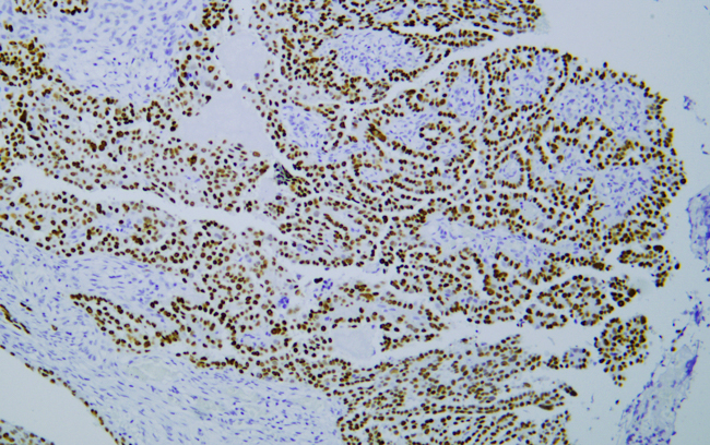
IHC (Immunohistochemistry) (Immunohistochemical analysis of paraffin-embedded human breast carcinoma tissue using anti-hnRNP C1+C2 antibody. Counter stained with hematoxylin.)
WB (Western Blot) (Western blot analysis of hnRNP C1+C2 on different lysates using anti-hnRNP C1+C2 antibody at 1/1, 000 dilution. Positive control: Lane 1: Hela Lane 2: MCF-7 Lane 3: HepG2)
WB (Western Blot) (DDX54 monoclonal antibody, Western Blot analysis of DDX54 expression in HeLa.)
Application Data (Detection limit for recombinant GST tagged DDX54 is 0.03ng/ml as a capture antibody.)
IF (Immunofluorescence) (Immunofluorescence of monoclonal antibody to DDX54 on HeLa cell. [antibody concentration 10ug/ml].)
IHC (Immunohistochemistry) (Immunoperoxidase of monoclonal antibody to DDX54 on formalin-fixed paraffin-embedded human ovary, clear cell carcinoma. [antibody concentration 3ug/ml].)
WB (Western Blot) (Western Blot analysis of DDX54 expression in transfected 293T cell line by DDX54 monoclonal antibody. Lane 1: DDX54 transfected lysate (98.6kD). Lane 2: Non-transfected lysate.)
WB (Western Blot) (Western Blot detection against Immunogen (37.55kD).)
ICC (Immunocytochemistry) (ICC staining p23 in Hela cells (green). The nuclear counter stain is DAPI (blue). Cells were fixed in paraformaldehyde, permeabilised with 0.25% Triton X100/PBS.)
ICC (Immunocytochemistry) (ICC staining p23 in A431 cells (green). The nuclear counter stain is DAPI (blue). Cells were fixed in paraformaldehyde, permeabilised with 0.25% Triton X100/PBS.)
IHC (Immunohistochemistry) (Immunohistochemical analysis of paraffin-embedded rat testis tissue using anti-p23 antibody. Counter stained with hematoxylin.)
IHC (Immunohistochemistry) (Immunohistochemical analysis of paraffin-embedded mouse fallopian tube tissue using anti-p23 antibody. Counter stained with hematoxylin.)
IHC (Immunohistochemistry) (Immunohistochemical analysis of paraffin-embedded human colon cancer tissue using anti-p23 antibody. Counter stained with hematoxylin.)
IHC (Immunohistochemistry) (Immunohistochemical analysis of paraffin-embedded human tonsil tissue using anti-p23 antibody. Counter stained with hematoxylin.)
WB (Western Blot) (Western blot analysis of p23 on different lysates using anti-p23 antibody at 1/500 dilution. Positive control: Lane 1: Mouse brain tissue Lane 2: SK-Br-3 Lane 3: Rat lung tissue)
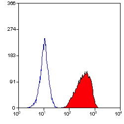
FCM (Flow Cytometry) (Flow cytometric analysis of Hela cells with CK5 antibody at 1/100 dilution (green) compared with an unlabelled control (cells without incubation with primary antibody; red).)
ICC (Immunocytochemistry) (ICC staining CK5 (green) and Actin filaments (red) in MCF-7 cells. The nuclear counter stain is DAPI (blue). Cells were fixed in paraformaldehyde, permeabilised with 0.25% Triton X100/PBS.)
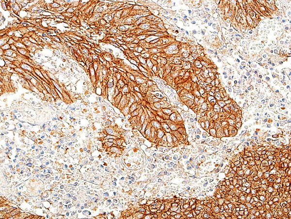
IHC (Immunohistochemistry) (Immunohistochemical analysis of paraffin-embedded human ovarian cancer tissue using anti- CK5 antibody. Counter stained with hematoxylin.)
WB (Western Blot) (Western blot analysis of CK5 on HEK293 (1) and CK5-hIgGFc transfected HEK293 (2) cell lysate using anti-CK5 antibody at 1/1, 000 dilution.)
WB (Western Blot) (Western blot analysis of CK5 on human CK5 recombinant protein using anti-CK5 antibody at 1/1, 000 dilution.)
WB (Western Blot) (Western blot analysis of CK5 on different cells lysates using anti-CK5 antibody at 1/1, 000 dilution. Positive control�� Line1: A431 Line2: 3T3-L1 Line3: COS7 Line4: MCF-7 Line5: SK-Br-3 Line6: Hela Line7: Lncap Line8: HepG2)
WB (Western Blot) (Western blot analysis of UGP2 over-expressed 293 cell line, cotransfected with UGP2 Validated Chimera RNAi (Lane 2) or non-transfected control (Lane 1). Blot probed with UGP2 monoclonal antibody. GAPDH (36.1kD) used as specificity and loading control.)
Application Data (Detection limit for recombinant GST tagged UGP2 is ~0.1ng/ml as a capture antibody.)
WB (Western Blot) (Western Blot analysis of UGP2 expression in transfected 293T cell line by UGP2 monoclonal antibody (M01). Lane 1: UGP2 transfected lysate (56.9kD). Lane 2: Non-transfected lysate.)
WB (Western Blot) (UGP2 monoclonal antibody Western Blot analysis of UGP2 expression in NIH/3T3.)
WB (Western Blot) (UGP2 monoclonal antibody Western Blot analysis of UGP2 expression in Raw 264.7)
WB (Western Blot) (UGP2 monoclonal antibody Western Blot analysis of UGP2 expression in PC-12)
WB (Western Blot) (UGP2 monoclonal antibody Western Blot analysis of UGP2 expression in HeLa)
Application Data (Proximity Ligation Analysis of protein-protein interactions between TP53 and PML HeLa cells were stained with anti-TP53 rabbit purified polyclonal 1:1200 and anti-PML mouse monoclonal antibody 1:50. Each red dot represents the detection of protein-protein interaction complex, and nuclei were counterstained with DAPI (blue).)
Application Data (Detection limit for recombinant GST tagged PML is ~0.3ng/ml as a capture antibody.)
IF (Immunofluorescence) (Immunofluorescence of monoclonal antibody to PML on HeLa cell. [antibody concentration 10ug/ml])
WB (Western Blot) (PML monoclonal antibody Western Blot analysis of PML expression in Hela NE.)
WB (Western Blot) (PML monoclonal antibody Western Blot analysis of PML expression in PC-12.)
WB (Western Blot) (Western Blot detection against Immunogen (36.63kD).)
WB (Western Blot) (Western Blot analysis of TRIM28 expression in transfected 293T cell line by TRIM28 monoclonal antibody (M02), clone 1D11.Lane 1: TRIM28 transfected lysate (Predicted MW: 88.5 KDa).Lane 2: Non-transfected lysate.)
WB (Western Blot) (TRIM28 monoclonal antibody (M02), clone 1D11. Western Blot analysis of TRIM28 expression in MCF-7.)
WB (Western Blot) (TRIM28 monoclonal antibody (M02), clone 1D11. Western Blot analysis of TRIM28 expression in PC-12 (Cat # L012V1).)
WB (Western Blot) (TRIM28 monoclonal antibody (M02), clone 1D11. Western Blot analysis of TRIM28 expression in NIH/3T3 (Cat # L018V1).)
IHC (Immunohistochemistry) (Immunoperoxidase of monoclonal antibody to TRIM28 on formalin-fixed paraffin-embedded human liver. [antibody concentration 3 ug/ml])
Application Data (Immunoperoxidase of monoclonal antibody to TRIM28 on formalin-fixed paraffin-embedded human liver. [antibody concentration 3 ug/ml])
MARCH7 (E3 Ubiquitin-protein Ligase MARCH7, Axotrophin, Membrane-associated RING Finger Protein 7, Membrane-associated RING-CH Protein VII, MARCH-VII, RING Finger Protein 177, AXOT, RNF177, DKFZp586F1122) (AP)
Application Data (Detection limit for recombinant GST tagged PGR is approximately 0.1ng/ml as a capture antibody.)
WB (Western Blot) (PGR monoclonal antibody (M04), clone 2C2 Western Blot analysis of PGR expression in A-431.)
IF (Immunofluorescence) (Immunofluorescence of monoclonal antibody to PGR on A-431 cell. [antibody concentration 10 ug/ml])
IHC (Immunohistochemistry) (Immunoperoxidase of monoclonal antibody to PGR on formalin-fixed paraffin-embedded human endometrium. [antibody concentration 1.5 ug/ml])
Application Data (Immunoperoxidase of monoclonal antibody to PGR on formalin-fixed paraffin-embedded human endometrium. [antibody concentration 1.5 ug/ml])
WB (Western Blot) (Western blot analysis of lysate from HepG2 cell line using PPT1 Antibody. AAA28640 was diluted at 1:1000 at each lane. A goat anti-mouse IgG H&L(HRP) at 1:3000 dilution was used as the secondary antibody. Lysate at 35ug per lane.)
IHC (Immunohistochemistry) (Immunohistochemical analysis of paraffin-embedded R. cerebellum section using PPT1 Antibody (C-term). AAA28640 was diluted at 1:25 dilution. A peroxidase-conjugated goat anti-rabbit IgG at 1:400 dilution was used as the secondary antibody, followed by DAB staining.)
IHC (Immunohistochemistry) (Immunohistochemical analysis of paraffin-embedded M. cerebellum section using PPT1 Antibody (C-term). AAA28640 was diluted at 1:25 dilution. A peroxidase-conjugated goat anti-rabbit IgG at 1:400 dilution was used as the secondary antibody, followed by DAB staining.)
IHC (Immunohistochemistry) (Immunohistochemical analysis of paraffin-embedded H. cerebellum section using PPT1 Antibody (C-term). AAA28640 was diluted at 1:25 dilution. A peroxidase-conjugated goat anti-rabbit IgG at 1:400 dilution was used as the secondary antibody, followed by DAB staining.)
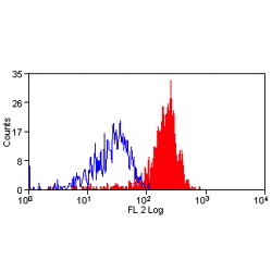
FCM (Flow Cytometry) (Flow cytometric analysis of HepG2 cells using PPT1 Antibody (C-term)(green) compared to an isotype control of mouse IgG1(blue). AAA28640 was diluted at 1:25 dilution. An Alexa Fluor 488 goat anti-mouse lgG at 1:400 dilution was used as the secondary antibody.)
WB (Western Blot) (All lanes : Anti-PPT1 Antibody (C-term) at 1:2000 dilutionLane 1: Hela whole cell lysatesLane 2: HepG2 whole cell lysatesLysates/proteins at 20 ug per lane.SecondaryGoat Anti-Rabbit IgG, (H+L), Peroxidase conjugated at 1/10000 dilutionPredicted band size : 34 kDaBlocking/Dilution buffer: 5% NFDM/TBST.)
ICC (Immunocytochemistry) (ICC staining PSME1 in PC-3M cells (green). The nuclear counter stain is DAPI (blue). Cells were fixed in paraformaldehyde, permeabilised with 0.25% Triton X100/PBS.)
ICC (Immunocytochemistry) (ICC staining PSME1 in MCF-7 cells (green). The nuclear counter stain is DAPI (blue). Cells were fixed in paraformaldehyde, permeabilised with 0.25% Triton X100/PBS.)
ICC (Immunocytochemistry) (ICC staining PSME1 in HepG2 cells (green). The nuclear counter stain is DAPI (blue). Cells were fixed in paraformaldehyde, permeabilised with 0.25% Triton X100/PBS.)
IHC (Immunohistchemistry) (Immunohistochemical analysis of paraffin-embedded mouse small intestine tissue using anti-PSME1 antibody. Counter stained with hematoxylin.)
IHC (Immunohistochemistry) (Immunohistochemical analysis of paraffin-embedded human prostate tissue using anti-PSME1 antibody. Counter stained with hematoxylin.)
IHC (Immunohistochemistry) (Immunohistochemical analysis of paraffin-embedded human liver cancer tissue using anti-PSME1 antibody. Counter stained with hematoxylin.)
IHC (Immunohistochemistry) (Immunohistochemical analysis of paraffin-embedded human tonsil tissue using anti-PSME1 antibody. Counter stained with hematoxylin.)
IHC (Immunohistochemistry) (Immunohistochemical analysis of paraffin-embedded rat kidney tissue using anti-PSME1 antibody. Counter stained with hematoxylin.)
WB (Western Blot) (Western blot analysis of PSME1 on MCF-7 cell (1) and rat lung tissue (2) lysate using anti-PSME1 antibody at 1/500 dilution.)
Application Data (Published Customer Image:Rat anti Mouse Gr-1 antibody, clone RB6-8C5 used for the identification of neutrophils by immunofluorescence.Image caption:S. typhimurium-Infected Macrophages Containing Phagocytosed Neutrophils and T Cells Confocal fluorescence microscopy of 50-mum-thick liver sections from 1-wk-infected Slc11a1 wild-type mice. (A-C) S.Typhimurium (O-antigen, arrows) are red, macrophages (F4-80 and MOMA-2) are blue, DNA (DAPI) is gray, phalloidin is green, and neutrophils (Gr-1/Ly-6G/RB6-8C5) are pink (arrowheads). (A) Collapsed image from a 40-mum Z-stack. Scale bar is 20 mum. (B and C) Sections from (A) that are 4 mum apart. The video from which (A-C) were derived (Video S2) is available online. (D-G) T cells within multinucleate macrophages.Macrophages (F4-80 and MOMA-2) are blue (D, G, and H), T cells (CD3zeta) are red (D, G, arrowheads), DAPI is gray (E, G), actin-bound phalloidin is green (F, G). (G) Is a composite of (D, E, and F). Scale bars are 16 mum. (H) An image from a different mouse stained and labeled as described for (D-G). Scale bar is 8 mum. A video showing a T cell inside of a macrophage is available online (Video S3).From: Nix RN, Altschuler SE, Henson PM, Detweiler CS (2007) Hemophagocytic Macrophages Harbor Salmonella enterica during Persistent Infection.PLoS Pathog 3(12): e193.)
Application Data (Published customer image:Rat anti Mouse Gr-1 antibody, clone RB6-8C5 used for the evaluation of Gr-1 expression on circulating monocytes from mouse blood by flow cytometry.Image caption:Effect of CD206+ M2 macrophage depletion on collagen deposition and cell infiltration within the infarct 2 weeks post MI in MAFIA mice. A. Representative Sirius Red staining on histological sections from MAFIA mice 2 weeks post MI. B. Quantification of collagen staining as a percentage of LV from MAFIA mice 2 weeks post MI (Scale bar: 1 mm, n = 6 animals per group, >=8 images per animal). C. Representative hematoxilin and eosin staining on 20x infarct or remote histological section from MAFIA mice 2 weeks post MI showing an increase in inflammatory infiltrates in animals treated with GW2580. D. Quantification of nuclei number per mm2 in MAFIA remote and infarct zone in MAFIA mice 2 weeks post MI. (n = 4-6 animals per group). E. Representative images of immunofluorescent staining of CD206+ M2 macrophages, Gr1+ M1 macrophages and Ly6G+ neutrophils within the infarct zone from MAFIA mice 2 weeks post MI. Quantification of Ly6G+ neutrophil, Gr1+ M1 macrophage and CD206+ M2 macrophage infiltration within the infarct zone from MAFIA mice 2 weeks post MI (n = 4 animals per group, 10 images/animal). * p)
Application Data (Published customer image:Rat anti Mouse Gr-1 antibody, clone RB6-8C5 used for the evaluation of Gr-1 expression on circulating monocytes from mouse blood by flow cytometry.Image caption:Depletion of the circulating monocyte and Gr1lo and F4/80hi populations following 1 week GW2580 treatment. A. Identification of total monocyte population in mouse blood using MAFIA-GFP. B Following one week of GW2580 treatment, no difference in total circulating monocytes was observed. FACS quantification of Gr1hi (M1) and Gr1lo (M2; C,D) and F4/80hi (E,F) in total monocytes following 1 week GW2580 treatment (n = 6 and n = 4 animals per group, respectively).From: Leblond A-L, Klinkert K, Martin K, Turner EC, Kumar AH, Browne T, et al. (2015) Systemic and Cardiac Depletion of M2 Macrophage through CSF-1R Signaling Inhibition Alters Cardiac Function Post Myocardial Infarction.PLoS ONE 10(9): e0137515.)
Application Data (Figure A : RPE conjugated Rat anti Mouse CD11b and Alexa Fluor 647 conjugated Rat IgG2b isotype control . Figure B. RPE conjugated Rat anti Mouse CD11b and Alexa Fluor 647 conjugated Rat anti Mouse Gr-1 . All experiments performed on murine bone marrow in the presence of Murine SeroBlock (BUF041A).)
Application Data (Figure A : Pacific Blue conjugated Rat anti Mouse CD11b and FITC conjugated Rat IgG2b isotype control . Figure B. Pacific Blue conjugated Rat anti Mouse CD11b and FITC conjugated Rat anti Mouse GR-1 . All experiments performed on red cell lysed mouse bone marrow gated on mononuclear cells in the presence of Mouse Seroblock (BUF041A). Data acquired on the ZE5 cell analyzer.)
Application Data (Figure A. Pacific Blue conjugated Rat anti Mouse CD11b and Alexa Fluor700 conjugated Rat IgG2b isotype control . Figure B. Pacific Blue conjugated Rat anti Mouse CD11b and Alexa Fluor700 conjugated Rat anti Mouse GR-1 . All experiments performed on red cell lysed mouse bone marrow gated on mononuclear cells in the presence of Mouse Seroblock (BUF041A). Data acquired on the ZE5 cell analyzer.)
Application Data (Figure A. RPE conjugated Rat anti Mouse CD11b and Pacific Blue conjugated Rat IgG2b isotype control . Figure B. RPE conjugated Rat anti Mouse CD11b and Pacific Blue conjugated Rat anti Mouse GR-1 . All experiments performed on red cell lysed mouse bone marrow gated on mononuclear cells in the presence of Mouse Seroblock (BUF041A). Data acquired on the ZE5 cell analyzer.)
WB (Western Blot) (NEK9 monoclonal antibody. Western Blot analysis of NEK9 expression in PC-12.)
WB (Western Blot) (NEK9 monoclonal antibody Western Blot analysis of NEK9 expression in HeLa.)
Application Data (Detection limit for recombinant GST tagged NEK9 is ~0.03ng/ml as a capture antibody.)
IF (Immunofluorescence) (Immunofluorescence of monoclonal antibody to NEK9 on HeLa cell. [antibody concentration 10ug/ml].)
WB (Western Blot) (Western Blot analysis of NEK9 expression in transfected 293T cell line by NEK9 monoclonal antibody. Lane 1: NEK9 transfected lysate (107.2kD). Lane 2: Non-transfected lysate.)
WB (Western Blot) (NEK9 monoclonal antibody. Western Blot analysis of NEK9 expression in NIH/3T3.)
WB (Western Blot) (Western Blot detection against Immunogen (37.11kD).)
IF (Immunofluorescence) (Immunofluorescence of monoclonal antibody to TOMM20 on HeLa cell. [antibody concentration 10ug/ml])
Application Data (Detection limit for recombinant GST tagged TOMM20 is ~0.1ng/ml as a capture antibody.)
IHC (Immunohistochemistry) (Immunoperoxidase on formalin-fixed paraffin-embedded human small Intestine using 1346004 (3ug/ml).)
WB (Western Blot) (Western Blot analysis in transfected 293T cell line using 134604. Lane 1: 134604 transfected lysate (16.3kD). Lane 2: Non-transfected lysate.)
WB (Western Blot) (Western Blot analysis in NIH/3T3 using 134604.)
WB (Western Blot) (Western Blot analysis of in PC-12 using 134604.)
WB (Western Blot) (Western Blot analysis in HeLa using 134604.)
Application Data (Proximity Ligation Analysis of protein-protein interactions between MAPK14 and MKNK2 HeLa cells were stained with anti-MAPK14 rabbit purified polyclonal 1:1200 and anti-MKNK2 mouse monoclonal antibody 1:50. Each red dot represents the detection of protein-protein interaction complex, and nuclei were counterstained with DAPI (blue).)
WB (Western Blot) (Western Blot analysis of MKNK2 expression in transfected 293T cell line by MKNK2 monoclonal antibody (M07), clone 2A10.Lane 1: MKNK2 transfected lysate (46.7 KDa).Lane 2: Non-transfected lysate.)
WB (Western Blot) (MKNK2 monoclonal antibody (M07), clone 2A10. Western Blot analysis of MKNK2 expression in PC-12 (Cat # L012V1).)
WB (Western Blot) (MKNK2 monoclonal antibody (M07), clone 2A10. Western Blot analysis of MKNK2 expression in NIH/3T3 (Cat # L018V1).)
Application Data (Detection limit for recombinant GST tagged MKNK2 is approximately 0.3ng/ml as a capture antibody.)
WB (Western Blot) (MKNK2 monoclonal antibody (M07), clone 2A10 Western Blot analysis of MKNK2 expression in K-562 (Cat # L009V1).)
Application Data (Proximity Ligation Analysis (PLA) of protein-protein interactions between FGA and F2. HeLa cells were stained with FGA rabbit purified polyclonal 1:1200 and F2 mouse monoclonal antibody 1:50. Signals were detected by 30 Detection Kit 613 (red), and nuclei were counterstained with DAPI (blue). Each red dot represents the detection of protein-protein interaction complex.)
WB (Western Blot) (Western blot analysis of F2 over-expressed 293 cell line, cotransfected with F2 Validated Chimera RNAi (Lane 2) or non-transfected control (Lane 1). Blot probed with F2 monoclonal antibody. GAPDH (36.1kD) used as specificity and loading control.)
Application Data (Detection limit for recombinant GST tagged F2 is ~1ng/ml as a capture antibody.)
IP (Immunoprecipitation) (Immunoprecipitation of F2 transfected lysate using F2 monoclonal antibody and Protein A Magnetic Bead and immunoblotted with F2 rabbit polyclonal antibody.)
WB (Western Blot) (Western Blot analysis of F2 expression in transfected 293T cell line by F2 monoclonal antibody. Lane 1: F2 transfected lysate (70kD). Lane 2: Non-transfected lysate.)
WB (Western Blot) (Western Blot detection against Immunogen (36.74kD).)
WB (Western Blot) (EGFR Antibody (C-term) western blot analysis in Hela cell line lysates (35ug/lane).This demonstrates the EGFR antibody detected the EGFR protein (arrow).)
WB (Western Blot) (Western blot analysis of lysates from A431, MCF-7 cell line (from left to right), using EGFR Antibody (C-term). AAA28637 was diluted at 1:1000 at each lane. A goat anti-mouse IgG H&L(HRP) at 1:3000 dilution was used as the secondary antibody. Lysates at 35ug per lane.)
IHC (Immunohistochemistry) (Immunohistochemical analysis of paraffin-embedded R. esophagus section using EGFR Antibody (C-term). AAA28637 was diluted at 1:25 dilution. A peroxidase-conjugated goat anti-rabbit IgG at 1:400 dilution was used as the secondary antibody, followed by DAB staining.)
IHC (Immunohistochemistry) (Immunohistochemical analysis of paraffin-embedded M. esophagus section using EGFR Antibody (C-term). AAA28637 was diluted at 1:25 dilution. A peroxidase-conjugated goat anti-rabbit IgG at 1:400 dilution was used as the secondary antibody, followed by DAB staining.)
IHC (Immunohistochemistry) (Immunohistochemical analysis of paraffin-embedded H. stomach section using EGFR Antibody (C-term). AAA28637 was diluted at 1:25 dilution. A peroxidase-conjugated goat anti-rabbit IgG at 1:400 dilution was used as the secondary antibody, followed by DAB staining.)
IHC (Immunohistochemistry) (Immunohistochemical analysis of paraffin-embedded H. esophagus section using EGFR Antibody (C-term). AAA28637 was diluted at 1:25 dilution. A peroxidase-conjugated goat anti-rabbit IgG at 1:400 dilution was used as the secondary antibody, followed by DAB staining.)
ICC (Immunocytochemistry) (ICC staining CPT2 in SW480 cells (green). The nuclear counter stain is DAPI (blue). Cells were fixed in paraformaldehyde, permeabilised with 0.25% Triton X100/PBS.)
ICC (Immunocytochemistry) (ICC staining CPT2 in HepG2 cells (green). The nuclear counter stain is DAPI (blue). Cells were fixed in paraformaldehyde, permeabilised with 0.25% Triton X100/PBS.)
IHC (Immunohistochemistry) (Immunohistochemical analysis of paraffin-embedded mouse kidney tissue using anti-CPT2 antibody. Counter stained with hematoxylin.)
IHC (Immunohistochemistry) (Immunohistochemical analysis of paraffin-embedded human kidney tissue using anti-CPT2 antibody. Counter stained with hematoxylin.)
IHC (Immunohistochemistry) (Immunohistochemical analysis of paraffin-embedded human liver cancer tissue using anti-CPT2 antibody. Counter stained with hematoxylin.)
WB (Western Blot) (Western blot analysis of CPT2 on different lysates using anti-CPT2 antibody at 1/1, 000 dilution. Positive control: Lane 1: Hela Lane 2: 293 Lane 3: HepG2 Lane 4: NIH/3T3)
FCM (Flow Cytometry) (Flow cytometric analysis of LOVO cells with Matrin 3 antibody at 1/100 dilution (red) compared with an unlabelled control (cells without incubation with primary antibody; black). Alexa Fluor 488-conjugated goat anti-rabbit IgG was used as the secondary antibody.)
ICC (Immunocytochemistry) (ICC staining Matrin 3 in SH-SY-5Y cells (green). The nuclear counter stain is DAPI (blue). Cells were fixed in paraformaldehyde, permeabilised with 0.25% Triton X100/PBS.)
ICC (Immunocytochemistry) (ICC staining Matrin 3 in LOVO cells (green). The nuclear counter stain is DAPI (blue). Cells were fixed in paraformaldehyde, permeabilised with 0.25% Triton X100/PBS.)
ICC (Immunocytochemistry) (ICC staining Matrin 3 in Hela cells (green). The nuclear counter stain is DAPI (blue). Cells were fixed in paraformaldehyde, permeabilised with 0.25% Triton X100/PBS.)
IHC (Immunohistchemistry) (Immunohistochemical analysis of paraffin-embedded mouse testis tissue using anti-Matrin 3 antibody. Counter stained with hematoxylin.)
IHC (Immunohistochemistry) (Immunohistochemical analysis of paraffin-embedded human liver tissue using anti-Matrin 3 antibody. Counter stained with hematoxylin.)
IHC (Immunohistochemistry) (Immunohistochemical analysis of paraffin-embedded human lung cancer tissue using anti-Matrin 3 antibody. Counter stained with hematoxylin.)
IHC (Immunohistochemistry) (Immunohistochemical analysis of paraffin-embedded human tonsil tissue using anti-Matrin 3 antibody. Counter stained with hematoxylin.)
IHC (Immunohistochemistry) (Immunohistochemical analysis of paraffin-embedded rat brain tissue using anti-Matrin 3 antibody. Counter stained with hematoxylin.)
WB (Western Blot) (Western blot analysis of Matrin 3 on different lysates using anti-Matrin 3 antibody at 1/1, 000 dilution. Positive control: Lane 1: Daudi Lane 2: Mouse brain tissue)
WB (Western Blot) (Western Blot analysis of NDUFV2 expression in transfected 293T cell line by NDUFV2 monoclonal antibody. Lane 1: NDUFV2 transfected lysate (Predicted MW: 27.4kD). Lane 2: Non-transfected lysate.)
WB (Western Blot) (NDUFV2 monoclonal antibody Western Blot analysis of NDUFV2 expression in K-562.)
WB (Western Blot) (NDUFV2 monoclonal antibody Western Blot analysis of NDUFV2 expression in NIH/3T3.)
WB (Western Blot) (NDUFV2 monoclonal antibody Western Blot analysis of NDUFV2 expression in Raw 264.7.)
WB (Western Blot) (NDUFV2 monoclonal antibody Western Blot analysis of NDUFV2 expression in PC-12.)
WB (Western Blot) (Western Blot detection against Immunogen (36.74kD).)
WB (Western Blot) (Western Blot analysis of MPP1 expression in transfected 293T cell line by MPP1 monoclonal antibody (M01), clone 1E11-1G11.Lane 1: MPP1 transfected lysate (Predicted MW: 52.3 KDa).Lane 2: Non-transfected lysate.)
IP (Immunoprecipitation) (Immunoprecipitation of MPP1 transfected lysate using anti-MPP1 monoclonal antibody and Protein A Magnetic Bead , and immunoblotted with MPP1 monoclonal antibody.)
IF (Immunofluorescence) (Immunofluorescence of monoclonal antibody to MPP1 on HeLa cell. [antibody concentration 10 ug/ml])
WB (Western Blot) (MPP1 monoclonal antibody (M01), clone 1E11-1G11. Western Blot analysis of MPP1 expression in human placenta.)
Application Data (Detection limit for recombinant GST tagged MPP1 is approximately 1ng/ml as a capture antibody.)
WB (Western Blot) (FG Pancreatic Carcinoma Cell Lines stably expressing vector along (FG-V) the b3 integrin subunit (FG-b3) or a b3 truncation mutant (FG-759x). Src Mab (AAA28639) was diluted 1:500 in 1% BSA/TBST and incubated Overnight at 4 degree C. After washing 3x 5 min. with TBST the blots were incubated with 1:5000 Goat anti-mouse or Goat anti-rabbit secondary antibody for 1 hr at Room temperature. The blots were again washed 3x 5 min. with TBST and developed using ECL reagent.Data and protocol kindly provided by Dr. Weis of Cheresh Lab, UCSD.)
WB (Western Blot) (The anti-Src Mab is used in Western blot to detect Src in Jurkat cell lysate.)
IF (Immunofluorescence) (Fluorescent image of A549 cell stained with SRC Antibody. A549 cells were fixed with 4% PFA (20 min), permeabilized with Triton X-100 (0.1%, 10 min), then incubated with SRC primary antibody (1:25, 1 h at 37 degree). For secondary antibody, Alexa Fluor 488 conjugated donkey anti-mouse antibody (green) was used (1:400, 50 min at 37 degree).Cytoplasmic actin was counterstained with Alexa Fluor 555 (red) conjugated Phalloidin (7units/ml, 1 h at 37 degree).SRC immunoreactivity is localized to Cytoplasm significantly.)
WB (Western Blot) (Western blot analysis of lysates from HT29, Jurkat cell line (from left to right), using SRC Antibody. AAA28639 was diluted at 1:1000 at each lane. A goat anti-mouse IgG H&L(HRP) at 1:3000 dilution was used as the secondary antibody. Lysates at 35ug per lane.)
WB (Western Blot) (Anti-SRC Antibody at 1:500 dilution + HT-29 whole cell lysate Lysates/proteins at 20 ug per lane. Secondary Goat Anti-Rabbit IgG, (H+L), Peroxidase conjugated at 1/10000 dilution. Predicted band size :60 kDa Blocking/Dilution buffer:5% NFDM/TBST.)
WB (Western Blot) (Anti-SRC Antibody at 1:2000 dilution + HT-29 whole cell lysate Lysates/proteins at 20 ug per lane. Secondary Goat Anti-Rabbit IgG, (H+L), Peroxidase conjugated at 1/10000 dilution. Predicted band size :60 kDa Blocking/Dilution buffer:5% NFDM/TBST.)
ICC (Immunocytochemistry) (ICC staining FANCD2 in MCF-7 cells (green). The nuclear counter stain is DAPI (blue). Cells were fixed in paraformaldehyde, permeabilised with 0.25% Triton X100/PBS.)
ICC (Immunocytochemistry) (ICC staining FANCD2 in Hela cells (green). The nuclear counter stain is DAPI (blue). Cells were fixed in paraformaldehyde, permeabilised with 0.25% Triton X100/PBS.)
ICC (Immunocytochemistry) (ICC staining FANCD2 in SW480 cells (green). The nuclear counter stain is DAPI (blue). Cells were fixed in paraformaldehyde, permeabilised with 0.25% Triton X100/PBS.)
IHC (Immunohistochemistry) (Immunohistochemical analysis of paraffin-embedded mouse stomach tissue using anti-FANCD2 antibody. Counter stained with hematoxylin.)
IHC (Immunohistochemistry) (Immunohistochemical analysis of paraffin-embedded rat testis tissue using anti-FANCD2 antibody. Counter stained with hematoxylin.)
IHC (Immunohistochemistry) (Immunohistochemical analysis of paraffin-embedded rat stomach tissue using anti-FANCD2 antibody. Counter stained with hematoxylin.)
IHC (Immunohistochemistry) (Immunohistochemical analysis of paraffin-embedded mouse testis tissue using anti-FANCD2 antibody. Counter stained with hematoxylin.)
FCM (Flow Cytometry) (Flow cytometric analysis of SH-SY-5Y cells with Fructose 6 Phosphate Kinase antibody at 1/100 dilution (red) compared with an unlabelled control (cells without incubation with primary antibody; black). Alexa Fluor 488-conjugated goat anti-rabbit IgG was used as the secondary antibody.)
ICC (Immunocytochemistry) (ICC staining Fructose 6 Phosphate Kinase in SH-SY-5Y cells (green). The nuclear counter stain is DAPI (blue). Cells were fixed in paraformaldehyde, permeabilised with 0.25% Triton X100/PBS.)
ICC (Immunocytochemistry) (ICC staining Fructose 6 Phosphate Kinase in PC-3M cells (green). The nuclear counter stain is DAPI (blue). Cells were fixed in paraformaldehyde, permeabilised with 0.25% Triton X100/PBS.)
ICC (Immunocytochemistry) (ICC staining Fructose 6 Phosphate Kinase in LOVO cells (green). The nuclear counter stain is DAPI (blue). Cells were fixed in paraformaldehyde, permeabilised with 0.25% Triton X100/PBS.)
IHC (Immunohistchemistry) (Immunohistochemical analysis of paraffin-embedded human kidney tissue using anti-Fructose 6 Phosphate Kinase antibody. Counter stained with hematoxylin.)
IHC (Immunohistochemistry) (Immunohistochemical analysis of paraffin-embedded human thyroid gland tissue using anti-Fructose 6 Phosphate Kinase antibody. Counter stained with hematoxylin.)
IHC (Immunohistochemistry) (Immunohistochemical analysis of paraffin-embedded mouse skeletal muscle tissue using anti-Fructose 6 Phosphate Kinase antibody. Counter stained with hematoxylin.)
IHC (Immunohistochemistry) (Immunohistochemical analysis of paraffin-embedded human liver cancer tissue using anti-Fructose 6 Phosphate Kinase antibody. Counter stained with hematoxylin.)
IHC (Immunohistochemistry) (Immunohistochemical analysis of paraffin-embedded rat brain tissue using anti-Fructose 6 Phosphate Kinase antibody. Counter stained with hematoxylin.)
WB (Western Blot) (Western blot analysis of Fructose 6 Phosphate Kinase on PC-3M cell lysate using anti-Fructose 6 Phosphate Kinase antibody at 1/500 dilution.)
WB (Western Blot) (CFL1 monoclonal antibody. Western Blot analysis of CFL1 expression in PC-12.)
WB (Western Blot) (CFL1 monoclonal antibody. Western Blot analysis of CFL1 expression in Raw 264.7.)
Application Data (Detection limit for recombinant GST tagged CFL1 is ~10ng/ml as a capture antibody.)
IF (Immunofluorescence) (Immunofluorescence of monoclonal antibody to CFL1 on HeLa cell. [antibody concentration 10ug/ml].)
IHC (Immunohistochemistry) (Immunoperoxidase of monoclonal antibody to CFL1 on formalin-fixed paraffin-embedded human breast cancer. [antibody concentration 1.5ug/ml].)
WB (Western Blot) (CFL1 monoclonal antibody. Western Blot analysis of CFL1 expression in NIH/3T3.)
WB (Western Blot) (Western Blot detection against Immunogen (44kD).)
Application Data (Detection limit for 123091 is ~0.03ng/ml as a capture antibody.)
IF (Immunofluorescence) (Immunofluorescence to AHR on HeLa cell using 123091 (10ug/ml).)
IP (Immunoprecipitation) (Immunoprecipitation of AHR transfected lysate using 123091 and Protein A Magnetic Bead and immunoblotted with AHR rabbit polyclonal antibody.)
WB (Western Blot) (Western Blot analysis of AHR over-expressed 293 cell line, cotransfected with AHR Validated Chimera RNAi (Lane 2) or non-transfected control (Lane 1). Blot probed with 123091. GAPDH (36.1kD) used as specificity and loading control.)
WB (Western Blot) (Western Blot detection against immunogen (36.74kD).)
WB (Western Blot) (TIMM9 monoclonal antibody. Western Blot analysis of TIMM9 expression in Raw 264.7.)
WB (Western Blot) (TIMM9 monoclonal antibody Western Blot analysis of TIMM9 expression in IMR-32.)
Application Data (Detection limit for recombinant GST tagged TIMM9 is ~0.1ng/ml as a capture antibody.)
IHC (Immunohistochemistry) (Immunoperoxidase of monoclonal antibody to TIMM9 on formalin-fixed paraffin-embedded human small Intestine. [antibody concentration 3ug/ml])
WB (Western Blot) (Western Blot analysis of TIMM9 expression in transfected 293T cell line by TIMM9 monoclonal antibody. Lane 1: TIMM9 transfected lysate (10.4kD). Lane 2: Non-transfected lysate.)
WB (Western Blot) (TIMM9 monoclonal antibody. Western Blot analysis of TIMM9 expression in PC-12.)
WB (Western Blot) (Western Blot detection against Immunogen (35.9kD).)
WB (Western Blot) (PBK monoclonal antibody (M07), clone 3A7. Western Blot analysis of PBK expression in 293.)
WB (Western Blot) (PBK monoclonal antibody (M07), clone 3A7 Western Blot analysis of PBK expression in Hela S3 NE.)
IF (Immunofluorescence) (Immunofluorescence of monoclonal antibody to PBK on HeLa cell. [antibody concentration 10 ug/ml])
IHC (Immunohistochemistry) (Immunoperoxidase of monoclonal antibody to PBK on formalin-fixed paraffin-embedded human testis. [antibody concentration 3 ug/ml])
Application Data (Immunoperoxidase of monoclonal antibody to PBK on formalin-fixed paraffin-embedded human testis. [antibody concentration 3 ug/ml])
WB (Western Blot) (SMAD1 monoclonal antibody (M03), clone 2E9. Western Blot analysis of SMAD1 expression in IMR-32 (Cat # L008V1).)
IF (Immunofluorescence) (Immunofluorescence of monoclonal antibody to SMAD1 on HeLa cell. [antibody concentration 10 ug/ml])
WB (Western Blot) (SMAD1 monoclonal antibody (M03), clone 2E9 Western Blot analysis of SMAD1 expression in HeLa (Cat # L013V1).)
IHC (Immunohistochemistry) (Immunoperoxidase of monoclonal antibody to SMAD1 on formalin-fixed paraffin-embedded human colon. [antibody concentration 3 ug/ml])
Application Data (Immunoperoxidase of monoclonal antibody to SMAD1 on formalin-fixed paraffin-embedded human colon. [antibody concentration 3 ug/ml])
SMAD1 (SMAD Family Member 1, BSP1, JV4-1, JV41, MADH1, MADR1) (PE)
Gene Names
SMAD1; BSP1; JV41; BSP-1; JV4-1; MADH1; MADR1
Applications
Immunofluorescence (IF), Immunohistochemistry (IHC), Western Blot (WB)
Purity
Purified
Pricing
What are Monoclonal Antibodies?
Monoclonal antibodies are specialized laboratory-produced proteins developed for binding to specific biological antigens or other molecular targets. Since they come from a single cell (or clone), they are especially consistent and accurate in the data they are involved in producing.
This type of antibody material has been shown to be a powerful tool in finding and subsequently destroying harmful cells in an organism, such as those found in cancers or various autoimmune diseases. This makes them excellent aids in medical testing and research, which is why they are so widely used.
AAA Biotech offers a comprehensive range of high-quality monoclonal antibodies that perform effectively in various laboratory tests, including (amongst others) ELISA, western blotting, immunohistochemistry, and flow cytometry. All of the products in our catalog are thoroughly quality tested to make sure that they are reliable and will consistently perform well in your research.
What Are The Uses of Monoclonal Antibodies
Monoclonal antibodies are used in many lab tests, including (amongst others) ELISA, western blotting, immunohistochemistry, and flow cytometry.
ELISA is a test that helps detect a specific substance/analyte in a sample. It uses antibodies (often monoclonal) bound to a solid surface (such as the well of a microplate) to “capture” the substance/analyte in the sample and immobilize it so that the detection antibody component can then bind to it and produce a signal, which can then be measured.
Western blotting identifies specific proteins in a sample. The sample is first separated on a gel, and then antibodies are applied that will typically bind to the target, which will all be localized to a single band in a lane.
Immunohistochemistry helps locate specific proteins in cells or tissue samples using antibodies.
Flow cytometry looks at and sorts cells. It uses antibodies that are conjugated to reporter molecules called “fluorophores”, which, under special lights, emit light themselves, which can then be measured by a detector instrument.
How Monoclonal Antibodies Are Used as Medicine?
Please note that all of the products listed in AAA Biotech’s catalog are strictly for research-use only (RUO).
Monoclonal antibodies can also be used as therapeutic/medical treatments, particularly in the context of cancers. They are designed to find and bind to specific cells or proteins, helping the immune system recognize and attack the cancer. These treatments work in different ways, such as:
Radioimmunotherapy attaches a small amount of radioactive molecule to the antibody, so it delivers the radiation directly to the cancer cells that the antibody is specifically binding to.
Antibody-directed enzyme prodrug therapy uses antibodies that are specifically bound to special enzymes. These enzymes activate a harmless drug in the body and turn it into a cancer-killing drug only near the cancer cells—this helps avoid harming healthy cells.
Immunoliposomes are tiny “bubbles” filled with medicine/drug and coated with antibodies. They carry the drug straight to the cancer cells.
Why Buy Monoclonal Antibodies From Us?
At AAA Biotech, we provide high-performance monoclonal antibodies designed to support a wide range of research needs.
1. Validated for Versatile Applications
The antibodies in our catalog are extensively validated and compatible with multiple techniques, including (but not limited to) ELISA, flow cytometry (FC), immunocytochemistry (ICC), immunofluorescence (IF), immunohistochemistry (IHC), immunoprecipitation (IP), and western blotting (WB).
2. Wide Selection & Specialized Options
We offer antibodies for common and rare species, that are available in various conjugated forms, and also in recombinant formats. Essentially, there is almost anything one might need to meet their experimental model’s requirements.
3. High-Quality Proteins
Our proteins meet high purity standards—90% or more as confirmed by SDS-PAGE. Many are available with tags like His, Flag, GST, or MBP, and we also supply native and biologically active proteins for functional studies.
Frequently Asked Questions
1. Are your monoclonal antibodies validated for specific applications?
Yes, our antibodies are tested and validated for use in methods such as ELISA, western blot, IHC, flow cytometry, and more. Refer to specific product pages or datasheets for individual product information.
2. How do I choose the right monoclonal antibody for my application?
Review the product details directly for application validation, species reactivity, and target information. You may also contact our support team at any time for help.
3. How quickly can I receive my order?
Most orders are processed and shipped within 1–3 business days, depending on product availability and your shipping location.
Submit a Question
Please complete the form below and a representative will contact you as soon as possible.
Request more Information
Please complete the form below and a representative will contact you as soon as possible.
Request a Manual
Please complete the form below and a representative will contact you as soon as possible.
Request a Quote
Please complete the form below and a representative will contact you as soon as possible.