Get accurate results in your research with our Monoclonal Antibodies, which are specially made to target exactly what you require for your research, and will produce consistent, reliable performance in lab tests.
WB (Western Blot) (ERO1L monoclonal antibody. Western Blot analysis of ERO1L expression in PC-12.)
Application Data (Detection limit for recombinant GST tagged ERO1L is ~3ng/ml as a capture antibody.)
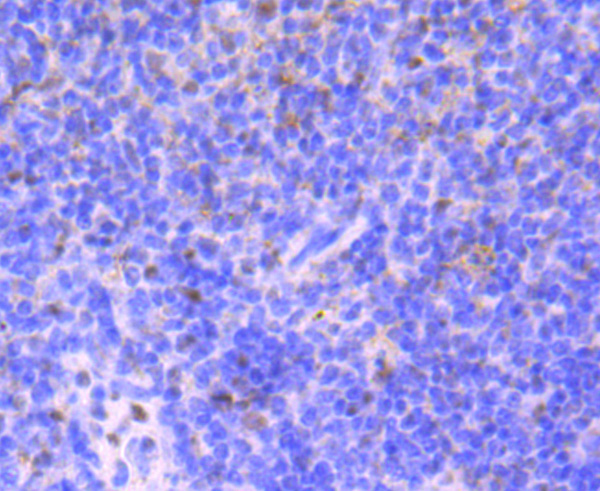
IHC (Immunohistochemistry) (Immunoperoxidase of monoclonal antibody to ERO1L on formalin-fixed paraffin-embedded human small Intestine. [antibody concentration 1.5ug/ml].)
WB (Western Blot) (Western Blot analysis of ERO1L expression in transfected 293T cell line by ERO1L monoclonal antibody. Lane 1: ERO1L transfected lysate (54kD). Lane 2: Non-transfected lysate.)
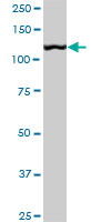
WB (Western Blot) (ERO1L monoclonal antibody, Western Blot analysis of ERO1L expression in HeLa.)
WB (Western Blot) (Western Blot detection against Immunogen (35.9kD).)
WB (Western Blot) (Western Blot analysis of ACY1 expression in transfected 293T cell line by ACY1 monoclonal antibody. Lane 1: ACY1 transfected lysate (45.9kD). Lane 2: Non-transfected lysate.)
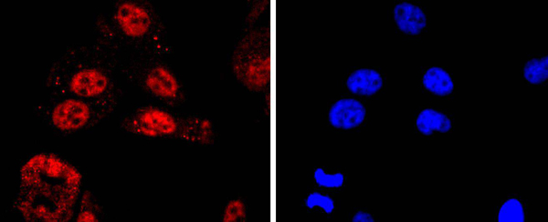
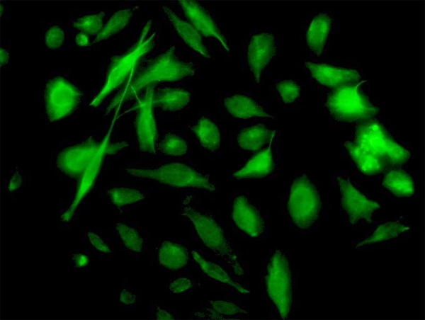
IF (Immunofluorescence) (Immunofluorescence of monoclonal antibody to ACY1 on HeLa cell. [antibody concentration 10ug/ml].)
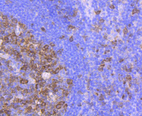
IHC (Immunohistochemistry) (Immunoperoxidase of monoclonal antibody to ACY1 on formalin-fixed paraffin-embedded human malignant lymphoma, diffuse large B tissue. [antibody concentration 3ug/ml])
IP (Immunoprecipitation) (Immunoprecipitation of ACY1 transfected lysate using ACY1 monoclonal antibody and Protein A Magnetic Bead and immunoblotted with ACY1 rabbit polyclonal antibody.)
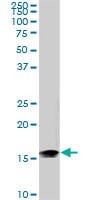
WB (Western Blot) (ACY1 monoclonal antibody Western Blot analysis of ACY1 expression in K-562.)
WB (Western Blot) (Western Blot detection against Immunogen (70.62kD).)
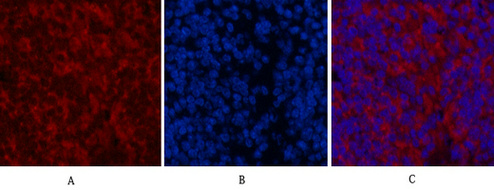
IF (Immunofluorescence) (Immunofluorescence analysis of rat brain using GFAP Rabbit mAb at dilution of 1:100 (40x lens). Blue: DAPI for nuclear staining.)
IF (Immunofluorescence) (Immunofluorescence analysis of mouse brain using GFAP Rabbit mAb at dilution of 1:100 (40x lens). Blue: DAPI for nuclear staining.)
IHC (Immunohistochemistry) (Immunohistochemistry of paraffin-embedded mouse brain using GFAP Rabbit mAb at dilution of 1:100 (40x lens).Perform microwave antigen retrieval with 10 mM PBS buffer pH 7.2 before commencing with IHC staining protocol.)
IHC (Immunohistochemistry) (Immunohistochemistry of paraffin-embedded human brain using GFAP Rabbit mAb at dilution of 1:100 (40x lens).Perform microwave antigen retrieval with 10 mM PBS buffer pH 7.2 before commencing with IHC staining protocol.)
IHC (Immunohistochemistry) (Immunohistochemistry of paraffin-embedded rat brain using GFAP Rabbit mAb at dilution of 1:100 (40x lens).Perform microwave antigen retrieval with 10 mM PBS buffer pH 7.2 before commencing with IHC staining protocol.)
WB (Western Blot) (Western blot analysis of extracts of U-251MG cells, using GFAP antibody at 1:1000 dilution.Secondary antibody: HRP Goat Anti-Rabbit IgG (H+L) (AS014) at 1:10000 dilution.Lysates/proteins: 25ug per lane.Blocking buffer: 3% nonfat dry milk in TBST.Detection: ECL Basic Kit (RM00020).Exposure time: 3min.)
WB (Western Blot) (Western blot analysis of extracts of various cell lines, using GFAP antibody at 1:1000 dilution.Secondary antibody: HRP Goat Anti-Rabbit IgG (H+L) (AS014) at 1:10000 dilution.Lysates/proteins: 25ug per lane.Blocking buffer: 3% nonfat dry milk in TBST.Detection: ECL Basic Kit (RM00020).Exposure time: 1s.)
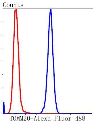
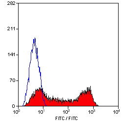
FCM (Flow Cytometry) (Flow cytometric analysis of Hela cells with Beta-actin antibody at 1/100 dilution (blue) compared with an unlabelled control (cells without incubation with primary antibody; red). Goat anti mouse IgG (FITC) was used as the secondary antibody.)
ICC (Immunocytochemistry) (ICC staining of Beta-actin in NIH-3T3 cells (red). Cells were fixed in paraformaldehyde, permeabilised with 0.25% Triton X100/PBS.)
ICC (Immunocytochemistry) (ICC staining of Beta-actin in A549 cells (red). Cells were fixed in paraformaldehyde, permeabilised with 0.25% Triton X100/PBS.)
ICC (Immunocytochemistry) (ICC staining of Beta-actin in Hela cells (red). Cells were fixed in paraformaldehyde, permeabilised with 0.25% Triton X100/PBS.)
IHC (Immunohistochemistry) (Immunohistochemical analysis of paraffin-embedded mouse kidney tissue using anti-Beta-actin antibody. Counter stained with hematoxylin.)
IHC (Immunohistochemistry) (Immunohistochemical analysis of paraffin-embedded mouse prostate tissue using anti-Beta-actin antibody. Counter stained with hematoxylin.)
IHC (Immunohistochemistry) (Immunohistochemical analysis of paraffin-embedded human colon carcinoma tissue using anti-Beta-actin antibody. Counter stained with hematoxylin.)
WB (Western Blot) (Western blot analysis of Beta-Western blot analysis of Beta-actin on different lysates using anti-Beta-actin antibody at 1/10, 000 dilution. Positive control: Lane 1: NIH/3T3 Lane 2: PC12 Lane 3: MCF-7 Lane 4: HepG2 Lane 5: Hela Lane 6: Mouse lung)
FCM (Flow Cytometry) (Flow cytometric analysis of SH-SY5Y cells with PPP1R1A antibody at 1/50 dilution (red) compared with an unlabelled control (cells without incubation with primary antibody; black). Alexa Fluor 488-conjugated goat anti rabbit IgG was used as the secondary antibody.)
ICC (Immunocytochemistry) (ICC staining PPP1R1A in SHG-44 cells (green). The nuclear counter stain is DAPI (blue). Cells were fixed in paraformaldehyde, permeabilised with 0.25% Triton X100/PBS.)
ICC (Immunocytochemistry) (ICC staining PPP1R1A in SH-SY5Y cells (green). The nuclear counter stain is DAPI (blue). Cells were fixed in paraformaldehyde, permeabilised with 0.25% Triton X100/PBS.)
ICC (Immunocytochemistry) (ICC staining PPP1R1A in N2A cells (green). The nuclear counter stain is DAPI (blue). Cells were fixed in paraformaldehyde, permeabilised with 0.25% Triton X100/PBS.)
IHC (Immunohistochemistry) (Immunohistochemical analysis of paraffin-embedded mouse kidney tissue using anti-PPP1R1A antibody. Counter stained with hematoxylin.)
IHC (Immunohistochemistry) (Immunohistochemical analysis of paraffin-embedded mouse brain tissue using anti-PPP1R1A antibody. Counter stained with hematoxylin.)
IHC (Immunohistochemistry) (Immunohistochemical analysis of paraffin-embedded human kidney tissue using anti-PPP1R1A antibody. Counter stained with hematoxylin.)
WB (Western Blot) (Western blot analysis of PPP1R1A on Rat brain lysates using anti-PPP1R1A antibody at 1/1, 000 dilution.)
FCM (Flow Cytometry) (Flow cytometric analysis of LOVO cells with Cellular Apoptosis Susceptibility antibody at 1/100 dilution (red) compared with an unlabelled control (cells without incubation with primary antibody; black). Alexa Fluor 488-conjugated goat anti-rabbit IgG was used as the secondary antibody.)
ICC (Immunocytochemistry) (ICC staining Cellular Apoptosis Susceptibility in SH-SY5Y cells (green). The nuclear counter stain is DAPI (blue). Cells were fixed in paraformaldehyde, permeabilised with 0.25% Triton X100/PBS.)
ICC (Immunocytochemistry) (ICC staining Cellular Apoptosis Susceptibility in PC-3M cells (green). The nuclear counter stain is DAPI (blue). Cells were fixed in paraformaldehyde, permeabilised with 0.25% Triton X100/PBS.)
ICC (Immunocytochemistry) (ICC staining Cellular Apoptosis Susceptibility in LOVO cells (green). The nuclear counter stain is DAPI (blue). Cells were fixed in paraformaldehyde, permeabilised with 0.25% Triton X100/PBS.)
IHC (Immunohistochemistry) (Immunohistochemical analysis of paraffin-embedded mouse testis tissue using anti-Cellular Apoptosis Susceptibility antibody. Counter stained with hematoxylin.)
IHC (Immunohistochemistry) (Immunohistochemical analysis of paraffin-embedded human colon cancer tissue using anti-Cellular Apoptosis Susceptibility antibody. Counter stained with hematoxylin.)
WB (Western Blot) (Western blot analysis of Cellular Apoptosis Susceptibility on different lysates using anti-Cellular Apoptosis Susceptibility antibody at 1/500 dilution. Positive control: Lane 1: Mouse testis Lane 2: SiHa Lane 3: SK-BR-3 Lane 4: PC-3M)
FCM (Flow Cytometry) (Dual staining of pig peripheral blood lymphocytes with Mouse anti Pig CD335 detected with Goat anti Mouse IgG (H/L):FITC (STAR117F), and Mouse anti Pig wCD8a:RPE)
Application Data (Published investigator image: Mouse anti Pig CD335 antibody, clone VIV-KM1 used for the identification of NK cells in influenza infected pigs by flow cytometry.Image caption: NK cell numbers in the blood of influenza infected pigs. Blood was taken from influenza A virus infected (n = 12) and control pigs (n = 9) on day 0-3 pi in a second experiment. (A) Isolated PBMCs were analysed by flow cytometry and live CD3- lymphocytes were gated as NKp46- or NKp46+ NK cells according to CD8a and NKp46 expression. Plots are taken from a representative control animal. Absolute numbers of (B) NKp46+ NK cells in PBMC were analysed by flow cytometry in control (left) and infected animals (right). (C) Results for the NKp46+ cells in the two groups were compared on each sampling day. (D) NKp46- NK cells in PBMC in control (left) and infected animals (right). (E) NKp46- NK cells were compared for the two groups. Each line in (B) and (D) represents one animal. **p=0.01.From: Forberg H, Hauge AG, Valheim M, Garcon F, Nunez A, et al. (2014) Early Responses of Natural Killer Cells in Pigs Experimentally Infected with 2009 Pandemic H1N1 Influenza A Virus.PLoS ONE 9(6): e100619.)
Application Data (Published investigator image: Mouse anti Pig CD335 antibody, clone VIV-KM1 used for the identification of NK cells in influenza infected pigs by immunofluorescence.Image caption:Staining for apoptosis in the lungs. Lung tissue sections from animals infected with influenza A virus (n = 12) were stained with immunofluorescence markers against (A) NKp46(green), (B) influenza A NP(red) and (C) the apoptosis marker caspase-3(blue). (D) Overlay displaying simultaneously influenza A virus NP+ and caspase-3+ cells as purple (arrows). Representative of virus infected bronchiole at day 1 pi. Immunofluorescence staining, 400x.From: Forberg H, Hauge AG, Valheim M, Garcon F, Nunez A, et al. (2014) Early Responses of Natural Killer Cells in Pigs Experimentally Infected with 2009 Pandemic H1N1 Influenza A Virus.PLoS ONE 9(6): e100619.)
Application Data (Published investigator image: Mouse anti Pig CD335 antibody, clone VIV-KM1 used for the identification of NK cells in influenza infected pigs by immunofluorescence.Image caption:NKp46+ cells in the lungs of influenza virus infected pigs.Lung tissue sections from pigs infected with influenza A virus and control pigs were stained with immunofluorescence markers for cytokeratin (blue), NKp46 (green) and influenza A virus NP (red). NKp46+ cells were counted in areas were influenza A virus NP was (A) detected and (B) not detected. Representative pictures taken from the same animal on day 1 pi are shown. Arrows point at NKp46+ cells. Immunofluorescence staining, 200x. (C) Plot shows number of NKp46+ cells per 0,1 mm2 in sections (n = 24 per animal) from control animals (n = 6) and in areas with and without virus in infected animals (n = 4 per day) calculated as described in Material and Methods. Groups with different letters differ significantly (p=0.05). (D) NKp46+ cells in the lumen of a bronchus (BL). Arrows point at the epithelial lining. Representative picture of luminal exudate, taken from an infected animal on day 2 pi. Insert shows NKp46+ and influenza A virus NP+ cell in the lung tissue of an infected animal on day 1 pi. Immunofluorescence staining, 400x.From: Forberg H, Hauge AG, Valheim M, Garcon F, Nunez A, et al. (2014) Early Responses of Natural Killer Cells in Pigs Experimentally Infected with 2009 Pandemic H1N1 Influenza A Virus.PLoS ONE 9(6): e100619.)
Application Data (Published investigator image: Mouse anti Pig CD335 antibody, clone VIV-KM1 used for the identification of NK cells in influenza infected pigs by flow cytometry.Image caption: Percentages of NK cells and expression of NKp46 and CD25 in lung tissue. Mononuclear cells were isolated from lung tissue of pigs infected with influenza A virus (n = 12) and control animals (n = 6) during the first 3 days pi and analysed by flow cytometry. (A) Live CD3- lymphocytes were gated as described in Fig 2. NK cells were gated according to CD8a and NKp46 expression and defined as NKp46-, NKp46int or NKp46high cells. Plot shown is from a representative control animal. (B) Proportions of NKp46- (green), NKp46int (blue) and NKp46high (purple) NK cells in individual animals, shown as percentages of gated cells in lymphocytes. (C) Percentages of NKp46- (left), NKp46int (middle) and NKp46high (right) NK cells among lymphocytes. (D) Median fluorescence intensity (MFI) in the NKp46- gate (left), the NKp46+ gate (middle) and in the NKp46high gate (right) are shown. (E) CD25+ cells were gated in the NKp46- and NKp46int NK cells (left) and in the NKp46high NK cells (right). Plots shown are from a representative control animal. (F) The percentages of CD25+ cells in each gate were calculated in control animals (green), infected animals from day 1 (purple), day 2 (blue) and day 3 (pink) pi. *p=0,05, **p=0.01.From: Forberg H, Hauge AG, Valheim M, Garcon F, Nunez A, et al. (2014) Early Responses of Natural Killer Cells in Pigs Experimentally Infected with 2009 Pandemic H1N1 Influenza A Virus.PLoS ONE 9(6): e100619.)
Application Data (Published investigator image: Mouse anti Pig CD335 antibody, clone VIV-KM1 used for the identification of NK cells in influenza infected pigs by immunofluorescence.Image caption:Counting of NKp46+ cells. Numbers of NKp46+ cells per area were counted in lung tissue sections from control animals and influenza A virus infected animals as described in Material and Methods. Representative picture of area with virus from infected animal. Immunofluorescence staining, 200x.From: Forberg H, Hauge AG, Valheim M, Garcon F, Nunez A, et al. (2014) Early Responses of Natural Killer Cells in Pigs Experimentally Infected with 2009 Pandemic H1N1 Influenza A Virus.PLoS ONE 9(6): e100619.)
Application Data (Published investigator image: Mouse anti Pig CD335 antibody, clone VIV-KM1 used for the evaluation of CD335 expression on porcine cells by flow cytometry.Image caption:Splenic NKp46high NK cells produced the highest levels of IFN-?. (A + B) Intracellular cytokine staining for IFN-? production in NKp46-defined NK cells within PBMC (upper graphs) and splenocytes (lower graphs) by four-colour FCM after 24 h in vitro stimulation with rhIL-2, rpIL-12 and rpIL-18 or medium. CD3- lymphocytes were gated (not shown) and NKp46/CD8a defined total NK cells were further subgated for IFN-? production. IFN-? producing NK cells were gated according to NKp46 expression levels (CD8a+NKp46-: blue, CD8a+NKp46+: green, CD8adim/-NKp46high: red). (A) Numbers indicate the percentage of IFN-?+ NK cells within respective gates. Results are representative for experiments with four different animals. (B) Percentage of IFN-?+ cells (left graph) and mean fluorescence intensity of IFN-?+ cells (right graph) within the different NK-cell subsets in blood and spleen of four animals are shown. (C) Supernatants of cytokine stimulated FACS-sorted CD3-CD8a+NKp46- and CD3-CD8a+NKp46+ NK cells from blood and CD3-CD8a+NKp46-, CD3-CD8a+NKp46+ and CD3-CD8adim/-NKp46high NK cells from spleen were tested for IFN-? production in ELISA following 24 h in vitro stimulation with rhIL-2, rpIL-12 and rpIL-18. Data on the left are from one representative animal and displayed as the mean of duplicates +/- SD. IFN-? production in experiments with four animals analysed is shown on the right. Mean values are represented by a black bar. (B + C) Significant differences between the subsets in blood or spleen are indicated (* = p < 0.05, ** = p < 0.01).From: Mair KH, Mllebner A, Essler SE, Duvigneau JC, Storset AK, Saalmller A, Gerner W. Porcine CD8adim/-NKp46high NK cells are in a highly activated state. Vet Res. 2013 Mar 1;44:13.)
Application Data (Published investigator image: Mouse anti Pig CD335 antibody, clone VIV-KM1 used for the evaluation of CD335 expression on porcine cells by flow cytometry.Image caption:Varying expression of NKp46, CD8a, CD16 and CD27 on NK-cell subsets in blood and spleen. (A) Following five-colour staining, PBMC and splenocytes were gated on CD3- cells and further subgated according to their NKp46/CD8a expression pattern in NKp46- and NKp46+ NK cells in blood (upper graph) and NKp46-, NKp46+ and NKp46high NK cells in spleen (lower graph). (B) 14 healthy 6 -7 month old pigs were investigated for NKp46 and CD8a expression levels in the NKp46-defined NK-cell subsets. Box-plots show the mean fluorescence intensity of the two markers. (C) NK-cell subsets defined in (A) were further analysed for their expression of the surface markers CD16 and CD27. Histograms show the expression of the two markers within the respective NKp46-defined subsets (CD8a+NKp46-: blue histograms, CD8a+NKp46+: green histograms, CD8adim/-NKp46high: red histograms) in blood (upper graphs) and spleen (lower graphs) according to the corresponding isotype control (grey histrograms with dotted lines). Box-plots show the mean fluorescence intensity of CD16 and CD27 of the NKp46-defined NK-cell subsets in blood and spleen of 14 healthy 6 -7 month old pigs. (B + C) Significant differences between the subsets in blood or spleen are indicated (* = p < 0.05, ** = p < 0.01, *** = p < 0.001).From: Mair KH, Mllebner A, Essler SE, Duvigneau JC, Storset AK, Saalmller A, Gerner W. Porcine CD8adim/-NKp46high NK cells are in a highly activated state. Vet Res. 2013 Mar 1;44:13.)
Application Data (Dual staining of pig peripheral blood lymphocytes with Mouse anti Pig CD335:Alexa Fluor488 and Mouse anti Pig wCD8a:RPE)
Application Data (Dual staining of pig peripheral blood lymphocytes with Mouse anti Pig CD335:APC and Mouse anti Pig wCD8a:RPE)
Application Data (Published customer image: Mouse anti Human CD49d antibody, clone HP2/1 used for binding efficiency determinationImage caption:Binding efficiencies (BE) of different a4beta7 molecules composed of distinct a4 mutants to monoclonal antibodies against a4, beta7 or the a4beta7 heterodimer. Binding efficiency is determined by the ratio between the mean fluorescence of antibody binding to each a4 molecule and of the binding in a mock-transfected cell culture (see Materials and Methods for details). Dark gray bars represent binding to the human (wild type) a4 clone, whereas light gray bars are those of binding to the different a4 mutants (as shown in the x-axis). a4 mutants which included substitutions at codon 201 are boxed. A, binding of anti-a4 2b4 antibody. B, binding of anti-a4 HP2/1 antibody. C, BE of different anti-a4 and beta7 antibodies to the human a4 and the quintuple a4 mutant (5 aa mut). Bars represent the range of standard errors deduced from triplicate experiments. p-values of Student's t tests are shown above each comparison. NS, non-significant (> 0.05).From:Darc M, Hait SH, Soares EA, Cicala C, Seuanez HN, et al. (2011) Polymorphisms in the a4 Integrin of Neotropical Primates: Insights for Binding of Natural Ligands and HIV-1 gp120 to the Human a4beta7. PLoS ONE 6(9): e24461.)
Application Data (Published customer image: Mouse anti Human CD49d antibody, clone HP2/1 used for blocking studies.Image caption: Blocking experiments with anti-beta7, anti-a4 and anti-aVbeta3 antibodies for adhesion to HS-5 stromal cells. Panel A, Blocking with anti-beta7. HMCLs were stimulated with Pam3CSK4 for 24 hours and then treated with anti-beta7 antibody before adhesion to HS5-coated wells. Panel B, Blocking experiments with anti-a4 and anti-aVbeta3 antibodies for adhesion to HS-5. HMCLs were stimulated with Pam3CSK4 for 24 hours and then treated with anti-a4 and anti-aVbeta3 antibodies before adhesion to HS5-coated wells. The results are the statistical analyses of data in 3 separate experiments expressed as mean +/- SEM, *P)
Application Data (Published customer image: Mouse anti Human CD49d antibody, clone HP2/1 used for immunoprecipitationImage caption:All anti-CD81, anti-CD82 and anti-CD151 clones co-precipitate beta1 and beta3 integrins from normal and leukemic proerythroblasts. A. CD81 clones. B. CD82 clones. C. CD151 clones. ERB, day 6 proerythroblasts. a4, HP2/1; a5, IIA1; aL, TS1/22; aIIb, PAB-1. Integrins in 7.5% NR gels, tetraspanins in 12% non-reduced gels. More beta3 integrins are co-precipitated from HEL cells as they express aIIbbeta3 and aVbeta3; proerythroblasts express only aIIbbeta3.From: Spring FA, Griffiths RE, Mankelow TJ, Agnew C, Parsons SF, et al. (2013) Tetraspanins CD81 and CD82 Facilitate a4beta1-Mediated Adhesion of Human Erythroblasts to Vascular Cell Adhesion Molecule-1. PLoS ONE 8(5): e62654.)
Application Data (Published customer image: Mouse anti Human CD49d antibody, clone HP2/1 used for western blotting and immunoprecipitationImage caption:Tetraspanins CD81, CD82 and CD151 are associated with a4beta1 throughout erythroid maturation and with beta3 in proerythroblasts and basophilic erythroblasts. A. CD81, CD82 and CD151 precipitates from Mn2+-activated proerythroblasts (ProEB, day 5), basophilic (BasoEB, day 8) and polychromatic (PolyEB, day 12) erythroblasts were successively probed with anti-a4, anti-beta1 and anti-beta3 antibodies; tetraspanin controls from each time point are also illustrated. All tetraspanins co-precipitated a4 and beta1 from erythroblasts B. Tetraspanin precipitates from day 6 proerythroblasts (ProEB) solubilised in the presence of EDTA or different cations, and from Mn2+-activated basophilic erythroblasts (BasoEB, day 8) were probed with a mix of antibodies to a5, beta1, beta2 and beta3 integrins while the control samples were probed with the relevant tetraspanin antibodies. For clarity, integrin controls are illustrated for the EDTA blot but were present on all blots. beta1 and beta3 integrins were precipitated well only in the presence of Mn2+. C. CD81 and CD82 precipitates from day 5 proerythroblasts were successively probed with different anti-integrin subunit antibodies and demonstrate co-precipitation of beta1 and beta3 but not a5 or beta2 integrins. D. CD81 (454720) and CD82 (53H5) precipitates from day 6 proerythroblasts (ProEB) and HEL cells (HEL) solubilised in the presence of EDTA, Ca2++Mg2+ or Mn2+ probed with anti-CD82 and anti-CD81 antibodies. Each tetraspanin co-precipitates the other most strongly in the presence of Mn2+ from proerythroblasts while any cation permits co-precipitation in HEL cells. Integrins were analysed on 7.5% gels, tetraspanins on 12% gels; non-reducing conditions. Unless stated, the following clones were used: CD53, MEM-53; CD63, MEM-259; CD81, 454720; CD82, TS82b; CD151, IIG5a; a4, HP2/1; a5, IIA1; aL, TS1/22; aIIb, PAB-1. All day 5 and 6 cultures comprised 90 -95% proerythroblasts; day 8 culture comprised 5% proerythroblasts, 81% basophilic erythroblasts and 14% polychromatic erythroblasts; day 12 culture comprised 41% polychromatic erythroblasts, 15% orthochromatic erythroblasts and 41% reticulocytes. In the day 5 and 6 cultures 15 -34% of cells were GPA+ and 28 -35% of cells were aIIb+. Day 8 and day 12 cultures had 77% and 97% GPA+ cells, respectively, and 9% and 0% aIIb+ cells, respectively.From: Spring FA, Griffiths RE, Mankelow TJ, Agnew C, Parsons SF, et al. (2013) Tetraspanins CD81 and CD82 Facilitate a4beta1-Mediated Adhesion of Human Erythroblasts to Vascular Cell Adhesion Molecule-1. PLoS ONE 8(5): e62654.)
Application Data (Published customer image: Mouse anti Human CD49d antibody, clone HP2/1 used for immunoprecipitationImage caption:Several anti-tetraspanin antibodies co-precipitate beta1 integrins from HEL cells solubilised in Brij-97. Precipitates were prepared from HEL cells solubilised in different detergents in the presence of Mn2+. CD53, MEM-53; CD63, MEM-259; CD81, 454720; CD82, TS82b; CD151, IIG5a; a4, HP2/1; aL, TS1/22; aIIb, PAB-1. Precipitates were run on 7.5% non-reduced gels.From: Spring FA, Griffiths RE, Mankelow TJ, Agnew C, Parsons SF, et al. (2013) Tetraspanins CD81 and CD82 Facilitate a4beta1-Mediated Adhesion of Human Erythroblasts to Vascular Cell Adhesion Molecule-1. PLoS ONE 8(5): e62654.)
Application Data (Published Customer Image:Mouse CD31 antibody, clone ER-MP12 used for the demonstration of vasculature in mouse brain by immunofluorescence.Image caption:Inhibition of 2-AG hydrolysis reduces LPS-induced BBB permeability. a, b Fibrinogen levels in b plasma and the a ratio of brain to plasma fibrinogen were assessed by ELISA. n?=?5/7 mice per group. c, d Fluorescent immunostaining in the striatum for fibrinogen (red) and vascular marker (CD31; green) demonstrated leakage of fibrinogen into the brain with vehicle treatment, whereas vascular integrity was preserved when (e, f) MAGL was inhibited. g Extravascular fibrinogen was semi-quantitated in fluorescently labeled sections of the striatum. Bar graphs were plotted with mean?+/-SEM and data analyzed using one-way analysis of variance (ANOVA) with Tukey post-hoc comparisons. n = 5/7 mice per group. Significance is shown as *p?)
Application Data (Rat anti Mouse CD31 antibody, clone ER-MP12 used for the detection of blood vessels in an experimental murine tumor model by immunohistofluorescence.Image caption:Percentage of pixels positive for NG2 in 4T1 (n = 5) and RM11 (n = 4 and n = 6) tumors (A,D) from WT and beta3-KO mice were calculated using immunofluorescent images. No statistical differences in 4T1 (p = 0.90) or RM11 (p = 0.23) were found. Mean +/- SD. Representative images of NG2-staining from both genotypes of 4T1 (B,C) (NG2 green, CD31 red) and RM11 (E,F) (NG2 red) tumors are shown. Scale bars indicate 50 mum.From: Reigstad I, Sortland K, Skogstrand T, Reed RK, Stuhr L.The Effect of Stromal Integrin beta3-Deficiency on Two Different Tumors in Mice.Cancers (Basel). 2016 Jan 12;8(1). pii: E14.)
Application Data (Immunoperoxidase staining of a mouse lymph node cryosection with Rat anti Mouse CD31 antibody, clone ER-MP12 followed by horseradish peroxidase conjugated Goat anti Rat IgG antibody . High power)
Application Data (Immunoperoxidase staining of a mouse lymph node cryosection with Rat anti Mouse CD31 antibody, clone ER-MP12 followed by horseradish peroxidase conjugated Goat anti Rat IgG antibody . Medium power)
Application Data (Immunoperoxidase staining of a mouse lymph node cryosection with Rat anti Mouse CD31 antibody, clone ER-MP12 followed by horseradish peroxidase conjugated Goat anti Rat IgG antibody . Low power)
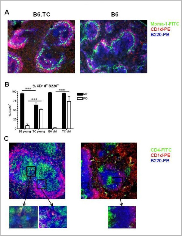
Application Data (Figure A. RPE conjugated rat anti mouse CD45R and FITC conjugated rat IgG2a isotype control Figure B. RPE conjugated rat anti mouse CD45R and Pacific Blue conjugated rat anti mouse CD31 . All experiments performed on red cell lysed murine splenocytes gated on mononuclear cells. Data acquired on the ZE5 cell analyzer.)
Application Data (Figure A. Alexa647 conjugated rat anti mouse CD22 and Alexa488 conjugated Rat IgG2a isotype control . Figure B. Alexa647 conjugated rat anti mouse CD22 and Alexa488 conjugated rat anti mouse CD31 . All experiments performed on red cell lysed murine peripheral blood in the presence of murine SeroBlock (BUF041A).)
Application Data (Figure A. RPE conjugated rat anti mouse CD45R and FITC conjugated rat IgG2a isotype control Figure B. RPE conjugated rat anti mouse CD45R and FITC conjugated rat anti mouse CD31 . All experiments performed on red cell lysed murine splenocytes gated on mononuclear cells. Data acquired on the ZE5 cell analyzer.)
WB (Western Blot) (AKAP8 monoclonal antibody (Western Blot analysis of AKAP8 expression in HeLa NE)
IP (Immunoprecipitation) (Immunoprecipitation of AKAP8 transfected lysate using AKAP8 monoclonal antibody and Protein A Magnetic Bead, and immunoblotted with AKAP8 monoclonal antibody.)
IF (Immunofluorescence) (Immunofluorescence of monoclonal antibody to AKAP8 on HeLa cell. [antibody concentration 10ug/ml])
IHC (Immunohistochemistry) (Immunoperoxidase of monoclonal antibody to AKAP8 on formalin-fixed paraffin-embedded human pancreas. [antibody concentration 3ug/ml])
WB (Western Blot) (Western Blot analysis of AKAP8 expression in transfected 293T cell line by AKAP8 monoclonal antibody Lane 1: AKAP8 transfected lysate (76.2kD). Lane 2: Non-transfected lysate.)
WB (Western Blot) (Western Blot detection against Immunogen (38.43kD).)
WB (Western Blot) (Western Blot analysis of TOMM22 expression in transfected 293T cell line by TOMM22 monoclonal antibody. Lane 1: TOMM22 transfected lysate (15.5kD). Lane 2: Non-transfected lysate.)
WB (Western Blot) (TOMM22 monoclonal antibody. Western Blot analysis of TOMM22 expression in PC-12.)
WB (Western Blot) (Western blot analysis of TOMM22 over-expressed 293 cell line, cotransfected with TOMM22 Validated Chimera RNAi (Lane 2) or non-transfected control (Lane 1). Blot probed with TOMM22 monoclonal antibody. GAPDH (36.1kD) used as specificity and loading control.)
Application Data (Detection limit for recombinant GST tagged TOMM22 is 0.1ng/ml as a capture antibody.)
IF (Immunofluorescence) (Immunofluorescence of monoclonal antibody to TOMM22 on HeLa cell. [antibody concentration 10ug/ml])
IHC (Immunohistochemistry) (Immunoperoxidase of monoclonal antibody to TOMM22 on formalin-fixed paraffin-embedded human small Intestine. [antibody concentration 3ug/ml].)
WB (Western Blot) (Western Blot detection against Immunogen (41.73kD).)
WB (Western Blot) (RUNX1 monoclonal antibody Western Blot analysis of RUNX1 expression in Hela NE.)
Application Data (Detection limit for recombinant GST tagged RUNX1 is ~1ng/ml as a capture antibody.)
IF (Immunofluorescence) (Immunofluorescence of monoclonal antibody to RUNX1 on HeLa cell. [antibody concentration 10ug/ml].)
IHC (Immunohistochemistry) (Immunoperoxidase of monoclonal antibody to RUNX1 on formalin-fixed paraffin-embedded human placenta. [antibody concentration 3ug/ml].)
WB (Western Blot) (RUNX1 monoclonal antibody Western Blot analysis of RUNX1 expression in NIH/3T3.)
WB (Western Blot) (Western Blot detection against Immunogen (36.85kD).)
WB (Western Blot) (RIPK2 monoclonal antibody (M02), clone 6F7. Western Blot analysis of RIPK2 expression in PC-12 (Cat # L012V1).)
WB (Western Blot) (RIPK2 monoclonal antibody (M02), clone 6F7. Western Blot analysis of RIPK2 expression in NIH/3T3 (Cat # L018V1).)
Application Data (Detection limit for recombinant GST tagged RIPK2 is approximately 0.1ng/ml as a capture antibody.)
IHC (Immunohistochemistry) (Immunoperoxidase of monoclonal antibody to RIPK2 on formalin-fixed paraffin-embedded human prostate. [antibody concentration 1.2 ug/ml])
WB (Western Blot) (RIPK2 monoclonal antibody (M02), clone 6F7 Western Blot analysis of RIPK2 expression in HeLa (Cat # L013V1).)
Application Data (Detection limit for recombinant GST tagged EIF2AK2 is approximately 0.03ng/ml as a capture antibody.)
IHC (Immunohistochemistry) (Immunoperoxidase of monoclonal antibody to EIF2AK2 on formalin-fixed paraffin-embedded human adrenal gland. [antibody concentration 1.5 ug/ml])
IF (Immunofluorescence) (Immunofluorescence of monoclonal antibody to EIF2AK2 on HeLa cell. [antibody concentration 30 ug/ml])
WB (Western Blot) (EIF2AK2 monoclonal antibody (M02), clone 1D11 Western Blot analysis of EIF2AK2 expression in K-562 (Cat # L009V1).)
WB (Western Blot) (HSPA1B monoclonal antibody Western Blot analysis of HSPA1B expression in Raw 264.7.)
WB (Western Blot) (HSPA1B monoclonal antibody Western Blot analysis of HSPA1B expression in NIH/3T3)
Application Data (Proximity Ligation Analysis (PLA) of protein-protein interactions between TP53 and HSPA1B. HeLa cells were stained with TP53 rabbit purified polyclonal 1:1200 and HSPA1B mouse monoclonal antibody 1:50. Signals were detected 30 Detection Kit 613 (red), and nuclei were counterstained with DAPI (blue). Each red dot represents the detection of protein-protein interaction complex.)
Application Data (Detection limit for recombinant GST tagged HSPA1B is ~0.03ng/ml as a capture antibody.)
IF (Immunofluorescence) (Immunofluorescence of monoclonal antibody to HSPA1B on HeLa cell. [antibody concentration 10ug/ml])
IHC (Immunohistochemistry) (Immunoperoxidase of monoclonal antibody to HSPA1B on formalin-fixed paraffin-embedded human testis. [antibody concentration 6ug/ml])
WB (Western Blot) (Western Blot detection against Immunogen (34.65kD).)
WB (Western Blot) (Western Blot detection against Immunogen (77.26kD).)
IP (Immunoprecipitation) (Immunoprecipitation of STK38 transfected lysate using STK38 monoclonal antibody and Protein A Magnetic Bead, and immunoblotted with STK38 rabbit polyclonal antibody.)
IF (Immunofluorescence) (Immunofluorescence of monoclonal antibody to STK38 on HeLa cell. [antibody concentration 10ug/ml])
IHC (Immunohistochemistry) (Immunoperoxidase of monoclonal antibody to STK38 on formalin-fixed paraffin-embedded human malignant lymphoma, diffuse large B tissue [antibody concentration 5ug/ml])
WB (Western Blot) (STK38 monoclonal antibody Western Blot analysis of STK38 expression in human kidney.)
WB (Western Blot) (Western Blot analysis of STK38 expression in transfected 293T cell line by STK38 monoclonal antibody Lane 1: STK38 transfected lysate (54.2kD). Lane 2: Non-transfected lysate.)
WB (Western Blot) (NME2 monoclonal antibody (M06), clone 1D3. Western Blot analysis of NME2 expression in NIH/3T3.)
IHC (Immunohistochemistry) (Immunoperoxidase of monoclonal antibody to NME2 on formalin-fixed paraffin-embedded human testis. [antibody concentration 1.5 ug/ml])
IF (Immunofluorescence) (Immunofluorescence of monoclonal antibody to NME2 on HeLa cell. [antibody concentration 10 ug/ml])
WB (Western Blot) (NME2 monoclonal antibody (M06), clone 1D3 Western Blot analysis of NME2 expression in HeLa.)
IHC (Immunohistchemistry) (Immunoperoxidase of monoclonal antibody to PDK2 on formalin-fixed paraffin-embedded human pancreas. [antibody concentration 2 ug/ml])
IHC (Immunohistochemistry) (Immunoperoxidase of monoclonal antibody to PDK2 on formalin-fixed paraffin-embedded human pancreas. [antibody concentration 2 ug/ml])
Application Data (Detection limit for recombinant GST tagged PDK2 is approximately 0.03ng/ml as a capture antibody.)
WB (Western Blot) (PDK2 monoclonal antibody (M02), clone 5F8. Western Blot analysis of PDK2 expression in A-431 (Cat # L015V1).)
WB (Western Blot) (PDK2 monoclonal antibody (M02), clone 5F8. Western Blot analysis of PDK2 expression in 293 (Cat # L026V1).)
WB (Western Blot) (PDK2 monoclonal antibody (M02), clone 5F8 Western Blot analysis of PDK2 expression in HeLa (Cat # L013V1).)
Application Data (Detection limit for recombinant GST tagged HDAC1 is approximately 0.1ng/ml as a capture antibody.)
WB (Western Blot) (HDAC1 monoclonal antibody (M06), clone 1D6 Western Blot analysis of HDAC1 expression in Hela S3 NE (Cat # L013V3).)
IF (Immunofluorescence) (Immunofluorescence of monoclonal antibody to HDAC1 on HeLa cell. [antibody concentration 10 ug/ml])
IHC (Immunohistochemistry) (Immunoperoxidase of monoclonal antibody to HDAC1 on formalin-fixed paraffin-embedded human liver. [antibody concentration 3 ug/ml])
Application Data (Immunoperoxidase of monoclonal antibody to HDAC1 on formalin-fixed paraffin-embedded human liver. [antibody concentration 3 ug/ml])
Application Data (Detection limit for recombinant GST tagged TCEA3 is approximately 0.03ng/ml as a capture antibody.)
WB (Western Blot) (TCEA3 monoclonal antibody (M08), clone 4E11 Western Blot analysis of TCEA3 expression in HepG2.)
IF (Immunofluorescence) (Immunofluorescence of monoclonal antibody to TCEA3 on HepG2 cell. [antibody concentration 10 ug/ml])
IHC (Immunohistochemistry) (Immunoperoxidase of monoclonal antibody to TCEA3 on formalin-fixed paraffin-embedded human salivary gland. [antibody concentration 3 ug/ml])
Application Data (Immunoperoxidase of monoclonal antibody to TCEA3 on formalin-fixed paraffin-embedded human salivary gland. [antibody concentration 3 ug/ml])
ICC (Immunocytochemistry) (ICC staining ABCF1 in Hela cells (green). The nuclear counter stain is DAPI (blue). Cells were fixed in paraformaldehyde, permeabilised with 0.25% Triton X100/PBS.)
ICC (Immunocytochemistry) (ICC staining ABCF1 in PC-3M cells (green). The nuclear counter stain is DAPI (blue). Cells were fixed in paraformaldehyde, permeabilised with 0.25% Triton X100/PBS.)
ICC (Immunocytochemistry) (ICC staining ABCF1 in HepG2 cells (green). The nuclear counter stain is DAPI (blue). Cells were fixed in paraformaldehyde, permeabilised with 0.25% Triton X100/PBS.)
IHC (Immunohistochemistry) (Immunohistochemical analysis of paraffin-embedded human kidney tissue using anti-ABCF1 antibody. Counter stained with hematoxylin.)
IHC (Immunohistochemistry) (Immunohistochemical analysis of paraffin-embedded human liver cancer tissue using anti-ABCF1 antibody. Counter stained with hematoxylin.)
IHC (Immunohistochemistry) (Immunohistochemical analysis of paraffin-embedded human colon cancer tissue using anti-ABCF1 antibody. Counter stained with hematoxylin.)
IHC (Immunohistochemistry) (Immunohistochemical analysis of paraffin-embedded rat stomach tissue using anti-ABCF1 antibody. Counter stained with hematoxylin.)
WB (Western Blot) (Western blot analysis of ABCF1 on Hela lysates using anti-ABCF1 antibody at 1/1, 000 dilution.)
FCM (Flow Cytometry) (Dual staining of pig peripheral blood lymphocytes with Mouse anti Pig CD335 detected with Goat anti Mouse IgG (H/L):FITC (STAR117F), and Mouse anti Pig wCD8a:RPE)
Application Data (Published investigator image: Mouse anti Pig CD335 antibody, clone VIV-KM1 used for the identification of NK cells in influenza infected pigs by flow cytometry.Image caption: NK cell numbers in the blood of influenza infected pigs. Blood was taken from influenza A virus infected (n = 12) and control pigs (n = 9) on day 0-3 pi in a second experiment. (A) Isolated PBMCs were analysed by flow cytometry and live CD3- lymphocytes were gated as NKp46- or NKp46+ NK cells according to CD8a and NKp46 expression. Plots are taken from a representative control animal. Absolute numbers of (B) NKp46+ NK cells in PBMC were analysed by flow cytometry in control (left) and infected animals (right). (C) Results for the NKp46+ cells in the two groups were compared on each sampling day. (D) NKp46- NK cells in PBMC in control (left) and infected animals (right). (E) NKp46- NK cells were compared for the two groups. Each line in (B) and (D) represents one animal. **p=0.01.From: Forberg H, Hauge AG, Valheim M, Garcon F, Nunez A, et al. (2014) Early Responses of Natural Killer Cells in Pigs Experimentally Infected with 2009 Pandemic H1N1 Influenza A Virus.PLoS ONE 9(6): e100619.)
Application Data (Published investigator image: Mouse anti Pig CD335 antibody, clone VIV-KM1 used for the identification of NK cells in influenza infected pigs by immunofluorescence.Image caption:Staining for apoptosis in the lungs. Lung tissue sections from animals infected with influenza A virus (n = 12) were stained with immunofluorescence markers against (A) NKp46(green), (B) influenza A NP(red) and (C) the apoptosis marker caspase-3(blue). (D) Overlay displaying simultaneously influenza A virus NP+ and caspase-3+ cells as purple (arrows). Representative of virus infected bronchiole at day 1 pi. Immunofluorescence staining, 400x.From: Forberg H, Hauge AG, Valheim M, Garcon F, Nunez A, et al. (2014) Early Responses of Natural Killer Cells in Pigs Experimentally Infected with 2009 Pandemic H1N1 Influenza A Virus.PLoS ONE 9(6): e100619.)
Application Data (Published investigator image: Mouse anti Pig CD335 antibody, clone VIV-KM1 used for the identification of NK cells in influenza infected pigs by immunofluorescence.Image caption:NKp46+ cells in the lungs of influenza virus infected pigs.Lung tissue sections from pigs infected with influenza A virus and control pigs were stained with immunofluorescence markers for cytokeratin (blue), NKp46 (green) and influenza A virus NP (red). NKp46+ cells were counted in areas were influenza A virus NP was (A) detected and (B) not detected. Representative pictures taken from the same animal on day 1 pi are shown. Arrows point at NKp46+ cells. Immunofluorescence staining, 200x. (C) Plot shows number of NKp46+ cells per 0,1 mm2 in sections (n = 24 per animal) from control animals (n = 6) and in areas with and without virus in infected animals (n = 4 per day) calculated as described in Material and Methods. Groups with different letters differ significantly (p=0.05). (D) NKp46+ cells in the lumen of a bronchus (BL). Arrows point at the epithelial lining. Representative picture of luminal exudate, taken from an infected animal on day 2 pi. Insert shows NKp46+ and influenza A virus NP+ cell in the lung tissue of an infected animal on day 1 pi. Immunofluorescence staining, 400x.From: Forberg H, Hauge AG, Valheim M, Garcon F, Nunez A, et al. (2014) Early Responses of Natural Killer Cells in Pigs Experimentally Infected with 2009 Pandemic H1N1 Influenza A Virus.PLoS ONE 9(6): e100619.)
Application Data (Published investigator image: Mouse anti Pig CD335 antibody, clone VIV-KM1 used for the identification of NK cells in influenza infected pigs by flow cytometry.Image caption: Percentages of NK cells and expression of NKp46 and CD25 in lung tissue. Mononuclear cells were isolated from lung tissue of pigs infected with influenza A virus (n = 12) and control animals (n = 6) during the first 3 days pi and analysed by flow cytometry. (A) Live CD3- lymphocytes were gated as described in Fig 2. NK cells were gated according to CD8a and NKp46 expression and defined as NKp46-, NKp46int or NKp46high cells. Plot shown is from a representative control animal. (B) Proportions of NKp46- (green), NKp46int (blue) and NKp46high (purple) NK cells in individual animals, shown as percentages of gated cells in lymphocytes. (C) Percentages of NKp46- (left), NKp46int (middle) and NKp46high (right) NK cells among lymphocytes. (D) Median fluorescence intensity (MFI) in the NKp46- gate (left), the NKp46+ gate (middle) and in the NKp46high gate (right) are shown. (E) CD25+ cells were gated in the NKp46- and NKp46int NK cells (left) and in the NKp46high NK cells (right). Plots shown are from a representative control animal. (F) The percentages of CD25+ cells in each gate were calculated in control animals (green), infected animals from day 1 (purple), day 2 (blue) and day 3 (pink) pi. *p=0,05, **p=0.01.From: Forberg H, Hauge AG, Valheim M, Garcon F, Nunez A, et al. (2014) Early Responses of Natural Killer Cells in Pigs Experimentally Infected with 2009 Pandemic H1N1 Influenza A Virus.PLoS ONE 9(6): e100619.)
Application Data (Published investigator image: Mouse anti Pig CD335 antibody, clone VIV-KM1 used for the identification of NK cells in influenza infected pigs by immunofluorescence.Image caption:Counting of NKp46+ cells. Numbers of NKp46+ cells per area were counted in lung tissue sections from control animals and influenza A virus infected animals as described in Material and Methods. Representative picture of area with virus from infected animal. Immunofluorescence staining, 200x.From: Forberg H, Hauge AG, Valheim M, Garcon F, Nunez A, et al. (2014) Early Responses of Natural Killer Cells in Pigs Experimentally Infected with 2009 Pandemic H1N1 Influenza A Virus.PLoS ONE 9(6): e100619.)
Application Data (Published investigator image: Mouse anti Pig CD335 antibody, clone VIV-KM1 used for the evaluation of CD335 expression on porcine cells by flow cytometry.Image caption:Splenic NKp46high NK cells produced the highest levels of IFN-?. (A + B) Intracellular cytokine staining for IFN-? production in NKp46-defined NK cells within PBMC (upper graphs) and splenocytes (lower graphs) by four-colour FCM after 24 h in vitro stimulation with rhIL-2, rpIL-12 and rpIL-18 or medium. CD3- lymphocytes were gated (not shown) and NKp46/CD8a defined total NK cells were further subgated for IFN-? production. IFN-? producing NK cells were gated according to NKp46 expression levels (CD8a+NKp46-: blue, CD8a+NKp46+: green, CD8adim/-NKp46high: red). (A) Numbers indicate the percentage of IFN-?+ NK cells within respective gates. Results are representative for experiments with four different animals. (B) Percentage of IFN-?+ cells (left graph) and mean fluorescence intensity of IFN-?+ cells (right graph) within the different NK-cell subsets in blood and spleen of four animals are shown. (C) Supernatants of cytokine stimulated FACS-sorted CD3-CD8a+NKp46- and CD3-CD8a+NKp46+ NK cells from blood and CD3-CD8a+NKp46-, CD3-CD8a+NKp46+ and CD3-CD8adim/-NKp46high NK cells from spleen were tested for IFN-? production in ELISA following 24 h in vitro stimulation with rhIL-2, rpIL-12 and rpIL-18. Data on the left are from one representative animal and displayed as the mean of duplicates +/- SD. IFN-? production in experiments with four animals analysed is shown on the right. Mean values are represented by a black bar. (B + C) Significant differences between the subsets in blood or spleen are indicated (* = p < 0.05, ** = p < 0.01).From: Mair KH, Mllebner A, Essler SE, Duvigneau JC, Storset AK, Saalmller A, Gerner W. Porcine CD8adim/-NKp46high NK cells are in a highly activated state. Vet Res. 2013 Mar 1;44:13.)
Application Data (Published investigator image: Mouse anti Pig CD335 antibody, clone VIV-KM1 used for the evaluation of CD335 expression on porcine cells by flow cytometry.Image caption:Varying expression of NKp46, CD8a, CD16 and CD27 on NK-cell subsets in blood and spleen. (A) Following five-colour staining, PBMC and splenocytes were gated on CD3- cells and further subgated according to their NKp46/CD8a expression pattern in NKp46- and NKp46+ NK cells in blood (upper graph) and NKp46-, NKp46+ and NKp46high NK cells in spleen (lower graph). (B) 14 healthy 6 -7 month old pigs were investigated for NKp46 and CD8a expression levels in the NKp46-defined NK-cell subsets. Box-plots show the mean fluorescence intensity of the two markers. (C) NK-cell subsets defined in (A) were further analysed for their expression of the surface markers CD16 and CD27. Histograms show the expression of the two markers within the respective NKp46-defined subsets (CD8a+NKp46-: blue histograms, CD8a+NKp46+: green histograms, CD8adim/-NKp46high: red histograms) in blood (upper graphs) and spleen (lower graphs) according to the corresponding isotype control (grey histrograms with dotted lines). Box-plots show the mean fluorescence intensity of CD16 and CD27 of the NKp46-defined NK-cell subsets in blood and spleen of 14 healthy 6 -7 month old pigs. (B + C) Significant differences between the subsets in blood or spleen are indicated (* = p < 0.05, ** = p < 0.01, *** = p < 0.001).From: Mair KH, Mllebner A, Essler SE, Duvigneau JC, Storset AK, Saalmller A, Gerner W. Porcine CD8adim/-NKp46high NK cells are in a highly activated state. Vet Res. 2013 Mar 1;44:13.)
Application Data (Dual staining of pig peripheral blood lymphocytes with Mouse anti Pig CD335:Alexa Fluor488 and Mouse anti Pig wCD8a:RPE)
Application Data (Dual staining of pig peripheral blood lymphocytes with Mouse anti Pig CD335:APC and Mouse anti Pig wCD8a:RPE)
IF (Immunofluorescence) (Immunofluorescence to CDKN1B on HeLa cell using 124808 (10ug/ml).)
Application Data (Proximity Ligation Analysis (PLA) of protein-protein interactions between AKT1 and CDKN1B HeLa cells were stained with AKT1 rabbit purified polyclonal (1:1200) and 124808 (1:50). Signals were detected by 30 Detection Kit 613 (red), and nuclei were counterstained with DAPI (blue). Each red dot represents the detection of protein-protein interaction complex.)
IHC (Immunohistochemistry) (Immunoperoxidase to CDKN1B on formalin-fixed paraffin-embedded human ovary, clear cell carcinoma tissue using 124808 (5ug/ml).)
WB (Western Blot) (Western Blot analysis of CDKN1B expression in transfected 293T cell line by 124808 Lane 1: CDKN1B transfected lysate (22.1kD). Lane 2: Non-transfected lysate.)
WB (Western Blot) (Western Blot detection against Immunogen (47.89kD).)
Application Data (Detection limit for 124808 is 10ng/ml as a capture antibody.)
FCM (Flow Cytometry) (Flow cytometric analysis of SH-SY-5Y cells with ILF2 antibody at 1/100 dilution (red) compared with an unlabelled control (cells without incubation with primary antibody; black). Alexa Fluor 488-conjugated goat anti rabbit IgG was used as the secondary antibody.)
IHC (Immunohistchemistry) (Immunohistochemical analysis of paraffin-embedded mouse testis tissue using anti-ILF2 antibody. Counter stained with hematoxylin.)
IHC (Immunohistochemistry) (Immunohistochemical analysis of paraffin-embedded human kidney tissue using anti-ILF2 antibody. Counter stained with hematoxylin.)
IHC (Immunohistochemistry) (Immunohistochemical analysis of paraffin-embedded human liver cancer tissue using anti-ILF2 antibody. Counter stained with hematoxylin.)
IHC (Immunohistochemistry) (Immunohistochemical analysis of paraffin-embedded human tonsil tissue using anti-ILF2 antibody. Counter stained with hematoxylin.)
IHC (Immunohistochemistry) (Immunohistochemical analysis of paraffin-embedded rat brain tissue using anti-ILF2 antibody. Counter stained with hematoxylin.)
WB (Western Blot) (Western blot analysis of ILF2 on different cell lysates using anti-ILF2 antibody at 1/500 dilution. Positive control: Lane 1: Mouse testis tissue Lane 2: HL-60 Lane 3: SH-SY-5Y)
Application Data (Detection limit for recombinant GST tagged HDAC1 is approximately 0.1ng/ml as a capture antibody.)
WB (Western Blot) (HDAC1 monoclonal antibody (M02), clone 3E1 Western Blot analysis of HDAC1 expression in Hela S3 NE (Cat # L013V3).)
IF (Immunofluorescence) (Immunofluorescence of monoclonal antibody to HDAC1 on HeLa cell. [antibody concentration 10 ug/ml])
IHC (Immunohistochemistry) (Immunoperoxidase of monoclonal antibody to HDAC1 on formalin-fixed paraffin-embedded human testis. [antibody concentration 3 ug/ml])
Application Data (Immunoperoxidase of monoclonal antibody to HDAC1 on formalin-fixed paraffin-embedded human testis. [antibody concentration 3 ug/ml])
Application Data (The phagocytosis ratio lemzoparlimab by CCRF-CEM increased with the increase of antibody concentration, and the phagocytosis Rate (%) reached 43%.)
FCM (Flow Cytometry) (Anti-CD47 Reference Antibody (lemzoparlimab) FACS Blocking was evaluated using CCRF-CEM. The IC50 was approximately 0.4557 ug/mL.)
FCM (Flow Cytometry) (PANC-1 cells were stained with Anti-CD47 Reference Antibody (lemzoparlimab) and negative control protein respectively, washed and then followed by PE and analyzed with FACS, EC317=0.265 ug/mL)
Application Data (Immobilized Recomvinant human CD47(c His) at1.5 ug/mL can bind Anti-CD47 Reference Antibody (lemzoparlimab), EC50=0.0163ug/mL)
Application Data (The purity of Anti-CD47 Reference Antibody (lemzoparlimab)is more than 100% ,determined by SEC-HPLC.)
SDS-PAGE (Anti-CD47 Reference Antibody (lemzoparlimab) on SDS-PAGE under reducing (R) condition. The gel was stained with Coomassie Blue. The purity of the protein is greater than 95%)
ICC (Immunocytochemistry) (ICC staining CD13 in PANC-1 cells (green). The nuclear counter stain is DAPI (blue). Cells were fixed in paraformaldehyde, permeabilised with 0.25% Triton X100/PBS.)
ICC (Immunocytochemistry) (ICC staining CD13 in HepG2 cells (green). The nuclear counter stain is DAPI (blue). Cells were fixed in paraformaldehyde, permeabilised with 0.25% Triton X100/PBS.)
ICC (Immunocytochemistry) (ICC staining CD13 in Hela cells (green). The nuclear counter stain is DAPI (blue). Cells were fixed in paraformaldehyde, permeabilised with 0.25% Triton X100/PBS)
IHC (Immunohistchemistry) (Immunohistochemical analysis of paraffin-embedded mouse pancreas tissue using anti-CD13 antibody. Counter stained with hematoxylin.)
IHC (Immunohistochemistry) (Immunohistochemical analysis of paraffin-embedded human kidney tissue using anti-CD13 antibody. Counter stained with hematoxylin.)
IHC (Immunohistochemistry) (Immunohistochemical analysis of paraffin-embedded human breast tissue using anti-CD13 antibody. Counter stained with hematoxylin.)
IHC (Immunohistochemistry) (Immunohistochemical analysis of paraffin-embedded human liver tissue using anti-CD13 antibody. Counter stained with hematoxylin.)
IHC (Immunohistochemistry) (Immunohistochemical analysis of paraffin-embedded human tonsil tissue using anti-CD13 antibody. Counter stained with hematoxylin.)
WB (Western Blot) (Western blot analysis of CD13 on different lysates using anti-CD13 antibody at 1/1, 000 dilution. Positive control: Lane 1: THP-1 Lane 2: Human kidney)
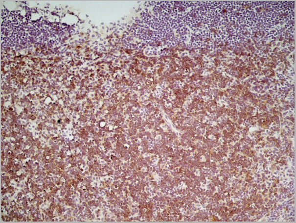
Application Data (Formalin fixed, paraffin embedded human breast cancer biopsy stained with Mouse anti Human CD44 antibody followed by HRP-polymer detection and DAB substrate development (high power) following antigen retrieval using citrate buffer at pH6.2)
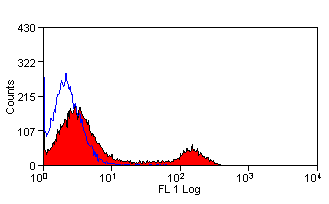
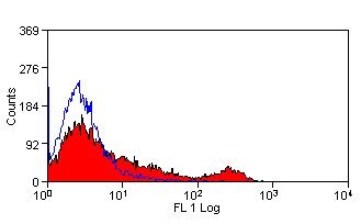
Application Data (Staining of human peripheral blood lymphocytes with Mouse anti Human CD44:FITC)
Application Data (Staining of human peripheral blood lymphocytes with Mouse anti Human CD44:Azide free)
Application Data (Staining of human peripheral blood lymphocytes with Mouse anti Human CD44:RPE)
Application Data (Staining of human peripheral blood granulocytes with Mouse anti Human CD44:Alexa Fluor 647 (AAA12016A647))
Application Data (Staining of human peripheral blood lymphocytes with Mouse anti Human CD44:Biotin)
Application Data (Formalin fixed, paraffin embedded human breast cancer biopsy stained with Mouse anti Human CD44 antibody followed by HRP-polymer detection and DAB substrate development (low power) following antigen retrieval using citrate buffer at pH6.2)
Application Data (Detection limit for recombinant GST tagged MAPK3 is ~0.1ng/ml as a capture antibody.)
IP (Immunoprecipitation) (Immunoprecipitation of MAPK3 transfected lysate using MAPK3 monoclonal antibody and Protein A Magnetic Bead, and immunoblotted with MAPK3 monoclonal antibody)
IF (Immunofluorescence) (Immunofluorescence of monoclonal antibody to MAPK3 on HeLa cell. [antibody concentration 25ug/ml])
IHC (Immunohistochemistry) (Immunoperoxidase of monoclonal antibody to MAPK3 on formalin-fixed paraffin-embedded human placenta. [antibody concentration 3ug/ml])
WB (Western Blot) (Western Blot analysis of MAPK3 expression in transfected 293T cell line by MAPK3 monoclonal antibody. Lane 1: MAPK3 transfected lysate (43.1kD). Lane 2: Non-transfected lysate.)
WB (Western Blot) (MAPK3 monoclonal antibody Western Blot analysis of MAPK3 expression in A-431.)
WB (Western Blot) (Western Blot analysis of ATP6V1G2 expression in transfected 293T cell line by ATP6V1G2 monoclonal antibody. Lane 1: ATP6V1G2 transfected lysate (13.6kD). Lane 2: Non-transfected lysate.)
WB (Western Blot) (ATP6V1G2 monoclonal antibody Western Blot analysis of ATP6V1G2 expression in NIH/3T3.)
WB (Western Blot) (ATP6V1G2 monoclonal antibody Western Blot analysis of ATP6V1G2 expression in Raw 264.7.)
WB (Western Blot) (ATP6V1G2 monoclonal antibody, Western Blot analysis of ATP6V1G2 expression in HepG2.)
WB (Western Blot) (ATP6V1G2 monoclonal antibody. Western Blot analysis of ATP6V1G2 expression in PC-12.)
WB (Western Blot) (Western Blot detection against Immunogen (34.32kD).)
WB (Western Blot) (SMAD3 monoclonal antibody. Western Blot analysis of SMAD3 expression in human colon.)
WB (Western Blot) (SMAD3 monoclonal antibody. Western Blot analysis of SMAD3 expression in HeLa.)
Application Data (Proximity Ligation Analysis (PLA) of protein-protein interactions between MAPK8 and SMAD3. HeLa cells were stained with MAPK8 rabbit purified polyclonal 1:1200 and 133539 1:50. Signals were detected 30 Detection Kit 613 (red), and nuclei were counterstained with DAPI (blue). Each red dot represents the detection of protein-protein interaction complex.)
Application Data (Detection limit for recombinant GST tagged SMAD3 is ~0.1ng/ml as a capture antibody.)
IHC (Immunohistochemistry) (Immunoperoxidase of 133539 on formalin-fixed paraffin-embedded human placenta. [antibody concentration 3ug/ml])
WB (Western Blot) (Western Blot analysis of SMAD3 expression in transfected 293T cell line by 133539. Lane 1: SMAD3 transfected lysate (48kD). Lane 2: Non-transfected lysate.)
WB (Western Blot) (Western Blot analysis of SMAD3 expression in PC-12 using 133539.)
WB (Western Blot) (Detection of mouse PTPN12 by western blot. Samples: Whole cell lysate (50 ug) from NIH 3T3 cells prepared using NETN lysis buffer. Antibody: Rabbit anti-PTPN12 recombinant monoclonal antibody [BL-5-2F8] (AAA23788 lot 1) used at 1:1000. Secondary: HRP-conjugated goat anti-rabbit IgG . Detection: Chemiluminescence with an exposure time of 30 seconds.)
WB (Western Blot) (Detection of human PTPN12 by western blot. Samples: Whole cell lysate (5 ug) from HeLa, HEK293T, Jurkat, MCF-7, Hep-G2, A-549, SW620, SK-MEL-28 [SK], and HCT 116 prepared using NETN lysis buffer. Antibody: Rabbit anti-PTPN12 recombinant monoclonal antibody [BL-5-2F8] (AAA23788 lot 1) used at 1:1000. Secondary: HRP-conjugated goat anti-rabbit IgG . Detection: Chemiluminescence with an exposure time of 10 seconds.)
IP (Immunoprecipitation) (Detection of human PTPN12 by western blot of immunoprecipitates. Samples: Whole cell lysate (0.5 mg per IP reaction; 20% of IP loaded) from HeLa cells prepared using NETN lysis buffer. Antibodies: Rabbit anti-PTPN12 recombinant monoclonal antibody [BL-5-2F8] (AAA23788 lot 1) used for IP at 20 ul per reaction. PTPN12 was also immunoprecipitated by rabbit anti-PTPN12 antibody For blotting immunoprecipitated PTPN12, AAA23788 was used at 1:1000. Detection: Chemiluminescence with an exposure time of 30 seconds.)
IHC (Immunohistochemistry) (Detection of human PTPN12 by immunohistochemistry. Sample: FFPE section of non-small cell lung cancer. Antibody: Rabbit anti-PTPN12 recombinant monoclonal antibody [BL-5-2F8] (AAA23788 lot 1) used at 1:100. Secondary: HRP-conjugated goat anti-rabbit IgG . Substrate: DAB.)
IHC (Immunohistochemistry) (Detection of human PTPN12 by immunohistochemistry. Sample: FFPE section of mammary gland. Antibody: Rabbit anti-PTPN12 recombinant monoclonal antibody [BL-5-2F8] (AAA23788 lot 1) used at 1:100. Secondary: HRP-conjugated goat anti-rabbit IgG . Substrate: DAB.)
ICC (Immunocytochemistry) (Detection of human PTPN12 by immunocytochemistry. Sample: FFPE section of SW620 cells. Antibody: Rabbit anti-PTPN12 recombinant monoclonal antibody [BL-5-2F8] (AAA23788 lot 1) used at 1:40. Secondary:HRP-conjugated goat anti-rabbit IgG . Substrate: DAB.)
WB (Western Blot) (SAV1 monoclonal antibody (M02), Western Blot analysis of SAV1 expression in Hela NE.)
Application Data (Detection limit for recombinant GST tagged SAV1 is ~0.1ng/ml as a capture antibody.)
IP (Immunoprecipitation) (Immunoprecipitation of SAV1 transfected lysate using SAV1 monoclonal antibody and Protein A Magnetic Bead and immunoblotted with rabbit polyclonal antibody.)
IF (Immunofluorescence) (Immunofluorescence of monoclonal antibody to SAV1 on HeLa cell. [antibody concentration 60ug/ml].)
WB (Western Blot) (SAV1 monoclonal antibody. Western Blot analysis of SAV1 expression in HepG2.)
WB (Western Blot) (Western Blot detection against Immunogen (35.35kD).)
Application Data (Detection limit for recombinant GST tagged UBE2C is ~0.03ng/ml as a capture antibody.)
IF (Immunofluorescence) (Immunofluorescence of monoclonal antibody to UBE2C on HeLa cell. [antibody concentration 10ug/ml].)
IHC (Immunohistochemistry) (Immunoperoxidase of monoclonal antibody to UBE2C on formalin-fixed paraffin-embedded human placenta. [antibody concentration 3ug/ml].)
WB (Western Blot) (Western Blot analysis of UBE2C expression in transfected 293T cell line by UBE2C monoclonal antibody. Lane 1: UBE2C transfected lysate (19.7kD). Lane 2: Non-transfected lysate.)
WB (Western Blot) (UBE2C monoclonal antibody, Western Blot analysis of UBE2C expression in HeLa.)
WB (Western Blot) (Western Blot detection against Immunogen (38.21kD).)
Application Data (Proximity Ligation Analysis (PLA) of protein-protein interactions between PIK3R1 and SHC1 Huh7 cells were stained with PIK3R1 rabbit purified polyclonal 1:1200 and anti-SHC1 mouse monoclonal antibody 1:50. Signals were detected 30 Detection Kit 613 (red), and nuclei were counterstained with DAPI (blue). Each red dot represents the detection of protein-protein interaction complex.)
Application Data (Proximity Ligation Analysis (PLA) of protein-protein interactions between PIK3R1 and SHC1 HeLa cells were stained with PIK3R1 rabbit purified polyclonal 1:1200 and SHC1 mouse monoclonal antibody 1:50. Signals were detected by 30 Detection Kit 613 (red), and nuclei were counterstained with DAPI (blue). Each red dot represents the detection of protein-protein interaction complex.)
Application Data (Detection limit for recombinant GST tagged SHC1 is ~0.03ng/ml as a capture antibody.)
IF (Immunofluorescence) (Immunofluorescence of monoclonal antibody to SHC1 on A-431 cell. [antibody concentration 10ug/ml].)
IHC (Immunohistochemistry) (Immunoperoxidase of monoclonal antibody to SHC1 on formalin-fixed paraffin-embedded human stomach. [antibody concentration 1ug/ml].)
WB (Western Blot) (SHC1 monoclonal antibody, Western Blot analysis of SHC1 expression in A-431.)
WB (Western Blot) (Western Blot detection against Immunogen (38.1kD).)
SHC (SHC-transforming Protein 1, SH2 Domain Protein C1, SHC1, SHC-transforming Protein 3, SHC-transforming Protein A, SHCA, Src Homology 2 Domain-containing-transforming Protein C1, FLJ26504) (PE)
Gene Names
SHC1; SHC; SHCA
Reactivity
Human
Applications
ELISA (EIA), Immunofluorescence (IF), Immunohistochemistry (IHC), Western Blot (WB)
Application Data (Detection limit for recombinant GST tagged HMGB2 is approximately 0.03ng/ml as a capture antibody.)
IF (Immunofluorescence) (Immunofluorescence of monoclonal antibody to HMGB2 on HeLa cell. [antibody concentration 10 ug/ml])
WB (Western Blot) (HMGB2 monoclonal antibody (M04), clone 3D2 Western Blot analysis of HMGB2 expression in Hela S3 NE.)
IHC (Immunohistochemistry) (Immunoperoxidase of monoclonal antibody to HMGB2 on formalin-fixed paraffin-embedded human placenta. [antibody concentration 3 ug/ml])
Application Data (Immunoperoxidase of monoclonal antibody to HMGB2 on formalin-fixed paraffin-embedded human placenta. [antibody concentration 3 ug/ml])
Application Data (Detection limit for recombinant GST tagged RUNX1 is approximately 1ng/ml as a capture antibody.)
IF (Immunofluorescence) (Immunofluorescence of monoclonal antibody to RUNX1 on HeLa cell. [antibody concentration 40 ug/ml])
WB (Western Blot) (RUNX1 monoclonal antibody (M06), clone 2C10 Western Blot analysis of RUNX1 expression in Hela S3 NE (Cat # L013V3).)
IHC (Immunohistochemistry) (Immunoperoxidase of monoclonal antibody to RUNX1 on formalin-fixed paraffin-embedded human stomach. [antibody concentration 1.5 ug/ml])
Application Data (Immunoperoxidase of monoclonal antibody to RUNX1 on formalin-fixed paraffin-embedded human stomach. [antibody concentration 1.5 ug/ml])
WB (Western Blot) (NME2 monoclonal antibody (M06), clone 1D3. Western Blot analysis of NME2 expression in NIH/3T3 (Cat # L018V1).)
IHC (Immunohistochemistry) (Immunoperoxidase of monoclonal antibody to NME2 on formalin-fixed paraffin-embedded human testis. [antibody concentration 1.5 ug/ml])
IF (Immunofluorescence) (Immunofluorescence of monoclonal antibody to NME2 on HeLa cell. [antibody concentration 10 ug/ml])
WB (Western Blot) (NME2 monoclonal antibody (M06), clone 1D3 Western Blot analysis of NME2 expression in HeLa (Cat # L013V1).)
FCM (Flow Cytometry) (Flow cytometric analysis of LOVO cells with Dcp1a antibody at 1/100 dilution (red) compared with an unlabelled control (cells without incubation with primary antibody; black). Alexa Fluor 488-conjugated goat anti-rabbit IgG was used as the secondary antibody.)
ICC (Immunocytochemistry) (ICC staining Dcp1a in LOVO cells (green). The nuclear counter stain is DAPI (blue). Cells were fixed in paraformaldehyde, permeabilised with 0.25% Triton X100/PBS.)
ICC (Immunocytochemistry) (ICC staining Dcp1a in SiHa cells (green). The nuclear counter stain is DAPI (blue). Cells were fixed in paraformaldehyde, permeabilised with 0.25% Triton X100/PBS.)
ICC (Immunocytochemistry) (ICC staining Dcp1a in SH-SY-5Y cells (green). The nuclear counter stain is DAPI (blue). Cells were fixed in paraformaldehyde, permeabilised with 0.25% Triton X100/PBS.)
IHC (Immunohistochemistry) (Immunohistochemical analysis of paraffin-embedded human liver tissue using anti-Dcp1a antibody. Counter stained with hematoxylin.)
IHC (Immunohistochemistry) (Immunohistochemical analysis of paraffin-embedded human placenta tissue using anti-Dcp1a antibody. Counter stained with hematoxylin.)
WB (Western Blot) (Western blot analysis of Dcp1a on human liver tissue lysates using anti-Dcp1a antibody at 1/500 dilution.)
WB (Western Blot) (Detection of human Phospho KAP-1 (S824) by western blot. Samples: Whole cell lysate (25 ug) from HEK293T cells treated with 100 uM etoposide (+) or mock treated (-) prepared using NETN lysis buffer. Antibody: Rabbit anti-Phospho KAP-1 (S824) recombinant monoclonal antibody [BL-246-7B5] (AAA23791 lot 4) used at 1:1000. Secondary: HRP-conjugated goat anti-rabbit IgG . Chemiluminescence with an exposure time of 3 seconds. Lower Panel: Rabbit anti-KAP1 recombinant monoclonal antibody [BL-248-2G6] .)
WB (Western Blot) (Detection of mouse Phospho KAP-1 (S824) by western blot. Samples: Whole cell lysate (50 ug) from NIH3T3 cells treated with 100 uM etoposide (+) or mock treated (-) prepared using NETN lysis buffer. Antibody: Rabbit anti-Phospho KAP-1 (S824) recombinant monoclonal antibody [BL-246-7B5] (AAA23791 lot 4) used at 1:1000. Secondary: HRP-conjugated goat anti-rabbit IgG . Chemiluminescence with an exposure time of 3 seconds. Lower Panel: Rabbit anti-KAP1 recombinant monoclonal antibody [BL-248-2G6] .)
Simple Western (Detection of human Phospho KAP-1 (S824) by Simple Western. Samples: Whole cell lysate (0.4 mg/mL) from HEK293T cells treated with 100 uM etoposide prepared using NETN lysis buffer. Antibody: Rabbit anti-Phospho KAP-1 (S824) recombinant monoclonal antibody [BL-246-7B5] (AAA23791) used at 1:10, 1:50, and 1:250. Separation and Detection: SallySue ProteinSimple instrument with the 12-230 kDa separation module and anti-Rabbit detection module. Left Panel: Virtual Lane View. Right Panel: Electropherogram.)
IP (Immunoprecipitation) (Detection of human Phospho KAP-1 (S824) by western blot of immunoprecipitates. Samples: Whole cell lysate (1.0 mg per IP reaction; 5% of IP loaded) from HEK293T cells prepared using NETN lysis buffer that were treated with 100 uM etoposide (+) or mock treated (-). Antibodies: Rabbit anti-Phospho KAP-1 (S824) recombinant monoclonal antibody [BL-246-7B5] (AAA23791 lot 4) used for IP at 20 ul/mg lysate. Phospho KAP-1 (S824) was also immunoprecipitated by a previous lot of this antibody (AAA23791 lot 3) and rabbit anti-KAP-1 recombinant monoclonal antibody [BL-248-2G6] . For blotting immunoprecipitated Phospho KAP-1 (S824), AAA23791 was used at 1:1000. Chemiluminescence with an exposure time of 1 second. Lower Panel: Rabbit anti-KAP1 recombinant monoclonal antibody [BL-248-2G6] .)
IHC (Immunohistochemistry) (Detection of human Phospho KAP-1 (S824) in FFPE prostate carcinoma by IHC. Mock phosphatase treated section (left) and calf intestinal phosphatase-treated section (right). Antibody: Rabbit anti-Phospho KAP-1 (S824) recombinant monoclonal [BL-246-7B5] (AAA23791). Secondary: HRP-conjugated goat anti-rabbit IgG . Substrate: DAB.)
ICC (Immunocytochemistry) (Detection of human Phospho KAP-1 (S824) by immunocytochemistry. Samples: Formaldehyde-fixed asynchronous HeLa cells grown in chambered microscope slides and treated with etoposide (right) or untreated (left). Antibody: Rabbit anti-Phospho KAP-1 (S824) recombinant monoclonal antibody [BL-246-7B5] (AAA23791-T lot 1) used at of 1:100. Secondary: DyLight 594-conjugated goat anti-rabbit IgG . Counterstain: Phalloidin conjugated Alexa Fluor 488 (green).)
ICC (Immunocytochemistry) (Detection of human Phospho KAP-1 (S824) in FFPE etoposide treated HeLa cells by ICC. Mock phosphatase treated section (left) and calf intestinal phosphatase-treated section (right). Antibody: Rabbit anti-Phospho KAP-1 (S824) recombinant monoclonal [BL-246-7B5] (AAA23791). Secondary: HRP-conjugated goat anti-rabbit IgG . Substrate: DAB.)
FCM (Flow Cytometry) (Detection of human phospho KAP-1 (shaded) in etoposide treated HEK293T cells (right) and untreated HEK293T cells (left) by flow cytometry. Antibody: Rabbit anti-phospho KAP-1 recombinant monoclonal [BL-246-7B5] (AAA23791) or isotype control (unshaded). Secondary: DyLight 488-conjugated goat anti-rabbit IgG .)
WB (Western Blot) (Western blot analysis of PCDH8 over-expressed 293 cell line, cotransfected with PCDH8 Validated Chimera RNAi (Lane 2) or non-transfected control (Lane 1). Blot probed with PCDH8 monoclonal antibody. GAPDH (36.1kD) used as specificity and loading control.)
Application Data (Detection limit for recombinant GST tagged PCDH8 is ~0.1ng/ml as a capture antibody.)
IHC (Immunohistochemistry) (Immunoperoxidase of monoclonal antibody to PCDH8 on formalin-fixed paraffin-embedded human tonsil. [antibody concentration 3ug/ml])
WB (Western Blot) (Western Blot analysis of PCDH8 expression in transfected 293T cell line by PCDH8 monoclonal antibody. Lane 1: PCDH8 transfected lysate (113kD). Lane 2: Non-transfected lysate.)
WB (Western Blot) (PCDH8 monoclonal antibody Western Blot analysis of PCDH8 expression in COLO 320 HSR.)
WB (Western Blot) (Western Blot detection against Immunogen (35.53kD).)
IHC (Immunohistchemistry) (Immunoperoxidase of monoclonal antibody to USF2 on formalin-fixed paraffin-embedded human esophagus. [antibody concentration 1 ug/ml])
IHC (Immunohistochemistry) (Immunoperoxidase of monoclonal antibody to USF2 on formalin-fixed paraffin-embedded human esophagus. [antibody concentration 1 ug/ml])
IF (Immunofluorescence) (Immunofluorescence of monoclonal antibody to USF2 on HeLa cell. [antibody concentration 10 ug/ml])
WB (Western Blot) (USF2 monoclonal antibody (M02), clone 5F2. Western Blot analysis of USF2 expression in different cell lines.)
WB (Western Blot) (USF2 monoclonal antibody (M02), clone 5F2. Western Blot analysis of USF2 expression in Hela S3 NE.)
Immunofluorescence (IF), Immunohistochemistry (IHC), Western Blot (WB)
Purity
Purified
Pricing
What are Monoclonal Antibodies?
Monoclonal antibodies are specialized laboratory-produced proteins developed for binding to specific biological antigens or other molecular targets. Since they come from a single cell (or clone), they are especially consistent and accurate in the data they are involved in producing.
This type of antibody material has been shown to be a powerful tool in finding and subsequently destroying harmful cells in an organism, such as those found in cancers or various autoimmune diseases. This makes them excellent aids in medical testing and research, which is why they are so widely used.
AAA Biotech offers a comprehensive range of high-quality monoclonal antibodies that perform effectively in various laboratory tests, including (amongst others) ELISA, western blotting, immunohistochemistry, and flow cytometry. All of the products in our catalog are thoroughly quality tested to make sure that they are reliable and will consistently perform well in your research.
What Are The Uses of Monoclonal Antibodies
Monoclonal antibodies are used in many lab tests, including (amongst others) ELISA, western blotting, immunohistochemistry, and flow cytometry.
ELISA is a test that helps detect a specific substance/analyte in a sample. It uses antibodies (often monoclonal) bound to a solid surface (such as the well of a microplate) to “capture” the substance/analyte in the sample and immobilize it so that the detection antibody component can then bind to it and produce a signal, which can then be measured.
Western blotting identifies specific proteins in a sample. The sample is first separated on a gel, and then antibodies are applied that will typically bind to the target, which will all be localized to a single band in a lane.
Immunohistochemistry helps locate specific proteins in cells or tissue samples using antibodies.
Flow cytometry looks at and sorts cells. It uses antibodies that are conjugated to reporter molecules called “fluorophores”, which, under special lights, emit light themselves, which can then be measured by a detector instrument.
How Monoclonal Antibodies Are Used as Medicine?
Please note that all of the products listed in AAA Biotech’s catalog are strictly for research-use only (RUO).
Monoclonal antibodies can also be used as therapeutic/medical treatments, particularly in the context of cancers. They are designed to find and bind to specific cells or proteins, helping the immune system recognize and attack the cancer. These treatments work in different ways, such as:
Radioimmunotherapy attaches a small amount of radioactive molecule to the antibody, so it delivers the radiation directly to the cancer cells that the antibody is specifically binding to.
Antibody-directed enzyme prodrug therapy uses antibodies that are specifically bound to special enzymes. These enzymes activate a harmless drug in the body and turn it into a cancer-killing drug only near the cancer cells—this helps avoid harming healthy cells.
Immunoliposomes are tiny “bubbles” filled with medicine/drug and coated with antibodies. They carry the drug straight to the cancer cells.
Why Buy Monoclonal Antibodies From Us?
At AAA Biotech, we provide high-performance monoclonal antibodies designed to support a wide range of research needs.
1. Validated for Versatile Applications
The antibodies in our catalog are extensively validated and compatible with multiple techniques, including (but not limited to) ELISA, flow cytometry (FC), immunocytochemistry (ICC), immunofluorescence (IF), immunohistochemistry (IHC), immunoprecipitation (IP), and western blotting (WB).
2. Wide Selection & Specialized Options
We offer antibodies for common and rare species, that are available in various conjugated forms, and also in recombinant formats. Essentially, there is almost anything one might need to meet their experimental model’s requirements.
3. High-Quality Proteins
Our proteins meet high purity standards—90% or more as confirmed by SDS-PAGE. Many are available with tags like His, Flag, GST, or MBP, and we also supply native and biologically active proteins for functional studies.
Frequently Asked Questions
1. Are your monoclonal antibodies validated for specific applications?
Yes, our antibodies are tested and validated for use in methods such as ELISA, western blot, IHC, flow cytometry, and more. Refer to specific product pages or datasheets for individual product information.
2. How do I choose the right monoclonal antibody for my application?
Review the product details directly for application validation, species reactivity, and target information. You may also contact our support team at any time for help.
3. How quickly can I receive my order?
Most orders are processed and shipped within 1–3 business days, depending on product availability and your shipping location.
Submit a Question
Please complete the form below and a representative will contact you as soon as possible.
Request more Information
Please complete the form below and a representative will contact you as soon as possible.
Request a Manual
Please complete the form below and a representative will contact you as soon as possible.
Request a Quote
Please complete the form below and a representative will contact you as soon as possible.