Filters
▼Clonality
▼Type
▼Reactivity
▼Gene Name
▼Isotype
▼Host
▼Application
▼Clone
▼Monoclonal Antibodies
Get accurate results in your research with our Monoclonal Antibodies, which are specially made to target exactly what you require for your research, and will produce consistent, reliable performance in lab tests.
Viewing 2150-2200 of 27645 product results
HLA-A,B,C, Monoclonal Antibody (Cat# AAA174660)
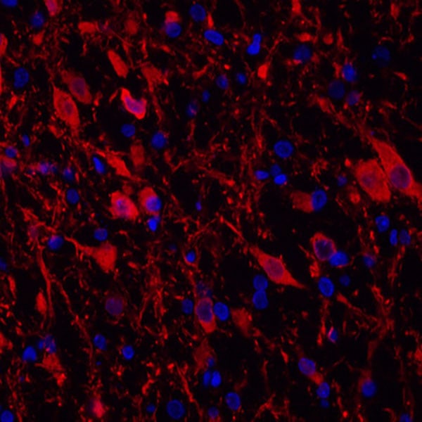
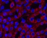
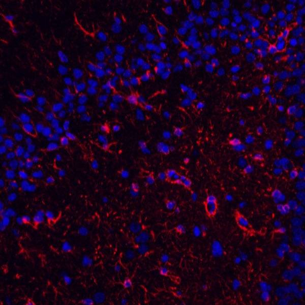 IF (Immunofluorescence)
(Immunofluorescence analysis of paraffin-embedded rat brain using GFAP Monoclonal Antibody at dilution of 1:400.)
IF (Immunofluorescence)
(Immunofluorescence analysis of paraffin-embedded rat brain using GFAP Monoclonal Antibody at dilution of 1:400.)
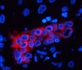
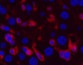
GFAP, Monoclonal Antibody (Cat# AAA174477)
 IF (Immunofluorescence)
(Immunofluorescence analysis of paraffin-embedded rat brain using TH Monoclonal Antibody at dilution of 1:400.)
IF (Immunofluorescence)
(Immunofluorescence analysis of paraffin-embedded rat brain using TH Monoclonal Antibody at dilution of 1:400.)
TH, Monoclonal Antibody (Cat# AAA174478)
 IF (Immunofluorescence)
(Immunofluorescence analysis of Mouse kidney tissue using COX IV Monoclonal Antibody at dilution of 1:200.)
IF (Immunofluorescence)
(Immunofluorescence analysis of Mouse kidney tissue using COX IV Monoclonal Antibody at dilution of 1:200.)
COX IV, Monoclonal Antibody (Cat# AAA171593)
 IF (Immunofluorescence)
(Immunofluorescence analysis of Mouse liver tissue using ACTA1 Monoclonal Antibody at dilution of 1:200.)
IF (Immunofluorescence)
(Immunofluorescence analysis of Mouse liver tissue using ACTA1 Monoclonal Antibody at dilution of 1:200.)
alpha skeletal muscle actin, Monoclonal Antibody (Cat# AAA171595)
 IF (Immunofluorescence)
(Immunofluorescence analysis of Rat testis tissue using PCNA Monoclonal Antibody at dilution of 1:200.)
IF (Immunofluorescence)
(Immunofluorescence analysis of Rat testis tissue using PCNA Monoclonal Antibody at dilution of 1:200.)
PCNA, Monoclonal Antibody (Cat# AAA171599)
 IF (Immunofluorescence)
(Immunofluorescence analysis of Hela tissue using EFHD1 Monoclonal Antibody at dilution of 1:100.)
IF (Immunofluorescence)
(Immunofluorescence analysis of Hela tissue using EFHD1 Monoclonal Antibody at dilution of 1:100.)
EFHD1, Monoclonal Antibody (Cat# AAA171600)
 IF (Immunofluorescence)
(Immunofluorescence analysis of Mouse colon tissue using Kif 7 Monoclonal Antibody at dilution of 1:200.)
IF (Immunofluorescence)
(Immunofluorescence analysis of Mouse colon tissue using Kif 7 Monoclonal Antibody at dilution of 1:200.)
Kif 7, Monoclonal Antibody (Cat# AAA171601)
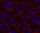
 IF (Immunofluorescence)
(Immunofluorescence analysis of Human liver cancer tissue using FH Monoclonal Antibody at dilution of 1:200.)
IF (Immunofluorescence)
(Immunofluorescence analysis of Human liver cancer tissue using FH Monoclonal Antibody at dilution of 1:200.)
FH, Monoclonal Antibody (Cat# AAA171604)
 IF (Immunofluorescence)
(Immunofluorescence analysis of Human liver cancer tissue using CA IX Monoclonal Antibody at dilution of 1:200.)
IF (Immunofluorescence)
(Immunofluorescence analysis of Human liver cancer tissue using CA IX Monoclonal Antibody at dilution of 1:200.)
CA IX, Monoclonal Antibody (Cat# AAA171605)
 IF (Immunofluorescence)
(Immunofluorescence analysis of Mouse liver tissue using ?-tubulin Monoclonal Antibody at dilution of 1:200.)
IF (Immunofluorescence)
(Immunofluorescence analysis of Mouse liver tissue using ?-tubulin Monoclonal Antibody at dilution of 1:200.)
alpha-tubulin, Monoclonal Antibody (Cat# AAA171611)
 IF (Immunofluorescence)
(Immunofluorescence analysis of Mouse spleen tissue using NSE Monoclonal Antibody at dilution of 1:200.)
IF (Immunofluorescence)
(Immunofluorescence analysis of Mouse spleen tissue using NSE Monoclonal Antibody at dilution of 1:200.)
NSE, Monoclonal Antibody (Cat# AAA171565)
 IF (Immunofluorescence)
(Immunofluorescence analysis of Mouse spleen tissue using eIF4A1 Monoclonal Antibody at dilution of 1:200.)
IF (Immunofluorescence)
(Immunofluorescence analysis of Mouse spleen tissue using eIF4A1 Monoclonal Antibody at dilution of 1:200.)
eIF4A1, Monoclonal Antibody (Cat# AAA171568)
 IF (Immunofluorescence)
(Immunofluorescence analysis of Hela tissue using Peroxiredoxin 1 Monoclonal Antibody at dilution of 1:100.)
IF (Immunofluorescence)
(Immunofluorescence analysis of Hela tissue using Peroxiredoxin 1 Monoclonal Antibody at dilution of 1:100.)
Peroxiredoxin 1, Monoclonal Antibody (Cat# AAA171574)
 IF (Immunofluorescence)
(Immunofluorescence analysis of Mouse kidney tissue using Histone H2B Monoclonal Antibody at dilution of 1:200.)
IF (Immunofluorescence)
(Immunofluorescence analysis of Mouse kidney tissue using Histone H2B Monoclonal Antibody at dilution of 1:200.)
Histone H2B, Monoclonal Antibody (Cat# AAA171576)
 IF (Immunofluorescence)
(Immunofluorescence analysis of Human liver tissue using CK7 Monoclonal Antibody at dilution of 1:200.)
IF (Immunofluorescence)
(Immunofluorescence analysis of Human liver tissue using CK7 Monoclonal Antibody at dilution of 1:200.)
CK7, Monoclonal Antibody (Cat# AAA171578)
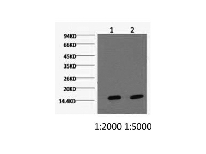
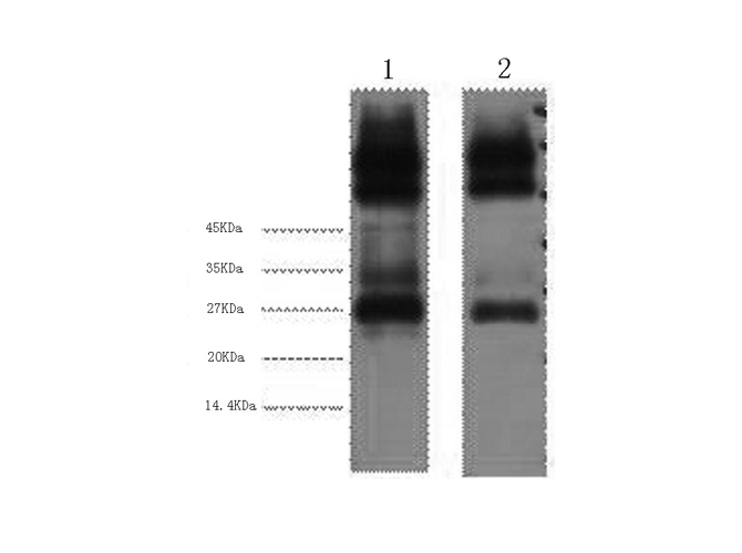
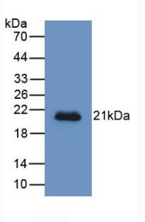
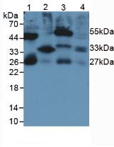
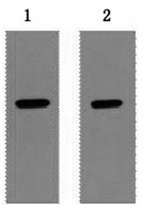 WB (Western Blot)
(Western Blot analysis of 1ug Strep II fusion protein using Strep-Tag Monoclonal Antibody at dilution of 1) 1:5000 2) 1:10000.)
WB (Western Blot)
(Western Blot analysis of 1ug Strep II fusion protein using Strep-Tag Monoclonal Antibody at dilution of 1) 1:5000 2) 1:10000.)
Strep-Tag, Monoclonal Antibody (Cat# AAA171591)
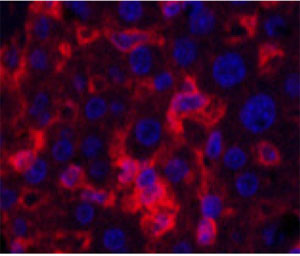
 IF (Immunofluorescence)
(Immunofluorescence analysis of Mouse liver tissue using alpha-SMA Monoclonal Antibody at dilution of 1:200.)
IF (Immunofluorescence)
(Immunofluorescence analysis of Mouse liver tissue using alpha-SMA Monoclonal Antibody at dilution of 1:200.)
alpha-SMA, Monoclonal Antibody (Cat# AAA173647)
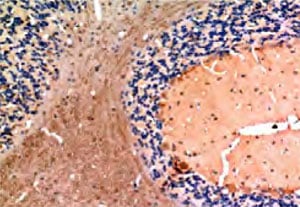
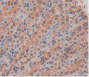
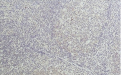 IHC (Immunohiostchemistry)
(Immunohistochemical analysis of paraffin-embedded Human tonsil tissue using TNF alpha Monoclonal Antibody at dilution of 1:50.)
IHC (Immunohiostchemistry)
(Immunohistochemical analysis of paraffin-embedded Human tonsil tissue using TNF alpha Monoclonal Antibody at dilution of 1:50.)
TNF alpha, Monoclonal Antibody (Cat# AAA173655)
 IHC (Immunohiostchemistry)
(Immunohistochemistry of paraffin-embedded Human colon carcinoma tissue using HSP90 alpha Monoclonal Antibody at dilution of 1:200.)
IHC (Immunohiostchemistry)
(Immunohistochemistry of paraffin-embedded Human colon carcinoma tissue using HSP90 alpha Monoclonal Antibody at dilution of 1:200.)
HSP90 alpha, Monoclonal Antibody (Cat# AAA173656)
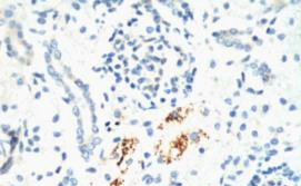
 IHC (Immunohiostchemistry)
(Immunohistochemistry of paraffin-embedded Human colon carcinoma tissue using GRP78/Bip Monoclonal Antibody at dilution of 1:200.)
IHC (Immunohiostchemistry)
(Immunohistochemistry of paraffin-embedded Human colon carcinoma tissue using GRP78/Bip Monoclonal Antibody at dilution of 1:200.)
GRP78/Bip, Monoclonal Antibody (Cat# AAA173659)

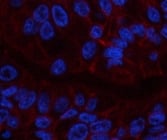
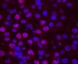
 IF (Immunofluorescence)
(Immunofluorescence analysis of Human liver cancer tissue using Cystatin C Monoclonal Antibody at dilution of 1:200.)
IF (Immunofluorescence)
(Immunofluorescence analysis of Human liver cancer tissue using Cystatin C Monoclonal Antibody at dilution of 1:200.)
Cystatin C, Monoclonal Antibody (Cat# AAA173666)
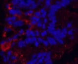
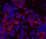
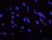 IF (Immunofluorescence)
(Immunofluorescence analysis of Human uterus tissue using HP-1alpha Monoclonal Antibody at dilution of 1:200.)
IF (Immunofluorescence)
(Immunofluorescence analysis of Human uterus tissue using HP-1alpha Monoclonal Antibody at dilution of 1:200.)
HP-1alpha, Monoclonal Antibody (Cat# AAA173679)
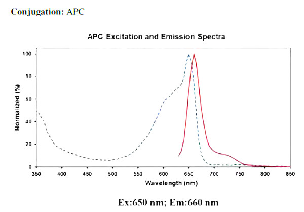
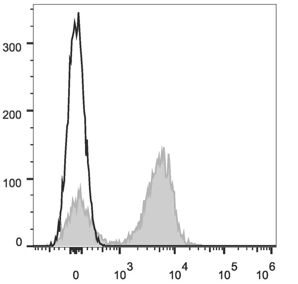
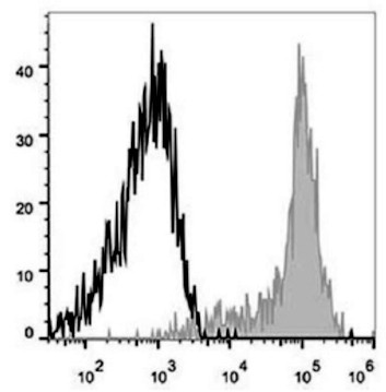 FCM/FACS (Flow Cytometry)
(Human peripheral blood monocytes are stained with APC Anti-Human CD11c Antibody (filled gray histogram). Unstained monocytes (empty black histogram) are used as control.)
FCM/FACS (Flow Cytometry)
(Human peripheral blood monocytes are stained with APC Anti-Human CD11c Antibody (filled gray histogram). Unstained monocytes (empty black histogram) are used as control.)
CD11c, Monoclonal Antibody (Cat# AAA174051)
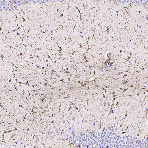
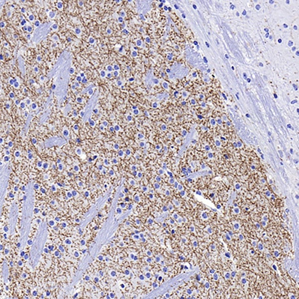
 IHC (Immunohiostchemistry)
(Immunohistochemistry of paraffinembedded Rat brain tissue using TGFB1 Monoclonal Antibody at dilution of 1:200.)
IHC (Immunohiostchemistry)
(Immunohistochemistry of paraffinembedded Rat brain tissue using TGFB1 Monoclonal Antibody at dilution of 1:200.)
TGFbeta1, Monoclonal Antibody (Cat# AAA173686)
CD1d, Monoclonal Antibody (Cat# AAA174737)
 FCM/FACS (Flow Cytometry)
CD162, Monoclonal Antibody (Cat# AAA174738)
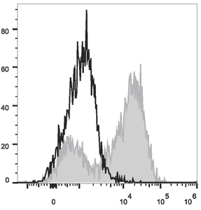
 FCM/FACS (Flow Cytometry)
(Human platelets are stained with FITC Anti-Human CD9 Antibody (filled gray histogram). Unstained platelets (empty black histogram) are used as control.)
FCM/FACS (Flow Cytometry)
(Human platelets are stained with FITC Anti-Human CD9 Antibody (filled gray histogram). Unstained platelets (empty black histogram) are used as control.)
CD9, Monoclonal Antibody (Cat# AAA174742)
 Application Data
(C57BL/6 murine splenocytes are stained with Anti-Mouse CD3epsilon Monoclonal Antibody(APC Conjugated)(filled gray histogram). Unstained splenocytes (empty black histogram) are used as control.)
Application Data
(C57BL/6 murine splenocytes are stained with Anti-Mouse CD3epsilon Monoclonal Antibody(APC Conjugated)(filled gray histogram). Unstained splenocytes (empty black histogram) are used as control.)
CD3epsilon, Monoclonal Antibody (Cat# AAA174745)
Ly6G, Monoclonal Antibody (Cat# AAA174747)
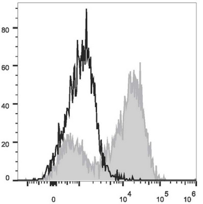
 FCM/FACS (Flow Cytometry)
(Recommended Usage: Each lot of this antibody is quality control tested by flow cytometric analysis. The amount of the reagent is suggested to be used 5 uL of the antibody per test (million cells in 100 uL staining volume or per 100 uL of whole blood). Please check your vial before the experiment. Since applications vary, the appropriate dilutions must be determined for individual use.Mouse abdominal macrophages elicited by starch broth are stained with PE Anti-Mouse CD14 Antibody (filled gray histogram). Unstained macrophages (blank black histogram) are used as control.)
FCM/FACS (Flow Cytometry)
(Recommended Usage: Each lot of this antibody is quality control tested by flow cytometric analysis. The amount of the reagent is suggested to be used 5 uL of the antibody per test (million cells in 100 uL staining volume or per 100 uL of whole blood). Please check your vial before the experiment. Since applications vary, the appropriate dilutions must be determined for individual use.Mouse abdominal macrophages elicited by starch broth are stained with PE Anti-Mouse CD14 Antibody (filled gray histogram). Unstained macrophages (blank black histogram) are used as control.)
CD14, Monoclonal Antibody (Cat# AAA174759)
 FCM/FACS (Flow Cytometry)
(Mouse abdominal macrophages elicited by starch broth are stained with Anti-Mouse CD14 Monoclonal Antibody(PE Conjugated)(filled gray curve). Unstained macrophages (blank black curve) are used as control.)
FCM/FACS (Flow Cytometry)
(Mouse abdominal macrophages elicited by starch broth are stained with Anti-Mouse CD14 Monoclonal Antibody(PE Conjugated)(filled gray curve). Unstained macrophages (blank black curve) are used as control.)
CD14, Monoclonal Antibody (Cat# AAA174760)
IgM, Monoclonal Secondary Antibody (Cat# AAA174767)
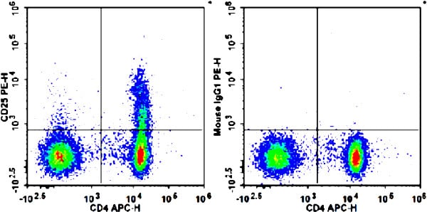 Application Data
(Human peripheral blood lymphocytes are stained with APC Anti-Human CD4 Antibody and PE Anti-Human CD25 Antibody (Left). Lymphocytes are stained with APC Anti-Human CD4 Antibody and PE Mouse IgG1, κ Isotype Control (Right).)
Application Data
(Human peripheral blood lymphocytes are stained with APC Anti-Human CD4 Antibody and PE Anti-Human CD25 Antibody (Left). Lymphocytes are stained with APC Anti-Human CD4 Antibody and PE Mouse IgG1, κ Isotype Control (Right).)
CD25, Monoclonal Antibody (Cat# AAA174768)
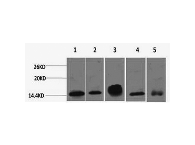
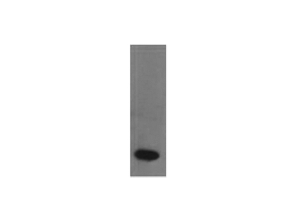
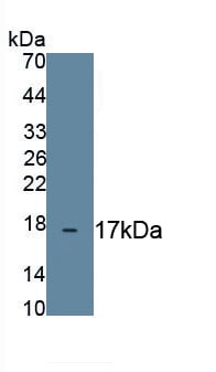
 WB (Western Blot)
(Western Blot;Sample: Recombinant PTHrP, Human.)
WB (Western Blot)
(Western Blot;Sample: Recombinant PTHrP, Human.)
Parathyroid Hormone, Monoclonal Antibody (Cat# AAA141258)
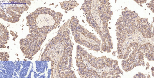
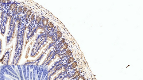
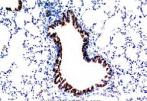
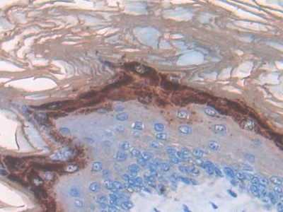
 IHC (Immunohistochemisry)
(DAB staining on IHC-P;Samples: Human Small intestine Tissue;Primary Ab: 30ug/ml Mouse Anti-Human MUC5B AntibodySecond Ab: 2ug/mL HRPLinked Caprine Anti-Mouse IgG Polyclonal Antibody)
IHC (Immunohistochemisry)
(DAB staining on IHC-P;Samples: Human Small intestine Tissue;Primary Ab: 30ug/ml Mouse Anti-Human MUC5B AntibodySecond Ab: 2ug/mL HRPLinked Caprine Anti-Mouse IgG Polyclonal Antibody)
Mucin 5 Subtype B, Monoclonal Antibody (Cat# AAA141263)
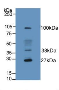
 WB (Western Blot)
(Sample: U87MG cell lysatePrimary Ab: 0.5ug/ml Mouse Anti-Human COL4a5 AntibodySecond Ab: 0.2ug/ml HRP-Linked Caprine Anti-Mouse IgG Polyclonal Antibody)
WB (Western Blot)
(Sample: U87MG cell lysatePrimary Ab: 0.5ug/ml Mouse Anti-Human COL4a5 AntibodySecond Ab: 0.2ug/ml HRP-Linked Caprine Anti-Mouse IgG Polyclonal Antibody)
Collagen Type IV Alpha 5, Monoclonal Antibody (Cat# AAA141299)
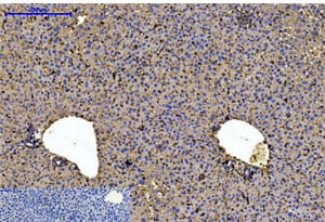
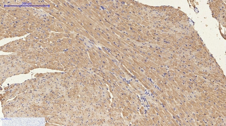
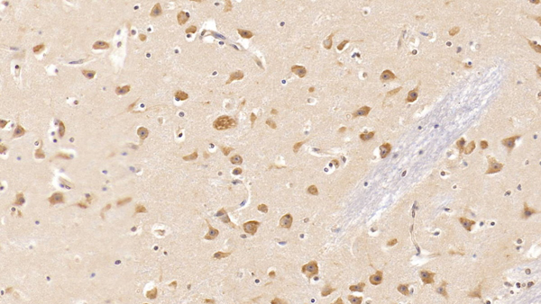
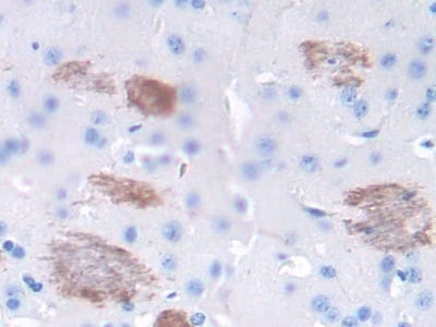 IHC (Immunohistochemistry)
(DAB staining on IHC-P;Samples: Mouse Cerebrum Tissue;Primary Ab: 20ug/ml Mouse Anti-Mouse TNFa AntibodySecond Ab: 2ug/mL HRP-Linked Caprine Anti-Mouse IgG Polyclonal Antibody)
IHC (Immunohistochemistry)
(DAB staining on IHC-P;Samples: Mouse Cerebrum Tissue;Primary Ab: 20ug/ml Mouse Anti-Mouse TNFa AntibodySecond Ab: 2ug/mL HRP-Linked Caprine Anti-Mouse IgG Polyclonal Antibody)
Tumor Necrosis Factor Alpha, Monoclonal Antibody (Cat# AAA141303)
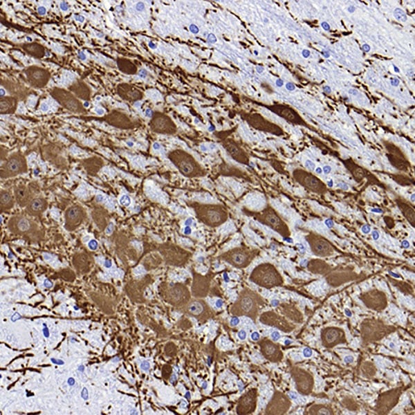
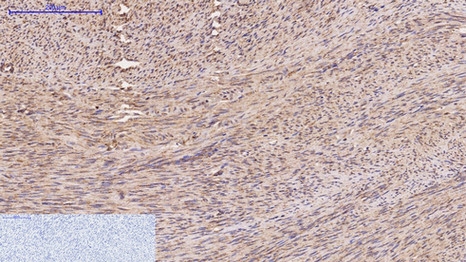
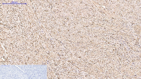
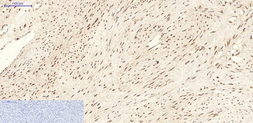
 IHC (Immunohistochemistry)
(DAB staining on IHC-P; Samples: Human Glioma Tissue)
IHC (Immunohistochemistry)
(DAB staining on IHC-P; Samples: Human Glioma Tissue)
Hemojuvelin, Monoclonal Antibody (Cat# AAA141313)
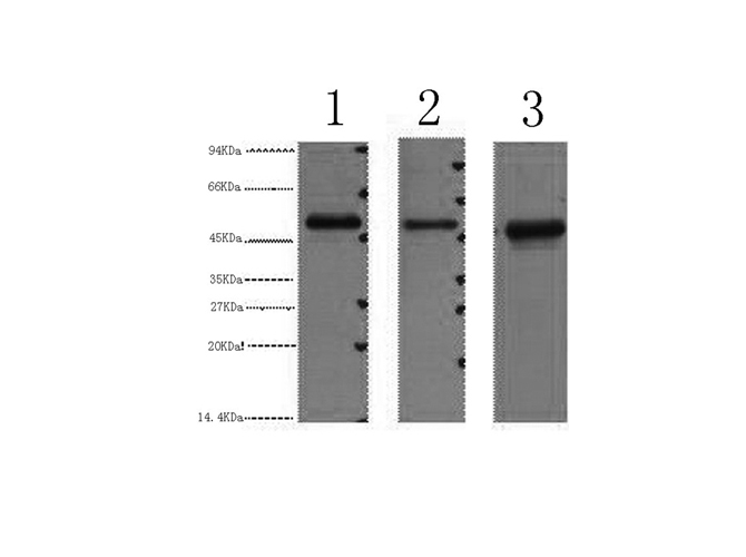
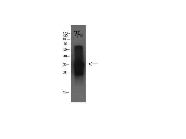
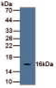
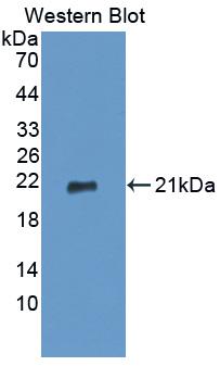
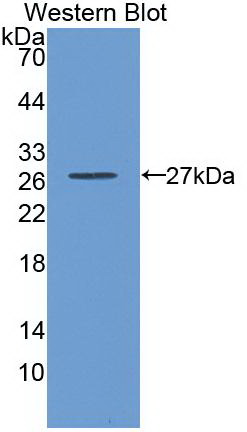
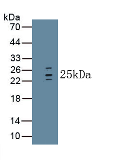
 WB (Western Blot)
(Western Blot: Sample: Recombinant protein.)
WB (Western Blot)
(Western Blot: Sample: Recombinant protein.)
Keratin 18, Monoclonal Antibody (Cat# AAA141319)
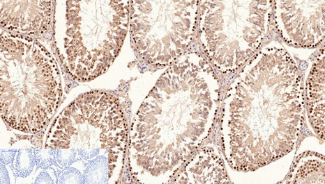
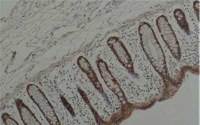
 IHC (Immunohistochemistry)
(DAB staining on IHC-P; Samples: Rat Stomach Tissue.)
IHC (Immunohistochemistry)
(DAB staining on IHC-P; Samples: Rat Stomach Tissue.)
Interleukin 33, Monoclonal Antibody (Cat# AAA141339)
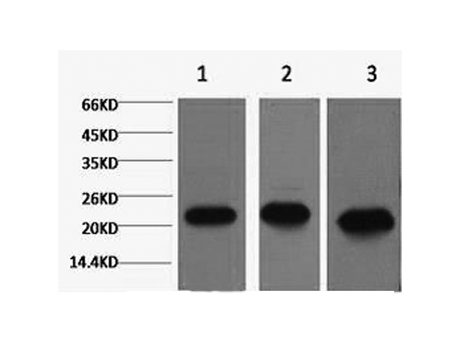
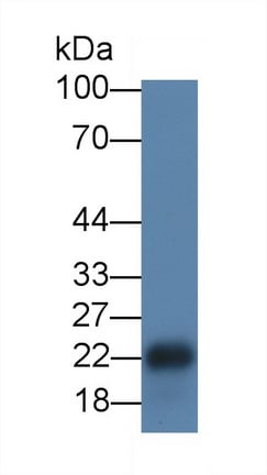
 WB (Western Blot)
(Western Blot: Sample: Recombinant protein.)
WB (Western Blot)
(Western Blot: Sample: Recombinant protein.)
Glutathione S Transferase Alpha 3, Monoclonal Antibody (Cat# AAA141359)
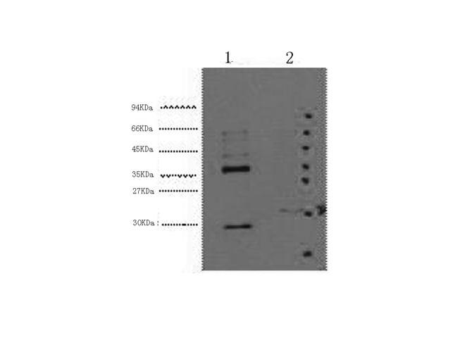
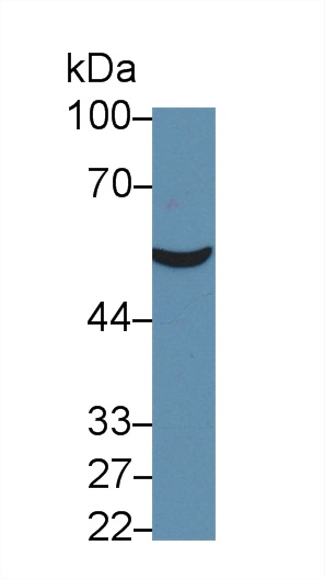
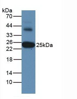
 WB (Western Blot)
(Western Blot;Sample: Recombinant OCTN1, Human.)
WB (Western Blot)
(Western Blot;Sample: Recombinant OCTN1, Human.)
Organic Cation/Ergothioneine Transporter (OCTN1), Monoclonal Antibody (Cat# AAA149460)
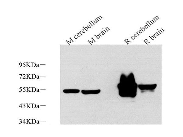
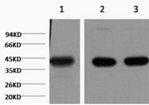
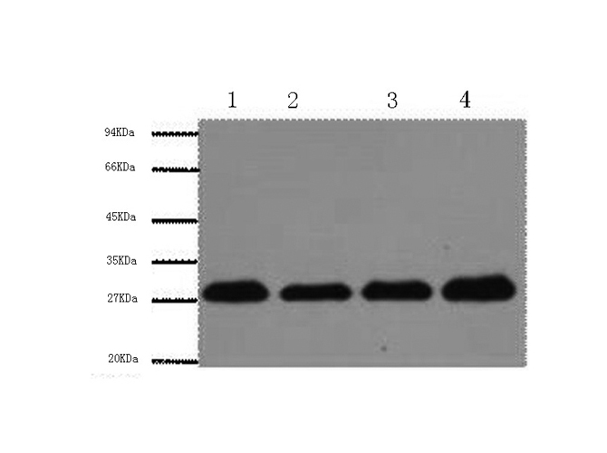
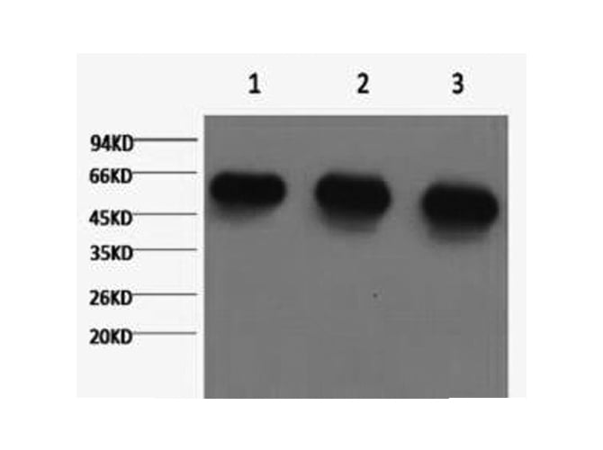
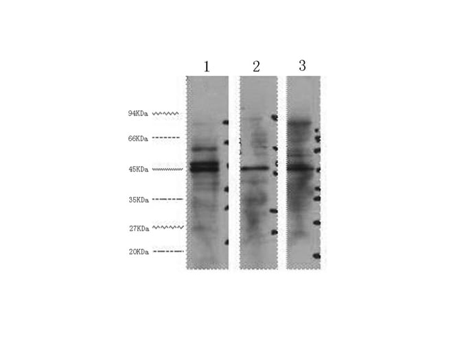
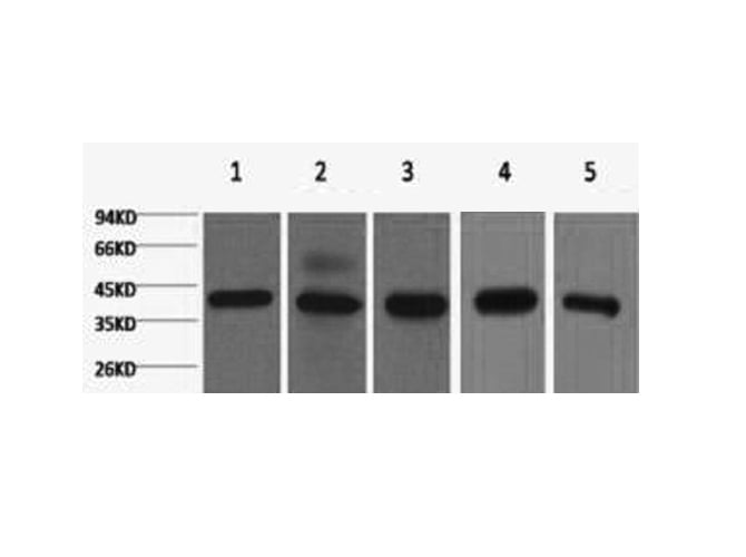
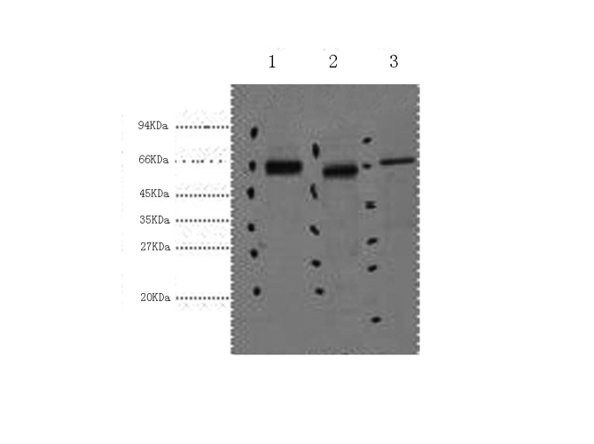
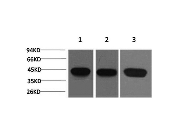
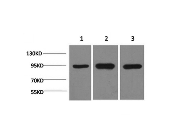
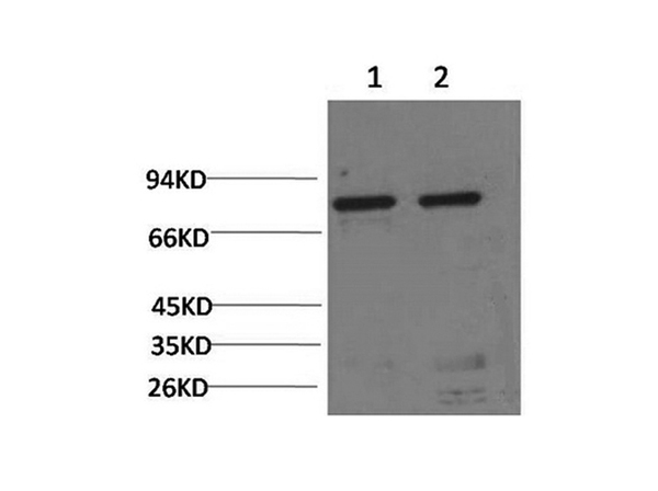
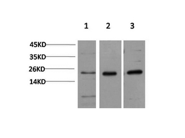
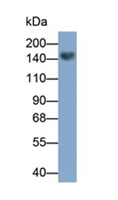
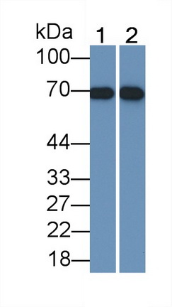
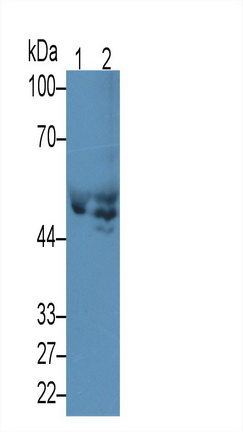
 WB (Western Blot)
(Western Blot Lane1: Mouse Liver TissueLane2: Mouse Lung TissuePrimary Ab: 1:800 Dilution of Mouse Anti-Human CTNNb1 AbSecond Ab: 1:5000 Dilution of HRP-Linked Rabbit Anti-Mouse IgG Ab)
WB (Western Blot)
(Western Blot Lane1: Mouse Liver TissueLane2: Mouse Lung TissuePrimary Ab: 1:800 Dilution of Mouse Anti-Human CTNNb1 AbSecond Ab: 1:5000 Dilution of HRP-Linked Rabbit Anti-Mouse IgG Ab)
Beta Catenin (beta-catenin), Monoclonal Antibody (Cat# AAA149499)
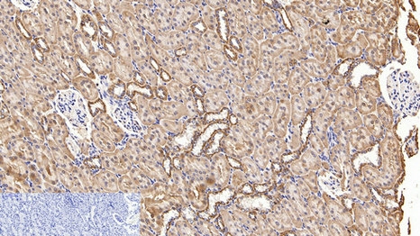
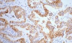
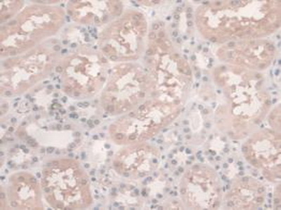
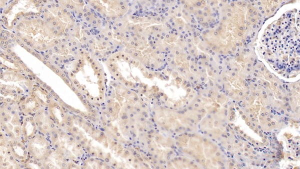 IHC (Immunohiostchemistry)
(DAB staining on IHC-P; Samples: Human Kidney Tissue; Primary Ab: 20ug/ml Mouse Anti-Human Tie1 AntibodySecond Ab: 2ug/mL HRP-Linked Caprine Anti-Mouse IgG Polyclonal Antibody)
IHC (Immunohiostchemistry)
(DAB staining on IHC-P; Samples: Human Kidney Tissue; Primary Ab: 20ug/ml Mouse Anti-Human Tie1 AntibodySecond Ab: 2ug/mL HRP-Linked Caprine Anti-Mouse IgG Polyclonal Antibody)
Tyrosine Kinase With Immunoglobulin Like And EGF Like Domains Protein 1 (Tie1), Monoclonal Antibody (Cat# AAA149510)
Carcinoembryonic Antigen (CEA), Monoclonal Antibody (Cat# AAA149512)
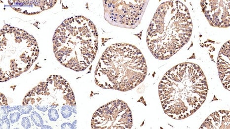
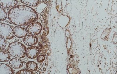
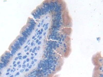
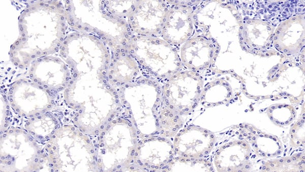
 IHC (Immunohiostchemistry)
(DAB staining on IHC-P; Samples: Human Stomach Tissue; Primary Ab: 30ug/ml Mouse Anti-Human IL1RA Antibody Second Ab: 2ug/mL HRP-Linked Caprine Anti-Mouse IgG Polyclonal Antibody)
IHC (Immunohiostchemistry)
(DAB staining on IHC-P; Samples: Human Stomach Tissue; Primary Ab: 30ug/ml Mouse Anti-Human IL1RA Antibody Second Ab: 2ug/mL HRP-Linked Caprine Anti-Mouse IgG Polyclonal Antibody)
Interleukin 1 Receptor Antagonist (IL1RA), Monoclonal Antibody (Cat# AAA149515)
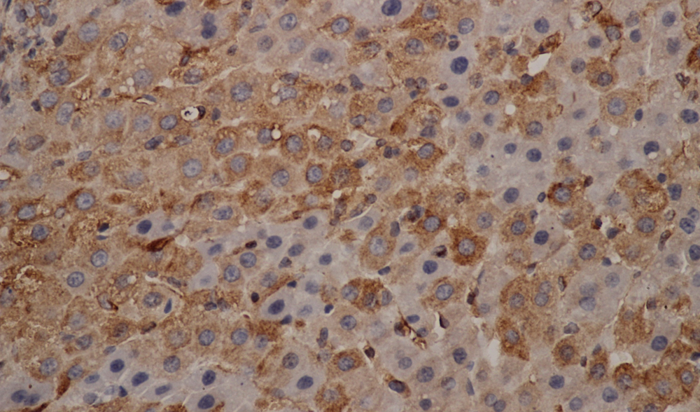
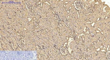
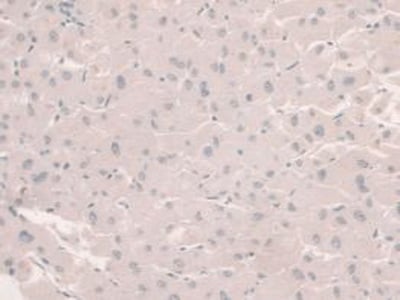
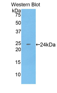
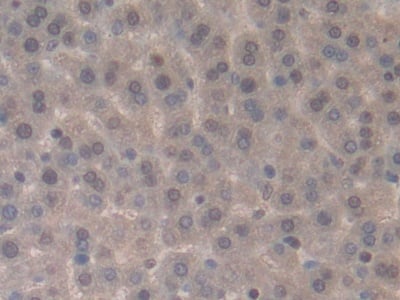
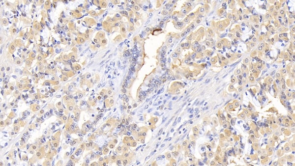
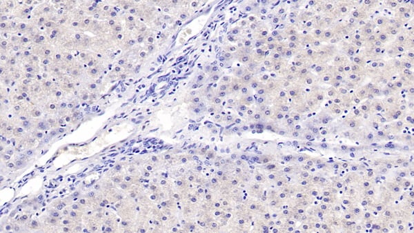
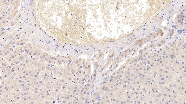 IHC (Immunohiostchemistry)
(DAB staining on IHC-P; Samples: Human Liver Tissue; Primary Ab: 10ug/ml Mouse Anti-Human SHBG Antibody Second Ab: 2ug/mL HRP-Linked Caprine Anti-Mouse IgG Polyclonal Antibody)
IHC (Immunohiostchemistry)
(DAB staining on IHC-P; Samples: Human Liver Tissue; Primary Ab: 10ug/ml Mouse Anti-Human SHBG Antibody Second Ab: 2ug/mL HRP-Linked Caprine Anti-Mouse IgG Polyclonal Antibody)
Sex Hormone Binding Globulin (SHBG), Monoclonal Antibody (Cat# AAA149520)
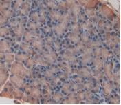
 IHC (Immunohistochemisry)
(DAB staining on IHC-P; Samples: Human Liver Tissue; Primary Ab: 40ug/ml Mouse Anti-Human aHSP AntibodySecond Ab: 2ug/mL HRP-Linked Caprine Anti-Mouse IgG Polyclonal Antibody)
IHC (Immunohistochemisry)
(DAB staining on IHC-P; Samples: Human Liver Tissue; Primary Ab: 40ug/ml Mouse Anti-Human aHSP AntibodySecond Ab: 2ug/mL HRP-Linked Caprine Anti-Mouse IgG Polyclonal Antibody)
Alpha-Hemoglobin Stabilizing Protein (aHSP), Monoclonal Antibody (Cat# AAA149521)
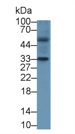
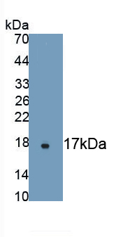
 WB (Western Blot)
(Western Blot; Sample: Human MilkPrimary Ab: 2?g/ml Mouse Anti-Human MUC1 AntibodySecond Ab: 0.2ug/mL HRP-Linked Rabbit Anti-Mouse IgG Polyclonal Antibody)
WB (Western Blot)
(Western Blot; Sample: Human MilkPrimary Ab: 2?g/ml Mouse Anti-Human MUC1 AntibodySecond Ab: 0.2ug/mL HRP-Linked Rabbit Anti-Mouse IgG Polyclonal Antibody)
Mucin 1 (MUC1), Monoclonal Antibody (Cat# AAA149522)
What are Monoclonal Antibodies?
Monoclonal antibodies are specialized laboratory-produced proteins developed for binding to specific biological antigens or other molecular targets. Since they come from a single cell (or clone), they are especially consistent and accurate in the data they are involved in producing.
This type of antibody material has been shown to be a powerful tool in finding and subsequently destroying harmful cells in an organism, such as those found in cancers or various autoimmune diseases. This makes them excellent aids in medical testing and research, which is why they are so widely used.
AAA Biotech offers a comprehensive range of high-quality monoclonal antibodies that perform effectively in various laboratory tests, including (amongst others) ELISA, western blotting, immunohistochemistry, and flow cytometry. All of the products in our catalog are thoroughly quality tested to make sure that they are reliable and will consistently perform well in your research.
What Are The Uses of Monoclonal Antibodies
Monoclonal antibodies are used in many lab tests, including (amongst others) ELISA, western blotting, immunohistochemistry, and flow cytometry.
ELISA is a test that helps detect a specific substance/analyte in a sample. It uses antibodies (often monoclonal) bound to a solid surface (such as the well of a microplate) to “capture” the substance/analyte in the sample and immobilize it so that the detection antibody component can then bind to it and produce a signal, which can then be measured.
Western blotting identifies specific proteins in a sample. The sample is first separated on a gel, and then antibodies are applied that will typically bind to the target, which will all be localized to a single band in a lane.
Immunohistochemistry helps locate specific proteins in cells or tissue samples using antibodies.
Flow cytometry looks at and sorts cells. It uses antibodies that are conjugated to reporter molecules called “fluorophores”, which, under special lights, emit light themselves, which can then be measured by a detector instrument. For a deeper understanding of these techniques, explore our complete guide to monoclonal antibodies and their benefits.
How Monoclonal Antibodies Are Used as Medicine?
Please note that all of the products listed in AAA Biotech’s also known as AAA Bio or AAABio catalog are strictly for research-use only (RUO).
Monoclonal antibodies can also be used as therapeutic/medical treatments, particularly in the context of cancers. They are designed to find and bind to specific cells or proteins, helping the immune system recognize and attack the cancer. These treatments work in different ways, such as:
- Radioimmunotherapy attaches a small amount of radioactive molecule to the antibody, so it delivers the radiation directly to the cancer cells that the antibody is specifically binding to.
- Antibody-directed enzyme prodrug therapy uses antibodies that are specifically bound to special enzymes. These enzymes activate a harmless drug in the body and turn it into a cancer-killing drug only near the cancer cells—this helps avoid harming healthy cells.
- Immunoliposomes are tiny “bubbles” filled with medicine/drug and coated with antibodies. They carry the drug straight to the cancer cells.
Why Buy Monoclonal Antibodies From Us?
At AAA Biotech, we provide high-performance monoclonal antibodies designed to support a wide range of research needs.
1. Validated for Versatile Applications
The antibodies in our catalog are extensively validated and compatible with multiple techniques, including (but not limited to) ELISA, flow cytometry (FC), immunocytochemistry (ICC), immunofluorescence (IF), immunohistochemistry (IHC), immunoprecipitation (IP), and western blotting (WB).
2. Wide Selection & Specialized Options
We offer antibodies for common and rare species, that are available in various conjugated forms, and also in recombinant formats. Essentially, there is almost anything one might need to meet their experimental model’s requirements.
3. High-Quality Proteins
Our proteins meet high purity standards—90% or more as confirmed by SDS-PAGE. Many are available with tags like His, Flag, GST, or MBP, and we also supply native and biologically active proteins for functional studies.
Frequently Asked Questions
1. Are your monoclonal antibodies validated for specific applications?
Yes, our antibodies are tested and validated for use in methods such as ELISA, western blot, IHC, flow cytometry, and more. Refer to specific product pages or datasheets for individual product information.
2. How do I choose the right monoclonal antibody for my application?
Review the product details directly for application validation, species reactivity, and target information. You may also contact our support team at any time for help.
3. How quickly can I receive my order?
Most orders are processed and shipped within 1–3 business days, depending on product availability and your shipping location.