Filters
▼Clonality
▼Type
▼Reactivity
▼Gene Name
▼Isotype
▼Host
▼Application
▼Clone
▼Monoclonal Antibodies
Get accurate results in your research with our Monoclonal Antibodies, which are specially made to target exactly what you require for your research, and will produce consistent, reliable performance in lab tests.
Viewing 2350-2400 of 27645 product results
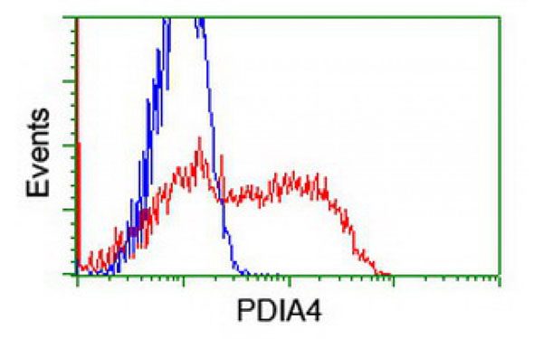
 WB (Western Blot)
(Western Blot analysis of HEK293T cell lysates (5 ug) transfected with either recombinant PDIA4 protein (Right) or empty vector (Left) detected with PDIA4 antibody)
WB (Western Blot)
(Western Blot analysis of HEK293T cell lysates (5 ug) transfected with either recombinant PDIA4 protein (Right) or empty vector (Left) detected with PDIA4 antibody)
PDIA4, Monoclonal Antibody (Cat# AAA107084)
 WB (Western Blot)
(Western Blot analysis of HEK293T cell lysates (5 ug) transfected with either recombinant SOX17 protein (Right) or empty vector (Left) detected with SOX17 antibody)
WB (Western Blot)
(Western Blot analysis of HEK293T cell lysates (5 ug) transfected with either recombinant SOX17 protein (Right) or empty vector (Left) detected with SOX17 antibody)
SOX17, Monoclonal Antibody (Cat# AAA107093)
 IF (Immunofluorescence)
(Immunofluorescent staining of COS7 cells transiently transfected with recombinant SMS protein using SMS antibody)
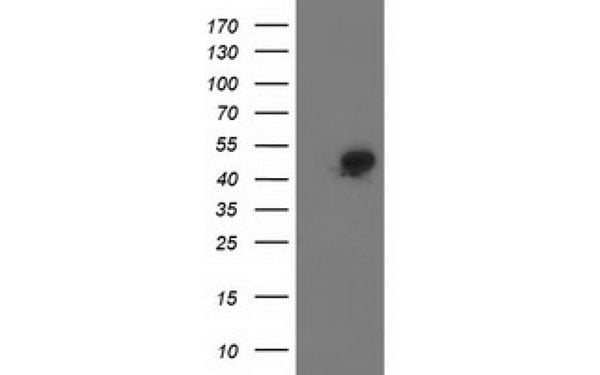
IF (Immunofluorescence)
(Immunofluorescent staining of COS7 cells transiently transfected with recombinant SMS protein using SMS antibody)
SMS, Monoclonal Antibody (Cat# AAA107097)
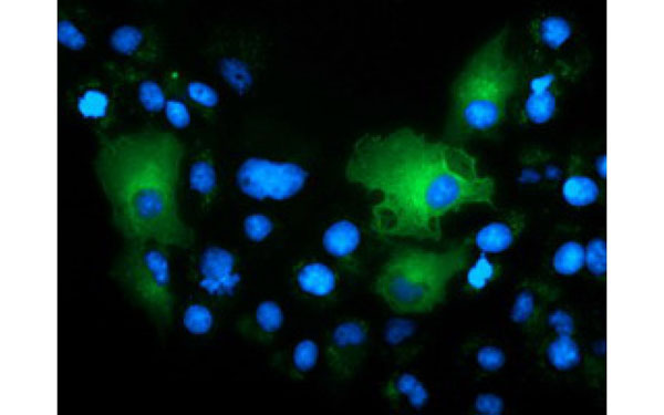
 WB (Western Blot)
(Western Blot analysis using PACSIN3 antibodyWestern Blot analysis of HEK293T cell lysates (5 ug) transfected with either recombinant PACSIN3 protein (Right) or empty vector (Left) detected with PACSIN3 antibody)
WB (Western Blot)
(Western Blot analysis using PACSIN3 antibodyWestern Blot analysis of HEK293T cell lysates (5 ug) transfected with either recombinant PACSIN3 protein (Right) or empty vector (Left) detected with PACSIN3 antibody)
PACSIN3, Monoclonal Antibody (Cat# AAA107099)
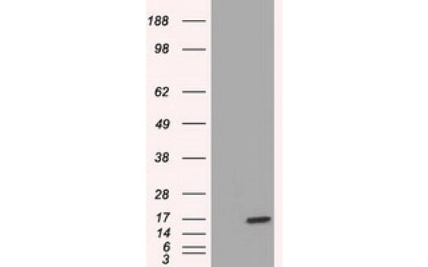
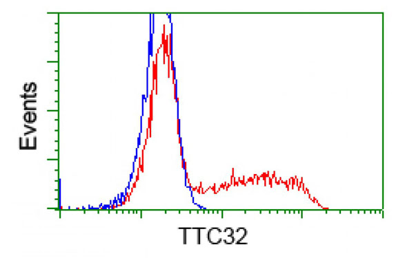
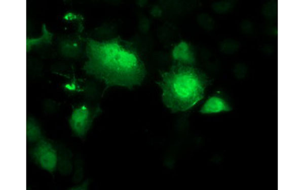
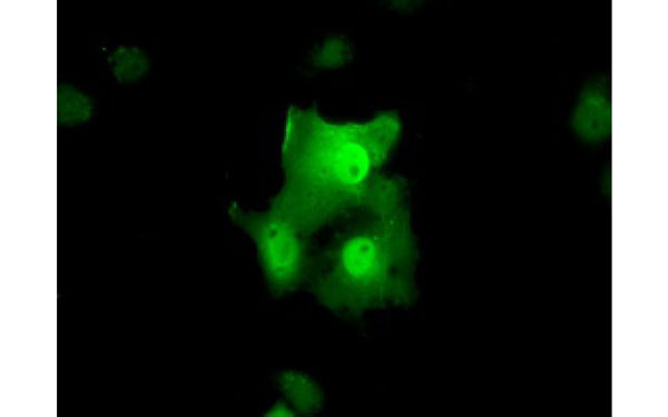 IF (Immunofluorescence)
(Immunofluorescent staining of COS7 cells transiently transfected with recombinant TTC32 protein using TTC32 antibody)
IF (Immunofluorescence)
(Immunofluorescent staining of COS7 cells transiently transfected with recombinant TTC32 protein using TTC32 antibody)
TTC32, Monoclonal Antibody (Cat# AAA107108)
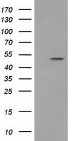
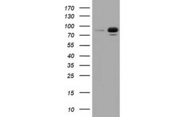
 WB (Western Blot)
(Western Blot analysis of HEK293T cell lysates (5 ug) transfected with either recombinant SLC2A5 protein (Right) or empty vector (Left) detected with SLC2A5 antibody)
WB (Western Blot)
(Western Blot analysis of HEK293T cell lysates (5 ug) transfected with either recombinant SLC2A5 protein (Right) or empty vector (Left) detected with SLC2A5 antibody)
SLC2A5, Monoclonal Antibody (Cat# AAA107110)
 WB (Western Blot)
(Western Blot analysis of HEK293T cell lysates (5 ug) transfected with either recombinant SIRT5 protein (Right) or empty vector (Left) detected with SIRT5 antibody)
WB (Western Blot)
(Western Blot analysis of HEK293T cell lysates (5 ug) transfected with either recombinant SIRT5 protein (Right) or empty vector (Left) detected with SIRT5 antibody)
SIRT5, Monoclonal Antibody (Cat# AAA106944)

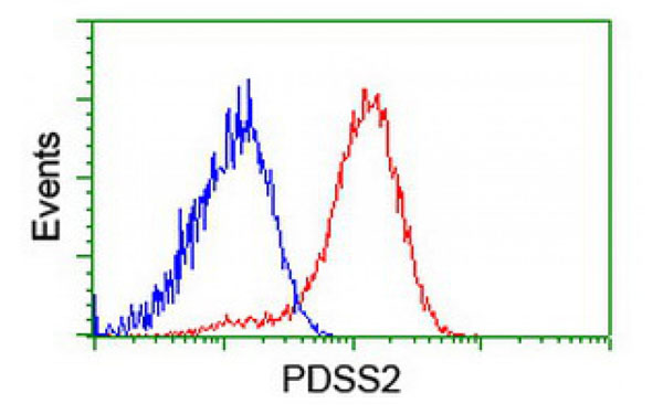
 IHC (Immunohistochemisry)
(Immunohistochemical analysis of PDSS2 protein in paraffin embedded Human endometrium tissue using PDSS2 antibody)
IHC (Immunohistochemisry)
(Immunohistochemical analysis of PDSS2 protein in paraffin embedded Human endometrium tissue using PDSS2 antibody)
PDSS2, Monoclonal Antibody (Cat# AAA106946)
 WB (Western Blot)
(Western Blot analysis of HEK293T cell lysates (5 ug) transfected with either recombinant VWA5A protein (Right) or empty vector (Left) detected with VWA5A antibody)
WB (Western Blot)
(Western Blot analysis of HEK293T cell lysates (5 ug) transfected with either recombinant VWA5A protein (Right) or empty vector (Left) detected with VWA5A antibody)
VWA5A, Monoclonal Antibody (Cat# AAA107017)
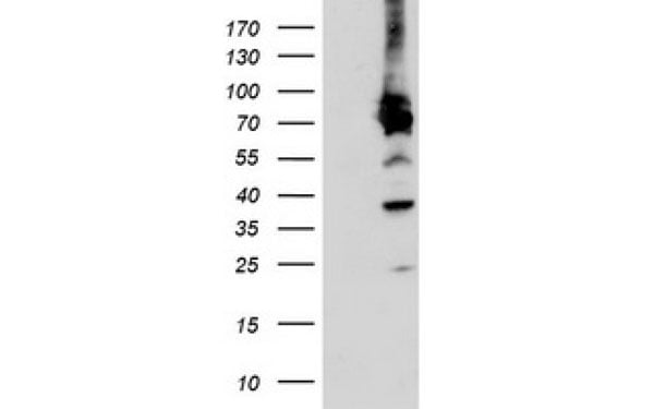
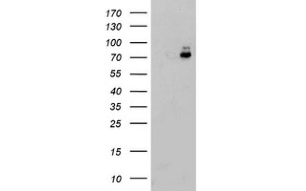 WB (Western Blot)
(Western Blot analysis of HEK293T cell lysates (5 ug) transfected with either recombinant PDIA4 protein (Right) or empty vector (Left) detected with PDIA4 antibody)
WB (Western Blot)
(Western Blot analysis of HEK293T cell lysates (5 ug) transfected with either recombinant PDIA4 protein (Right) or empty vector (Left) detected with PDIA4 antibody)
PDIA4, Monoclonal Antibody (Cat# AAA107239)
 IF (Immunofluorescence)
(Immunofluorescent staining of COS7 cells transiently transfected with recombinant LOX protein using LOX antibody)
IF (Immunofluorescence)
(Immunofluorescent staining of COS7 cells transiently transfected with recombinant LOX protein using LOX antibody)
LOX, Monoclonal Antibody (Cat# AAA107254)
 IF (Immunofluorescence)
(Immunofluorescent staining of COS7 cells transiently transfected with recombinant NPR3 protein using NPR3 antibody)
IF (Immunofluorescence)
(Immunofluorescent staining of COS7 cells transiently transfected with recombinant NPR3 protein using NPR3 antibody)
NPR3, Monoclonal Antibody (Cat# AAA107278)
 IF (Immunofluorescence)
(Immunofluorescent staining of COS7 cells transiently transfected with recombinant PFKP protein using PFKP antibody)
IF (Immunofluorescence)
(Immunofluorescent staining of COS7 cells transiently transfected with recombinant PFKP protein using PFKP antibody)
PFKP, Monoclonal Antibody (Cat# AAA107136)
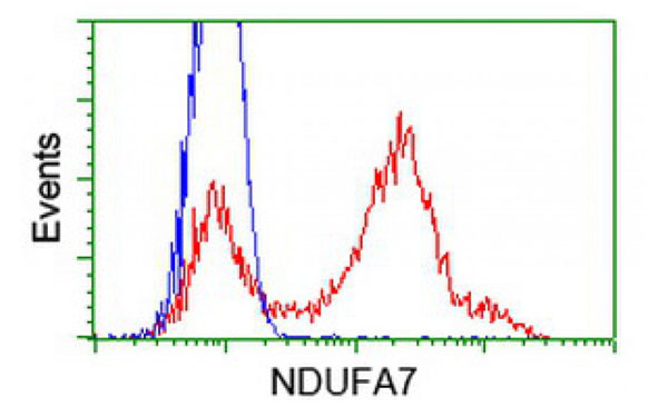
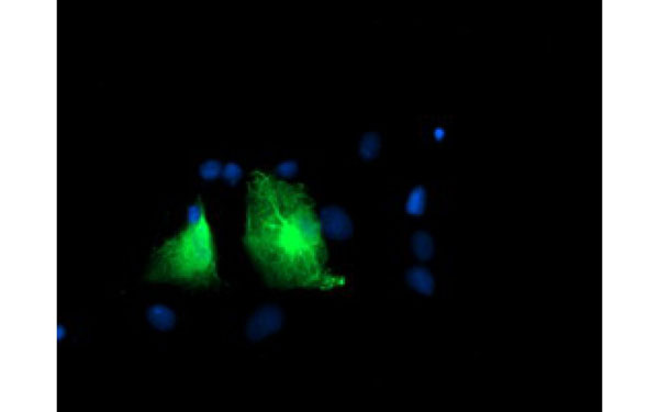
 IF (Immunofluorescence)
(Immunofluorescent staining of COS7 cells transiently transfected with recombinant NDUFA7 protein using NDUFA7 antibody)
IF (Immunofluorescence)
(Immunofluorescent staining of COS7 cells transiently transfected with recombinant NDUFA7 protein using NDUFA7 antibody)
NDUFA7, Monoclonal Antibody (Cat# AAA107182)
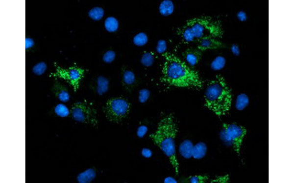
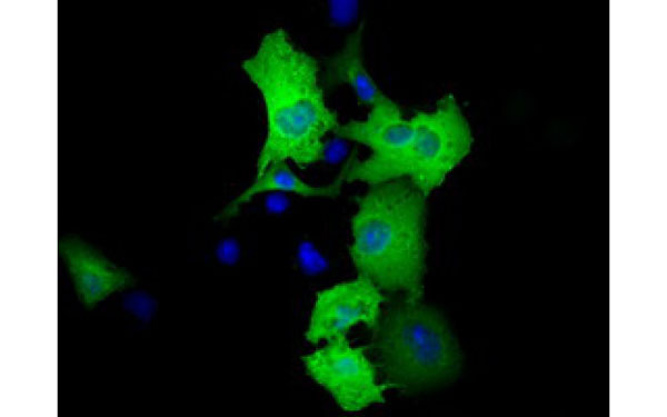 IF (Immunofluorescence)
(Immunofluorescent staining of COS7 cells transiently transfected with recombinant RALBP1 protein using RALBP1 antibody)
IF (Immunofluorescence)
(Immunofluorescent staining of COS7 cells transiently transfected with recombinant RALBP1 protein using RALBP1 antibody)
RALBP1, Monoclonal Antibody (Cat# AAA106875)
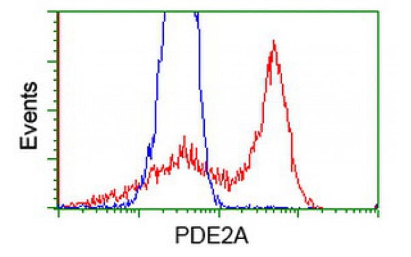
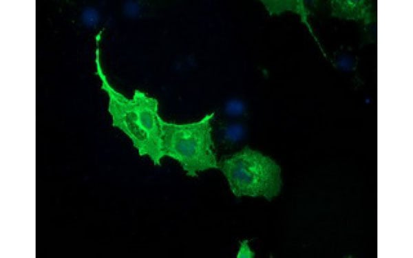 IF (Immunofluorescence)
(Immunofluorescent staining of COS7 cells transiently transfected with recombinant PDE2A protein using PDE2A antibody)
IF (Immunofluorescence)
(Immunofluorescent staining of COS7 cells transiently transfected with recombinant PDE2A protein using PDE2A antibody)
PDE2A, Monoclonal Antibody (Cat# AAA106902)
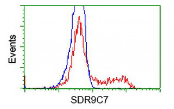
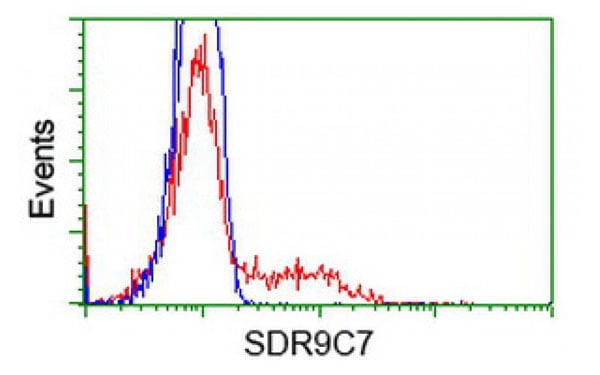
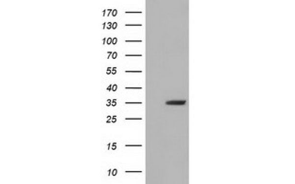 WB (Western Blot)
(Western Blot analysis of HEK293T cell lysates (5 ug) transfected with either recombinant SDR9C7 protein (Right) or empty vector (Left) detected with SDR9C7 antibody)
WB (Western Blot)
(Western Blot analysis of HEK293T cell lysates (5 ug) transfected with either recombinant SDR9C7 protein (Right) or empty vector (Left) detected with SDR9C7 antibody)
SDR9C7, Monoclonal Antibody (Cat# AAA106924)
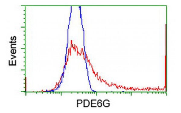
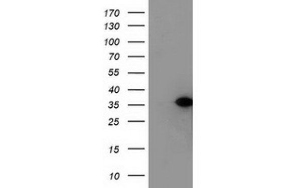
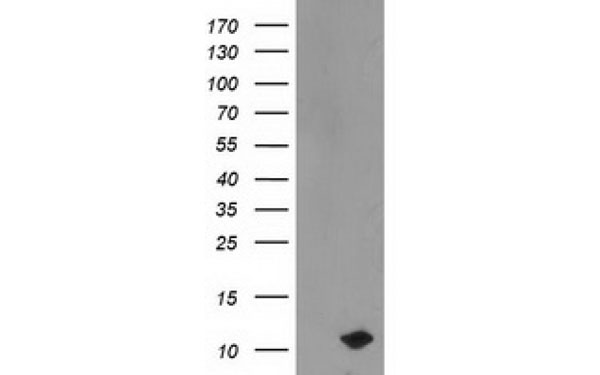 WB (Western Blot)
(Western Blot analysis of HEK293T cell lysates (5 ug) transfected with either recombinant PDE6G protein (Right) or empty vector (Left) detected with PDE6G antibody)
WB (Western Blot)
(Western Blot analysis of HEK293T cell lysates (5 ug) transfected with either recombinant PDE6G protein (Right) or empty vector (Left) detected with PDE6G antibody)
PDE6G, Monoclonal Antibody (Cat# AAA107385)
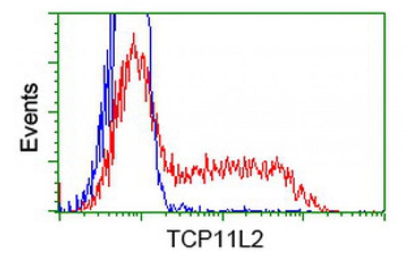
 IF (Immunofluorescence)
(Immunofluorescent staining of COS7 cells transiently transfected with recombinant TCP11L2 protein using TCP11L2 antibody)
IF (Immunofluorescence)
(Immunofluorescent staining of COS7 cells transiently transfected with recombinant TCP11L2 protein using TCP11L2 antibody)
TCP11L2, Monoclonal Antibody (Cat# AAA107406)
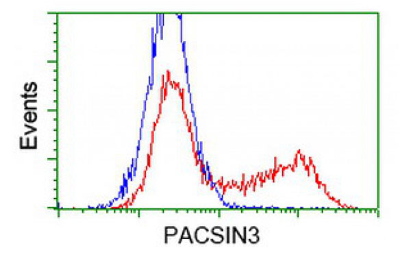
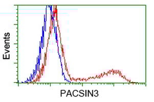
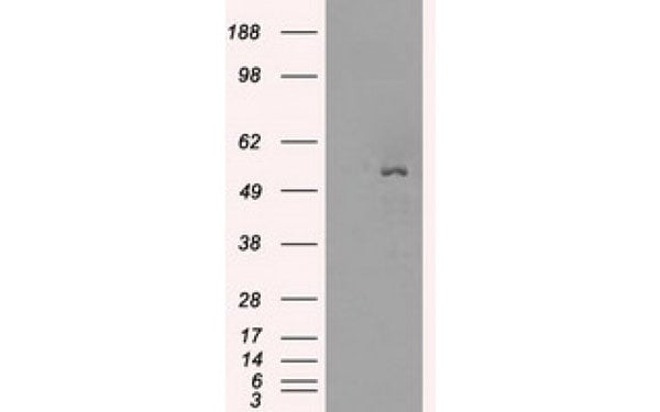
 WB (Western Blot)
(Western Blot analysis of HEK293T cell lysates (5 ug) transfected with either recombinant PACSIN3 protein (Right) or empty vector (Left) detected with PACSIN3 antibody)
WB (Western Blot)
(Western Blot analysis of HEK293T cell lysates (5 ug) transfected with either recombinant PACSIN3 protein (Right) or empty vector (Left) detected with PACSIN3 antibody)
PACSIN3, Monoclonal Antibody (Cat# AAA107413)
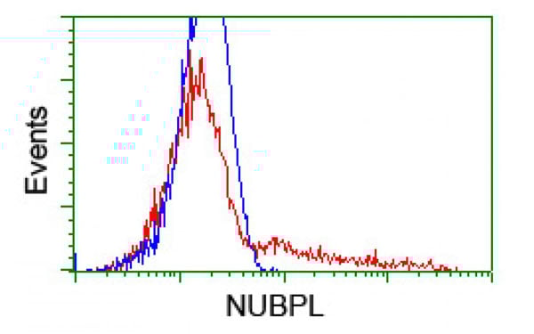
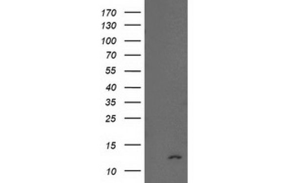
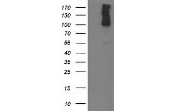
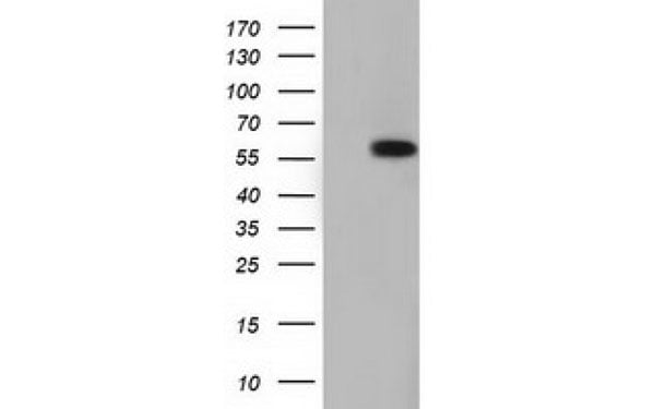
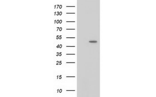
 WB (Western Blot)
(Western Blot analysis of HEK293T cell lysates (5 ug) transfected with either recombinant NUBPL protein (Right) or empty vector (Left) detected with NUBPL antibody)
WB (Western Blot)
(Western Blot analysis of HEK293T cell lysates (5 ug) transfected with either recombinant NUBPL protein (Right) or empty vector (Left) detected with NUBPL antibody)
NUBPL, Monoclonal Antibody (Cat# AAA107431)
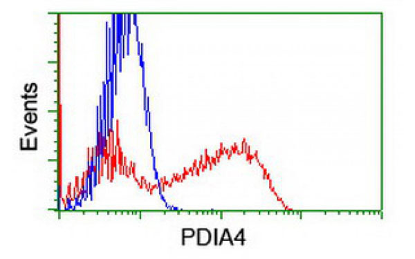
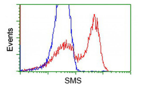
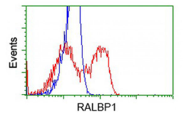
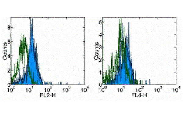 FCM/FACS (Flow Cytometry)
(Surface staining of normal human peripheral blood cells with CD284 antibody (PE) (left), and CD284 antibody (APC) (right).Appropriate Isotype Controls were used (open histogram). Cells in the monocyte population were used for analysis.)
FCM/FACS (Flow Cytometry)
(Surface staining of normal human peripheral blood cells with CD284 antibody (PE) (left), and CD284 antibody (APC) (right).Appropriate Isotype Controls were used (open histogram). Cells in the monocyte population were used for analysis.)
Toll-like receptor 4, Monoclonal Antibody (Cat# AAA107432)
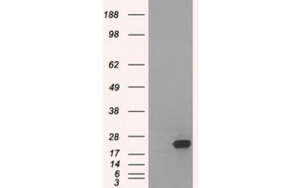
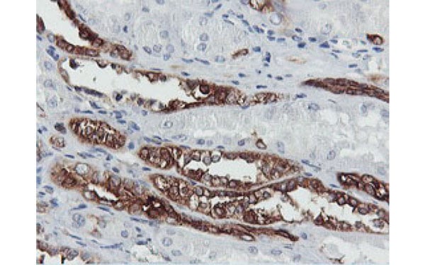
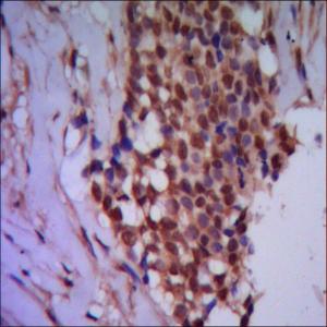
 IHC (Immunohiostchemistry)
(Immunohistochemical analysis of SSX2 protein in paraffin embedded Human colon tissue using SSX2 antibody)
IHC (Immunohiostchemistry)
(Immunohistochemical analysis of SSX2 protein in paraffin embedded Human colon tissue using SSX2 antibody)
SSX2, Monoclonal Antibody (Cat# AAA107291)
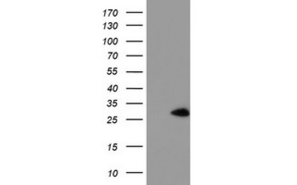
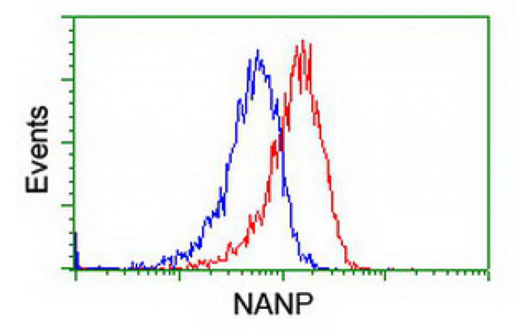
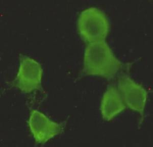
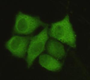
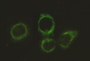
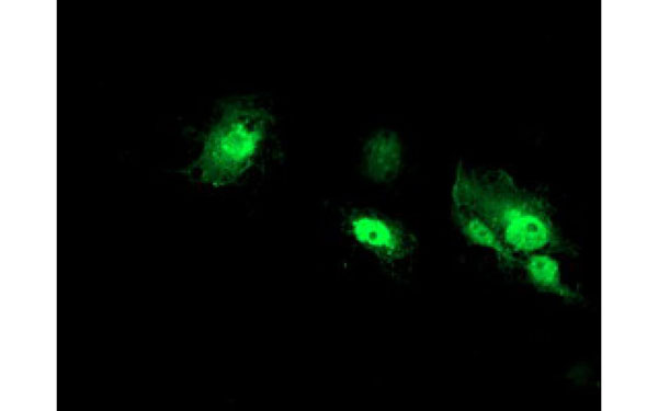 IF (Immunofluorescence)
(Immunofluorescent staining of COS7 cells transiently transfected with recombinant NANP protein using NANP antibody)
IF (Immunofluorescence)
(Immunofluorescent staining of COS7 cells transiently transfected with recombinant NANP protein using NANP antibody)
NANP, Monoclonal Antibody (Cat# AAA107302)
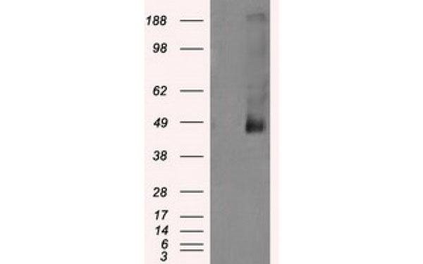
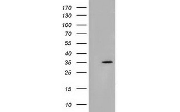
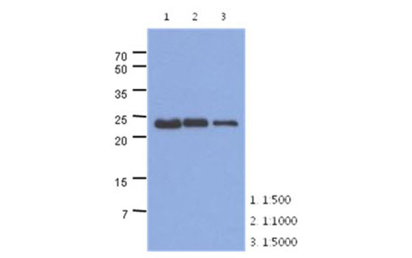 WB (Western Blot)
(The extracts of mouse muscle (40 ug) were resolved by SDS-PAGE, transferred to PVDF membrane and probed with TNNI1 antibody (1:500 - 1:5000). Proteins were visualized using a goat anti-mouse secondary antibody conjugated to HRP and an ECL detection system.)
WB (Western Blot)
(The extracts of mouse muscle (40 ug) were resolved by SDS-PAGE, transferred to PVDF membrane and probed with TNNI1 antibody (1:500 - 1:5000). Proteins were visualized using a goat anti-mouse secondary antibody conjugated to HRP and an ECL detection system.)
TNNI1, Monoclonal Antibody (Cat# AAA107309)
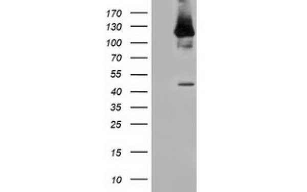
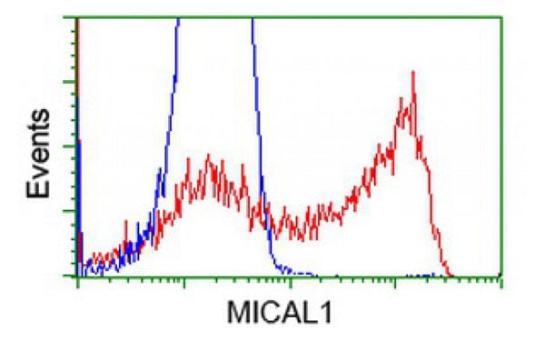
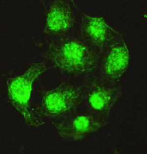
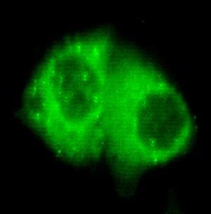
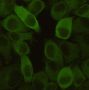
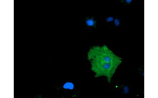 IF (Immunofluorescence)
(Immunofluorescent staining of COS7 cells transiently transfected with recombinant MICAL1 protein using MICAL1 antibody)
IF (Immunofluorescence)
(Immunofluorescent staining of COS7 cells transiently transfected with recombinant MICAL1 protein using MICAL1 antibody)
MICAL1, Monoclonal Antibody (Cat# AAA107352)
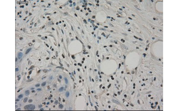
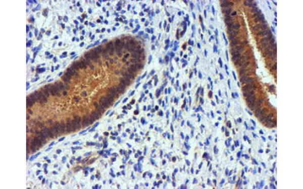
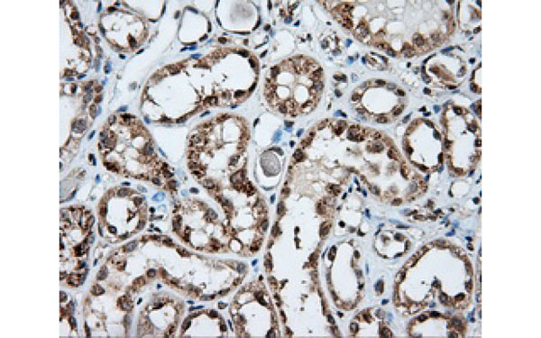
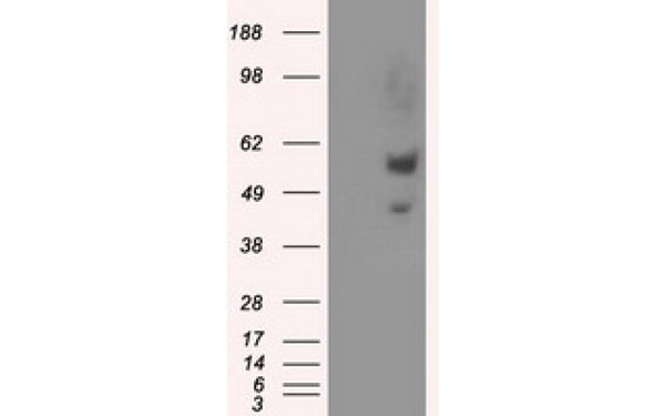
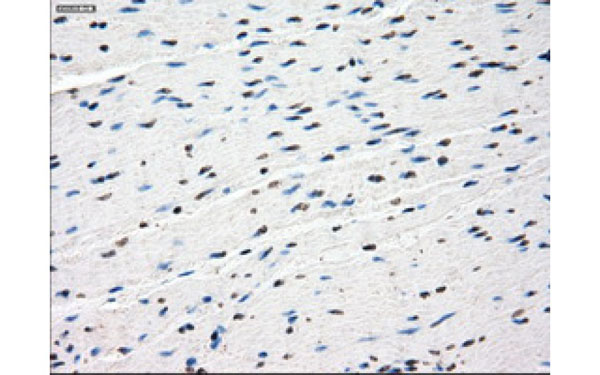
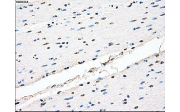
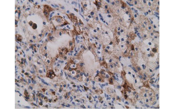 IHC (Immunohistochemisry)
(Immunohistochemical analysis of SDR9C7 protein in paraffin embedded Carcinoma of Human kidney tissue using SDR9C7 antibody)
IHC (Immunohistochemisry)
(Immunohistochemical analysis of SDR9C7 protein in paraffin embedded Carcinoma of Human kidney tissue using SDR9C7 antibody)
SDR9C7, Monoclonal Antibody (Cat# AAA107481)
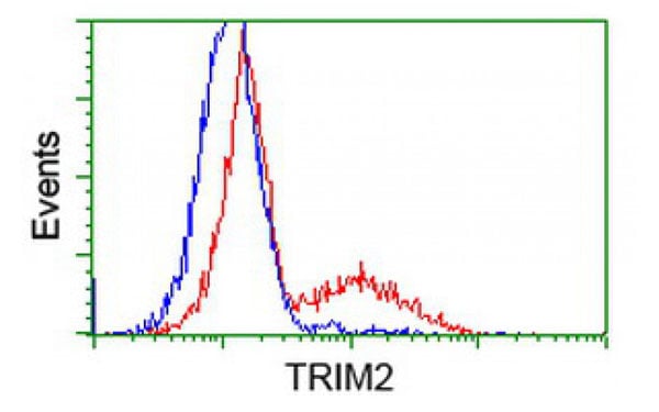
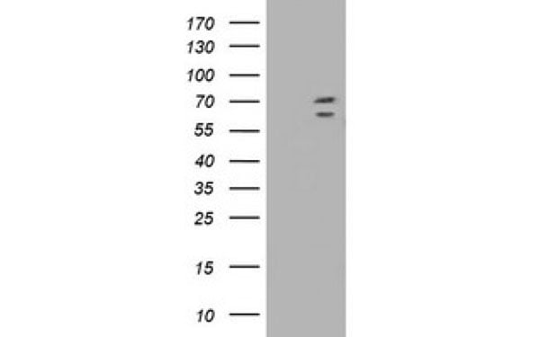 WB (Western Blot)
(Western Blot analysis of HEK293T cell lysates (5 ug) transfected with either recombinant TRIM2 protein (Right) or empty vector (Left) detected with TRIM2 antibody)
WB (Western Blot)
(Western Blot analysis of HEK293T cell lysates (5 ug) transfected with either recombinant TRIM2 protein (Right) or empty vector (Left) detected with TRIM2 antibody)
TRIM2, Monoclonal Antibody (Cat# AAA107497)

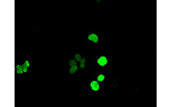
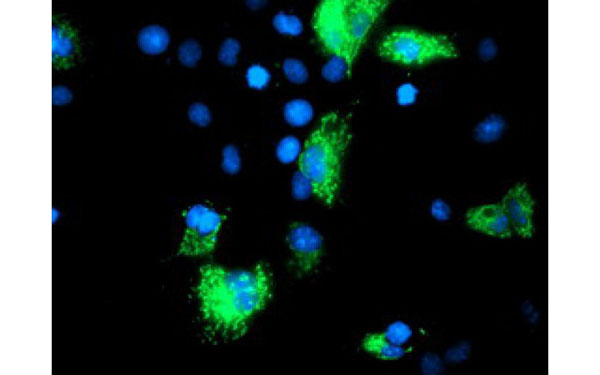
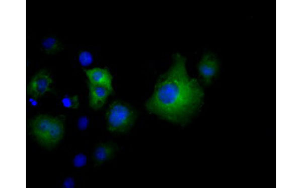
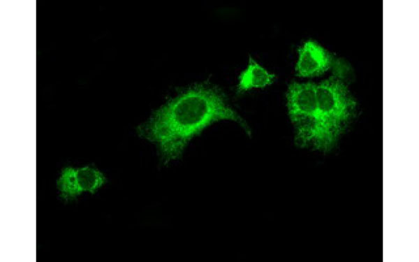
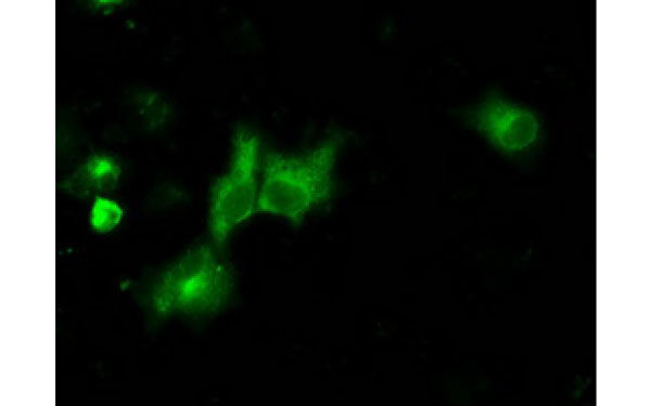
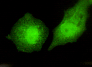
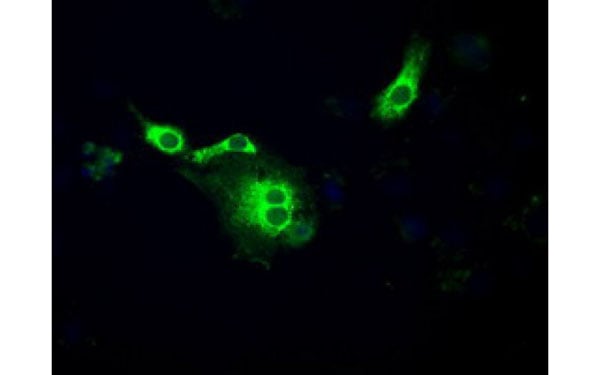 IF (Immunofluorescence)
(Immunofluorescent staining of COS7 cells transiently transfected with recombinant POR protein using POR antibody)
IF (Immunofluorescence)
(Immunofluorescent staining of COS7 cells transiently transfected with recombinant POR protein using POR antibody)
POR, Monoclonal Antibody (Cat# AAA107503)
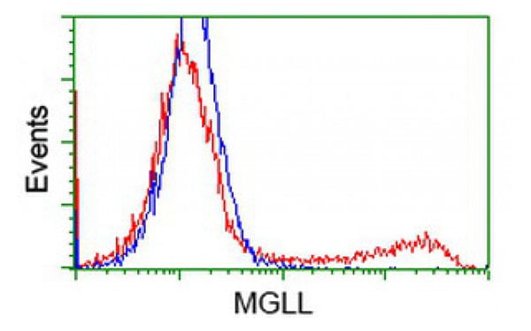
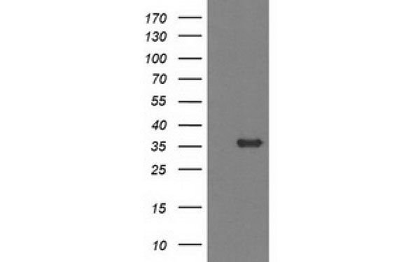 WB (Western Blot)
(Western Blot analysis of HEK293T cell lysates (5 ug) transfected with either recombinant MGLL protein (Right) or empty vector (Left) detected with MGLL antibody)
WB (Western Blot)
(Western Blot analysis of HEK293T cell lysates (5 ug) transfected with either recombinant MGLL protein (Right) or empty vector (Left) detected with MGLL antibody)
MGLL, Monoclonal Antibody (Cat# AAA107455)
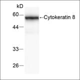
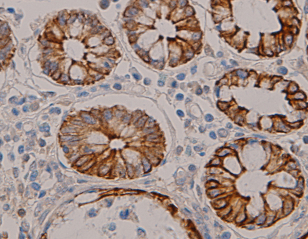 Application Data
CK8, Monoclonal Antibody (Cat# AAA109983)
 WB (Western Blot)
(WB (1:1000) analysis of recombinant protein MIP1P (CCL4) with Anti-MIPl 3 (CCL4).)
WB (Western Blot)
(WB (1:1000) analysis of recombinant protein MIP1P (CCL4) with Anti-MIPl 3 (CCL4).)
MIP1beta, Monoclonal Antibody (Cat# AAA109494)
 Application Data
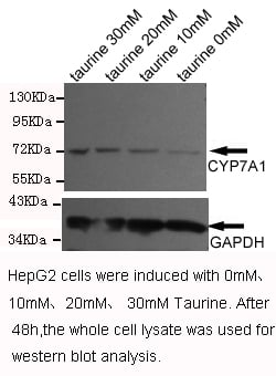
(Western blot detection of CYP7A1(C-terminus) antibody in HepG2 cell lysates using CYP7A1(C-terminus) antibody (1:1000 diluted). Predicted band size: 58KDa Observed band size: 72KDa.)
Application Data
(Western blot detection of CYP7A1(C-terminus) antibody in HepG2 cell lysates using CYP7A1(C-terminus) antibody (1:1000 diluted). Predicted band size: 58KDa Observed band size: 72KDa.)
CYP7A1-C, Monoclonal Antibody (Cat# AAA111288)
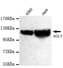
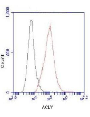 Application Data
(Flow Cytometry analysis of HeLa cells stained with ATP-Citrate Lyase (red, 1/100 dilution), followed by FITC-conjugated goat anti-mouse IgG. Black line histogram represents the isotype control, normal mouse IgG.)
Application Data
(Flow Cytometry analysis of HeLa cells stained with ATP-Citrate Lyase (red, 1/100 dilution), followed by FITC-conjugated goat anti-mouse IgG. Black line histogram represents the isotype control, normal mouse IgG.)
ATP-Citrate Lyase, Monoclonal Antibody (Cat# AAA111291)
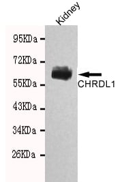 Application Data
(Western blot detection of CHRDL1 in Rat kidney tissue lysates using CHRDL1 antibody (1:1000 diluted). Predicted band size: 57kDa Observed band size: 57kDa.)
Application Data
(Western blot detection of CHRDL1 in Rat kidney tissue lysates using CHRDL1 antibody (1:1000 diluted). Predicted band size: 57kDa Observed band size: 57kDa.)
CHRDL1, Monoclonal Antibody (Cat# AAA111300)
 Application Data
(IHC of paraffin-embedded human breast cancer using anti-Protein Phosphatase 4C diluted 1/500-1/1000.)
Application Data
(IHC of paraffin-embedded human breast cancer using anti-Protein Phosphatase 4C diluted 1/500-1/1000.)
Protein Phosphatase 4C (PPP4C), Monoclonal Antibody (Cat# AAA111302)
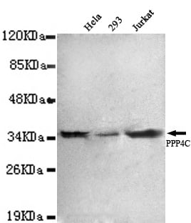
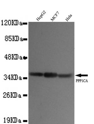 Application Data
(Western blot detection of PP1A(N-terminus) antibody in HepG2,MCF7&Hela cell lysates using PP1A(N-terminus) antibody (1:1000 diluted). Predicted band size:37KDa. Observed band size:37KDa.)
Application Data
(Western blot detection of PP1A(N-terminus) antibody in HepG2,MCF7&Hela cell lysates using PP1A(N-terminus) antibody (1:1000 diluted). Predicted band size:37KDa. Observed band size:37KDa.)
protein phosphatase 1A (PPP1A), Monoclonal Antibody (Cat# AAA111307)
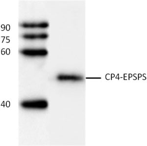 WB (Western Blot)
(WB:(1:1000) analysis of CP4-EPSPS fusion protien with Anti-CP4-EPSPS.)
WB (Western Blot)
(WB:(1:1000) analysis of CP4-EPSPS fusion protien with Anti-CP4-EPSPS.)
CP4-EPSPS, Monoclonal Antibody (Cat# AAA111308)
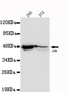 Application Data
(Western blot detection of JUN in :293 &3T3 cell lysates(1:1000 diluted). Predicted band size: 36KDa Observed band size: 45KDa.)
Application Data
(Western blot detection of JUN in :293 &3T3 cell lysates(1:1000 diluted). Predicted band size: 36KDa Observed band size: 45KDa.)
JUN, Monoclonal Antibody (Cat# AAA111311)
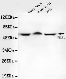
 Application Data
(IHC of paraffin-embedded huma breast cancer using anti- TBLR1 diluted 1/500-1/1000.)
Application Data
(IHC of paraffin-embedded huma breast cancer using anti- TBLR1 diluted 1/500-1/1000.)
TBLR1, Monoclonal Antibody (Cat# AAA111314)
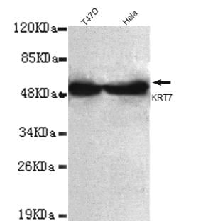 Application Data
(Western blot detection of Keratin 7(C-terminus) antibody in T47D&Hela Lung cell lysates using Keratin 7(C-terminus) antibody (1:1000 diluted). Predicted band size: 51KDa Observed band size: 55KDa.)
Application Data
(Western blot detection of Keratin 7(C-terminus) antibody in T47D&Hela Lung cell lysates using Keratin 7(C-terminus) antibody (1:1000 diluted). Predicted band size: 51KDa Observed band size: 55KDa.)
Keratin 7 (KRT7), Monoclonal Antibody (Cat# AAA111316)
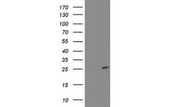
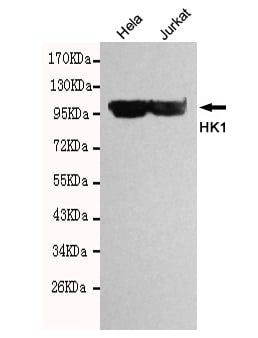 Application Data
(Western blot detection of HK1 in Hela&Jurkat cell lysates using HK1 antibody (1:10000 diluted). Predicted band size: 102kDa Observed band size: 102kDa)
Application Data
(Western blot detection of HK1 in Hela&Jurkat cell lysates using HK1 antibody (1:10000 diluted). Predicted band size: 102kDa Observed band size: 102kDa)
HK1, Monoclonal Antibody (Cat# AAA111319)
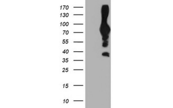
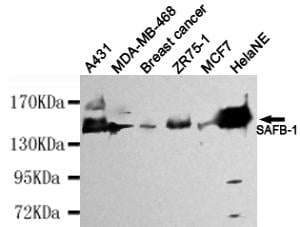 Application Data
(Western blot detection of SAFB-1 in HelaNE, A431, MDA-MB-468, Breast cancer, ZR75-1&MCF7 cell lysates using SAFB-1 antibody (1:4000 diluted). Predicted band size: 130kDa Observed band size: 130kDa.)
Application Data
(Western blot detection of SAFB-1 in HelaNE, A431, MDA-MB-468, Breast cancer, ZR75-1&MCF7 cell lysates using SAFB-1 antibody (1:4000 diluted). Predicted band size: 130kDa Observed band size: 130kDa.)
SAFB-1, Monoclonal Antibody (Cat# AAA111320)
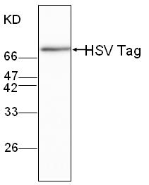 WB (Western Blot)
(WB (1:1000) analysis of HSV-tagged fusion protein with Anti-HSV Tag (AAA111366))
WB (Western Blot)
(WB (1:1000) analysis of HSV-tagged fusion protein with Anti-HSV Tag (AAA111366))
HSV Tag, Monoclonal Antibody (Cat# AAA111366)
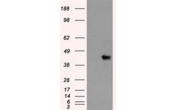
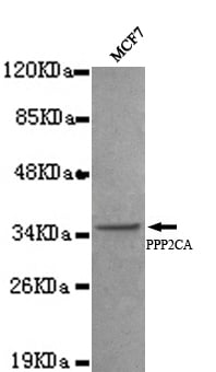 Application Data
(Western blot detection of PP2A-alpha(N-terminus) antibody in MCF7 cell lysates using PP2A-alpha(N-terminus) antibody (1:1000 diluted). Predicted band size:35KDa. Observed band size:35KDa.)
Application Data
(Western blot detection of PP2A-alpha(N-terminus) antibody in MCF7 cell lysates using PP2A-alpha(N-terminus) antibody (1:1000 diluted). Predicted band size:35KDa. Observed band size:35KDa.)
protein phosphatase 2A, Monoclonal Antibody (Cat# AAA111275)
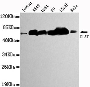
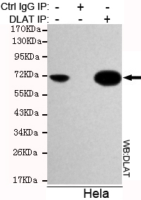 Application Data
(Immunoprecipitation analysis of Hela cell lysates using DLAT antibody.)
Application Data
(Immunoprecipitation analysis of Hela cell lysates using DLAT antibody.)
DLAT, Monoclonal Antibody (Cat# AAA111276)
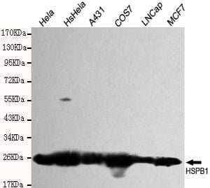 Application Data
(Western blot detection of HSPB1 in Hela, HsHela, A431, COS7, LNCAP&MCF7 cell lysates and using HSPB1 antibody (1:1000 diluted). Predicted band size: 23KDa Observed band size:27KDa.)
Application Data
(Western blot detection of HSPB1 in Hela, HsHela, A431, COS7, LNCAP&MCF7 cell lysates and using HSPB1 antibody (1:1000 diluted). Predicted band size: 23KDa Observed band size:27KDa.)
HSPB1, Monoclonal Antibody (Cat# AAA111278)
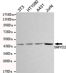 Application Data
(Western blot detection of SMYD2 in 3T3,HT1080, A431&Jurkat cell lysates and using SMYD2 antibody (1:1000 diluted). Predicted band size: 50KDa Observed band size: 50KDa)
Application Data
(Western blot detection of SMYD2 in 3T3,HT1080, A431&Jurkat cell lysates and using SMYD2 antibody (1:1000 diluted). Predicted band size: 50KDa Observed band size: 50KDa)
SMYD2, Monoclonal Antibody (Cat# AAA111282)
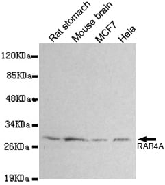 Application Data
(Western blot detection of RAB4A in Rat stomach, Mouse brain, Hela&MCF7 cell lysates and using RAB4A antibody (1:1000 diluted). Predicted band size: 24KDa Observed band size: 30KDa.)
Application Data
(Western blot detection of RAB4A in Rat stomach, Mouse brain, Hela&MCF7 cell lysates and using RAB4A antibody (1:1000 diluted). Predicted band size: 24KDa Observed band size: 30KDa.)
RAB4A-N, Monoclonal Antibody (Cat# AAA111283)
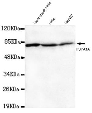 Application Data
(Western blot detection of Hsp70(N-terminus) antibody in Heat shcok Hela,Hela&HepG2 lysates using Hsp70(N-terminus) antibody (1:1000 diluted). Predicted band size: 70KDa Observed band size: 70KDa.)
Application Data
(Western blot detection of Hsp70(N-terminus) antibody in Heat shcok Hela,Hela&HepG2 lysates using Hsp70(N-terminus) antibody (1:1000 diluted). Predicted band size: 70KDa Observed band size: 70KDa.)
Hsp70, Monoclonal Antibody (Cat# AAA111284)
What are Monoclonal Antibodies?
Monoclonal antibodies are specialized laboratory-produced proteins developed for binding to specific biological antigens or other molecular targets. Since they come from a single cell (or clone), they are especially consistent and accurate in the data they are involved in producing.
This type of antibody material has been shown to be a powerful tool in finding and subsequently destroying harmful cells in an organism, such as those found in cancers or various autoimmune diseases. This makes them excellent aids in medical testing and research, which is why they are so widely used.
AAA Biotech offers a comprehensive range of high-quality monoclonal antibodies that perform effectively in various laboratory tests, including (amongst others) ELISA, western blotting, immunohistochemistry, and flow cytometry. All of the products in our catalog are thoroughly quality tested to make sure that they are reliable and will consistently perform well in your research.
What Are The Uses of Monoclonal Antibodies
Monoclonal antibodies are used in many lab tests, including (amongst others) ELISA, western blotting, immunohistochemistry, and flow cytometry.
ELISA is a test that helps detect a specific substance/analyte in a sample. It uses antibodies (often monoclonal) bound to a solid surface (such as the well of a microplate) to “capture” the substance/analyte in the sample and immobilize it so that the detection antibody component can then bind to it and produce a signal, which can then be measured.
Western blotting identifies specific proteins in a sample. The sample is first separated on a gel, and then antibodies are applied that will typically bind to the target, which will all be localized to a single band in a lane.
Immunohistochemistry helps locate specific proteins in cells or tissue samples using antibodies.
Flow cytometry looks at and sorts cells. It uses antibodies that are conjugated to reporter molecules called “fluorophores”, which, under special lights, emit light themselves, which can then be measured by a detector instrument. For a deeper understanding of these techniques, explore our complete guide to monoclonal antibodies and their benefits.
How Monoclonal Antibodies Are Used as Medicine?
Please note that all of the products listed in AAA Biotech’s also known as AAA Bio or AAABio catalog are strictly for research-use only (RUO).
Monoclonal antibodies can also be used as therapeutic/medical treatments, particularly in the context of cancers. They are designed to find and bind to specific cells or proteins, helping the immune system recognize and attack the cancer. These treatments work in different ways, such as:
- Radioimmunotherapy attaches a small amount of radioactive molecule to the antibody, so it delivers the radiation directly to the cancer cells that the antibody is specifically binding to.
- Antibody-directed enzyme prodrug therapy uses antibodies that are specifically bound to special enzymes. These enzymes activate a harmless drug in the body and turn it into a cancer-killing drug only near the cancer cells—this helps avoid harming healthy cells.
- Immunoliposomes are tiny “bubbles” filled with medicine/drug and coated with antibodies. They carry the drug straight to the cancer cells.
Why Buy Monoclonal Antibodies From Us?
At AAA Biotech, we provide high-performance monoclonal antibodies designed to support a wide range of research needs.
1. Validated for Versatile Applications
The antibodies in our catalog are extensively validated and compatible with multiple techniques, including (but not limited to) ELISA, flow cytometry (FC), immunocytochemistry (ICC), immunofluorescence (IF), immunohistochemistry (IHC), immunoprecipitation (IP), and western blotting (WB).
2. Wide Selection & Specialized Options
We offer antibodies for common and rare species, that are available in various conjugated forms, and also in recombinant formats. Essentially, there is almost anything one might need to meet their experimental model’s requirements.
3. High-Quality Proteins
Our proteins meet high purity standards—90% or more as confirmed by SDS-PAGE. Many are available with tags like His, Flag, GST, or MBP, and we also supply native and biologically active proteins for functional studies.
Frequently Asked Questions
1. Are your monoclonal antibodies validated for specific applications?
Yes, our antibodies are tested and validated for use in methods such as ELISA, western blot, IHC, flow cytometry, and more. Refer to specific product pages or datasheets for individual product information.
2. How do I choose the right monoclonal antibody for my application?
Review the product details directly for application validation, species reactivity, and target information. You may also contact our support team at any time for help.
3. How quickly can I receive my order?
Most orders are processed and shipped within 1–3 business days, depending on product availability and your shipping location.