Filters
▼Clonality
▼Type
▼Reactivity
▼Gene Name
▼Isotype
▼Host
▼Application
▼Clone
▼Monoclonal Antibodies
Get accurate results in your research with our Monoclonal Antibodies, which are specially made to target exactly what you require for your research, and will produce consistent, reliable performance in lab tests.
Viewing 3500-3550 of 27645 product results
 IF (Immunofluorescence)
(Immunofluorescence analysis of Mouse liver tissue using Caspase-8 Monoclonal Antibody at dilution of 1:200.)
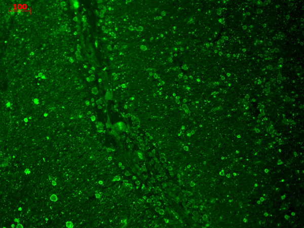
IF (Immunofluorescence)
(Immunofluorescence analysis of Mouse liver tissue using Caspase-8 Monoclonal Antibody at dilution of 1:200.)
Caspase-8, Monoclonal Antibody (Cat# AAA173648)
 IHC (Immunohiostchemistry)
(Immunohistochemistry of paraffin-embedded Rat brain tissue with GAP-43 Monoclonal Antibody.)
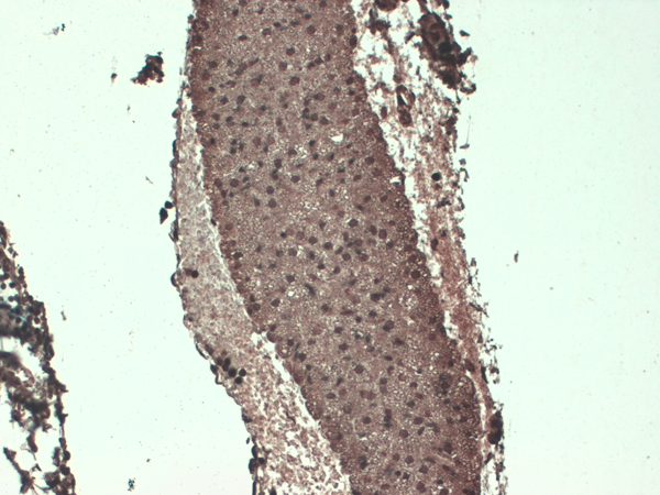
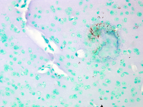
IHC (Immunohiostchemistry)
(Immunohistochemistry of paraffin-embedded Rat brain tissue with GAP-43 Monoclonal Antibody.)
GAP-43, Monoclonal Antibody (Cat# AAA173649)
 IF (Immunofluorescence)
(Immunofluorescence analysis of Human appendix tissue using MICU1 Monoclonal Antibody at dilution of 1:200.)
IF (Immunofluorescence)
(Immunofluorescence analysis of Human appendix tissue using MICU1 Monoclonal Antibody at dilution of 1:200.)
MICU1, Monoclonal Antibody (Cat# AAA173654)
 IF (Immunofluorescence)
(Immunofluorescence analysis of Human lung tissue using Cyclophilin B Monoclonal Antibody at dilution of 1:200.)
IF (Immunofluorescence)
(Immunofluorescence analysis of Human lung tissue using Cyclophilin B Monoclonal Antibody at dilution of 1:200.)
Cyclophilin B, Monoclonal Antibody (Cat# AAA173661)
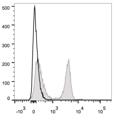
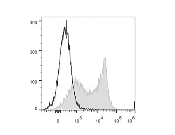
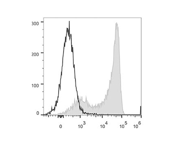
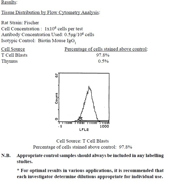
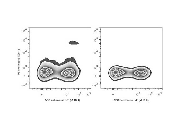 FCM/FACS (Flow Cytometry)
(C57BL/6 murine splenocytes are stained with Anti-Mouse CD11c Monoclonal Antibody(PE Conjugated) and Anti-Mouse MHC II (I-A/I-E) Monoclonal Antibody(APC Conjugated). Splenocytes stained with Anti-Mouse MHC II (I-A/I-E) Monoclonal Antibody(APC Conjugated)(Right) are used as control)
FCM/FACS (Flow Cytometry)
(C57BL/6 murine splenocytes are stained with Anti-Mouse CD11c Monoclonal Antibody(PE Conjugated) and Anti-Mouse MHC II (I-A/I-E) Monoclonal Antibody(APC Conjugated). Splenocytes stained with Anti-Mouse MHC II (I-A/I-E) Monoclonal Antibody(APC Conjugated)(Right) are used as control)
CD11c, Monoclonal Antibody (Cat# AAA174728)
CD3, Monoclonal Antibody (Cat# AAA174732)
 Application Data
CD74, Monoclonal Antibody (Cat# AAA174741)
 Application Data
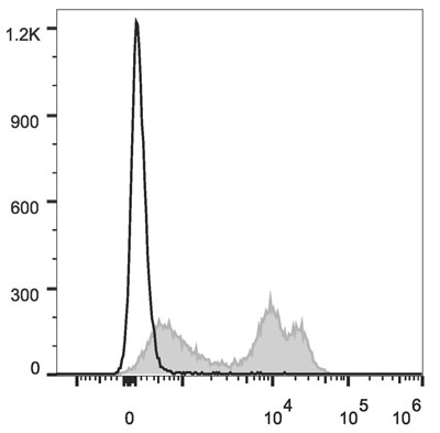
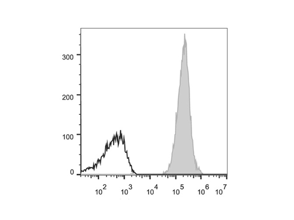
(Rat splenocytes are stained with PE/Cyanine7 Anti-Rat CD8a Antibody (filled gray histogram). Unstained splenocytes (empty black histogram) are used as control.)
Application Data
(Rat splenocytes are stained with PE/Cyanine7 Anti-Rat CD8a Antibody (filled gray histogram). Unstained splenocytes (empty black histogram) are used as control.)
CD8a, Monoclonal Antibody (Cat# AAA174743)
 Application Data
(C57BL/6 murine splenocytes are stained with PerCP/Cyanine5.5 Anti-Rat CD4(domain 1) Antibody (filled gray histogram). Unstained splenocytes (empty black histogram) are used as control.)
Application Data
(C57BL/6 murine splenocytes are stained with PerCP/Cyanine5.5 Anti-Rat CD4(domain 1) Antibody (filled gray histogram). Unstained splenocytes (empty black histogram) are used as control.)
CD4, Monoclonal Antibody (Cat# AAA174746)
 Application Data
(Human peripheral blood lymphocytes are stained with PE/Cyanine5.5 Anti-Human CD45RO Antibody[UCHL1] (filled gray histogram) or PE/Cyanine5.5 Mouse IgG2a, kappa Isotype Control (empty black histogram).)
Application Data
(Human peripheral blood lymphocytes are stained with PE/Cyanine5.5 Anti-Human CD45RO Antibody[UCHL1] (filled gray histogram) or PE/Cyanine5.5 Mouse IgG2a, kappa Isotype Control (empty black histogram).)
CD45RO, Monoclonal Antibody (Cat# AAA174751)
CD11b, Monoclonal Antibody (Cat# AAA174752)
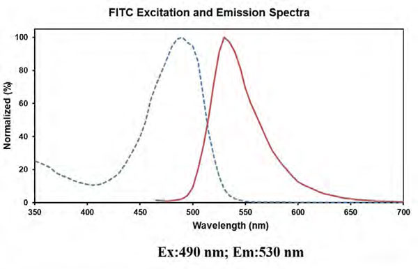 Fluorophore
(Conjugation: FITC)
Fluorophore
(Conjugation: FITC)
CD183, Monoclonal Antibody (Cat# AAA174755)
 FCM/FACS (Flow Cytometry)
(C57BL/6 murine splenocytes are stained with APC Anti-Mouse CD31 Antibody(filled gray histogram). Unstained splenocytes (empty black histogram) are used as control.)
FCM/FACS (Flow Cytometry)
(C57BL/6 murine splenocytes are stained with APC Anti-Mouse CD31 Antibody(filled gray histogram). Unstained splenocytes (empty black histogram) are used as control.)
CD31, Monoclonal Antibody (Cat# AAA174762)
CD31, Monoclonal Antibody (Cat# AAA174763)
H-2, Monoclonal Antibody (Cat# AAA174770)
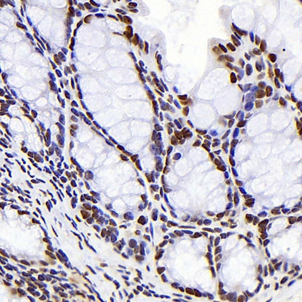
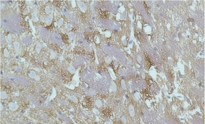
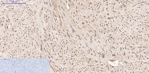
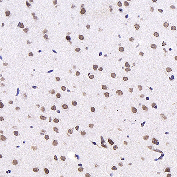
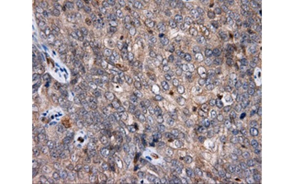
 IHC (Immunohistochemisry)
(Immunohistochemistry analysis of paraffin-embedded Rat brain using S100A4 Monoclonal Antibody at dilution of 1:300.)
IHC (Immunohistochemisry)
(Immunohistochemistry analysis of paraffin-embedded Rat brain using S100A4 Monoclonal Antibody at dilution of 1:300.)
S100A4, Monoclonal Antibody (Cat# AAA174556)

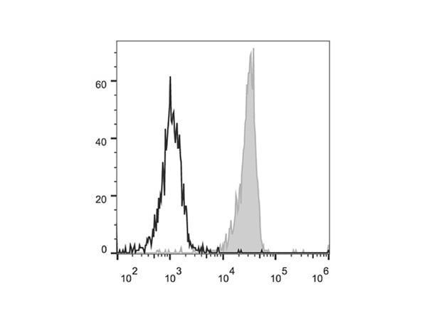
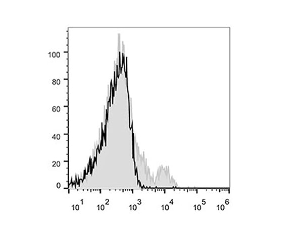 FCM/FACS (Flow Cytometry)
(C57BL/6 murine splenocytes are stained with Anti-Mouse CD122 Monoclonal Antibody(Alexa Fluor 647 Conjuaged)(filled gray histogram) or isotype control(empty black histogram).)
FCM/FACS (Flow Cytometry)
(C57BL/6 murine splenocytes are stained with Anti-Mouse CD122 Monoclonal Antibody(Alexa Fluor 647 Conjuaged)(filled gray histogram) or isotype control(empty black histogram).)
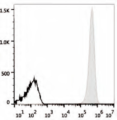
CD122, Monoclonal Antibody (Cat# AAA174615)
 FCM/FACS (Flow Cytometry)
(Human peripheral blood lymphocytes are stained with Anti-Human CD62L Monoclonal Antibody(PerCP/Cy5.5 Conjugated)(filled gray histogram). Unstained lymphocytes (empty black histogram) are used as control.)
FCM/FACS (Flow Cytometry)
(Human peripheral blood lymphocytes are stained with Anti-Human CD62L Monoclonal Antibody(PerCP/Cy5.5 Conjugated)(filled gray histogram). Unstained lymphocytes (empty black histogram) are used as control.)
CD62L, Monoclonal Antibody (Cat# AAA174622)
 FCM/FACS (Flow Cytometry)
(Human peripheral blood lymphocytes are stained with Anti-Human CD45RA Monoclonal Antibody(PerCP/Cy5.5 Conjugated)(filled gray histogram). Unstained lymphocytes (empty black histogram) are used as control.)
FCM/FACS (Flow Cytometry)
(Human peripheral blood lymphocytes are stained with Anti-Human CD45RA Monoclonal Antibody(PerCP/Cy5.5 Conjugated)(filled gray histogram). Unstained lymphocytes (empty black histogram) are used as control.)
CD45RA, Monoclonal Antibody (Cat# AAA174623)
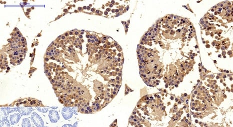
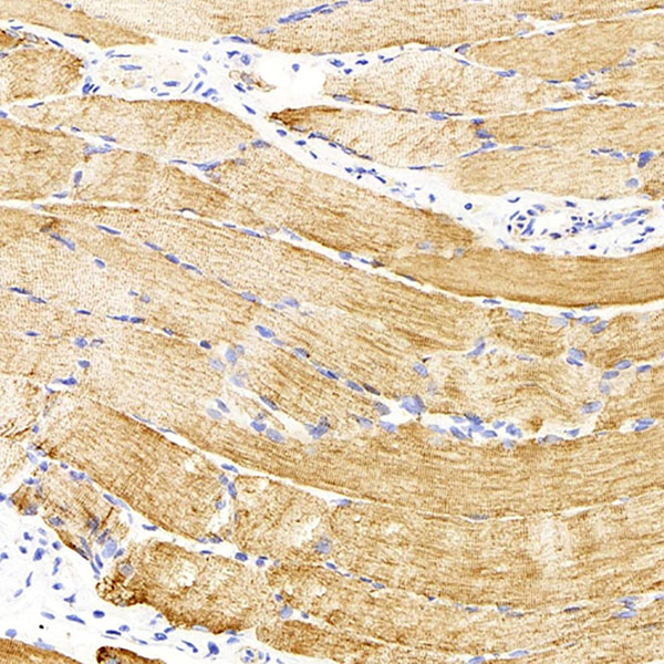
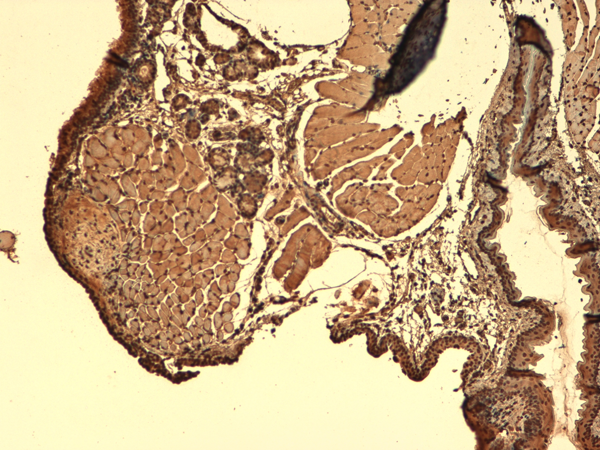
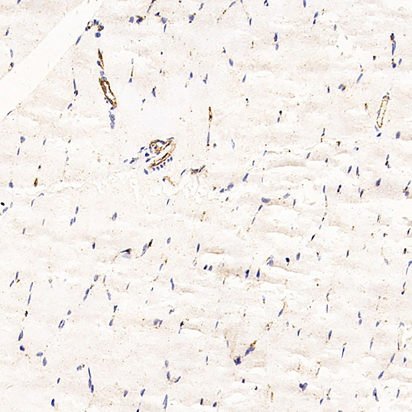 IHC (Immunohistochemistry)
(Immunohistochemistry analysis of paraffin-embedded Rat skeletal muscle using CD31 Monoclonal Antibody at dilution of 1:200.)
IHC (Immunohistochemistry)
(Immunohistochemistry analysis of paraffin-embedded Rat skeletal muscle using CD31 Monoclonal Antibody at dilution of 1:200.)
CD31, Monoclonal Antibody (Cat# AAA174457)
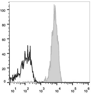
 FCM/FACS (Flow Cytometry)
(Human peripheral blood monocytes are stained with Anti-Human CD33 Monoclonal Antibody(PerCP/Cy5.5 Conjugated)(filled gray histogram). Unstained monocytes (empty black histogram) are used as control.)
FCM/FACS (Flow Cytometry)
(Human peripheral blood monocytes are stained with Anti-Human CD33 Monoclonal Antibody(PerCP/Cy5.5 Conjugated)(filled gray histogram). Unstained monocytes (empty black histogram) are used as control.)
CD33, Monoclonal Antibody (Cat# AAA174632)
 FCM/FACS (Flow Cytometry)
(Human ErythroLeukemia cell line HEL are stained with PE Anti-Human CD41 Antibody (filled gray histogram).Unstained Human ErythroLeukemia cell line HEL (empty black histogram) are used as control.)
FCM/FACS (Flow Cytometry)
(Human ErythroLeukemia cell line HEL are stained with PE Anti-Human CD41 Antibody (filled gray histogram).Unstained Human ErythroLeukemia cell line HEL (empty black histogram) are used as control.)
CD41, Monoclonal Antibody (Cat# AAA174633)
 FCM/FACS (Flow Cytometry)
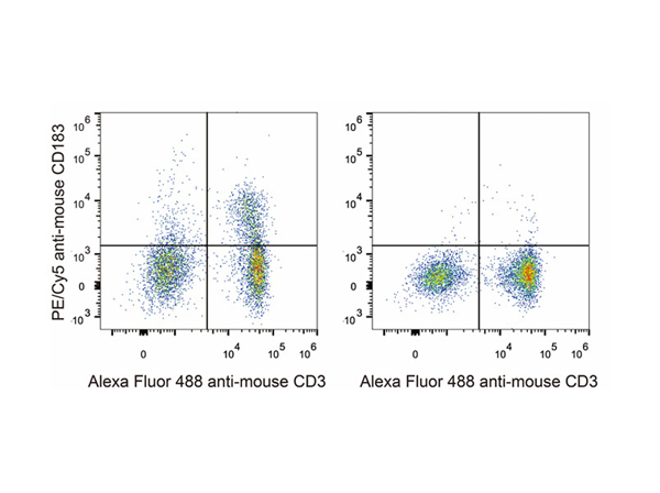
(C57BL/6 murine splenocytes are stained with Anti-Mouse CD183 Monoclonal Antibody(PE/Cy5 Conjugated)(left). Splenocytes stained with Anti-Mouse CD3 Monoclonal Antibody(Alexa Fluor 488 Conjuaged)(right) are used as control.)
FCM/FACS (Flow Cytometry)
(C57BL/6 murine splenocytes are stained with Anti-Mouse CD183 Monoclonal Antibody(PE/Cy5 Conjugated)(left). Splenocytes stained with Anti-Mouse CD3 Monoclonal Antibody(Alexa Fluor 488 Conjuaged)(right) are used as control.)
CD183, Monoclonal Antibody (Cat# AAA174639)
TER-119, Monoclonal Antibody (Cat# AAA174659)

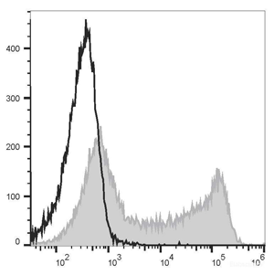
 FCM/FACS (Flow Cytometry)
(Human peripheral blood lymphocytes are stained with Anti-Human HLA-A,B,C Monoclonal Antibody(Alexa Fluor 647 Conjugated)(filled gray histogram). Unstained lymphocytes (empty black histogram) are used as control.)
FCM/FACS (Flow Cytometry)
(Human peripheral blood lymphocytes are stained with Anti-Human HLA-A,B,C Monoclonal Antibody(Alexa Fluor 647 Conjugated)(filled gray histogram). Unstained lymphocytes (empty black histogram) are used as control.)
HLA-A,B,C, Monoclonal Antibody (Cat# AAA174664)
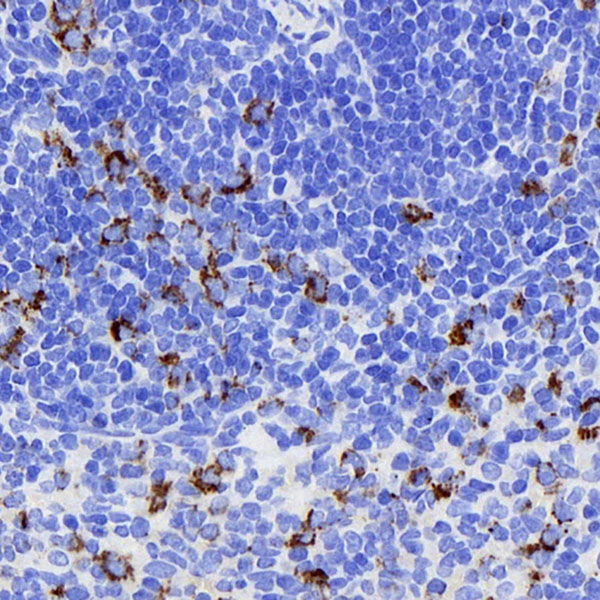
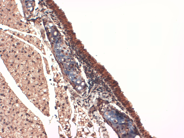
 IHC (Immunohistochemisry)
(Immunohistochemistry analysis of parafffin-embedded rat skeletal muscle using Desmin Monoclonal Antibody at dilution of 1:300.)
IHC (Immunohistochemisry)
(Immunohistochemistry analysis of parafffin-embedded rat skeletal muscle using Desmin Monoclonal Antibody at dilution of 1:300.)
Desmin, Monoclonal Antibody (Cat# AAA174489)
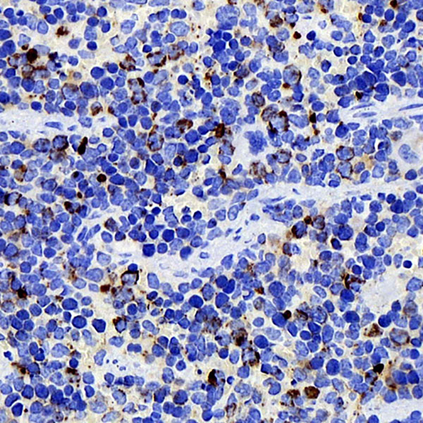
 IHC (Immunohiostchemistry)
(Immunohistochemistry analysis of paraffin-embedded rat spleen using CD284 Monoclonal Antibody at dilution of 1:400.)
IHC (Immunohiostchemistry)
(Immunohistochemistry analysis of paraffin-embedded rat spleen using CD284 Monoclonal Antibody at dilution of 1:400.)
CD284, Monoclonal Antibody (Cat# AAA174490)
 IF (Immunofluorescence)
(Immunofluorescence analysis of paraffin-embedded rat brain using c-Fos Monoclonal Antibody at dilution of 1:400.)
IF (Immunofluorescence)
(Immunofluorescence analysis of paraffin-embedded rat brain using c-Fos Monoclonal Antibody at dilution of 1:400.)
c-Fos, Monoclonal Antibody (Cat# AAA174476)
Desmoplakin 1 + 2, Monoclonal Antibody (Cat# AAA74518)
 Application Data
CD25, Monoclonal Antibody (Cat# AAA74138)
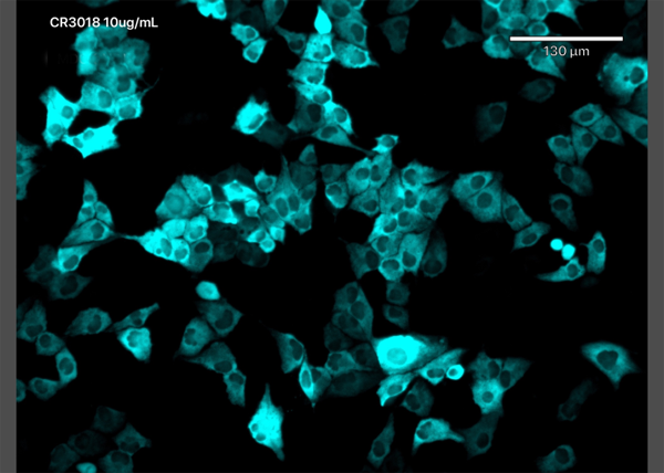 IF (Immunofluorescence)
(Immunofluorescence staining of MDCK-SIAT1 cells transfected with SARS-CoV-2 NP with anti-Covid-19 & SARS-CoV Nucleoprotein antibody CR3018 (03-018) Immunofluorescence analysis of MDCK-SIAT1 cells stably transfected with SARS-CoV-2 NP. The cells were seeded in a flat bottomed 96 well plate overnight, fixed in 10% formalin at 4C for 30min, permeabilised for 20min at RT and then stained with the human IgG1 version of CR3018 (03-018) in PBS/0.1% BSA at 10ug/ml for 1 hour followed by a goat anti-human Alexa Fluor 647 (Invitrogen) secondary antibody. The image is courtesy of Jack Tan, Radcliffe Department of Medicine, University of Oxford.)
IF (Immunofluorescence)
(Immunofluorescence staining of MDCK-SIAT1 cells transfected with SARS-CoV-2 NP with anti-Covid-19 & SARS-CoV Nucleoprotein antibody CR3018 (03-018) Immunofluorescence analysis of MDCK-SIAT1 cells stably transfected with SARS-CoV-2 NP. The cells were seeded in a flat bottomed 96 well plate overnight, fixed in 10% formalin at 4C for 30min, permeabilised for 20min at RT and then stained with the human IgG1 version of CR3018 (03-018) in PBS/0.1% BSA at 10ug/ml for 1 hour followed by a goat anti-human Alexa Fluor 647 (Invitrogen) secondary antibody. The image is courtesy of Jack Tan, Radcliffe Department of Medicine, University of Oxford.)
COVID 19 Nucleocapsid (NP) Coronavirus, Monoclonal Antibody (Cat# AAA72197)
 FCM/FACS (Flow Cytometry)
(Flowcytrometryusinganti-IL-18antibodyABT-325(AAA72552). Humanbloodleucocyteswererestedfor4hat37°C,fixedwith2%PFA,permeabilizedwith0.5%Triton,andstainedwiththeanti-unknownspecificityantibodyortherabbitIgGversionofABT-325(AAA72552,left)atadilutionof1:100overnightat4°C.Afterwashing,theboundantibodywasdetectedusingagoatanti-rabbitIgGAlexaFluor488antibodyatadilutionof1:1000,andthecellswereanalyzedusingaFACSCantoflow-cytometer.)
FCM/FACS (Flow Cytometry)
(Flowcytrometryusinganti-IL-18antibodyABT-325(AAA72552). Humanbloodleucocyteswererestedfor4hat37°C,fixedwith2%PFA,permeabilizedwith0.5%Triton,andstainedwiththeanti-unknownspecificityantibodyortherabbitIgGversionofABT-325(AAA72552,left)atadilutionof1:100overnightat4°C.Afterwashing,theboundantibodywasdetectedusingagoatanti-rabbitIgGAlexaFluor488antibodyatadilutionof1:1000,andthecellswereanalyzedusingaFACSCantoflow-cytometer.)
IL-18, Monoclonal Recombinant Antibody (Cat# AAA72552)

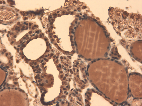
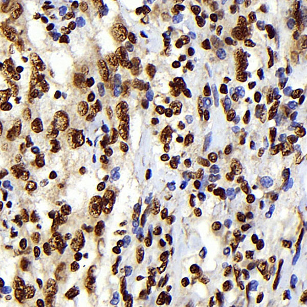
 IHC (Immunohistochemistry)
(Immunohistochemistry analysis using Mouse Anti-Sodium Iodide Symporter Monoclonal Antibody, Clone 14F. Tissue: Thyroid. Species: Mouse. Fixation: 10% Formalin Solution for 12-24 hours at RT. Primary Antibody: Mouse Anti-Sodium Iodide Symporter Monoclonal Antibody at 1:1000 for 1 hour at RT. Secondary Antibody: HRP/DAB Detection System: Biotinylated Goat Anti-Mouse, Streptavidin Peroxidase, DAB Chromogen (brown) for 30 minutes at RT. Counterstain: Mayer Hematoxylin (purple/blue) nuclear stain at 250-500 ul for 5 minutes at RT.)
IHC (Immunohistochemistry)
(Immunohistochemistry analysis using Mouse Anti-Sodium Iodide Symporter Monoclonal Antibody, Clone 14F. Tissue: Thyroid. Species: Mouse. Fixation: 10% Formalin Solution for 12-24 hours at RT. Primary Antibody: Mouse Anti-Sodium Iodide Symporter Monoclonal Antibody at 1:1000 for 1 hour at RT. Secondary Antibody: HRP/DAB Detection System: Biotinylated Goat Anti-Mouse, Streptavidin Peroxidase, DAB Chromogen (brown) for 30 minutes at RT. Counterstain: Mayer Hematoxylin (purple/blue) nuclear stain at 250-500 ul for 5 minutes at RT.)
Sodium-Iodide Symporter, Monoclonal Antibody (Cat# AAA103013)
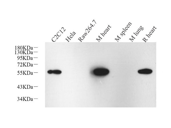
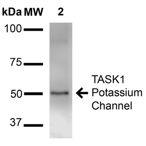 WB (Western Blot)
(Western Blot analysis of Rat Brain Membrane showing detection of ~50 kDa TASK1 Potassium Channel protein using Mouse Anti-TASK1 Potassium Channel Monoclonal Antibody, Clone S374-48 . Lane 1: Molecular Weight Ladder (MW). Lane 2: Rat brain membrane. Load: 15 ug. Block: 2% BSA and 2% Skim Milk in 1X TBST. Primary Antibody: Mouse Anti-TASK1 Potassium Channel Monoclonal Antibody at 1:1000 for 16 hours at 4 degree C. Secondary Antibody: Goat Anti-Mouse IgG: HRP at 1:2000 for 60 min at RT. Color Development: ECL solution for 6 min at RT. Predicted/Observed Size: ~50 kDa.)
WB (Western Blot)
(Western Blot analysis of Rat Brain Membrane showing detection of ~50 kDa TASK1 Potassium Channel protein using Mouse Anti-TASK1 Potassium Channel Monoclonal Antibody, Clone S374-48 . Lane 1: Molecular Weight Ladder (MW). Lane 2: Rat brain membrane. Load: 15 ug. Block: 2% BSA and 2% Skim Milk in 1X TBST. Primary Antibody: Mouse Anti-TASK1 Potassium Channel Monoclonal Antibody at 1:1000 for 16 hours at 4 degree C. Secondary Antibody: Goat Anti-Mouse IgG: HRP at 1:2000 for 60 min at RT. Color Development: ECL solution for 6 min at RT. Predicted/Observed Size: ~50 kDa.)
TASK1 Potassium Channel, Monoclonal Antibody (Cat# AAA103015)
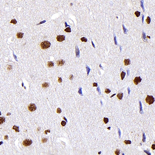
 IHC (Immunohistochemistry)
(Immunohistochemistry analysis using Mouse Anti-CaV1.2 Calcium channel Monoclonal Antibody, Clone S57-47. Tissue: Brain Tissue. Species: Mouse. Fixation: Formalin. Primary Antibody: Mouse Anti-CaV1.2 Calcium channel Monoclonal Antibody at 1:10000 for 12 hours at 4 degree C. Secondary Antibody: Biotin Goat Anti-Mouse at 1:2000 for 1 hour at RT. Counterstain: Mayer Hematoxylin (purple/blue) nuclear stain at 200 ul for 2 minutes at RT. Magnification: 40x.)
IHC (Immunohistochemistry)
(Immunohistochemistry analysis using Mouse Anti-CaV1.2 Calcium channel Monoclonal Antibody, Clone S57-47. Tissue: Brain Tissue. Species: Mouse. Fixation: Formalin. Primary Antibody: Mouse Anti-CaV1.2 Calcium channel Monoclonal Antibody at 1:10000 for 12 hours at 4 degree C. Secondary Antibody: Biotin Goat Anti-Mouse at 1:2000 for 1 hour at RT. Counterstain: Mayer Hematoxylin (purple/blue) nuclear stain at 200 ul for 2 minutes at RT. Magnification: 40x.)
Cav1.2, Monoclonal Antibody (Cat# AAA103035)
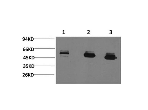
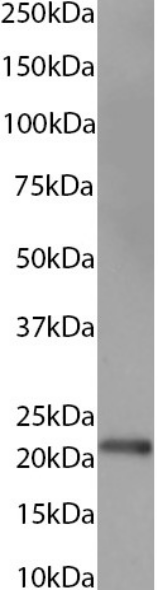
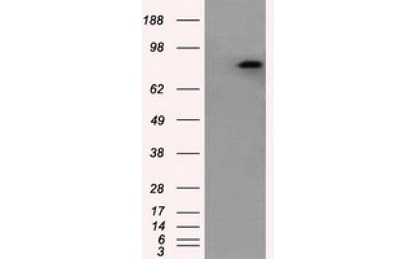
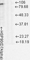 WB (Western Blot)
(Western Blot analysis of Rat cell lysates showing detection of Hsp90 protein using Mouse Anti-Hsp90 Monoclonal Antibody, Clone D7Alpha. Load: 15 ug. Block: 1.5% BSA for 30 minutes at RT. Primary Antibody: Mouse Anti-Hsp90 Monoclonal Antibody at 1:1000 for 2 hours at RT. Secondary Antibody: Sheep Anti-Mouse IgG: HRP for 1 hour at RT.)
WB (Western Blot)
(Western Blot analysis of Rat cell lysates showing detection of Hsp90 protein using Mouse Anti-Hsp90 Monoclonal Antibody, Clone D7Alpha. Load: 15 ug. Block: 1.5% BSA for 30 minutes at RT. Primary Antibody: Mouse Anti-Hsp90 Monoclonal Antibody at 1:1000 for 2 hours at RT. Secondary Antibody: Sheep Anti-Mouse IgG: HRP for 1 hour at RT.)
Hsp90, Monoclonal Antibody (Cat# AAA103037)
 WB (Western Blot)
(Western Blot analysis of Human Cell lysates showing detection of Hsp70 protein using Mouse Anti-Hsp70 Monoclonal Antibody, Clone N27. Load: 15 ug. Block: 1.5% BSA for 30 minutes at RT. Primary Antibody: Mouse Anti-Hsp70 Monoclonal Antibody at 1:1000 for 2 hours at RT. Secondary Antibody: Sheep Anti-Mouse IgG: HRP for 1 hour at RT.)
WB (Western Blot)
(Western Blot analysis of Human Cell lysates showing detection of Hsp70 protein using Mouse Anti-Hsp70 Monoclonal Antibody, Clone N27. Load: 15 ug. Block: 1.5% BSA for 30 minutes at RT. Primary Antibody: Mouse Anti-Hsp70 Monoclonal Antibody at 1:1000 for 2 hours at RT. Secondary Antibody: Sheep Anti-Mouse IgG: HRP for 1 hour at RT.)
HSP70/HSC70, Monoclonal Antibody (Cat# AAA103057)
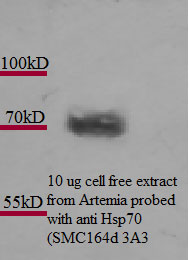
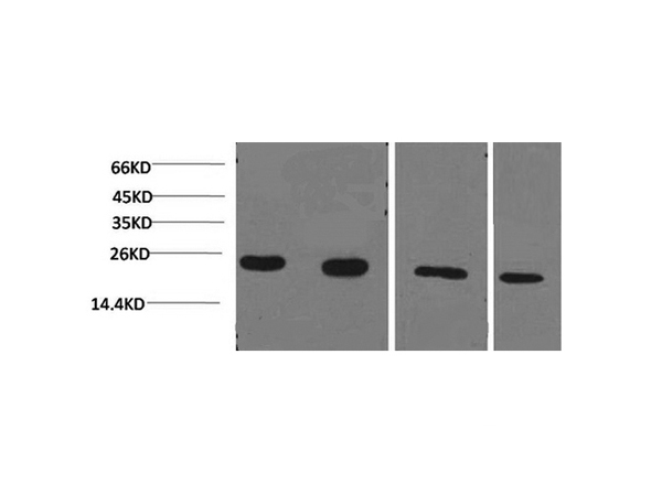
 WB (Western Blot)
(Western Blot analysis of Rat cell lysates showing detection of Hsp70 protein using Mouse Anti-Hsp70 Monoclonal Antibody, Clone 3A3. Load: 15 ug. Block: 1.5% BSA for 30 minutes at RT. Primary Antibody: Mouse Anti-Hsp70 Monoclonal Antibody at 1:1000 for 2 hours at RT. Secondary Antibody: Sheep Anti-Mouse IgG: HRP for 1 hour at RT.)
WB (Western Blot)
(Western Blot analysis of Rat cell lysates showing detection of Hsp70 protein using Mouse Anti-Hsp70 Monoclonal Antibody, Clone 3A3. Load: 15 ug. Block: 1.5% BSA for 30 minutes at RT. Primary Antibody: Mouse Anti-Hsp70 Monoclonal Antibody at 1:1000 for 2 hours at RT. Secondary Antibody: Sheep Anti-Mouse IgG: HRP for 1 hour at RT.)
Hsp70, Monoclonal Antibody (Cat# AAA103081)
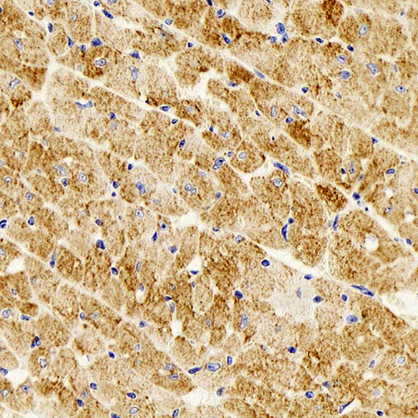
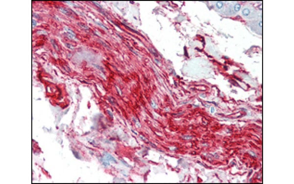
 IHC (Immunohiostchemistry)
(Immunohistochemical analysis of paraffin-embedded human skeletal muscle tissues using CRYAB antibody.)
IHC (Immunohiostchemistry)
(Immunohistochemical analysis of paraffin-embedded human skeletal muscle tissues using CRYAB antibody.)
CRYAB, Monoclonal Antibody (Cat# AAA106286)
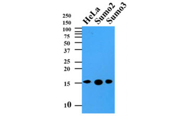
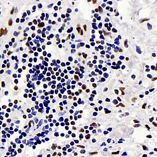
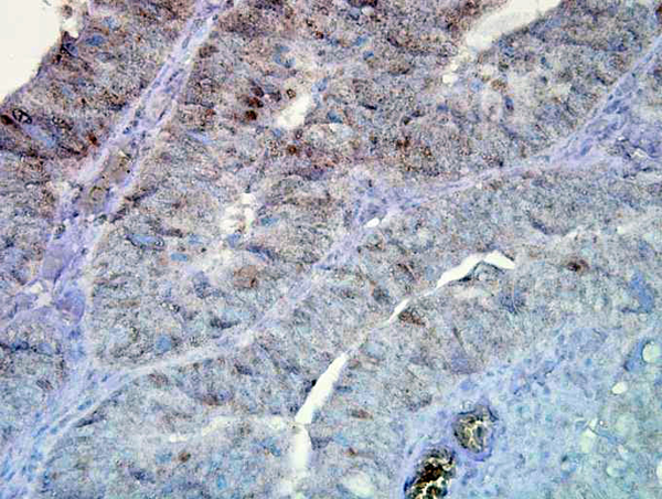
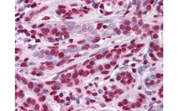
 IHC (Immunohiostchemistry)
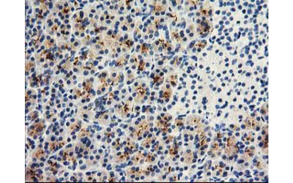
(Paraffin embedded sections of colorectal canitrocelluloseer tissue were initrocelluloseubated with anti-human SUMO2/3 antibody (1:50) for 2 hours at room temperature. Antigen retrieval was performed in 0.1M sodium citrate buffer and detected using Diaminobenzidine (DAB).)
IHC (Immunohiostchemistry)
(Paraffin embedded sections of colorectal canitrocelluloseer tissue were initrocelluloseubated with anti-human SUMO2/3 antibody (1:50) for 2 hours at room temperature. Antigen retrieval was performed in 0.1M sodium citrate buffer and detected using Diaminobenzidine (DAB).)
SUMO2, Monoclonal Antibody (Cat# AAA106295)
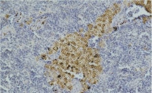
 IHC (Immunohiostchemistry)
(Immunohistochemical analysis of paraffin-embedded human Breast tissues using anti-ZBTB7B antibody)
IHC (Immunohiostchemistry)
(Immunohistochemical analysis of paraffin-embedded human Breast tissues using anti-ZBTB7B antibody)
ZBTB7B, Monoclonal Antibody (Cat# AAA106302)
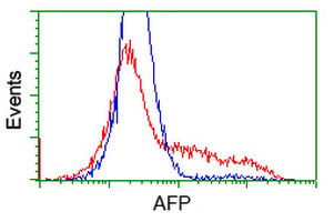
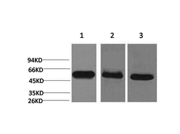
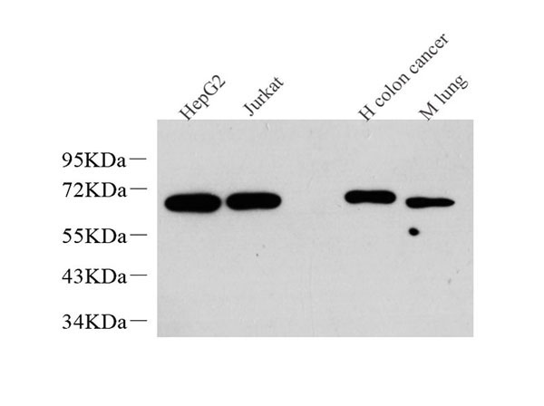
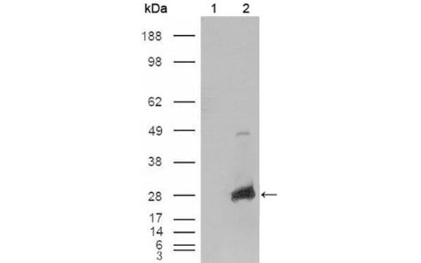
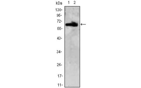
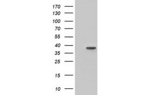
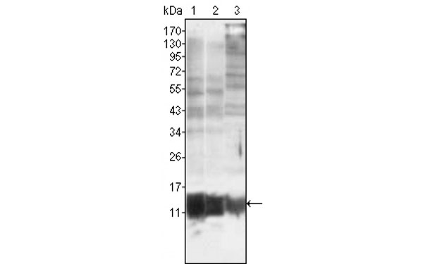
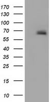 WB (Western Blot)
(Western Blot analysis using AFP antibodyWestern Blot analysis of HEK293T cell lysates (5 ug) transfected with either recombinant AFP protein (Right) or empty vector (Left) detected with AFP antibody)
WB (Western Blot)
(Western Blot analysis using AFP antibodyWestern Blot analysis of HEK293T cell lysates (5 ug) transfected with either recombinant AFP protein (Right) or empty vector (Left) detected with AFP antibody)
AFP, Monoclonal Antibody (Cat# AAA106308)
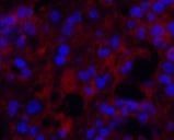
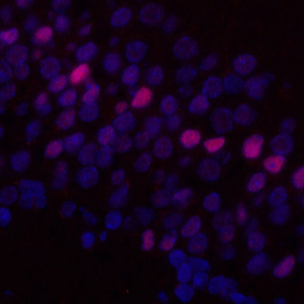
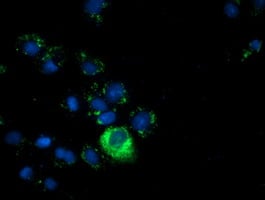
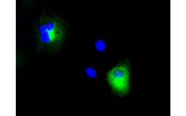
 IF (Immunofluorescence)
(Immunofluorescent staining of COS7 cells transiently transfected with recombinant BTK protein using BTK antibody)
IF (Immunofluorescence)
(Immunofluorescent staining of COS7 cells transiently transfected with recombinant BTK protein using BTK antibody)
BTK, Monoclonal Antibody (Cat# AAA106313)
Rab11B, Monoclonal Antibody (Cat# AAA106331)
 IF (Immunofluorescence)
(Immunofluorescent staining of COS7 cells transiently transfected with recombinant HIBCH protein using HIBCH antibody)
IF (Immunofluorescence)
(Immunofluorescent staining of COS7 cells transiently transfected with recombinant HIBCH protein using HIBCH antibody)
HIBCH, Monoclonal Antibody (Cat# AAA106338)

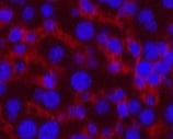
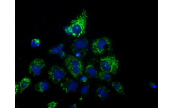
 IF (Immunofluorescence)
(Immunofluorescent staining of COS7 cells transiently transfected with recombinant BHMT protein using BHMT antibody)
IF (Immunofluorescence)
(Immunofluorescent staining of COS7 cells transiently transfected with recombinant BHMT protein using BHMT antibody)
BHMT, Monoclonal Antibody (Cat# AAA106201)
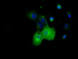
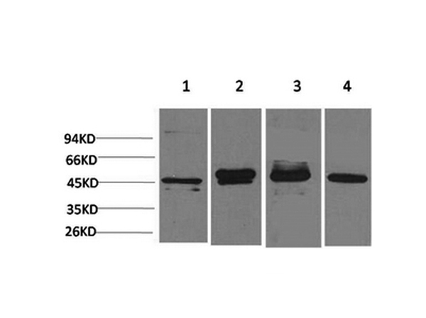
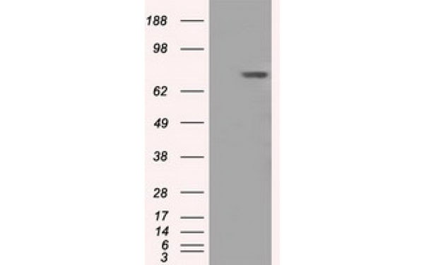
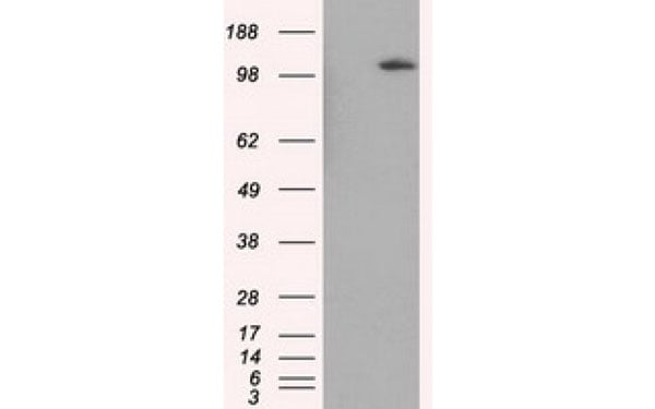
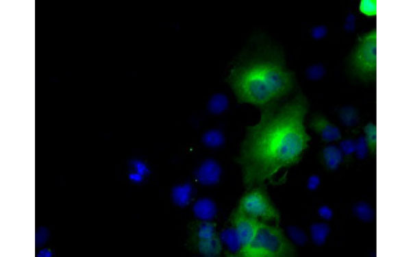
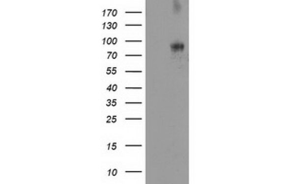
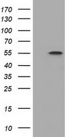 WB (Western Blot)
(Western Blot analysis using DYNC1LI1 antibodyWestern Blot analysis of HEK293T cell lysates (5 ug) transfected with either recombinant DYNC1LI1 protein (Right) or empty vector (Left) detected with DYNC1LI1 antibody)
WB (Western Blot)
(Western Blot analysis using DYNC1LI1 antibodyWestern Blot analysis of HEK293T cell lysates (5 ug) transfected with either recombinant DYNC1LI1 protein (Right) or empty vector (Left) detected with DYNC1LI1 antibody)
DYNC1LI1, Monoclonal Antibody (Cat# AAA106206)
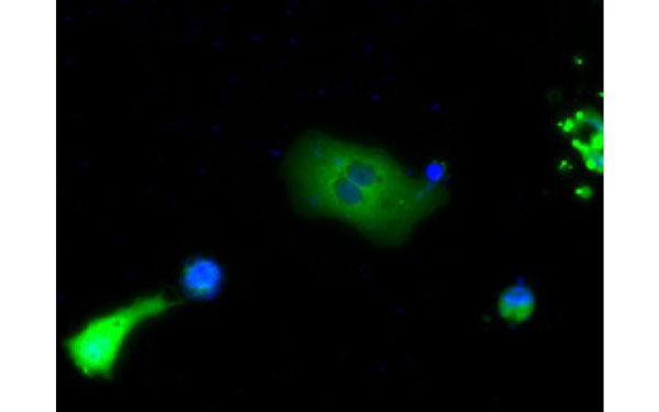
 IF (Immunofluorescence)
(Immunofluorescent staining of COS7 cells transiently transfected with recombinant APP protein using APP antibody)
IF (Immunofluorescence)
(Immunofluorescent staining of COS7 cells transiently transfected with recombinant APP protein using APP antibody)
APP, Monoclonal Antibody (Cat# AAA106367)
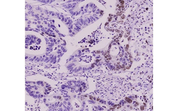
 IHC (Immunohiostchemistry)
(Immunohistochemical analysis of DPP10 protein in paraffin embedded Human pancreas tissue using DPP10 antibody)
IHC (Immunohiostchemistry)
(Immunohistochemical analysis of DPP10 protein in paraffin embedded Human pancreas tissue using DPP10 antibody)
DPP10, Monoclonal Antibody (Cat# AAA106380)
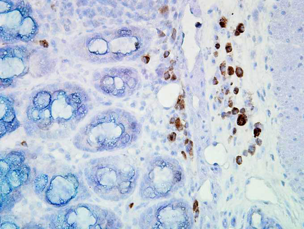
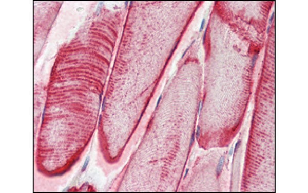
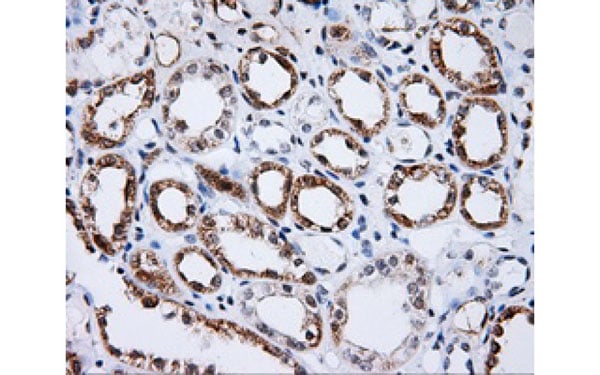
 IHC (Immunohiostchemistry)
(Immunohistochemical analysis of paraffin-embedded human nerve and ganglion cells using S100A10/P11 antibody.)
IHC (Immunohiostchemistry)
(Immunohistochemical analysis of paraffin-embedded human nerve and ganglion cells using S100A10/P11 antibody.)
p11, Monoclonal Antibody (Cat# AAA106381)
What are Monoclonal Antibodies?
Monoclonal antibodies are specialized laboratory-produced proteins developed for binding to specific biological antigens or other molecular targets. Since they come from a single cell (or clone), they are especially consistent and accurate in the data they are involved in producing.
This type of antibody material has been shown to be a powerful tool in finding and subsequently destroying harmful cells in an organism, such as those found in cancers or various autoimmune diseases. This makes them excellent aids in medical testing and research, which is why they are so widely used.
AAA Biotech offers a comprehensive range of high-quality monoclonal antibodies that perform effectively in various laboratory tests, including (amongst others) ELISA, western blotting, immunohistochemistry, and flow cytometry. All of the products in our catalog are thoroughly quality tested to make sure that they are reliable and will consistently perform well in your research.
What Are The Uses of Monoclonal Antibodies
Monoclonal antibodies are used in many lab tests, including (amongst others) ELISA, western blotting, immunohistochemistry, and flow cytometry.
ELISA is a test that helps detect a specific substance/analyte in a sample. It uses antibodies (often monoclonal) bound to a solid surface (such as the well of a microplate) to “capture” the substance/analyte in the sample and immobilize it so that the detection antibody component can then bind to it and produce a signal, which can then be measured.
Western blotting identifies specific proteins in a sample. The sample is first separated on a gel, and then antibodies are applied that will typically bind to the target, which will all be localized to a single band in a lane.
Immunohistochemistry helps locate specific proteins in cells or tissue samples using antibodies.
Flow cytometry looks at and sorts cells. It uses antibodies that are conjugated to reporter molecules called “fluorophores”, which, under special lights, emit light themselves, which can then be measured by a detector instrument. For a deeper understanding of these techniques, explore our complete guide to monoclonal antibodies and their benefits.
How Monoclonal Antibodies Are Used as Medicine?
Please note that all of the products listed in AAA Biotech’s also known as AAA Bio or AAABio catalog are strictly for research-use only (RUO).
Monoclonal antibodies can also be used as therapeutic/medical treatments, particularly in the context of cancers. They are designed to find and bind to specific cells or proteins, helping the immune system recognize and attack the cancer. These treatments work in different ways, such as:
- Radioimmunotherapy attaches a small amount of radioactive molecule to the antibody, so it delivers the radiation directly to the cancer cells that the antibody is specifically binding to.
- Antibody-directed enzyme prodrug therapy uses antibodies that are specifically bound to special enzymes. These enzymes activate a harmless drug in the body and turn it into a cancer-killing drug only near the cancer cells—this helps avoid harming healthy cells.
- Immunoliposomes are tiny “bubbles” filled with medicine/drug and coated with antibodies. They carry the drug straight to the cancer cells.
Why Buy Monoclonal Antibodies From Us?
At AAA Biotech, we provide high-performance monoclonal antibodies designed to support a wide range of research needs.
1. Validated for Versatile Applications
The antibodies in our catalog are extensively validated and compatible with multiple techniques, including (but not limited to) ELISA, flow cytometry (FC), immunocytochemistry (ICC), immunofluorescence (IF), immunohistochemistry (IHC), immunoprecipitation (IP), and western blotting (WB).
2. Wide Selection & Specialized Options
We offer antibodies for common and rare species, that are available in various conjugated forms, and also in recombinant formats. Essentially, there is almost anything one might need to meet their experimental model’s requirements.
3. High-Quality Proteins
Our proteins meet high purity standards—90% or more as confirmed by SDS-PAGE. Many are available with tags like His, Flag, GST, or MBP, and we also supply native and biologically active proteins for functional studies.
Frequently Asked Questions
1. Are your monoclonal antibodies validated for specific applications?
Yes, our antibodies are tested and validated for use in methods such as ELISA, western blot, IHC, flow cytometry, and more. Refer to specific product pages or datasheets for individual product information.
2. How do I choose the right monoclonal antibody for my application?
Review the product details directly for application validation, species reactivity, and target information. You may also contact our support team at any time for help.
3. How quickly can I receive my order?
Most orders are processed and shipped within 1–3 business days, depending on product availability and your shipping location.