Filters
▼Clonality
▼Type
▼Reactivity
▼Gene Name
▼Isotype
▼Host
▼Application
▼Clone
▼Monoclonal Antibodies
Get accurate results in your research with our Monoclonal Antibodies, which are specially made to target exactly what you require for your research, and will produce consistent, reliable performance in lab tests.
Viewing 3350-3400 of 27645 product results
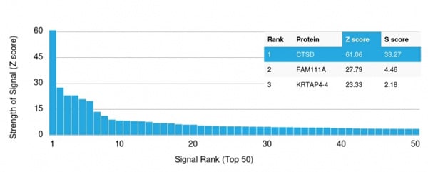 Application Data
(Analysis of Protein Array containing more than 19,000 full-length human proteins using Cathepsin D Mouse Monoclonal Antibody (CTSD/3083) Z- and S- Score: The Z-score represents the strength of a signal that a monoclonal antibody (MAb) (in combination with a fluorescently-tagged anti-IgG secondary antibody) produces when binding to a particular protein on the HuProtTM array. Z-scores are described in units of standard deviations (SD's) above the mean value of all signals generated on that array. If targets on HuProtTM are arranged in descending order of the Z-score, the S-score is the difference (also in units of SD's) between the Z-score. S-score therefore represents the relative target specificity of a MAb to its intended target. A MAb is considered to specific to its intended target, if the MAb has an S-score of at least 2.5. For example, if a MAb binds to protein X with a Z-score of 43 and to protein Y with a Z-score of 14, then the S-score for the binding of that MAb to protein X is equal to 29.)
Application Data
(Analysis of Protein Array containing more than 19,000 full-length human proteins using Cathepsin D Mouse Monoclonal Antibody (CTSD/3083) Z- and S- Score: The Z-score represents the strength of a signal that a monoclonal antibody (MAb) (in combination with a fluorescently-tagged anti-IgG secondary antibody) produces when binding to a particular protein on the HuProtTM array. Z-scores are described in units of standard deviations (SD's) above the mean value of all signals generated on that array. If targets on HuProtTM are arranged in descending order of the Z-score, the S-score is the difference (also in units of SD's) between the Z-score. S-score therefore represents the relative target specificity of a MAb to its intended target. A MAb is considered to specific to its intended target, if the MAb has an S-score of at least 2.5. For example, if a MAb binds to protein X with a Z-score of 43 and to protein Y with a Z-score of 14, then the S-score for the binding of that MAb to protein X is equal to 29.)
Cathepsin D, Monoclonal Antibody (Cat# AAA214911)
 Application Data
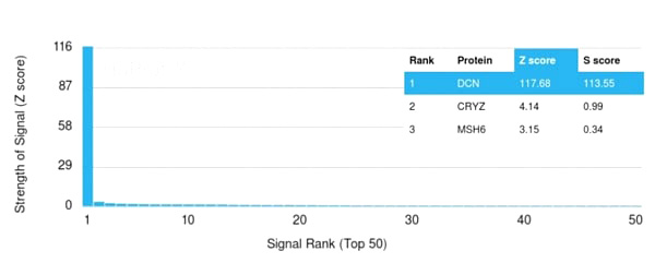
(Analysis of Protein Array containing more than 19,000 full-length human proteins using Monospecific Mouse Monoclonal Antibody to Decorin (DCN/3523). Z- and S- Score: The Z-score represents the strength of a signal that a monoclonal antibody (Monoclonal Antibody) (in combination with a fluorescently-tagged anti-IgG secondary antibody) produces when binding to a particular protein on the HuProtTM array. Z-scores are described in units of standard deviations (SD’s) above the mean value of all signals generated on that array. If targets on HuProtTM are arranged in descending order of the Z-score, the S-score is the difference (also in units of SD’s) between the Z-score. S-score therefore represents the relative target specificity of a Monoclonal Antibody to its intended target. A Monoclonal Antibody is considered to specific to its intended target, if the Monoclonal Antibody has an S-score of at least 2.5. For example, if a Monoclonal Antibody binds to protein X with a Z-score of 43 and to protein Y with a Z-score of 14, then the S-score for the binding of that Monoclonal Antibody to protein X is equal to 29.)
Application Data
(Analysis of Protein Array containing more than 19,000 full-length human proteins using Monospecific Mouse Monoclonal Antibody to Decorin (DCN/3523). Z- and S- Score: The Z-score represents the strength of a signal that a monoclonal antibody (Monoclonal Antibody) (in combination with a fluorescently-tagged anti-IgG secondary antibody) produces when binding to a particular protein on the HuProtTM array. Z-scores are described in units of standard deviations (SD’s) above the mean value of all signals generated on that array. If targets on HuProtTM are arranged in descending order of the Z-score, the S-score is the difference (also in units of SD’s) between the Z-score. S-score therefore represents the relative target specificity of a Monoclonal Antibody to its intended target. A Monoclonal Antibody is considered to specific to its intended target, if the Monoclonal Antibody has an S-score of at least 2.5. For example, if a Monoclonal Antibody binds to protein X with a Z-score of 43 and to protein Y with a Z-score of 14, then the S-score for the binding of that Monoclonal Antibody to protein X is equal to 29.)
Decorin, Monoclonal Antibody (Cat# AAA214919)

 Application Data
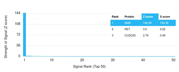
(Analysis of Protein Array containing more than 19,000 full-length human proteins using Dystrophin Monospecific Mouse Monoclonal Antibody (DMD/3241). Z- and S- Score: The Z-score represents the strength of a signal that a monoclonal antibody (Monoclonal Antibody) (in combination with a fluorescently-tagged anti-IgG secondary antibody) produces when binding to a particular protein on the HuProtTM array. Z-scores are described in units of standard deviations (SD’s) above the mean value of all signals generated on that array. If targets on HuProtTM are arranged in descending order of the Z-score, the S-score is the difference (also in units of SD’s) between the Z-score. S-score therefore represents the relative target specificity of a Monoclonal Antibody to its intended target. A Monoclonal Antibody is considered to specific to its intended target, if the Monoclonal Antibody has an S-score of at least 2.5. For example, if a Monoclonal Antibody binds to protein X with a Z-score of 43 and to protein Y with a Z-score of 14, then the S-score for the binding of that Monoclonal Antibody to protein X is equal to 29.)
Application Data
(Analysis of Protein Array containing more than 19,000 full-length human proteins using Dystrophin Monospecific Mouse Monoclonal Antibody (DMD/3241). Z- and S- Score: The Z-score represents the strength of a signal that a monoclonal antibody (Monoclonal Antibody) (in combination with a fluorescently-tagged anti-IgG secondary antibody) produces when binding to a particular protein on the HuProtTM array. Z-scores are described in units of standard deviations (SD’s) above the mean value of all signals generated on that array. If targets on HuProtTM are arranged in descending order of the Z-score, the S-score is the difference (also in units of SD’s) between the Z-score. S-score therefore represents the relative target specificity of a Monoclonal Antibody to its intended target. A Monoclonal Antibody is considered to specific to its intended target, if the Monoclonal Antibody has an S-score of at least 2.5. For example, if a Monoclonal Antibody binds to protein X with a Z-score of 43 and to protein Y with a Z-score of 14, then the S-score for the binding of that Monoclonal Antibody to protein X is equal to 29.)
Dystrophin (DMD), Monoclonal Antibody (Cat# AAA214922)
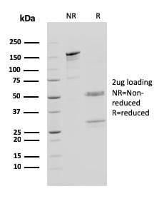
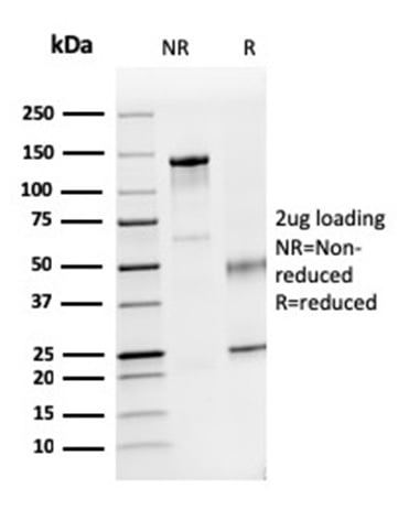
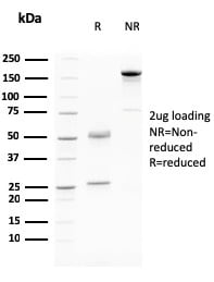
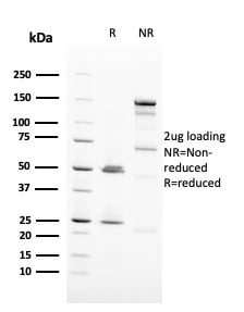
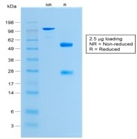
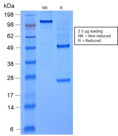
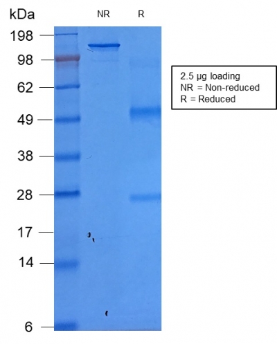
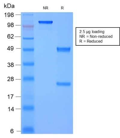
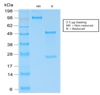
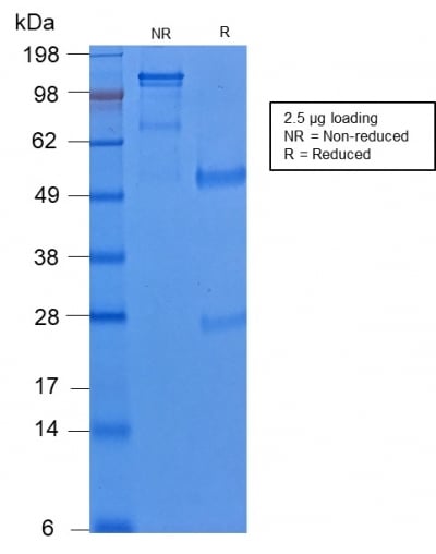
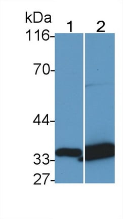
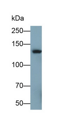
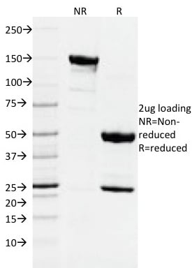 SDS-PAGE
(SDS-PAGE Analysis Purified DNMT1 Mouse Monoclonal Antibody (DNMT1/2061). Confirmation of Integrity and Purity of Antibody.)
SDS-PAGE
(SDS-PAGE Analysis Purified DNMT1 Mouse Monoclonal Antibody (DNMT1/2061). Confirmation of Integrity and Purity of Antibody.)
DNMT1/DNA Methyltransferase 1, Monoclonal Antibody (Cat# AAA214927)
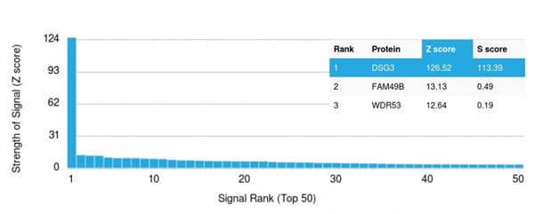 Application Data
(Analysis of Protein Array containing more than 19,000 full-length human proteins using Desmoglein-3 Mouse Monoclonal Antibody (DSG3/2796) Z- and S- Score: The Z-score represents the strength of a signal that a monoclonal antibody (Monoclonal Antibody) (in combination with a fluorescently-tagged anti-IgG secondary antibody) produces when binding to a particular protein on the HuProtTM array. Z-scores are described in units of standard deviations (SD's) above the mean value of all signals generated on that array. If targets on HuProtTM are arranged in descending order of the Z-score, the S-score is the difference (also in units of SD's) between the Z-score. S-score therefore represents the relative target specificity of a Monoclonal Antibody to its intended target. A Monoclonal Antibody is considered to specific to its intended target, if the Monoclonal Antibody has an S-score of at least 2.5. For example, if a Monoclonal Antibody binds to protein X with a Z-score of 43 and to protein Y with a Z-score of 14, then the S-score for the binding of that Monoclonal Antibody to protein X is equal to 29.)
Application Data
(Analysis of Protein Array containing more than 19,000 full-length human proteins using Desmoglein-3 Mouse Monoclonal Antibody (DSG3/2796) Z- and S- Score: The Z-score represents the strength of a signal that a monoclonal antibody (Monoclonal Antibody) (in combination with a fluorescently-tagged anti-IgG secondary antibody) produces when binding to a particular protein on the HuProtTM array. Z-scores are described in units of standard deviations (SD's) above the mean value of all signals generated on that array. If targets on HuProtTM are arranged in descending order of the Z-score, the S-score is the difference (also in units of SD's) between the Z-score. S-score therefore represents the relative target specificity of a Monoclonal Antibody to its intended target. A Monoclonal Antibody is considered to specific to its intended target, if the Monoclonal Antibody has an S-score of at least 2.5. For example, if a Monoclonal Antibody binds to protein X with a Z-score of 43 and to protein Y with a Z-score of 14, then the S-score for the binding of that Monoclonal Antibody to protein X is equal to 29.)
Desmoglein-3, Monoclonal Antibody (Cat# AAA214932)
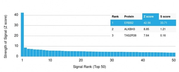 Application Data
(Analysis of Protein Array containing more than 19,000 full-length human proteins using HER-2 Mouse Monoclonal Antibody (ERBB2/3078). Z- and S- Score: The Z-score represents the strength of a signal that a monoclonal antibody (MAb) (in combination with a fluorescently-tagged anti-IgG secondary antibody) produces when binding to a particular protein on the HuProtTM array. Z-scores are described in units of standard deviations (SD's) above the mean value of all signals generated on that array. If targets on HuProtTM are arranged in descending order of the Z-score, the S-score is the difference (also in units of SD's) between the Z-score. S-score therefore represents the relative target specificity of a MAb to its intended target. A MAb is considered to specific to its intended target, if the MAb has an S-score of at least 2.5. For example, if a MAb binds to protein X with a Z-score of 43 and to protein Y with a Z-score of 14, then the S-score for the binding of that MAb to protein X is equal to 29.)
Application Data
(Analysis of Protein Array containing more than 19,000 full-length human proteins using HER-2 Mouse Monoclonal Antibody (ERBB2/3078). Z- and S- Score: The Z-score represents the strength of a signal that a monoclonal antibody (MAb) (in combination with a fluorescently-tagged anti-IgG secondary antibody) produces when binding to a particular protein on the HuProtTM array. Z-scores are described in units of standard deviations (SD's) above the mean value of all signals generated on that array. If targets on HuProtTM are arranged in descending order of the Z-score, the S-score is the difference (also in units of SD's) between the Z-score. S-score therefore represents the relative target specificity of a MAb to its intended target. A MAb is considered to specific to its intended target, if the MAb has an S-score of at least 2.5. For example, if a MAb binds to protein X with a Z-score of 43 and to protein Y with a Z-score of 14, then the S-score for the binding of that MAb to protein X is equal to 29.)
HER-2/c-erbB-2/neu/CD340, Monoclonal Antibody (Cat# AAA214944)
 Application Data
(Analysis of Protein Array containing more than 19,000 full-length human proteins using HER-2 Mouse Monoclonal Antibody (ERBB2/3093). Z- and S- Score: The Z-score represents the strength of a signal that a monoclonal antibody (MAb) (in combination with a fluorescently-tagged anti-IgG secondary antibody) produces when binding to a particular protein on the HuProtTM array. Z-scores are described in units of standard deviations (SD's) above the mean value of all signals generated on that array. If targets on HuProtTM are arranged in descending order of the Z-score, the S-score is the difference (also in units of SD's) between the Z-score. S-score therefore represents the relative target specificity of a MAb to its intended target. A MAb is considered to specific to its intended target, if the MAb has an S-score of at least 2.5. For example, if a MAb binds to protein X with a Z-score of 43 and to protein Y with a Z-score of 14, then the S-score for the binding of that MAb to protein X is equal to 29.)
Application Data
(Analysis of Protein Array containing more than 19,000 full-length human proteins using HER-2 Mouse Monoclonal Antibody (ERBB2/3093). Z- and S- Score: The Z-score represents the strength of a signal that a monoclonal antibody (MAb) (in combination with a fluorescently-tagged anti-IgG secondary antibody) produces when binding to a particular protein on the HuProtTM array. Z-scores are described in units of standard deviations (SD's) above the mean value of all signals generated on that array. If targets on HuProtTM are arranged in descending order of the Z-score, the S-score is the difference (also in units of SD's) between the Z-score. S-score therefore represents the relative target specificity of a MAb to its intended target. A MAb is considered to specific to its intended target, if the MAb has an S-score of at least 2.5. For example, if a MAb binds to protein X with a Z-score of 43 and to protein Y with a Z-score of 14, then the S-score for the binding of that MAb to protein X is equal to 29.)
HER-2/c-erbB-2/neu/CD340, Monoclonal Antibody (Cat# AAA214946)
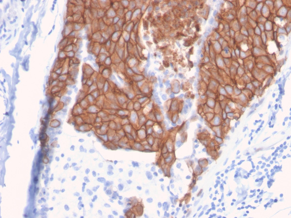
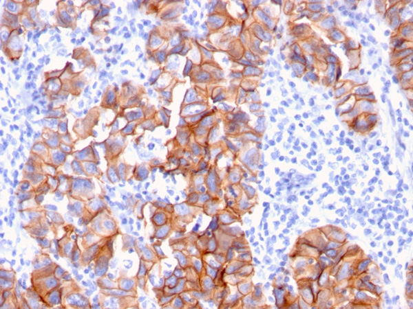
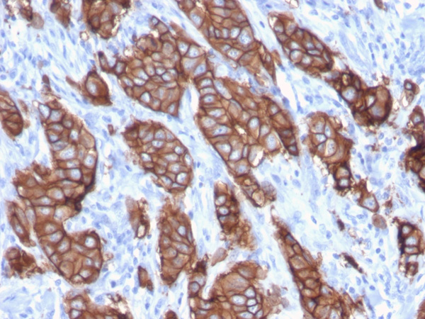
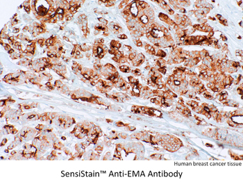 Application Data
EMA, Monoclonal Antibody (Cat# AAA214371)
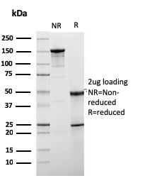
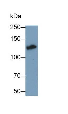
 SDS-PAGE
(SDS-PAGE Analysis of Purified Histone H1 Rabbit Recombinant Monoclonal Antibody (HH1/1784R).)
SDS-PAGE
(SDS-PAGE Analysis of Purified Histone H1 Rabbit Recombinant Monoclonal Antibody (HH1/1784R).)
Histone H1, Monoclonal Antibody (Cat# AAA214377)
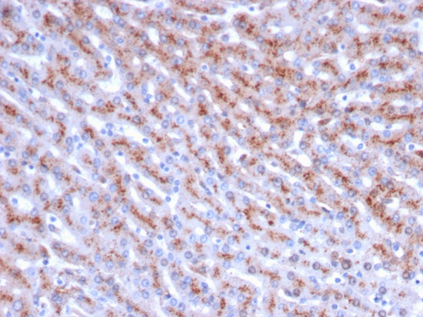
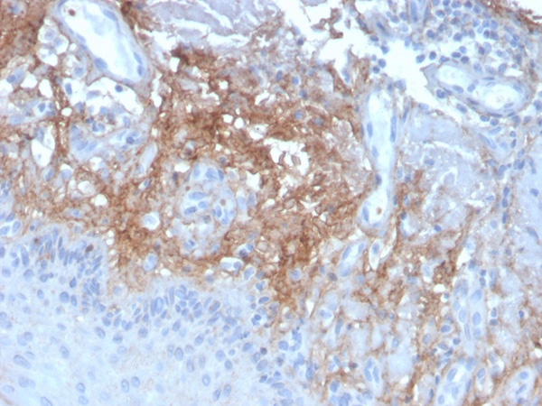
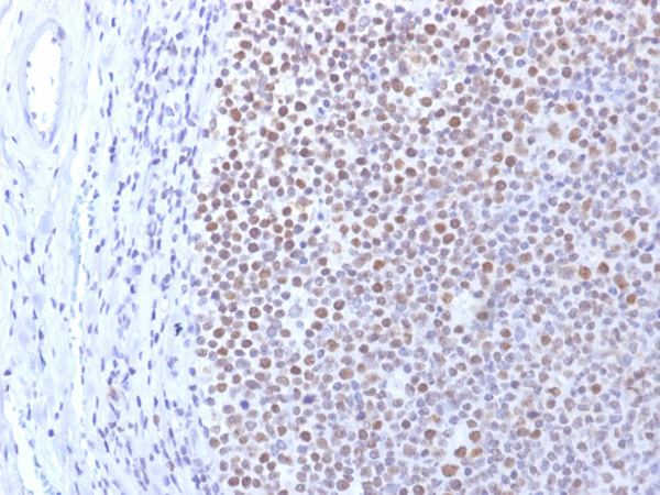
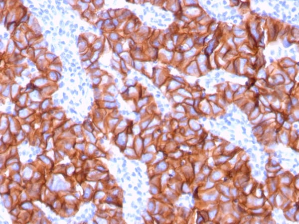
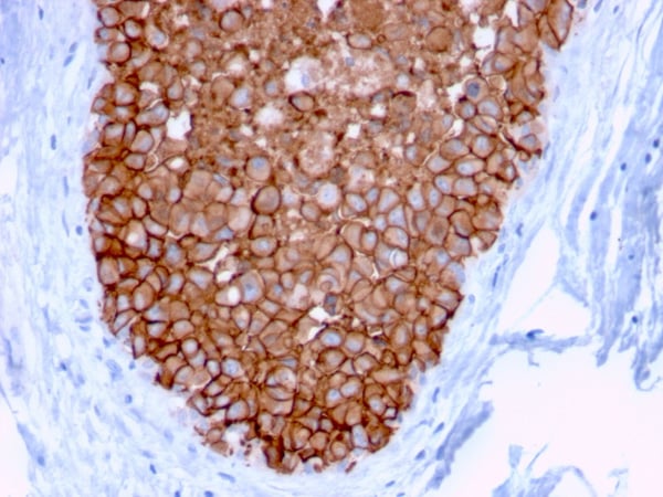
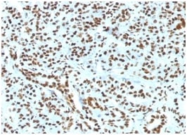
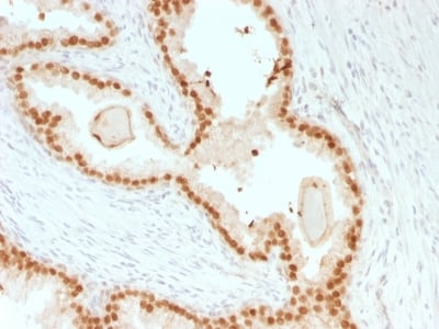
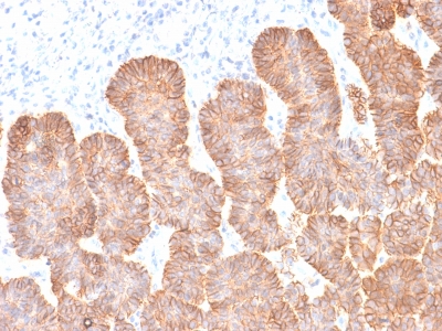
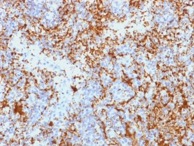
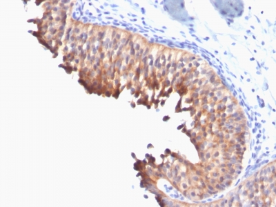
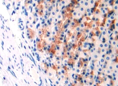
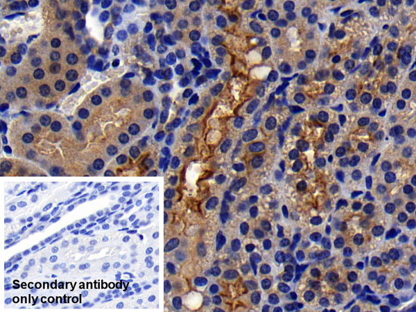
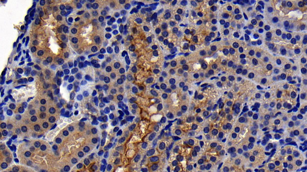
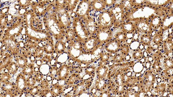
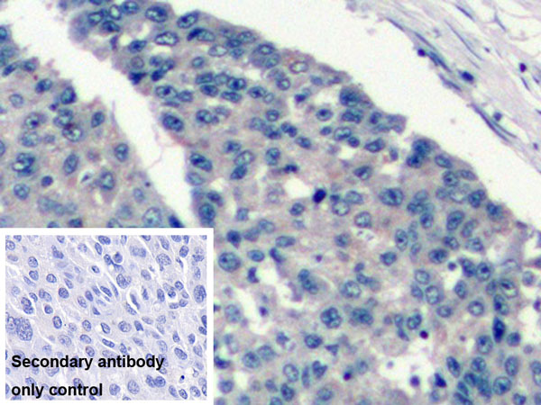
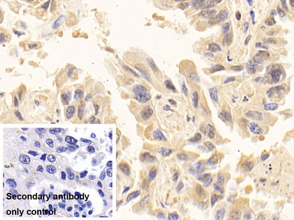
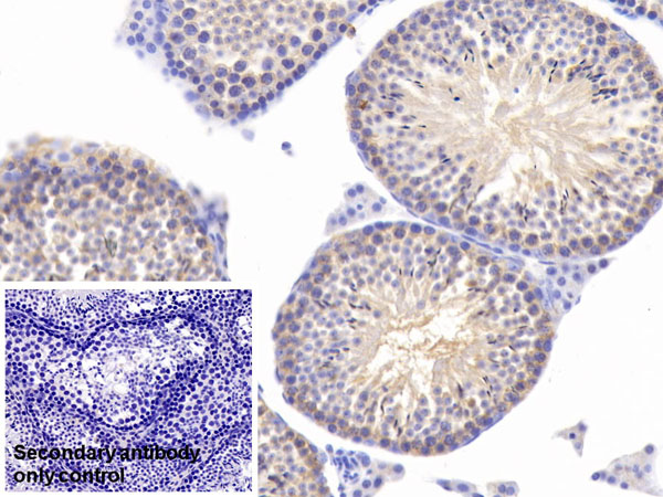
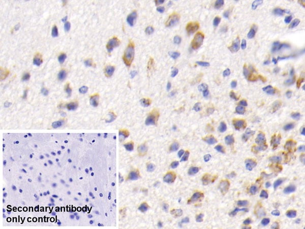
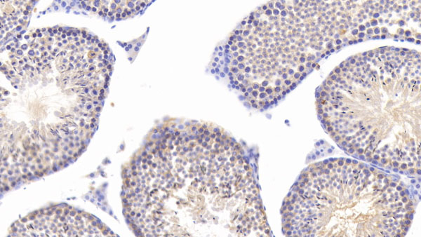
 IHC (Immunohiostchemistry)
(Formalin-paraffin human Bladder Carcinoma stained with FOXA1 Monoclonal Antibody (FOXA1/1241).)
IHC (Immunohiostchemistry)
(Formalin-paraffin human Bladder Carcinoma stained with FOXA1 Monoclonal Antibody (FOXA1/1241).)
FOXA1/HNF3A, Monoclonal Antibody (Cat# AAA214381)
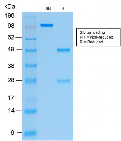
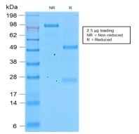
 SDS-PAGE
(SDS-PAGE Analysis of Purified FOXA1 Mouse Recombinant Monoclonal Antibody (rFOXA1/1515).)
SDS-PAGE
(SDS-PAGE Analysis of Purified FOXA1 Mouse Recombinant Monoclonal Antibody (rFOXA1/1515).)
FOXA1/HNF3A, Monoclonal Antibody (Cat# AAA214382)
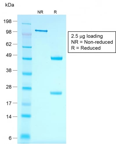
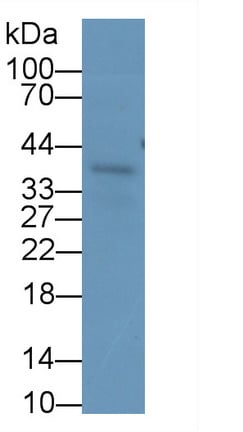
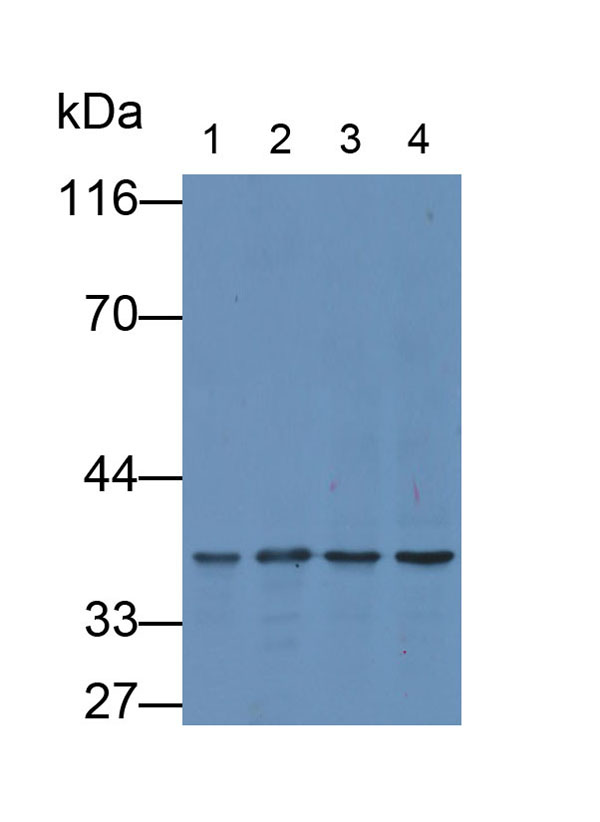
 SDS-PAGE
(SDS-PAGE Analysis Purified Cytokeratin 19 Mouse Recombinant Monoclonal Antibody (rKRT19/799).)
SDS-PAGE
(SDS-PAGE Analysis Purified Cytokeratin 19 Mouse Recombinant Monoclonal Antibody (rKRT19/799).)
Cytokeratin 19 (KRT19), Monoclonal Antibody (Cat# AAA214428)
Does not react with Rat and Ferret. Others not known.
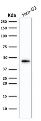 WB (Western Blot)
(Western Blot Analysis of Hep-G2 cell lysate using Cytokeratin 19 Rabbit Recombinant Monoclonal Antibody)
WB (Western Blot)
(Western Blot Analysis of Hep-G2 cell lysate using Cytokeratin 19 Rabbit Recombinant Monoclonal Antibody)
Cytokeratin 19 (KRT19), Monoclonal Antibody (Cat# AAA214430)
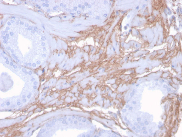
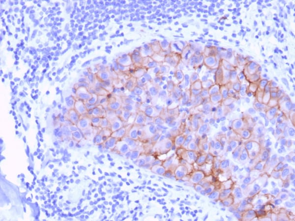
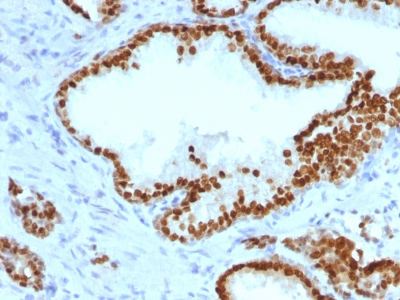
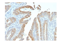
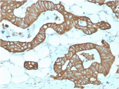
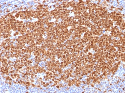
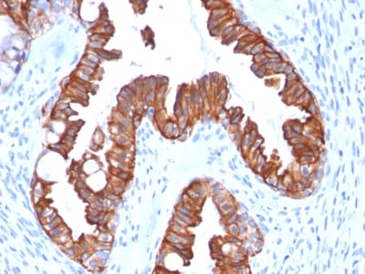
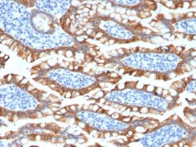
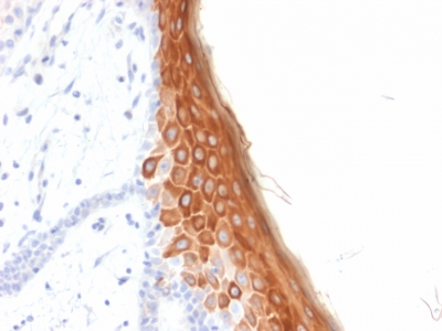
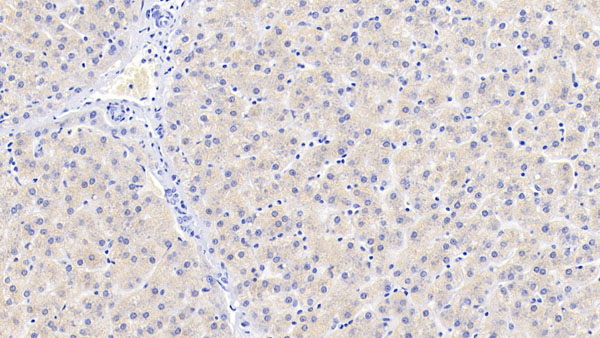
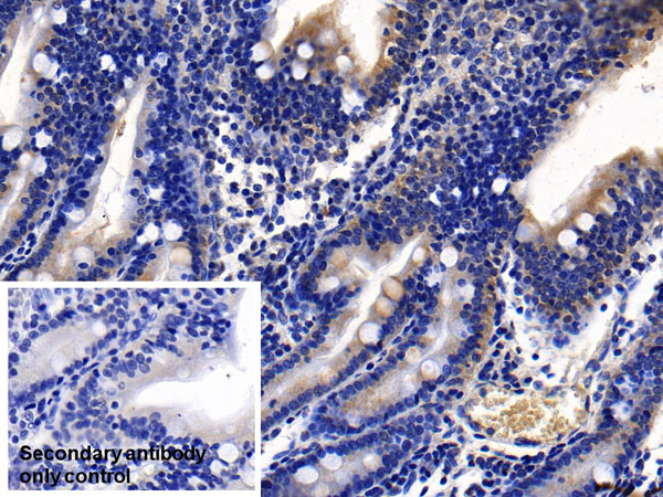
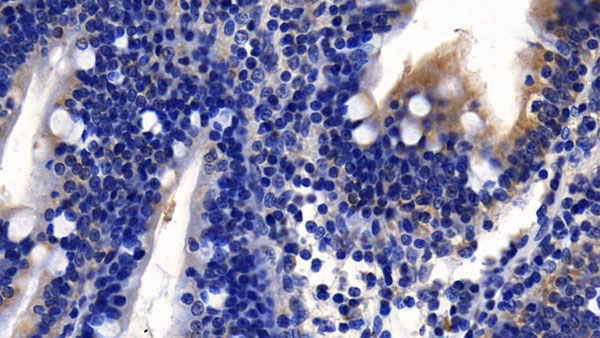
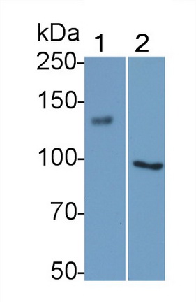
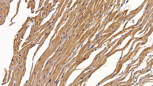
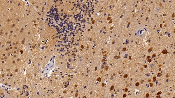
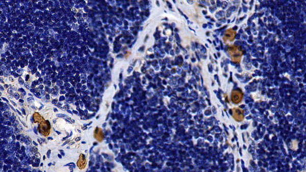
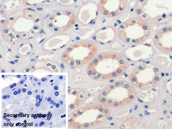
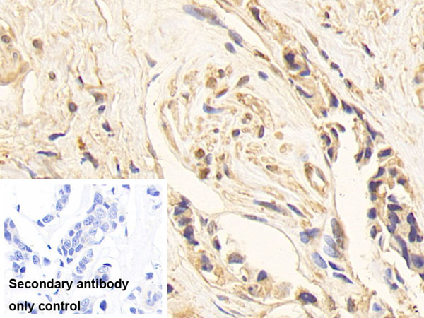
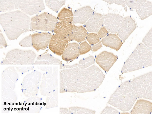
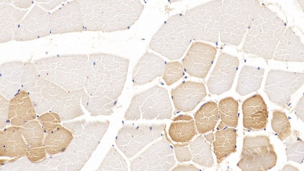
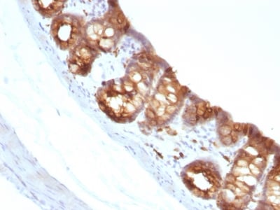 IHC (Immunohiostchemistry)
(Formalin-fixed, paraffin-embedded Rat Colon stained with Cytokeratin 18 Monoclonal Antibody (KRT18/1190).)
IHC (Immunohiostchemistry)
(Formalin-fixed, paraffin-embedded Rat Colon stained with Cytokeratin 18 Monoclonal Antibody (KRT18/1190).)
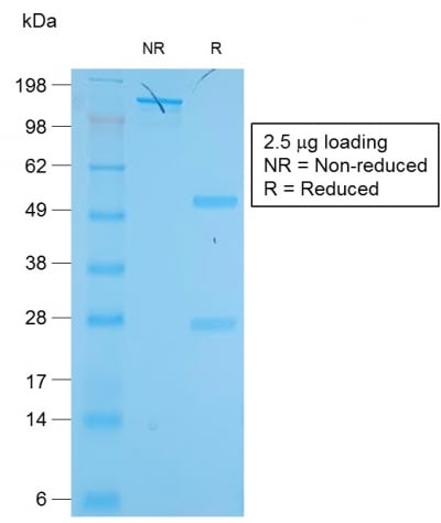
Ep-CAM/CD326, Monoclonal Antibody (Cat# AAA214434)
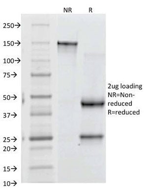
 SDS-PAGE
(SDS-PAGE Analysis Purified EpCAM Mouse Recombinant Monoclonal Antibody (rEGP40/1372).)
SDS-PAGE
(SDS-PAGE Analysis Purified EpCAM Mouse Recombinant Monoclonal Antibody (rEGP40/1372).)
Ep-CAM/CD326, Monoclonal Antibody (Cat# AAA214437)
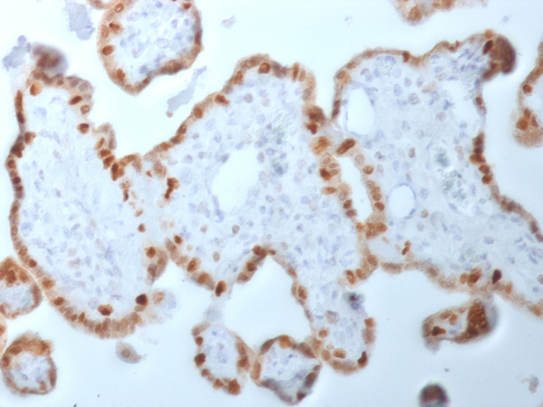
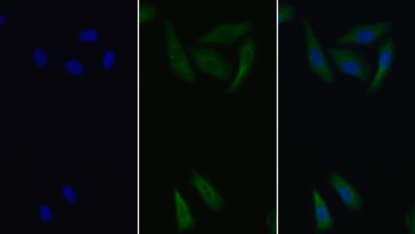
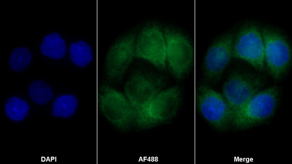
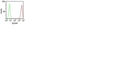 Application Data
(Surface staining of HT29 cells using Ep-CAM Monoclonal Antibody (VU-1D9) (red) and isotype control antibody (green).)
Application Data
(Surface staining of HT29 cells using Ep-CAM Monoclonal Antibody (VU-1D9) (red) and isotype control antibody (green).)
MCM7, Monoclonal Antibody (Cat# AAA214444)
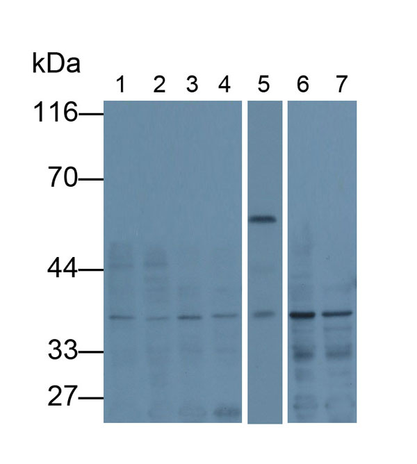
 SDS-PAGE
(SDS-PAGE Analysis Purified Moesin Mouse Recombinant Monoclonal Antibody (rMSN/492).)
SDS-PAGE
(SDS-PAGE Analysis Purified Moesin Mouse Recombinant Monoclonal Antibody (rMSN/492).)
Moesin, Monoclonal Antibody (Cat# AAA214454)

 SDS-PAGE
(SDS-PAGE Analysis Purified CD31 Rabbit Recombinant Monoclonal Antibody (C31/1395R).)
SDS-PAGE
(SDS-PAGE Analysis Purified CD31 Rabbit Recombinant Monoclonal Antibody (C31/1395R).)
CD31/PECAM-1, Monoclonal Antibody (Cat# AAA214475)

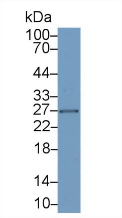
 SDS-PAGE
(SDS-PAGE Analysis of Purified Kappa Light Chain Rabbit Recombinant Monoclonal (KLC2289R).)
SDS-PAGE
(SDS-PAGE Analysis of Purified Kappa Light Chain Rabbit Recombinant Monoclonal (KLC2289R).)
Kappa LightChain/IGKC, Monoclonal Antibody (Cat# AAA214400)
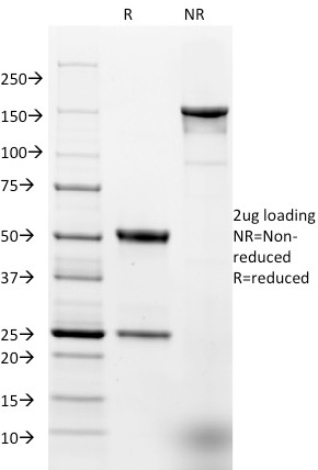
 SDS-PAGE
(SDS-PAGE Analysis Purified CD61 Monoclonal Antibody (Y2/51).Confirmation of Purity and Integrity of Antibody.)
SDS-PAGE
(SDS-PAGE Analysis Purified CD61 Monoclonal Antibody (Y2/51).Confirmation of Purity and Integrity of Antibody.)
CD61/Integrin beta-3/Platelet Glycoprotein IIIa, Monoclonal Antibody (Cat# AAA214405)

 SDS-PAGE
(SDS-PAGE Analysis of Purified Involucrin Rabbit Recombinant Monoclonal Antibody (IVRN/2113R).)
SDS-PAGE
(SDS-PAGE Analysis of Purified Involucrin Rabbit Recombinant Monoclonal Antibody (IVRN/2113R).)
Involucrin, Monoclonal Antibody (Cat# AAA214408)
Does not react with Mouse. Others not known.
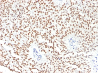
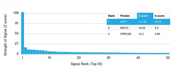 Application Data
(Analysis of Protein Array containing more than 19, 000 full-length human proteins using Cytokeratin 7 Mouse Recombinant Monoclonal Antibody (rOV-TL12/30).Z- and S- Score: The Z-score represents the strength of a signal that a monoclonal antibody (MAb) (in combination with a fluorescently-tagged anti-IgG secondary antibody) produces when binding to a particular protein on the HuProtTM array. Z-scores are described in units of standard deviations (SD's) above the mean value of all signals generated on that array. If targets on HuProtTM are arranged in descending order of the Z-score, the S-score is the difference (also in units of SD's) between the Z-score. S-score therefore represents the relative target specificity of a MAb to its intended target. A MAb is considered to specific to its intended target, if the MAb has an S-score of at least 2.5. For example, if a MAb binds to protein X with a Z-score of 43 and to protein Y with a Z-score of 14, then the S-score for the binding of that MAb to protein X is equal to 29.)
Application Data
(Analysis of Protein Array containing more than 19, 000 full-length human proteins using Cytokeratin 7 Mouse Recombinant Monoclonal Antibody (rOV-TL12/30).Z- and S- Score: The Z-score represents the strength of a signal that a monoclonal antibody (MAb) (in combination with a fluorescently-tagged anti-IgG secondary antibody) produces when binding to a particular protein on the HuProtTM array. Z-scores are described in units of standard deviations (SD's) above the mean value of all signals generated on that array. If targets on HuProtTM are arranged in descending order of the Z-score, the S-score is the difference (also in units of SD's) between the Z-score. S-score therefore represents the relative target specificity of a MAb to its intended target. A MAb is considered to specific to its intended target, if the MAb has an S-score of at least 2.5. For example, if a MAb binds to protein X with a Z-score of 43 and to protein Y with a Z-score of 14, then the S-score for the binding of that MAb to protein X is equal to 29.)
Cytokeratin 7, Monoclonal Antibody (Cat# AAA214413)
 SDS-PAGE
(SDS-PAGE Analysis of Purified Cytokeratin 8 Rabbit Recombinant Monoclonal Antibody (KRT8/2174R).)
SDS-PAGE
(SDS-PAGE Analysis of Purified Cytokeratin 8 Rabbit Recombinant Monoclonal Antibody (KRT8/2174R).)
Cytokeratin 8 (KRT8), Monoclonal Antibody (Cat# AAA214416)
Does not react with Mouse, Rat, Chicken and Xenopus laevis.
 SDS-PAGE
(SDS-PAGE Analysis of Purified Involucrin Rabbit Recombinant Monoclonal Antibody (IVRN/2113R).)
SDS-PAGE
(SDS-PAGE Analysis of Purified Involucrin Rabbit Recombinant Monoclonal Antibody (IVRN/2113R).)
Cytokeratin 10, Monoclonal Antibody (Cat# AAA214417)
 SDS-PAGE
(SDS-PAGE Analysis of Purified Cytokeratin 10 Mouse Recombinant Monoclonal Antibody (rKRT10/844).)
SDS-PAGE
(SDS-PAGE Analysis of Purified Cytokeratin 10 Mouse Recombinant Monoclonal Antibody (rKRT10/844).)
Cytokeratin 10 (KRT10), Monoclonal Antibody (Cat# AAA214420)
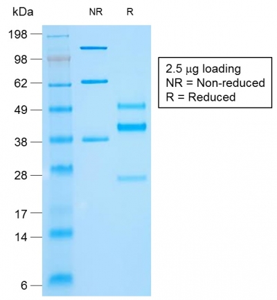
 Application Data
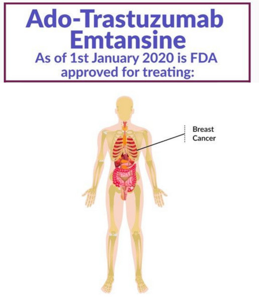
Ado-trastuzumab emtansine, Monoclonal Antibody (Cat# AAA197048)
This monoclonal antibody was purified using multi-step affinity chromatography methods such as Protein A or G depending on the species and isotype.
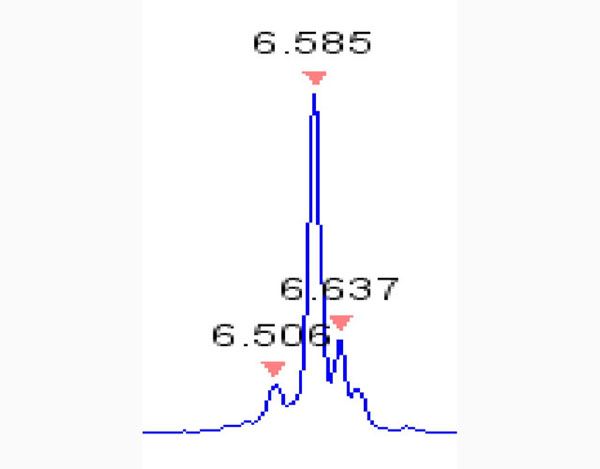
Evolocumab, Monoclonal Antibody (Cat# AAA197050)
This monoclonal antibody was purified using multi-step affinity chromatography methods such as Protein A or G depending on the species and isotype.
 Application Data
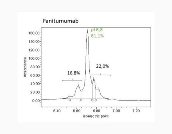
Panitumumab, Monoclonal Antibody (Cat# AAA197051)
This monoclonal antibody was purified using multi-step affinity chromatography methods such as Protein A or G depending on the species and isotype.
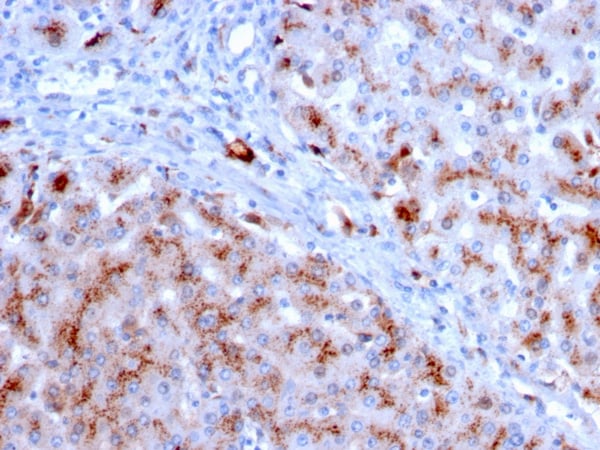
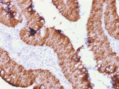
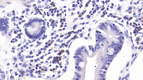
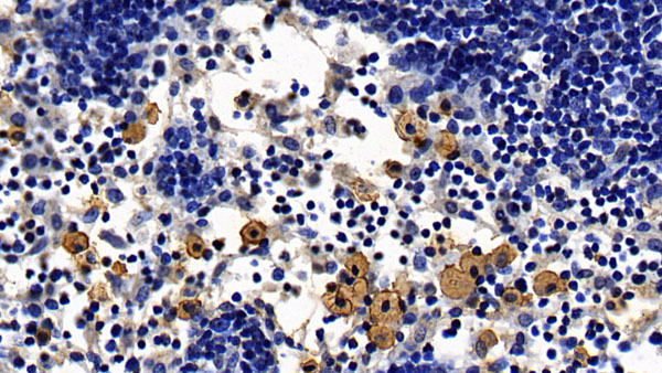
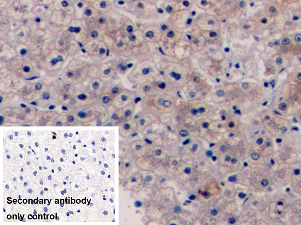
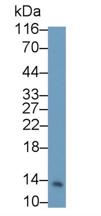
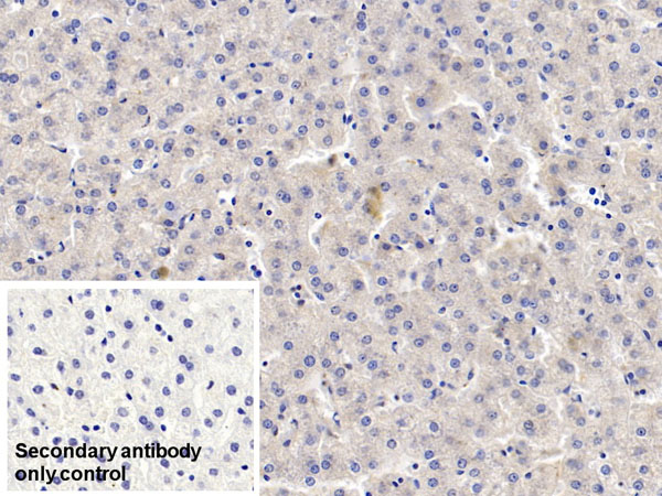
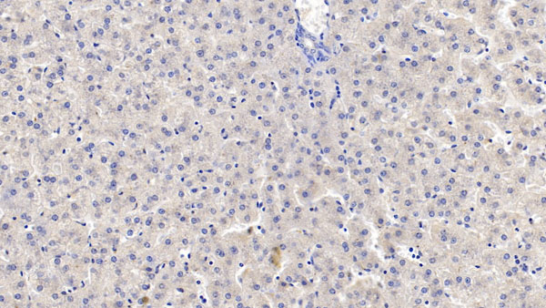
 IHC (Immunohistochemisry)
(DAB staining on IHC-P; Recombinant IL10, Rat. Sample: Rat Stomach Tissue; Primary Ab: 40ug/ml Mouse Anti-Rat IL10 Antibody Second Ab: 2ug/mL HRP-Linked Caprine Anti-Mouse IgG Polyclonal Antibody)
IHC (Immunohistochemisry)
(DAB staining on IHC-P; Recombinant IL10, Rat. Sample: Rat Stomach Tissue; Primary Ab: 40ug/ml Mouse Anti-Rat IL10 Antibody Second Ab: 2ug/mL HRP-Linked Caprine Anti-Mouse IgG Polyclonal Antibody)
Interleukin 10, Monoclonal Antibody (Cat# AAA144653)
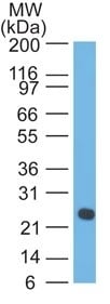
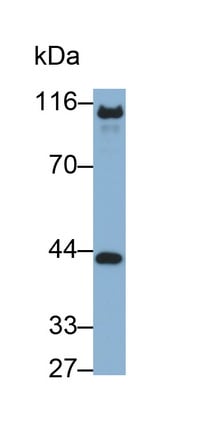
 WB (Western Blot)
(Western Blot: Sample: Recombinant PCT, Human.)
WB (Western Blot)
(Western Blot: Sample: Recombinant PCT, Human.)
Procalcitonin, Monoclonal Antibody (Cat# AAA144661)
Toll Like Receptor 4 (TLR4), Monoclonal Antibody (Cat# AAA149819)
Mucin 5 Subtype AC (MUC5AC), Monoclonal Antibody (Cat# AAA149820)
Procollagen I N-Terminal Propeptide (PINP), Monoclonal Antibody (Cat# AAA149821)
Plasminogen Activator, Urokinase Receptor (uPAR), Monoclonal Antibody (Cat# AAA149854)
Procollagen III N-Terminal Propeptide (PIIINP), Monoclonal Antibody (Cat# AAA149784)
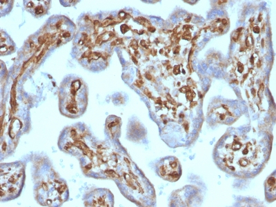
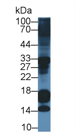
 WB (Western Blot)
(Western Blot; Sample: human Placenta lysatePrimary Ab: 0.05ug/ml Mouse Anti-human PL AntibodySecond Ab: 0.2ug/mL HRP-Linked Caprine Anti-Mouse IgG Polyclonal Antibody)
WB (Western Blot)
(Western Blot; Sample: human Placenta lysatePrimary Ab: 0.05ug/ml Mouse Anti-human PL AntibodySecond Ab: 0.2ug/mL HRP-Linked Caprine Anti-Mouse IgG Polyclonal Antibody)
Placental Lactogen (PL), Monoclonal Antibody (Cat# AAA152590)
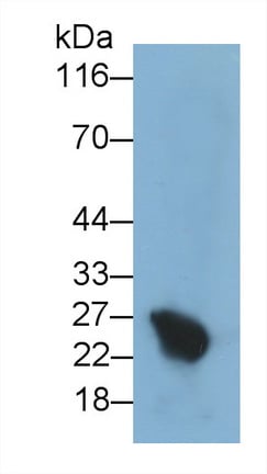
 WB (Western Blot)
(Western Blot; Sample: MCF7 cell lysate Primary Ab: 2ug/ml Mouse Anti-human IGFBP3 Antibody Second Ab: 0.2ug/mL HRP-Linked Caprine Anti-Mouse IgG Polyclonal Antibody)
WB (Western Blot)
(Western Blot; Sample: MCF7 cell lysate Primary Ab: 2ug/ml Mouse Anti-human IGFBP3 Antibody Second Ab: 0.2ug/mL HRP-Linked Caprine Anti-Mouse IgG Polyclonal Antibody)
Inulin Like Growth Factor Binding Protein 3 (IGFBP3), Monoclonal Antibody (Cat# AAA152641)
 WB (Western Blot)
(Western Blot; Sample: Rat Spleen lysate Primary Ab: 1ug/ml Mouse Anti-Rabbit IL6 Antibody Second Ab: 0.2ug/mL HRP-Linked Caprine Anti-Mouse IgG Polyclonal Antibody)
WB (Western Blot)
(Western Blot; Sample: Rat Spleen lysate Primary Ab: 1ug/ml Mouse Anti-Rabbit IL6 Antibody Second Ab: 0.2ug/mL HRP-Linked Caprine Anti-Mouse IgG Polyclonal Antibody)
Interleukin 6 (IL6), Monoclonal Antibody (Cat# AAA152646)
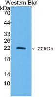
 WB (Western Blot)
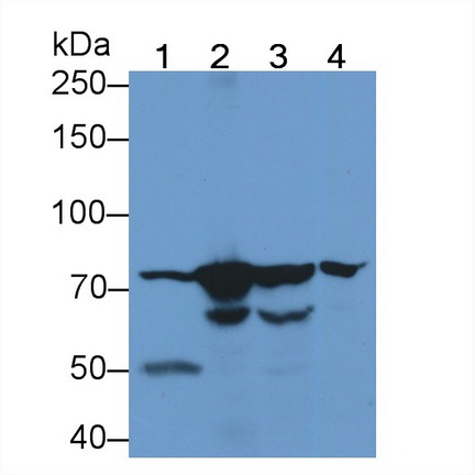
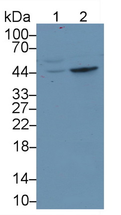
(Western Blot; Sample: Lane1: Porcine Kidney lysate; Lane2: Mouse Kidney lysate Primary Ab: 0.6ug/ml Mouse Anti-human ACE2 Antibody Second Ab: 0.2ug/mL HRP-Linked Caprine Anti-Mouse IgG Polyclonal Antibody)
WB (Western Blot)
(Western Blot; Sample: Lane1: Porcine Kidney lysate; Lane2: Mouse Kidney lysate Primary Ab: 0.6ug/ml Mouse Anti-human ACE2 Antibody Second Ab: 0.2ug/mL HRP-Linked Caprine Anti-Mouse IgG Polyclonal Antibody)
Angiotensin I Converting Enzyme 2 (ACE2), Monoclonal Antibody (Cat# AAA152718)
 WB (Western Blot)
(Western Blot; Sample: Lane1: Rat Placenta lysate; Lane2: HepG2 cell lysate; Lane3: HepG2 Primary Ab: 0.05 ug/ml Mouse Anti-Rat TFPI Antibody Second Ab: 0.2ug/mL HRP-Linked Caprine Anti-Mouse IgG Polyclonal Antibody)
WB (Western Blot)
(Western Blot; Sample: Lane1: Rat Placenta lysate; Lane2: HepG2 cell lysate; Lane3: HepG2 Primary Ab: 0.05 ug/ml Mouse Anti-Rat TFPI Antibody Second Ab: 0.2ug/mL HRP-Linked Caprine Anti-Mouse IgG Polyclonal Antibody)
Tissue Factor Pathway Inhibitor (TFPI), Monoclonal Antibody (Cat# AAA152723)
 WB (Western Blot)
(Western Blot; Sample: Lane1: Hela cell lysate; Lane2: human Lung lysate; Lane3: Mouse Thymus lysate Primary Ab: 1ug/ml Mouse Anti-ulti-species STAT3 Antibody Second Ab: 0.2ug/mL HRP-Linked Caprine Anti-Mouse IgG Polyclonal Antibody)
WB (Western Blot)
(Western Blot; Sample: Lane1: Hela cell lysate; Lane2: human Lung lysate; Lane3: Mouse Thymus lysate Primary Ab: 1ug/ml Mouse Anti-ulti-species STAT3 Antibody Second Ab: 0.2ug/mL HRP-Linked Caprine Anti-Mouse IgG Polyclonal Antibody)
Signal Transducer And Activator Of Transcription 3 (STAT3), Monoclonal Antibody (Cat# AAA152738)
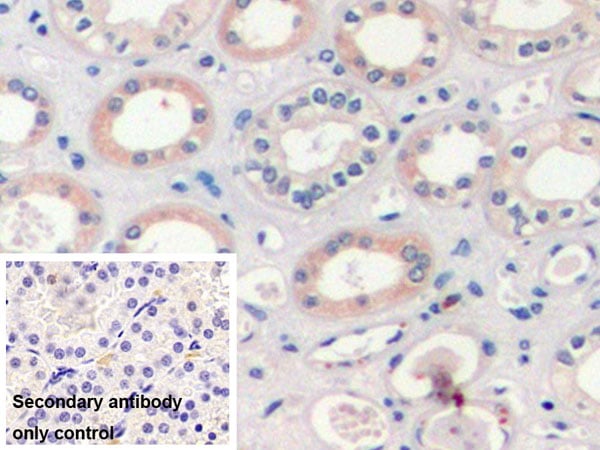
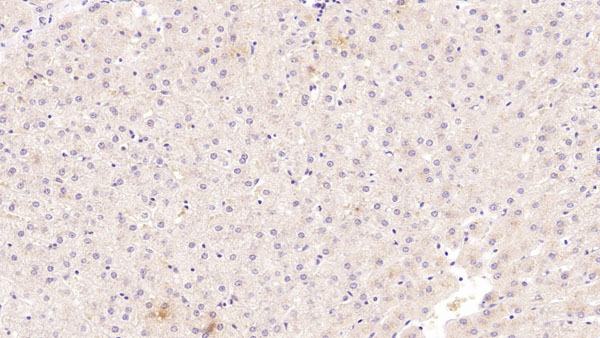
 IHC (Immunohiostchemistry)
(DAB staining on IHC-P;Sample: Rat Lymph node Tissue; Primary Ab: 30ug/ml Mouse Anti-human TLR2 AntibodySecond Ab: 2ug/mL HRP-Linked Caprine Anti-Mouse IgG Polyclonal Antibody)
IHC (Immunohiostchemistry)
(DAB staining on IHC-P;Sample: Rat Lymph node Tissue; Primary Ab: 30ug/ml Mouse Anti-human TLR2 AntibodySecond Ab: 2ug/mL HRP-Linked Caprine Anti-Mouse IgG Polyclonal Antibody)
Toll Like Receptor 2 (TLR2), Monoclonal Antibody (Cat# AAA152770)
 WB (Western Blot)
(Western Blot; Sample: Rat Small intestine lysatePrimary Ab: 2ug/ml Mouse Anti-human SI AntibodySecond Ab: 0.2ug/mL HRP-Linked Caprine Anti-Mouse IgG Polyclonal Antibody)
WB (Western Blot)
(Western Blot; Sample: Rat Small intestine lysatePrimary Ab: 2ug/ml Mouse Anti-human SI AntibodySecond Ab: 0.2ug/mL HRP-Linked Caprine Anti-Mouse IgG Polyclonal Antibody)
Sucrase Isomaltase (SI), Monoclonal Antibody (Cat# AAA152813)
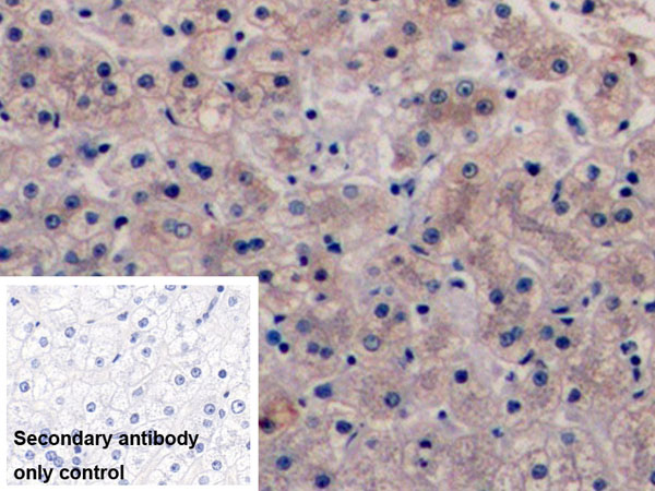
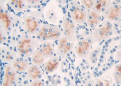
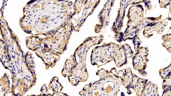
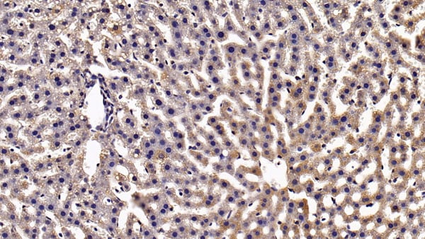
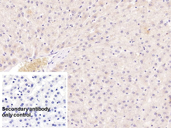
 IHC (Immunohistochemistry)
(DAB staining on IHC-P;Sample: human Kidney TissuePrimary Ab: 10ug/ml Mouse Anti-human VEGF165 AntibodyControl: Used PBS instead of primary antibodySecond Ab: 2 ug/ml HRP-Linked Caprine Anti-Mouse IgG Polyclonal Antibody)
IHC (Immunohistochemistry)
(DAB staining on IHC-P;Sample: human Kidney TissuePrimary Ab: 10ug/ml Mouse Anti-human VEGF165 AntibodyControl: Used PBS instead of primary antibodySecond Ab: 2 ug/ml HRP-Linked Caprine Anti-Mouse IgG Polyclonal Antibody)
Vasular Endothelial Growth Factor 165 (VEGF165), Monoclonal Antibody (Cat# AAA152872)
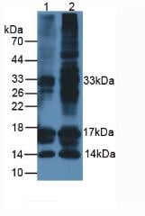
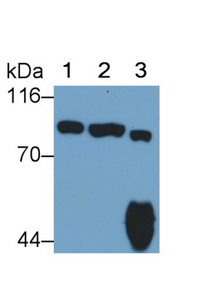
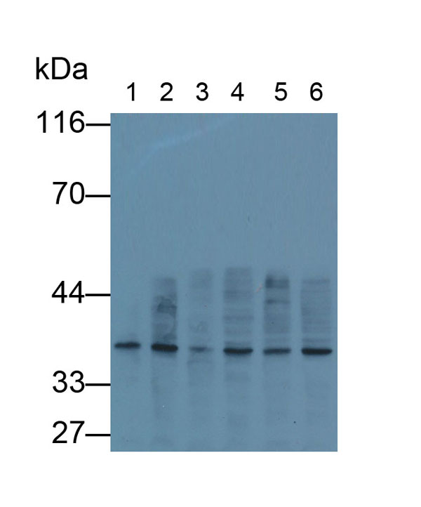 WB (Western Blot)
(Western Blot; Samples: Lane1: human Lung lysate; Lane2: 293T cell lysate; Lane3: Cavia Liver lysate; Lane4: Cavia Kidney lysate; Lane5: Porcine Liver lysate; Lane6: Porcine Kidney lysate;Primary Ab: 0.5ug/ml Mouse Anti-human GAPDH AntibodySecond Ab: 0.2 ug/ml HRP-Linked Caprine Anti-Mouse IgG Polyclonal Antibody)
WB (Western Blot)
(Western Blot; Samples: Lane1: human Lung lysate; Lane2: 293T cell lysate; Lane3: Cavia Liver lysate; Lane4: Cavia Kidney lysate; Lane5: Porcine Liver lysate; Lane6: Porcine Kidney lysate;Primary Ab: 0.5ug/ml Mouse Anti-human GAPDH AntibodySecond Ab: 0.2 ug/ml HRP-Linked Caprine Anti-Mouse IgG Polyclonal Antibody)
Glyceraldehyde-3-Phosphate Dehydrogenase (GAPDH), Monoclonal Antibody (Cat# AAA152883)
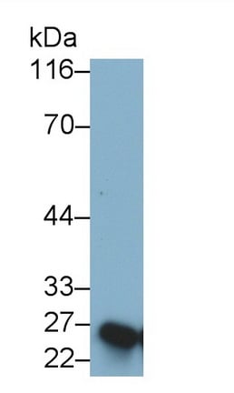
 WB (Western Blot)
(Western Blot; Sample: A431 cell lysate Primary Ab: 0.3ug/ml Mouse Anti-human E-cadherin Antibody Second Ab: 0.2ug/mL HRP-Linked Caprine Anti-Mouse IgG Polyclonal Antibody)
WB (Western Blot)
(Western Blot; Sample: A431 cell lysate Primary Ab: 0.3ug/ml Mouse Anti-human E-cadherin Antibody Second Ab: 0.2ug/mL HRP-Linked Caprine Anti-Mouse IgG Polyclonal Antibody)
E-cadherin, Monoclonal Antibody (Cat# AAA152938)
 WB (Western Blot)
(Western Blot; Sample: Lane1: Rat CerebuM lysate; Lane2: Rat Liver lysate; Lane3: Rat Testis lysate; Lane4: A549 cell lysate Primary Ab: 2 ug/ml Mouse Anti-Mouse HSPA5 Antibody Second Ab: 0.2ug/mL HRP-Linked Caprine Anti-Mouse IgG Polyclonal Antibody)
WB (Western Blot)
(Western Blot; Sample: Lane1: Rat CerebuM lysate; Lane2: Rat Liver lysate; Lane3: Rat Testis lysate; Lane4: A549 cell lysate Primary Ab: 2 ug/ml Mouse Anti-Mouse HSPA5 Antibody Second Ab: 0.2ug/mL HRP-Linked Caprine Anti-Mouse IgG Polyclonal Antibody)
Heat Shock 70kDa Protein 5 (HSPA5), Monoclonal Antibody (Cat# AAA152649)
 WB (Western Blot)
(Western Blot; Sample: human SeuM Primary Ab: 2ug/ml Mouse Anti-human SAA Antibody Second Ab: 0.2ug/mL HRP-Linked Caprine Anti-Mouse IgG Polyclonal Antibody)
WB (Western Blot)
(Western Blot; Sample: human SeuM Primary Ab: 2ug/ml Mouse Anti-human SAA Antibody Second Ab: 0.2ug/mL HRP-Linked Caprine Anti-Mouse IgG Polyclonal Antibody)
SeuM Amyloid A (SAA), Monoclonal Antibody (Cat# AAA152676)
 WB (Western Blot)
(Western Blot; Sample: Lane1: Porcine Liver lysate; Lane2: Rat Liver lysate Primary Ab: 2ug/ml Mouse Anti-human TDO Antibody Second Ab: 0.2ug/mL HRP-Linked Caprine Anti-Mouse IgG Polyclonal Antibody)
WB (Western Blot)
(Western Blot; Sample: Lane1: Porcine Liver lysate; Lane2: Rat Liver lysate Primary Ab: 2ug/ml Mouse Anti-human TDO Antibody Second Ab: 0.2ug/mL HRP-Linked Caprine Anti-Mouse IgG Polyclonal Antibody)
Tryptophan-2,3-dioxygenase (TDO), Monoclonal Antibody (Cat# AAA152686)
What are Monoclonal Antibodies?
Monoclonal antibodies are specialized laboratory-produced proteins developed for binding to specific biological antigens or other molecular targets. Since they come from a single cell (or clone), they are especially consistent and accurate in the data they are involved in producing.
This type of antibody material has been shown to be a powerful tool in finding and subsequently destroying harmful cells in an organism, such as those found in cancers or various autoimmune diseases. This makes them excellent aids in medical testing and research, which is why they are so widely used.
AAA Biotech offers a comprehensive range of high-quality monoclonal antibodies that perform effectively in various laboratory tests, including (amongst others) ELISA, western blotting, immunohistochemistry, and flow cytometry. All of the products in our catalog are thoroughly quality tested to make sure that they are reliable and will consistently perform well in your research.
What Are The Uses of Monoclonal Antibodies
Monoclonal antibodies are used in many lab tests, including (amongst others) ELISA, western blotting, immunohistochemistry, and flow cytometry.
ELISA is a test that helps detect a specific substance/analyte in a sample. It uses antibodies (often monoclonal) bound to a solid surface (such as the well of a microplate) to “capture” the substance/analyte in the sample and immobilize it so that the detection antibody component can then bind to it and produce a signal, which can then be measured.
Western blotting identifies specific proteins in a sample. The sample is first separated on a gel, and then antibodies are applied that will typically bind to the target, which will all be localized to a single band in a lane.
Immunohistochemistry helps locate specific proteins in cells or tissue samples using antibodies.
Flow cytometry looks at and sorts cells. It uses antibodies that are conjugated to reporter molecules called “fluorophores”, which, under special lights, emit light themselves, which can then be measured by a detector instrument. For a deeper understanding of these techniques, explore our complete guide to monoclonal antibodies and their benefits.
How Monoclonal Antibodies Are Used as Medicine?
Please note that all of the products listed in AAA Biotech’s also known as AAA Bio or AAABio catalog are strictly for research-use only (RUO).
Monoclonal antibodies can also be used as therapeutic/medical treatments, particularly in the context of cancers. They are designed to find and bind to specific cells or proteins, helping the immune system recognize and attack the cancer. These treatments work in different ways, such as:
- Radioimmunotherapy attaches a small amount of radioactive molecule to the antibody, so it delivers the radiation directly to the cancer cells that the antibody is specifically binding to.
- Antibody-directed enzyme prodrug therapy uses antibodies that are specifically bound to special enzymes. These enzymes activate a harmless drug in the body and turn it into a cancer-killing drug only near the cancer cells—this helps avoid harming healthy cells.
- Immunoliposomes are tiny “bubbles” filled with medicine/drug and coated with antibodies. They carry the drug straight to the cancer cells.
Why Buy Monoclonal Antibodies From Us?
At AAA Biotech, we provide high-performance monoclonal antibodies designed to support a wide range of research needs.
1. Validated for Versatile Applications
The antibodies in our catalog are extensively validated and compatible with multiple techniques, including (but not limited to) ELISA, flow cytometry (FC), immunocytochemistry (ICC), immunofluorescence (IF), immunohistochemistry (IHC), immunoprecipitation (IP), and western blotting (WB).
2. Wide Selection & Specialized Options
We offer antibodies for common and rare species, that are available in various conjugated forms, and also in recombinant formats. Essentially, there is almost anything one might need to meet their experimental model’s requirements.
3. High-Quality Proteins
Our proteins meet high purity standards—90% or more as confirmed by SDS-PAGE. Many are available with tags like His, Flag, GST, or MBP, and we also supply native and biologically active proteins for functional studies.
Frequently Asked Questions
1. Are your monoclonal antibodies validated for specific applications?
Yes, our antibodies are tested and validated for use in methods such as ELISA, western blot, IHC, flow cytometry, and more. Refer to specific product pages or datasheets for individual product information.
2. How do I choose the right monoclonal antibody for my application?
Review the product details directly for application validation, species reactivity, and target information. You may also contact our support team at any time for help.
3. How quickly can I receive my order?
Most orders are processed and shipped within 1–3 business days, depending on product availability and your shipping location.