Filters
▼Clonality
▼Type
▼Reactivity
▼Gene Name
▼Isotype
▼Host
▼Application
▼Clone
▼Monoclonal Antibodies
Get accurate results in your research with our Monoclonal Antibodies, which are specially made to target exactly what you require for your research, and will produce consistent, reliable performance in lab tests.
Viewing 3550-3600 of 27645 product results

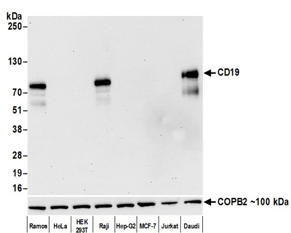 WB (Western Blot)
(Detection of human CD19 by western blot. Samples: Whole cell lysate (10 ug) from Ramos, HeLa, HEK293T, Raji, Hep-G2, MCF-7, Jurkat, and Daudi cells prepared using NETN lysis buffer. Antibody: Rabbit anti-CD19 recombinant monoclonal antibody (AAA213598 Lot 1) used at 1:1000. Secondary: HRP-conjugated goat anti-rabbit IgG . Detection: Chemiluminescence with an exposure time of 30 seconds. Lower Panel: Rabbit anti-COPB2 antibody .)
WB (Western Blot)
(Detection of human CD19 by western blot. Samples: Whole cell lysate (10 ug) from Ramos, HeLa, HEK293T, Raji, Hep-G2, MCF-7, Jurkat, and Daudi cells prepared using NETN lysis buffer. Antibody: Rabbit anti-CD19 recombinant monoclonal antibody (AAA213598 Lot 1) used at 1:1000. Secondary: HRP-conjugated goat anti-rabbit IgG . Detection: Chemiluminescence with an exposure time of 30 seconds. Lower Panel: Rabbit anti-COPB2 antibody .)
CD19, Monoclonal Recombinant Antibody (Cat# AAA213598)
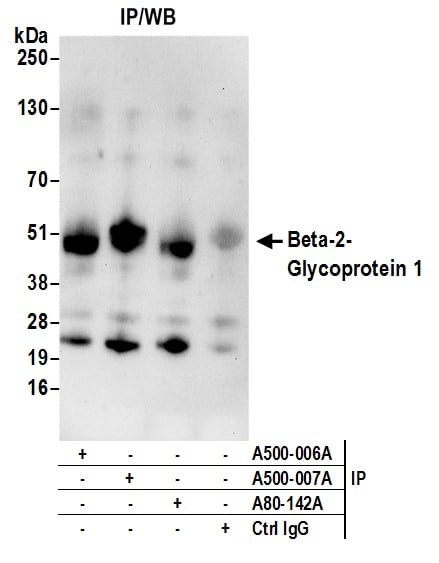
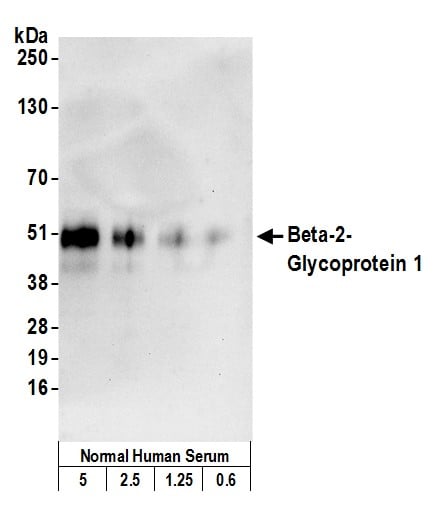 WB (Western Blot)
(Detection of human Beta-2-Glycoprotein 1 by western blot. Samples: Normal human serum (5, 2.5, 1.25 and 0.6 ul of a 1:20 dilution). Antibody: Mouse monoclonal anti-Beta-2-Glycoprotein 1 antibody [7B8] (AAA213494 lot 1) used at 1:1000. Secondary: HRP-conjugated goat anti-mouse IgG . Detection: Chemiluminescence with an exposure time of 10 seconds.)
WB (Western Blot)
(Detection of human Beta-2-Glycoprotein 1 by western blot. Samples: Normal human serum (5, 2.5, 1.25 and 0.6 ul of a 1:20 dilution). Antibody: Mouse monoclonal anti-Beta-2-Glycoprotein 1 antibody [7B8] (AAA213494 lot 1) used at 1:1000. Secondary: HRP-conjugated goat anti-mouse IgG . Detection: Chemiluminescence with an exposure time of 10 seconds.)
Beta-2-Glycoprotein 1, Monoclonal Antibody (Cat# AAA213494)
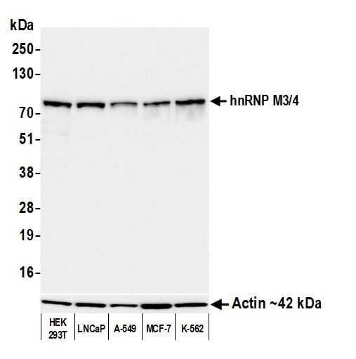
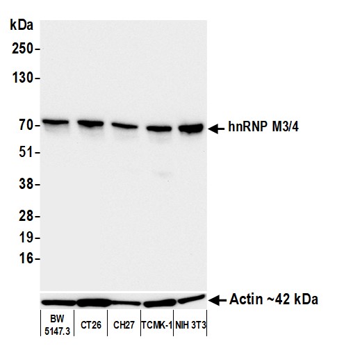 WB (Western Blot)
(Detection of mouse hnRNP M3/4 by western blot. Samples: Whole cell lysate (10 ug) from BW5147.3, CT26, CH27, TCMK-1, and NIH 3T3 cells prepared using NETN lysis buffer. Antibody: Mouse anti-hnRNP M3/4 monoclonal antibody [2A6-2H3] (AAA213497 lot 3) used at 1:1000. Secondary: HRP-conjugated goat anti-mouse IgG . Detection: Chemiluminescence with an exposure time of 10 seconds. Lower Panel: Rabbit anti-Actin recombinant monoclonal antibody .)
WB (Western Blot)
(Detection of mouse hnRNP M3/4 by western blot. Samples: Whole cell lysate (10 ug) from BW5147.3, CT26, CH27, TCMK-1, and NIH 3T3 cells prepared using NETN lysis buffer. Antibody: Mouse anti-hnRNP M3/4 monoclonal antibody [2A6-2H3] (AAA213497 lot 3) used at 1:1000. Secondary: HRP-conjugated goat anti-mouse IgG . Detection: Chemiluminescence with an exposure time of 10 seconds. Lower Panel: Rabbit anti-Actin recombinant monoclonal antibody .)
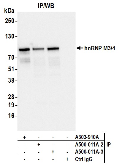
hnRNP M3/4, Monoclonal Antibody (Cat# AAA213497)
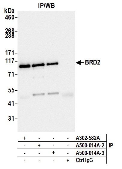
 WB (Western Blot)
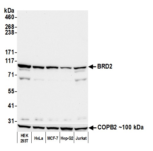
(Detection of human BRD2 by western blot. Samples: Whole cell lysate (50 ug) from HEK293T, HeLa, MCF-7, Hep-G2, and Jurkat cells prepared using NETN lysis buffer. Antibody: Mouse anti-BRD2 monoclonal antibody [12C2E4] (AAA213500 lot 3) used at 1:1000. Secondary: HRP-conjugated goat anti-mouse IgG . Detection: Chemiluminescence with an exposure time of 30 seconds. Lower Panel: Rabbit anti-COPB2 antibody .)
WB (Western Blot)
(Detection of human BRD2 by western blot. Samples: Whole cell lysate (50 ug) from HEK293T, HeLa, MCF-7, Hep-G2, and Jurkat cells prepared using NETN lysis buffer. Antibody: Mouse anti-BRD2 monoclonal antibody [12C2E4] (AAA213500 lot 3) used at 1:1000. Secondary: HRP-conjugated goat anti-mouse IgG . Detection: Chemiluminescence with an exposure time of 30 seconds. Lower Panel: Rabbit anti-COPB2 antibody .)
BRD2, Monoclonal Antibody (Cat# AAA213500)
 WB (Western Blot)
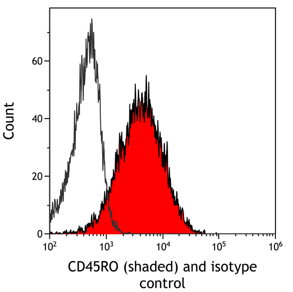
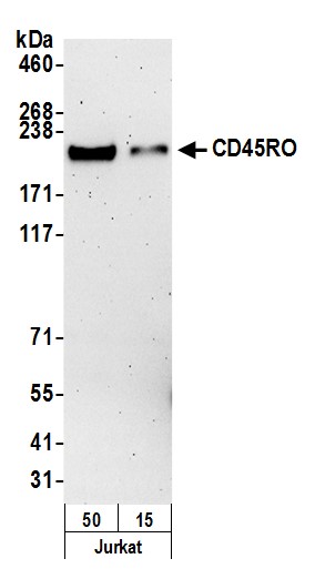
(Detection of human CD45RO by western blot. Samples: Whole cell lysate (15 and 50 ug) from Jurkat cells prepared using NETN lysis buffer. Antibody: mouse anti-CD45RO antibody [UCHL-1] (AAA213504 Lot 1) used at 1:250. Secondary: HRP-conjugated goat anti-mouse IgG . Detection: Chemiluminescence with an exposure time of 3 minutes.)
WB (Western Blot)
(Detection of human CD45RO by western blot. Samples: Whole cell lysate (15 and 50 ug) from Jurkat cells prepared using NETN lysis buffer. Antibody: mouse anti-CD45RO antibody [UCHL-1] (AAA213504 Lot 1) used at 1:250. Secondary: HRP-conjugated goat anti-mouse IgG . Detection: Chemiluminescence with an exposure time of 3 minutes.)
CD45RO, Monoclonal Antibody (Cat# AAA213504)
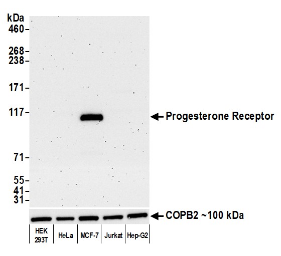 WB (Western Blot)
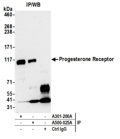
(Detection of human Progesterone Receptor by western blot. Samples: Whole cell lysate (10 ug) from HEK293T, HeLa, MCF-7, Jurkat, and Hep-G2 cells prepared using NETN lysis buffer. Antibody: Mouse anti-Progesterone Receptor monoclonal antibody [188] (AAA213506 lot 1) used at 1:1000. Secondary: HRP-conjugated goat anti-mouse IgG . Detection: Chemiluminescence with an exposure time of 3 minutes. Lower Panel: Rabbit anti-COPB2 antibody .)
WB (Western Blot)
(Detection of human Progesterone Receptor by western blot. Samples: Whole cell lysate (10 ug) from HEK293T, HeLa, MCF-7, Jurkat, and Hep-G2 cells prepared using NETN lysis buffer. Antibody: Mouse anti-Progesterone Receptor monoclonal antibody [188] (AAA213506 lot 1) used at 1:1000. Secondary: HRP-conjugated goat anti-mouse IgG . Detection: Chemiluminescence with an exposure time of 3 minutes. Lower Panel: Rabbit anti-COPB2 antibody .)
Progesterone Receptor, Monoclonal Antibody (Cat# AAA213506)
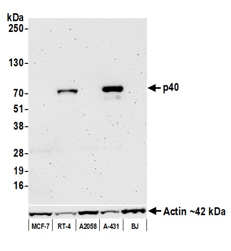 WB (Western Blot)
(Detection of human p40 by western blot. Samples: Whole cell lysate (50 ug) from MCF-7, RT-4, A2058, A-431, and BJ cells prepared using NETN lysis buffer. Antibody: Mouse anti-p40 monoclonal antibody [BC28] (AAA213508 lot 1) used at 1:1000. Secondary: HRP-conjugated goat anti-mouse IgG . Detection: Chemiluminescence with an exposure time of 3 minutes. Lower Panel: Rabbit anti-Actin recombinant monoclonal antibody .)
WB (Western Blot)
(Detection of human p40 by western blot. Samples: Whole cell lysate (50 ug) from MCF-7, RT-4, A2058, A-431, and BJ cells prepared using NETN lysis buffer. Antibody: Mouse anti-p40 monoclonal antibody [BC28] (AAA213508 lot 1) used at 1:1000. Secondary: HRP-conjugated goat anti-mouse IgG . Detection: Chemiluminescence with an exposure time of 3 minutes. Lower Panel: Rabbit anti-Actin recombinant monoclonal antibody .)
p40, Monoclonal Antibody (Cat# AAA213508)
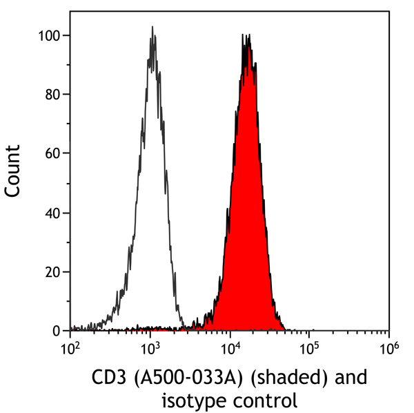
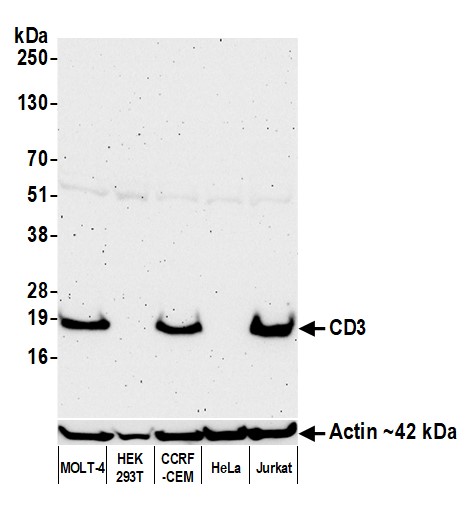 WB (Western Blot)
(Detection of human CD3E by western blot. Samples: Whole cell lysate (50 ug) from MOLT-4, HEK293T, CCRF-CEM, HeLa, and Jurkat cells prepared using NETN lysis buffer. Antibody: Mouse anti-CD3E monoclonal antibody [UCHT1] (AAA213512 lot 1) used at 1:1000. Secondary: HRP-conjugated goat anti-mouse IgG . Detection: Chemiluminescence with an exposure time of 3 minutes. Lower Panel: Rabbit anti-Actin recombinant monoclonal antibody .)
WB (Western Blot)
(Detection of human CD3E by western blot. Samples: Whole cell lysate (50 ug) from MOLT-4, HEK293T, CCRF-CEM, HeLa, and Jurkat cells prepared using NETN lysis buffer. Antibody: Mouse anti-CD3E monoclonal antibody [UCHT1] (AAA213512 lot 1) used at 1:1000. Secondary: HRP-conjugated goat anti-mouse IgG . Detection: Chemiluminescence with an exposure time of 3 minutes. Lower Panel: Rabbit anti-Actin recombinant monoclonal antibody .)
CD3E, Monoclonal Antibody (Cat# AAA213512)
 WB (Western Blot)
(Detection of human HLA-DR/DP/DQ by western blot. Samples: Whole cell lysate (50 ug) from Ramos, KG-1, and Raji (10 ug) cells prepared using NETN lysis buffer. Antibody: Mouse anti-HLA-DR/DP/DQ monoclonal antibody [CR3-43] (AAA213513 lot 1) used at 1:1000. Secondary: HRP-conjugated goat anti-mouse IgG . Detection: Chemiluminescence with an exposure time of 30 seconds. Lower Panel: Rabbit anti-Actin recombinant monoclonal antibody .)
WB (Western Blot)
(Detection of human HLA-DR/DP/DQ by western blot. Samples: Whole cell lysate (50 ug) from Ramos, KG-1, and Raji (10 ug) cells prepared using NETN lysis buffer. Antibody: Mouse anti-HLA-DR/DP/DQ monoclonal antibody [CR3-43] (AAA213513 lot 1) used at 1:1000. Secondary: HRP-conjugated goat anti-mouse IgG . Detection: Chemiluminescence with an exposure time of 30 seconds. Lower Panel: Rabbit anti-Actin recombinant monoclonal antibody .)
HLA-DR/DP/DQ, Monoclonal Antibody (Cat# AAA213513)
 WB (Western Blot)
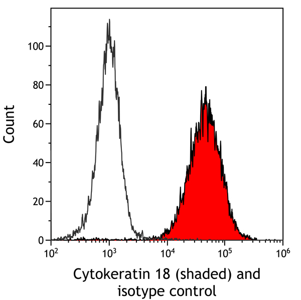
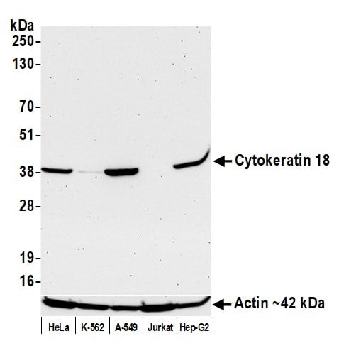
(Detection of human Cytokeratin 18 by western blot. Samples: Whole cell lysate (50 ug) from HeLa, K-562, A-549, Jurkat, and Hep-G2 cells prepared using NETN lysis buffer. Antibody: Mouse anti-Cytokeratin 18 monoclonal antibody [LDK18] (AAA213514 lot 1) used at 1:1000. Secondary: HRP-conjugated goat anti-mouse IgG . Detection: Chemiluminescence with an exposure time of 75 seconds. Lower Panel: Rabbit anti-Actin recombinant monoclonal antibody .)
WB (Western Blot)
(Detection of human Cytokeratin 18 by western blot. Samples: Whole cell lysate (50 ug) from HeLa, K-562, A-549, Jurkat, and Hep-G2 cells prepared using NETN lysis buffer. Antibody: Mouse anti-Cytokeratin 18 monoclonal antibody [LDK18] (AAA213514 lot 1) used at 1:1000. Secondary: HRP-conjugated goat anti-mouse IgG . Detection: Chemiluminescence with an exposure time of 75 seconds. Lower Panel: Rabbit anti-Actin recombinant monoclonal antibody .)
Cytokeratin 18, Monoclonal Antibody (Cat# AAA213514)

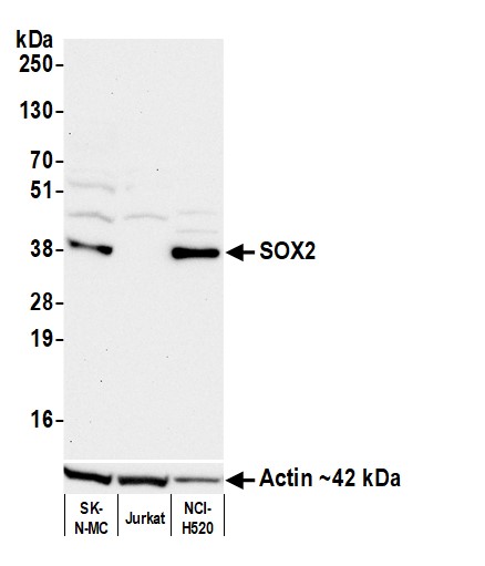 WB (Western Blot)
(Detection of human SOX2 by western blot. Samples: Whole cell lysate (50 ug) from SK-N-MC, Jurkat, and NCI-H520 (10 ug) cells prepared using NETN lysis buffer. Antibody: Mouse anti-SOX2 monoclonal antibody [BC36] (AAA213516 lot 1) used at 1:1000. Secondary: HRP-conjugated goat anti-mouse IgG . Detection: Chemiluminescence with an exposure time of 75 seconds. Lower Panel: Rabbit anti-Actin recombinant monoclonal antibody .)
WB (Western Blot)
(Detection of human SOX2 by western blot. Samples: Whole cell lysate (50 ug) from SK-N-MC, Jurkat, and NCI-H520 (10 ug) cells prepared using NETN lysis buffer. Antibody: Mouse anti-SOX2 monoclonal antibody [BC36] (AAA213516 lot 1) used at 1:1000. Secondary: HRP-conjugated goat anti-mouse IgG . Detection: Chemiluminescence with an exposure time of 75 seconds. Lower Panel: Rabbit anti-Actin recombinant monoclonal antibody .)
SOX2, Monoclonal Antibody (Cat# AAA213516)
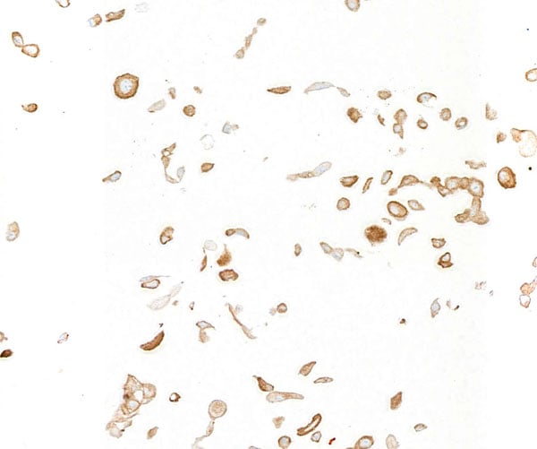
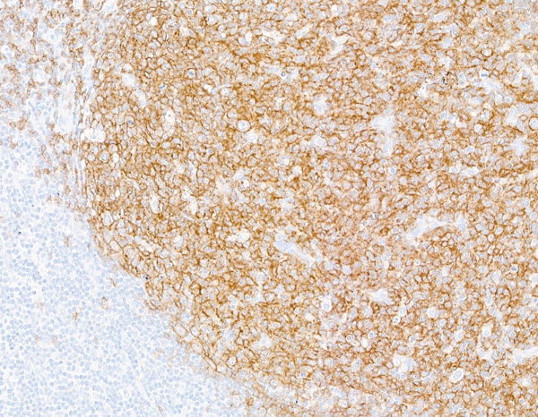
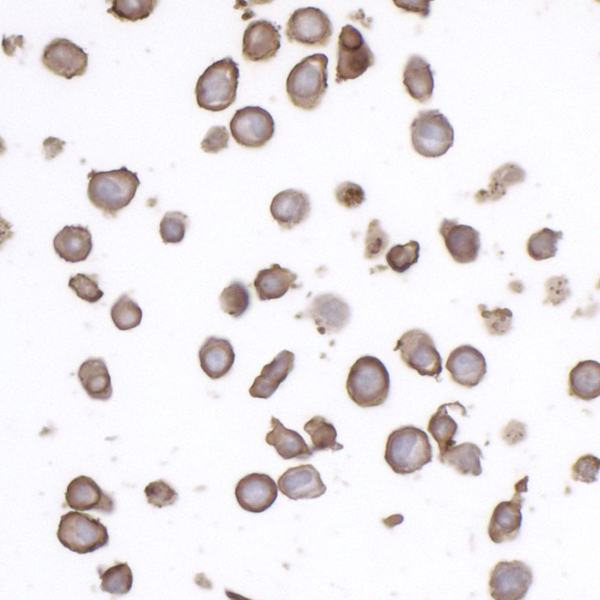
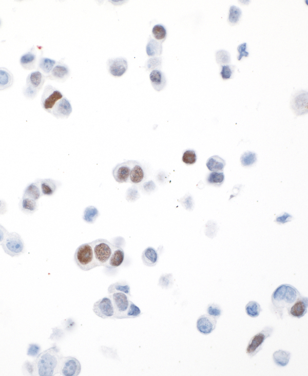
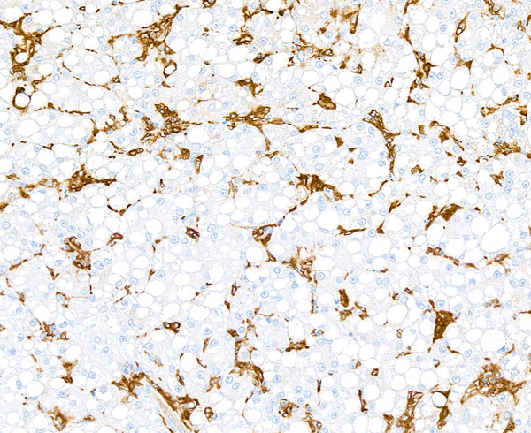
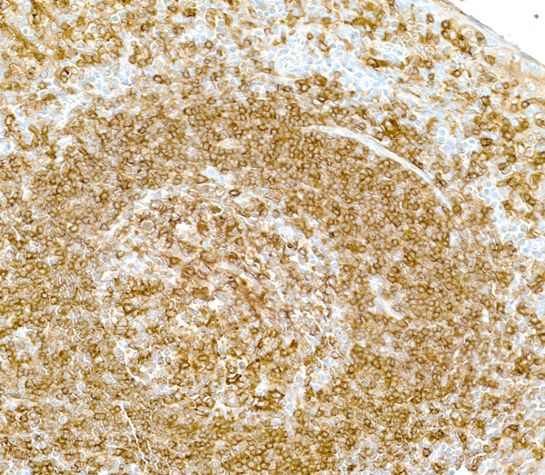
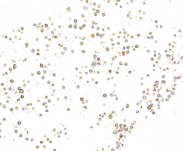
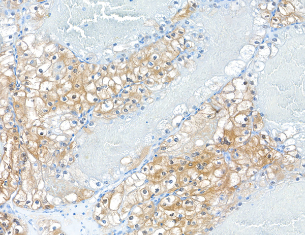
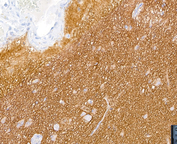
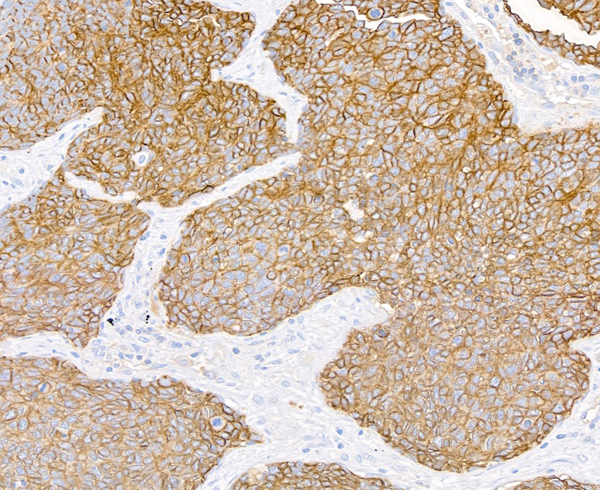
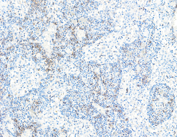
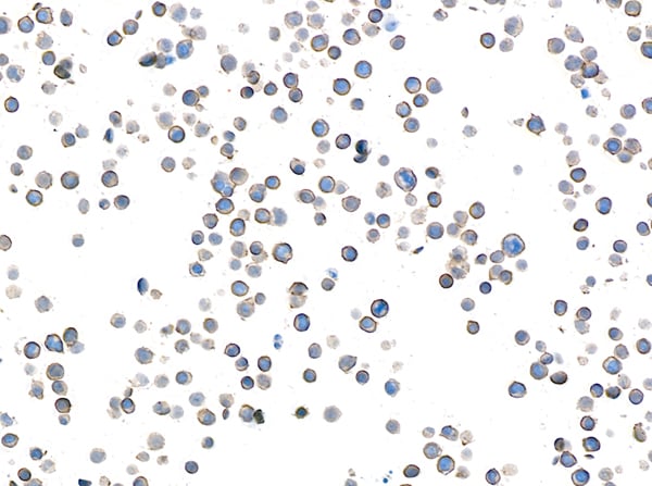
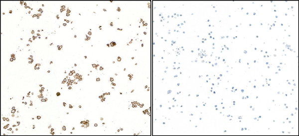
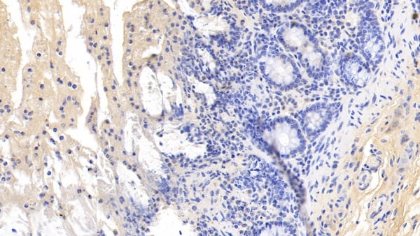
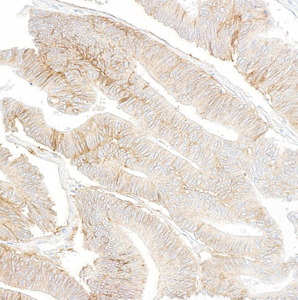 IHC (Immunohiostchemistry)
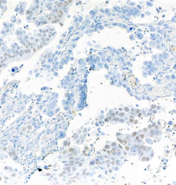
(Detection of human IGF1R by immunohistochemistry. Sample: FFPE section of colon carcinoma. Antibody: Mouse anti-IGF1R monoclonal antibody [BC10] (AAA213517 lot 1). Secondary: HRP-conjugated goat anti-mouse IgG .)
IHC (Immunohiostchemistry)
(Detection of human IGF1R by immunohistochemistry. Sample: FFPE section of colon carcinoma. Antibody: Mouse anti-IGF1R monoclonal antibody [BC10] (AAA213517 lot 1). Secondary: HRP-conjugated goat anti-mouse IgG .)
IGF1R, Monoclonal Antibody (Cat# AAA213517)
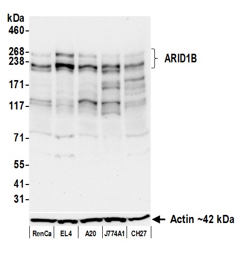
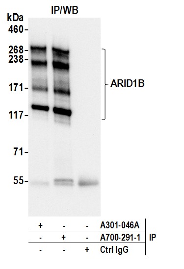
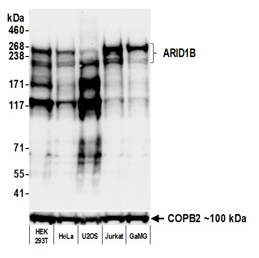 WB (Western Blot)
(Detection of human ARID1B by western blot. Samples: Whole cell lysate (50 ug) from HEK293T, HeLa, U2OS, Jurkat, and GaMG cells prepared using NETN lysis buffer. Antibody: Rabbit anti-ARID1B recombinant monoclonal antibody (AAA213690 lot 1) used at 1:1000. Secondary: HRP-conjugated goat anti-rabbit IgG . Detection: Chemiluminescence with an exposure time of 30 seconds. Lower Panel: Rabbit anti-COPB2 antibody .)
WB (Western Blot)
(Detection of human ARID1B by western blot. Samples: Whole cell lysate (50 ug) from HEK293T, HeLa, U2OS, Jurkat, and GaMG cells prepared using NETN lysis buffer. Antibody: Rabbit anti-ARID1B recombinant monoclonal antibody (AAA213690 lot 1) used at 1:1000. Secondary: HRP-conjugated goat anti-rabbit IgG . Detection: Chemiluminescence with an exposure time of 30 seconds. Lower Panel: Rabbit anti-COPB2 antibody .)
ARID1B, Monoclonal Recombinant Antibody (Cat# AAA213690)

 WB (Western Blot)
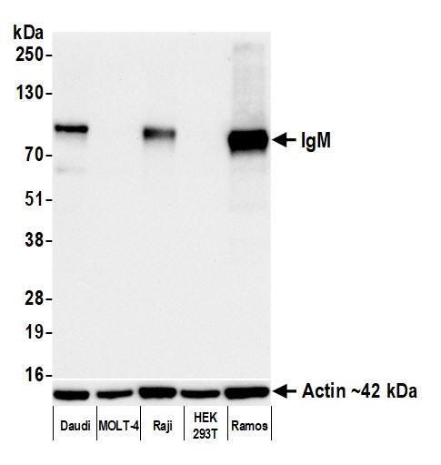
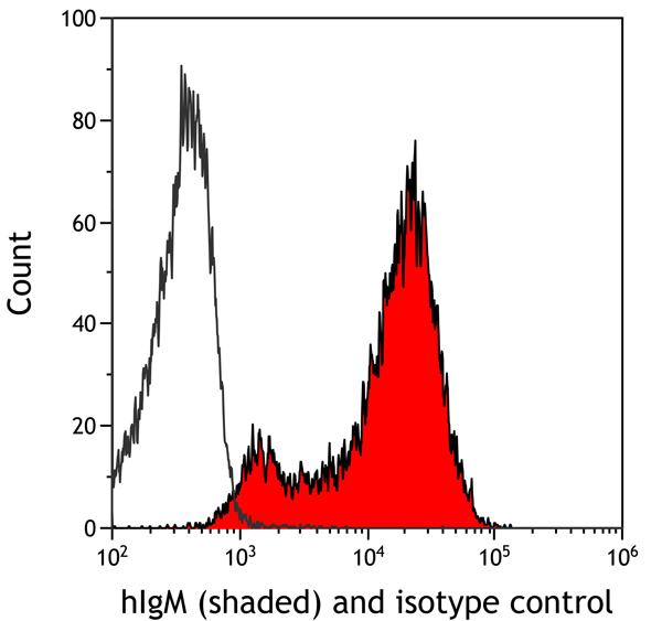
(Detection of mouse IgM by western blot. Samples: Whole cell lysate (50 ug) from NIH 3T3, WEHI-231, CT26, CH27, and TCMK-1 cells prepared using NETN lysis buffer. Antibody: Rabbit anti-IgM recombinant monoclonal antibody (AAA213693 lot 1) used at 1:1000. Secondary: HRP-conjugated goat anti-rabbit IgG . Detection: Chemiluminescence with an exposure time of 1 second. Lower Panel: Rabbit anti-Actin recombinant monoclonal antibody .)
WB (Western Blot)
(Detection of mouse IgM by western blot. Samples: Whole cell lysate (50 ug) from NIH 3T3, WEHI-231, CT26, CH27, and TCMK-1 cells prepared using NETN lysis buffer. Antibody: Rabbit anti-IgM recombinant monoclonal antibody (AAA213693 lot 1) used at 1:1000. Secondary: HRP-conjugated goat anti-rabbit IgG . Detection: Chemiluminescence with an exposure time of 1 second. Lower Panel: Rabbit anti-Actin recombinant monoclonal antibody .)
IgM, Monoclonal Recombinant Antibody (Cat# AAA213693)
 WB (Western Blot)
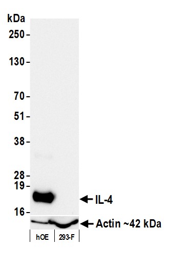
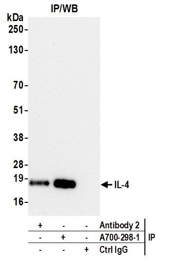
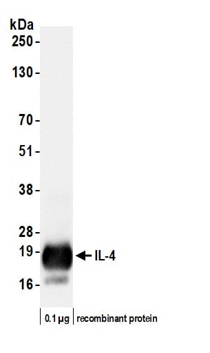
(Detection of human IL-4 by western blot. Samples: Human recombinant IL-4 (0.1 ug) protein. Antibody: Rabbit anti-IL-4 recombinant monoclonal antibody (AAA213696 lot 1) used at 1:1000. Secondary: HRP-conjugated goat anti-rabbit IgG . Detection: Chemiluminescence with an exposure time of 1 second.)
WB (Western Blot)
(Detection of human IL-4 by western blot. Samples: Human recombinant IL-4 (0.1 ug) protein. Antibody: Rabbit anti-IL-4 recombinant monoclonal antibody (AAA213696 lot 1) used at 1:1000. Secondary: HRP-conjugated goat anti-rabbit IgG . Detection: Chemiluminescence with an exposure time of 1 second.)
IL-4, Monoclonal Recombinant Antibody (Cat# AAA213696)

 WB (Western Blot)
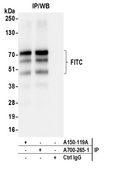
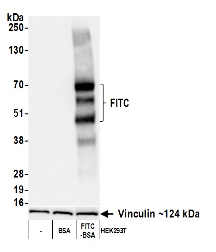
(Detection of FITC by western blot. Samples: Whole cell lysate (25 ug) from HEK293T cells (-), HEK293T + BSA and HEK293T + FITC conjugated BSA. Antibody: Rabbit anti-FITC recombinant monoclonal antibody (AAA213701 lot 1) used at 1:1000. Secondary: HRP-conjugated goat anti-rabbit IgG . Detection: Chemiluminescence with an exposure time of 1 second. Lower Panel: Rabbit anti-Vinculin antibody .)
WB (Western Blot)
(Detection of FITC by western blot. Samples: Whole cell lysate (25 ug) from HEK293T cells (-), HEK293T + BSA and HEK293T + FITC conjugated BSA. Antibody: Rabbit anti-FITC recombinant monoclonal antibody (AAA213701 lot 1) used at 1:1000. Secondary: HRP-conjugated goat anti-rabbit IgG . Detection: Chemiluminescence with an exposure time of 1 second. Lower Panel: Rabbit anti-Vinculin antibody .)
FITC, Monoclonal Recombinant Antibody (Cat# AAA213701)
 WB (Western Blot)
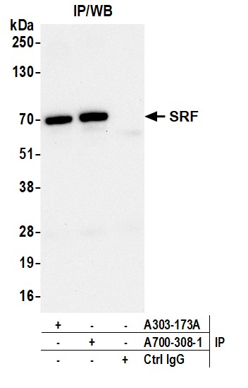
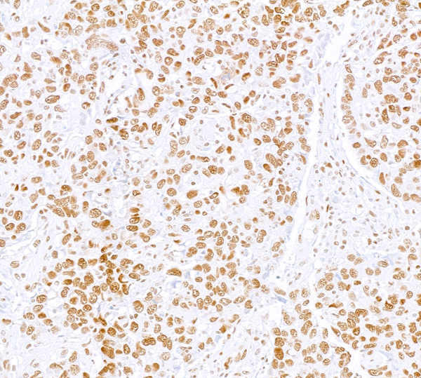
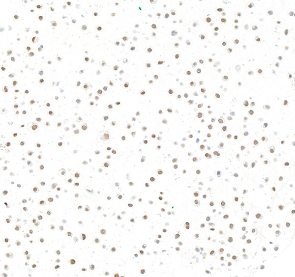
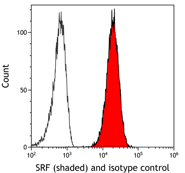
(Detection of human SRF by western blot. Samples: Whole cell lysate (10 ug) from Jurkat, Hep-G2, HeLa, RKO, and MDA-MB-231 cells prepared using NETN lysis buffer. Antibody: Rabbit anti-SRF recombinant monoclonal antibody (AAA213704 lot 1) used at 1:1000. Secondary: HRP-conjugated goat anti-rabbit IgG . Detection: Chemiluminescence with an exposure time of 3 minutes. Lower Panel: Rabbit anti-Actin recombinant monoclonal antibody .)
WB (Western Blot)
(Detection of human SRF by western blot. Samples: Whole cell lysate (10 ug) from Jurkat, Hep-G2, HeLa, RKO, and MDA-MB-231 cells prepared using NETN lysis buffer. Antibody: Rabbit anti-SRF recombinant monoclonal antibody (AAA213704 lot 1) used at 1:1000. Secondary: HRP-conjugated goat anti-rabbit IgG . Detection: Chemiluminescence with an exposure time of 3 minutes. Lower Panel: Rabbit anti-Actin recombinant monoclonal antibody .)
SRF, Monoclonal Recombinant Antibody (Cat# AAA213704)
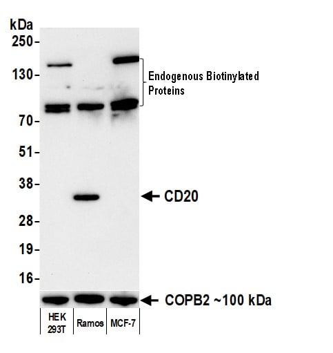
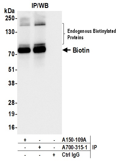
 WB (Western Blot)
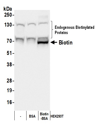
(Detection of Biotin by western blot. Samples: Whole cell lysate (50 ug) from HEK293T cells with or without BSA (0.01 ug) or Biotin-BSA (0.01 ug) added. Antibody: Rabbit anti-Biotin recombinant monoclonal antibody (AAA213706 lot 1) used at 1:1000. Secondary: HRP-conjugated goat anti-rabbit IgG . Detection: Chemiluminescence with an exposure time of 3 seconds.)
WB (Western Blot)
(Detection of Biotin by western blot. Samples: Whole cell lysate (50 ug) from HEK293T cells with or without BSA (0.01 ug) or Biotin-BSA (0.01 ug) added. Antibody: Rabbit anti-Biotin recombinant monoclonal antibody (AAA213706 lot 1) used at 1:1000. Secondary: HRP-conjugated goat anti-rabbit IgG . Detection: Chemiluminescence with an exposure time of 3 seconds.)
Biotin, Monoclonal Recombinant Antibody (Cat# AAA213706)
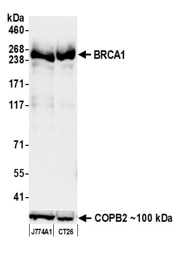 WB (Western Blot)
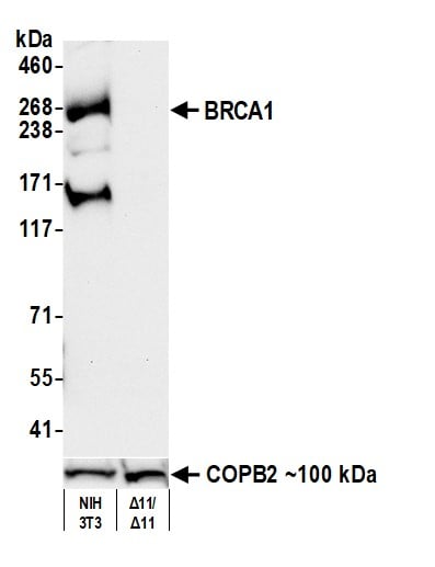
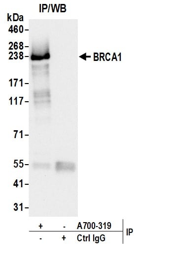
(Detection of mouse BRCA1 by western blot. Samples: Whole cell lysate (50 ug) from J774A1 and CT26 cells prepared using NETN lysis buffer. Antibody: Rabbit anti-BRCA1 recombinant monoclonal antibody (AAA213708 lot 1) used at 1:1000. Secondary: HRP-conjugated goat anti-rabbit IgG . Detection: Chemiluminescence with an exposure time of 30 seconds. Lower Panel: Rabbit anti-COPB2 antibody .)
WB (Western Blot)
(Detection of mouse BRCA1 by western blot. Samples: Whole cell lysate (50 ug) from J774A1 and CT26 cells prepared using NETN lysis buffer. Antibody: Rabbit anti-BRCA1 recombinant monoclonal antibody (AAA213708 lot 1) used at 1:1000. Secondary: HRP-conjugated goat anti-rabbit IgG . Detection: Chemiluminescence with an exposure time of 30 seconds. Lower Panel: Rabbit anti-COPB2 antibody .)
BRCA1, Monoclonal Recombinant Antibody (Cat# AAA213708)
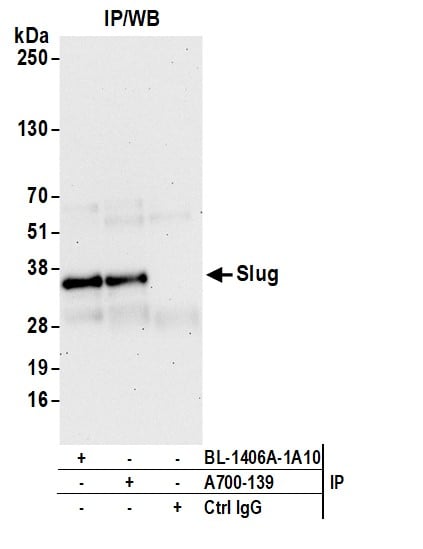
 WB (Western Blot)
(Detection of human Slug by western blot. Samples: Whole cell lysate (50 ug) from MCF-7, HeLa, HEK293T, MDA-MB-435, Jurkat, MDA-MB-231, Hep-G2, and K-562 cells prepared using NETN lysis buffer. Antibody: Rabbit anti-Slug recombinant monoclonal antibody (AAA213600 Lot 1) used at 1:1000. Secondary: HRP-conjugated goat anti-rabbit IgG . Detection: Chemiluminescence with an exposure time of 3 seconds. Lower Panel: Rabbit anti-COPB2 antibody .)
WB (Western Blot)
(Detection of human Slug by western blot. Samples: Whole cell lysate (50 ug) from MCF-7, HeLa, HEK293T, MDA-MB-435, Jurkat, MDA-MB-231, Hep-G2, and K-562 cells prepared using NETN lysis buffer. Antibody: Rabbit anti-Slug recombinant monoclonal antibody (AAA213600 Lot 1) used at 1:1000. Secondary: HRP-conjugated goat anti-rabbit IgG . Detection: Chemiluminescence with an exposure time of 3 seconds. Lower Panel: Rabbit anti-COPB2 antibody .)
Slug, Monoclonal Recombinant Antibody (Cat# AAA213600)
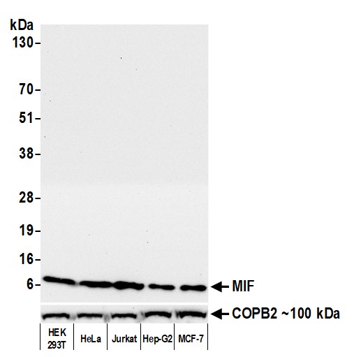 WB (Western Blot)
(Detection of human MIF by western blot. Samples: Whole cell lysate (25 ug) from HEK293T, HeLa, Jurkat, Hep-G2, and MCF-7 cells prepared using NETN lysis buffer. Antibody: Rabbit anti-MIF recombinant monoclonal antibody (AAA213602 lot 1) used at 1:1000. Secondary: HRP-conjugated goat anti-rabbit IgG . Detection: Chemiluminescence with an exposure time of 10 seconds. Lower Panel: Rabbit anti-COPB2 antibody .)
WB (Western Blot)
(Detection of human MIF by western blot. Samples: Whole cell lysate (25 ug) from HEK293T, HeLa, Jurkat, Hep-G2, and MCF-7 cells prepared using NETN lysis buffer. Antibody: Rabbit anti-MIF recombinant monoclonal antibody (AAA213602 lot 1) used at 1:1000. Secondary: HRP-conjugated goat anti-rabbit IgG . Detection: Chemiluminescence with an exposure time of 10 seconds. Lower Panel: Rabbit anti-COPB2 antibody .)
MIF, Monoclonal Recombinant Antibody (Cat# AAA213602)
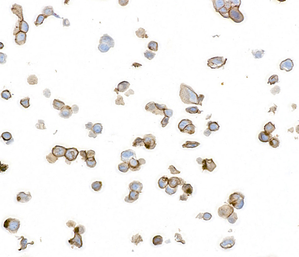
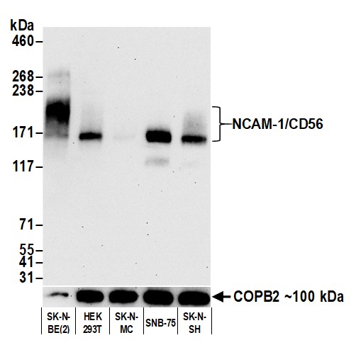 WB (Western Blot)
(Detection of human NCAM-1/CD56 by western blot. Samples: Whole cell lysate from SK-N-BE(2), HEK293T, SK-N-MC, SNB-75, and SK-N-SH cells prepared using NETN lysis buffer. Antibody: Rabbit anti-NCAM-1/CD56 recombinant monoclonal antibody (AAA213610 lot 1) used at 1:1000. Secondary: HRP-conjugated goat anti-rabbit IgG . Detection: Chemiluminescence with an exposure time of 30 seconds. Lower Panel: Rabbit anti-COPB2 antibody .)
WB (Western Blot)
(Detection of human NCAM-1/CD56 by western blot. Samples: Whole cell lysate from SK-N-BE(2), HEK293T, SK-N-MC, SNB-75, and SK-N-SH cells prepared using NETN lysis buffer. Antibody: Rabbit anti-NCAM-1/CD56 recombinant monoclonal antibody (AAA213610 lot 1) used at 1:1000. Secondary: HRP-conjugated goat anti-rabbit IgG . Detection: Chemiluminescence with an exposure time of 30 seconds. Lower Panel: Rabbit anti-COPB2 antibody .)
NCAM-1/CD56, Monoclonal Recombinant Antibody (Cat# AAA213610)
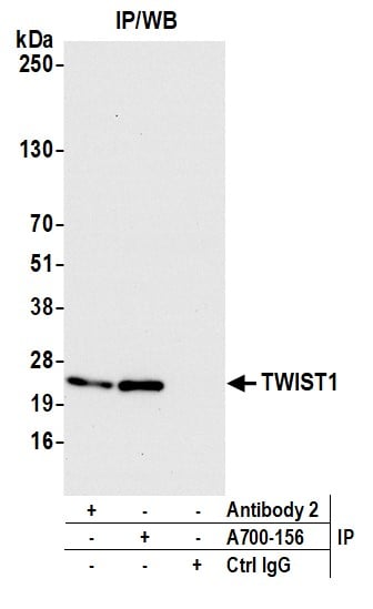
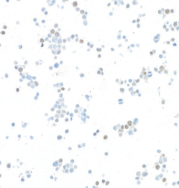
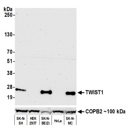 WB (Western Blot)
(Detection of human TWIST1 by western blot. Samples: Whole cell lysate (50 ug) from SK-N-SH, HEK293T, SK-N-BE(2), HeLa, and SK-N-MC cells prepared using NETN lysis buffer. Antibody: Rabbit anti-TWIST1 recombinant monoclonal antibody (AAA213613 lot 1) used at 1:1000. Secondary: HRP-conjugated goat anti-rabbit IgG . Detection: Chemiluminescence with an exposure time of 75 seconds. Lower Panel: Rabbit anti-COPB2 antibody .)
WB (Western Blot)
(Detection of human TWIST1 by western blot. Samples: Whole cell lysate (50 ug) from SK-N-SH, HEK293T, SK-N-BE(2), HeLa, and SK-N-MC cells prepared using NETN lysis buffer. Antibody: Rabbit anti-TWIST1 recombinant monoclonal antibody (AAA213613 lot 1) used at 1:1000. Secondary: HRP-conjugated goat anti-rabbit IgG . Detection: Chemiluminescence with an exposure time of 75 seconds. Lower Panel: Rabbit anti-COPB2 antibody .)
TWIST, Monoclonal Recombinant Antibody (Cat# AAA213613)
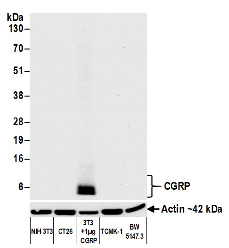
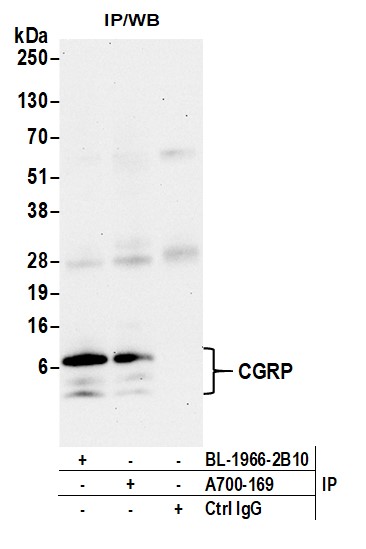
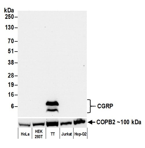 WB (Western Blot)
(Detection of human CGRP by western blot. Samples: Whole cell lysate (50 ug) from HeLa, HEK293T, TT, Jurkat, and Hep-G2 cells prepared using NETN lysis buffer. Antibody: Rabbit anti-CGRP recombinant monoclonal antibody (AAA213623 lot 1) used at 1:1000. Secondary: HRP-conjugated goat anti-rabbit IgG . Detection: Chemiluminescence with an exposure time of 3 seconds. Lower Panel: Rabbit anti-COPB2 antibody .)
WB (Western Blot)
(Detection of human CGRP by western blot. Samples: Whole cell lysate (50 ug) from HeLa, HEK293T, TT, Jurkat, and Hep-G2 cells prepared using NETN lysis buffer. Antibody: Rabbit anti-CGRP recombinant monoclonal antibody (AAA213623 lot 1) used at 1:1000. Secondary: HRP-conjugated goat anti-rabbit IgG . Detection: Chemiluminescence with an exposure time of 3 seconds. Lower Panel: Rabbit anti-COPB2 antibody .)
CGRP, Monoclonal Recombinant Antibody (Cat# AAA213623)
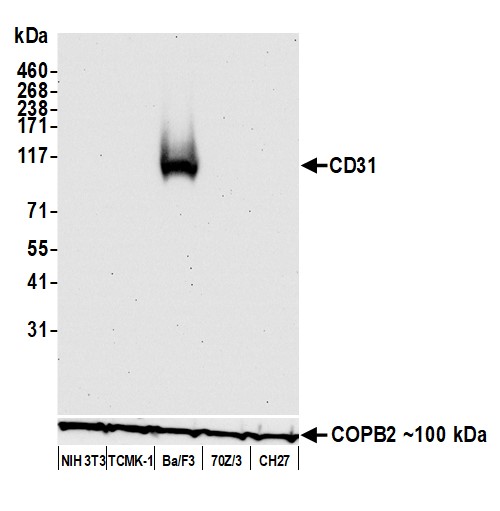
 WB (Western Blot)
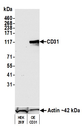
(Detection of mouse CD31 by western blot. Samples: Whole cell lysate (50 ug) from HEK293f and HEK293f overexpressing mouse CD31 prepared using NETN lysis buffer. Antibody: Rabbit anti-CD31 recombinant monoclonal antibody (AAA213629 lot 1) used at 1:1000. Secondary: HRP-conjugated goat anti-rabbit IgG . Detection: Chemiluminescence with an exposure time of 30 seconds. Lower Panel: Rabbit anti-Actin recombinant monoclonal antibody .)
WB (Western Blot)
(Detection of mouse CD31 by western blot. Samples: Whole cell lysate (50 ug) from HEK293f and HEK293f overexpressing mouse CD31 prepared using NETN lysis buffer. Antibody: Rabbit anti-CD31 recombinant monoclonal antibody (AAA213629 lot 1) used at 1:1000. Secondary: HRP-conjugated goat anti-rabbit IgG . Detection: Chemiluminescence with an exposure time of 30 seconds. Lower Panel: Rabbit anti-Actin recombinant monoclonal antibody .)
CD31, Monoclonal Recombinant Antibody (Cat# AAA213629)
 WB (Western Blot)
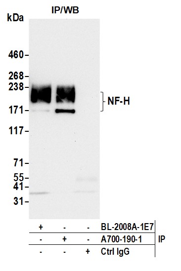
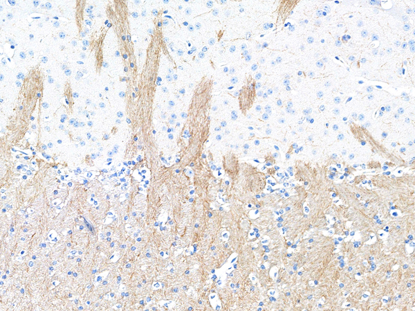
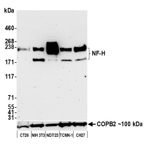
(Detection of mouse NF-H by western blot. Samples: Whole cell lysate (50 ug) from CT26, NIH 3T3, ND7/23 (10 ug), TCMK-1, and CH27 cells prepared using NETN lysis buffer. Antibody: Rabbit anti-NF-H recombinant monoclonal antibody (AAA213634 lot 1) used at 1:1000. Secondary: HRP-conjugated goat anti-rabbit IgG . Detection: Chemiluminescence with an exposure time of 10 seconds. Lower Panel: Rabbit anti-COPB2 antibody .)
WB (Western Blot)
(Detection of mouse NF-H by western blot. Samples: Whole cell lysate (50 ug) from CT26, NIH 3T3, ND7/23 (10 ug), TCMK-1, and CH27 cells prepared using NETN lysis buffer. Antibody: Rabbit anti-NF-H recombinant monoclonal antibody (AAA213634 lot 1) used at 1:1000. Secondary: HRP-conjugated goat anti-rabbit IgG . Detection: Chemiluminescence with an exposure time of 10 seconds. Lower Panel: Rabbit anti-COPB2 antibody .)
NF-H, Monoclonal Recombinant Antibody (Cat# AAA213634)
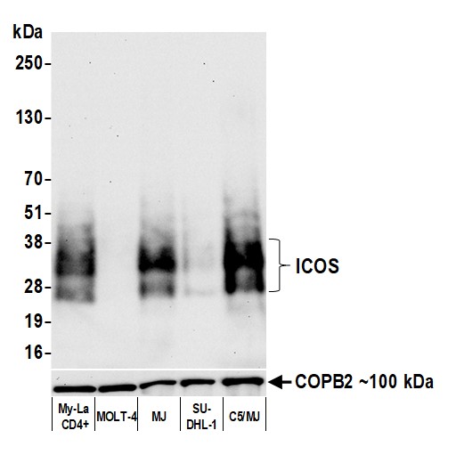 WB (Western Blot)
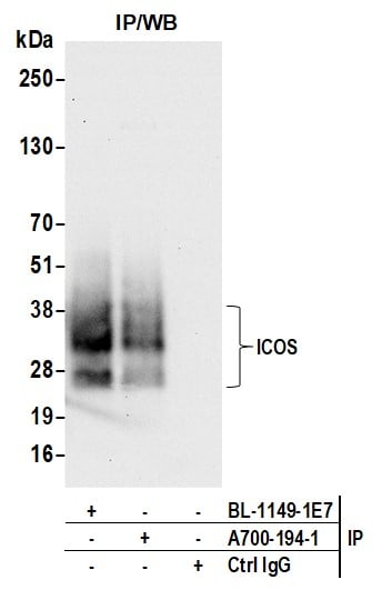
(Detection of human ICOS by western blot. Samples: Whole cell lysate (10 ug) from My-La CD4+, MOLT-4, MJ, SU-DHL-1, and C5/MJ cells prepared using NETN lysis buffer. Antibody: Rabbit anti-ICOS recombinant monoclonal antibody (AAA213635 lot 1) used at 1:1000. Secondary: HRP-conjugated goat anti-rabbit IgG . Detection: Chemiluminescence with an exposure time of 10 seconds. Lower Panel: Rabbit anti-COPB2 antibody .)
WB (Western Blot)
(Detection of human ICOS by western blot. Samples: Whole cell lysate (10 ug) from My-La CD4+, MOLT-4, MJ, SU-DHL-1, and C5/MJ cells prepared using NETN lysis buffer. Antibody: Rabbit anti-ICOS recombinant monoclonal antibody (AAA213635 lot 1) used at 1:1000. Secondary: HRP-conjugated goat anti-rabbit IgG . Detection: Chemiluminescence with an exposure time of 10 seconds. Lower Panel: Rabbit anti-COPB2 antibody .)
ICOS, Monoclonal Recombinant Antibody (Cat# AAA213635)
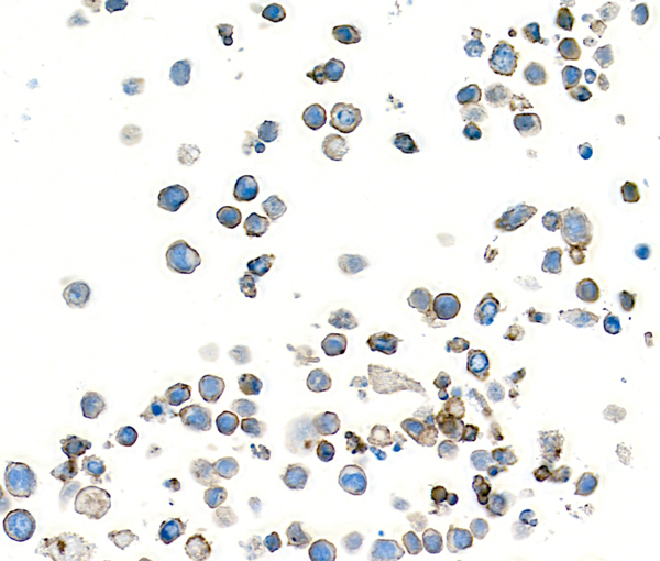
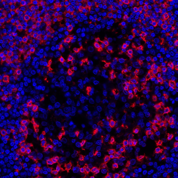
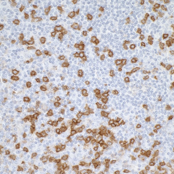
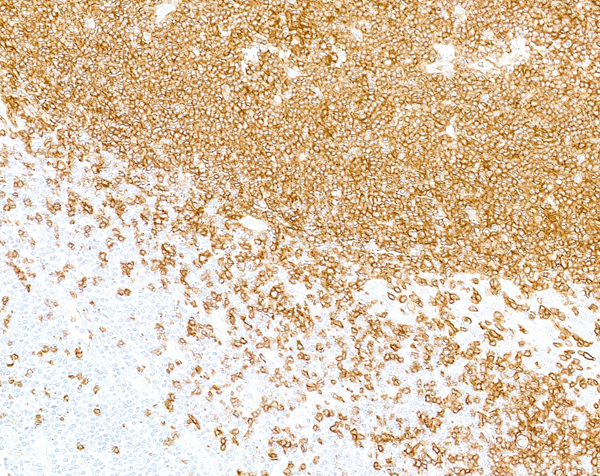
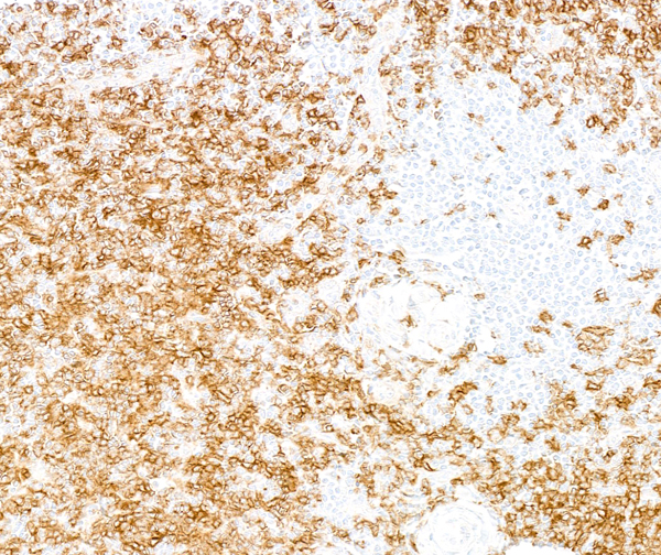
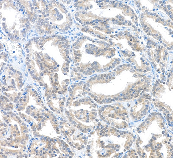
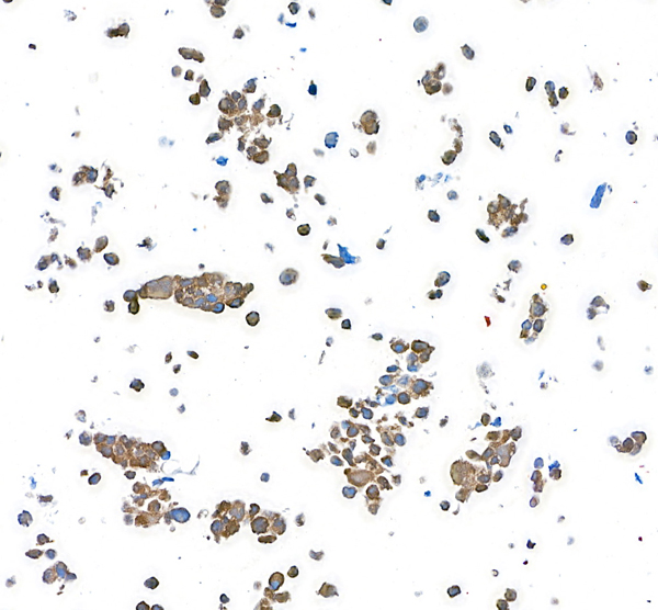
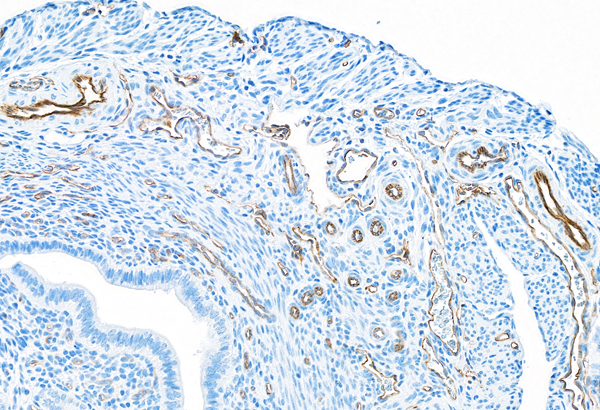
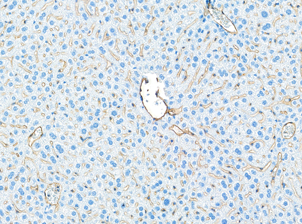
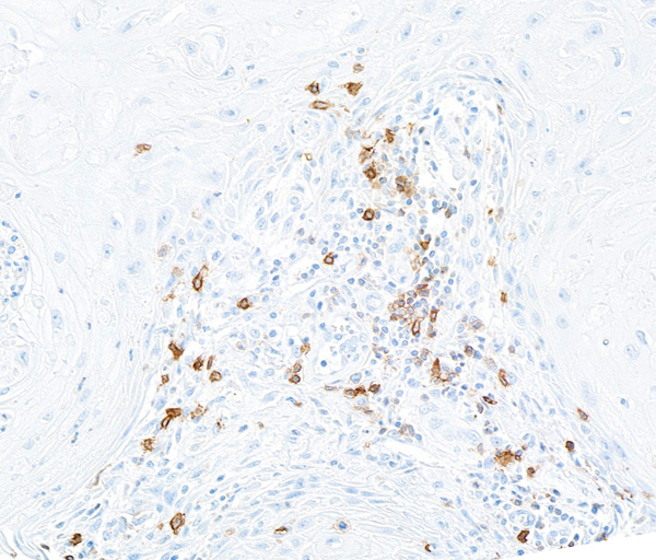
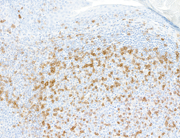
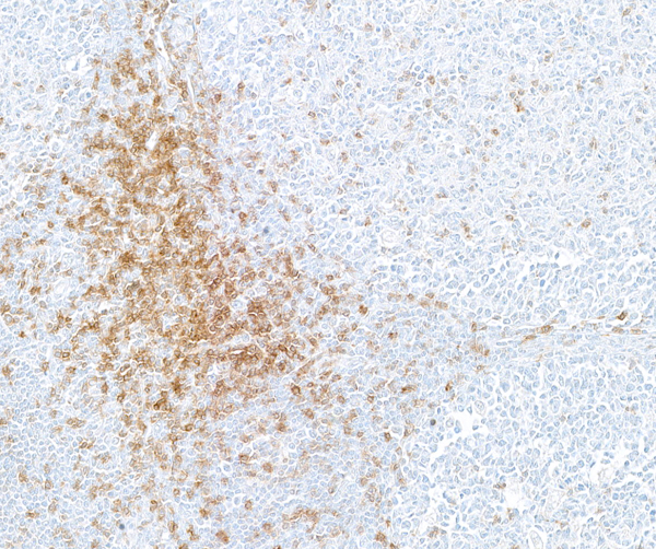
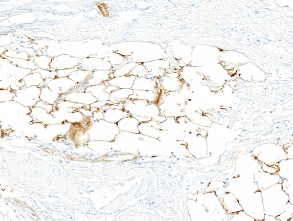
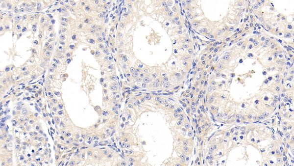
 IHC (Immunohistochemistry)
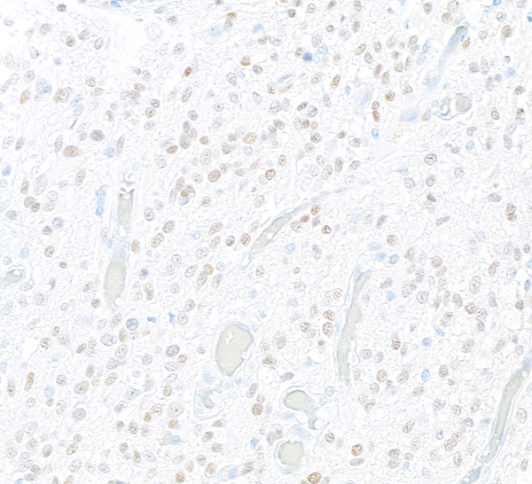
(Detection of human CD28 by immunohistochemistry. Sample: FFPE section of tonsil. Antibody: Rabbit anti-CD28 recombinant monoclonal antibody (AAA213637 lot 1). Secondary: HRP-conjugated goat anti-rabbit IgG .)
IHC (Immunohistochemistry)
(Detection of human CD28 by immunohistochemistry. Sample: FFPE section of tonsil. Antibody: Rabbit anti-CD28 recombinant monoclonal antibody (AAA213637 lot 1). Secondary: HRP-conjugated goat anti-rabbit IgG .)
CD28, Monoclonal Recombinant Antibody (Cat# AAA213637)
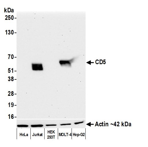 WB (Western Blot)
(Detection of human CD5 by western blot. Samples: Whole cell lysate (10 ug) from HeLa, Jurkat, HEK293T, MOLT-4, and Hep-G2 cells prepared using NETN lysis buffer. Antibody: Rabbit anti-CD5 recombinant monoclonal antibody (AAA213645 lot 1) used at 1:1000. Secondary: HRP-conjugated goat anti-rabbit IgG . Detection: Chemiluminescence with an exposure time of 30 seconds. Lower Panel: Rabbit anti-Actin recombinant monoclonal antibody .)
WB (Western Blot)
(Detection of human CD5 by western blot. Samples: Whole cell lysate (10 ug) from HeLa, Jurkat, HEK293T, MOLT-4, and Hep-G2 cells prepared using NETN lysis buffer. Antibody: Rabbit anti-CD5 recombinant monoclonal antibody (AAA213645 lot 1) used at 1:1000. Secondary: HRP-conjugated goat anti-rabbit IgG . Detection: Chemiluminescence with an exposure time of 30 seconds. Lower Panel: Rabbit anti-Actin recombinant monoclonal antibody .)
CD5, Monoclonal Recombinant Antibody (Cat# AAA213645)
TIF1 Alpha/TRIM24, Monoclonal Recombinant Antibody (Cat# AAA213648)
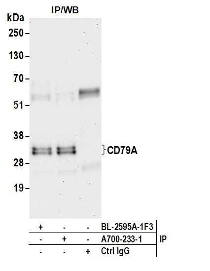

 WB (Western Blot)
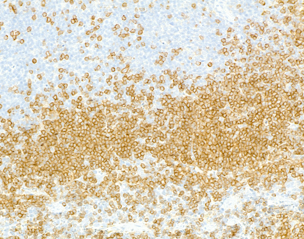
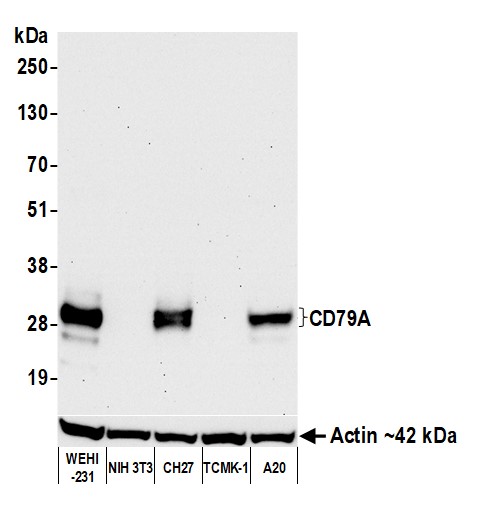
(Detection of mouse CD79A by western blot. Samples: Whole cell lysate (10 ug) from WEHI-231, NIH 3T3, CH27, TCMK-1, and A20 cells prepared using NETN lysis buffer. Antibody: Rabbit anti-CD79A recombinant monoclonal antibody (AAA213652 lot 1) used at 1:1000. Secondary: HRP-conjugated goat anti-rabbit IgG . Detection: Chemiluminescence with an exposure time of 30 seconds. Lower Panel: Rabbit anti-Actin recombinant monoclonal antibody .)
WB (Western Blot)
(Detection of mouse CD79A by western blot. Samples: Whole cell lysate (10 ug) from WEHI-231, NIH 3T3, CH27, TCMK-1, and A20 cells prepared using NETN lysis buffer. Antibody: Rabbit anti-CD79A recombinant monoclonal antibody (AAA213652 lot 1) used at 1:1000. Secondary: HRP-conjugated goat anti-rabbit IgG . Detection: Chemiluminescence with an exposure time of 30 seconds. Lower Panel: Rabbit anti-Actin recombinant monoclonal antibody .)
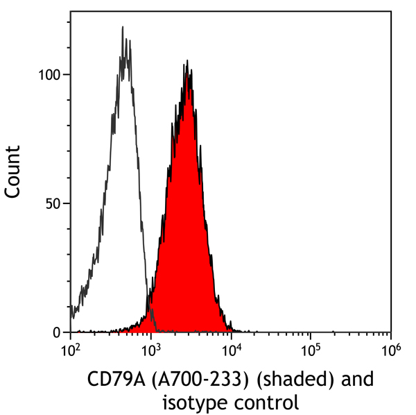
CD79A, Monoclonal Recombinant Antibody (Cat# AAA213652)
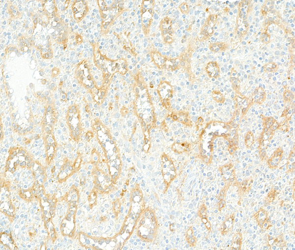

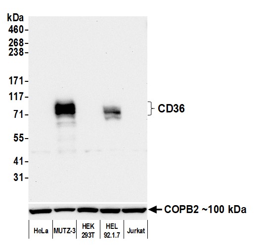 WB (Western Blot)
(Detection of human CD36 by western blot. Samples: Whole cell lysate (50 ug) from HeLa, MUTZ-3, HEK293T, HEL 92.1.7, and Jurkat cells prepared using NETN lysis buffer. Antibody: Rabbit anti-CD36 recombinant monoclonal antibody (AAA213654 lot 1) used at 1:1000. Secondary: HRP-conjugated goat anti-rabbit IgG . Detection: Chemiluminescence with an exposure time of 30 seconds. Lower Panel: Rabbit anti-COPB2 antibody .)
WB (Western Blot)
(Detection of human CD36 by western blot. Samples: Whole cell lysate (50 ug) from HeLa, MUTZ-3, HEK293T, HEL 92.1.7, and Jurkat cells prepared using NETN lysis buffer. Antibody: Rabbit anti-CD36 recombinant monoclonal antibody (AAA213654 lot 1) used at 1:1000. Secondary: HRP-conjugated goat anti-rabbit IgG . Detection: Chemiluminescence with an exposure time of 30 seconds. Lower Panel: Rabbit anti-COPB2 antibody .)
CD36, Monoclonal Recombinant Antibody (Cat# AAA213654)
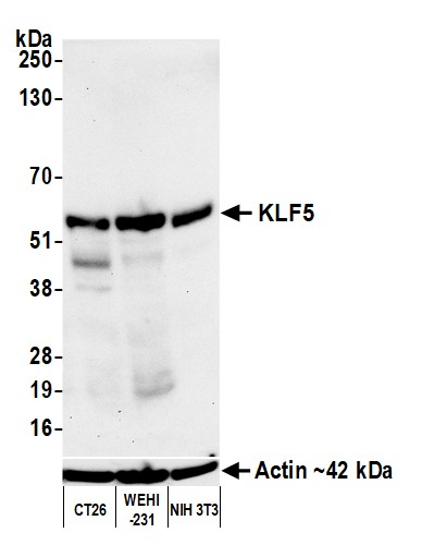
 WB (Western Blot)
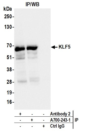
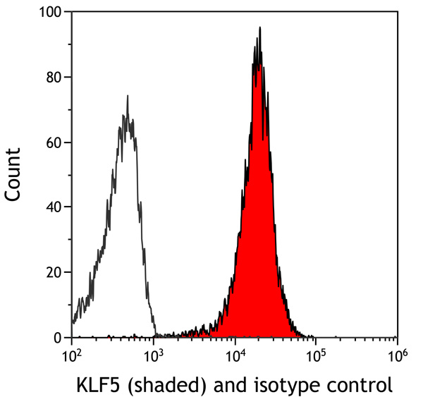
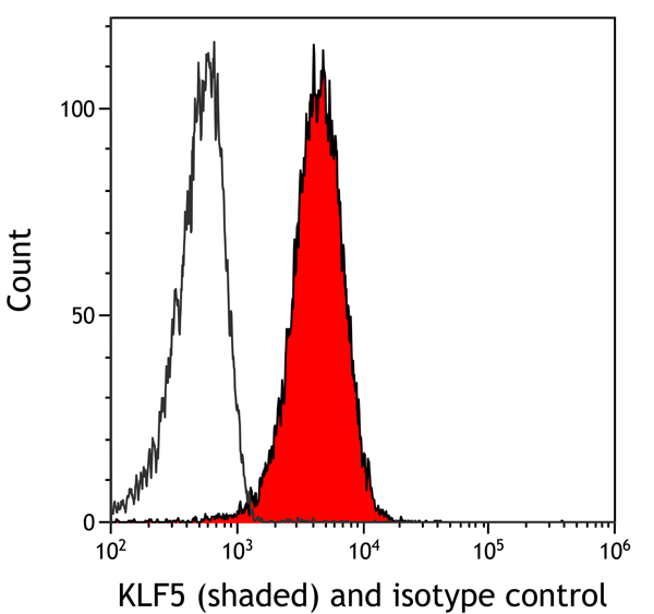
(Detection of human KLF5 by western blot. Samples: Whole cell lysate (25 ug) from HeLa, SW620, MCF-7, and KM12 cells prepared using NETN lysis buffer. Antibody: Rabbit anti-KLF5 recombinant monoclonal antibody (AAA213659 lot 1) used at 1:1000. Secondary: HRP-conjugated goat anti-rabbit IgG . Chemiluminescence with an exposure time of 10 seconds. Lower Panel: Rabbit anti-Actin recombinant monoclonal antibody .)
WB (Western Blot)
(Detection of human KLF5 by western blot. Samples: Whole cell lysate (25 ug) from HeLa, SW620, MCF-7, and KM12 cells prepared using NETN lysis buffer. Antibody: Rabbit anti-KLF5 recombinant monoclonal antibody (AAA213659 lot 1) used at 1:1000. Secondary: HRP-conjugated goat anti-rabbit IgG . Chemiluminescence with an exposure time of 10 seconds. Lower Panel: Rabbit anti-Actin recombinant monoclonal antibody .)
KLF5, Monoclonal Recombinant Antibody (Cat# AAA213659)
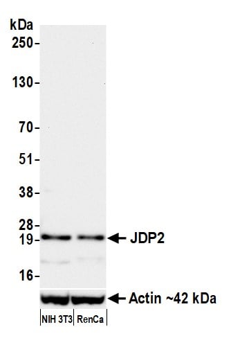
 WB (Western Blot)
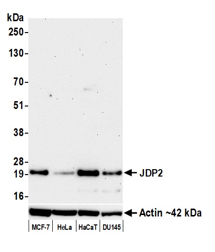
(Detection of human JDP2 by western blot. Samples: Whole cell lysate (25 ug) from MCF-7, HeLa, HaCaT, and DU145 cells prepared using NETN lysis buffer. Antibody: Rabbit anti-JDP2 recombinant monoclonal antibody (AAA213661 lot 1) used at 1:1000. Secondary: HRP-conjugated goat anti-rabbit IgG . Detection: Chemiluminescence with an exposure time of 3 minutes. Lower Panel: Rabbit anti-Actin recombinant monoclonal antibody .)
WB (Western Blot)
(Detection of human JDP2 by western blot. Samples: Whole cell lysate (25 ug) from MCF-7, HeLa, HaCaT, and DU145 cells prepared using NETN lysis buffer. Antibody: Rabbit anti-JDP2 recombinant monoclonal antibody (AAA213661 lot 1) used at 1:1000. Secondary: HRP-conjugated goat anti-rabbit IgG . Detection: Chemiluminescence with an exposure time of 3 minutes. Lower Panel: Rabbit anti-Actin recombinant monoclonal antibody .)
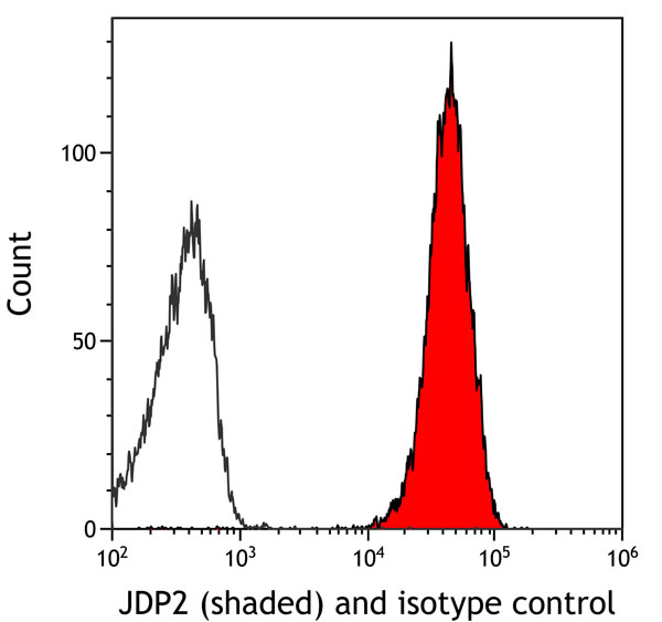
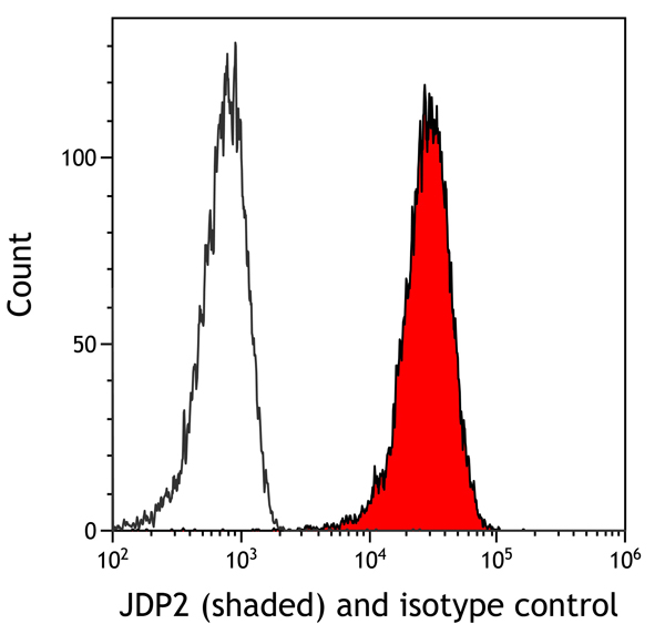
JDP2, Monoclonal Recombinant Antibody (Cat# AAA213661)
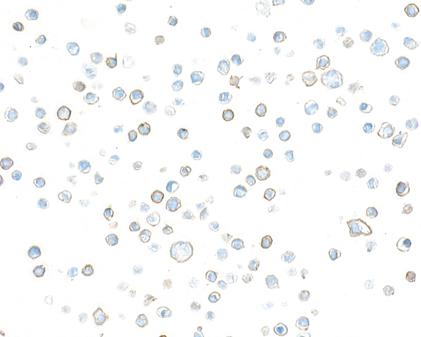
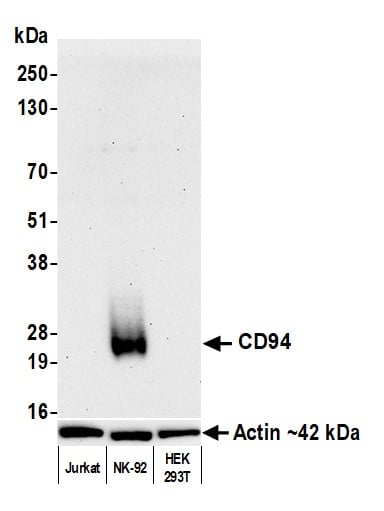 WB (Western Blot)
(Detection of human CD94 by western blot. Samples: Whole cell lysate (10 ug) from Jurkat, NK-92, and HEK293T cells prepared using NETN lysis buffer. Antibody: Rabbit anti-CD94 recombinant monoclonal antibody (AAA213672 lot 1) used at 1:1000. Secondary: HRP-conjugated goat anti-rabbit IgG . Detection: Chemiluminescence with an exposure time of 3 minutes. Lower Panel: Rabbit anti-Actin recombinant monoclonal antibody .)
WB (Western Blot)
(Detection of human CD94 by western blot. Samples: Whole cell lysate (10 ug) from Jurkat, NK-92, and HEK293T cells prepared using NETN lysis buffer. Antibody: Rabbit anti-CD94 recombinant monoclonal antibody (AAA213672 lot 1) used at 1:1000. Secondary: HRP-conjugated goat anti-rabbit IgG . Detection: Chemiluminescence with an exposure time of 3 minutes. Lower Panel: Rabbit anti-Actin recombinant monoclonal antibody .)
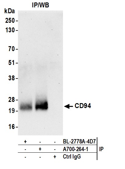
CD94, Monoclonal Recombinant Antibody (Cat# AAA213672)
Lambda (lambda chain), Monoclonal Antibody (Cat# AAA214173)
 Application Data
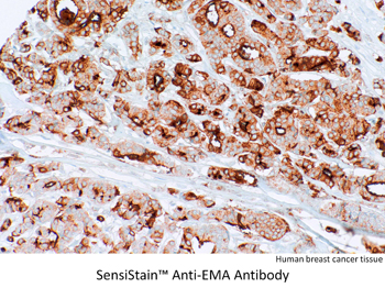
EMA, Monoclonal Antibody (Cat# AAA214371)
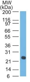
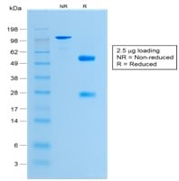 SDS-PAGE
(SDS-PAGE Analysis of Purified Histone H1 Rabbit Recombinant Monoclonal Antibody (HH1/1784R).)
SDS-PAGE
(SDS-PAGE Analysis of Purified Histone H1 Rabbit Recombinant Monoclonal Antibody (HH1/1784R).)
Histone H1, Monoclonal Antibody (Cat# AAA214377)
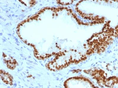
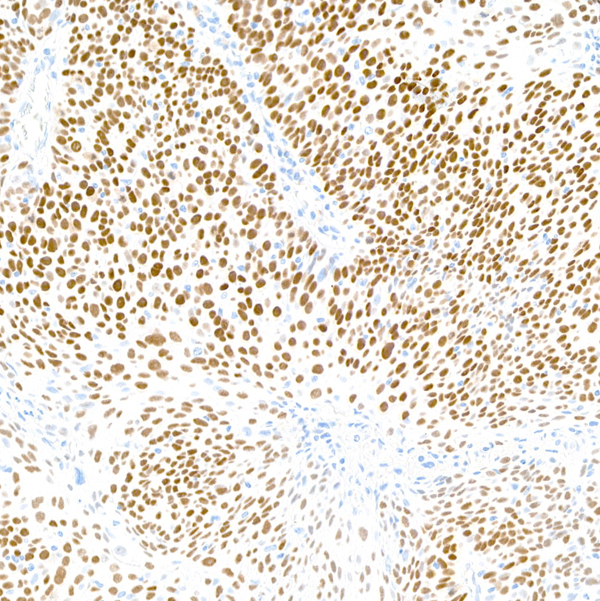
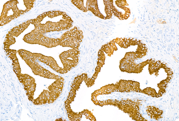
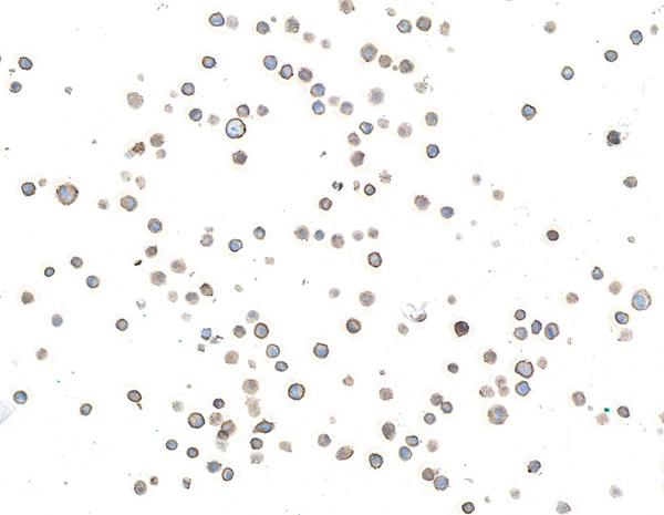
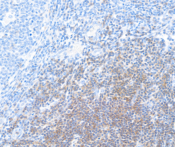
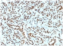
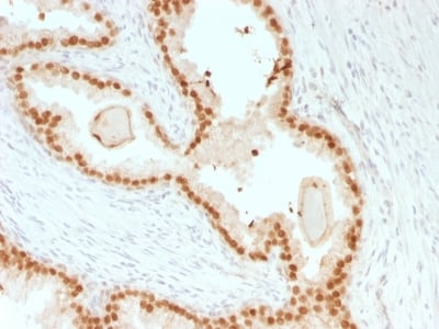
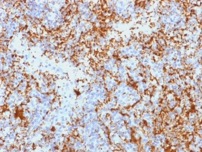
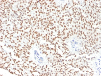 IHC (Immunohiostchemistry)
(Formalin-paraffin human Bladder Carcinoma stained with FOXA1 Monoclonal Antibody (FOXA1/1241).)
IHC (Immunohiostchemistry)
(Formalin-paraffin human Bladder Carcinoma stained with FOXA1 Monoclonal Antibody (FOXA1/1241).)
FOXA1/HNF3A, Monoclonal Antibody (Cat# AAA214381)
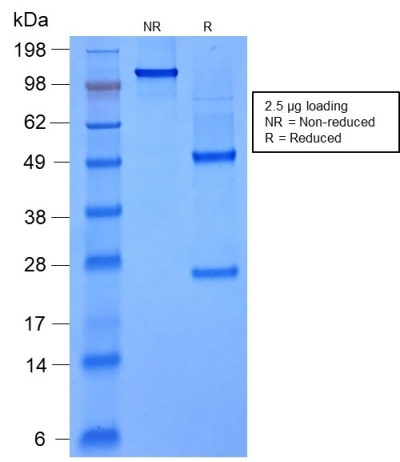 SDS-PAGE
(SDS-PAGE Analysis of Purified FOXA1 Mouse Recombinant Monoclonal Antibody (rFOXA1/1515).)
SDS-PAGE
(SDS-PAGE Analysis of Purified FOXA1 Mouse Recombinant Monoclonal Antibody (rFOXA1/1515).)
FOXA1/HNF3A, Monoclonal Antibody (Cat# AAA214382)

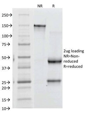
 SDS-PAGE
(SDS-PAGE Analysis of Purified Kappa Light Chain Rabbit Recombinant Monoclonal (KLC2289R).)
SDS-PAGE
(SDS-PAGE Analysis of Purified Kappa Light Chain Rabbit Recombinant Monoclonal (KLC2289R).)
Kappa LightChain/IGKC, Monoclonal Antibody (Cat# AAA214400)
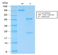
 SDS-PAGE
(SDS-PAGE Analysis Purified CD61 Monoclonal Antibody (Y2/51).Confirmation of Purity and Integrity of Antibody.)
SDS-PAGE
(SDS-PAGE Analysis Purified CD61 Monoclonal Antibody (Y2/51).Confirmation of Purity and Integrity of Antibody.)
CD61/Integrin beta-3/Platelet Glycoprotein IIIa, Monoclonal Antibody (Cat# AAA214405)

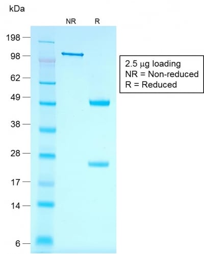
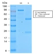 SDS-PAGE
(SDS-PAGE Analysis of Purified Involucrin Rabbit Recombinant Monoclonal Antibody (IVRN/2113R).)
SDS-PAGE
(SDS-PAGE Analysis of Purified Involucrin Rabbit Recombinant Monoclonal Antibody (IVRN/2113R).)
Involucrin, Monoclonal Antibody (Cat# AAA214408)
Does not react with Mouse. Others not known.
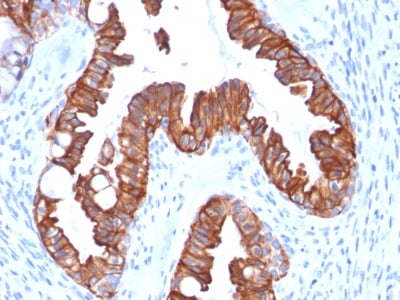
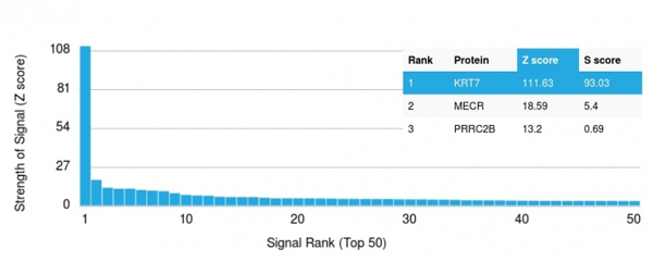 Application Data
(Analysis of Protein Array containing more than 19, 000 full-length human proteins using Cytokeratin 7 Mouse Recombinant Monoclonal Antibody (rOV-TL12/30).Z- and S- Score: The Z-score represents the strength of a signal that a monoclonal antibody (MAb) (in combination with a fluorescently-tagged anti-IgG secondary antibody) produces when binding to a particular protein on the HuProtTM array. Z-scores are described in units of standard deviations (SD's) above the mean value of all signals generated on that array. If targets on HuProtTM are arranged in descending order of the Z-score, the S-score is the difference (also in units of SD's) between the Z-score. S-score therefore represents the relative target specificity of a MAb to its intended target. A MAb is considered to specific to its intended target, if the MAb has an S-score of at least 2.5. For example, if a MAb binds to protein X with a Z-score of 43 and to protein Y with a Z-score of 14, then the S-score for the binding of that MAb to protein X is equal to 29.)
Application Data
(Analysis of Protein Array containing more than 19, 000 full-length human proteins using Cytokeratin 7 Mouse Recombinant Monoclonal Antibody (rOV-TL12/30).Z- and S- Score: The Z-score represents the strength of a signal that a monoclonal antibody (MAb) (in combination with a fluorescently-tagged anti-IgG secondary antibody) produces when binding to a particular protein on the HuProtTM array. Z-scores are described in units of standard deviations (SD's) above the mean value of all signals generated on that array. If targets on HuProtTM are arranged in descending order of the Z-score, the S-score is the difference (also in units of SD's) between the Z-score. S-score therefore represents the relative target specificity of a MAb to its intended target. A MAb is considered to specific to its intended target, if the MAb has an S-score of at least 2.5. For example, if a MAb binds to protein X with a Z-score of 43 and to protein Y with a Z-score of 14, then the S-score for the binding of that MAb to protein X is equal to 29.)
Cytokeratin 7, Monoclonal Antibody (Cat# AAA214413)
Cyclin D1, Monoclonal Antibody (Cat# AAA214177)
Neurofilament, Monoclonal Antibody (Cat# AAA214178)
SLC10A1, Monoclonal Antibody (Cat# AAA214179)
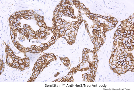
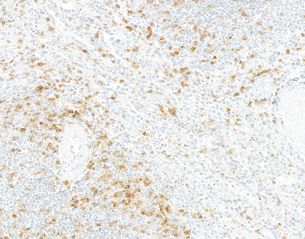
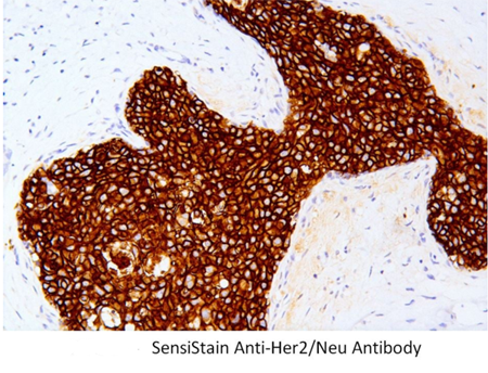 IHC (Immunohiostchemistry)
Her2/Neu, Monoclonal Antibody (Cat# AAA214200)
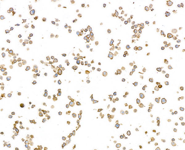
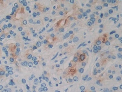 IHC (Immunohiostchemistry)
(DAB staining on IHCP;Sample: Human Prostate Tissue; Primary Ab: 30ug/ml Mouse AntiHuman IL2 AntibodySecond Ab: 2ug/mL HRPLinked Caprine AntiMouse IgG Polyclonal Antibody(Catalog: SAA544Mu19))
IHC (Immunohiostchemistry)
(DAB staining on IHCP;Sample: Human Prostate Tissue; Primary Ab: 30ug/ml Mouse AntiHuman IL2 AntibodySecond Ab: 2ug/mL HRPLinked Caprine AntiMouse IgG Polyclonal Antibody(Catalog: SAA544Mu19))
Interleukin 2 (IL2), Monoclonal Antibody (Cat# AAA151460)
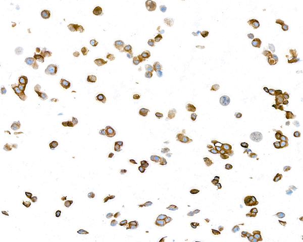
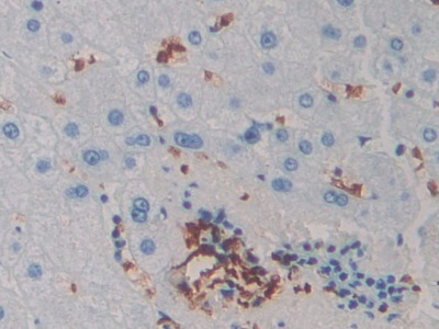
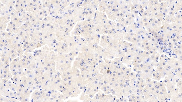 IHC (Immunohistochemisry)
(DAB staining on IHCP;Samples: Bovine Liver Tissue; Primary Ab: 10ug/ml Mouse AntiBovine TNFa AntibodySecond Ab: 2ug/mL HRPLinked Caprine AntiMouse IgG Polyclonal Antibody(Catalog: SAA544Mu19))
IHC (Immunohistochemisry)
(DAB staining on IHCP;Samples: Bovine Liver Tissue; Primary Ab: 10ug/ml Mouse AntiBovine TNFa AntibodySecond Ab: 2ug/mL HRPLinked Caprine AntiMouse IgG Polyclonal Antibody(Catalog: SAA544Mu19))
Tumor Necrosis Factor Alpha (TNFa), Monoclonal Antibody (Cat# AAA151490)
What are Monoclonal Antibodies?
Monoclonal antibodies are specialized laboratory-produced proteins developed for binding to specific biological antigens or other molecular targets. Since they come from a single cell (or clone), they are especially consistent and accurate in the data they are involved in producing.
This type of antibody material has been shown to be a powerful tool in finding and subsequently destroying harmful cells in an organism, such as those found in cancers or various autoimmune diseases. This makes them excellent aids in medical testing and research, which is why they are so widely used.
AAA Biotech offers a comprehensive range of high-quality monoclonal antibodies that perform effectively in various laboratory tests, including (amongst others) ELISA, western blotting, immunohistochemistry, and flow cytometry. All of the products in our catalog are thoroughly quality tested to make sure that they are reliable and will consistently perform well in your research.
What Are The Uses of Monoclonal Antibodies
Monoclonal antibodies are used in many lab tests, including (amongst others) ELISA, western blotting, immunohistochemistry, and flow cytometry.
ELISA is a test that helps detect a specific substance/analyte in a sample. It uses antibodies (often monoclonal) bound to a solid surface (such as the well of a microplate) to “capture” the substance/analyte in the sample and immobilize it so that the detection antibody component can then bind to it and produce a signal, which can then be measured.
Western blotting identifies specific proteins in a sample. The sample is first separated on a gel, and then antibodies are applied that will typically bind to the target, which will all be localized to a single band in a lane.
Immunohistochemistry helps locate specific proteins in cells or tissue samples using antibodies.
Flow cytometry looks at and sorts cells. It uses antibodies that are conjugated to reporter molecules called “fluorophores”, which, under special lights, emit light themselves, which can then be measured by a detector instrument. For a deeper understanding of these techniques, explore our complete guide to monoclonal antibodies and their benefits.
How Monoclonal Antibodies Are Used as Medicine?
Please note that all of the products listed in AAA Biotech’s also known as AAA Bio or AAABio catalog are strictly for research-use only (RUO).
Monoclonal antibodies can also be used as therapeutic/medical treatments, particularly in the context of cancers. They are designed to find and bind to specific cells or proteins, helping the immune system recognize and attack the cancer. These treatments work in different ways, such as:
- Radioimmunotherapy attaches a small amount of radioactive molecule to the antibody, so it delivers the radiation directly to the cancer cells that the antibody is specifically binding to.
- Antibody-directed enzyme prodrug therapy uses antibodies that are specifically bound to special enzymes. These enzymes activate a harmless drug in the body and turn it into a cancer-killing drug only near the cancer cells—this helps avoid harming healthy cells.
- Immunoliposomes are tiny “bubbles” filled with medicine/drug and coated with antibodies. They carry the drug straight to the cancer cells.
Why Buy Monoclonal Antibodies From Us?
At AAA Biotech, we provide high-performance monoclonal antibodies designed to support a wide range of research needs.
1. Validated for Versatile Applications
The antibodies in our catalog are extensively validated and compatible with multiple techniques, including (but not limited to) ELISA, flow cytometry (FC), immunocytochemistry (ICC), immunofluorescence (IF), immunohistochemistry (IHC), immunoprecipitation (IP), and western blotting (WB).
2. Wide Selection & Specialized Options
We offer antibodies for common and rare species, that are available in various conjugated forms, and also in recombinant formats. Essentially, there is almost anything one might need to meet their experimental model’s requirements.
3. High-Quality Proteins
Our proteins meet high purity standards—90% or more as confirmed by SDS-PAGE. Many are available with tags like His, Flag, GST, or MBP, and we also supply native and biologically active proteins for functional studies.
Frequently Asked Questions
1. Are your monoclonal antibodies validated for specific applications?
Yes, our antibodies are tested and validated for use in methods such as ELISA, western blot, IHC, flow cytometry, and more. Refer to specific product pages or datasheets for individual product information.
2. How do I choose the right monoclonal antibody for my application?
Review the product details directly for application validation, species reactivity, and target information. You may also contact our support team at any time for help.
3. How quickly can I receive my order?
Most orders are processed and shipped within 1–3 business days, depending on product availability and your shipping location.