Filters
▼Clonality
▼Type
▼Reactivity
▼Gene Name
▼Isotype
▼Host
▼Application
▼Clone
▼Polyclonal Antibodies
At AAA Biotech also known as AAA Bio or AAABio, we provide a broad range of purified polyclonal antibodies (pAbs) that are able to all be browsed online through our website. Due to their high specificity and strong binding affinity, these antibodies are ideal for wide swathes of research and experimental applications.
Our polyclonal antibodies can easily support your work, whether you use them for Western Blotting, Immunocytochemistry (with or without Immunofluorescence used in conjunction), Immunohistochemistry, Immunoprecipitation, and ELISA tests. We highly encourage you to browse our range of pAbs and choose the one that best suits your experimental model.
Viewing 300-350 of 118597 product results

 IF (Immunofluorescence)
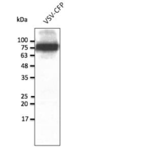
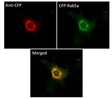
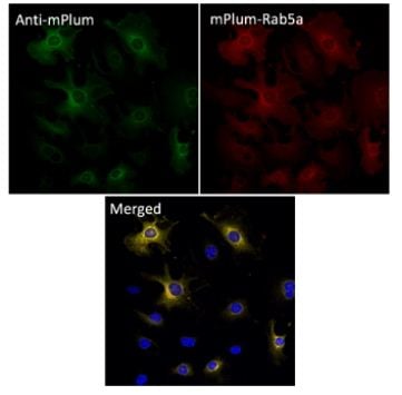
(Immunofluorescence - anti-CFP Ab using NIH3T3 cells transduced with CFP-Rab5a; cells were fixed with methanol and anti-CFP at 1/250;)
IF (Immunofluorescence)
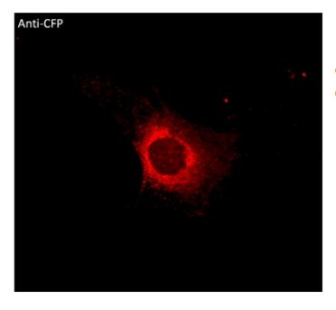
(Immunofluorescence - anti-CFP Ab using NIH3T3 cells transduced with CFP-Rab5a; cells were fixed with methanol and anti-CFP at 1/250;)
CFP, Polyclonal Antibody (Cat# AAA63258)

 Application Data
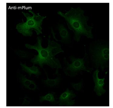
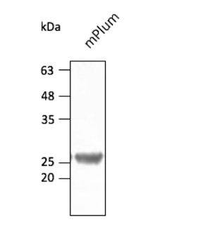
(Anti-mPlum Ab at 1/2,500 dilution using HEK293 transfected cell lysates at 50 ug per lane; rabbit polyclonal to goat IgG (HRP) at 1/10,000 dilution;)
Application Data
(Anti-mPlum Ab at 1/2,500 dilution using HEK293 transfected cell lysates at 50 ug per lane; rabbit polyclonal to goat IgG (HRP) at 1/10,000 dilution;)
mPlum, Polyclonal Antibody (Cat# AAA63259)
 IF (Immunofluorescence)
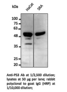
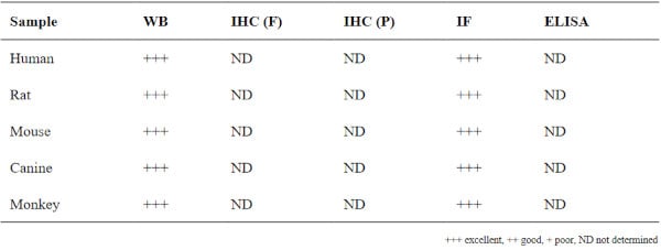
P53, Polyclonal Antibody (Cat# AAA63066)
 Application Data
(AtT-20 transfected with GFP-Rab27a and immunostained with GFP Ab)
Application Data
(AtT-20 transfected with GFP-Rab27a and immunostained with GFP Ab)
GFP, Polyclonal Antibody (Cat# AAA63068)
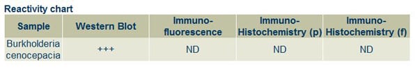
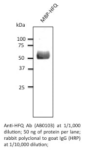 Application Data
HQ, Polyclonal Antibody (Cat# AAA63080)
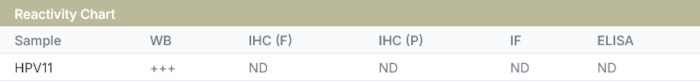
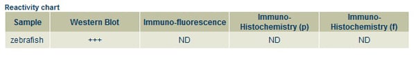
 Application Data
HPV11 E6, Polyclonal Antibody (Cat# AAA63113)
 Application Data
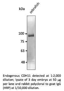
CDH11, Polyclonal Antibody (Cat# AAA63115)

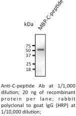 Application Data
C-Peptide, Polyclonal Antibody (Cat# AAA63126)

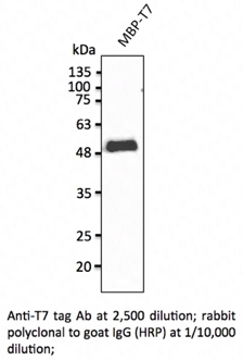 WB (Western Blot)
T7 Epitope Tag, Polyclonal Antibody (Cat# AAA63136)
 WB (Western Blot)
STUB1, Polyclonal Antibody (Cat# AAA63141)
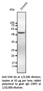 WB (Western Blot)
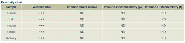
Vimentin, Polyclonal Antibody (Cat# AAA63144)
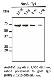 WB (Western Blot)
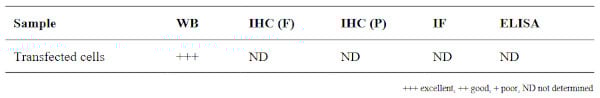
Ty1, Polyclonal Antibody (Cat# AAA63148)
 WB (Western Blot)
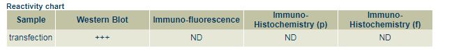
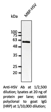
HSV, Polyclonal Antibody (Cat# AAA63151)
 WB (Western Blot)
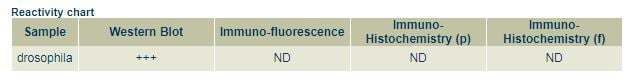
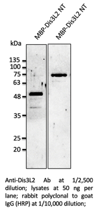
Dis3L2, Polyclonal Antibody (Cat# AAA63152)

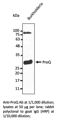 WB (Western Blot)
ProQ, Polyclonal Antibody (Cat# AAA63157)
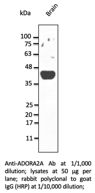 WB (Western Blot)
ADORA2A, Polyclonal Antibody (Cat# AAA63159)

 Application Data
(Anti-Nucleocapsid Ab at 1/2,500 dilution; lane with 30 ng of recombinant fusion protein; rabbit polyclonal to goat IgG (HRP) at 1/10,000 dilution;)
Application Data
(Anti-Nucleocapsid Ab at 1/2,500 dilution; lane with 30 ng of recombinant fusion protein; rabbit polyclonal to goat IgG (HRP) at 1/10,000 dilution;)
COVID 19 Nucleocapsid (NP) Coronavirus, Polyclonal Antibody (Cat# AAA63173)

 Application Data
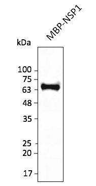
(Anti-NSP1 Ab at 1/2,500 dilution; lane with 30 ng of recombinant fusion protein; rabbit polyclonal to goat IgG (HRP) at 1/10,000 dilution;)
Application Data
(Anti-NSP1 Ab at 1/2,500 dilution; lane with 30 ng of recombinant fusion protein; rabbit polyclonal to goat IgG (HRP) at 1/10,000 dilution;)
COVID 19 NSP1 Coronavirus, Polyclonal Antibody (Cat# AAA63179)

 Application Data
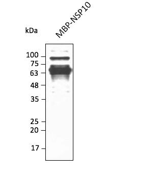
(Anti-NSP10 Ab at 1/2,500 dilution; lane with 30 ng of recombinant fusion protein; rabbit polyclonal to goat IgG (HRP) at 1/10,000 dilution;)
Application Data
(Anti-NSP10 Ab at 1/2,500 dilution; lane with 30 ng of recombinant fusion protein; rabbit polyclonal to goat IgG (HRP) at 1/10,000 dilution;)
COVID 19 NSP10 Coronavirus, Polyclonal Antibody (Cat# AAA63186)

 Application Data
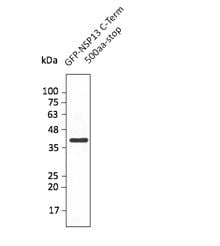
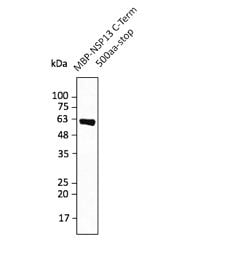
(Anti-NSP13 Ab at 1/2,500 dilution; lane with 30 ng of recombinant fusion protein; rabbit polyclonal to goat IgG (HRP) at 1/10,000 dilution;)
Application Data
(Anti-NSP13 Ab at 1/2,500 dilution; lane with 30 ng of recombinant fusion protein; rabbit polyclonal to goat IgG (HRP) at 1/10,000 dilution;)
COVID 19 NSP13 Coronavirus, Polyclonal Antibody (Cat# AAA63188)

 Application Data
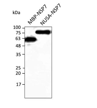
(Anti-NSP7 protein Ab at 1/2,500 dilution; lane with 30 ng of recombinant fusion protein; rabbit polyclonal to goat IgG (HRP) at 1/10,000 dilution;)
Application Data
(Anti-NSP7 protein Ab at 1/2,500 dilution; lane with 30 ng of recombinant fusion protein; rabbit polyclonal to goat IgG (HRP) at 1/10,000 dilution;)
COVID 19 NSP7 Coronavirus, Polyclonal Antibody (Cat# AAA63199)

 Application Data
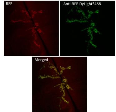
(Anti-RFP Ab conjugated to DyLight 488 at 1/2,500 dilution using HEK293 transfected cell lysates at 50 ug per lane;)
Application Data
(Anti-RFP Ab conjugated to DyLight 488 at 1/2,500 dilution using HEK293 transfected cell lysates at 50 ug per lane;)
RFP, Polyclonal Antibody (Cat# AAA63201)
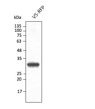 Application Data
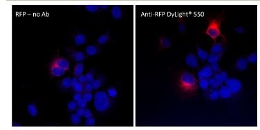
(Anti-RFP Ab conjugated to DyLight 550 at 1/2,500 dilution using HEK293 transfected cell lysates at 50 ug per lane;)
Application Data
(Anti-RFP Ab conjugated to DyLight 550 at 1/2,500 dilution using HEK293 transfected cell lysates at 50 ug per lane;)
RFP, Polyclonal Antibody (Cat# AAA63202)
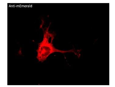
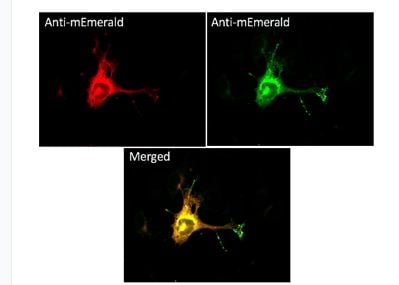

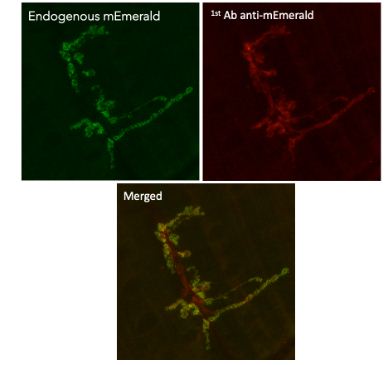 IF (Immunofluorescence)
(Immunofluorescence in Drosophila larvaenmJ muscle 6/7 expressing GluRIIA-mEmerald in neurons (GluRIIA ia a post-synaptic protein) using 1st Ab anti-mEmerald at 1/1,000 and 2nd Ab anti-goat IgY conjugated to DyLight550 at 1/500;)
IF (Immunofluorescence)
(Immunofluorescence in Drosophila larvaenmJ muscle 6/7 expressing GluRIIA-mEmerald in neurons (GluRIIA ia a post-synaptic protein) using 1st Ab anti-mEmerald at 1/1,000 and 2nd Ab anti-goat IgY conjugated to DyLight550 at 1/500;)
mEmerald, Polyclonal Antibody (Cat# AAA63214)
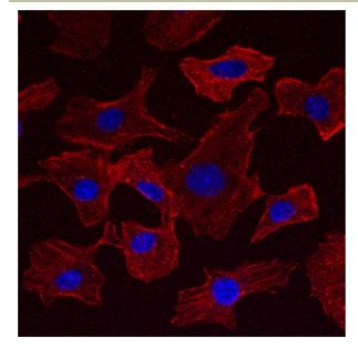
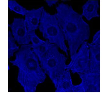 IF (Immunofluorescence)
(Immunofluorescence using hCEC cells, 1st Ab (anti-beta-Actin at 1/250) and 2nd Ab (anti-mouse AB27405 at 1/1,000); cells were fixed with methanol;)
IF (Immunofluorescence)
(Immunofluorescence using hCEC cells, 1st Ab (anti-beta-Actin at 1/250) and 2nd Ab (anti-mouse AB27405 at 1/1,000); cells were fixed with methanol;)
IgG, Polyclonal Secondary Antibody (Cat# AAA63217)
 IF (Immunofluorescence)
(Immunofluorescence using hCEC cells, 1st Ab (anti-beta-Actin at 1/250) and 2nd Ab (anti-mouse AB27633 at 1/1,000); cells were fixed with methanol;)
IF (Immunofluorescence)
(Immunofluorescence using hCEC cells, 1st Ab (anti-beta-Actin at 1/250) and 2nd Ab (anti-mouse AB27633 at 1/1,000); cells were fixed with methanol;)
IgG, Polyclonal Secondary Antibody (Cat# AAA63220)
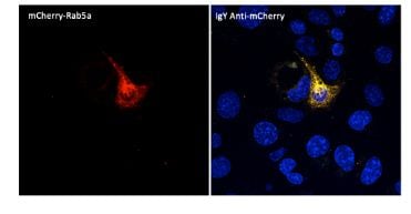 IF (Immunofluorescence)
(Immunofluorescence -Anti-mCherry Ab (AB9770) using hCEC cells transduced with mCherry-Rab5a; cells were fixed with methanol and mCherry Ab at 1/250; 2nd Ab goatAnti-IgY (AB307633) at 1:1,000;)
IF (Immunofluorescence)
(Immunofluorescence -Anti-mCherry Ab (AB9770) using hCEC cells transduced with mCherry-Rab5a; cells were fixed with methanol and mCherry Ab at 1/250; 2nd Ab goatAnti-IgY (AB307633) at 1:1,000;)
IgY, Polyclonal Secondary Antibody (Cat# AAA63225)

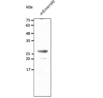
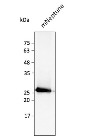 Application Data
(Anti-mNeptune Ab at 1/2,500 dilution using HEK293 transfected cell lysates at 40 ug per lane; rabbit polyclonal to goat IgG (HRP) at 1/10,000 dilution;)
Application Data
(Anti-mNeptune Ab at 1/2,500 dilution using HEK293 transfected cell lysates at 40 ug per lane; rabbit polyclonal to goat IgG (HRP) at 1/10,000 dilution;)
mNeptune, Polyclonal Antibody (Cat# AAA63229)
 Application Data
(Anti-GAPDH Ab conjugated to DyLight633 (AB49633) at 1:2,000 using lysates at 30 ug per lane;)
Application Data
(Anti-GAPDH Ab conjugated to DyLight633 (AB49633) at 1:2,000 using lysates at 30 ug per lane;)
GAPDH, Polyclonal Antibody (Cat# AAA63241)
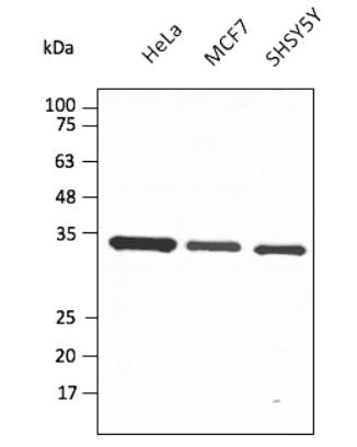
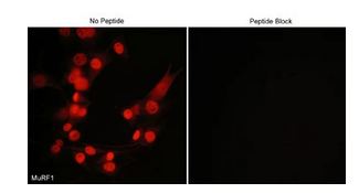 ICC (Immunocytochemistry)
(Immunocytochemical labeling of MuRF1 in mouse C2C12 cells. The cells were labeled with rabbit polyclonal MuRF1 antibody, then detected using appropriate secondary antibody conjugated to Cy3. The antibody was used in the absence (left) or presence (right) of blocking peptide (MX3405).)
ICC (Immunocytochemistry)
(Immunocytochemical labeling of MuRF1 in mouse C2C12 cells. The cells were labeled with rabbit polyclonal MuRF1 antibody, then detected using appropriate secondary antibody conjugated to Cy3. The antibody was used in the absence (left) or presence (right) of blocking peptide (MX3405).)
MuRF1, Polyclonal Antibody (Cat# AAA71659)
 ICC (Immunocytochemistry)
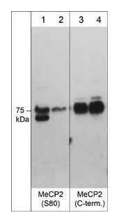
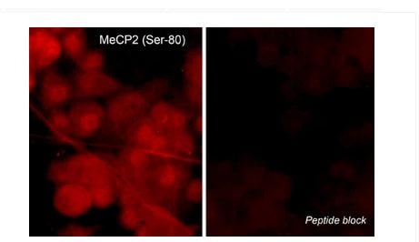
(Immunocytochemical labeling of MeCP2 phosphorylation in rat PC12 cells differentiated with NGF. The cells were probed with MeCP2 (Ser-80) rabbit polyclonal antibody (MP4601) in the absence (left) or presence (right) of blocking peptide (MX4605). The antibody was detected using appropriate secondary antibody conjugated to DyLight 594.)
ICC (Immunocytochemistry)
(Immunocytochemical labeling of MeCP2 phosphorylation in rat PC12 cells differentiated with NGF. The cells were probed with MeCP2 (Ser-80) rabbit polyclonal antibody (MP4601) in the absence (left) or presence (right) of blocking peptide (MX4605). The antibody was detected using appropriate secondary antibody conjugated to DyLight 594.)
MeCP2, Polyclonal Antibody (Cat# AAA71669)
 ICC (Immunocytochemistry)
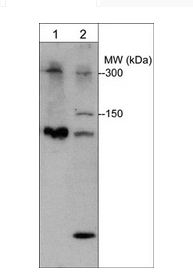
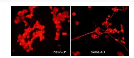
(Immunocytochemical labeling in rat PC12 cells differentiated with NGF. The cells were probed with rabbit polyclonal Plexin B1 (PP1841) and mouse monoclonal Sema-4D (SM1881) antibodies, then the antibodies were detected using appropriate secondary antibody conjugated to DyLight 594.)
ICC (Immunocytochemistry)
(Immunocytochemical labeling in rat PC12 cells differentiated with NGF. The cells were probed with rabbit polyclonal Plexin B1 (PP1841) and mouse monoclonal Sema-4D (SM1881) antibodies, then the antibodies were detected using appropriate secondary antibody conjugated to DyLight 594.)
Plexin B1, Polyclonal Antibody (Cat# AAA71691)
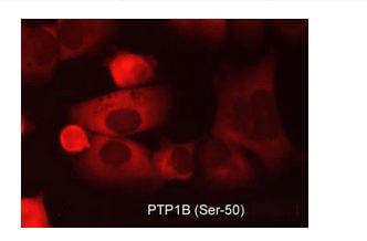 ICC (Immunocytochemistry)
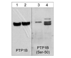
(Immunocytochemical labeling of PTP1B in aldehyde-fixed and NP-40 permeabilized human NCI-H1915 lung carcinoma cells. The cells were labeled with rabbit polyclonal anti-PTP1B (Ser-50) (PP2411) phosphospecific antibody. The antibody was detected using appropriate secondary antibody conjugated to DyLight 594.)
ICC (Immunocytochemistry)
(Immunocytochemical labeling of PTP1B in aldehyde-fixed and NP-40 permeabilized human NCI-H1915 lung carcinoma cells. The cells were labeled with rabbit polyclonal anti-PTP1B (Ser-50) (PP2411) phosphospecific antibody. The antibody was detected using appropriate secondary antibody conjugated to DyLight 594.)
PTP1B, Polyclonal Antibody (Cat# AAA71693)
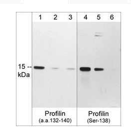
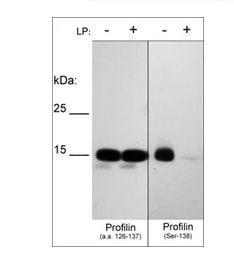 WB (Western Blot)
(Western blot of human recombinant Profilin-1 phosphorylated in vitro with PKCalpha kinase then untreated (-) or treated with lambda phosphatase (+). The blots were probed with anti-Profilin (a.a. 126-137) (left panel) or anti-Profilin (Ser-138) phospho-specific (right panel) antibodies at 1:1000.)
WB (Western Blot)
(Western blot of human recombinant Profilin-1 phosphorylated in vitro with PKCalpha kinase then untreated (-) or treated with lambda phosphatase (+). The blots were probed with anti-Profilin (a.a. 126-137) (left panel) or anti-Profilin (Ser-138) phospho-specific (right panel) antibodies at 1:1000.)
Profilin, Polyclonal Antibody (Cat# AAA71697)
 ICC (Immunocytochemistry)
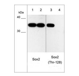
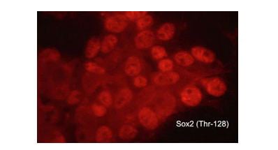
(Immunocytochemical labeling of phosphorylated Sox2 in aldehyde fixed and NP-40 permeabilized human NCI-H446 lung carcinoma cells. The cells were labeled with rabbit polyclonal anti-Sox2 (Thr-128) phospho-specific (SP0381). The antibody was detected using goat anti-rabbit Ig:DyLight 594.)
ICC (Immunocytochemistry)
(Immunocytochemical labeling of phosphorylated Sox2 in aldehyde fixed and NP-40 permeabilized human NCI-H446 lung carcinoma cells. The cells were labeled with rabbit polyclonal anti-Sox2 (Thr-128) phospho-specific (SP0381). The antibody was detected using goat anti-rabbit Ig:DyLight 594.)
Sox2, Polyclonal Antibody (Cat# AAA71709)
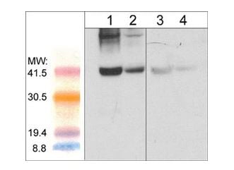
 WB (Western Blot)
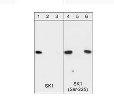
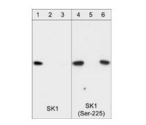
(Western blot of HeLa stimulated with calyculin A (lanes 1-4). The blots were untreated (lane 1 & 3) or treated with lambda phosphatase (lane 2 & 4), then probed with anti-SK1 (Central region) SP1621 (lanes 1 & 2) or anti-SK1 (Ser-225) SP1641 (lanes 3 & 4).)
WB (Western Blot)
(Western blot of HeLa stimulated with calyculin A (lanes 1-4). The blots were untreated (lane 1 & 3) or treated with lambda phosphatase (lane 2 & 4), then probed with anti-SK1 (Central region) SP1621 (lanes 1 & 2) or anti-SK1 (Ser-225) SP1641 (lanes 3 & 4).)
Sphingosine Kinase 1, Polyclonal Antibody (Cat# AAA71710)
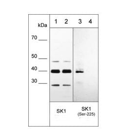
 WB (Western Blot)
(Western blot of HeLa stimulated with calyculin A (lanes 1-4). The blots were untreated (lane 1 & 3) or treated with lambda phosphatase (lane 2 & 4), then probed with anti-SK1 (Central region) SP1621 (lanes 1 & 2) or anti-SK1 (Ser-225) SP1641 (lanes 3 & 4).)
WB (Western Blot)
(Western blot of HeLa stimulated with calyculin A (lanes 1-4). The blots were untreated (lane 1 & 3) or treated with lambda phosphatase (lane 2 & 4), then probed with anti-SK1 (Central region) SP1621 (lanes 1 & 2) or anti-SK1 (Ser-225) SP1641 (lanes 3 & 4).)
Sphingosine Kinase 1, Polyclonal Antibody (Cat# AAA71711)
 IHC (Immunohiostchemistry)
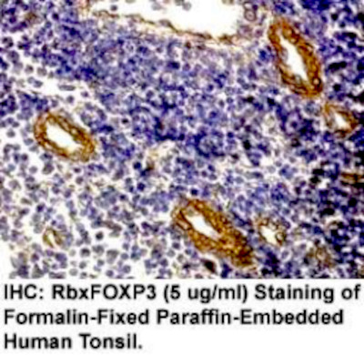
(Immunohistochemistry: Human Tonsil carcinoma (FFPE) stained with Rabbit anti-FOXP3 (Cat# AAA71424) at 1:200 for 10 min @ RT. Staining of formalin-fixed tissue requires boiling tissue sections in 10 mM Citrate Buffer, pH 6.0 for 10 min followed by cooling at RT for 20 min.)
IHC (Immunohiostchemistry)
(Immunohistochemistry: Human Tonsil carcinoma (FFPE) stained with Rabbit anti-FOXP3 (Cat# AAA71424) at 1:200 for 10 min @ RT. Staining of formalin-fixed tissue requires boiling tissue sections in 10 mM Citrate Buffer, pH 6.0 for 10 min followed by cooling at RT for 20 min.)
FOXP3, Polyclonal Antibody (Cat# AAA71424)
 IHC (Immunohiostchemistry)
(Immunohistochemistry: Human Tonsil (FFPE) stained with Rabbit anti-PDGF-alpha (Cat# AAA71430) at 1:200 for 10 min RT. Staining of formalin-fixed tissue requires boiling tissue sections in 10 mM Citrate Buffer, pH 6.0 for 10 min followed by cooling at RT for 20 min)
IHC (Immunohiostchemistry)
(Immunohistochemistry: Human Tonsil (FFPE) stained with Rabbit anti-PDGF-alpha (Cat# AAA71430) at 1:200 for 10 min RT. Staining of formalin-fixed tissue requires boiling tissue sections in 10 mM Citrate Buffer, pH 6.0 for 10 min followed by cooling at RT for 20 min)
PDGF, Polyclonal Antibody (Cat# AAA71430)
 Application Data
IkBa (NFKB1A), Polyclonal Antibody (Cat# AAA71433)
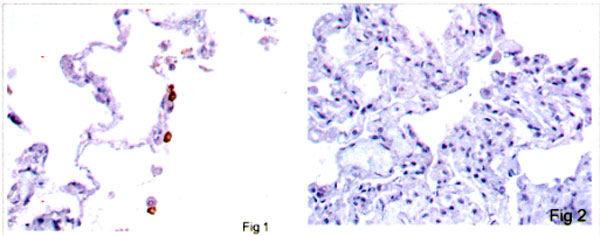 IHC (Immunohistochemistry)
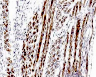
(IHC: Human Lung Tissue stained with Rabbit anti SARS-Cov2 (N) antibody, AAA71443 at 1:100 for 10 minutes at room tempeture.Staining of formalin-fixed tissue requires boiling tissue sections in 10mM Citrate Buffer, pH6.0 for 10 minutes followed by cooling at room tempeture for 20 minutes.Fig 1 Positive alveolar macrophages in the lung tissue;Figure 2 is a negative staining of Lung tissue.)
IHC (Immunohistochemistry)
(IHC: Human Lung Tissue stained with Rabbit anti SARS-Cov2 (N) antibody, AAA71443 at 1:100 for 10 minutes at room tempeture.Staining of formalin-fixed tissue requires boiling tissue sections in 10mM Citrate Buffer, pH6.0 for 10 minutes followed by cooling at room tempeture for 20 minutes.Fig 1 Positive alveolar macrophages in the lung tissue;Figure 2 is a negative staining of Lung tissue.)
COVID 19 Nucleocapsid (NP) Coronavirus, Polyclonal Antibody (Cat# AAA71443)
 IHC (Immunohiostchemistry)
(Formalin fixed, citric acid treated paraffin sections of E16 mouse skeletal muscle. Sections were probed with anti-Atrogin-1 (AAA71564) then anti-Rabbit:HRP before detection using DAB. (Images provided by Carl Hobbs and Dr. Pat Doherty at Wolfson Centre for Age-Related Diseases, King's College London).)
IHC (Immunohiostchemistry)
(Formalin fixed, citric acid treated paraffin sections of E16 mouse skeletal muscle. Sections were probed with anti-Atrogin-1 (AAA71564) then anti-Rabbit:HRP before detection using DAB. (Images provided by Carl Hobbs and Dr. Pat Doherty at Wolfson Centre for Age-Related Diseases, King's College London).)
Atrogin-1, Polyclonal Antibody (Cat# AAA71564)
 ICC (Immunocytochemistry)
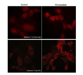
(Immunocytochemical labeling of phosphorylated Exphexin-1 in pervanadate-treated mouse C2C12. The cells were labeled with rabbit polyclonal Ephexin-1 (C-terminal region) and Ephexin-1 (Tyr-87) antibodies, then the antibodies were detected using appropriate secondary antibodies conjugated to Cy3.)
ICC (Immunocytochemistry)
(Immunocytochemical labeling of phosphorylated Exphexin-1 in pervanadate-treated mouse C2C12. The cells were labeled with rabbit polyclonal Ephexin-1 (C-terminal region) and Ephexin-1 (Tyr-87) antibodies, then the antibodies were detected using appropriate secondary antibodies conjugated to Cy3.)
Ephexin-1, Polyclonal Antibody (Cat# AAA71632)
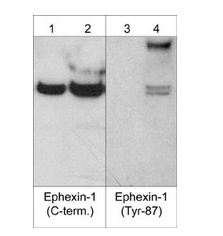
 WB (Western Blot)
(Western blot analysis of A431 cells treated with pervanadate (1 mM) for 30 min. Blots were probed with anti-IKBa (lane 1), anti-IKBa (Tyr-42) (IP1 031; lanes 2-5), or anti-IKBa (Tyr-305) (IP1 041 ; lanes 6-9). In some lanes, the antibodies were used in the absence (lane 2 & 6) or presence of IKBa (Tyr-42) (lane 3 & 8) or IKBa (Tyr-305) (lane 4 & 7) blocking peptides, or BSA conjugated to phospho-tyrosine (lane 5 & 9).)
WB (Western Blot)
(Western blot analysis of A431 cells treated with pervanadate (1 mM) for 30 min. Blots were probed with anti-IKBa (lane 1), anti-IKBa (Tyr-42) (IP1 031; lanes 2-5), or anti-IKBa (Tyr-305) (IP1 041 ; lanes 6-9). In some lanes, the antibodies were used in the absence (lane 2 & 6) or presence of IKBa (Tyr-42) (lane 3 & 8) or IKBa (Tyr-305) (lane 4 & 7) blocking peptides, or BSA conjugated to phospho-tyrosine (lane 5 & 9).)
I-kappa-Ba (Tyr-42), Polyclonal Antibody (Cat# AAA71521)
 Application Data
Inducible Nitric Oxide Synthase, Polyclonal Antibody (Cat# AAA71522)
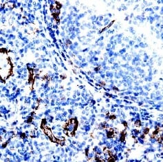
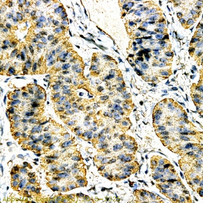 IHC (Immunohistochemistry)
(Immunohistochemistry: Human Colon carcinoma (FFPE) stained with Rabbit anti-Calcitonin (Cat# AAA71301) at 1:200 for 10 min @ RT. Staining of formalin- fixed tissue requires boiling tissue sections in 10 mM Citrate Buffer, pH 6.0 for 10 min followed by cooling at RT for 20 min.)
IHC (Immunohistochemistry)
(Immunohistochemistry: Human Colon carcinoma (FFPE) stained with Rabbit anti-Calcitonin (Cat# AAA71301) at 1:200 for 10 min @ RT. Staining of formalin- fixed tissue requires boiling tissue sections in 10 mM Citrate Buffer, pH 6.0 for 10 min followed by cooling at RT for 20 min.)
Calcitonin, Polyclonal Antibody (Cat# AAA71301)
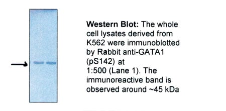 WB (Western Blot)
GATA1, Polyclonal Antibody (Cat# AAA71302)
Parathyroid Hormone (PTH), Polyclonal Antibody (Cat# AAA71308)
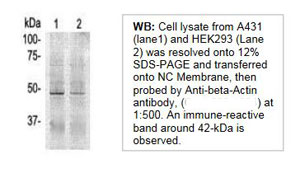 WB (Western Blot)
Actin-beta, Polyclonal Antibody (Cat# AAA71314)
FGF4, Polyclonal Antibody (Cat# AAA71328)
What are Polyclonal Antibodies?
Polyclonal antibodies are antibodies that come from multiple B cell clones of a host animal. The typical hosts used for the majority of polyclonal antibody production are rabbits, goats, sheep, and donkeys. These polyclonal antibodies, once having identified their target, will bind to different epitopes located at different regions or sequences on the same protein/antigen. This ability to bind multiple epitopes is what makes polyclonal antibodies highly sensitive, as explained in our detailed guide on polyclonal antibodies and why they matter.
As a result, they are ideal at locating and binding to the target, even if the target is in very low concentrations (due to many different antibodies being able to bind to the same target molecule, which allows for significant amplification of a downstream signal).
Polyclonal antibodies are typically produced by injecting an antigen into a host animal, which causes the animal’s immune system to attack the foreign antigen by mass generating antibodies against it. After a period of time, serum is collected from the animal and purified using physicochemical fractionation, class-specific affinity purification, and/or antigen-affinity purification.
Key Uses of Polyclonal Antibodies
- Western Blotting: This method is used to find specific proteins in biological samples after separating them by size.
- Immunohistochemistry: IHC helps visualize the location of proteins in tissue sections using various staining techniques.
- ELISA: (Enzyme-Linked Immunosorbent Assay) is typically used to identify specific protein quantities in a sample. ELISAs can be either “Quantitative” or “Qualitative”.
- Flow Cytometry: technique that identifies and measures the specific protein on the surface or inside the cells in a fluid suspension.
- Immunoprecipitation: IP isolates and studies a specific protein from a complex mixture using antibodies.
Why Buy Polyclonal Antibodies from AAA Biotech?
1. Ideal for Various Applications
Our antibodies are generally going to be validated for use in multiple types of assays, including ELISA, Western Blotting, Immunohistochemistry, Immunoprecipitation, amongst others. They are ideal for a wide range of research applications.
2. Rigorous Quality Control
All of the antibodies in our catalog undergo strict quality testing to ensure specificity, sensitivity, and consistent performance. We are confident in the ability of our antibodies to provide you with accurate results.
3. Wide Assortment of Antibodies
Antibodies in our catalog can be found for both common and exotic species, and these antibodies are also available in both conjugated and recombinant forms to suit many diverse experimental needs.
4. Highly Purified
Our antibodies are available in purified forms with over 85% purity, as confirmed by SDS-PAGE. They are also available with tags such as His, Flag, GST, or MBP. We cater to customers worldwide.
FAQ
1. How are polyclonal antibodies produced?
Traditionally, polyclonal antibodies are produced by injecting an antigen into a host animal (such as a rabbit or goat), which then triggers an immune response from the host animal. The animal’s B cells produce antibodies that will recognize different parts of the injected antigen. These antibodies are then collected from the animal’s blood and purified for use.
2. How do polyclonal antibodies differ from monoclonal antibodies?
Polyclonal antibodies are a mix of antibodies that bind to different locations (epitopes) of the same antigen, while monoclonal antibodies are identical and bind to just one specific epitope. This makes polyclonal antibodies more versatile and better at detecting proteins that may be present in low quantities or in altered/modified forms.
3. How should I store polyclonal antibodies?
Polyclonal antibodies should be stored at 4°C for short-term use (up to a few weeks) and at -20°C or -80°C for long-term storage. Avoid repeated freeze-thaw cycles by dividing them into small aliquots. Always check the datasheet for specific storage instructions.