
Filters
Clonality
Type
Reactivity
Gene Name
Isotype
Host
Application
Clone
10000 results for "Recombinant Proteins" - showing 8650-8700
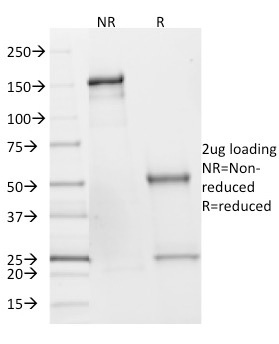
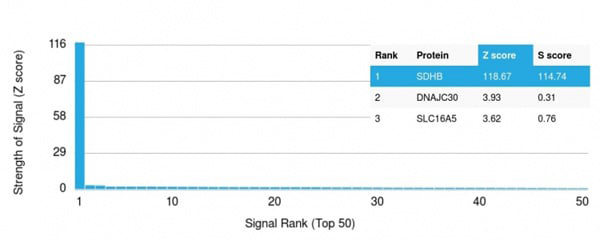 Application Data
(Analysis of Protein Array containing more than 19,000 full-length human proteins using SDHB Mouse Monoclonal Antibody (SDHB/2382). Z- and S- Score: The Z-score represents the strength of a signal that a monoclonal antibody (Monoclonal Antibody) (in combination with a fluorescently-tagged anti-IgG secondary antibody) produces when binding to a particular protein on the HuProtTM array. Z-scores are described in units of standard deviations (SD's) above the mean value of all signals generated on that array. If targets on HuProtTM are arranged in descending order of the Z-score, the S-score is the difference (also in units of SD's) between the Z-score. S-score therefore represents the relative target specificity of a Monoclonal Antibody to its intended target. A Monoclonal Antibody is considered to specific to its intended target, if the Monoclonal Antibody has an S-score of at least 2.5. For example, if a Monoclonal Antibody binds to protein X with a Z-score of 43 and to protein Y with a Z-score of 14, then the S-score for the binding of that Monoclonal Antibody to protein X is equal to 29.)
Application Data
(Analysis of Protein Array containing more than 19,000 full-length human proteins using SDHB Mouse Monoclonal Antibody (SDHB/2382). Z- and S- Score: The Z-score represents the strength of a signal that a monoclonal antibody (Monoclonal Antibody) (in combination with a fluorescently-tagged anti-IgG secondary antibody) produces when binding to a particular protein on the HuProtTM array. Z-scores are described in units of standard deviations (SD's) above the mean value of all signals generated on that array. If targets on HuProtTM are arranged in descending order of the Z-score, the S-score is the difference (also in units of SD's) between the Z-score. S-score therefore represents the relative target specificity of a Monoclonal Antibody to its intended target. A Monoclonal Antibody is considered to specific to its intended target, if the Monoclonal Antibody has an S-score of at least 2.5. For example, if a Monoclonal Antibody binds to protein X with a Z-score of 43 and to protein Y with a Z-score of 14, then the S-score for the binding of that Monoclonal Antibody to protein X is equal to 29.)
SDHB (Succinate Dehydrogenase B), Monoclonal Antibody (Cat# AAA215191)
Full Name
SDHB (Succinate Dehydrogenase B) (Pheochromocytoma Marker)
Gene Names
SDHB; IP; SDH; CWS2; PGL4; SDH1; SDH2; SDHIP
Reactivity
Human
Applications
Western Blot
Purity
Purified Ab with BSA and Azide at 200ug/ml OR Purified Ab WITHOUT BSA and Azide at 1.0mg/ml
Pricing
 FCM/FACS (Flow Cytometry)
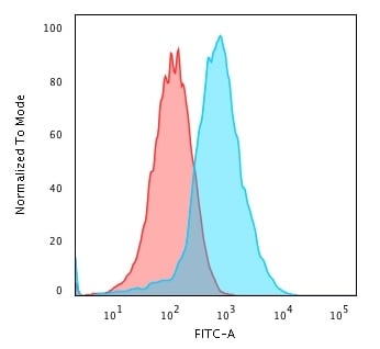
(Flow Cytometric Analysis of PFA-fixed HePG2 cells using GRP94 Recombinant Rabbit Monoclonal Antibody (HSP90B1/3168R) followed by Goat anti-Rat- IgG-CF488 (Blue); Isotype Control (Red).)
FCM/FACS (Flow Cytometry)
(Flow Cytometric Analysis of PFA-fixed HePG2 cells using GRP94 Recombinant Rabbit Monoclonal Antibody (HSP90B1/3168R) followed by Goat anti-Rat- IgG-CF488 (Blue); Isotype Control (Red).)
GRP94/HSP90B1, Antibody (Cat# AAA215236)
Full Name
GRP94/HSP90B1 (Endoplasmic Reticulum Marker)
Gene Names
HSP90B1; ECGP; GP96; TRA1; GRP94; HEL35; HEL-S-125m
Reactivity
Human, Monkey, Horse, Cow, Sheep, Pig, Rabbit, Dog, Rat, Mouse, Hamster, Guinea Pig, Chicken, Xenopus laevis
Applications
Immunohistochemistry, Flow Cytometry
Purity
Purified Ab with BSA and Azide at 200ug/ml OR Purified Ab WITHOUT BSA at 1.0mg/ml
Pricing
 Bioactivity
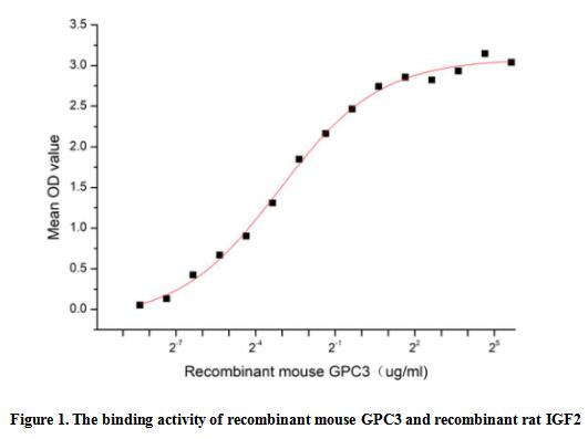
(Glypican-3 (GPC3), a 70 kDa protein, is a member of the glypican family that attaches to the cell surface by a glycosylphosphatidylinositol anchor, is specifically up-regulated in hepatocellular carcinoma (HCC) although rarely or not expressed in normal liver tissues, making it a perfect test and treatment target for HCC. GPC3 is also a negative transcriptional regulator and tumor suppressor that inhibits the growth of breast, ovary, and lung cancer cells. It is reported that GPC3 can form a complex with insulin-like growth factor 2 (IGF2), and might thereby modulate IGF2 action. Thus a functional binding ELISA assay was conducted to detect the interaction of recombinant mouse GPC3 and recombinant rat IGF2. Briefly, GPC3 was diluted serially in PBS with 0.01% BSA (pH 7.4). Duplicate samples of 100 ul were then transferred to IGF2-coated microtiter wells and incubated for 1h at 37 degree C. Wells were washed with PBST and incubated for 1h with anti-GPC3 pAb, then aspirated and washed 3 times. After incubation with HRP labelled secondary antibody for 1h at 37 degree C, wells were aspirated and washed 5 times. With the addition of substrate solution, wells were incubated 15-25 minutes at 37 degree C. Finally, add 50 uL stop solution to the wells and read at 450/630 nm immediately. The binding activity of recombinant mouse GPC3 and recombinant rat IGF2 was shown in Figure 1, the EC50 for this effect is 0.12 ug/mL.)
Bioactivity
(Glypican-3 (GPC3), a 70 kDa protein, is a member of the glypican family that attaches to the cell surface by a glycosylphosphatidylinositol anchor, is specifically up-regulated in hepatocellular carcinoma (HCC) although rarely or not expressed in normal liver tissues, making it a perfect test and treatment target for HCC. GPC3 is also a negative transcriptional regulator and tumor suppressor that inhibits the growth of breast, ovary, and lung cancer cells. It is reported that GPC3 can form a complex with insulin-like growth factor 2 (IGF2), and might thereby modulate IGF2 action. Thus a functional binding ELISA assay was conducted to detect the interaction of recombinant mouse GPC3 and recombinant rat IGF2. Briefly, GPC3 was diluted serially in PBS with 0.01% BSA (pH 7.4). Duplicate samples of 100 ul were then transferred to IGF2-coated microtiter wells and incubated for 1h at 37 degree C. Wells were washed with PBST and incubated for 1h with anti-GPC3 pAb, then aspirated and washed 3 times. After incubation with HRP labelled secondary antibody for 1h at 37 degree C, wells were aspirated and washed 5 times. With the addition of substrate solution, wells were incubated 15-25 minutes at 37 degree C. Finally, add 50 uL stop solution to the wells and read at 450/630 nm immediately. The binding activity of recombinant mouse GPC3 and recombinant rat IGF2 was shown in Figure 1, the EC50 for this effect is 0.12 ug/mL.)
Glypican 3 (GPC3), Active Protein (Cat# AAA161815)
Full Name
Active Glypican 3 (GPC3)
Gene Names
Gpc3; OCI-5
Applications
Activity Assay, Cell Culture
Purity
Greater than 90% by SDS-PAGE
Pricing
 WB (Western Blot)
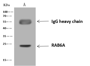
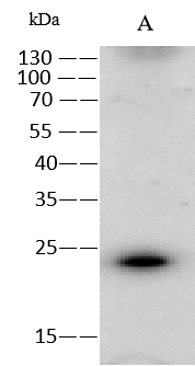
(Anti-RAB6A rabbit polyclonal antibody at 1:500 dilutionLane A: H1299 Whole Cell LysateLysates/proteins at 30 ug per lane.SecondaryGoat Anti-Rabbit IgG (H+L)/HRP at 1/10000 dilution.Developed using the ECL technique.Performed under reducing conditions.Predicted band size:24 kDaObserved band size:24 kDa)
WB (Western Blot)
(Anti-RAB6A rabbit polyclonal antibody at 1:500 dilutionLane A: H1299 Whole Cell LysateLysates/proteins at 30 ug per lane.SecondaryGoat Anti-Rabbit IgG (H+L)/HRP at 1/10000 dilution.Developed using the ECL technique.Performed under reducing conditions.Predicted band size:24 kDaObserved band size:24 kDa)
RAB6A, Polyclonal Antibody (Cat# AAA258930)
Full Name
Anti-RAB6A Antibody, Rabbit Polyclonal
Reactivity
Human
Applications
Immunoprecipitation, Western Blot
Purity
Protein A & Antigen Affinity
Pricing

 SDS-PAGE
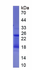
(SDS-PAGE)
SDS-PAGE
(SDS-PAGE)
Cluster Of Differentiation 38 (CD38), Recombinant Protein (Cat# AAA152038)
Full Name
Eukaryotic Cluster Of Differentiation 38 (CD38)
Gene Names
CD38; T10; ADPRC 1
Applications
Western Blot
Purity
Greater than 80% by SDS-PAGE
Pricing

 ELISA
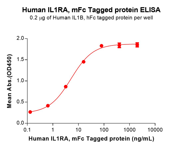
(Figure 2. ELISA plate pre-coated by 2 ug/mL (100 uL/well) Human IL1B Protein, hFc Tag can bind Human IL1RA Protein, mFc Tag (AAA129488) in a linear range of 0.64–80 ng/mL.)
ELISA
(Figure 2. ELISA plate pre-coated by 2 ug/mL (100 uL/well) Human IL1B Protein, hFc Tag can bind Human IL1RA Protein, mFc Tag (AAA129488) in a linear range of 0.64–80 ng/mL.)
IL1RA, Recombinant Protein (Cat# AAA129488)
Full Name
Human IL1RA Protein, mFc Tag
Gene Names
IL1R1; P80; IL1R; IL1RA; CD121A; D2S1473; IL-1R-alpha
Purity
The purity of the protein is greater than 95% as determined by SDS-PAGE and Coomassie blue staining.
Pricing
Fc epsilon RI alpha, Active Protein (Cat# AAA48987)
Full Name
Recombinant Human Fc epsilon RI alpha protein
Applications
Bioactivity, SDS-PAGE
Purity
> 90% by SDS-PAGE
Pricing
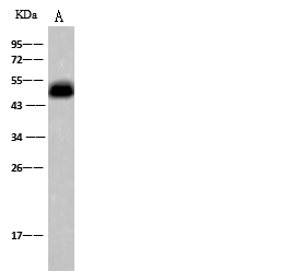
 IP (Immunoprecipitation)
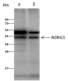
(NDRG1 was immunoprecipitated using:Lane A:0.5 mg HeLa Whole Cell LysateLane B:0.5 mg HepG2 Whole Cell Lysate2 uL anti-NDRG1 rabbit polyclonal antibody and 60 ug of Immunomagnetic beads Protein A/G.Primary antibody:Anti-NDRG1 rabbit polyclonal antibody,at 1:100 dilutionSecondary antibody:Clean-Blot IP Detection Reagent (HRP) at 1:1000dilutionDeveloped using the ECL technique.Performed under reducing conditions.Predicted band size: 43 kDaObserved band size :43 kDa)
IP (Immunoprecipitation)
(NDRG1 was immunoprecipitated using:Lane A:0.5 mg HeLa Whole Cell LysateLane B:0.5 mg HepG2 Whole Cell Lysate2 uL anti-NDRG1 rabbit polyclonal antibody and 60 ug of Immunomagnetic beads Protein A/G.Primary antibody:Anti-NDRG1 rabbit polyclonal antibody,at 1:100 dilutionSecondary antibody:Clean-Blot IP Detection Reagent (HRP) at 1:1000dilutionDeveloped using the ECL technique.Performed under reducing conditions.Predicted band size: 43 kDaObserved band size :43 kDa)
NDRG1, Polyclonal Antibody (Cat# AAA255720)
Full Name
Anti-NDRG1 Antibody, Rabbit Polyclonal
Gene Names
NDRG1; GC4; RTP; DRG1; NDR1; NMSL; TDD5; CAP43; CMT4D; DRG-1; HMSNL; RIT42; TARG1; PROXY1
Reactivity
Human
Applications
Immunoprecipitation, Immunofluorescence, Immunocytochemistry, Immunohistochemistry, Western Blot
Purity
Protein A & Antigen Affinity
Pricing
 FCM/FACS (Flow Cytometry)
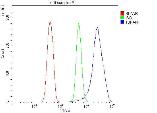
(Figure 2. Flow Cytometry analysis of RH35 cells using anti-Tspan1 antibody (AAA125799).Overlay histogram showing RH35 cells stained with AAA125799 (Blue line). The cells were blocked with 10% normal goat serum. And then incubated with rabbit anti-Tspan1 Antibody (AAA125799, 1μg/1x106 cells) for 30 min at 20 degree C. DyLight®488 conjugated goat anti-rabbit IgG (5-10μg/1x106 cells) was used as secondary antibody for 30 minutes at 20 degree C. Isotype control antibody (Green line) was rabbit IgG (1μg/1x106) used under the same conditions. Unlabelled sample (Red line) was also used as a control.)
FCM/FACS (Flow Cytometry)
(Figure 2. Flow Cytometry analysis of RH35 cells using anti-Tspan1 antibody (AAA125799).Overlay histogram showing RH35 cells stained with AAA125799 (Blue line). The cells were blocked with 10% normal goat serum. And then incubated with rabbit anti-Tspan1 Antibody (AAA125799, 1μg/1x106 cells) for 30 min at 20 degree C. DyLight®488 conjugated goat anti-rabbit IgG (5-10μg/1x106 cells) was used as secondary antibody for 30 minutes at 20 degree C. Isotype control antibody (Green line) was rabbit IgG (1μg/1x106) used under the same conditions. Unlabelled sample (Red line) was also used as a control.)
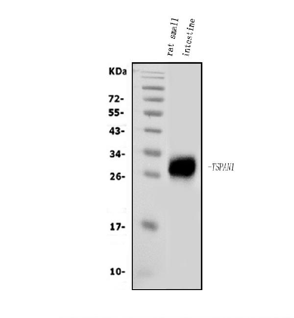
Tspan1, Polyclonal Antibody (Cat# AAA125799)
Full Name
Anti-Tspan1 Antibody
Reactivity
Rat
Applications
Direct ELISA, Flow Cytometry, Western Blot
Purity
Immunogen affinity purified.
Pricing
 FCM/FACS (Flow Cytometry)
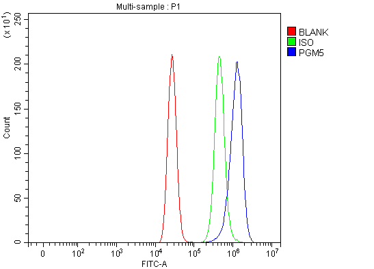
(Figure 2. Flow Cytometry analysis of MCF-7 cells using anti-PGM5 antibody (AAA127727).Overlay histogram showing MCF-7 cells stained with AAA127727 (Blue line). To facilitate intracellular staining, cells were fixed with 4% paraformaldehyde and permeabilized with permeabilization buffer. The cells were blocked with 10% normal goat serum. And then incubated with rabbit anti-PGM5 Antibody (AAA127727, 1ug/1x106 cells) for 30 min at 20 degree C. DyLight488 conjugated goat anti-rabbit IgG was used as secondary antibody for 30 minutes at 20 degree C. Isotype control antibody (Green line) was rabbit IgG (1ug/1x106) used under the same conditions. Unlabelled sample (Red line) was also used as a control.)
FCM/FACS (Flow Cytometry)
(Figure 2. Flow Cytometry analysis of MCF-7 cells using anti-PGM5 antibody (AAA127727).Overlay histogram showing MCF-7 cells stained with AAA127727 (Blue line). To facilitate intracellular staining, cells were fixed with 4% paraformaldehyde and permeabilized with permeabilization buffer. The cells were blocked with 10% normal goat serum. And then incubated with rabbit anti-PGM5 Antibody (AAA127727, 1ug/1x106 cells) for 30 min at 20 degree C. DyLight488 conjugated goat anti-rabbit IgG was used as secondary antibody for 30 minutes at 20 degree C. Isotype control antibody (Green line) was rabbit IgG (1ug/1x106) used under the same conditions. Unlabelled sample (Red line) was also used as a control.)
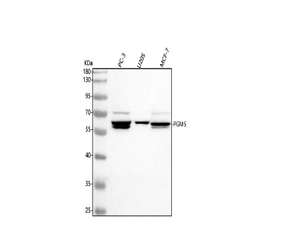
PGM5, Polyclonal Antibody (Cat# AAA127727)
Full Name
Anti-PGM5 Antibody Picoband
Gene Names
PGM5; PGMRP
Reactivity
Human
Applications
Flow Cytometry, Western Blot
Purity
Immunogen affinity purified.
Pricing
 IF (Immunofluorescence)
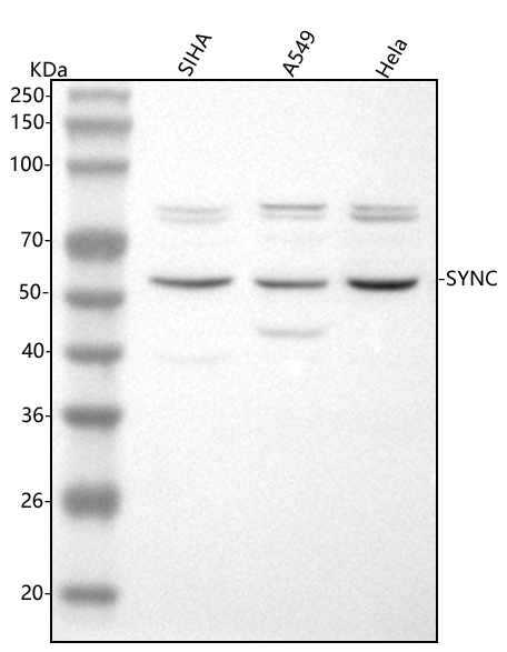
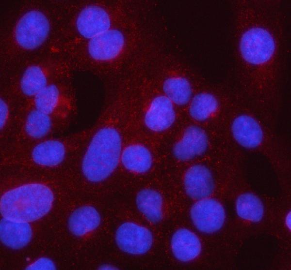
(Figure 2. IF analysis of Syncoilin/SYNC using anti-Syncoilin/SYNC antibody (AAA127346).Syncoilin/SYNC was detected in an immunocytochemical section of U2OS cells. Enzyme antigen retrieval was performed using IHC enzyme antigen retrieval reagent for 15 mins. The cells were blocked with 10% goat serum. And then incubated with 5ug/mL rabbit anti-Syncoilin/SYNC Antibody (AAA127346) overnight at 4 degree C. Cy3 Conjugated Goat Anti-Rabbit IgG (BA1032) was used as secondary antibody at 1:500 dilution and incubated for 30 minutes at 37 degree C. The section was counterstained with DAPI. Visualize using a fluorescence microscope and filter sets appropriate for the label used.)
IF (Immunofluorescence)
(Figure 2. IF analysis of Syncoilin/SYNC using anti-Syncoilin/SYNC antibody (AAA127346).Syncoilin/SYNC was detected in an immunocytochemical section of U2OS cells. Enzyme antigen retrieval was performed using IHC enzyme antigen retrieval reagent for 15 mins. The cells were blocked with 10% goat serum. And then incubated with 5ug/mL rabbit anti-Syncoilin/SYNC Antibody (AAA127346) overnight at 4 degree C. Cy3 Conjugated Goat Anti-Rabbit IgG (BA1032) was used as secondary antibody at 1:500 dilution and incubated for 30 minutes at 37 degree C. The section was counterstained with DAPI. Visualize using a fluorescence microscope and filter sets appropriate for the label used.)
Syncoilin/SYNC, Polyclonal Antibody (Cat# AAA127346)
Full Name
Anti-Syncoilin/SYNC Antibody Picoband
Gene Names
SYNC; SYNC1; SYNCOILIN
Reactivity
Human
Applications
Western Blot, Immunofluorescence, Immunocytochemistry
Purity
Immunogen affinity purified.
Pricing
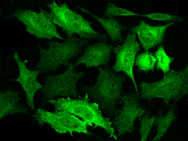
 ICC (Immunocytochemistry)
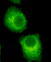
(Immunocytochemistry of HeLa cells using anti-Rab2 mouse mAb diluted 1:200)
ICC (Immunocytochemistry)
(Immunocytochemistry of HeLa cells using anti-Rab2 mouse mAb diluted 1:200)
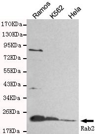
Rab2, Monoclonal Antibody (Cat# AAA320329)
Full Name
Rab2 Mouse mAb
Gene Names
RAB2A; LHX; RAB2
Reactivity
Human
Applications
Immunocytochemistry, Western Blot
Purity
The antibody was affinity-purified from mouse ascites by affinity-chromatography using epitope-specific immunogen.
Pricing
 Bioactivity
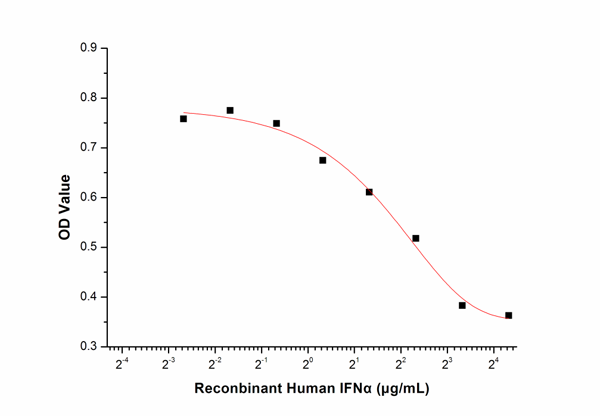
(Figure 2. Inhibition of A549 cells proliferation after stimulated with IFN-.)
Bioactivity
(Figure 2. Inhibition of A549 cells proliferation after stimulated with IFN-.)
Interferon Alpha, Active Protein (Cat# AAA150049)
Full Name
Active Interferon Alpha (IFNa)
Applications
Activity Assay, Cell Culture
Purity
>90%
Pricing
 FCM/FACS (Flow Cytometry)
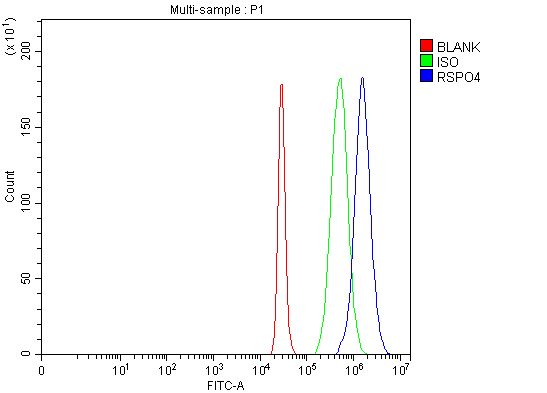
(Figure 2. Flow Cytometry analysis of 293T cells using anti-RSPO4 antibody (AAA127594).Overlay histogram showing 293T cells stained with AAA127594 (Blue line). The cells were fixed with 4% paraformaldehyde and blocked with 10% normal goat serum. And then incubated with rabbit anti-RSPO4 Antibody (AAA127594, 1ug/1x106 cells) for 30 min at 20 degree C. DyLight488 conjugated goat anti-rabbit IgG was used as secondary antibody for 30 minutes at 20 degree C. Isotype control antibody (Green line) was rabbit IgG (1ug/1x106) used under the same conditions. Unlabelled sample (Red line) was also used as a control.)
FCM/FACS (Flow Cytometry)
(Figure 2. Flow Cytometry analysis of 293T cells using anti-RSPO4 antibody (AAA127594).Overlay histogram showing 293T cells stained with AAA127594 (Blue line). The cells were fixed with 4% paraformaldehyde and blocked with 10% normal goat serum. And then incubated with rabbit anti-RSPO4 Antibody (AAA127594, 1ug/1x106 cells) for 30 min at 20 degree C. DyLight488 conjugated goat anti-rabbit IgG was used as secondary antibody for 30 minutes at 20 degree C. Isotype control antibody (Green line) was rabbit IgG (1ug/1x106) used under the same conditions. Unlabelled sample (Red line) was also used as a control.)
RSPO4, Polyclonal Antibody (Cat# AAA127594)
Full Name
Anti-RSPO4 Antibody Picoband
Gene Names
RSPO4; CRISTIN4; C20orf182
Reactivity
Human, Mouse, Rat
Applications
Flow Cytometry, Western Blot
Purity
Immunogen affinity purified.
Pricing
 IF (Immunofluorescence)
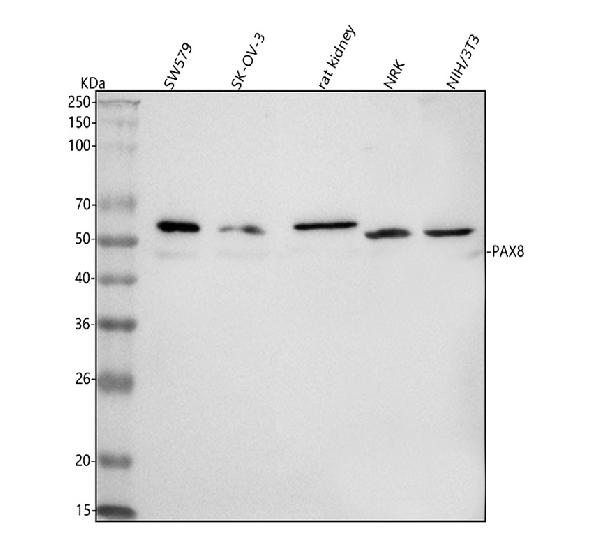
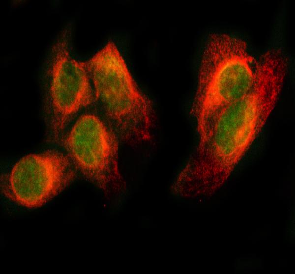
(Figure 2. IF analysis of PAX8 using anti-PAX8 antibody (AAA127022) and anti-Beta Tubulin antibody (M01857-3).PAX8 was detected in immunocytochemical section of HELA cell. Enzyme antigen retrieval was performed using IHC enzyme antigen retrieval reagent for 15 mins. The cells were blocked with 10% goat serum. And then incubated with 5ug/mL rabbit anti-PAX8 Antibody (AAA127022) and mouse anti-Beta Tubulin antibody (M01857-3) overnight at 4 degree C. DyLight488 Conjugated Goat Anti-Rabbit IgG and Cy3 Conjugated Goat Anti-Mouse IgG (BA1031) were used as secondary antibody at 1:500 dilution and incubated for 30 minutes at 37 degree C. Visualize using a fluorescence microscope and filter sets appropriate for the label used.)
IF (Immunofluorescence)
(Figure 2. IF analysis of PAX8 using anti-PAX8 antibody (AAA127022) and anti-Beta Tubulin antibody (M01857-3).PAX8 was detected in immunocytochemical section of HELA cell. Enzyme antigen retrieval was performed using IHC enzyme antigen retrieval reagent for 15 mins. The cells were blocked with 10% goat serum. And then incubated with 5ug/mL rabbit anti-PAX8 Antibody (AAA127022) and mouse anti-Beta Tubulin antibody (M01857-3) overnight at 4 degree C. DyLight488 Conjugated Goat Anti-Rabbit IgG and Cy3 Conjugated Goat Anti-Mouse IgG (BA1031) were used as secondary antibody at 1:500 dilution and incubated for 30 minutes at 37 degree C. Visualize using a fluorescence microscope and filter sets appropriate for the label used.)
PAX8, Polyclonal Antibody (Cat# AAA127022)
Full Name
Anti-PAX8 Antibody Picoband
Reactivity
Human, Mouse, Rat
Applications
Immunofluorescence, Immunocytochemistry, Western Blot
Purity
Immunogen affinity purified.
Pricing
EphA3, Active Protein (Cat# AAA48999)
Full Name
Recombinant mouse EphA3 protein
Gene Names
Epha3; Hek; Cek4; ETK1; End3; Hek4; Mek4; Tyro4; AW492086
Applications
Bioactivity, SDS-PAGE
Purity
> 95% by SDS-PAGE
Pricing
 Application Data
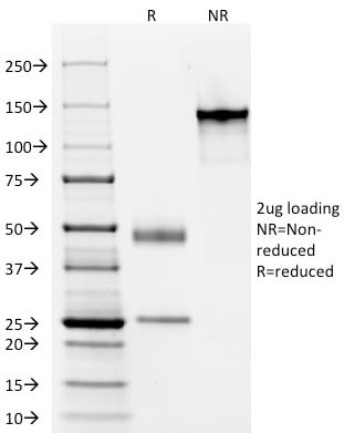
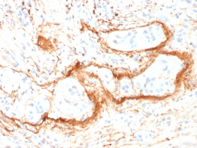
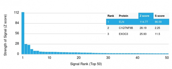
(Analysis of Protein Array containing >19, 000 full-length human proteins using Elastin (ELN) Mouse Monoclonal Antibody (ELN/1981) Z- and S- Score: The Z-score represents the strength of a signal that a monoclonal antibody (MAb) (in combination with a fluorescently-tagged anti-IgG secondary antibody) produces when binding to a particular protein on the HuProtTM array. Z-scores are described in units of standard deviations (SD's) above the mean value of all signals generated on that array. If targets on HuProtTM are arranged in descending order of the Z-score, the S-score is the difference (also in units of SD's) between the Z-score. S-score therefore represents the relative target specificity of a MAb to its intended target. A MAb is considered to specific to its intended target, if the MAb has an S-score of at least 2.5. For example, if a MAb binds to protein X with a Z-score of 43 and to protein Y with a Z-score of 14, then the S-score for the binding of that MAb to protein X is equal to 29.)
Application Data
(Analysis of Protein Array containing >19, 000 full-length human proteins using Elastin (ELN) Mouse Monoclonal Antibody (ELN/1981) Z- and S- Score: The Z-score represents the strength of a signal that a monoclonal antibody (MAb) (in combination with a fluorescently-tagged anti-IgG secondary antibody) produces when binding to a particular protein on the HuProtTM array. Z-scores are described in units of standard deviations (SD's) above the mean value of all signals generated on that array. If targets on HuProtTM are arranged in descending order of the Z-score, the S-score is the difference (also in units of SD's) between the Z-score. S-score therefore represents the relative target specificity of a MAb to its intended target. A MAb is considered to specific to its intended target, if the MAb has an S-score of at least 2.5. For example, if a MAb binds to protein X with a Z-score of 43 and to protein Y with a Z-score of 14, then the S-score for the binding of that MAb to protein X is equal to 29.)
Elastin (ELN), Monoclonal Antibody (Cat# AAA214642)
Full Name
Elastin (ELN) (Marker of Arterial Stiffness and Atherosclerosis)
Gene Names
ELN; WS; WBS; SVAS; ADCL1
Reactivity
Human. Others not known.
Applications
Immunohistochemistry
Pricing
 IHC (Immunohistochemisry)
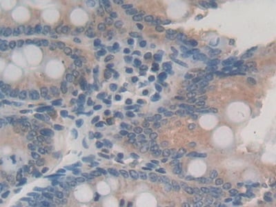
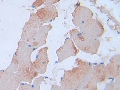
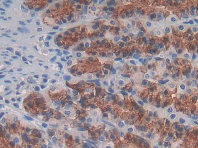
(DAB staining on IHCP;Sample: Rat Stomach Tissue; Primary Ab: 30ug/ml Mouse AntiRat IL24 AntibodySecond Ab: 2ug/mL HRPLinked Caprine AntiMouse IgG Polyclonal Antibody(Catalog: SAA544Mu19))
IHC (Immunohistochemisry)
(DAB staining on IHCP;Sample: Rat Stomach Tissue; Primary Ab: 30ug/ml Mouse AntiRat IL24 AntibodySecond Ab: 2ug/mL HRPLinked Caprine AntiMouse IgG Polyclonal Antibody(Catalog: SAA544Mu19))
Interleukin 24 (IL24), Monoclonal Antibody (Cat# AAA151787)
Full Name
Monoclonal Antibody to Interleukin 24 (IL24)
Gene Names
Il24; Mda7; Mda-7
Reactivity
Rat
Applications
Immunoprecipitation, Immunocytochemistry, Immunohistochemistry, Western Blot
Purity
Protein A + Protein G affinity chromatography
Pricing
 Application Data
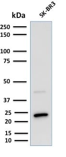
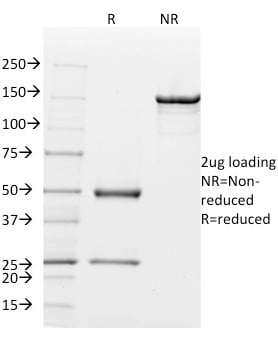
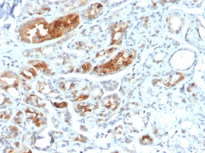
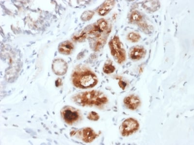
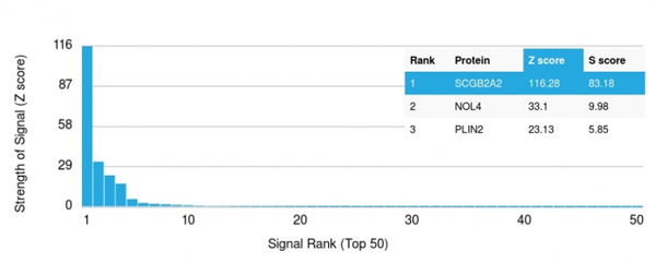
(Analysis of Protein Array containing >19, 000 full-length human proteins using Mammaglobin (SCGB2A2) Mouse Monoclonal Antibody (MGB1/2000) Z- and S- Score: The Z-score represents the strength of a signal that a monoclonal antibody (MAb) (in combination with a fluorescently-tagged anti-IgG secondary antibody) produces when binding to a particular protein on the HuProtTM array. Z-scores are described in units of standard deviations (SD's) above the mean value of all signals generated on that array. If targets on HuProtTM are arranged in descending order of the Z-score, the S-score is the difference (also in units of SD's) between the Z-score. S-score therefore represents the relative target specificity of a MAb to its intended target. A MAb is considered to specific to its intended target, if the MAb has an S-score of at least 2.5. For example, if a MAb binds to protein X with a Z-score of 43 and to protein Y with a Z-score of 14, then the S-score for the binding of that MAb to protein X is equal to 29.)
Application Data
(Analysis of Protein Array containing >19, 000 full-length human proteins using Mammaglobin (SCGB2A2) Mouse Monoclonal Antibody (MGB1/2000) Z- and S- Score: The Z-score represents the strength of a signal that a monoclonal antibody (MAb) (in combination with a fluorescently-tagged anti-IgG secondary antibody) produces when binding to a particular protein on the HuProtTM array. Z-scores are described in units of standard deviations (SD's) above the mean value of all signals generated on that array. If targets on HuProtTM are arranged in descending order of the Z-score, the S-score is the difference (also in units of SD's) between the Z-score. S-score therefore represents the relative target specificity of a MAb to its intended target. A MAb is considered to specific to its intended target, if the MAb has an S-score of at least 2.5. For example, if a MAb binds to protein X with a Z-score of 43 and to protein Y with a Z-score of 14, then the S-score for the binding of that MAb to protein X is equal to 29.)
Mammaglobin, Monoclonal Antibody (Cat# AAA214664)
Full Name
Mammaglobin (SCGB2A2) (Breast Cancer Marker)
Gene Names
SCGB2A2; MGB1; UGB2
Reactivity
Human. Others not known.
Applications
Immunohistochemistry
Pricing
 IHC (Immunohiostchemistry)
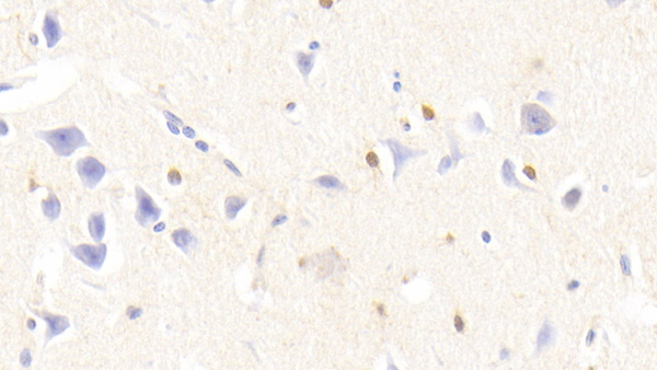
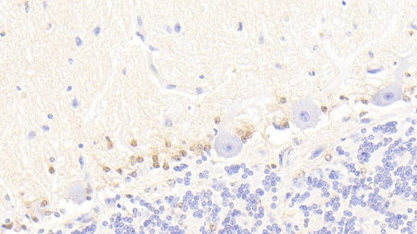
(DAB staining on IHCP;Samples: Human Cerebellum Tissue; Primary Ab: 20ug/ml Mouse AntiHuman S100B AntibodySecond Ab: 2ug/mL HRPLinked Caprine AntiMouse IgG Polyclonal Antibody(Catalog: SAA544Mu19))
IHC (Immunohiostchemistry)
(DAB staining on IHCP;Samples: Human Cerebellum Tissue; Primary Ab: 20ug/ml Mouse AntiHuman S100B AntibodySecond Ab: 2ug/mL HRPLinked Caprine AntiMouse IgG Polyclonal Antibody(Catalog: SAA544Mu19))
S100 Calcium Binding Protein B (S100B), Monoclonal Antibody (Cat# AAA151573)
Full Name
Monoclonal Antibody to S100 Calcium Binding Protein B (S100B)
Gene Names
S100B; NEF; S100; S100-B; S100beta
Reactivity
Human, Pig
Applications
Immunoprecipitation, Immunocytochemistry, Immunohistochemistry, Western Blot
Purity
Protein A + Protein G affinity chromatography
Pricing
 IF (Immunofluorescence)
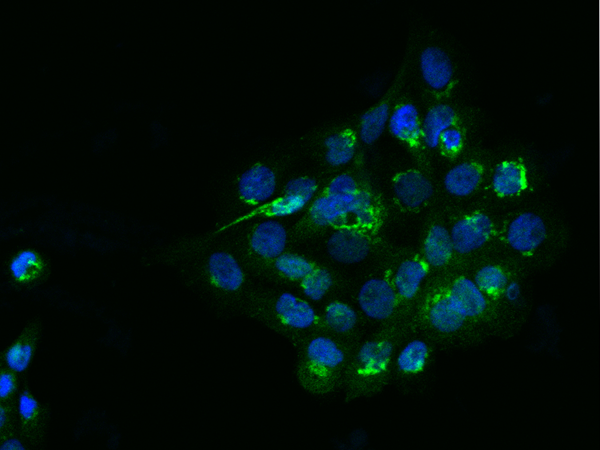
(Immunofluorescence staining of FAM3C-His in A431 cells. Cells were fixed with 4% PFA, permeabilzed with 0.3% Triton X-100 in PBS,blocked with 10% serum, and incubated with rabbit anti-Human FAM3C-His polyclonal antibody (dilution ratio 1:100) at 4 degree C overnight. Then cells were stained with the Alexa Fluor488-conjugated Goat Anti-rabbit IgG secondary antibody (green) and counterstained with DAPI (blue).Positive staining was localized to Cytoplasm.)
IF (Immunofluorescence)
(Immunofluorescence staining of FAM3C-His in A431 cells. Cells were fixed with 4% PFA, permeabilzed with 0.3% Triton X-100 in PBS,blocked with 10% serum, and incubated with rabbit anti-Human FAM3C-His polyclonal antibody (dilution ratio 1:100) at 4 degree C overnight. Then cells were stained with the Alexa Fluor488-conjugated Goat Anti-rabbit IgG secondary antibody (green) and counterstained with DAPI (blue).Positive staining was localized to Cytoplasm.)
FAM3C, Polyclonal Antibody (Cat# AAA258645)
Full Name
FAM3C Antibody, Rabbit PAb, Antigen Affinity Purified
Gene Names
FAM3C; ILEI; GS3786
Reactivity
Human
Applications
Immunofluorescence, Immunocytochemistry, Immunohistochemistry, Western Blot
Pricing
 FCM/FACS (Flow Cytometry)
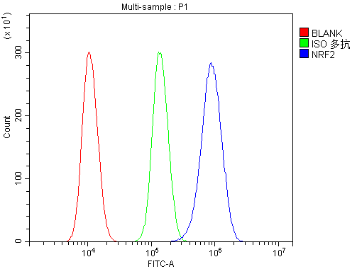
(Figure 4. Flow Cytometry analysis of 293T cells using anti-NFE2L2 antibody (AAA125514).Overlay histogram showing 293T cells stained with AAA125514 (Blue line). The cells were blocked with 10% normal goat serum. And then incubated with rabbit anti-NFE2L2 Antibody (AAA125514, 1μg/1x106 cells) for 30 min at 20 degree C. DyLight®488 conjugated goat anti-rabbit IgG (5-10μg/1x106 cells) was used as secondary antibody for 30 minutes at 20 degree C. Isotype control antibody (Green line) was rabbit IgG (1μg/1x106) used under the same conditions. Unlabelled sample (Red line) was also used as a control.)
FCM/FACS (Flow Cytometry)
(Figure 4. Flow Cytometry analysis of 293T cells using anti-NFE2L2 antibody (AAA125514).Overlay histogram showing 293T cells stained with AAA125514 (Blue line). The cells were blocked with 10% normal goat serum. And then incubated with rabbit anti-NFE2L2 Antibody (AAA125514, 1μg/1x106 cells) for 30 min at 20 degree C. DyLight®488 conjugated goat anti-rabbit IgG (5-10μg/1x106 cells) was used as secondary antibody for 30 minutes at 20 degree C. Isotype control antibody (Green line) was rabbit IgG (1μg/1x106) used under the same conditions. Unlabelled sample (Red line) was also used as a control.)
NFE2L2, Polyclonal Antibody (Cat# AAA125514)
Full Name
Anti-NFE2L2 Antibody
Gene Names
NFE2L2; NRF2
Reactivity
Human
Applications
Direct ELISA, Flow Cytometry, Immunofluorescence, Immunocytochemistry, Immunohistochemistry, Western Blot
Purity
Immunogen affinity purified.
Pricing
 Bioactivity
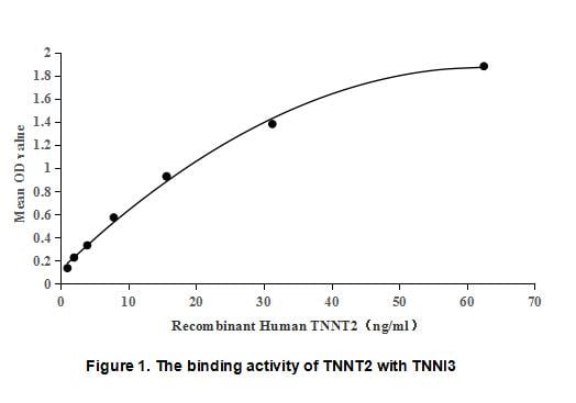
(Troponin T Type 2, Cardiac (TNNT2) is one of three troponin isoforms found in the tropomyosin-troponin complex. This complex is responsible for the calcium sensitivity of the contractile apparatus in the muscle. Cardiac Troponin T is used as a biological marker for cardiomyocytes and its level in serum is frequently used as an indicator of myocardial cell injury. Besides, Troponin I Type 3, Cardiac (TNNI3) has been identified as an interactor of TNNT2, thus a binding ELISA assay was conducted to detect the interaction of recombinant human TNNT2 and recombinant human TNNTI3. Briefly, TNNT2 was diluted serially in PBS with 0.01% BSA (pH 7.4). Duplicate samples of 100ul were then transferred to TNNI3-coated microtiter wells and incubated for 1h at 37 degree C. Wells were washed with PBST and incubated for 1h with anti-TNNT2 pAb, then aspirated and washed 3 times. After incubation with HRP labelled secondary antibody for 1h at 37 degree C, wells were aspirated and washed 5 times. With the addition of substrate solution, wells were incubated 15-25 minutes at 37 degree C. Finally, add 50uL stop solution to the wells and read at 450/630nm immediately. The binding activity of TNNT2 and TNNI3 was shown in Figure 1, and this effect was in a dose dependent manner.)
Bioactivity
(Troponin T Type 2, Cardiac (TNNT2) is one of three troponin isoforms found in the tropomyosin-troponin complex. This complex is responsible for the calcium sensitivity of the contractile apparatus in the muscle. Cardiac Troponin T is used as a biological marker for cardiomyocytes and its level in serum is frequently used as an indicator of myocardial cell injury. Besides, Troponin I Type 3, Cardiac (TNNI3) has been identified as an interactor of TNNT2, thus a binding ELISA assay was conducted to detect the interaction of recombinant human TNNT2 and recombinant human TNNTI3. Briefly, TNNT2 was diluted serially in PBS with 0.01% BSA (pH 7.4). Duplicate samples of 100ul were then transferred to TNNI3-coated microtiter wells and incubated for 1h at 37 degree C. Wells were washed with PBST and incubated for 1h with anti-TNNT2 pAb, then aspirated and washed 3 times. After incubation with HRP labelled secondary antibody for 1h at 37 degree C, wells were aspirated and washed 5 times. With the addition of substrate solution, wells were incubated 15-25 minutes at 37 degree C. Finally, add 50uL stop solution to the wells and read at 450/630nm immediately. The binding activity of TNNT2 and TNNI3 was shown in Figure 1, and this effect was in a dose dependent manner.)
Troponin T Type 2, Cardiac (TNNT2), Active Protein (Cat# AAA152235)
Full Name
Active Troponin T Type 2, Cardiac (TNNT2)
Gene Names
TNNT2; CMH2; RCM3; TnTC; cTnT; CMPD2; LVNC6
Applications
Activity Assay, Cell Culture
Purity
Greater than 95% by SDS-PAGE
Pricing
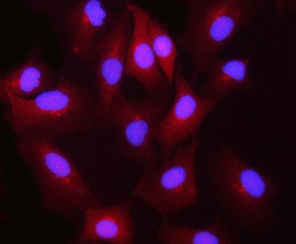
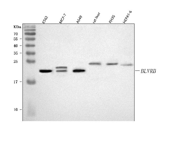
 FCM/FACS (Flow Cytometry)
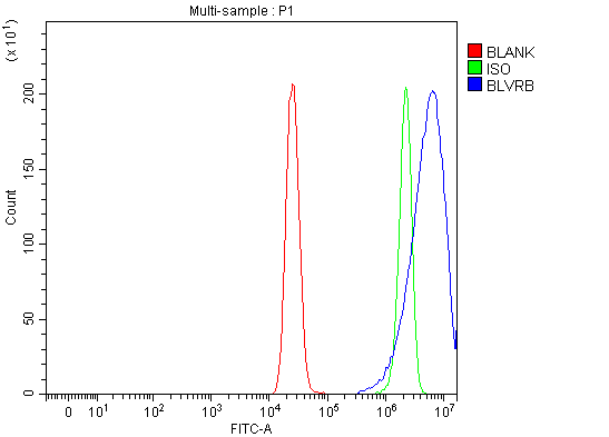
(Figure 3. Flow Cytometry analysis of MCF-7 cells using anti-BLVRB antibody (AAA127498).Overlay histogram showing MCF-7 cells stained with AAA127498 (Blue line). To facilitate intracellular staining, cells were fixed with 4% paraformaldehyde and permeabilized with permeabilization buffer. The cells were blocked with 10% normal goat serum. And then incubated with rabbit anti-BLVRB Antibody (AAA127498, 1ug/1x106 cells) for 30 min at 20 degree C. DyLight488 conjugated goat anti-rabbit IgG was used as secondary antibody for 30 minutes at 20 degree C. Isotype control antibody (Green line) was rabbit IgG (1ug/1x106) used under the same conditions. Unlabelled sample (Red line) was also used as a control.)
FCM/FACS (Flow Cytometry)
(Figure 3. Flow Cytometry analysis of MCF-7 cells using anti-BLVRB antibody (AAA127498).Overlay histogram showing MCF-7 cells stained with AAA127498 (Blue line). To facilitate intracellular staining, cells were fixed with 4% paraformaldehyde and permeabilized with permeabilization buffer. The cells were blocked with 10% normal goat serum. And then incubated with rabbit anti-BLVRB Antibody (AAA127498, 1ug/1x106 cells) for 30 min at 20 degree C. DyLight488 conjugated goat anti-rabbit IgG was used as secondary antibody for 30 minutes at 20 degree C. Isotype control antibody (Green line) was rabbit IgG (1ug/1x106) used under the same conditions. Unlabelled sample (Red line) was also used as a control.)
BLVRB, Polyclonal Antibody (Cat# AAA127498)
Full Name
Anti-BLVRB Antibody Picoband
Gene Names
BLVRB; FLR; BVRB; SDR43U1; HEL-S-10
Reactivity
Human, Mouse, Rat
Applications
Flow Cytometry, Immunofluorescence, Immunocytochemistry, Western Blot
Purity
Immunogen affinity purified.
Pricing
 WB (Western Blot)
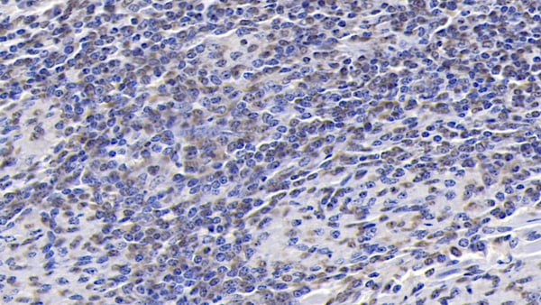
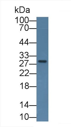
(Western Blot; Sample: Rat Placenta lysate Primary Ab: 2ug/mL Mouse AntiHuman IGFBP1 Antibody Second Ab: 0.2ug/mL HRPLinked Caprine AntiMouse IgG Polyclonal Antibody (Catalog: SAA544Mu19))
WB (Western Blot)
(Western Blot; Sample: Rat Placenta lysate Primary Ab: 2ug/mL Mouse AntiHuman IGFBP1 Antibody Second Ab: 0.2ug/mL HRPLinked Caprine AntiMouse IgG Polyclonal Antibody (Catalog: SAA544Mu19))
Insulin Like Growth Factor Binding Protein 1 (IGFBP1), Monoclonal Antibody (Cat# AAA151443)
Full Name
Monoclonal Antibody to Insulin Like Growth Factor Binding Protein 1 (IGFBP1)
Gene Names
IGFBP1; AFBP; IBP1; PP12; IGF-BP25; hIGFBP-1
Reactivity
Human, Rat, Pig
Applications
Immunoprecipitation, Immunocytochemistry, Immunohistochemistry, Western Blot
Purity
Protein A + Protein G affinity chromatography
Pricing
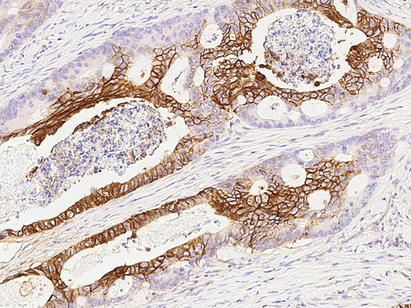
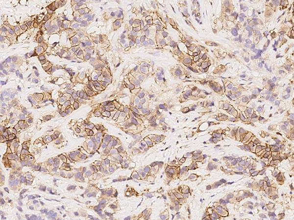
 IHC (Immunohistochemistry)
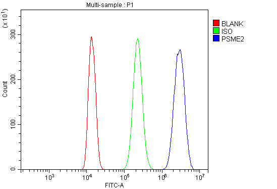
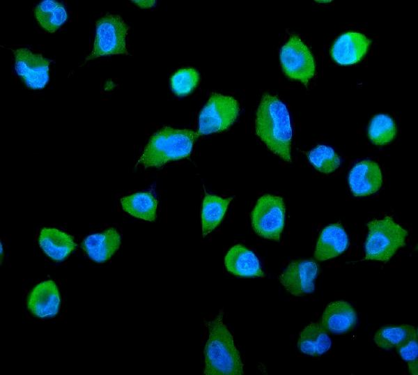
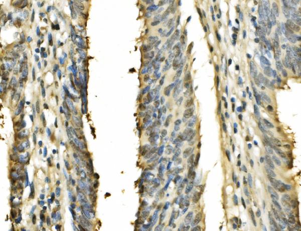
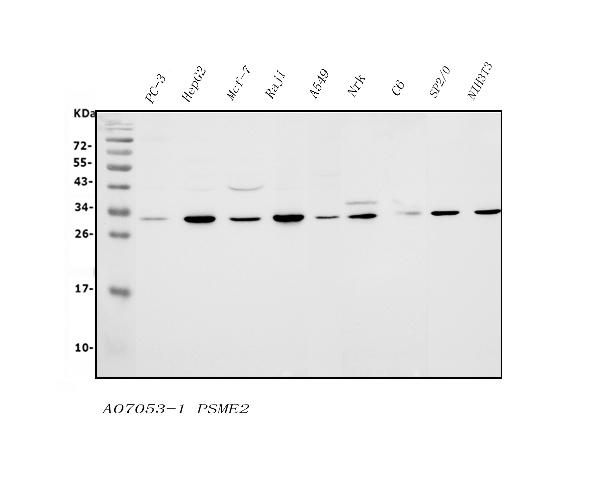
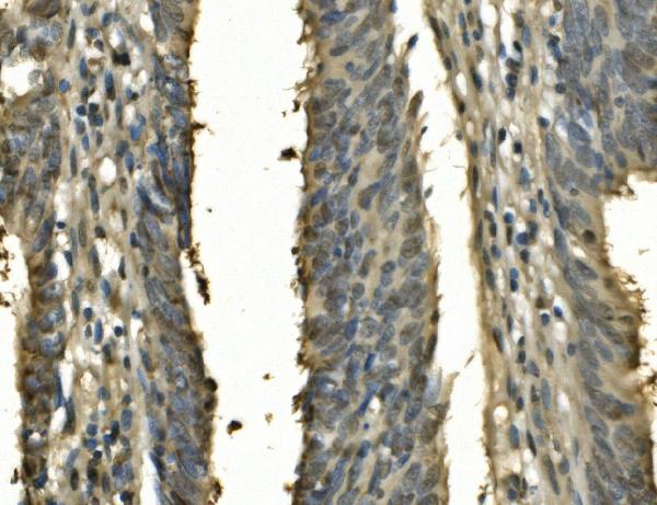
(Figure 5. IHC analysis of PSME2 using anti-PSME2 antibody (AAA125785).PSME2 was detected in paraffin-embedded section of human rectal cancer tissue. Heat mediated antigen retrieval was performed in EDTA buffer (pH8. 0, epitope retrieval solution). The tissue section was blocked with 10% goat serum. The tissue section was then incubated with 2μg/ml rabbit anti-PSME2 Antibody (AAA125785) overnight at 4 degree C. Biotinylated goat anti-rabbit IgG was used as secondary antibody and incubated for 30 minutes at 37 degree C. The tissue section was developed using Strepavidin-Biotin-Complex (SABC) with DAB as the chromogen.)
IHC (Immunohistochemistry)
(Figure 5. IHC analysis of PSME2 using anti-PSME2 antibody (AAA125785).PSME2 was detected in paraffin-embedded section of human rectal cancer tissue. Heat mediated antigen retrieval was performed in EDTA buffer (pH8. 0, epitope retrieval solution). The tissue section was blocked with 10% goat serum. The tissue section was then incubated with 2μg/ml rabbit anti-PSME2 Antibody (AAA125785) overnight at 4 degree C. Biotinylated goat anti-rabbit IgG was used as secondary antibody and incubated for 30 minutes at 37 degree C. The tissue section was developed using Strepavidin-Biotin-Complex (SABC) with DAB as the chromogen.)
PSME2, Polyclonal Antibody (Cat# AAA125785)
Full Name
Anti-PSME2 Antibody
Gene Names
PSME2; PA28B; REGbeta; PA28beta
Reactivity
Human, Mouse, Rat
Applications
Direct ELISA, Flow Cytometry, Immunofluorescence, Immunocytochemistry, Immunohistochemistry, Western Blot
Purity
Immunogen affinity purified.
Pricing
 FCM/FACS (Flow Cytometry)
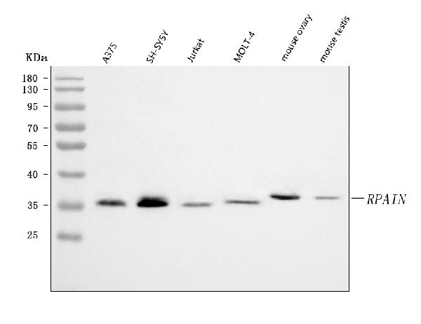
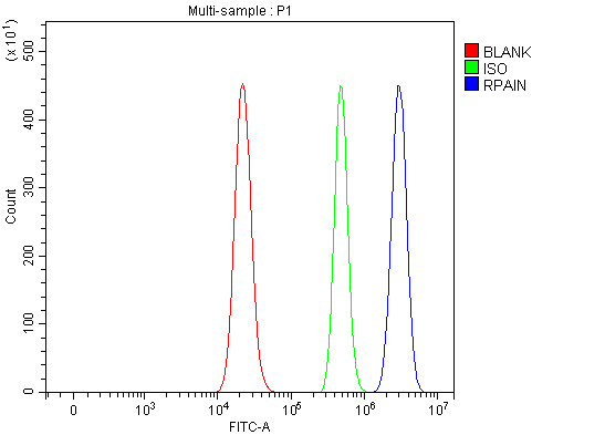
(Figure 2. Flow Cytometry analysis of RT4 cells using anti-RPAIN antibody (AAA127711).Overlay histogram showing RT4 cells stained with AAA127711 (Blue line). To facilitate intracellular staining, cells were fixed with 4% paraformaldehyde and permeabilized with permeabilization buffer. The cells were blocked with 10% normal goat serum. And then incubated with rabbit anti-RPAIN Antibody (AAA127711, 1ug/1x106 cells) for 30 min at 20 degree C. DyLight488 conjugated goat anti-rabbit IgG was used as secondary antibody for 30 minutes at 20 degree C. Isotype control antibody (Green line) was rabbit IgG (1ug/1x106) used under the same conditions. Unlabelled sample without incubation with primary antibody and secondary antibody (Red line) was used as a blank control.)
FCM/FACS (Flow Cytometry)
(Figure 2. Flow Cytometry analysis of RT4 cells using anti-RPAIN antibody (AAA127711).Overlay histogram showing RT4 cells stained with AAA127711 (Blue line). To facilitate intracellular staining, cells were fixed with 4% paraformaldehyde and permeabilized with permeabilization buffer. The cells were blocked with 10% normal goat serum. And then incubated with rabbit anti-RPAIN Antibody (AAA127711, 1ug/1x106 cells) for 30 min at 20 degree C. DyLight488 conjugated goat anti-rabbit IgG was used as secondary antibody for 30 minutes at 20 degree C. Isotype control antibody (Green line) was rabbit IgG (1ug/1x106) used under the same conditions. Unlabelled sample without incubation with primary antibody and secondary antibody (Red line) was used as a blank control.)
RPAIN, Polyclonal Antibody (Cat# AAA127711)
Full Name
Anti-RPAIN Antibody Picoband
Gene Names
RPAIN; RIP; HRIP
Reactivity
Human, Mouse
Applications
Flow Cytometry, Western Blot
Purity
Immunogen affinity purified.
Pricing
 FCM/FACS (Flow Cytometry)
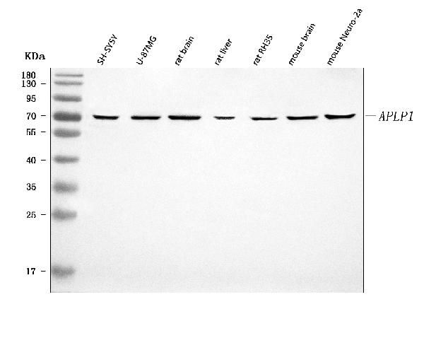
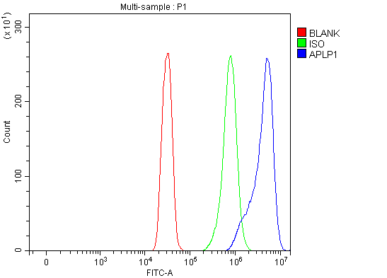
(Figure 2. Flow Cytometry analysis of SiHa cells using anti-APLP1 antibody (AAA127206).Overlay histogram showing SiHa cells stained with AAA127206 (Blue line). To facilitate intracellular staining, cells were fixed with 4% paraformaldehyde and permeabilized with permeabilization buffer. The cells were blocked with 10% normal goat serum. And then incubated with rabbit anti-APLP1 Antibody (AAA127206, 1ug/1x106 cells) for 30 min at 20 degree C. DyLight488 conjugated goat anti-rabbit IgG was used as secondary antibody for 30 minutes at 20 degree C. Isotype control antibody (Green line) was rabbit IgG (1ug/1x106) used under the same conditions. Unlabelled sample (Red line) was also used as a control.)
FCM/FACS (Flow Cytometry)
(Figure 2. Flow Cytometry analysis of SiHa cells using anti-APLP1 antibody (AAA127206).Overlay histogram showing SiHa cells stained with AAA127206 (Blue line). To facilitate intracellular staining, cells were fixed with 4% paraformaldehyde and permeabilized with permeabilization buffer. The cells were blocked with 10% normal goat serum. And then incubated with rabbit anti-APLP1 Antibody (AAA127206, 1ug/1x106 cells) for 30 min at 20 degree C. DyLight488 conjugated goat anti-rabbit IgG was used as secondary antibody for 30 minutes at 20 degree C. Isotype control antibody (Green line) was rabbit IgG (1ug/1x106) used under the same conditions. Unlabelled sample (Red line) was also used as a control.)
APLP1, Polyclonal Antibody (Cat# AAA127206)
Full Name
Anti-APLP1 Antibody Picoband
Gene Names
APLP1; APLP
Reactivity
Human, Mouse, Rat
Applications
Flow Cytometry, Western Blot
Purity
Immunogen affinity purified.
Pricing
 FCM/FACS (Flow Cytometry)
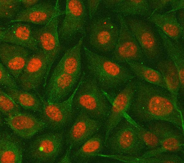
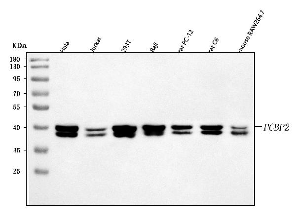
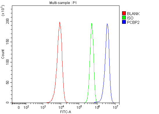
(Figure 3. Flow Cytometry analysis of Hela cells using anti-hnRNP E2/PCBP2 antibody (AAA127115).Overlay histogram showing Hela cells stained with AAA127115 (Blue line). To facilitate intracellular staining, cells were fixed with 4% paraformaldehyde and permeabilized with permeabilization buffer. The cells were blocked with 10% normal goat serum. And then incubated with rabbit anti-hnRNP E2/PCBP2 Antibody (AAA127115, 1ug/1x106 cells) for 30 min at 20 degree C. DyLight488 conjugated goat anti-rabbit IgG was used as secondary antibody for 30 minutes at 20 degree C. Isotype control antibody (Green line) was rabbit IgG (1ug/1x106) used under the same conditions. Unlabelled sample without incubation with primary antibody and secondary antibody (Red line) was used as a blank control.)
FCM/FACS (Flow Cytometry)
(Figure 3. Flow Cytometry analysis of Hela cells using anti-hnRNP E2/PCBP2 antibody (AAA127115).Overlay histogram showing Hela cells stained with AAA127115 (Blue line). To facilitate intracellular staining, cells were fixed with 4% paraformaldehyde and permeabilized with permeabilization buffer. The cells were blocked with 10% normal goat serum. And then incubated with rabbit anti-hnRNP E2/PCBP2 Antibody (AAA127115, 1ug/1x106 cells) for 30 min at 20 degree C. DyLight488 conjugated goat anti-rabbit IgG was used as secondary antibody for 30 minutes at 20 degree C. Isotype control antibody (Green line) was rabbit IgG (1ug/1x106) used under the same conditions. Unlabelled sample without incubation with primary antibody and secondary antibody (Red line) was used as a blank control.)
hnRNP E2/PCBP2, Polyclonal Antibody (Cat# AAA127115)
Full Name
Anti-hnRNP E2/PCBP2 Antibody Picoband
Gene Names
PCBP2; HNRPE2; HNRNPE2; hnRNP-E2
Reactivity
Human, Mouse, Rat
Applications
Western Blot, Immunofluorescence, Immunocytochemistry, Flow Cytometry
Purity
Immunogen affinity purified.
Pricing
 FCM/FACS (Flow Cytometry)
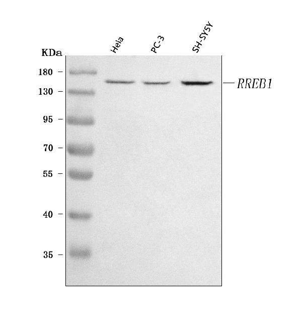
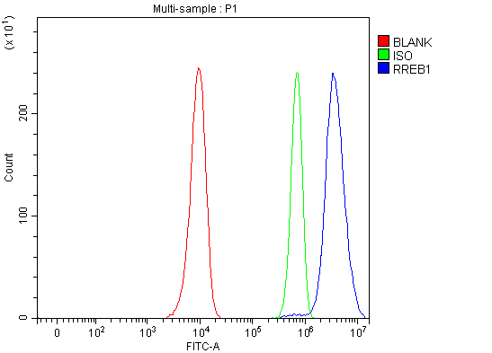
(Figure 2. Flow Cytometry analysis of Hela cells using anti-RREB1 antibody (AAA127204).Overlay histogram showing Hela cells stained with AAA127204 (Blue line). To facilitate intracellular staining, cells were fixed with 4% paraformaldehyde and permeabilized with permeabilization buffer. The cells were blocked with 10% normal goat serum. And then incubated with rabbit anti-RREB1 Antibody (AAA127204, 1ug/1x106 cells) for 30 min at 20 degree C. DyLight488 conjugated goat anti-rabbit IgG was used as secondary antibody for 30 minutes at 20 degree C. Isotype control antibody (Green line) was rabbit IgG (1ug/1x106) used under the same conditions. Unlabelled sample without incubation with primary antibody and secondary antibody (Red line) was used as a blank control.)
FCM/FACS (Flow Cytometry)
(Figure 2. Flow Cytometry analysis of Hela cells using anti-RREB1 antibody (AAA127204).Overlay histogram showing Hela cells stained with AAA127204 (Blue line). To facilitate intracellular staining, cells were fixed with 4% paraformaldehyde and permeabilized with permeabilization buffer. The cells were blocked with 10% normal goat serum. And then incubated with rabbit anti-RREB1 Antibody (AAA127204, 1ug/1x106 cells) for 30 min at 20 degree C. DyLight488 conjugated goat anti-rabbit IgG was used as secondary antibody for 30 minutes at 20 degree C. Isotype control antibody (Green line) was rabbit IgG (1ug/1x106) used under the same conditions. Unlabelled sample without incubation with primary antibody and secondary antibody (Red line) was used as a blank control.)
RREB1, Polyclonal Antibody (Cat# AAA127204)
Full Name
Anti-RREB1 Antibody Picoband
Gene Names
RREB1; HNT; FINB; LZ321; Zep-1; RREB-1
Reactivity
Human
Applications
Flow Cytometry, Western Blot
Purity
Immunogen affinity purified.
Pricing
 FCM/FACS (Flow Cytometry)
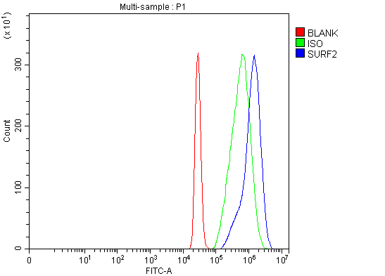
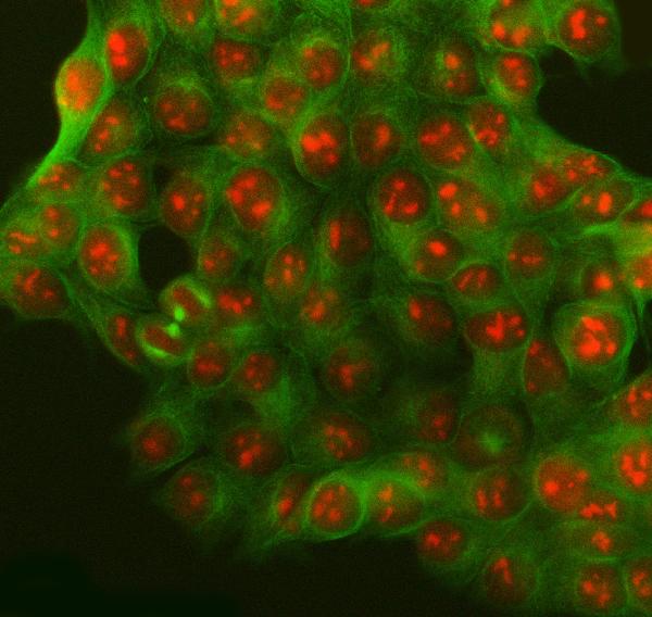
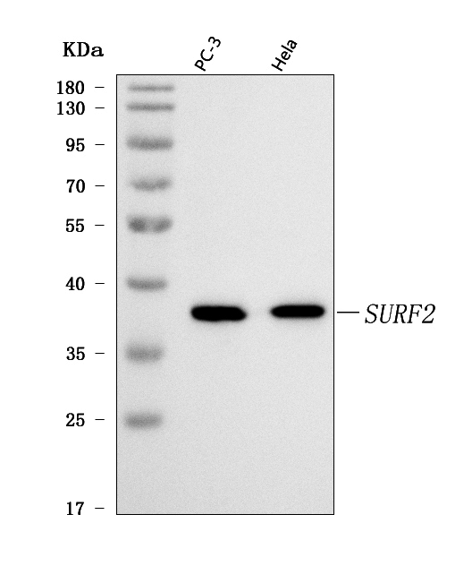
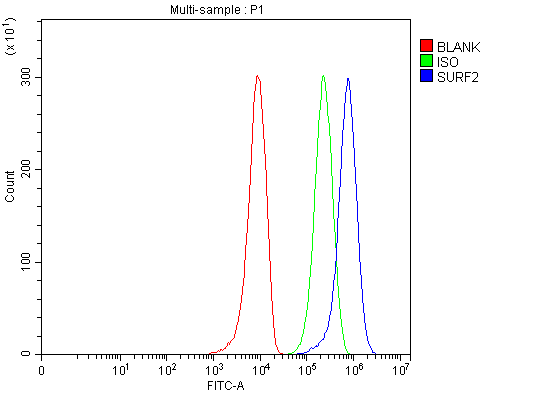
(Figure 4. Flow Cytometry analysis of 293T cells using anti-SURF2 antibody (AAA127842).Overlay histogram showing 293T cells stained with AAA127842 (Blue line). To facilitate intracellular staining, cells were fixed with 4% paraformaldehyde and permeabilized with permeabilization buffer. The cells were blocked with 10% normal goat serum. And then incubated with rabbit anti-SURF2 Antibody (AAA127842, 1ug/1x106 cells) for 30 min at 20 degree C. DyLight488 conjugated goat anti-rabbit IgG was used as secondary antibody for 30 minutes at 20 degree C. Isotype control antibody (Green line) was rabbit IgG (1ug/1x106) used under the same conditions. Unlabelled sample without incubation with primary antibody and secondary antibody (Red line) was used as a blank control.)
FCM/FACS (Flow Cytometry)
(Figure 4. Flow Cytometry analysis of 293T cells using anti-SURF2 antibody (AAA127842).Overlay histogram showing 293T cells stained with AAA127842 (Blue line). To facilitate intracellular staining, cells were fixed with 4% paraformaldehyde and permeabilized with permeabilization buffer. The cells were blocked with 10% normal goat serum. And then incubated with rabbit anti-SURF2 Antibody (AAA127842, 1ug/1x106 cells) for 30 min at 20 degree C. DyLight488 conjugated goat anti-rabbit IgG was used as secondary antibody for 30 minutes at 20 degree C. Isotype control antibody (Green line) was rabbit IgG (1ug/1x106) used under the same conditions. Unlabelled sample without incubation with primary antibody and secondary antibody (Red line) was used as a blank control.)
SURF2, Polyclonal Antibody (Cat# AAA127842)
Full Name
Anti-SURF2 Antibody Picoband
Gene Names
SURF2; SURF-2
Reactivity
Human
Applications
Flow Cytometry, Immunofluorescence, Immunocytochemistry, Western Blot
Purity
Immunogen affinity purified.
Pricing
 FCM/FACS (Flow Cytometry)
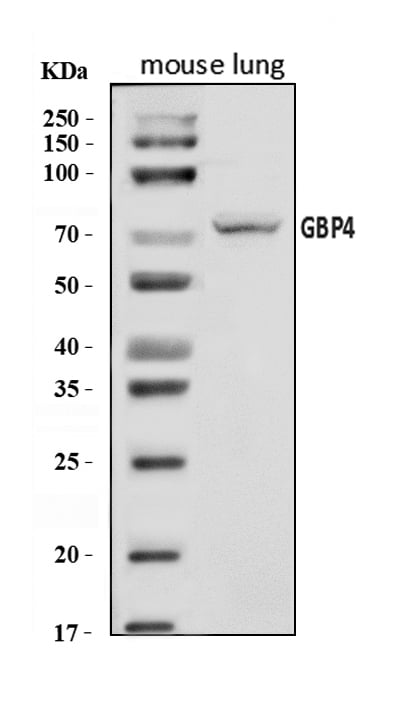
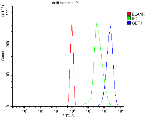
(Figure 2. Flow Cytometry analysis of ANA-1 cells using anti-Gbp4 antibody (AAA126587).Overlay histogram showing ANA-1 cells stained with AAA126587 (Blue line). The cells were blocked with 10% normal goat serum. And then incubated with rabbit anti-Gbp4 Antibody (AAA126587, 1 ug/1x10^6 cells) for 30 min at 20 degree C. DyLight488 conjugated goat anti-rabbit IgG was used as secondary antibody for 30 minutes at 20 degree C. Isotype control antibody (Green line) was rabbit IgG (1 ug/1x10^6) used under the same conditions. Unlabelled sample (Red line) was also used as a control.)
FCM/FACS (Flow Cytometry)
(Figure 2. Flow Cytometry analysis of ANA-1 cells using anti-Gbp4 antibody (AAA126587).Overlay histogram showing ANA-1 cells stained with AAA126587 (Blue line). The cells were blocked with 10% normal goat serum. And then incubated with rabbit anti-Gbp4 Antibody (AAA126587, 1 ug/1x10^6 cells) for 30 min at 20 degree C. DyLight488 conjugated goat anti-rabbit IgG was used as secondary antibody for 30 minutes at 20 degree C. Isotype control antibody (Green line) was rabbit IgG (1 ug/1x10^6) used under the same conditions. Unlabelled sample (Red line) was also used as a control.)
Gbp4, Polyclonal Antibody (Cat# AAA126587)
Full Name
Anti-Gbp4 Antibody Picoband
Gene Names
Gbp3; Gbp4; GBP-3; GBP-4; AW228655
Reactivity
Mouse
Applications
Flow Cytometry, Western Blot
Purity
Immunogen affinity purified.
Pricing
 FCM/FACS (Flow Cytometry)
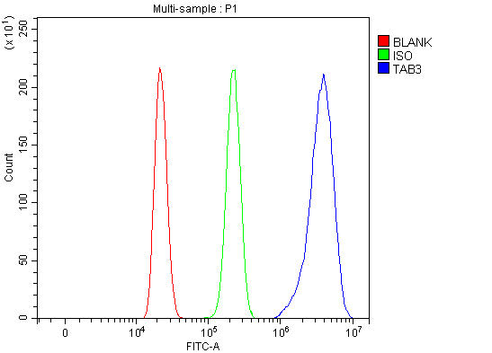
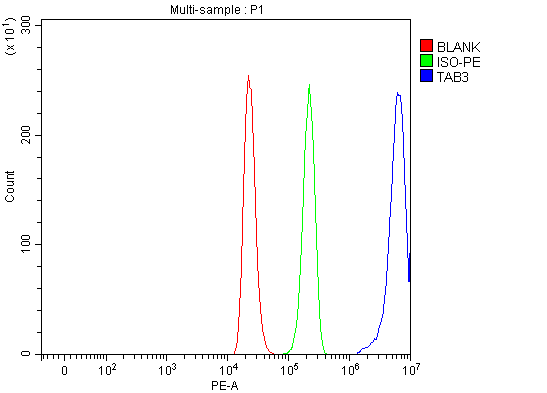
(Figure 3. Flow Cytometry analysis of HL-60 cells using anti-TAB3 antibody (AAA127275).Overlay histogram showing HL-60 cells stained with AAA127275 (Blue line). To facilitate intracellular staining, cells were fixed with 4% paraformaldehyde and permeabilized with permeabilization buffer. The cells were blocked with 10% normal goat serum. And then incubated with rabbit anti-TAB3 Antibody (AAA127275, 1ug/1x106 cells) for 30 min at 20 degree C. PE conjugated goat anti-rabbit IgG (5-10ug/1x106 cells) was used as secondary antibody for 30 minutes at 20 degree C. Isotype control antibody (Green line) was rabbit IgG (1ug/1x106) used under the same conditions. Unlabelled sample without incubation with primary antibody and secondary antibody (Red line) was used as a blank control.)
FCM/FACS (Flow Cytometry)
(Figure 3. Flow Cytometry analysis of HL-60 cells using anti-TAB3 antibody (AAA127275).Overlay histogram showing HL-60 cells stained with AAA127275 (Blue line). To facilitate intracellular staining, cells were fixed with 4% paraformaldehyde and permeabilized with permeabilization buffer. The cells were blocked with 10% normal goat serum. And then incubated with rabbit anti-TAB3 Antibody (AAA127275, 1ug/1x106 cells) for 30 min at 20 degree C. PE conjugated goat anti-rabbit IgG (5-10ug/1x106 cells) was used as secondary antibody for 30 minutes at 20 degree C. Isotype control antibody (Green line) was rabbit IgG (1ug/1x106) used under the same conditions. Unlabelled sample without incubation with primary antibody and secondary antibody (Red line) was used as a blank control.)
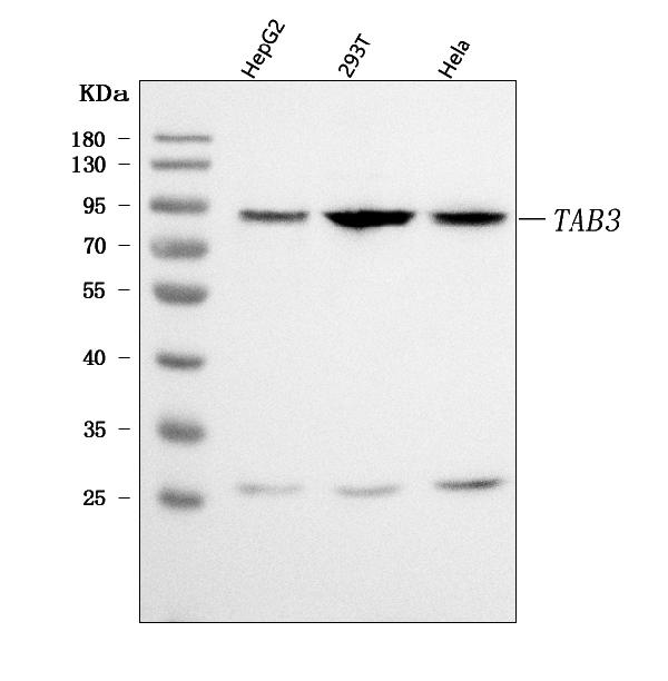
TAB3, Polyclonal Antibody (Cat# AAA127275)
Full Name
Anti-TAB3 Antibody Picoband
Gene Names
TAB3; NAP1; MAP3K7IP3
Reactivity
Human
Applications
Flow Cytometry, Western Blot
Purity
Immunogen affinity purified.
Pricing
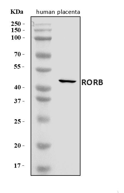
 FCM/FACS (Flow Cytometry)
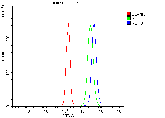
(Figure 2. Flow Cytometry analysis of JK cells using anti-ROR beta/RORB antibody (AAA126559).Overlay histogram showing JK cells stained with AAA126559 (Blue line). The cells were blocked with 10% normal goat serum. And then incubated with rabbit anti-ROR beta/RORB Antibody (AAA126559, 1 ug/1x10^6 cells) for 30 min at 20 degree C. DyLight488 conjugated goat anti-rabbit IgG was used as secondary antibody for 30 minutes at 20 degree C. Isotype control antibody (Green line) was rabbit IgG (1 ug/1x10^6) used under the same conditions. Unlabelled sample (Red line) was also used as a control.)
FCM/FACS (Flow Cytometry)
(Figure 2. Flow Cytometry analysis of JK cells using anti-ROR beta/RORB antibody (AAA126559).Overlay histogram showing JK cells stained with AAA126559 (Blue line). The cells were blocked with 10% normal goat serum. And then incubated with rabbit anti-ROR beta/RORB Antibody (AAA126559, 1 ug/1x10^6 cells) for 30 min at 20 degree C. DyLight488 conjugated goat anti-rabbit IgG was used as secondary antibody for 30 minutes at 20 degree C. Isotype control antibody (Green line) was rabbit IgG (1 ug/1x10^6) used under the same conditions. Unlabelled sample (Red line) was also used as a control.)
ROR beta/RORB, Polyclonal Antibody (Cat# AAA126559)
Full Name
Anti-ROR beta/RORB Antibody Picoband
Gene Names
RORB; RZRB; NR1F2; ROR-BETA; RZR-BETA; bA133M9.1
Reactivity
Human
Applications
Western Blot, Flow Cytometry
Purity
Immunogen affinity purified.
Pricing
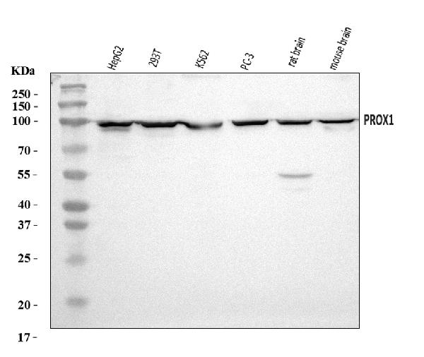
 FCM/FACS (Flow Cytometry)
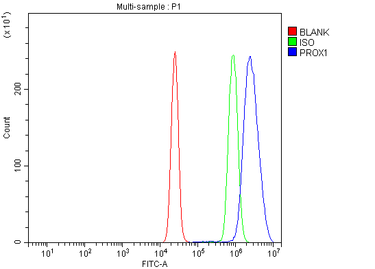
(Figure 2. Flow Cytometry analysis of K562 cells using anti-PROX1 antibody (AAA126192).Overlay histogram showing K562 cells stained with AAA126192 (Blue line). The cells were blocked with 10% normal goat serum. And then incubated with rabbit anti-PROX1 Antibody (AAA126192, 1 ug/1x10^6 cells) for 30 min at 20 degree C. DyLight488 conjugated goat anti-rabbit IgG was used as secondary antibody for 30 minutes at 20 degree C. Isotype control antibody (Green line) was rabbit IgG (1 ug/1x10^6) used under the same conditions. Unlabelled sample (Red line) was also used as a control.)
FCM/FACS (Flow Cytometry)
(Figure 2. Flow Cytometry analysis of K562 cells using anti-PROX1 antibody (AAA126192).Overlay histogram showing K562 cells stained with AAA126192 (Blue line). The cells were blocked with 10% normal goat serum. And then incubated with rabbit anti-PROX1 Antibody (AAA126192, 1 ug/1x10^6 cells) for 30 min at 20 degree C. DyLight488 conjugated goat anti-rabbit IgG was used as secondary antibody for 30 minutes at 20 degree C. Isotype control antibody (Green line) was rabbit IgG (1 ug/1x10^6) used under the same conditions. Unlabelled sample (Red line) was also used as a control.)
PROX1, Polyclonal Antibody (Cat# AAA126192)
Full Name
Anti-PROX1 Antibody Picoband
Reactivity
Human, Mouse, Rat
Applications
Flow Cytometry, Western Blot
Purity
Immunogen affinity purified.
Pricing
 IHC (Immunohistochemisry)
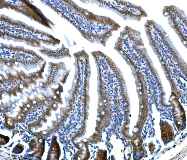
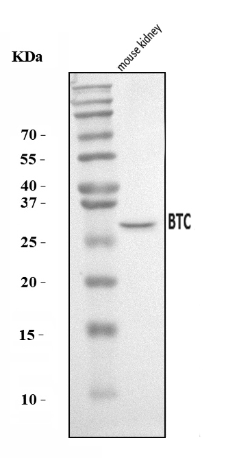
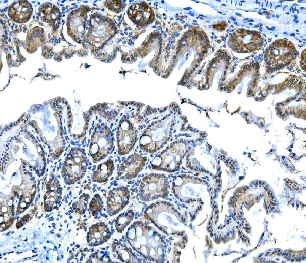
(Figure 3. IHC analysis of Btc using anti-Btc antibody (AAA126205).Btc was detected in a paraffin-embedded section of mouse stomach tissue. Heat mediated antigen retrieval was performed in EDTA buffer (pH 8.0, epitope retrieval solution). The tissue section was blocked with 10% goat serum. The tissue section was then incubated with 2 ug/ml rabbit anti-Btc Antibody (AAA126205) overnight at 4 degree C. Biotinylated goat anti-rabbit IgG was used as secondary antibody and incubated for 30 minutes at 37 degree C. The tissue section was developed using Strepavidin-Biotin-Complex (SABC) with DAB as the chromogen.)
IHC (Immunohistochemisry)
(Figure 3. IHC analysis of Btc using anti-Btc antibody (AAA126205).Btc was detected in a paraffin-embedded section of mouse stomach tissue. Heat mediated antigen retrieval was performed in EDTA buffer (pH 8.0, epitope retrieval solution). The tissue section was blocked with 10% goat serum. The tissue section was then incubated with 2 ug/ml rabbit anti-Btc Antibody (AAA126205) overnight at 4 degree C. Biotinylated goat anti-rabbit IgG was used as secondary antibody and incubated for 30 minutes at 37 degree C. The tissue section was developed using Strepavidin-Biotin-Complex (SABC) with DAB as the chromogen.)
Btc, Polyclonal Antibody (Cat# AAA126205)
Full Name
Anti-Btc Antibody Picoband
Gene Names
Btc; Bcn
Reactivity
Mouse
Applications
Immunohistochemistry, Western Blot
Purity
Immunogen affinity purified.
Pricing
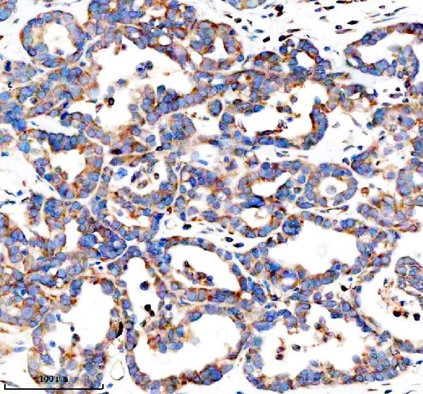
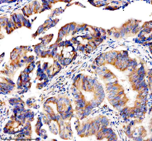
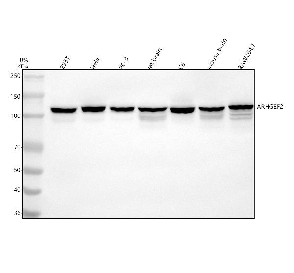
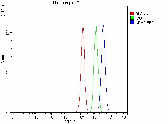 FCM/FACS (Flow Cytometry)
(Figure 5. Flow Cytometry analysis of PC-3 cells using anti-GEF-H1/ARHGEF2 antibody (AAA127130).Overlay histogram showing PC-3 cells stained with AAA127130 (Blue line). To facilitate intracellular staining, cells were fixed with 4% paraformaldehyde and permeabilized with permeabilization buffer. The cells were blocked with 10% normal goat serum. And then incubated with rabbit anti-GEF-H1/ARHGEF2 Antibody (AAA127130, 1ug/1x106 cells) for 30 min at 20 degree C. DyLight488 conjugated goat anti-rabbit IgG was used as secondary antibody for 30 minutes at 20 degree C. Isotype control antibody (Green line) was rabbit IgG (1ug/1x106) used under the same conditions. Unlabelled sample without incubation with primary antibody and secondary antibody (Red line) was used as a blank control.)
FCM/FACS (Flow Cytometry)
(Figure 5. Flow Cytometry analysis of PC-3 cells using anti-GEF-H1/ARHGEF2 antibody (AAA127130).Overlay histogram showing PC-3 cells stained with AAA127130 (Blue line). To facilitate intracellular staining, cells were fixed with 4% paraformaldehyde and permeabilized with permeabilization buffer. The cells were blocked with 10% normal goat serum. And then incubated with rabbit anti-GEF-H1/ARHGEF2 Antibody (AAA127130, 1ug/1x106 cells) for 30 min at 20 degree C. DyLight488 conjugated goat anti-rabbit IgG was used as secondary antibody for 30 minutes at 20 degree C. Isotype control antibody (Green line) was rabbit IgG (1ug/1x106) used under the same conditions. Unlabelled sample without incubation with primary antibody and secondary antibody (Red line) was used as a blank control.)
GEF-H1/ARHGEF2, Polyclonal Antibody (Cat# AAA127130)
Full Name
Anti-GEF-H1/ARHGEF2 Antibody Picoband
Gene Names
ARHGEF2; GEF; P40; GEFH1; LFP40; GEF-H1
Reactivity
Human, Mouse, Rat
Applications
Flow Cytometry, Immunohistochemistry, Western Blot
Purity
Immunogen affinity purified.
Pricing
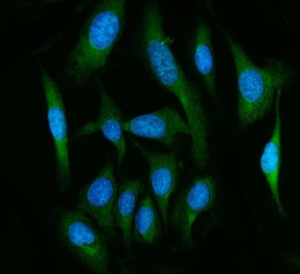
 FCM/FACS (Flow Cytometry)
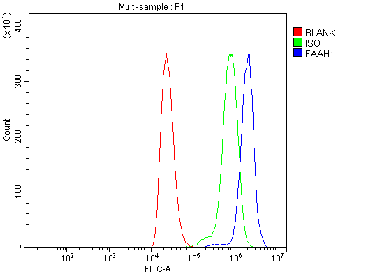
(Figure 3. Flow Cytometry analysis of THP-1 cells using anti-FAAH1/FAAH antibody (AAA126078).Overlay histogram showing THP-1 cells stained with AAA126078 (Blue line). The cells were blocked with 10% normal goat serum. And then incubated with rabbit anti-FAAH1/FAAH Antibody (AAA126078, 1 ug/1x10^6 cells) for 30 min at 20 degree C. DyLight488 conjugated goat anti-rabbit IgG was used as secondary antibody for 30 minutes at 20 degree C. Isotype control antibody (Green line) was rabbit IgG (1 ug/1x10^6) used under the same conditions. Unlabelled sample (Red line) was also used as a control.)
FCM/FACS (Flow Cytometry)
(Figure 3. Flow Cytometry analysis of THP-1 cells using anti-FAAH1/FAAH antibody (AAA126078).Overlay histogram showing THP-1 cells stained with AAA126078 (Blue line). The cells were blocked with 10% normal goat serum. And then incubated with rabbit anti-FAAH1/FAAH Antibody (AAA126078, 1 ug/1x10^6 cells) for 30 min at 20 degree C. DyLight488 conjugated goat anti-rabbit IgG was used as secondary antibody for 30 minutes at 20 degree C. Isotype control antibody (Green line) was rabbit IgG (1 ug/1x10^6) used under the same conditions. Unlabelled sample (Red line) was also used as a control.)
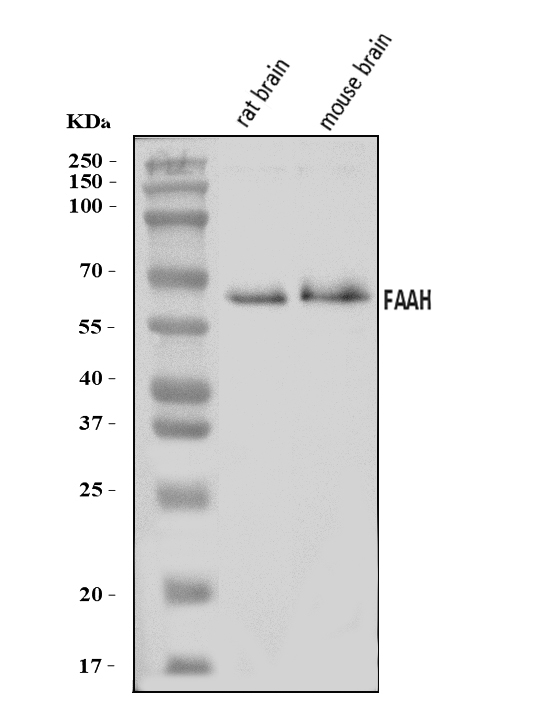
FAAH1/FAAH, Polyclonal Antibody (Cat# AAA126078)
Full Name
Anti-FAAH1/FAAH Antibody Picoband
Gene Names
FAAH; PSAB; FAAH-1
Reactivity
Human, Mouse, Rat
Applications
Flow Cytometry, Immunofluorescence, Immunocytochemistry, Western Blot
Purity
Immunogen affinity purified.
Pricing
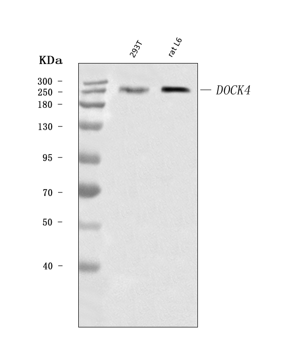
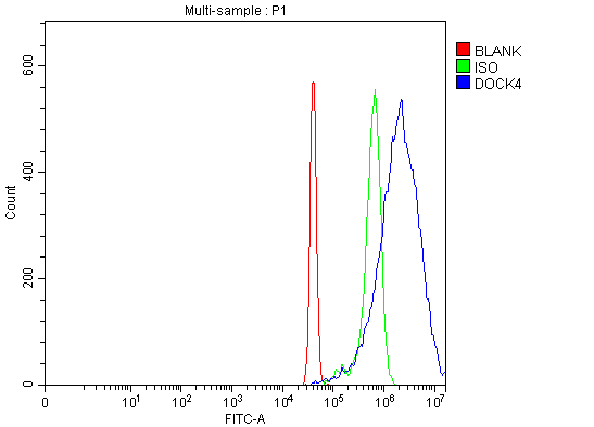 FCM/FACS (Flow Cytometry)
(Figure 3. Flow Cytometry analysis of SiHa cells using anti-DOCK4 antibody (AAA126444).Overlay histogram showing SiHa cells stained with AAA126444 (Blue line). The cells were blocked with 10% normal goat serum. And then incubated with rabbit anti-DOCK4 Antibody (AAA126444, 1 ug/1x10^6 cells) for 30 min at 20 degree C. DyLight488 conjugated goat anti-rabbit IgG was used as secondary antibody for 30 minutes at 20 degree C. Isotype control antibody (Green line) was rabbit IgG (1 ug/1x10^6) used under the same conditions. Unlabelled sample (Red line) was also used as a control.)
FCM/FACS (Flow Cytometry)
(Figure 3. Flow Cytometry analysis of SiHa cells using anti-DOCK4 antibody (AAA126444).Overlay histogram showing SiHa cells stained with AAA126444 (Blue line). The cells were blocked with 10% normal goat serum. And then incubated with rabbit anti-DOCK4 Antibody (AAA126444, 1 ug/1x10^6 cells) for 30 min at 20 degree C. DyLight488 conjugated goat anti-rabbit IgG was used as secondary antibody for 30 minutes at 20 degree C. Isotype control antibody (Green line) was rabbit IgG (1 ug/1x10^6) used under the same conditions. Unlabelled sample (Red line) was also used as a control.)
DOCK4, Polyclonal Antibody (Cat# AAA126444)
Full Name
Anti-DOCK4 Antibody Picoband
Reactivity
Human, Rat
Applications
Flow Cytometry, Immunohistochemistry, Western Blot
Purity
Immunogen affinity purified.
Pricing
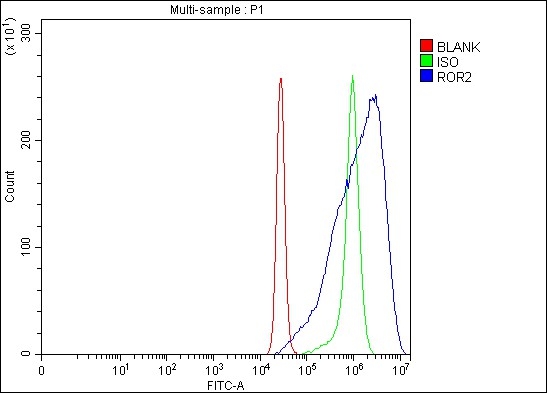
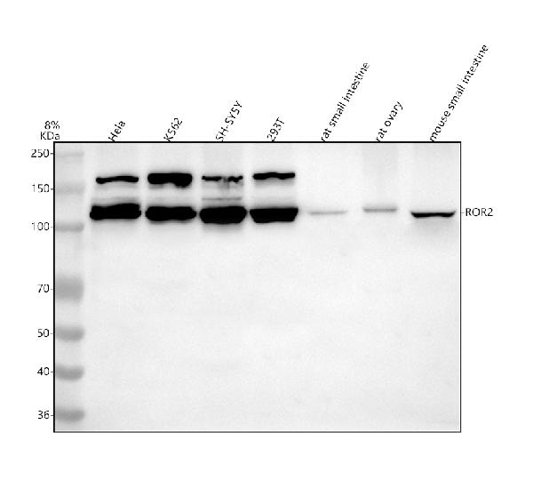
 FCM/FACS (Flow Cytometry)
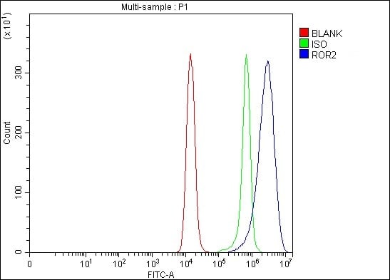
(Figure 3. Flow Cytometry analysis of Raji cells using anti-ROR2 antibody (AAA127084).Overlay histogram showing Raji cells stained with AAA127084 (Blue line). The cells were fixed with 4% paraformaldehyde and blocked with 10% normal goat serum. And then incubated with rabbit anti-ROR2 Antibody (AAA127084, 1ug/1x106 cells) for 30 min at 20 degree C. DyLight488 conjugated goat anti-rabbit IgG was used as secondary antibody for 30 minutes at 20 degree C. Isotype control antibody (Green line) was rabbit IgG (1ug/1x106) used under the same conditions. Unlabelled sample without incubation with primary antibody and secondary antibody (Red line) was used as a blank control.)
FCM/FACS (Flow Cytometry)
(Figure 3. Flow Cytometry analysis of Raji cells using anti-ROR2 antibody (AAA127084).Overlay histogram showing Raji cells stained with AAA127084 (Blue line). The cells were fixed with 4% paraformaldehyde and blocked with 10% normal goat serum. And then incubated with rabbit anti-ROR2 Antibody (AAA127084, 1ug/1x106 cells) for 30 min at 20 degree C. DyLight488 conjugated goat anti-rabbit IgG was used as secondary antibody for 30 minutes at 20 degree C. Isotype control antibody (Green line) was rabbit IgG (1ug/1x106) used under the same conditions. Unlabelled sample without incubation with primary antibody and secondary antibody (Red line) was used as a blank control.)
ROR2, Polyclonal Antibody (Cat# AAA127084)
Full Name
Anti-ROR2 Antibody Picoband
Gene Names
ROR2; BDB; BDB1; NTRKR2
Reactivity
Human, Mouse, Rat
Applications
Flow Cytometry, Western Blot
Purity
Immunogen affinity purified.
Pricing
 IHC (Immunohistochemistry)
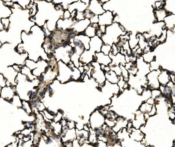
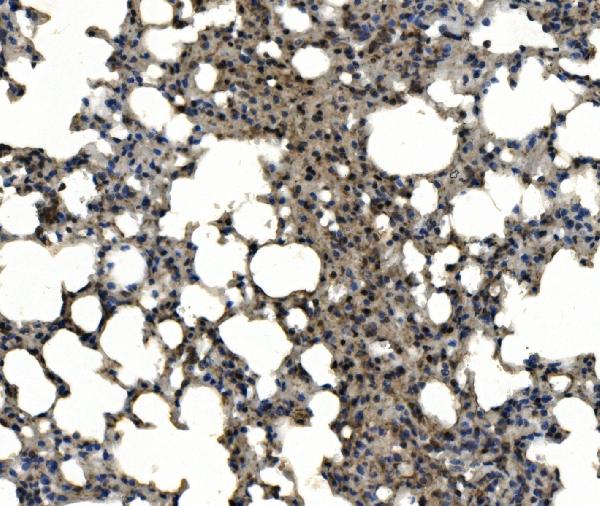
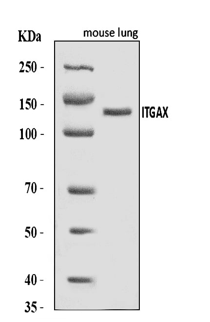
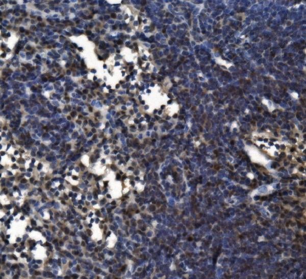
(Figure 4. IHC analysis of CD11c/Itgax using anti-CD11c/Itgax antibody (AAA126014).CD11c/Itgax was detected in a paraffin-embedded section of rat lymph node tissue. Heat mediated antigen retrieval was performed in EDTA buffer (pH 8.0, epitope retrieval solution). The tissue section was blocked with 10% goat serum. The tissue section was then incubated with 2 ug/ml rabbit anti-CD11c/Itgax Antibody (AAA126014) overnight at 4 degree C. Biotinylated goat anti-rabbit IgG was used as secondary antibody and incubated for 30 minutes at 37 degree C. The tissue section was developed using Strepavidin-Biotin-Complex (SABC) with DAB as the chromogen.)
IHC (Immunohistochemistry)
(Figure 4. IHC analysis of CD11c/Itgax using anti-CD11c/Itgax antibody (AAA126014).CD11c/Itgax was detected in a paraffin-embedded section of rat lymph node tissue. Heat mediated antigen retrieval was performed in EDTA buffer (pH 8.0, epitope retrieval solution). The tissue section was blocked with 10% goat serum. The tissue section was then incubated with 2 ug/ml rabbit anti-CD11c/Itgax Antibody (AAA126014) overnight at 4 degree C. Biotinylated goat anti-rabbit IgG was used as secondary antibody and incubated for 30 minutes at 37 degree C. The tissue section was developed using Strepavidin-Biotin-Complex (SABC) with DAB as the chromogen.)
CD11c/Itgax, Polyclonal Antibody (Cat# AAA126014)
Full Name
Anti-CD11c/Itgax Antibody Picoband
Gene Names
Itgax; Cr4; N418; Cd11c; AI449405
Reactivity
Mouse, Rat
Applications
Immunohistochemistry, Western Blot
Purity
Immunogen affinity purified.
Pricing
 FCM/FACS (Flow Cytometry)
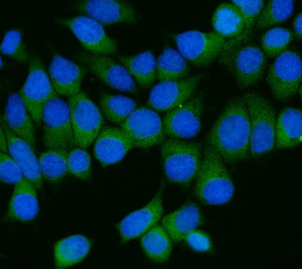
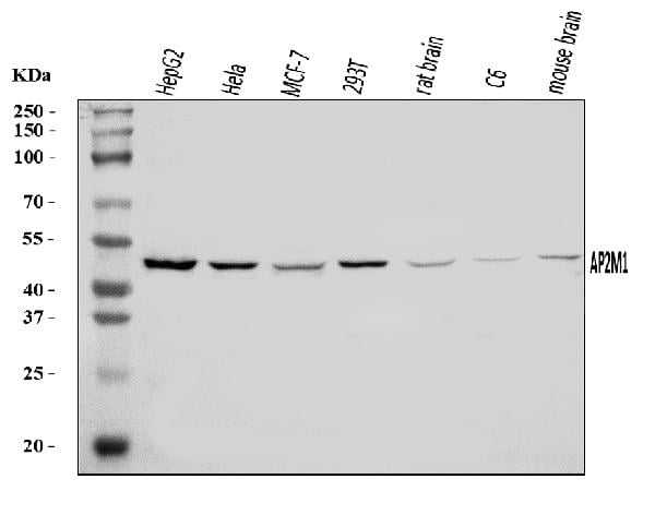
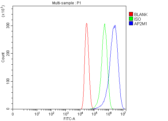
(Figure 3. Flow Cytometry analysis of MCF-7 cells using anti-AP2M1 antibody (AAA126452).Overlay histogram showing MCF-7 cells stained with AAA126452 (Blue line). The cells were blocked with 10% normal goat serum. And then incubated with rabbit anti-AP2M1 Antibody (AAA126452, 1 ug/1x10^6 cells) for 30 min at 20 degree C. DyLight488 conjugated goat anti-rabbit IgG was used as secondary antibody for 30 minutes at 20 degree C. Isotype control antibody (Green line) was rabbit IgG (1 ug/1x10^6) used under the same conditions. Unlabelled sample (Red line) was also used as a control.)
FCM/FACS (Flow Cytometry)
(Figure 3. Flow Cytometry analysis of MCF-7 cells using anti-AP2M1 antibody (AAA126452).Overlay histogram showing MCF-7 cells stained with AAA126452 (Blue line). The cells were blocked with 10% normal goat serum. And then incubated with rabbit anti-AP2M1 Antibody (AAA126452, 1 ug/1x10^6 cells) for 30 min at 20 degree C. DyLight488 conjugated goat anti-rabbit IgG was used as secondary antibody for 30 minutes at 20 degree C. Isotype control antibody (Green line) was rabbit IgG (1 ug/1x10^6) used under the same conditions. Unlabelled sample (Red line) was also used as a control.)
AP2M1, Polyclonal Antibody (Cat# AAA126452)
Full Name
Anti-AP2M1 Antibody Picoband
Gene Names
AP2M1; mu2; AP50; CLAPM1
Reactivity
Human, Mouse, Rat
Applications
Western Blot, Immunocytochemistry, Immunofluorescence, Flow Cytometry
Purity
Immunogen affinity purified.
Pricing
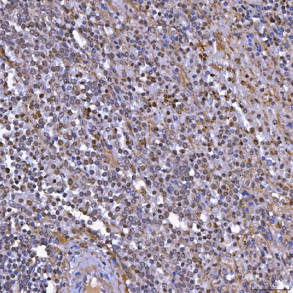
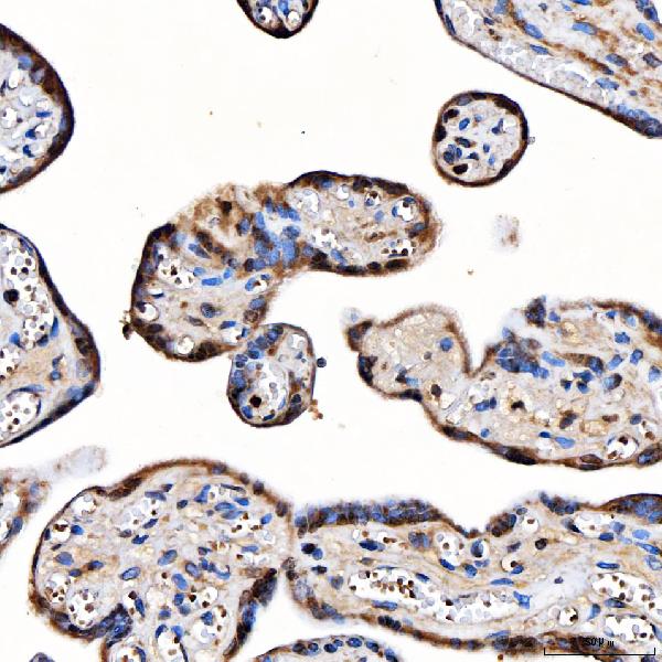
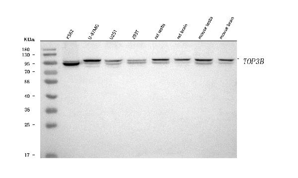
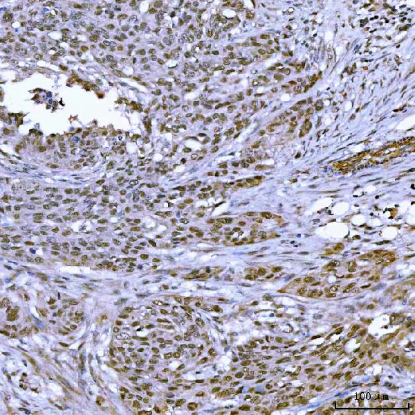 IHC (Immunohistochemistry)
(Figure 4. IHC analysis of TOP3B using anti-TOP3B antibody (AAA126576).TOP3B was detected in a paraffin-embedded section of human ovarian serous adenocarcinoma tissue. Heat mediated antigen retrieval was performed in EDTA buffer (pH 8.0, epitope retrieval solution). The tissue section was blocked with 10% goat serum. The tissue section was then incubated with 2 ug/ml rabbit anti-TOP3B Antibody (AAA126576) overnight at 4 degree C. Peroxidase Conjugated Goat Anti-rabbit IgG was used as secondary antibody and incubated for 30 minutes at 37 degree C. The tissue section was developed using HRP Conjugated Rabbit IgG Super Vision Assay Kit with DAB as the chromogen.)
IHC (Immunohistochemistry)
(Figure 4. IHC analysis of TOP3B using anti-TOP3B antibody (AAA126576).TOP3B was detected in a paraffin-embedded section of human ovarian serous adenocarcinoma tissue. Heat mediated antigen retrieval was performed in EDTA buffer (pH 8.0, epitope retrieval solution). The tissue section was blocked with 10% goat serum. The tissue section was then incubated with 2 ug/ml rabbit anti-TOP3B Antibody (AAA126576) overnight at 4 degree C. Peroxidase Conjugated Goat Anti-rabbit IgG was used as secondary antibody and incubated for 30 minutes at 37 degree C. The tissue section was developed using HRP Conjugated Rabbit IgG Super Vision Assay Kit with DAB as the chromogen.)
TOP3B, Polyclonal Antibody (Cat# AAA126576)
Full Name
Anti-TOP3B Antibody Picoband
Reactivity
Human, Mouse, Rat
Applications
Immunohistochemistry, Western Blot
Purity
Immunogen affinity purified.
Pricing
 IF (Immunofluorescence)
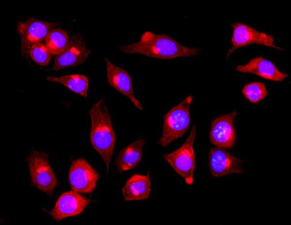
(Figure 4. IF analysis of PIST/GOPC using anti-PIST/GOPC antibody (AAA126329).PIST/GOPC was detected in an immunocytochemical section of SiHa cells. Enzyme antigen retrieval was performed using IHC enzyme antigen retrieval reagent (AR0022) for 15 mins. The cells were blocked with 10% goat serum. And then incubated with 5 ug/mL rabbit anti-PIST/GOPC Antibody (AAA126329) overnight at 4 degree C. DyLight594 Conjugated Goat Anti-Rabbit IgG (BA1142) was used as secondary antibody at 1:100 dilution and incubated for 30 minutes at 37 degree C. The section was counterstained with DAPI. Visualize using a fluorescence microscope and filter sets appropriate for the label used.)
IF (Immunofluorescence)
(Figure 4. IF analysis of PIST/GOPC using anti-PIST/GOPC antibody (AAA126329).PIST/GOPC was detected in an immunocytochemical section of SiHa cells. Enzyme antigen retrieval was performed using IHC enzyme antigen retrieval reagent (AR0022) for 15 mins. The cells were blocked with 10% goat serum. And then incubated with 5 ug/mL rabbit anti-PIST/GOPC Antibody (AAA126329) overnight at 4 degree C. DyLight594 Conjugated Goat Anti-Rabbit IgG (BA1142) was used as secondary antibody at 1:100 dilution and incubated for 30 minutes at 37 degree C. The section was counterstained with DAPI. Visualize using a fluorescence microscope and filter sets appropriate for the label used.)
PIST/GOPC, Polyclonal Antibody (Cat# AAA126329)
Full Name
Anti-PIST/GOPC Antibody Picoband
Gene Names
GOPC; CAL; FIG; PIST; GOPC1; dJ94G16.2
Reactivity
Human
Applications
Immunofluorescence, Immunocytochemistry, Immunohistochemistry, Western Blot
Purity
Immunogen affinity purified.
Pricing
 FCM/FACS (Flow Cytometry)
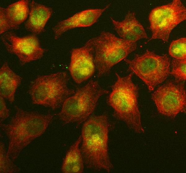
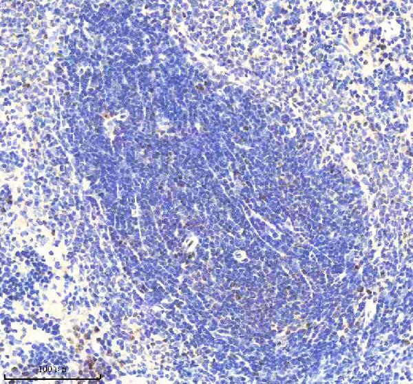
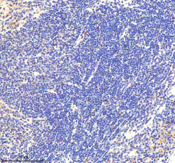
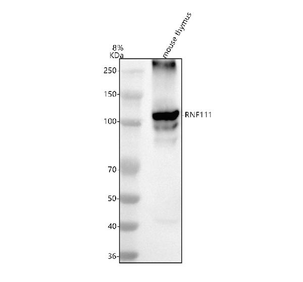
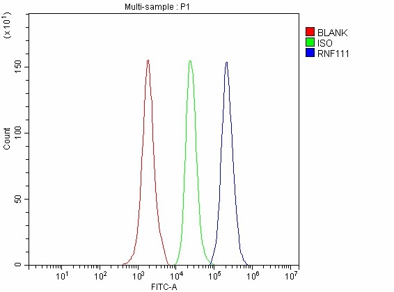
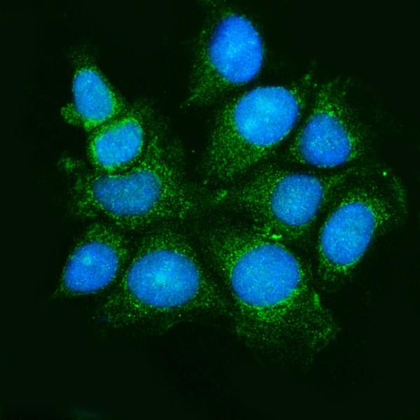
(Figure 5. Flow Cytometry analysis of SH-SY5Y cells using anti-RNF111 antibody (AAA127320).Overlay histogram showing SH-SY5Y cells stained with AAA127320 (Blue line). To facilitate intracellular staining, cells were fixed with 4% paraformaldehyde and permeabilized with permeabilization buffer. The cells were blocked with 10% normal goat serum. And then incubated with rabbit anti-RNF111 Antibody (AAA127320, 1ug/1x106 cells) for 30 min at 20 degree C. DyLight488 conjugated goat anti-rabbit IgG was used as secondary antibody for 30 minutes at 20 degree C. Isotype control antibody (Green line) was rabbit IgG (1ug/1x106) used under the same conditions. Unlabelled sample without incubation with primary antibody and secondary antibody (Red line) was used as a blank control.)
FCM/FACS (Flow Cytometry)
(Figure 5. Flow Cytometry analysis of SH-SY5Y cells using anti-RNF111 antibody (AAA127320).Overlay histogram showing SH-SY5Y cells stained with AAA127320 (Blue line). To facilitate intracellular staining, cells were fixed with 4% paraformaldehyde and permeabilized with permeabilization buffer. The cells were blocked with 10% normal goat serum. And then incubated with rabbit anti-RNF111 Antibody (AAA127320, 1ug/1x106 cells) for 30 min at 20 degree C. DyLight488 conjugated goat anti-rabbit IgG was used as secondary antibody for 30 minutes at 20 degree C. Isotype control antibody (Green line) was rabbit IgG (1ug/1x106) used under the same conditions. Unlabelled sample without incubation with primary antibody and secondary antibody (Red line) was used as a blank control.)
RNF111, Polyclonal Antibody (Cat# AAA127320)
Full Name
Anti-RNF111 Antibody Picoband
Gene Names
RNF111; ARK
Reactivity
Human, Mouse, Rat
Applications
Flow Cytometry, Immunofluorescence, Immunocytochemistry, Immunohistochemistry, Western Blot
Purity
Immunogen affinity purified.
Pricing
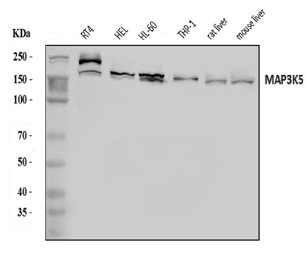
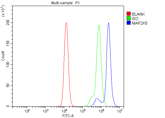 FCM/FACS (Flow Cytometry)
(Figure 5. Flow Cytometry analysis of Raji cells using anti-ASK1/MAP3K5 antibody (AAA126095).Overlay histogram showing Raji cells stained with AAA126095 (Blue line). The cells were blocked with 10% normal goat serum. And then incubated with rabbit anti-ASK1/MAP3K5 Antibody (AAA126095, 1 ug/1x10^6 cells) for 30 min at 20 degree C. DyLight488 conjugated goat anti-rabbit IgG was used as secondary antibody for 30 minutes at 20 degree C. Isotype control antibody (Green line) was rabbit IgG (1 ug/1x10^6) used under the same conditions. Unlabelled sample (Red line) was also used as a control.)
FCM/FACS (Flow Cytometry)
(Figure 5. Flow Cytometry analysis of Raji cells using anti-ASK1/MAP3K5 antibody (AAA126095).Overlay histogram showing Raji cells stained with AAA126095 (Blue line). The cells were blocked with 10% normal goat serum. And then incubated with rabbit anti-ASK1/MAP3K5 Antibody (AAA126095, 1 ug/1x10^6 cells) for 30 min at 20 degree C. DyLight488 conjugated goat anti-rabbit IgG was used as secondary antibody for 30 minutes at 20 degree C. Isotype control antibody (Green line) was rabbit IgG (1 ug/1x10^6) used under the same conditions. Unlabelled sample (Red line) was also used as a control.)
ASK1/MAP3K5, Polyclonal Antibody (Cat# AAA126095)
Full Name
Anti-ASK1/MAP3K5 Antibody Picoband
Gene Names
MAP3K5; ASK1; MEKK5; MAPKKK5
Reactivity
Human, Mouse, Rat
Applications
Western Blot, Immunocytochemistry, Immunohistochemistry, Immunofluorescence, Flow Cytometry
Purity
Immunogen affinity purified.
Pricing
Streptavidin, Recombinant Protein (Cat# AAA38638)
Full Name
Recombinant Streptavidin, His Tag
Purity
Greater than 95.0% as determined by analysis by SDS-PAGE.
Pricing
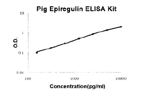 Standard Curve (Sample)
Epiregulin, ELISA Kit (Cat# AAA124844)
Full Name
Pig porcine Epiregulin PicoKine ELISA Kit
Gene Names
EREG; ER; Ep; EPR
Reactivity
Pig
There is no detectable cross-reactivity with other relevant proteins.
There is no detectable cross-reactivity with other relevant proteins.
Applications
ELISA
Pricing
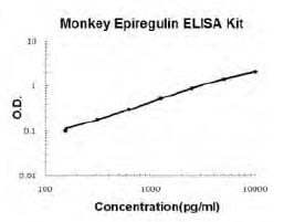 Standard Curve (Sample)
Epiregulin, ELISA Kit (Cat# AAA124845)
Full Name
Monkey primate Epiregulin PicoKine ELISA Kit
Gene Names
EREG; ER; Ep; EPR
Reactivity
Monkey
There is no detectable cross-reactivity with other relevant proteins.
There is no detectable cross-reactivity with other relevant proteins.
Applications
ELISA
Pricing