
Filters
Clonality
Type
Reactivity
Gene Name
Isotype
Host
Application
Clone
10000 results for "Recombinant Proteins" - showing 8500-8550
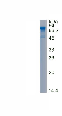
 Bioactivity
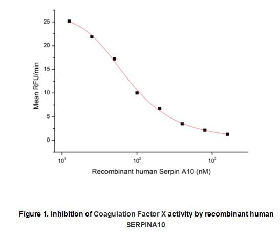
(Protein Z-dependent Protease Inhibitor (ZPI), also known as SerpinA10 (SERine Proteinase INhibitor-clade A10) is a monomeric, secreted member of the A (or extracellular) clade within the serpin superfamily of protease inhibitors. In general, members of this superfamily regulate multiple proteolytic cascades, and are particularly effective due to the fact that their inhibitory activities can be fine-tuned through the participation of discrete, non-serpin co-factors. Serpins are unusual in that they are one-time use, non-recyclable proteins whose native state is thermodynamically unstable. The activity of recombinant human SERPINA10 was measured by its ability to inhibit Coagulation Factor X cleavage of a fluorogenic peptide substrate Mca-RPKPVE-Nval-WRK(Dnp)-NH2 in the assay buffer 50 mM Tris, 10 mM CaCl2, 150 mM NaCl, 0.05% (w/v) Brij-35, pH 7.5. Coagulation Factor X was diluted to 5 ug/ml in the assay buffer and 25 ul different concentrations of recombinant human SERPINA10 (MW: 78.26 KD) was incubated with 25 ul diluted Coagulation Factor X at 37 degree C for 30 minutes. Loading 50 uL 20 uM substrate to start the reaction including a substrate blank containing 50 uL of assay buffer and 50 uL of 20 uM substrate. Then read at excitiation and emission wavelengths of 320 nm and 405 nm, respectively, in kinetic mode for 5 minutes. The result was shown in Figure 1 and it was obvious that recombinant human SERPINA10 significantly decreased Coagulation Factor X activity. The inhibition IC50 was )
Bioactivity
(Protein Z-dependent Protease Inhibitor (ZPI), also known as SerpinA10 (SERine Proteinase INhibitor-clade A10) is a monomeric, secreted member of the A (or extracellular) clade within the serpin superfamily of protease inhibitors. In general, members of this superfamily regulate multiple proteolytic cascades, and are particularly effective due to the fact that their inhibitory activities can be fine-tuned through the participation of discrete, non-serpin co-factors. Serpins are unusual in that they are one-time use, non-recyclable proteins whose native state is thermodynamically unstable. The activity of recombinant human SERPINA10 was measured by its ability to inhibit Coagulation Factor X cleavage of a fluorogenic peptide substrate Mca-RPKPVE-Nval-WRK(Dnp)-NH2 in the assay buffer 50 mM Tris, 10 mM CaCl2, 150 mM NaCl, 0.05% (w/v) Brij-35, pH 7.5. Coagulation Factor X was diluted to 5 ug/ml in the assay buffer and 25 ul different concentrations of recombinant human SERPINA10 (MW: 78.26 KD) was incubated with 25 ul diluted Coagulation Factor X at 37 degree C for 30 minutes. Loading 50 uL 20 uM substrate to start the reaction including a substrate blank containing 50 uL of assay buffer and 50 uL of 20 uM substrate. Then read at excitiation and emission wavelengths of 320 nm and 405 nm, respectively, in kinetic mode for 5 minutes. The result was shown in Figure 1 and it was obvious that recombinant human SERPINA10 significantly decreased Coagulation Factor X activity. The inhibition IC50 was )
Serpin A10 (SERPINA10), Active Protein (Cat# AAA161873)
Full Name
Active Serpin A10 (SERPINA10)
Gene Names
SERPINA10; PZI; ZPI
Applications
Activity Assay, Cell Culture
Purity
Greater than 80% by SDS-PAGE
Pricing
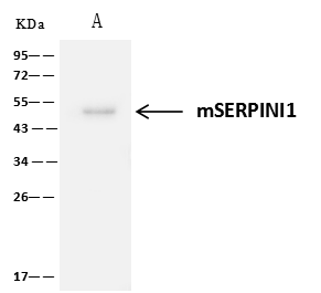 IP (Immunoprecipitation)
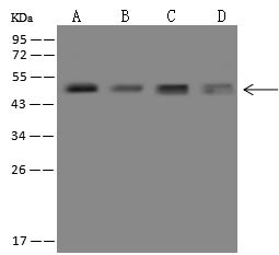
(mSERPINI1 was immunoprecipitated using:Lane A:0.5 mg Mouse brain Whole Cell Lysate1 uL anti-mSERPINI1 rabbit polyclonal antibody and 60 ug of Immunomagnetic beads Protein A/G.Primary antibody:Anti-mSERPINI1 rabbit polyclonal antibody,at 1:500 dilutionSecondary antibody:Clean-Blot IP Detection Reagent (HRP) at 1:1000dilutionDeveloped using the ECL technique.Performed under reducing conditions.Predicted band size: 50 kDaObserved band size :50 kDa)
IP (Immunoprecipitation)
(mSERPINI1 was immunoprecipitated using:Lane A:0.5 mg Mouse brain Whole Cell Lysate1 uL anti-mSERPINI1 rabbit polyclonal antibody and 60 ug of Immunomagnetic beads Protein A/G.Primary antibody:Anti-mSERPINI1 rabbit polyclonal antibody,at 1:500 dilutionSecondary antibody:Clean-Blot IP Detection Reagent (HRP) at 1:1000dilutionDeveloped using the ECL technique.Performed under reducing conditions.Predicted band size: 50 kDaObserved band size :50 kDa)
Neuroserpin, Polyclonal Antibody (Cat# AAA257243)
Full Name
Anti-Neuroserpin Antibody, Rabbit Polyclonal
Gene Names
Serpini1; Ns; PI12; PI-12; Spi17; AI837402
Reactivity
Mouse
Applications
Immunoprecipitation, Immunohistochemistry, Western Blot
Purity
Protein A & Antigen Affinity
Pricing
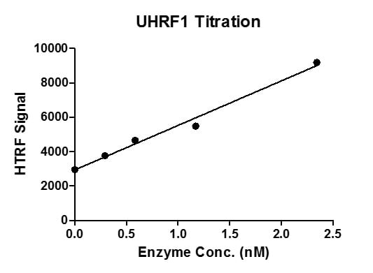
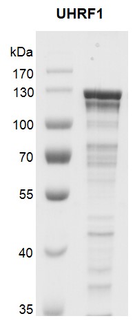
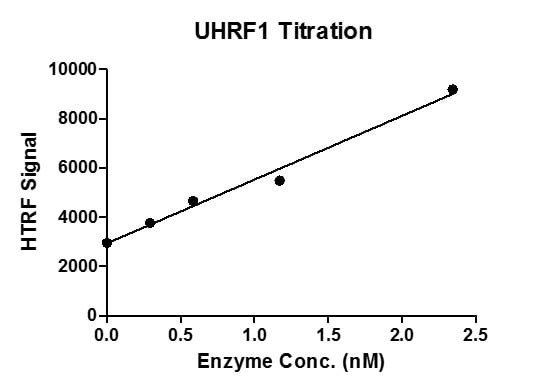 Application Data
UHRF1, Recombinant Protein (Cat# AAA60107)
Full Name
Recombinant UHRF1 protein
Gene Names
UHRF1; Np95; hNP95; ICBP90; RNF106; TDRD22; hUHRF1; huNp95
Pricing
 IF (Immunofluorescence)
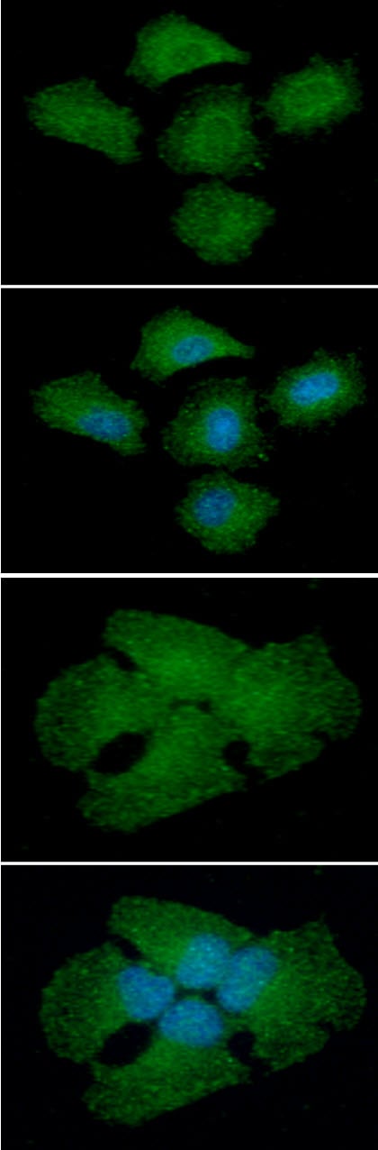
(ICC/IF analysis of NQO2 in A549 cell line, stained with DAPI (Blue) for nucleus staining and monoclonal anti-human NQO2 antibody (1:100) with goat anti-mouse IgG-Alexa fluor 488 conjugate (Green).)
IF (Immunofluorescence)
(ICC/IF analysis of NQO2 in A549 cell line, stained with DAPI (Blue) for nucleus staining and monoclonal anti-human NQO2 antibody (1:100) with goat anti-mouse IgG-Alexa fluor 488 conjugate (Green).)
NQO2, Monoclonal Antibody (Cat# AAA48081)
Full Name
NQO2 antibody
Gene Names
NQO2; QR2; DHQV; DIA6; NMOR2
Reactivity
Human, Mouse
Applications
Immunofluorescence, Immunocytochemistry, Western Blot
Purity
By protein-G affinity chromatography
Pricing
 SDS-PAGE
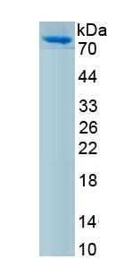
(Figure. SDS-PAGE)
SDS-PAGE
(Figure. SDS-PAGE)
Complement Component 1, S Subcomponent (C1s), Protein (Cat# AAA161950)
Full Name
Eukaryotic Complement Component 1, S Subcomponent (C1s)
Gene Names
C1s; r-gsp
Applications
Western Blot
Purity
Greater than 95% by SDS-PAGE
Pricing
 DB (Dot Blot)
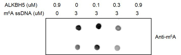
(Dot blot for Recombinant ALKBH5 protein activity. 3 uM m6A ssDNA oligos (sequence: 5’-GTTGCCTGTTCGTGTTGG/m6A/CTTGCCTGT-3’) were incubated with different concentrations of ALKBH5 proteins in a 30 ul reaction system including 50 mM HEPES-NaOH pH 7.5, 100 uM 2-oxoglutarate, 100 uM ascorbate, 50 ?M (NH4)2Fe(SO4)2·6H2O and 1 mM TCEP for 2 hours at room temperature. Then each reaction was concentrated to a final volume of 10 ul. 1 ul of each reaction was spotted onto a positively charged nylon membrane and products were detected by Dot blot assay with m6A antibody .)
DB (Dot Blot)
(Dot blot for Recombinant ALKBH5 protein activity. 3 uM m6A ssDNA oligos (sequence: 5’-GTTGCCTGTTCGTGTTGG/m6A/CTTGCCTGT-3’) were incubated with different concentrations of ALKBH5 proteins in a 30 ul reaction system including 50 mM HEPES-NaOH pH 7.5, 100 uM 2-oxoglutarate, 100 uM ascorbate, 50 ?M (NH4)2Fe(SO4)2·6H2O and 1 mM TCEP for 2 hours at room temperature. Then each reaction was concentrated to a final volume of 10 ul. 1 ul of each reaction was spotted onto a positively charged nylon membrane and products were detected by Dot blot assay with m6A antibody .)
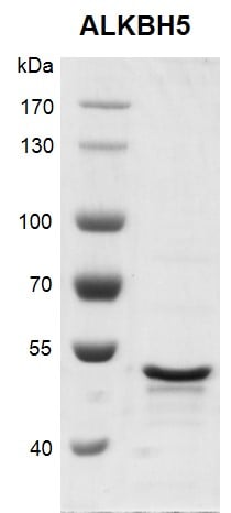
ALKBH5, Recombinant Protein (Cat# AAA59767)
Full Name
Recombinant ALKBH5 protein
Gene Names
ALKBH5; ABH5; OFOXD; OFOXD1
Pricing
 WB (Western Blot)
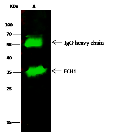
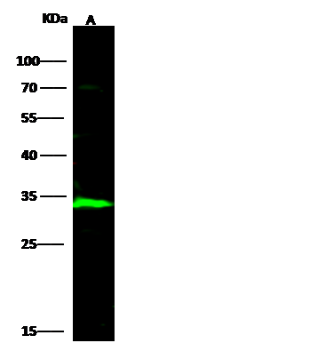
(Anti-ECH1 rabbit polyclonal antibody at 1:500 dilutionLane A: HepG2 Whole Cell LysateLysates/proteins at 30 ug per lane.SecondaryGoat Anti-Rabbit IgG H&L (Dylight800) at 1/10000 dilution.Developed using the Odyssey technique.Performed under reducing conditions.Predicted band size:36 kDaObserved band size:34 kDa)
WB (Western Blot)
(Anti-ECH1 rabbit polyclonal antibody at 1:500 dilutionLane A: HepG2 Whole Cell LysateLysates/proteins at 30 ug per lane.SecondaryGoat Anti-Rabbit IgG H&L (Dylight800) at 1/10000 dilution.Developed using the Odyssey technique.Performed under reducing conditions.Predicted band size:36 kDaObserved band size:34 kDa)
ECH1, Polyclonal Antibody (Cat# AAA255768)
Full Name
Anti-ECH1 Antibody, Rabbit Polyclonal
Gene Names
ECH1; HPXEL
Reactivity
Human
Applications
Immunoprecipitation, Immunohistochemistry, Western Blot
Purity
Protein A & Antigen Affinity
Pricing
 WB (Western Blot)
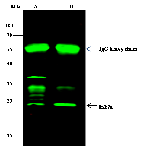
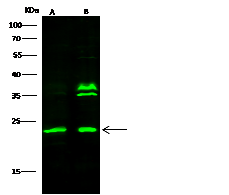
(Anti-RAB7A rabbit polyclonal antibody at 1:500 dilutionLane A: A431 Whole Cell LysateLane B: K562 Whole Cell LysateLysates/proteins at 30 ug per lane.SecondaryGoat Anti- Rabbit IgG H&L (Dylight 800) at 1/10000 dilution.Developed using the Odyssey technique.Performed under reducing conditions.Predicted band size:23 kDaObserved band size:23 kDa(We are unsure as to the identity of these extra bands.))
WB (Western Blot)
(Anti-RAB7A rabbit polyclonal antibody at 1:500 dilutionLane A: A431 Whole Cell LysateLane B: K562 Whole Cell LysateLysates/proteins at 30 ug per lane.SecondaryGoat Anti- Rabbit IgG H&L (Dylight 800) at 1/10000 dilution.Developed using the Odyssey technique.Performed under reducing conditions.Predicted band size:23 kDaObserved band size:23 kDa(We are unsure as to the identity of these extra bands.))
RAB7A, Polyclonal Antibody (Cat# AAA257338)
Full Name
Anti-RAB7A Antibody, Rabbit Polyclonal
Gene Names
Rab7a; Rab7
Reactivity
Rat
Applications
Immunoprecipitation, Immunohistochemistry, Western Blot
Purity
Protein A & Antigen Affinity
Pricing

 Application Data
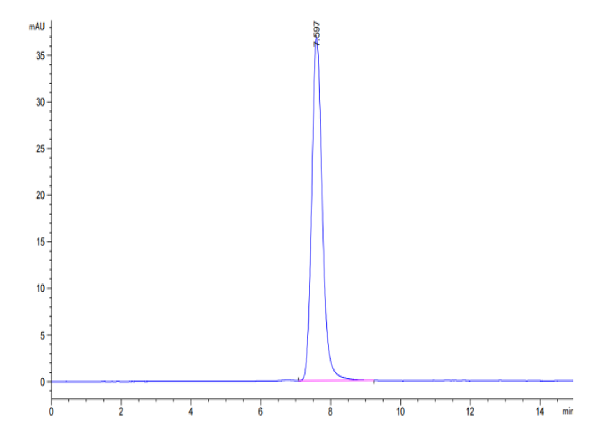
(Biotinylated Human IGF2R Protein SEC-HPLCThe purity of Biotinylated Human IGF2R is greater than 95% as determined by SEC-HPLC.)
Application Data
(Biotinylated Human IGF2R Protein SEC-HPLCThe purity of Biotinylated Human IGF2R is greater than 95% as determined by SEC-HPLC.)
IGF2R, Recombinant Protein (Cat# AAA76288)
Full Name
Biotinylated Human IGF2R Protein Recombinant
Gene Names
IGF2R; MPR1; MPRI; CD222; CIMPR; M6P-R
Purity
>95%, as determined by SDS-PAGE and HPLC
Pricing

 Application Data
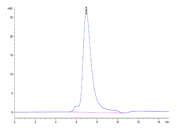
(Biotinylated Human LILRA1/CD85i/LIR-6 Protein SEC-HPLCThe purity of Biotinylated Human LILRA1 is greater than 95% as determined by SEC-HPLC.)
Application Data
(Biotinylated Human LILRA1/CD85i/LIR-6 Protein SEC-HPLCThe purity of Biotinylated Human LILRA1 is greater than 95% as determined by SEC-HPLC.)
LILRA1/CD85i/LIR-6, Recombinant Protein (Cat# AAA76278)
Full Name
Biotinylated Human LILRA1/CD85i/LIR-6 Protein Recombinant
Gene Names
LILRA1; LIR6; CD85I; LIR-6
Purity
>95%, as determined by SDS-PAGE and HPLC
Pricing
 Application Data
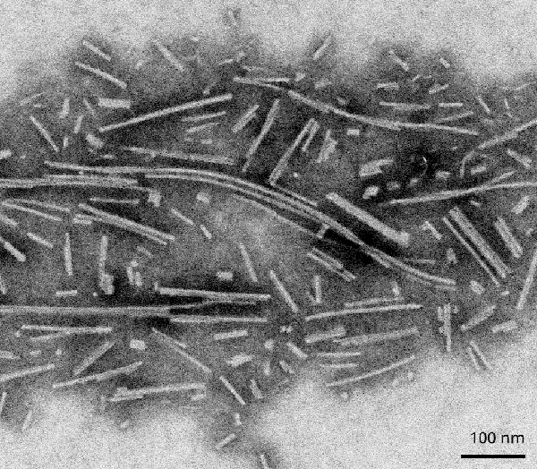
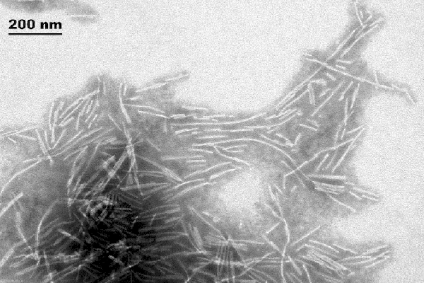
(TEMofActiveHumanRecombinantAlphaSynucleinProteinFilaments(AAA254088))
Application Data
(TEMofActiveHumanRecombinantAlphaSynucleinProteinFilaments(AAA254088))
Alpha Synuclein Protein Filaments, Recombinant Protein (Cat# AAA254088)
Full Name
Human Recombinant Alpha Synuclein Protein Filaments
Gene Names
SNCA; PD1; NACP; PARK1; PARK4
Applications
Western Blot
Purity
>95%; Ion-exchange Purified
Pricing
 IHC (Immunohiostchemistry)
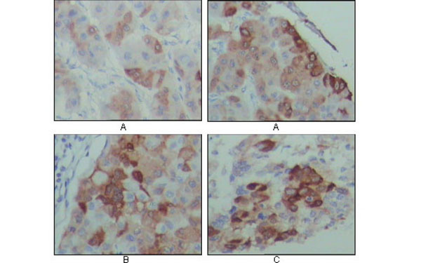
(Immunohistochemical analysis of paraffin-embedded human hepatocarcinoma (A), breast carcinoma (B) and lung cancer tissues (C), showing cytoplasmic localization with DAB staining using PEG10 antibody.)
IHC (Immunohiostchemistry)
(Immunohistochemical analysis of paraffin-embedded human hepatocarcinoma (A), breast carcinoma (B) and lung cancer tissues (C), showing cytoplasmic localization with DAB staining using PEG10 antibody.)
PEG10, Monoclonal Antibody (Cat# AAA74705)
Full Name
PEG10 antibody
Gene Names
PEG10; EDR; HB-1; Mar2; MEF3L; Mart2; RGAG3
Applications
Western Blot, Immunohistochemistry
Pricing
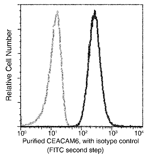
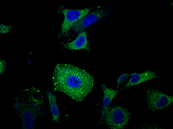 IF (Immunofluorescence)
(Immunofluorescence staining of Human CEACAM6 in SKBR3 cells. Cells were fixed with 4% PFA, permeabilzed with 0.3% Triton X-100 in PBS, blocked with 10% serum, and incubated with rabbit anti-Human CEACAM6 monoclonal antibody (1:300) at 4 degree C overnight. Then cells were stained with the Alexa Fluor 488-conjugated Goat Anti-rabbit IgG secondary antibody (green) and counterstained with DAPI (blue).)
IF (Immunofluorescence)
(Immunofluorescence staining of Human CEACAM6 in SKBR3 cells. Cells were fixed with 4% PFA, permeabilzed with 0.3% Triton X-100 in PBS, blocked with 10% serum, and incubated with rabbit anti-Human CEACAM6 monoclonal antibody (1:300) at 4 degree C overnight. Then cells were stained with the Alexa Fluor 488-conjugated Goat Anti-rabbit IgG secondary antibody (green) and counterstained with DAPI (blue).)
CEACAM6, Monoclonal Antibody (Cat# AAA255204)
Full Name
Recombinant Anti-CEACAM6 Antibody, Rabbit Monoclonal
Gene Names
CEACAM6; NCA; CEAL; CD66c
Reactivity
Human
Applications
Flow Cytometry
Purity
Protein A
Pricing
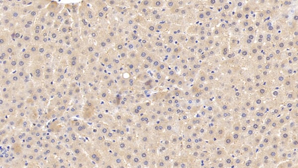 IHC (Immunohiostchemistry)
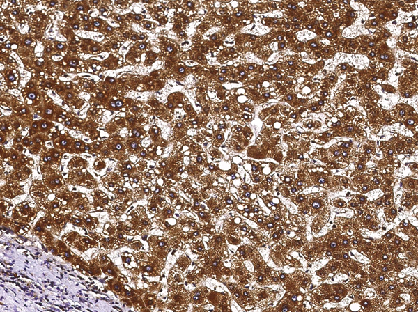
(DAB staining on IHCP; Samples: Rat Liver Tissue; Primary Ab: 10ug/ml Rabbit AntiRat APC Antibody Second Ab: 2ug/mL HRPLinked Caprine AntiRabbit IgG Polyclonal Antibody (Catalog: SAA544Rb19))
IHC (Immunohiostchemistry)
(DAB staining on IHCP; Samples: Rat Liver Tissue; Primary Ab: 10ug/ml Rabbit AntiRat APC Antibody Second Ab: 2ug/mL HRPLinked Caprine AntiRabbit IgG Polyclonal Antibody (Catalog: SAA544Rb19))
Activated Protein C (APC), Polyclonal Antibody (Cat# AAA151936)
Full Name
Polyclonal Antibody to Activated Protein C (APC)
Reactivity
Rat
Applications
Immunoprecipitation, Immunocytochemistry, Immunohistochemistry, Western Blot
Purity
Affinity Chromatography
Pricing
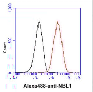
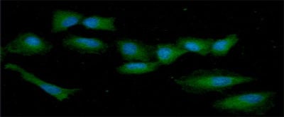
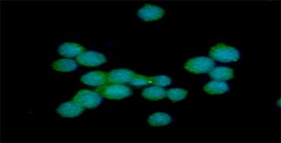
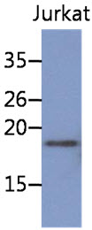
 FCM/FACS (Flow Cytometry)
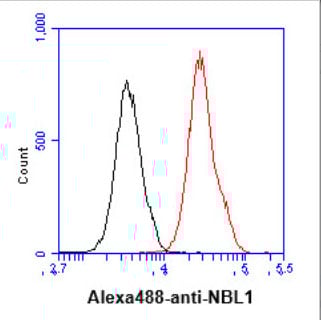
(Flow cytometry analysis of NBL1 in A549 cell line, staining at 2-5ug for 1x10^6cells (red line). The secondary antibody used goat anti-mouse IgG Alexa fluor 488 conjugate. Isotype control antibody was mouse IgG (black line).)
FCM/FACS (Flow Cytometry)
(Flow cytometry analysis of NBL1 in A549 cell line, staining at 2-5ug for 1x10^6cells (red line). The secondary antibody used goat anti-mouse IgG Alexa fluor 488 conjugate. Isotype control antibody was mouse IgG (black line).)
NBL1, Monoclonal Antibody (Cat# AAA48135)
Full Name
NBL1 antibody
Gene Names
NBL1; NB; DAN; NO3; DAND1; D1S1733E
Reactivity
Human
Applications
Immunofluorescence, Immunocytochemistry, Flow Cytometry, Western Blot
Purity
By protein-A affinity chromatography
Pricing
 Application Data
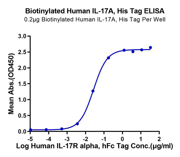
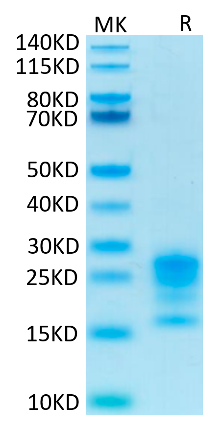
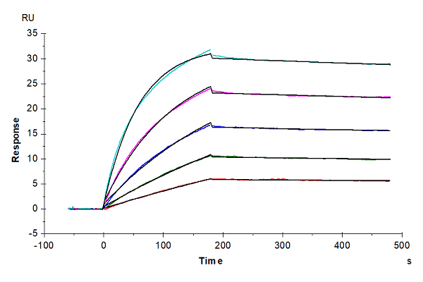
(Biotinylated Human IL-17A/CTLA-8 Protein SPR DataHuman IL-17R alpha, hFc Tag captured on CM5 Chip via protein A can bind Biotinylated Human IL-17A, His-Avi Tag with an affinity constant of 2.18 nM as determined in SPR assay (Biacore T200).)
Application Data
(Biotinylated Human IL-17A/CTLA-8 Protein SPR DataHuman IL-17R alpha, hFc Tag captured on CM5 Chip via protein A can bind Biotinylated Human IL-17A, His-Avi Tag with an affinity constant of 2.18 nM as determined in SPR assay (Biacore T200).)
IL-17A/CTLA-8, Recombinant Protein (Cat# AAA76135)
Full Name
Biotinylated Human IL-17A/CTLA-8 Protein Recombinant
Gene Names
IL17A; IL17; CTLA8; IL-17; IL-17A
Purity
>95%, as determined by SDS-PAGE and HPLC
Pricing
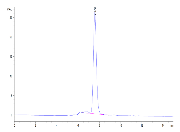
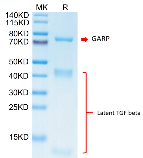
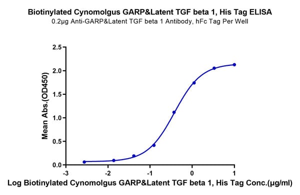 ELISA
(Biotinylated Cynomolgus GARP&Latent TGF beta 1 Complex Protein ELISA DataImmobilized Anti-GARP&Latent TGF beta 1 Antibody, hFc Tag at 2?g/ml (100?l/well) on the plate. Dose response curve for Biotinylated Cynomolgus GARP&Latent TGF beta 1 Complex, His Tag with the EC50 of 0.38?g/ml determined by ELISA.)
ELISA
(Biotinylated Cynomolgus GARP&Latent TGF beta 1 Complex Protein ELISA DataImmobilized Anti-GARP&Latent TGF beta 1 Antibody, hFc Tag at 2?g/ml (100?l/well) on the plate. Dose response curve for Biotinylated Cynomolgus GARP&Latent TGF beta 1 Complex, His Tag with the EC50 of 0.38?g/ml determined by ELISA.)
GARP&Latent TGF beta 1 Complex, Recombinant Protein (Cat# AAA76348)
Full Name
Biotinylated Human GARP&Latent TGF beta 1 Complex Protein Recombinant
Gene Names
LRRC32; GARP; D11S833E
Purity
>95%, as determined by SDS-PAGE and HPLC
Pricing
 Bioactivity
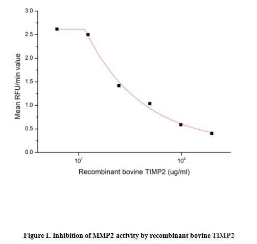
(Tissue inhibitors of metalloproteinases or TIMPs are a family of proteins that regulate the activation and proteolytic activity of the zinc enzymes known as matrix metalloproteinases (MMPs). There are four members of the family, TIMP-1, TIMP-2, TIMP-3, and TIMP-4. TIMP-2 is a non N-glycosylated protein with a molecular mass of 22 kDa produced by a wide range of cell types, which inhibits MMPs non-covalently by the formation of binary complexes. TIMP-2 also has erythroidpotentiating and cell growth promoting activities. The activity of recombinant bovine TIMP2 was measured by its ability to inhibit rhMMP2 cleavage of a fluorogenic peptide substrate MCA-Pro-Leu-Gly-Leu-DPA-Ala-Arg-NH2 in the assay buffer 50 mM Tris, 10 mM CaCl2, 150 mM NaCl, 0.05% (w/v) Brij-35, pH 7.5. rhMMP2 was diluted to 100 ug/ml and activated with 1 mM APMA at 37 degree C for 1 hour and rbTIMP2 (MW: 51.75 KD) was diluted to different concentrations with the assay buffer. Mix 8 ul of rhTIMP2 curve dilutions, 12.8 ul of activated rbMMP-2, and 59.2 ul of assay buffer, including a control containing assay buffer and the diluted rhMMP-2 and incubate the reactions for 2 hours at 37 degree C. Loading 50 ul of the incubated mixtures which were diluted five-fold in assay buffer into empty wells of a plate, and start the reaction by adding 50 ul of 20 uM substrate. Include a substrate blank containing 50 ul of assay buffer and 50 ul of 20 uM substrate. Then read at excitiation and emission wavelengths of 320 nm and 405 nm, respectively, in kinetic mode for 5 minutes. The result was shown in Figure 1 and it was obvious that recombinant bovine TIMP2 significantly decreased rhMMP2 activity. The inhibition IC50 was )
Bioactivity
(Tissue inhibitors of metalloproteinases or TIMPs are a family of proteins that regulate the activation and proteolytic activity of the zinc enzymes known as matrix metalloproteinases (MMPs). There are four members of the family, TIMP-1, TIMP-2, TIMP-3, and TIMP-4. TIMP-2 is a non N-glycosylated protein with a molecular mass of 22 kDa produced by a wide range of cell types, which inhibits MMPs non-covalently by the formation of binary complexes. TIMP-2 also has erythroidpotentiating and cell growth promoting activities. The activity of recombinant bovine TIMP2 was measured by its ability to inhibit rhMMP2 cleavage of a fluorogenic peptide substrate MCA-Pro-Leu-Gly-Leu-DPA-Ala-Arg-NH2 in the assay buffer 50 mM Tris, 10 mM CaCl2, 150 mM NaCl, 0.05% (w/v) Brij-35, pH 7.5. rhMMP2 was diluted to 100 ug/ml and activated with 1 mM APMA at 37 degree C for 1 hour and rbTIMP2 (MW: 51.75 KD) was diluted to different concentrations with the assay buffer. Mix 8 ul of rhTIMP2 curve dilutions, 12.8 ul of activated rbMMP-2, and 59.2 ul of assay buffer, including a control containing assay buffer and the diluted rhMMP-2 and incubate the reactions for 2 hours at 37 degree C. Loading 50 ul of the incubated mixtures which were diluted five-fold in assay buffer into empty wells of a plate, and start the reaction by adding 50 ul of 20 uM substrate. Include a substrate blank containing 50 ul of assay buffer and 50 ul of 20 uM substrate. Then read at excitiation and emission wavelengths of 320 nm and 405 nm, respectively, in kinetic mode for 5 minutes. The result was shown in Figure 1 and it was obvious that recombinant bovine TIMP2 significantly decreased rhMMP2 activity. The inhibition IC50 was )
Tissue Inhibitors Of Metalloproteinase 2 (TIMP2), Active Protein (Cat# AAA161695)
Full Name
Active Tissue Inhibitors Of Metalloproteinase 2 (TIMP2)
Applications
Activity Assay, Cell Culture
Purity
Greater than 90% by SDS-PAGE
Pricing
 Bioactivity
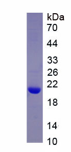
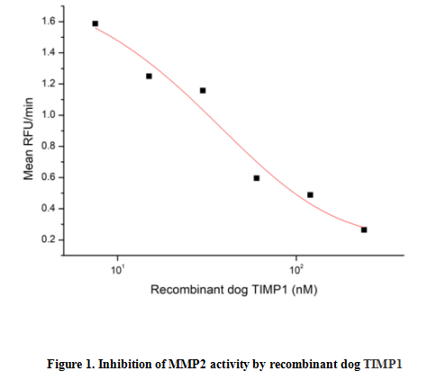
(Tissue inhibitors of metalloproteinase 1 (TIMP1) is a member of the family of proteins that regulate the activation and proteolytic activity of the zinc enzymes known as matrix metalloproteinases (MMPs). TIMP-1 is a glycoprotein with a molecular mass of 28 kDa produced by a wide range of cell types. TIMP-1 inhibits active MMP-mediated proteolysis by forming an N-terminal, non-covalent binary complex with the MMP active site. The activity of recombinant dog TIMP1 was measured by its ability to inhibit rhMMP2 cleavage of a fluorogenic peptide substrate MCA-Pro-Leu-Gly-Leu-DPA-Ala-Arg-NH2 in the assay buffer 50 mM Tris, 10 mM CaCl2, 150 mM NaCl, 0.05% (w/v) Brij-35, pH 7.5. rhMMP2 was diluted to 100 ug/ml and activated with 1 mM APMA at 37 degree C for 1 hour and TIMP1 (MW: 20.86 KD) was diluted to different concentrations with the assay buffer. Mix 8 ul of TIMP1 curve dilutions, 12.8 ul of activated rhMMP-2, and 59.2 ul of assay buffer, including a control containing assay buffer and the diluted rhMMP-2 and incubate the reactions for 2 hours at 37 degree C. Loading 50 ul of the incubated mixtures which were diluted five-fold in assay buffer into empty wells of a plate, and start the reaction by adding 50 ul of 20 uM substrate. Include a substrate blank containing 50 ul of assay buffer and 50 ul of 20 uM substrate. Then read at excitiation and emission wavelengths of 320 nm and 405 nm, respectively, in kinetic mode for 5 minutes. The result was shown in Figure 1 and it was obvious that recombinant dog TIMP1 significantly decreased rhMMP2 activity. The inhibition IC50 was )
Bioactivity
(Tissue inhibitors of metalloproteinase 1 (TIMP1) is a member of the family of proteins that regulate the activation and proteolytic activity of the zinc enzymes known as matrix metalloproteinases (MMPs). TIMP-1 is a glycoprotein with a molecular mass of 28 kDa produced by a wide range of cell types. TIMP-1 inhibits active MMP-mediated proteolysis by forming an N-terminal, non-covalent binary complex with the MMP active site. The activity of recombinant dog TIMP1 was measured by its ability to inhibit rhMMP2 cleavage of a fluorogenic peptide substrate MCA-Pro-Leu-Gly-Leu-DPA-Ala-Arg-NH2 in the assay buffer 50 mM Tris, 10 mM CaCl2, 150 mM NaCl, 0.05% (w/v) Brij-35, pH 7.5. rhMMP2 was diluted to 100 ug/ml and activated with 1 mM APMA at 37 degree C for 1 hour and TIMP1 (MW: 20.86 KD) was diluted to different concentrations with the assay buffer. Mix 8 ul of TIMP1 curve dilutions, 12.8 ul of activated rhMMP-2, and 59.2 ul of assay buffer, including a control containing assay buffer and the diluted rhMMP-2 and incubate the reactions for 2 hours at 37 degree C. Loading 50 ul of the incubated mixtures which were diluted five-fold in assay buffer into empty wells of a plate, and start the reaction by adding 50 ul of 20 uM substrate. Include a substrate blank containing 50 ul of assay buffer and 50 ul of 20 uM substrate. Then read at excitiation and emission wavelengths of 320 nm and 405 nm, respectively, in kinetic mode for 5 minutes. The result was shown in Figure 1 and it was obvious that recombinant dog TIMP1 significantly decreased rhMMP2 activity. The inhibition IC50 was )
Tissue Inhibitors Of Metalloproteinase 1 (TIMP1), Active Protein (Cat# AAA161742)
Full Name
Active Tissue Inhibitors Of Metalloproteinase 1 (TIMP1)
Gene Names
TIMP1; TIMP-1
Applications
Activity Assay, Cell Culture
Purity
Greater than 95% by SDS-PAGE
Pricing
 FCM/FACS (Flow Cytometry)
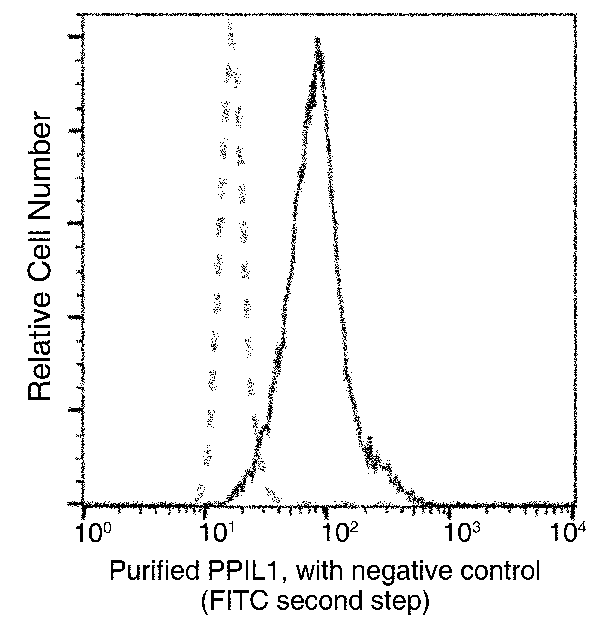
(Flow cytometric analysis of Human PPIL1 expression on MCF-7 cells. The cells were treated according to manufacturer's manual (BD Pharmingen'), stained with purified anti-Human PPIL1, then a FITC-conjugated second step antibody. The fluorescence histograms were derived from gated events with the forward and side light-scatter characteristics of intact cells.)
FCM/FACS (Flow Cytometry)
(Flow cytometric analysis of Human PPIL1 expression on MCF-7 cells. The cells were treated according to manufacturer's manual (BD Pharmingen'), stained with purified anti-Human PPIL1, then a FITC-conjugated second step antibody. The fluorescence histograms were derived from gated events with the forward and side light-scatter characteristics of intact cells.)
PPIL1, Monoclonal Antibody (Cat# AAA255724)
Full Name
Anti-PPIL1 Antibody, Mouse Monoclonal
Gene Names
PPIL1; CYPL1; hCyPX; PPIase; CGI-124
Reactivity
Human
Applications
Immunoprecipitation, Flow Cytometry, Western Blot
Purity
Protein A
Pricing
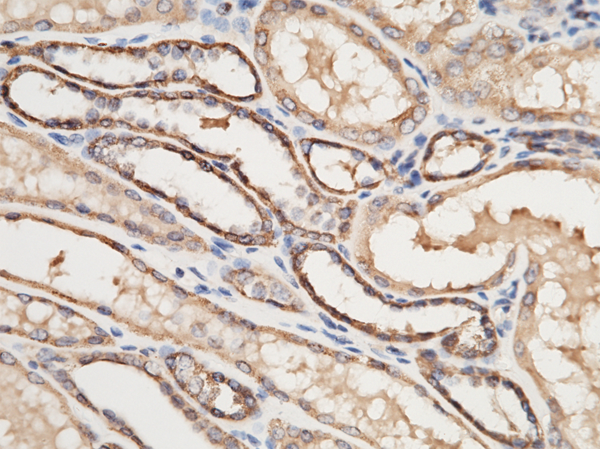
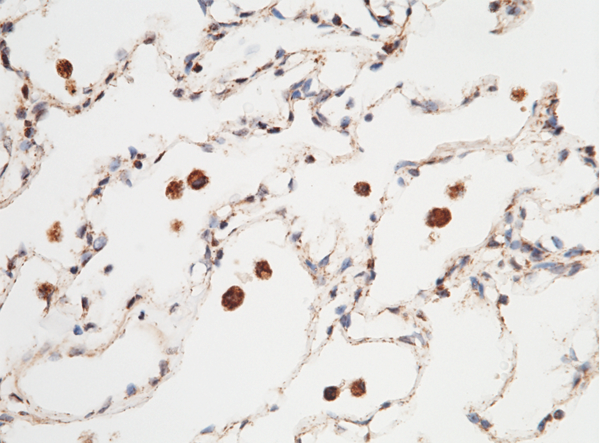
 WB (Western Blot)
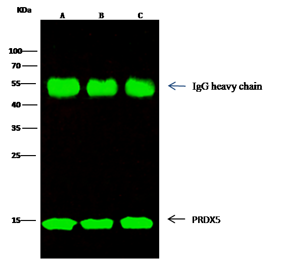
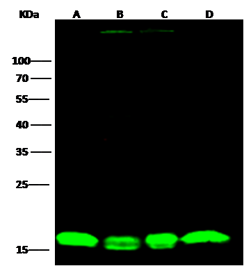
(Anti-PRDX5 rabbit polyclonal antibody at 1:2000 dilutionLane A: Hela Whole Cell LysateLane B: 293T Whole Cell LysateLane C: Jurkat Whole Cell LysateLane D: 293 Whole Cell LysateLysates/proteins at 30 ug per lane.SecondaryGoat Anti-Rabbit IgG H&L (Dylight800) at 1/10000 dilution.Developed using the Odyssey technique.Performed under reducing conditions.Predicted band size:22 kDaObserved band size:15 kDa)
WB (Western Blot)
(Anti-PRDX5 rabbit polyclonal antibody at 1:2000 dilutionLane A: Hela Whole Cell LysateLane B: 293T Whole Cell LysateLane C: Jurkat Whole Cell LysateLane D: 293 Whole Cell LysateLysates/proteins at 30 ug per lane.SecondaryGoat Anti-Rabbit IgG H&L (Dylight800) at 1/10000 dilution.Developed using the Odyssey technique.Performed under reducing conditions.Predicted band size:22 kDaObserved band size:15 kDa)
Peroxiredoxin 5, Polyclonal Antibody (Cat# AAA255377)
Full Name
Anti-Peroxiredoxin 5 Antibody, Rabbit Polyclonal
Gene Names
PRDX5; PLP; ACR1; B166; PRXV; PMP20; PRDX6; prx-V; SBBI10; AOEB166
Reactivity
Human
Applications
Immunoprecipitation, Immunohistochemistry, Western Blot
Purity
Protein A & Antigen Affinity
Pricing
 Bioactivity
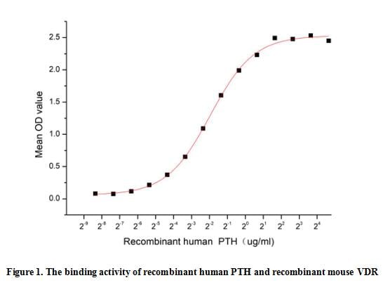
(Parathyroid hormone (PTH) is an 84-amino-acid peptide hormone that is secreted by the parathyroid gland. PTH plays an osteogenic role by acting directly on mesenchymal stem cells, cells with an osteoblastic lineage, osteocytes, and T cells. It also participates as an osteoclast by indirectly acting on osteoclast precursor cells and osteoclasts and directly acting on T cells. PTH is major endocrine regulators of vitamin D metabolism. It proves, with data, this hormone a potent down-regulator of VDR. A functional binding ELISA assay was conducted to detect the interaction of recombinant human PTH and recombinant mouse VDR. Briefly, PTH was diluted serially in PBS with 0.01% BSA (pH 7.4). Duplicate samples of 100 ul were then transferred to VDR-coated microtiter wells and incubated for 1h at 37 degree C. Wells were washed with PBST and incubated for 1h with anti-PTH pAb, then aspirated and washed 3 times. After incubation with HRP labelled secondary antibody for 1h at 37 degree C, wells were aspirated and washed 5 times. With the addition of substrate solution, wells were incubated 15-25 minutes at 37 degree C. Finally, add 50 uL stop solution to the wells and read at 450/630 nm immediately. The binding activity of recombinant human PTH and recombinant mouse VDR was shown in Figure 1, the EC50 for this effect is 0.26 ug/mL.)
Bioactivity
(Parathyroid hormone (PTH) is an 84-amino-acid peptide hormone that is secreted by the parathyroid gland. PTH plays an osteogenic role by acting directly on mesenchymal stem cells, cells with an osteoblastic lineage, osteocytes, and T cells. It also participates as an osteoclast by indirectly acting on osteoclast precursor cells and osteoclasts and directly acting on T cells. PTH is major endocrine regulators of vitamin D metabolism. It proves, with data, this hormone a potent down-regulator of VDR. A functional binding ELISA assay was conducted to detect the interaction of recombinant human PTH and recombinant mouse VDR. Briefly, PTH was diluted serially in PBS with 0.01% BSA (pH 7.4). Duplicate samples of 100 ul were then transferred to VDR-coated microtiter wells and incubated for 1h at 37 degree C. Wells were washed with PBST and incubated for 1h with anti-PTH pAb, then aspirated and washed 3 times. After incubation with HRP labelled secondary antibody for 1h at 37 degree C, wells were aspirated and washed 5 times. With the addition of substrate solution, wells were incubated 15-25 minutes at 37 degree C. Finally, add 50 uL stop solution to the wells and read at 450/630 nm immediately. The binding activity of recombinant human PTH and recombinant mouse VDR was shown in Figure 1, the EC50 for this effect is 0.26 ug/mL.)
Parathyroid Hormone (PTH), Active Protein (Cat# AAA161795)
Full Name
Active Parathyroid Hormone (PTH)
Gene Names
PTH; PTH1
Applications
Activity Assay, Cell Culture
Purity
Greater than 95% by SDS-PAGE
Pricing
 Bioactivity
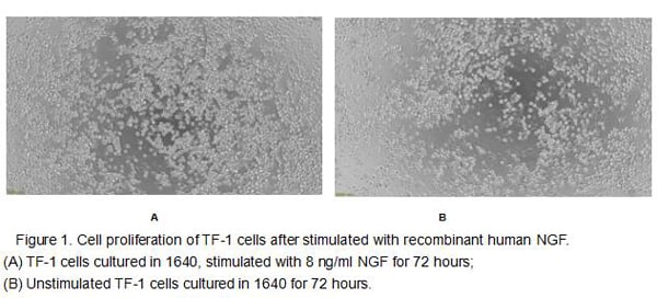
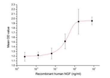
(Figure 2. Cell proliferation of TF-1 cells after stimulated with recombinant human NGF.)
Bioactivity
(Figure 2. Cell proliferation of TF-1 cells after stimulated with recombinant human NGF.)
Nerve Growth Factor (NGF), Active Protein (Cat# AAA161688)
Full Name
Active Nerve Growth Factor (NGF)
Gene Names
NGF; NGFB; HSAN5; Beta-NGF
Applications
Activity Assay, Cell Culture
Purity
Greater than 95% by SDS-PAGE
Pricing
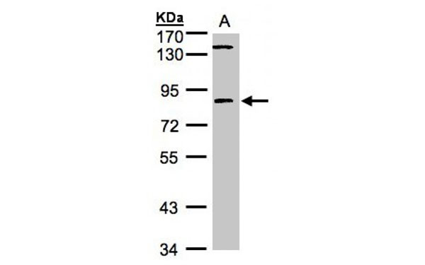
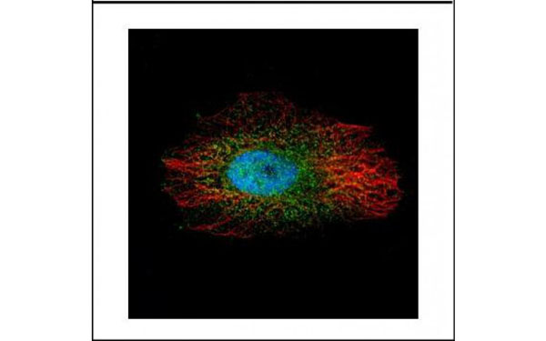
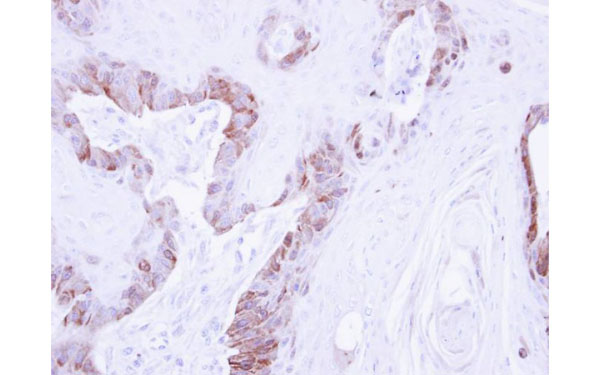 IHC (Immunohistochemisry)
(Immunohistochemical staining of paraffin-embedded CA922 xenograft using LIM kinase 2 antibody at a dilution of 1:500)
IHC (Immunohistochemisry)
(Immunohistochemical staining of paraffin-embedded CA922 xenograft using LIM kinase 2 antibody at a dilution of 1:500)
LIM kinase 2, Polyclonal Antibody (Cat# AAA107623)
Full Name
LIM kinase 2 antibody
Applications
Western Blot, Immunohistochemistry, Immunocytochemistry, Immunofluorescence
Purity
LIM kinase 2 antibody was purified by antigen-affinity chromatography
Pricing
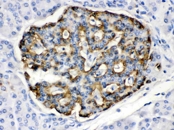
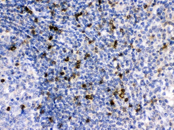
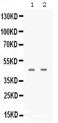
 FCM/FACS (Flow Cytometry)
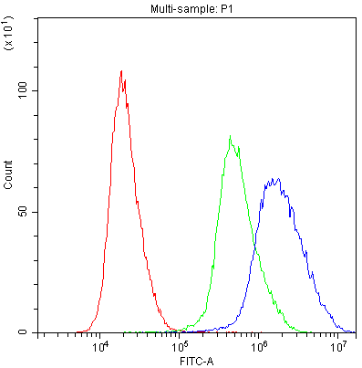
(Figure 4. Flow Cytometry analysis of THP-1 cells using anti-APOBEC3G antibody (AAA46636).Overlay histogram showing THP-1 cells stained with AAA46636 (Blue line).The cells were blocked with 10% normal goat serum. And then incubated with rabbit anti-APOBEC3G Antibody (AAA46636,1ug/1x10^6 cells) for 30 min at 20 degree C. DyLight®488 conjugated goat anti-rabbit IgG (5-10ug/1x10^6 cells) was used as secondary antibody for 30 minutes at 20 degree C. Isotype control antibody (Green line) was rabbit IgG (1ug/1x106) used under the same conditions. Unlabelled sample (Red line) was also used as a control.)
FCM/FACS (Flow Cytometry)
(Figure 4. Flow Cytometry analysis of THP-1 cells using anti-APOBEC3G antibody (AAA46636).Overlay histogram showing THP-1 cells stained with AAA46636 (Blue line).The cells were blocked with 10% normal goat serum. And then incubated with rabbit anti-APOBEC3G Antibody (AAA46636,1ug/1x10^6 cells) for 30 min at 20 degree C. DyLight®488 conjugated goat anti-rabbit IgG (5-10ug/1x10^6 cells) was used as secondary antibody for 30 minutes at 20 degree C. Isotype control antibody (Green line) was rabbit IgG (1ug/1x106) used under the same conditions. Unlabelled sample (Red line) was also used as a control.)
APOBEC3G, Polyclonal Antibody (Cat# AAA46636)
Full Name
Anti-APOBEC3G Antibody
Gene Names
APOBEC3G; A3G; ARCD; ARP9; ARP-9; CEM15; CEM-15; MDS019; bK150C2.7; dJ494G10.1
Reactivity
Human
No cross reactivity with other proteins.
No cross reactivity with other proteins.
Applications
Immunohistochemistry, Western Blot
Purity
Immunogen affinity purified.
Pricing
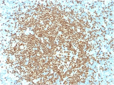
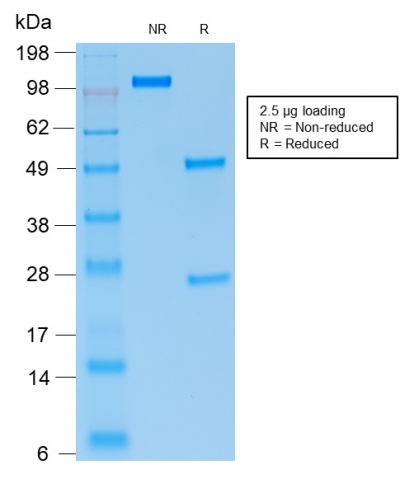 SDS-PAGE
(SDS-PAGE Analysis Purified PAX5 Mouse Recombinant Monoclonal Antibody (rPAX5/2060).)
SDS-PAGE
(SDS-PAGE Analysis Purified PAX5 Mouse Recombinant Monoclonal Antibody (rPAX5/2060).)
PAX5/BSAP, Monoclonal Antibody (Cat# AAA214472)
Full Name
PAX5/BSAP (Early B-Cell Marker)
Gene Names
PAX5; ALL3; BSAP
Reactivity
Human. Others not tested.
Applications
Immunohistochemistry, Western Blot, Immunoprecipitation
Pricing
 IHC (Immunohistochemisry)
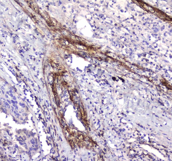
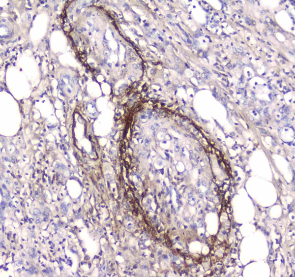
(Figure 3. IHC analysis of Serum Amyloid P using anti-Serum Amyloid P antibody (AAA124552).Serum Amyloid P was detected in paraffin-embedded section of human rectal cancer tissue. Heat mediated antigen retrieval was performed in citrate buffer (pH6, epitope retrieval solution) for 20 mins. The tissue section was blocked with 10% goat serum. The tissue section was then incubated with 1ug/ml rabbit anti-Serum Amyloid P Antibody (AAA124552) overnight at 4 degree C. Biotinylated goat anti-rabbit IgG was used as secondary antibody and incubated for 30 minutes at 37 degree C. The tissue section was developed using Strepavidin-Biotin-Complex (SABC) with DAB as the chromogen.)
IHC (Immunohistochemisry)
(Figure 3. IHC analysis of Serum Amyloid P using anti-Serum Amyloid P antibody (AAA124552).Serum Amyloid P was detected in paraffin-embedded section of human rectal cancer tissue. Heat mediated antigen retrieval was performed in citrate buffer (pH6, epitope retrieval solution) for 20 mins. The tissue section was blocked with 10% goat serum. The tissue section was then incubated with 1ug/ml rabbit anti-Serum Amyloid P Antibody (AAA124552) overnight at 4 degree C. Biotinylated goat anti-rabbit IgG was used as secondary antibody and incubated for 30 minutes at 37 degree C. The tissue section was developed using Strepavidin-Biotin-Complex (SABC) with DAB as the chromogen.)
Serum Amyloid P, Polyclonal Antibody (Cat# AAA124552)
Full Name
Anti-Serum Amyloid P Picoband antibody
Gene Names
APCS; SAP; PTX2; HEL-S-92n
Reactivity
Human
No cross reactivity with other proteins.
No cross reactivity with other proteins.
Applications
Western Blot, Immunohistochemistry
Pricing
 IF (Immunofluorescence)
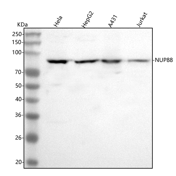
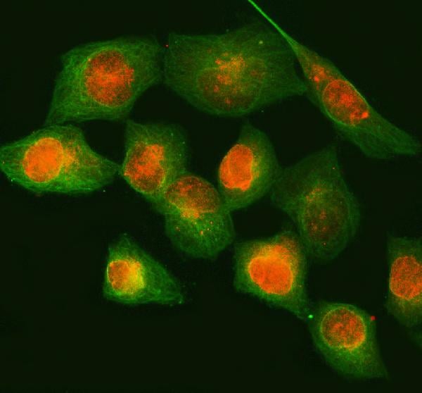
(Figure 2. IF analysis of NUP88 using anti-NUP88 antibody (AAA127293) and anti-Beta Tubulin antibody (M01857-3).NUP88 was detected in immunocytochemical section of HELA cell. Enzyme antigen retrieval was performed using IHC enzyme antigen retrieval reagent for 15 mins. The cells were blocked with 10% goat serum. And then incubated with 5ug/mL rabbit anti-NUP88 Antibody (AAA127293) and mouse anti-Beta Tubulin antibody (M01857-3) overnight at 4 degree C. Cy3 Conjugated Goat Anti-Rabbit IgG (BA1032) and FITC Conjugated Goat Anti-Mouse IgG (BA1101) were used as secondary antibody at 1:500 dilution and incubated for 30 minutes at 37 degree C. Visualize using a fluorescence microscope and filter sets appropriate for the label used.)
IF (Immunofluorescence)
(Figure 2. IF analysis of NUP88 using anti-NUP88 antibody (AAA127293) and anti-Beta Tubulin antibody (M01857-3).NUP88 was detected in immunocytochemical section of HELA cell. Enzyme antigen retrieval was performed using IHC enzyme antigen retrieval reagent for 15 mins. The cells were blocked with 10% goat serum. And then incubated with 5ug/mL rabbit anti-NUP88 Antibody (AAA127293) and mouse anti-Beta Tubulin antibody (M01857-3) overnight at 4 degree C. Cy3 Conjugated Goat Anti-Rabbit IgG (BA1032) and FITC Conjugated Goat Anti-Mouse IgG (BA1101) were used as secondary antibody at 1:500 dilution and incubated for 30 minutes at 37 degree C. Visualize using a fluorescence microscope and filter sets appropriate for the label used.)
NUP88, Polyclonal Antibody (Cat# AAA127293)
Full Name
Anti-NUP88 Antibody Picoband
Reactivity
Human
Applications
Immunofluorescence, Immunocytochemistry, Western Blot
Purity
Immunogen affinity purified.
Pricing
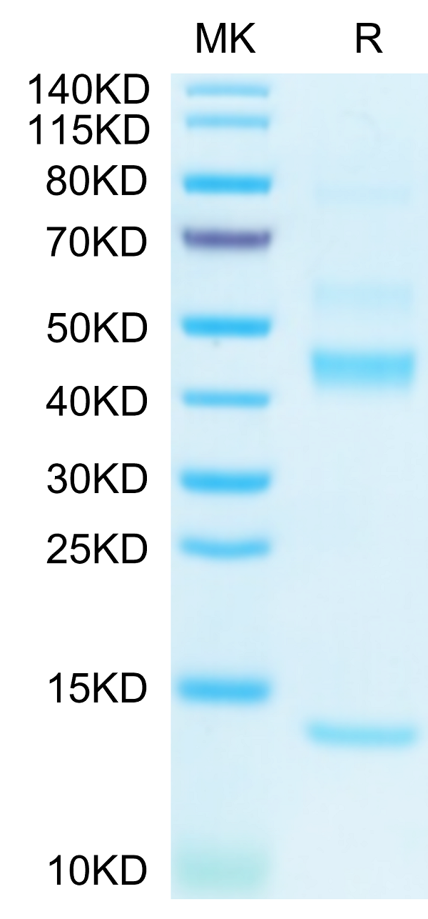
 ELISA
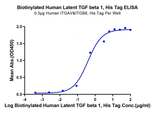
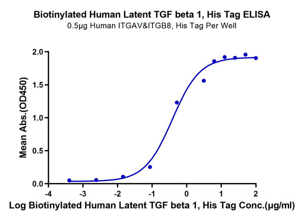
(Biotinylated Human Latent TGF beta 1/TGFB1 Protein ELISA DataImmobilized Human ITGAV&ITGB8, His Tag at 5?g/ml (100?l/well) on the plate. Dose response curve for Biotinylated Human Latent TGF beta 1, His Tag with the EC50 of 0.39ug/ml determined by ELISA.)
ELISA
(Biotinylated Human Latent TGF beta 1/TGFB1 Protein ELISA DataImmobilized Human ITGAV&ITGB8, His Tag at 5?g/ml (100?l/well) on the plate. Dose response curve for Biotinylated Human Latent TGF beta 1, His Tag with the EC50 of 0.39ug/ml determined by ELISA.)
Latent TGF beta 1/TGFB1, Recombinant Protein (Cat# AAA76216)
Full Name
Biotinylated Human Latent TGF beta 1/TGFB1 Protein Recombinant
Gene Names
TGFB1; CED; LAP; DPD1; TGFB; TGFbeta
Purity
>95%, as determined by SDS-PAGE and HPLC
Pricing
 IHC (Immunohiostchemistry)
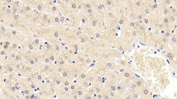
(DAB staining on IHCP;Sample: Human Liver Tissue; Primary Ab: 30ug/ml Mouse AntiHuman NT3 AntibodySecond Ab: 2ug/mL HRPLinked Caprine AntiMouse IgG Polyclonal Antibody(Catalog: SAA544Mu19))
IHC (Immunohiostchemistry)
(DAB staining on IHCP;Sample: Human Liver Tissue; Primary Ab: 30ug/ml Mouse AntiHuman NT3 AntibodySecond Ab: 2ug/mL HRPLinked Caprine AntiMouse IgG Polyclonal Antibody(Catalog: SAA544Mu19))
Neurotrophin 3 (NT3), Monoclonal Antibody (Cat# AAA151481)
Full Name
Monoclonal Antibody to Neurotrophin 3 (NT3)
Gene Names
NTF3; NT3; HDNF; NGF2; NGF-2
Reactivity
Human, Pig
Applications
Immunoprecipitation, Immunocytochemistry, Immunohistochemistry, Western Blot
Purity
Protein A + Protein G affinity chromatography
Pricing
 IHC (Immunohistochemisry)
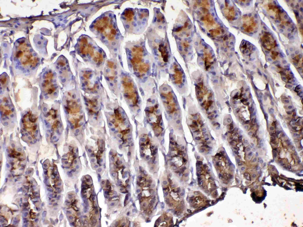
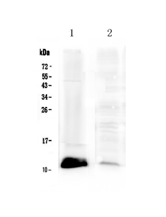
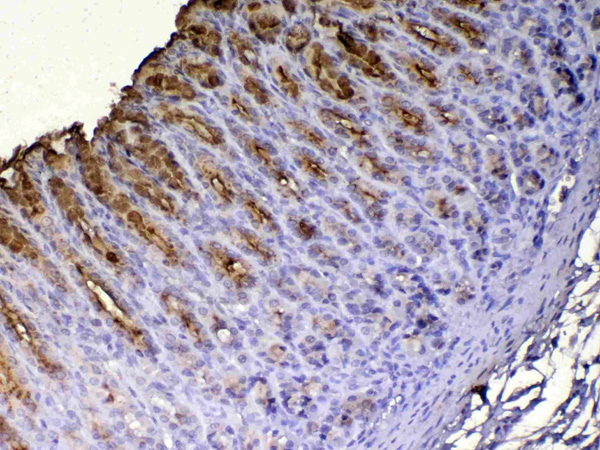
(Figure 3. IHC analysis of Tff1 using anti-Tff1 antibody (AAA124645).Tff1 was detected in paraffin-embedded section of rat gaster. Heat mediated antigen retrieval was performed in citrate buffer (pH6, epitope retrieval solution) for 20 mins. The tissue section was blocked with 10% goat serum. The tissue section was then incubated with 1ug/ml rabbit anti-Tff1 Antibody (AAA124645) overnight at 4 degree C. Biotinylated goat anti-rabbit IgG was used as secondary antibody and incubated for 30 minutes at 37 degree C. The tissue section was developed using Strepavidin-Biotin-Complex (SABC) with DAB as the chromogen.)
IHC (Immunohistochemisry)
(Figure 3. IHC analysis of Tff1 using anti-Tff1 antibody (AAA124645).Tff1 was detected in paraffin-embedded section of rat gaster. Heat mediated antigen retrieval was performed in citrate buffer (pH6, epitope retrieval solution) for 20 mins. The tissue section was blocked with 10% goat serum. The tissue section was then incubated with 1ug/ml rabbit anti-Tff1 Antibody (AAA124645) overnight at 4 degree C. Biotinylated goat anti-rabbit IgG was used as secondary antibody and incubated for 30 minutes at 37 degree C. The tissue section was developed using Strepavidin-Biotin-Complex (SABC) with DAB as the chromogen.)
Tff1/Ps2, Polyclonal Antibody (Cat# AAA124645)
Full Name
Anti-Tff1/Ps2 Picoband Antibody
Gene Names
Tff1; PS2; Bcei
Reactivity
Human, Mouse, Rat
No cross reactivity with other proteins.
No cross reactivity with other proteins.
Applications
Western Blot
Purity
Immunogen affinity purified
Pricing
 IHC (Immunohistochemisry)
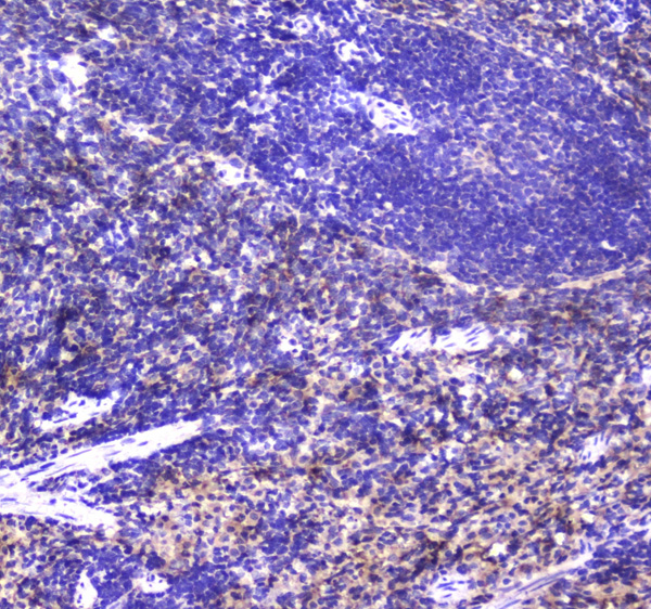
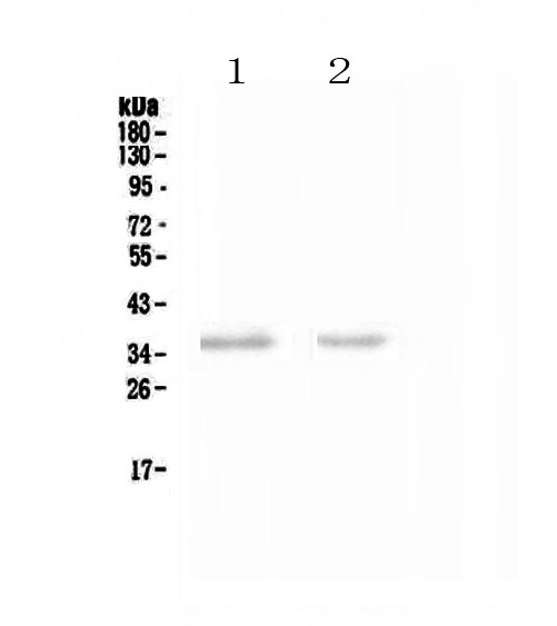
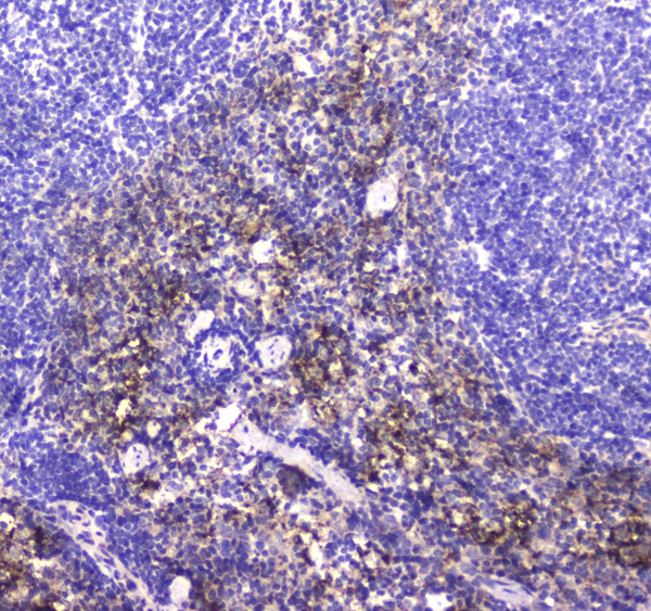
(Figure 3. IHC analysis of NCR1 using anti-NCR1 antibody (AAA124652).NCR1 was detected in paraffin-embedded section of mouse spleen tissue. Heat mediated antigen retrieval was performed in citrate buffer (pH6, epitope retrieval solution) for 20 mins. The tissue section was blocked with 10% goat serum. The tissue section was then incubated with 1ug/ml rabbit anti-NCR1 Antibody (AAA124652) overnight at 4 degree C. Biotinylated goat anti-rabbit IgG was used as secondary antibody and incubated for 30 minutes at 37 degree C. The tissue section was developed using Strepavidin-Biotin-Complex (SABC) with DAB as the chromogen.)
IHC (Immunohistochemisry)
(Figure 3. IHC analysis of NCR1 using anti-NCR1 antibody (AAA124652).NCR1 was detected in paraffin-embedded section of mouse spleen tissue. Heat mediated antigen retrieval was performed in citrate buffer (pH6, epitope retrieval solution) for 20 mins. The tissue section was blocked with 10% goat serum. The tissue section was then incubated with 1ug/ml rabbit anti-NCR1 Antibody (AAA124652) overnight at 4 degree C. Biotinylated goat anti-rabbit IgG was used as secondary antibody and incubated for 30 minutes at 37 degree C. The tissue section was developed using Strepavidin-Biotin-Complex (SABC) with DAB as the chromogen.)
NCR1, Polyclonal Antibody (Cat# AAA124652)
Full Name
Anti-NCR1 Picoband antibody
Gene Names
NCR1; LY94; CD335; NKP46; NK-p46
Reactivity
Human, Mouse, Rat
No cross reactivity with other proteins.
No cross reactivity with other proteins.
Applications
Western Blot, Immunohistochemistry
Pricing
 WB (Western Blot)
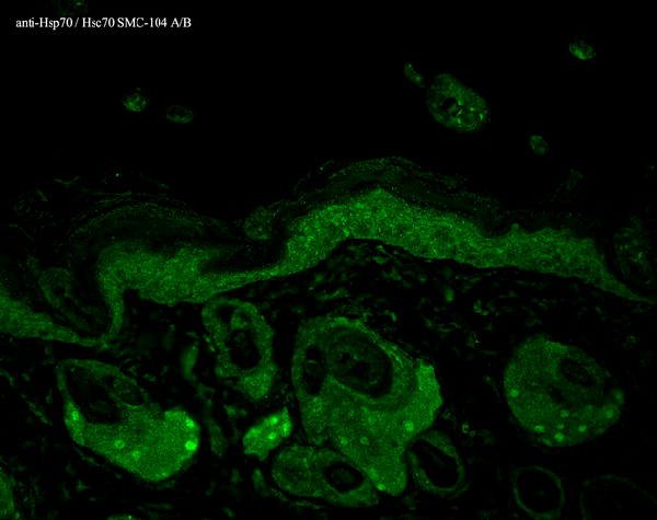
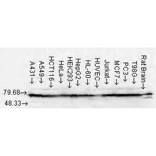
(Western Blot analysis of Human Cell lysates showing detection of Hsp70 protein using Mouse Anti-Hsp70 Monoclonal Antibody, Clone N27 . Load: 15 ug. Block: 1.5% BSA for 30 minutes at RT. Primary Antibody: Mouse Anti-Hsp70 Monoclonal Antibody at 1:1000 for 2 hours at RT. Secondary Antibody: Sheep Anti-Mouse IgG: HRP for 1 hour at RT.)
WB (Western Blot)
(Western Blot analysis of Human Cell lysates showing detection of Hsp70 protein using Mouse Anti-Hsp70 Monoclonal Antibody, Clone N27 . Load: 15 ug. Block: 1.5% BSA for 30 minutes at RT. Primary Antibody: Mouse Anti-Hsp70 Monoclonal Antibody at 1:1000 for 2 hours at RT. Secondary Antibody: Sheep Anti-Mouse IgG: HRP for 1 hour at RT.)
HSP70/HSC70, Monoclonal Antibody (Cat# AAA253927)
Full Name
HSP70/HSC70 Antibody
Gene Names
HSPA1A; HSP72; HSPA1; HSP70I; HSP70-1; HSP70-2; HSP70.1; HSP70.2; HSP70-1A; HEL-S-103
Reactivity
Human, Mouse, Rat, Bovine, Caenorhabditis elegans, Beluga, Dog, Chicken, Fruit Fly, Fish, Guinea Pig, Hamster, Monkey, Pig, Plant, Cucumber, Pea, Rabbit, Sheep, African Clawed Frog
Applications
Immuno-ElectronMicroscopy, Flow Cytometry, Immunoprecipitation, Immunofluorescence, Immunocytochemistry, Immunohistochemistry, Western Blot
Purity
Protein G Purified
Pricing
 IHC (Immunohiostchemistry)
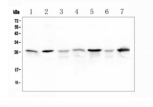
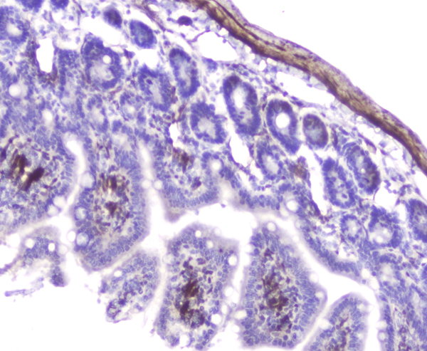
(Figure 3. IHC analysis of Hsp27 using anti-Hsp27 antibody.Hsp27 was detected in paraffin-embedded section of rat small intestine tissue. Heat mediated antigen retrieval was performed in citrate buffer (pH6, epitope retrieval solution) for 20 mins. The tissue section was blocked with 10% goat serum. The tissue section was then incubated with 2ug/ml rabbit anti-Hsp27 antibody.Hsp27 was detected in paraffin-embedded section of rat small intestine tissue. Heat mediated antigen retrieval was performed in citrate buffer (pH6, epitope retrieval solution) for 20 mins. The tissue section was blocked with 10% goat serum. The tissue section was then incubated with 2ug/ml rabbit anti-Hsp27 antibody.Hsp27 was detected in paraffin-embedded section of mouse small intestine tissue. Heat mediated antigen retrieval was performed in citrate buffer (pH6, epitope retrieval solution) for 20 mins. The tissue section was blocked with 10% goat serum. The tissue section was then incubated with 2ug/ml rabbit anti-Hsp27 Antibody overnight at 4 degree C. Biotinylated goat anti-rabbit IgG was used as secondary antibody and incubated for 30 minutes at 37 degree C. The tissue section was developed using Strepavidin-Biotin-Complex (SABC) with DAB as the chromogen.)
IHC (Immunohiostchemistry)
(Figure 3. IHC analysis of Hsp27 using anti-Hsp27 antibody.Hsp27 was detected in paraffin-embedded section of rat small intestine tissue. Heat mediated antigen retrieval was performed in citrate buffer (pH6, epitope retrieval solution) for 20 mins. The tissue section was blocked with 10% goat serum. The tissue section was then incubated with 2ug/ml rabbit anti-Hsp27 antibody.Hsp27 was detected in paraffin-embedded section of rat small intestine tissue. Heat mediated antigen retrieval was performed in citrate buffer (pH6, epitope retrieval solution) for 20 mins. The tissue section was blocked with 10% goat serum. The tissue section was then incubated with 2ug/ml rabbit anti-Hsp27 antibody.Hsp27 was detected in paraffin-embedded section of mouse small intestine tissue. Heat mediated antigen retrieval was performed in citrate buffer (pH6, epitope retrieval solution) for 20 mins. The tissue section was blocked with 10% goat serum. The tissue section was then incubated with 2ug/ml rabbit anti-Hsp27 Antibody overnight at 4 degree C. Biotinylated goat anti-rabbit IgG was used as secondary antibody and incubated for 30 minutes at 37 degree C. The tissue section was developed using Strepavidin-Biotin-Complex (SABC) with DAB as the chromogen.)
Hsp27/Hspb1, Polyclonal Antibody (Cat# AAA124908)
Full Name
Anti-Hsp27/Hspb1 Antibody
Gene Names
Hspb1; 27kDa; Hsp25
Reactivity
Mouse, Rat
Applications
Direct ELISA, Immunohistochemistry, Western Blot
Purity
Immunogen Affinity Purified
Pricing
 Bioactivity
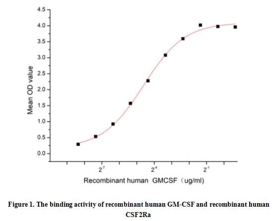
(Granulocyte-macrophage colony-stimulating factor (GM-CSF), also known as colony stimulating factor 2 (CSF2), is a monomeric glycoprotein secreted by macrophages, T cells, mast cells, natural killer cells, endothelial cells and fibroblasts that functions as a cytokine. GM-CSF is a monomeric glycoprotein that functions as a cytokine - it is a white blood cell growth factor. It also has some effects on mature cells of the immune system, such as inhibiting neutrophil migration and causing an alteration of the receptors expressed on the cells surface. GM-CSF signals via signal transducer and activator of transcription, STAT5. In macrophages, it has also been shown to signal via STAT3. Besides, Colony Stimulating Factor 2 Receptor Alpha (CSF2Ra) has been identified as an interactor of GM-CSF, thus a binding ELISA assay was conducted to detect the interaction of recombinant human GM-CSF and recombinant human CSF2Ra. Briefly, GM-CSF were diluted serially in PBS, with 0.01% BSA (pH 7.4). Duplicate samples of 100 ul were then transferred to CSF2Ra-coated microtiter wells and incubated for 1h at 37 degree C. Wells were washed with PBST and incubated for 1h with anti-GM-CSF pAb, then aspirated and washed 3 times. After incubation with HRP labelled secondary antibody, wells were aspirated and washed 3 times. With the addition of substrate solution, wells were incubated 15-25 minutes at 37 degree C. Finally, add 50 uL stop solution to the wells and read at 450 nm immediately. The binding activity of recombinant human GM-CSF and recombinant human CSF2Ra was shown in Figure 1, the EC50 for this effect is 0.04 ug/mL.)
Bioactivity
(Granulocyte-macrophage colony-stimulating factor (GM-CSF), also known as colony stimulating factor 2 (CSF2), is a monomeric glycoprotein secreted by macrophages, T cells, mast cells, natural killer cells, endothelial cells and fibroblasts that functions as a cytokine. GM-CSF is a monomeric glycoprotein that functions as a cytokine - it is a white blood cell growth factor. It also has some effects on mature cells of the immune system, such as inhibiting neutrophil migration and causing an alteration of the receptors expressed on the cells surface. GM-CSF signals via signal transducer and activator of transcription, STAT5. In macrophages, it has also been shown to signal via STAT3. Besides, Colony Stimulating Factor 2 Receptor Alpha (CSF2Ra) has been identified as an interactor of GM-CSF, thus a binding ELISA assay was conducted to detect the interaction of recombinant human GM-CSF and recombinant human CSF2Ra. Briefly, GM-CSF were diluted serially in PBS, with 0.01% BSA (pH 7.4). Duplicate samples of 100 ul were then transferred to CSF2Ra-coated microtiter wells and incubated for 1h at 37 degree C. Wells were washed with PBST and incubated for 1h with anti-GM-CSF pAb, then aspirated and washed 3 times. After incubation with HRP labelled secondary antibody, wells were aspirated and washed 3 times. With the addition of substrate solution, wells were incubated 15-25 minutes at 37 degree C. Finally, add 50 uL stop solution to the wells and read at 450 nm immediately. The binding activity of recombinant human GM-CSF and recombinant human CSF2Ra was shown in Figure 1, the EC50 for this effect is 0.04 ug/mL.)
Colony Stimulating Factor 2, Granulocyte Macrophage (GM-CSF), Active Protein (Cat# AAA161668)
Full Name
Active Colony Stimulating Factor 2, Granulocyte Macrophage (GM-CSF)
Gene Names
CSF2; GMCSF
Applications
Activity Assay, Cell Culture
Purity
Greater than 90% by SDS-PAGE
Pricing
CD4, Active Protein (Cat# AAA48979)
Full Name
Recombinant human CD4 protein
Gene Names
CD4; CD4mut
Applications
Bioactivity, SDS-PAGE
Purity
90% by SDS-PAGE
Pricing
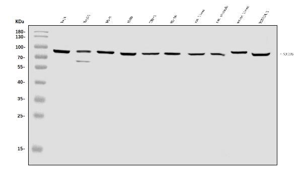
 FCM/FACS (Flow Cytometry)
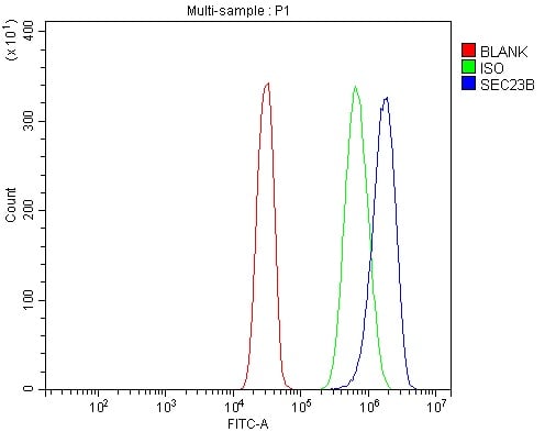
(Figure 2. Flow Cytometry analysis of SiHa cells using anti-SEC23B antibody (AAA125762).Overlay histogram showing SiHa cells stained with AAA125762 (Blue line). The cells were blocked with 10% normal goat serum. And then incubated with rabbit anti-SEC23B Antibody (AAA125762, 1μg/1x106 cells) for 30 min at 20 degree C. DyLight®488 conjugated goat anti-rabbit IgG (5-10μg/1x106 cells) was used as secondary antibody for 30 minutes at 20 degree C. Isotype control antibody (Green line) was rabbit IgG (1μg/1x106) used under the same conditions. Unlabelled sample (Red line) was also used as a control.)
FCM/FACS (Flow Cytometry)
(Figure 2. Flow Cytometry analysis of SiHa cells using anti-SEC23B antibody (AAA125762).Overlay histogram showing SiHa cells stained with AAA125762 (Blue line). The cells were blocked with 10% normal goat serum. And then incubated with rabbit anti-SEC23B Antibody (AAA125762, 1μg/1x106 cells) for 30 min at 20 degree C. DyLight®488 conjugated goat anti-rabbit IgG (5-10μg/1x106 cells) was used as secondary antibody for 30 minutes at 20 degree C. Isotype control antibody (Green line) was rabbit IgG (1μg/1x106) used under the same conditions. Unlabelled sample (Red line) was also used as a control.)
SEC23B, Polyclonal Antibody (Cat# AAA125762)
Full Name
Anti-SEC23B Antibody
Gene Names
SEC23B; CDAII; CDAN2; CDA-II; HEMPAS
Reactivity
Human, Mouse, Rat
Applications
Direct ELISA, Flow Cytometry, Western Blot
Purity
Immunogen affinity purified.
Pricing
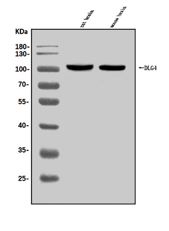
 FCM/FACS (Flow Cytometry)
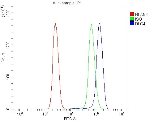
(Figure 2. Flow Cytometry analysis of U937 cells using anti-PSD95/DLG4 antibody (AAA125636).Overlay histogram showing U937 cells stained with AAA125636 (Blue line). The cells were blocked with 10% normal goat serum. And then incubated with rabbit anti-PSD95/DLG4 Antibody (AAA125636, 1μg/1x106 cells) for 30 min at 20 degree C. DyLight®488 conjugated goat anti-rabbit IgG (5-10μg/1x106 cells) was used as secondary antibody for 30 minutes at 20 degree C. Isotype control antibody (Green line) was rabbit IgG (1μg/1x106) used under the same conditions. Unlabelled sample (Red line) was also used as a control.)
FCM/FACS (Flow Cytometry)
(Figure 2. Flow Cytometry analysis of U937 cells using anti-PSD95/DLG4 antibody (AAA125636).Overlay histogram showing U937 cells stained with AAA125636 (Blue line). The cells were blocked with 10% normal goat serum. And then incubated with rabbit anti-PSD95/DLG4 Antibody (AAA125636, 1μg/1x106 cells) for 30 min at 20 degree C. DyLight®488 conjugated goat anti-rabbit IgG (5-10μg/1x106 cells) was used as secondary antibody for 30 minutes at 20 degree C. Isotype control antibody (Green line) was rabbit IgG (1μg/1x106) used under the same conditions. Unlabelled sample (Red line) was also used as a control.)
PSD95/DLG4, Polyclonal Antibody (Cat# AAA125636)
Full Name
Anti-PSD95/DLG4 Antibody
Gene Names
DLG4; PSD95; SAP90; SAP-90
Reactivity
Human, Mouse, Rat
Applications
Direct ELISA, Flow Cytometry, Western Blot
Purity
Immunogen affinity purified.
Pricing
 FCM/FACS (Flow Cytometry)
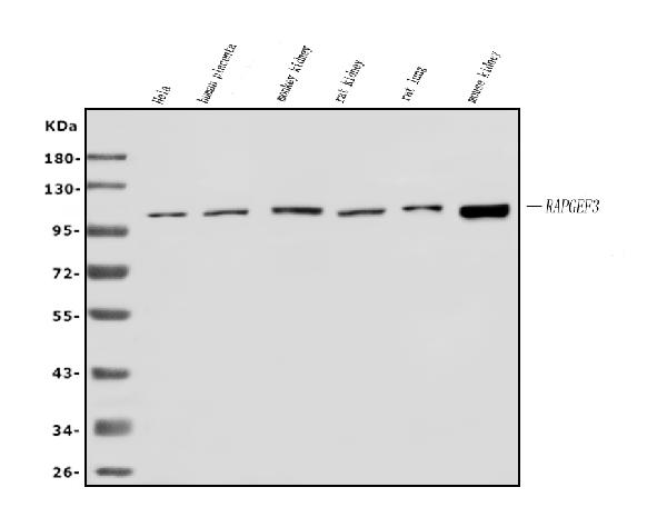
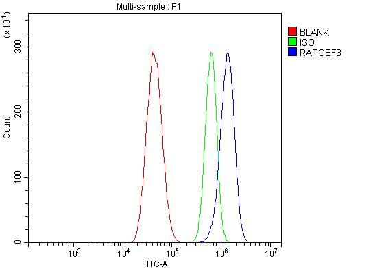
(Figure 2. Flow Cytometry analysis of U87 cells using anti-Epac1/RAPGEF3 antibody (AAA125656).Overlay histogram showing U87 cells stained with AAA125656 (Blue line). The cells were blocked with 10% normal goat serum. And then incubated with rabbit anti-Epac1/RAPGEF3 Antibody (AAA125656, 1μg/1x106 cells) for 30 min at 20 degree C. DyLight®488 conjugated goat anti-rabbit IgG (5-10μg/1x106 cells) was used as secondary antibody for 30 minutes at 20 degree C. Isotype control antibody (Green line) was rabbit IgG (1μg/1x106) used under the same conditions. Unlabelled sample (Red line) was also used as a control.)
FCM/FACS (Flow Cytometry)
(Figure 2. Flow Cytometry analysis of U87 cells using anti-Epac1/RAPGEF3 antibody (AAA125656).Overlay histogram showing U87 cells stained with AAA125656 (Blue line). The cells were blocked with 10% normal goat serum. And then incubated with rabbit anti-Epac1/RAPGEF3 Antibody (AAA125656, 1μg/1x106 cells) for 30 min at 20 degree C. DyLight®488 conjugated goat anti-rabbit IgG (5-10μg/1x106 cells) was used as secondary antibody for 30 minutes at 20 degree C. Isotype control antibody (Green line) was rabbit IgG (1μg/1x106) used under the same conditions. Unlabelled sample (Red line) was also used as a control.)
Epac1/RAPGEF3, Polyclonal Antibody (Cat# AAA125656)
Full Name
Anti-Epac1/RAPGEF3 Antibody
Gene Names
RAPGEF3; EPAC; EPAC1; bcm910; HSU79275; CAMP-GEFI
Reactivity
Human, Mouse, Rat, Monkey
Applications
Direct ELISA, Flow Cytometry, Western Blot
Purity
Immunogen affinity purified.
Pricing
 Application Data
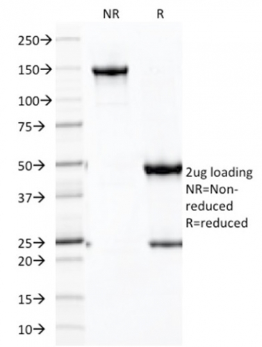
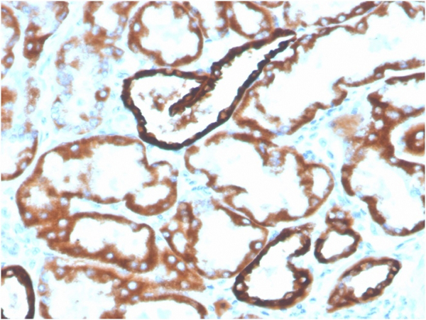
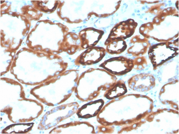
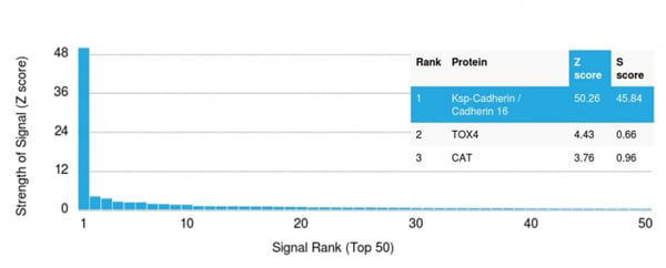
(Analysis of Protein Array containing more than 19,000 full-length human proteins using CDH16-Monospecific Mouse Monoclonal Antibody (CDH16/2125). Z- and S- Score: The Z-score represents the strength of a signal that a monoclonal antibody (MAb) (in combination with a fluorescently-tagged anti-IgG secondary antibody) produces when binding to a particular protein on the HuProtTM array. Z-scores are described in units of standard deviations (SD's) above the mean value of all signals generated on that array. If targets on HuProtTM are arranged in descending order of the Z-score, the S-score is the difference (also in units of SD's) between the Z-score. S-score therefore represents the relative target specificity of a MAb to its intended target. A MAb is considered to specific to its intended target, if the MAb has an S-score of at least 2.5. For example, if a MAb binds to protein X with a Z-score of 43 and to protein Y with a Z-score of 14, then the S-score for the binding of that MAb to protein X is equal to 29.)
Application Data
(Analysis of Protein Array containing more than 19,000 full-length human proteins using CDH16-Monospecific Mouse Monoclonal Antibody (CDH16/2125). Z- and S- Score: The Z-score represents the strength of a signal that a monoclonal antibody (MAb) (in combination with a fluorescently-tagged anti-IgG secondary antibody) produces when binding to a particular protein on the HuProtTM array. Z-scores are described in units of standard deviations (SD's) above the mean value of all signals generated on that array. If targets on HuProtTM are arranged in descending order of the Z-score, the S-score is the difference (also in units of SD's) between the Z-score. S-score therefore represents the relative target specificity of a MAb to its intended target. A MAb is considered to specific to its intended target, if the MAb has an S-score of at least 2.5. For example, if a MAb binds to protein X with a Z-score of 43 and to protein Y with a Z-score of 14, then the S-score for the binding of that MAb to protein X is equal to 29.)
Ksp-Cadherin/CDH16, Monoclonal Antibody (Cat# AAA215475)
Full Name
Ksp-Cadherin/CDH16 (Renal Cell Marker)
Reactivity
Human
Applications
Western Blot, Immunohistochemistry
Purity
Purified Ab conjugated to Biotin
Pricing
 IHC (Immunohiostchemistry)
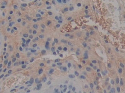
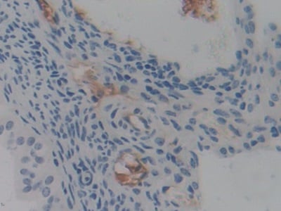
(DAB staining on IHCP;Samples: Mouse Ovary Tissue;Primary Ab: 10ug/ml Rabbit AntiMouse PTX3 AntibodySecond Ab: 2ug/mL HRPLinked Caprine AntiRabbit IgG Polyclonal Antibody(Catalog: SAA544Rb19))
IHC (Immunohiostchemistry)
(DAB staining on IHCP;Samples: Mouse Ovary Tissue;Primary Ab: 10ug/ml Rabbit AntiMouse PTX3 AntibodySecond Ab: 2ug/mL HRPLinked Caprine AntiRabbit IgG Polyclonal Antibody(Catalog: SAA544Rb19))
Pentraxin 3, Long (PTX3), Polyclonal Antibody (Cat# AAA151972)
Full Name
Polyclonal Antibody to Pentraxin 3, Long (PTX3)
Gene Names
PTX3; TSG-14; TNFAIP5
Reactivity
Human, Mouse
Applications
Immunoprecipitation, Immunocytochemistry, Immunohistochemistry, Western Blot
Purity
Affinity Chromatography
Pricing
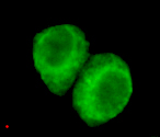
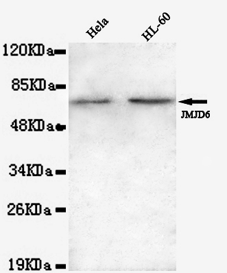 ICC (Immunocytochemistry)
(Immunocytochemistry of HeLa cells using anti-JMJD6(N-terminus) mouse mAb diluted 1:200.)
ICC (Immunocytochemistry)
(Immunocytochemistry of HeLa cells using anti-JMJD6(N-terminus) mouse mAb diluted 1:200.)
JMJD6, Monoclonal Antibody (Cat# AAA320291)
Full Name
JMJD6 (N-term) Mouse mAb
Gene Names
JMJD6; PSR; PTDSR; PTDSR1
Reactivity
Human
Applications
Immunocytochemistry, Western Blot
Purity
The antibody was affinity-purified from mouse ascites by affinity-chromatography using epitope-specific immunogen.
Pricing
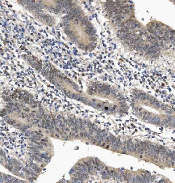
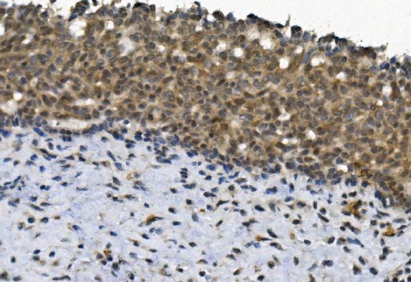
 FCM/FACS (Flow Cytometry)
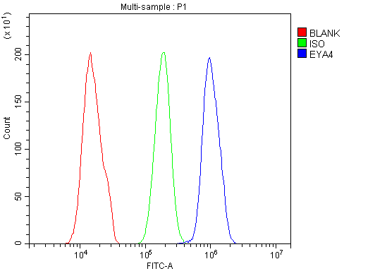
(Figure 4. Flow Cytometry analysis of HELA cells using anti-EYA4 antibody (AAA125725).Overlay histogram showing HELA cells stained with AAA125725 (Blue line). The cells were blocked with 10% normal goat serum. And then incubated with rabbit anti-EYA4 Antibody (AAA125725, 1μg/1x106 cells) for 30 min at 20 degree C. DyLight®488 conjugated goat anti-rabbit IgG (5-10μg/1x106 cells) was used as secondary antibody for 30 minutes at 20 degree C. Isotype control antibody (Green line) was rabbit IgG (1μg/1x106) used under the same conditions. Unlabelled sample (Red line) was also used as a control.)
FCM/FACS (Flow Cytometry)
(Figure 4. Flow Cytometry analysis of HELA cells using anti-EYA4 antibody (AAA125725).Overlay histogram showing HELA cells stained with AAA125725 (Blue line). The cells were blocked with 10% normal goat serum. And then incubated with rabbit anti-EYA4 Antibody (AAA125725, 1μg/1x106 cells) for 30 min at 20 degree C. DyLight®488 conjugated goat anti-rabbit IgG (5-10μg/1x106 cells) was used as secondary antibody for 30 minutes at 20 degree C. Isotype control antibody (Green line) was rabbit IgG (1μg/1x106) used under the same conditions. Unlabelled sample (Red line) was also used as a control.)
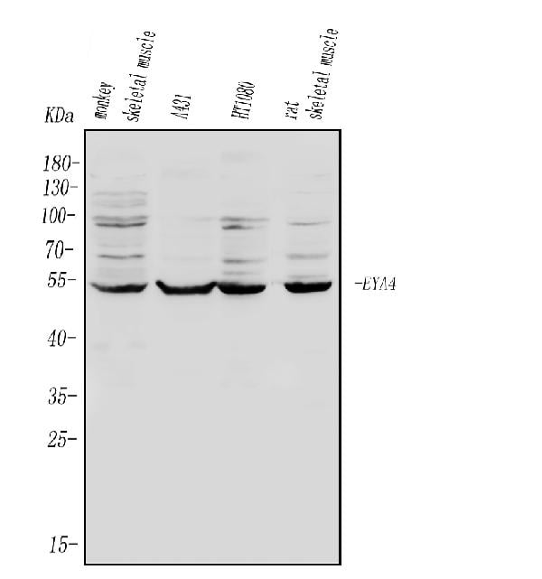
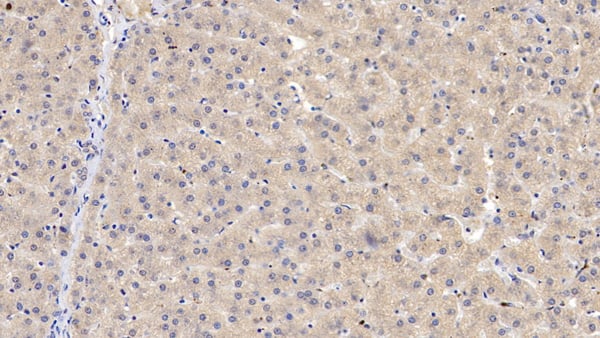
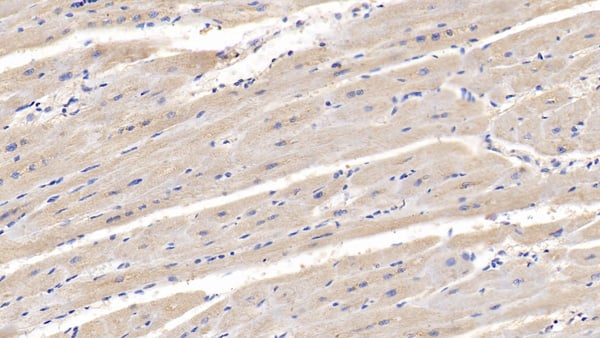
EYA4, Polyclonal Antibody (Cat# AAA125725)
Full Name
Anti-EYA4 Antibody
Gene Names
EYA4; CMD1J; DFNA10
Reactivity
Human, Monkey, Rat
Applications
Direct ELISA, Flow Cytometry, Immunohistochemistry, Western Blot
Purity
Immunogen affinity purified.
Pricing
 IHC (Immunohistochemistry)
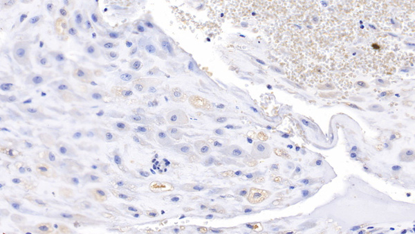
(DAB staining on IHCP;Sample: Human Stomach Tissue; Primary Ab: 10ug/ml Mouse AntiHuman LSR AntibodySecond Ab: 2ug/mL HRPLinked Caprine AntiMouse IgG Polyclonal Antibody(Catalog: SAA544Mu19))
IHC (Immunohistochemistry)
(DAB staining on IHCP;Sample: Human Stomach Tissue; Primary Ab: 10ug/ml Mouse AntiHuman LSR AntibodySecond Ab: 2ug/mL HRPLinked Caprine AntiMouse IgG Polyclonal Antibody(Catalog: SAA544Mu19))
Lipolysis Stimulated Lipoprotein Receptor (LSR), Monoclonal Antibody (Cat# AAA151841)
Full Name
Monoclonal Antibody to Lipolysis Stimulated Lipoprotein Receptor (LSR)
Gene Names
LSR; ILDR3; LISCH7
Reactivity
Human, Pig
Applications
Immunoprecipitation, Immunocytochemistry, Immunohistochemistry, Western Blot
Purity
Protein A + Protein G affinity chromatography
Pricing
 IHC (Immunohistochemisry)
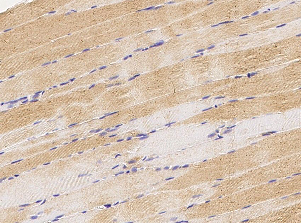
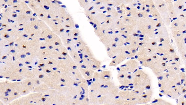
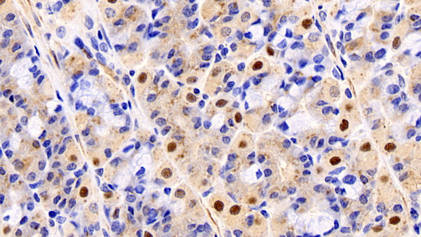
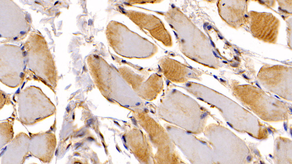
(DAB staining on IHCP;Sample: Human Skeletal muscle Tissue; Primary Ab: 30ug/ml Mouse AntiHuman NTProANP AntibodySecond Ab: 2ug/mL HRPLinked Caprine AntiMouse IgG Polyclonal Antibody(Catalog: SAA544Mu19))
IHC (Immunohistochemisry)
(DAB staining on IHCP;Sample: Human Skeletal muscle Tissue; Primary Ab: 30ug/ml Mouse AntiHuman NTProANP AntibodySecond Ab: 2ug/mL HRPLinked Caprine AntiMouse IgG Polyclonal Antibody(Catalog: SAA544Mu19))
NTerminal ProAtrial Natriuretic Peptide (NTProANP), Monoclonal Antibody (Cat# AAA151546)
Full Name
Monoclonal Antibody to NTerminal ProAtrial Natriuretic Peptide (NTProANP)
Gene Names
NPPA; ANF; ANP; PND; ATFB6; CDD-ANF
Reactivity
Human, Pig
Applications
Immunoprecipitation, Immunocytochemistry, Immunohistochemistry, Western Blot
Purity
Protein A + Protein G affinity chromatography
Pricing
 FCM/FACS (Flow Cytometry)
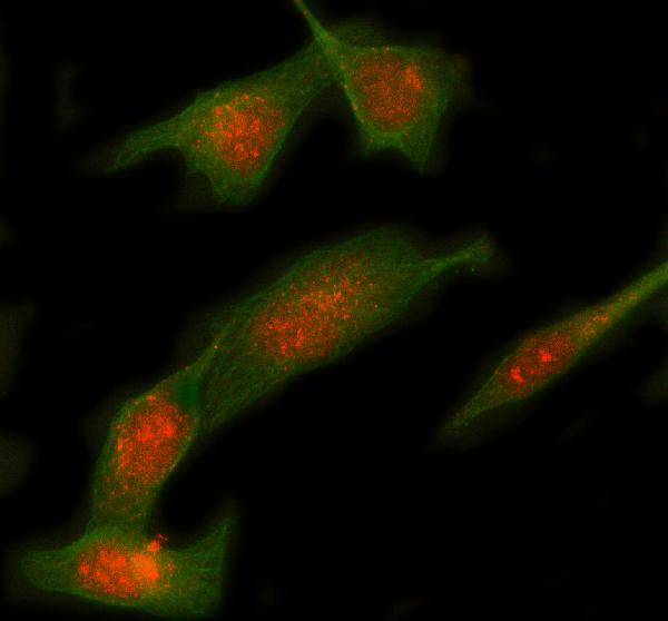
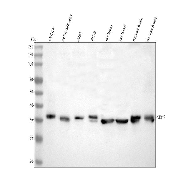
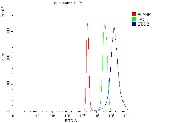
(Figure 3. Flow Cytometry analysis of 293T cells using anti-Syntaxin 12/STX12 antibody (AAA127574).Overlay histogram showing 293T cells stained with AAA127574 (Blue line). To facilitate intracellular staining, cells were fixed with 4% paraformaldehyde and permeabilized with permeabilization buffer. The cells were blocked with 10% normal goat serum. And then incubated with rabbit anti-Syntaxin 12/STX12 Antibody (AAA127574, 1ug/1x106 cells) for 30 min at 20 degree C. DyLight488 conjugated goat anti-rabbit IgG was used as secondary antibody for 30 minutes at 20 degree C. Isotype control antibody (Green line) was rabbit IgG (1ug/1x106) used under the same conditions. Unlabelled sample (Red line) was also used as a control.)
FCM/FACS (Flow Cytometry)
(Figure 3. Flow Cytometry analysis of 293T cells using anti-Syntaxin 12/STX12 antibody (AAA127574).Overlay histogram showing 293T cells stained with AAA127574 (Blue line). To facilitate intracellular staining, cells were fixed with 4% paraformaldehyde and permeabilized with permeabilization buffer. The cells were blocked with 10% normal goat serum. And then incubated with rabbit anti-Syntaxin 12/STX12 Antibody (AAA127574, 1ug/1x106 cells) for 30 min at 20 degree C. DyLight488 conjugated goat anti-rabbit IgG was used as secondary antibody for 30 minutes at 20 degree C. Isotype control antibody (Green line) was rabbit IgG (1ug/1x106) used under the same conditions. Unlabelled sample (Red line) was also used as a control.)
Syntaxin 12/STX12, Polyclonal Antibody (Cat# AAA127574)
Full Name
Anti-Syntaxin 12/STX12 Antibody Picoband
Gene Names
STX12; STX13; STX14
Reactivity
Human, Mouse, Rat
Applications
Flow Cytometry, Immunofluorescence, Immunocytochemistry, Western Blot
Purity
Immunogen affinity purified.
Pricing
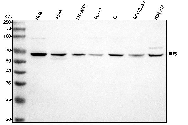
 FCM/FACS (Flow Cytometry)
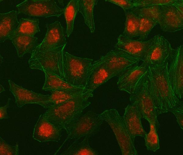
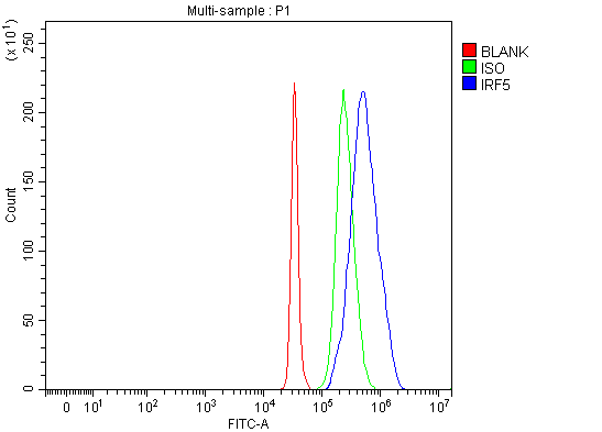
(Figure 3. Flow Cytometry analysis of SH-SY5Y cells using anti-IRF5 antibody (AAA127023).Overlay histogram showing SH-SY5Y cells stained with AAA127023 (Blue line). To facilitate intracellular staining, cells were fixed with 4% paraformaldehyde and permeabilized with permeabilization buffer. The cells were blocked with 10% normal goat serum. And then incubated with rabbit anti-IRF5 Antibody (AAA127023, 1ug/1x106 cells) for 30 min at 20 degree C. DyLight488 conjugated goat anti-rabbit IgG was used as secondary antibody for 30 minutes at 20 degree C. Isotype control antibody (Green line) was rabbit IgG (1ug/1x106) used under the same conditions. Unlabelled sample (Red line) was also used as a control.)
FCM/FACS (Flow Cytometry)
(Figure 3. Flow Cytometry analysis of SH-SY5Y cells using anti-IRF5 antibody (AAA127023).Overlay histogram showing SH-SY5Y cells stained with AAA127023 (Blue line). To facilitate intracellular staining, cells were fixed with 4% paraformaldehyde and permeabilized with permeabilization buffer. The cells were blocked with 10% normal goat serum. And then incubated with rabbit anti-IRF5 Antibody (AAA127023, 1ug/1x106 cells) for 30 min at 20 degree C. DyLight488 conjugated goat anti-rabbit IgG was used as secondary antibody for 30 minutes at 20 degree C. Isotype control antibody (Green line) was rabbit IgG (1ug/1x106) used under the same conditions. Unlabelled sample (Red line) was also used as a control.)
IRF5, Polyclonal Antibody (Cat# AAA127023)
Full Name
Anti-IRF5 Antibody Picoband
Gene Names
IRF5; SLEB10
Reactivity
Human, Mouse, Rat
Applications
Flow Cytometry, Immunofluorescence, Immunocytochemistry, Western Blot
Purity
Immunogen affinity purified.
Pricing
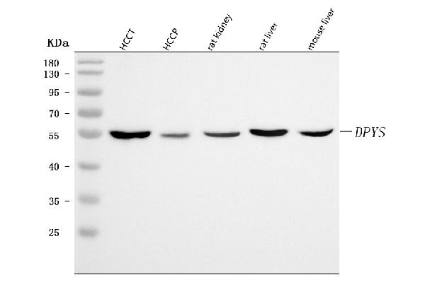
 FCM/FACS (Flow Cytometry)
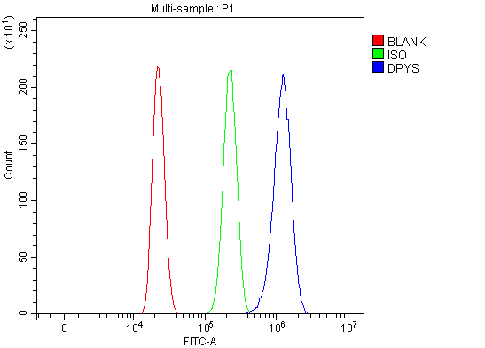
(Figure 2. Flow Cytometry analysis of HL-60 cells using anti-DPYS antibody (AAA127597).Overlay histogram showing HL-60 cells stained with AAA127597 (Blue line). To facilitate intracellular staining, cells were fixed with 4% paraformaldehyde and permeabilized with permeabilization buffer. The cells were blocked with 10% normal goat serum. And then incubated with rabbit anti-DPYS Antibody (AAA127597, 1ug/1x106 cells) for 30 min at 20 degree C. DyLight488 conjugated goat anti-rabbit IgG was used as secondary antibody for 30 minutes at 20 degree C. Isotype control antibody (Green line) was rabbit IgG (1ug/1x106) used under the same conditions. Unlabelled sample without incubation with primary antibody and secondary antibody (Red line) was used as a blank control.)
FCM/FACS (Flow Cytometry)
(Figure 2. Flow Cytometry analysis of HL-60 cells using anti-DPYS antibody (AAA127597).Overlay histogram showing HL-60 cells stained with AAA127597 (Blue line). To facilitate intracellular staining, cells were fixed with 4% paraformaldehyde and permeabilized with permeabilization buffer. The cells were blocked with 10% normal goat serum. And then incubated with rabbit anti-DPYS Antibody (AAA127597, 1ug/1x106 cells) for 30 min at 20 degree C. DyLight488 conjugated goat anti-rabbit IgG was used as secondary antibody for 30 minutes at 20 degree C. Isotype control antibody (Green line) was rabbit IgG (1ug/1x106) used under the same conditions. Unlabelled sample without incubation with primary antibody and secondary antibody (Red line) was used as a blank control.)
DPYS, Polyclonal Antibody (Cat# AAA127597)
Full Name
Anti-DPYS Antibody Picoband
Gene Names
DPYS; DHP; DHPase
Reactivity
Human, Mouse, Rat
Applications
Flow Cytometry, Western Blot
Purity
Immunogen affinity purified.
Pricing
 IF (Immunofluorescence)
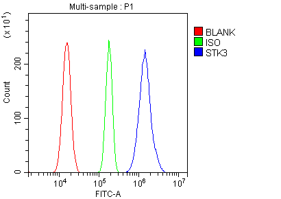
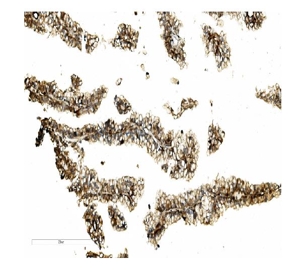
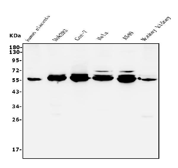
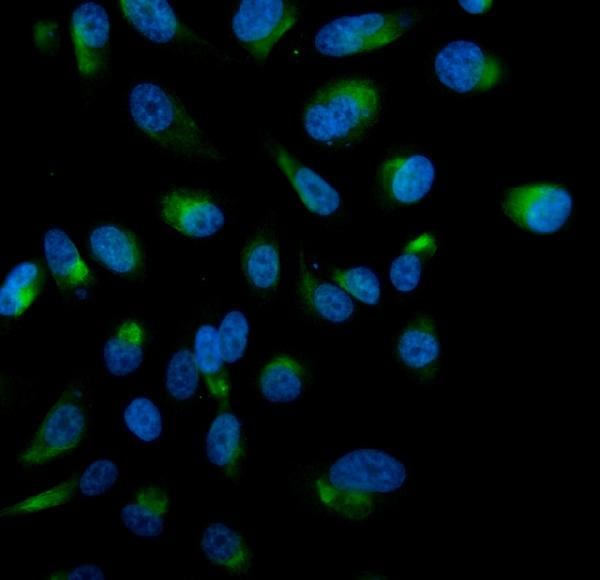
(Figure 4. IF analysis of STK3/MST-2 using anti- STK3/MST-2 antibody (AAA125642).STK3/MST-2 was detected in immunocytochemical section of PC-3 cells. Enzyme antigen retrieval was performed using IHC enzyme antigen retrieval reagent for 15 mins. The cells were blocked with 10% goat serum. And then incubated with 5μg/mL rabbit anti- STK3/MST-2 Antibody (AAA125642) overnight at 4 degree C. DyLight®488 Conjugated Goat Anti-Rabbit IgG was used as secondary antibody at 1:100 dilution and incubated for 30 minutes at 37 degree C. The section was counterstained with DAPI. Visualize using a fluorescence microscope and filter sets appropriate for the label used.)
IF (Immunofluorescence)
(Figure 4. IF analysis of STK3/MST-2 using anti- STK3/MST-2 antibody (AAA125642).STK3/MST-2 was detected in immunocytochemical section of PC-3 cells. Enzyme antigen retrieval was performed using IHC enzyme antigen retrieval reagent for 15 mins. The cells were blocked with 10% goat serum. And then incubated with 5μg/mL rabbit anti- STK3/MST-2 Antibody (AAA125642) overnight at 4 degree C. DyLight®488 Conjugated Goat Anti-Rabbit IgG was used as secondary antibody at 1:100 dilution and incubated for 30 minutes at 37 degree C. The section was counterstained with DAPI. Visualize using a fluorescence microscope and filter sets appropriate for the label used.)
STK3/MST-2, Polyclonal Antibody (Cat# AAA125642)
Full Name
Anti-STK3/MST-2 Antibody
Gene Names
STK3; KRS1; MST2
Reactivity
Human, Monkey
Applications
Direct ELISA, Flow Cytometry, Immunofluorescence, Immunocytochemistry, Immunohistochemistry, Western Blot
Purity
Immunogen affinity purified.
Pricing
 IHC (Immunohiostchemistry)
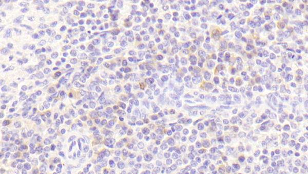
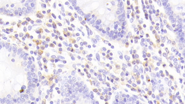
(DAB staining on IHCP;Sample: Human Colon Tissue; Primary Ab: 20ug/ml Rabbit AntiHuman IL8Rb AntibodySecond Ab: 2ug/mL HRPLinked Caprine AntiRabbit IgG Polyclonal Antibody(Catalog: SAA544Rb19))
IHC (Immunohiostchemistry)
(DAB staining on IHCP;Sample: Human Colon Tissue; Primary Ab: 20ug/ml Rabbit AntiHuman IL8Rb AntibodySecond Ab: 2ug/mL HRPLinked Caprine AntiRabbit IgG Polyclonal Antibody(Catalog: SAA544Rb19))
Interleukin 8 Receptor Beta (IL8Rb), Polyclonal Antibody (Cat# AAA151956)
Full Name
Polyclonal Antibody to Interleukin 8 Receptor Beta (IL8Rb)
Gene Names
CXCR2; CD182; IL8R2; IL8RA; IL8RB; CMKAR2; CDw128b
Reactivity
Human, Pig
Applications
Immunoprecipitation, Immunocytochemistry, Immunohistochemistry, Western Blot
Purity
Antigenspecific affinity chromatography followed by Protein A affinity chromatography
Pricing