Filters
▼Clonality
▼Type
▼Reactivity
▼Gene Name
▼Isotype
▼Host
▼Application
▼Clone
▼Monoclonal Antibodies
Get accurate results in your research with our Monoclonal Antibodies, which are specially made to target exactly what you require for your research, and will produce consistent, reliable performance in lab tests.
Viewing 200-250 of 27645 product results
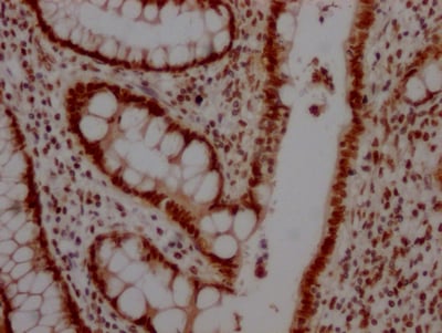
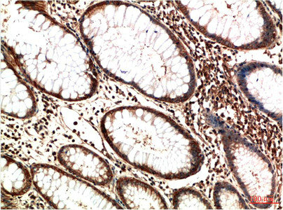
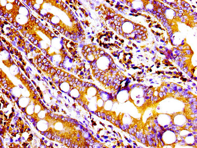
 IHC (Immunohiostchemistry)
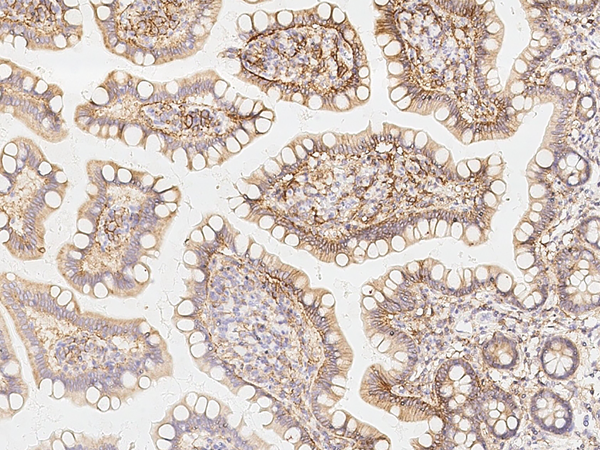
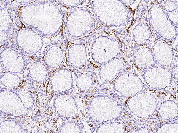
(Immunochemical staining of human SLC3A2 in human small intestine with mouse monoclonal antibody at 1:1000 dilution, formalin-fixed paraffin embedded sections.)
IHC (Immunohiostchemistry)
(Immunochemical staining of human SLC3A2 in human small intestine with mouse monoclonal antibody at 1:1000 dilution, formalin-fixed paraffin embedded sections.)
CD98, Monoclonal Antibody (Cat# AAA255536)
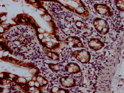
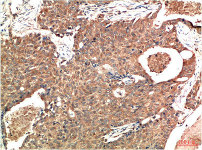
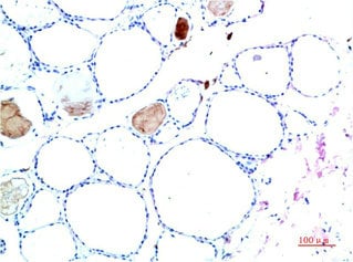
 IHC (Immunohistochemisry)
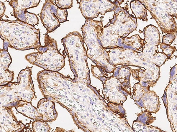
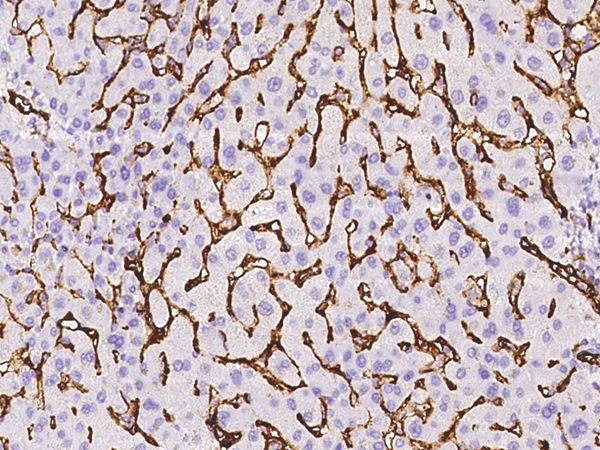
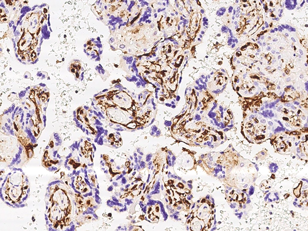
(Immunochemical staining of human CD36 in human placenta with rabbit monoclonal antibody at 1:200 dilution, formalin-fixed paraffin embedded sections.)
IHC (Immunohistochemisry)
(Immunochemical staining of human CD36 in human placenta with rabbit monoclonal antibody at 1:200 dilution, formalin-fixed paraffin embedded sections.)
CD36, Monoclonal Antibody (Cat# AAA255140)
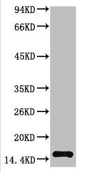
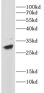 WB (Western Blot)
(MOLT-4 cell lysates were subjected to SDS PAGE followed by western blot with (CD8 antibody) at dilution of 1:1000)
WB (Western Blot)
(MOLT-4 cell lysates were subjected to SDS PAGE followed by western blot with (CD8 antibody) at dilution of 1:1000)
CD8, Monoclonal Antibody (Cat# AAA250101)
>=95% as determined by SDS-PAGE
progesterone, Monoclonal Antibody (Cat# AAA250102)
> 95% as determined by SDS-PAGE
 WB (Western Blot)
(Hela cells were subjected to SDS PAGE followed by western blot with (ANXA1 antibody) at dilution of 1:1000)
WB (Western Blot)
(Hela cells were subjected to SDS PAGE followed by western blot with (ANXA1 antibody) at dilution of 1:1000)
Annexin A1, Monoclonal Antibody (Cat# AAA250106)
>=95% as determined by SDS-PAGE
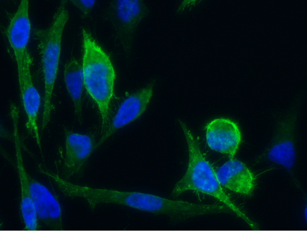 IF (Immunofluorescence)
(Immunofluorescence staining of Human CD171 in Hela cells. Cells were fixed with 4% PFA, blocked with 10% serum, and incubated with rabbit anti-Human CD171 monoclonal antibody (1:60) at 37 degree C 1 hour. Then cells were stained with the Alexa Fluor 488-conjugated Goat Anti-rabbit IgG secondary antibody (green) and counterstained with DAPI (blue). Positive staining was localized to cells membrane.)
IF (Immunofluorescence)
(Immunofluorescence staining of Human CD171 in Hela cells. Cells were fixed with 4% PFA, blocked with 10% serum, and incubated with rabbit anti-Human CD171 monoclonal antibody (1:60) at 37 degree C 1 hour. Then cells were stained with the Alexa Fluor 488-conjugated Goat Anti-rabbit IgG secondary antibody (green) and counterstained with DAPI (blue). Positive staining was localized to cells membrane.)
L1CAM, Monoclonal Antibody (Cat# AAA254552)
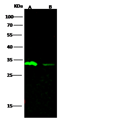
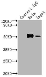
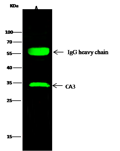 IP (Immunoprecipitation)
(CA3 was immunoprecipitated using:Lane A:0.5 mg HepG2 Whole Cell Lysate2 uL anti-CA3 rabbit monoclonal antibody and 15 ul of 50 % Protein G agarose.Primary antibody:Anti-CA3 rabbit monoclonal antibody,at 1:250 dilutionSecondary antibody:Dylight 800-labeled antibody to rabbit IgG (H+L), at 1:5000 dilutionDeveloped using the odssey technique.Performed under reducing conditions.Predicted band size: 30 kDaObserved band size: 30 kDa)
IP (Immunoprecipitation)
(CA3 was immunoprecipitated using:Lane A:0.5 mg HepG2 Whole Cell Lysate2 uL anti-CA3 rabbit monoclonal antibody and 15 ul of 50 % Protein G agarose.Primary antibody:Anti-CA3 rabbit monoclonal antibody,at 1:250 dilutionSecondary antibody:Dylight 800-labeled antibody to rabbit IgG (H+L), at 1:5000 dilutionDeveloped using the odssey technique.Performed under reducing conditions.Predicted band size: 30 kDaObserved band size: 30 kDa)
Carbonic Anhydrase III, Monoclonal Antibody (Cat# AAA254969)
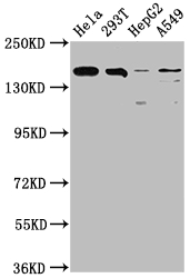
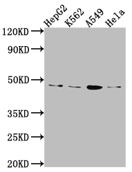
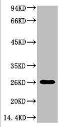 WB (Western Blot)
(Western blot analysis of Hela, diluted at 1:3000.)
WB (Western Blot)
(Western blot analysis of Hela, diluted at 1:3000.)
LGALS3, Monoclonal Antibody (Cat# AAA243570)

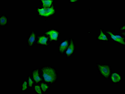
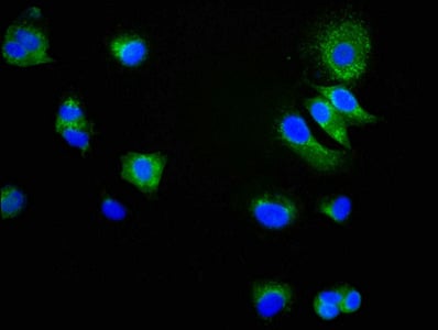
 IF (Immunofluorescence)
(Immunofluorescence staining of Hela Cells at 1?50, counter-stained with DAPI. The cells were fixed in 4% formaldehyde, permeated by 0.2% TritonX-100, and blocked in 10% normal Goat Serum. The cells were then incubated with the antibody overnight at 4 degree C. Nuclear DNA was labeled in blue with DAPI. The secondary antibody was FITC-conjugated AffiniPure Goat Anti-Rabbit IgG ?H+L?.)
IF (Immunofluorescence)
(Immunofluorescence staining of Hela Cells at 1?50, counter-stained with DAPI. The cells were fixed in 4% formaldehyde, permeated by 0.2% TritonX-100, and blocked in 10% normal Goat Serum. The cells were then incubated with the antibody overnight at 4 degree C. Nuclear DNA was labeled in blue with DAPI. The secondary antibody was FITC-conjugated AffiniPure Goat Anti-Rabbit IgG ?H+L?.)
CYP17A1, Monoclonal Recombinant Antibody (Cat# AAA243914)
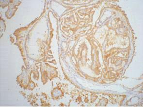
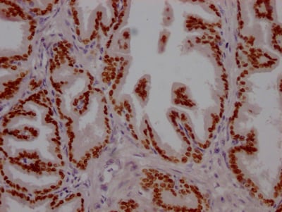
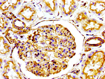
 IHC (Immunohiostchemistry)
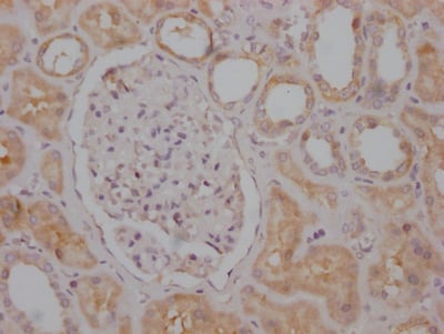
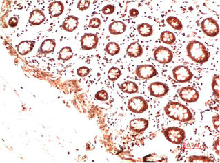
(IHC image diluted at 1:100 and staining in paraffin-embedded human kidney tissue performed on a Leica BondTM system. After dewaxing and hydration, antigen retrieval was mediated by high pressure in a citrate buffer (pH 6.0). Section was blocked with 10% normal goat serum 30min at RT. Then primary antibody (1% BSA) was incubated at 4 degree C overnight. The primary is detected by a Goat anti-rabbit IgG polymer labeled by HRP and visualized using 0.05% DAB.)
IHC (Immunohiostchemistry)
(IHC image diluted at 1:100 and staining in paraffin-embedded human kidney tissue performed on a Leica BondTM system. After dewaxing and hydration, antigen retrieval was mediated by high pressure in a citrate buffer (pH 6.0). Section was blocked with 10% normal goat serum 30min at RT. Then primary antibody (1% BSA) was incubated at 4 degree C overnight. The primary is detected by a Goat anti-rabbit IgG polymer labeled by HRP and visualized using 0.05% DAB.)
RPTOR, Monoclonal Recombinant Antibody (Cat# AAA243927)
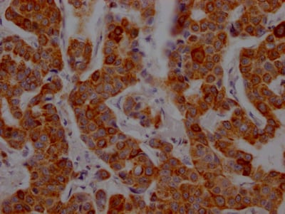
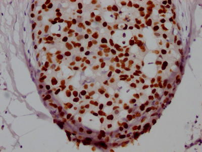
 IHC (Immunohiostchemistry)
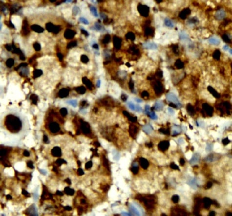
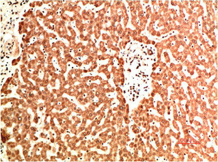
(IHC image diluted at 1:100 and staining in paraffin-embedded human liver cancer performed on a Leica BondTM system. After dewaxing and hydration, antigen retrieval was mediated by high pressure in a citrate buffer (pH 6.0). Section was blocked with 10% normal goat serum 30min at RT. Then primary antibody (1% BSA) was incubated at 4 degree C overnight. The primary is detected by a Goat anti-rabbit IgG polymer labeled by HRP and visualized using 0.05% DAB.)
IHC (Immunohiostchemistry)
(IHC image diluted at 1:100 and staining in paraffin-embedded human liver cancer performed on a Leica BondTM system. After dewaxing and hydration, antigen retrieval was mediated by high pressure in a citrate buffer (pH 6.0). Section was blocked with 10% normal goat serum 30min at RT. Then primary antibody (1% BSA) was incubated at 4 degree C overnight. The primary is detected by a Goat anti-rabbit IgG polymer labeled by HRP and visualized using 0.05% DAB.)
SOD2, Monoclonal Recombinant Antibody (Cat# AAA243937)
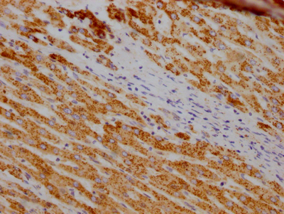
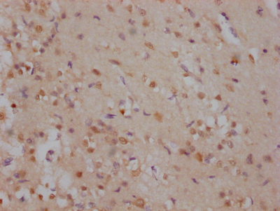
 IHC (Immunohiostchemistry)
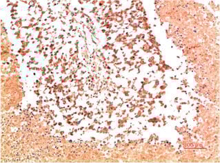
(IHC image diluted at 1:100 and staining in paraffin-embedded human brain tissue performed on a Leica BondTM system. After dewaxing and hydration, antigen retrieval was mediated by high pressure in a citrate buffer (pH 6.0). Section was blocked with 10% normal goat serum 30min at RT. Then primary antibody (1% BSA) was incubated at 4 degree C overnight. The primary is detected by a Goat anti-rabbit IgG polymer labeled by HRP and visualized using 0.05% DAB.)
IHC (Immunohiostchemistry)
(IHC image diluted at 1:100 and staining in paraffin-embedded human brain tissue performed on a Leica BondTM system. After dewaxing and hydration, antigen retrieval was mediated by high pressure in a citrate buffer (pH 6.0). Section was blocked with 10% normal goat serum 30min at RT. Then primary antibody (1% BSA) was incubated at 4 degree C overnight. The primary is detected by a Goat anti-rabbit IgG polymer labeled by HRP and visualized using 0.05% DAB.)
FYN, Monoclonal Recombinant Antibody (Cat# AAA243940)
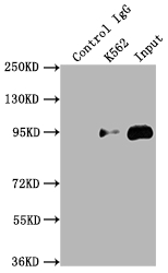 IP (Immunoprecipitation)
(Immunoprecipitating TOP1 in K562 whole cell lysateLane 1: Rabbit control IgG instead of in K562 whole cell lysate. For western blotting,a HRP-conjugated Protein G antibody was used as the secondary antibody (1/2000)Lane 2: K562 whole cell lysate?500ug?Lane 3: K562 whole cell lysate (10ug))
IP (Immunoprecipitation)
(Immunoprecipitating TOP1 in K562 whole cell lysateLane 1: Rabbit control IgG instead of in K562 whole cell lysate. For western blotting,a HRP-conjugated Protein G antibody was used as the secondary antibody (1/2000)Lane 2: K562 whole cell lysate?500ug?Lane 3: K562 whole cell lysate (10ug))
TOP1, Monoclonal Recombinant Antibody (Cat# AAA243952)
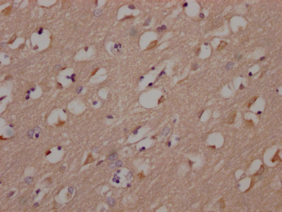
 IHC (Immunohiostchemistry)
(IHC image diluted at 1:100 and staining in paraffin-embedded human liver cancer performed on a Leica BondTM system. After dewaxing and hydration, antigen retrieval was mediated by high pressure in a citrate buffer (pH 6.0). Section was blocked with 10% normal goat serum 30min at RT. Then primary antibody (1% BSA) was incubated at 4 degree C overnight. The primary is detected by a Goat anti-rabbit IgG polymer labeled by HRP and visualized using 0.05% DAB.)
IHC (Immunohiostchemistry)
(IHC image diluted at 1:100 and staining in paraffin-embedded human liver cancer performed on a Leica BondTM system. After dewaxing and hydration, antigen retrieval was mediated by high pressure in a citrate buffer (pH 6.0). Section was blocked with 10% normal goat serum 30min at RT. Then primary antibody (1% BSA) was incubated at 4 degree C overnight. The primary is detected by a Goat anti-rabbit IgG polymer labeled by HRP and visualized using 0.05% DAB.)
ALDH2, Monoclonal Recombinant Antibody (Cat# AAA243957)
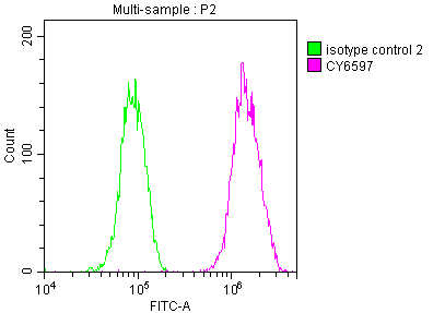
 FCM/FACS (Flow Cytometry)
(Overlay histogram showing Hela cells stained with (red line) at 1?50. The cells were fixed with 70% Ethylalcohol (18h) and then incubated in 10% normal goat serum to block non-specific protein-protein interactions followedby the antibody (1ug/1*106cells) for 1 h at 4?.The secondary antibody used was FITC-conjugated goat anti-rabbit IgG (H+L) at 1/200 dilution for 30min at 4?. Control antibody (green line) was Rabbit IgG (1ug/1*106cells) used under the same conditions. Acquisition of >10,000 events was performed.)
FCM/FACS (Flow Cytometry)
(Overlay histogram showing Hela cells stained with (red line) at 1?50. The cells were fixed with 70% Ethylalcohol (18h) and then incubated in 10% normal goat serum to block non-specific protein-protein interactions followedby the antibody (1ug/1*106cells) for 1 h at 4?.The secondary antibody used was FITC-conjugated goat anti-rabbit IgG (H+L) at 1/200 dilution for 30min at 4?. Control antibody (green line) was Rabbit IgG (1ug/1*106cells) used under the same conditions. Acquisition of >10,000 events was performed.)
E2F1, Monoclonal Recombinant Antibody (Cat# AAA243962)
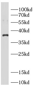
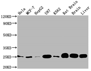
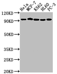
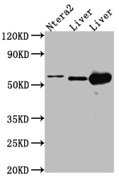
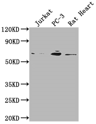
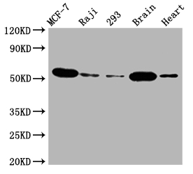
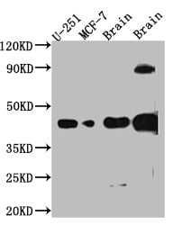
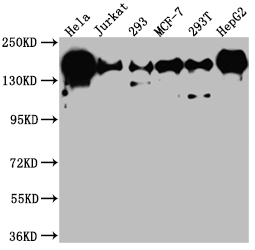
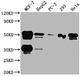
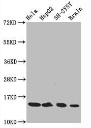
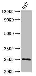
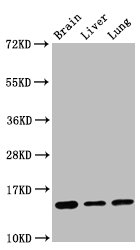
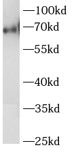
 WB (Western Blot)
(Western BlotPositive WB detected in: Hela whole cell lysate,MCF7 whole cell lysate, 293T whole cell lysate,U87 whole cell lysate, A549 whole cell lysate,Mouse Brain tissue, Rat Brain tissueAll lanes: GBA antibody at 1:500SecondaryGoat polyclonal to rabbit IgG at 1/50000 dilutionPredicted band size: 60 kDaObserved band size: 60 kDa)
WB (Western Blot)
(Western BlotPositive WB detected in: Hela whole cell lysate,MCF7 whole cell lysate, 293T whole cell lysate,U87 whole cell lysate, A549 whole cell lysate,Mouse Brain tissue, Rat Brain tissueAll lanes: GBA antibody at 1:500SecondaryGoat polyclonal to rabbit IgG at 1/50000 dilutionPredicted band size: 60 kDaObserved band size: 60 kDa)
GBA, Monoclonal Recombinant Antibody (Cat# AAA243967)
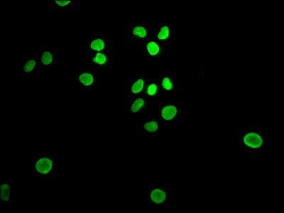
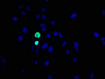
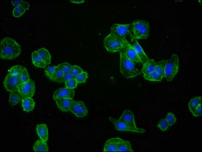
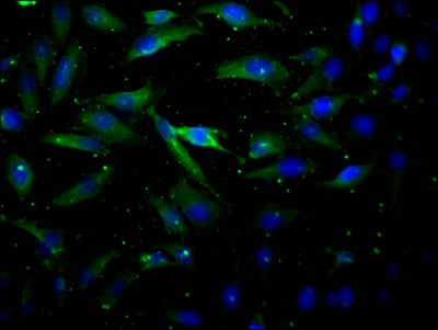 IF (Immunofluorescence)
(Immunofluorescence staining of Hela Cells at 1?50, counter-stained with DAPI. The cells were fixed in 4% formaldehyde, permeated by 0.2% TritonX-100, and blocked in 10% normal Goat Serum. The cells were then incubated with the antibody overnight at 4 degree C. Nuclear DNA was labeled in blue with DAPI. The secondary antibody was FITC-conjugated AffiniPure Goat Anti-Rabbit IgG ?H+L?.)
IF (Immunofluorescence)
(Immunofluorescence staining of Hela Cells at 1?50, counter-stained with DAPI. The cells were fixed in 4% formaldehyde, permeated by 0.2% TritonX-100, and blocked in 10% normal Goat Serum. The cells were then incubated with the antibody overnight at 4 degree C. Nuclear DNA was labeled in blue with DAPI. The secondary antibody was FITC-conjugated AffiniPure Goat Anti-Rabbit IgG ?H+L?.)
AKT1, Monoclonal Recombinant Antibody (Cat# AAA243980)
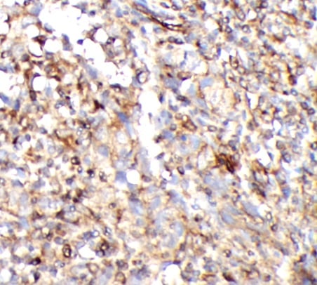
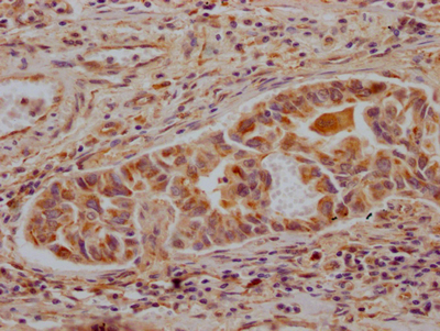
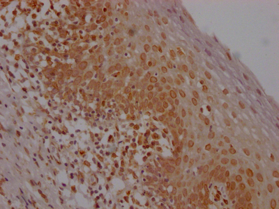
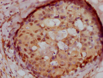
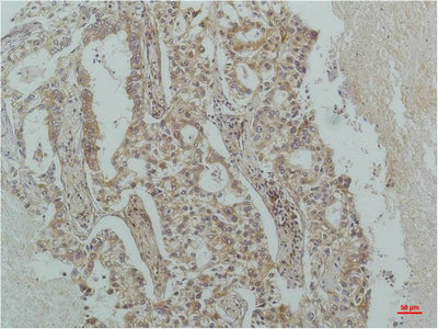
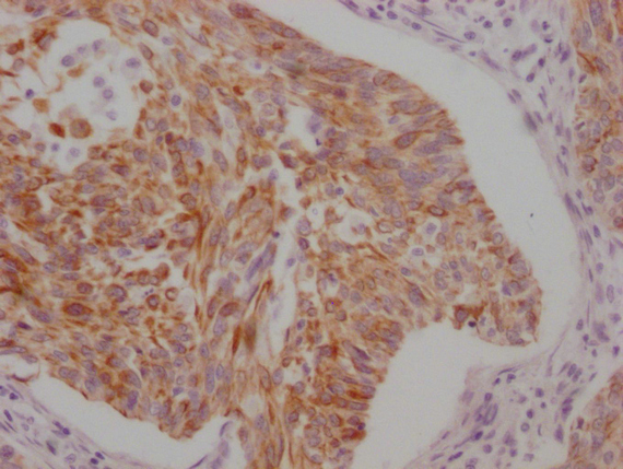
 IHC (Immunohistochemisry)
(IHC image diluted at 1:100 and staining in paraffin-embedded human breast cancer performed on a Leica BondTM system. After dewaxing and hydration, antigen retrieval was mediated by high pressure in a citrate buffer (pH 6.0). Section was blocked with 10% normal goat serum 30min at RT. Then primary antibody (1% BSA) was incubated at 4 degree C overnight. The primary is detected by a Goat anti-rabbit IgG polymer labeled by HRP and visualized using 0.05% DAB.)
IHC (Immunohistochemisry)
(IHC image diluted at 1:100 and staining in paraffin-embedded human breast cancer performed on a Leica BondTM system. After dewaxing and hydration, antigen retrieval was mediated by high pressure in a citrate buffer (pH 6.0). Section was blocked with 10% normal goat serum 30min at RT. Then primary antibody (1% BSA) was incubated at 4 degree C overnight. The primary is detected by a Goat anti-rabbit IgG polymer labeled by HRP and visualized using 0.05% DAB.)
BRD4, Monoclonal Recombinant Antibody (Cat# AAA243803)
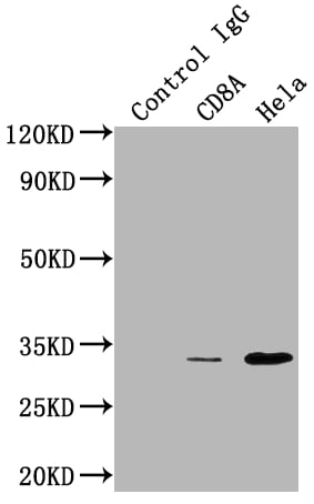 IP (Immunoprecipitation)
(Immunoprecipitating CD8A in HepG2 whole cell lysateLane 1: Rabbit control IgG instead of in HepG2 whole cell lysate. For western blotting,a HRP-conjugated Protein G antibody was used as the secondary antibody (1/2000)Lane 2: 3ug + HepG2 whole cell lysate?500ug?Lane 3: HepG2 whole cell lysate (10ug))
IP (Immunoprecipitation)
(Immunoprecipitating CD8A in HepG2 whole cell lysateLane 1: Rabbit control IgG instead of in HepG2 whole cell lysate. For western blotting,a HRP-conjugated Protein G antibody was used as the secondary antibody (1/2000)Lane 2: 3ug + HepG2 whole cell lysate?500ug?Lane 3: HepG2 whole cell lysate (10ug))
CD8A, Monoclonal Recombinant Antibody (Cat# AAA243987)
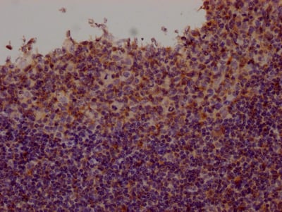
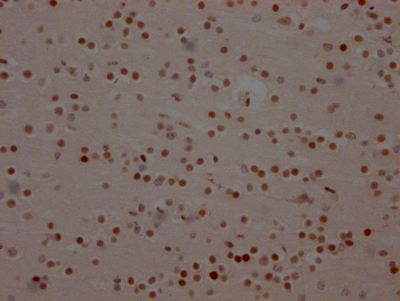
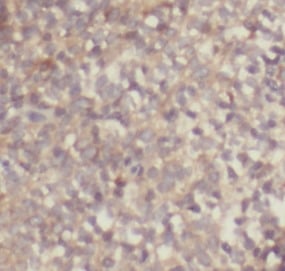
 IHC (Immunohiostchemistry)
(IHC image diluted at 1:100 and staining in paraffin-embedded human brain tissue performed on a Leica BondTM system. After dewaxing and hydration, antigen retrieval was mediated by high pressure in a citrate buffer (pH 6.0). Section was blocked with 10% normal goat serum 30min at RT. Then primary antibody (1% BSA) was incubated at 4 degree C overnight. The primary is detected by a Goat anti-rabbit IgG polymer labeled by HRP and visualized using 0.05% DAB.)
IHC (Immunohiostchemistry)
(IHC image diluted at 1:100 and staining in paraffin-embedded human brain tissue performed on a Leica BondTM system. After dewaxing and hydration, antigen retrieval was mediated by high pressure in a citrate buffer (pH 6.0). Section was blocked with 10% normal goat serum 30min at RT. Then primary antibody (1% BSA) was incubated at 4 degree C overnight. The primary is detected by a Goat anti-rabbit IgG polymer labeled by HRP and visualized using 0.05% DAB.)
SOX2, Monoclonal Recombinant Antibody (Cat# AAA244003)
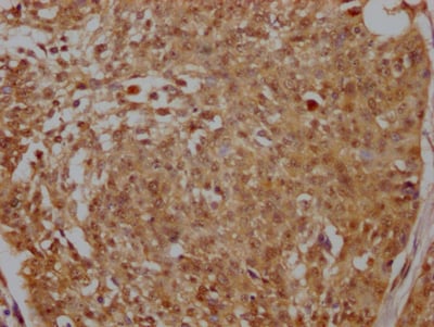
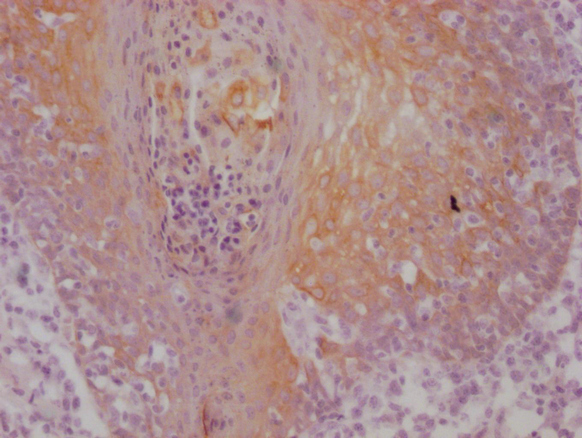
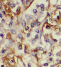
 IHC (Immunohiostchemistry)
(IHC image diluted at 1:100 and staining in paraffin-embedded human lung cancer performed on a Leica BondTM system. After dewaxing and hydration, antigen retrieval was mediated by high pressure in a citrate buffer (pH 6.0). Section was blocked with 10% normal goat serum 30min at RT. Then primary antibody (1% BSA) was incubated at 4 degree C overnight. The primary is detected by a Goat anti-rabbit IgG polymer labeled by HRP and visualized using 0.05% DAB.)
IHC (Immunohiostchemistry)
(IHC image diluted at 1:100 and staining in paraffin-embedded human lung cancer performed on a Leica BondTM system. After dewaxing and hydration, antigen retrieval was mediated by high pressure in a citrate buffer (pH 6.0). Section was blocked with 10% normal goat serum 30min at RT. Then primary antibody (1% BSA) was incubated at 4 degree C overnight. The primary is detected by a Goat anti-rabbit IgG polymer labeled by HRP and visualized using 0.05% DAB.)
CD274, Monoclonal Recombinant Antibody (Cat# AAA244004)
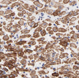
 IHC (Immunohistochemisry)
(IHC image diluted at 1:100 and staining in paraffin-embedded human cervical cancer performed on a Leica BondTM system. After dewaxing and hydration, antigen retrieval was mediated by high pressure in a citrate buffer (pH 6.0). Section was blocked with 10% normal goat serum 30min at RT. Then primary antibody (1% BSA) was incubated at 4 degree C overnight. The primary is detected by a Goat anti-rabbit IgG polymer labeled by HRP and visualized using 0.05% DAB.)
IHC (Immunohistochemisry)
(IHC image diluted at 1:100 and staining in paraffin-embedded human cervical cancer performed on a Leica BondTM system. After dewaxing and hydration, antigen retrieval was mediated by high pressure in a citrate buffer (pH 6.0). Section was blocked with 10% normal goat serum 30min at RT. Then primary antibody (1% BSA) was incubated at 4 degree C overnight. The primary is detected by a Goat anti-rabbit IgG polymer labeled by HRP and visualized using 0.05% DAB.)
PELP1, Monoclonal Recombinant Antibody (Cat# AAA244021)
C-reactive protein (CRP), Monoclonal Antibody (Cat# AAA244051)
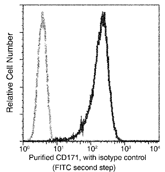
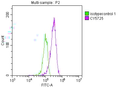
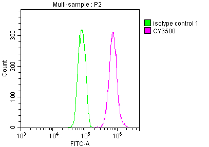
 FCM/FACS (Flow Cytometry)
(Overlay histogram showing A549 cells stained with (red line) at 1?50. The cells were fixed with 70% Ethylalcohol (18h) and then incubated in 10% normal goat serum to block non-specific protein-protein interactions followedby the antibody (1ug/1*106cells) for 1 h at 4?.The secondary antibody used was FITC-conjugated goat anti-rabbit IgG (H+L) at 1/200 dilution for 30min at 4?. Control antibody (green line) was Rabbit IgG (1ug/1*106cells) used under the same conditions. Acquisition of >10,000 events was performed.)
FCM/FACS (Flow Cytometry)
(Overlay histogram showing A549 cells stained with (red line) at 1?50. The cells were fixed with 70% Ethylalcohol (18h) and then incubated in 10% normal goat serum to block non-specific protein-protein interactions followedby the antibody (1ug/1*106cells) for 1 h at 4?.The secondary antibody used was FITC-conjugated goat anti-rabbit IgG (H+L) at 1/200 dilution for 30min at 4?. Control antibody (green line) was Rabbit IgG (1ug/1*106cells) used under the same conditions. Acquisition of >10,000 events was performed.)
FASN, Monoclonal Recombinant Antibody (Cat# AAA243862)
 IP (Immunoprecipitation)
(Immunoprecipitating FOXA1 in Hela whole cell lysateLane 1: Rabbit control IgG instead of in Hela whole cell lysate. For western blotting,a HRP-conjugated Protein G antibody was used as the secondary antibody (1/2000)Lane 2: Hela whole cell lysate?500ug?Lane 3: Hela whole cell lysate (10ug))
IP (Immunoprecipitation)
(Immunoprecipitating FOXA1 in Hela whole cell lysateLane 1: Rabbit control IgG instead of in Hela whole cell lysate. For western blotting,a HRP-conjugated Protein G antibody was used as the secondary antibody (1/2000)Lane 2: Hela whole cell lysate?500ug?Lane 3: Hela whole cell lysate (10ug))
FOXA1, Monoclonal Recombinant Antibody (Cat# AAA243867)

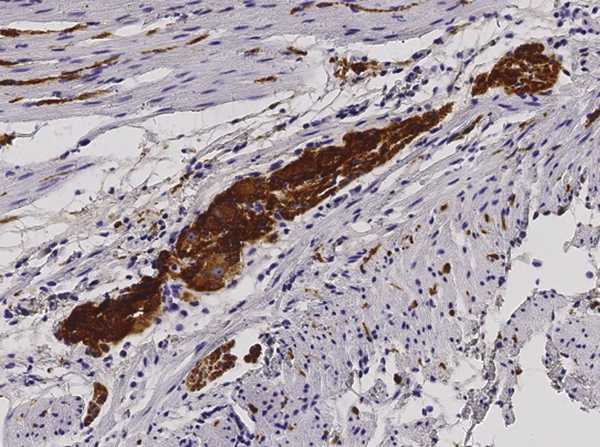
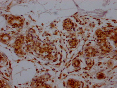
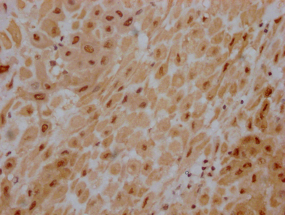
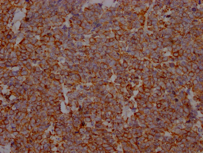
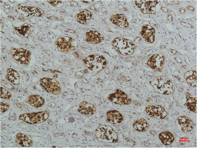
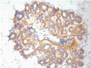
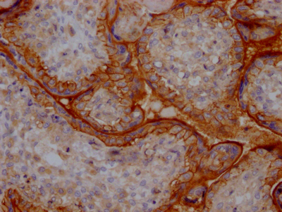 IHC (Immunohiostchemistry)
(IHC image diluted at 1:100 and staining in paraffin-embedded human placenta tissue performed on a Leica BondTM system. After dewaxing and hydration, antigen retrieval was mediated by high pressure in a citrate buffer (pH 6.0). Section was blocked with 10% normal goat serum 30min at RT. Then primary antibody (1% BSA) was incubated at 4 degree C overnight. The primary is detected by a Goat anti-rabbit IgG polymer labeled by HRP and visualized using 0.05% DAB.)
IHC (Immunohiostchemistry)
(IHC image diluted at 1:100 and staining in paraffin-embedded human placenta tissue performed on a Leica BondTM system. After dewaxing and hydration, antigen retrieval was mediated by high pressure in a citrate buffer (pH 6.0). Section was blocked with 10% normal goat serum 30min at RT. Then primary antibody (1% BSA) was incubated at 4 degree C overnight. The primary is detected by a Goat anti-rabbit IgG polymer labeled by HRP and visualized using 0.05% DAB.)
ITGAV, Monoclonal Recombinant Antibody (Cat# AAA243891)
 IHC (Immunohiostchemistry)
(Immunohistochemical analysis of paraffin-embedded Human Kidney Tissue using IkappaB betaMouse mAb diluted at 1:200.)
IHC (Immunohiostchemistry)
(Immunohistochemical analysis of paraffin-embedded Human Kidney Tissue using IkappaB betaMouse mAb diluted at 1:200.)
NFKBIB, Monoclonal Antibody (Cat# AAA243625)
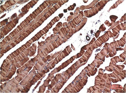 IHC (Immunohistochemisry)
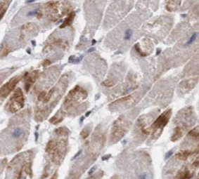
(Immunohistochemical analysis of paraffin-embedded Mouse Skeletal Muscle Tissue using Muscle Actin Mouse mAb diluted at 1:200.)
IHC (Immunohistochemisry)
(Immunohistochemical analysis of paraffin-embedded Mouse Skeletal Muscle Tissue using Muscle Actin Mouse mAb diluted at 1:200.)
Actin, Monoclonal Antibody (Cat# AAA243636)
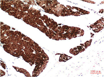
 IHC (Immunohiostchemistry)
(Immunohistochemical analysis of paraffin-embedded Human Breast Carcinoma Tissue using ATM Mouse mAb diluted at 1:200.)
IHC (Immunohiostchemistry)
(Immunohistochemical analysis of paraffin-embedded Human Breast Carcinoma Tissue using ATM Mouse mAb diluted at 1:200.)
ATM, Monoclonal Antibody (Cat# AAA243648)
 WB (Western Blot)
(Western blot analysis of Human Serum using TTR Mouse mAb diluted at 1:2000)
WB (Western Blot)
(Western blot analysis of Human Serum using TTR Mouse mAb diluted at 1:2000)
TTR, Monoclonal Antibody (Cat# AAA243675)
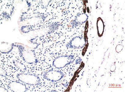
 IHC (Immunohiostchemistry)
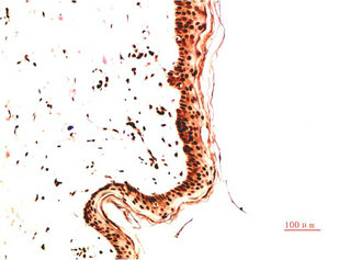
(Immunohistochemical analysis of paraffin-embedded Human Skin Carcinoma Tissue using Collagen IV Mouse mAb diluted at 1:2000)
IHC (Immunohiostchemistry)
(Immunohistochemical analysis of paraffin-embedded Human Skin Carcinoma Tissue using Collagen IV Mouse mAb diluted at 1:2000)
COL4A1, Monoclonal Antibody (Cat# AAA243689)
 IHC (Immunohiostchemistry)
(Immunohistochemical analysis of paraffin-embedded Human Colon Carcinoma Tissue using Collagen II Mouse mAb diluted at 1:2000)
IHC (Immunohiostchemistry)
(Immunohistochemical analysis of paraffin-embedded Human Colon Carcinoma Tissue using Collagen II Mouse mAb diluted at 1:2000)
COL2A1, Monoclonal Antibody (Cat# AAA243693)
 IHC (Immunohiostchemistry)
(IHC image of AAA243708 diluted at 1:100 and staining in paraffin-embedded human tonsil tissue performed on a Leica BondTM system. After dewaxing and hydration, antigen retrieval was mediated by high pressure in a citrate buffer (pH 6.0). Section was blocked with 10% normal goat serum 30min at RT. Then primary antibody (1% BSA) was incubated at 4 degree C overnight. The primary is detected by a Goat anti-mouse IgG polymer labeled by HRP and visualized using 0.05% DAB.)
IHC (Immunohiostchemistry)
(IHC image of AAA243708 diluted at 1:100 and staining in paraffin-embedded human tonsil tissue performed on a Leica BondTM system. After dewaxing and hydration, antigen retrieval was mediated by high pressure in a citrate buffer (pH 6.0). Section was blocked with 10% normal goat serum 30min at RT. Then primary antibody (1% BSA) was incubated at 4 degree C overnight. The primary is detected by a Goat anti-mouse IgG polymer labeled by HRP and visualized using 0.05% DAB.)
KRT5/KRT6A, Monoclonal Antibody (Cat# AAA243708)
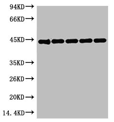 WB (Western Blot)
(Western blot analysis of 1) 293T, 2) Mouse Kidney tissue, 3) Hela, 4) Rat Heart tissue, 5) Rat Brain tissue, diluted at 1:5000.)
WB (Western Blot)
(Western blot analysis of 1) 293T, 2) Mouse Kidney tissue, 3) Hela, 4) Rat Heart tissue, 5) Rat Brain tissue, diluted at 1:5000.)
ACTB, Monoclonal Antibody (Cat# AAA243723)
 Application Data
(Overlay histogram showing A375 cells stained with AAA235164 (red line) at 1:50. The cells were fixed with 70% Ethylalcohol (18h) and then permeabilized with 0.3% Triton X-100 for 2 min.The cells were then incubated in 1x PBS /10% normal goat serum to block non-specific protein-protein interactions followed by primary antibody for 1 h at 4 degree C.The secondary antibody used was FITC goat anti-rabbit IgG (H+L) at 1/200 dilution for 1 h at 4 degree C. Control antibody (green line) was used under the same conditions. Acquisition of >10, 000 events was performed.)
Application Data
(Overlay histogram showing A375 cells stained with AAA235164 (red line) at 1:50. The cells were fixed with 70% Ethylalcohol (18h) and then permeabilized with 0.3% Triton X-100 for 2 min.The cells were then incubated in 1x PBS /10% normal goat serum to block non-specific protein-protein interactions followed by primary antibody for 1 h at 4 degree C.The secondary antibody used was FITC goat anti-rabbit IgG (H+L) at 1/200 dilution for 1 h at 4 degree C. Control antibody (green line) was used under the same conditions. Acquisition of >10, 000 events was performed.)
CD146, Monoclonal Recombinant Antibody (Cat# AAA235164)
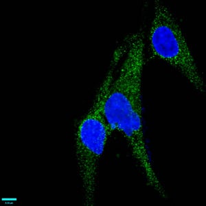
 IHC (Immunohistochemisry)
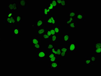
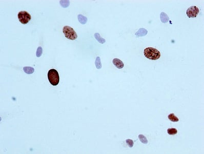
(Immunofluorescence staining of Hela cells (treated by 15mM sodium butyrate for 30min) with AAA235166 at 1:46, counter-stained with DAPI. The cells were fixed in 4% formaldehyde, permeabilized using 0.2% Triton X-100 and blocked in 10% normal Goat Serum. The cells were then incubated with the antibody overnight at 4 degree C.The secondary antibody was Alexa Fluor 488-congugated AffiniPure Goat Anti-Rabbit IgG (H+L).)
IHC (Immunohistochemisry)
(Immunofluorescence staining of Hela cells (treated by 15mM sodium butyrate for 30min) with AAA235166 at 1:46, counter-stained with DAPI. The cells were fixed in 4% formaldehyde, permeabilized using 0.2% Triton X-100 and blocked in 10% normal Goat Serum. The cells were then incubated with the antibody overnight at 4 degree C.The secondary antibody was Alexa Fluor 488-congugated AffiniPure Goat Anti-Rabbit IgG (H+L).)
Acetyl-Histone H3.1, Monoclonal Recombinant Antibody (Cat# AAA235166)
 Application Data
(Overlay histogram showing Hela cells stained with AAA235167 (red line) at 1:50. The cells were fixed with 70% Ethylalcohol (18h) and then permeabilized with 0.3% Triton X-100 for 2 min.The cells were then incubated in 1x PBS /10% normal goat serum to block non-specific protein-protein interactions followed by primary antibody for 1 h at 4 degree C.The secondary antibody used was FITC goat anti-rabbit IgG (H+L) at 1/200 dilution for 1 h at 4 degree C. Control antibody (green line) was used under the same conditions. Acquisition of >10, 000 events was performed.)
Application Data
(Overlay histogram showing Hela cells stained with AAA235167 (red line) at 1:50. The cells were fixed with 70% Ethylalcohol (18h) and then permeabilized with 0.3% Triton X-100 for 2 min.The cells were then incubated in 1x PBS /10% normal goat serum to block non-specific protein-protein interactions followed by primary antibody for 1 h at 4 degree C.The secondary antibody used was FITC goat anti-rabbit IgG (H+L) at 1/200 dilution for 1 h at 4 degree C. Control antibody (green line) was used under the same conditions. Acquisition of >10, 000 events was performed.)
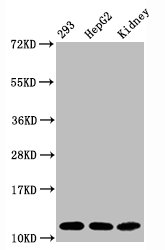
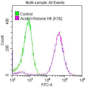
Acetyl-Histone H4, Monoclonal Recombinant Antibody (Cat# AAA235167)
 IHC (Immunohistochemisry)
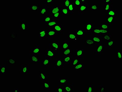
(Immunofluorescence staining of Hela cells with AAA235168 at 1:31, counter-stained with DAPI. The cells were fixed in 4% formaldehyde, permeabilized using 0.2% Triton X-100 and blocked in 10% normal Goat Serum. The cells were then incubated with the antibody overnight at 4 degree C.The secondary antibody was Alexa Fluor 488-congugated AffiniPure Goat Anti-Rabbit IgG (H+L).)
IHC (Immunohistochemisry)
(Immunofluorescence staining of Hela cells with AAA235168 at 1:31, counter-stained with DAPI. The cells were fixed in 4% formaldehyde, permeabilized using 0.2% Triton X-100 and blocked in 10% normal Goat Serum. The cells were then incubated with the antibody overnight at 4 degree C.The secondary antibody was Alexa Fluor 488-congugated AffiniPure Goat Anti-Rabbit IgG (H+L).)
Histone H3.1, Monoclonal Recombinant Antibody (Cat# AAA235168)
 Application Data
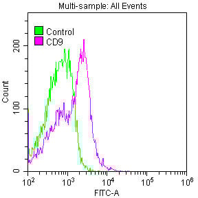
(Overlay histogram showing Jurkat cells stained with AAA235170 (red line) at 1:50. The cells were fixed with 70% Ethylalcohol (18h) and then permeabilized with 0.3% Triton X-100 for 2 min.The cells were then incubated in 1x PBS /10% normal goat serum to block non-specific protein-protein interactions followed by primary antibody for 1 h at 4 degree C.The secondary antibody used was FITC goat anti-rabbit IgG (H+L) at 1/200 dilution for 1 h at 4 degree C. Control antibody (green line) was used under the same conditions. Acquisition of >10, 000 events was performed.)
Application Data
(Overlay histogram showing Jurkat cells stained with AAA235170 (red line) at 1:50. The cells were fixed with 70% Ethylalcohol (18h) and then permeabilized with 0.3% Triton X-100 for 2 min.The cells were then incubated in 1x PBS /10% normal goat serum to block non-specific protein-protein interactions followed by primary antibody for 1 h at 4 degree C.The secondary antibody used was FITC goat anti-rabbit IgG (H+L) at 1/200 dilution for 1 h at 4 degree C. Control antibody (green line) was used under the same conditions. Acquisition of >10, 000 events was performed.)
CD9, Monoclonal Recombinant Antibody (Cat# AAA235170)
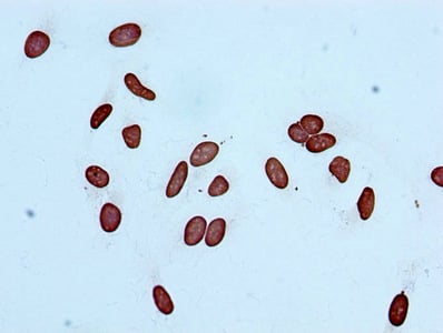
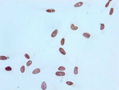
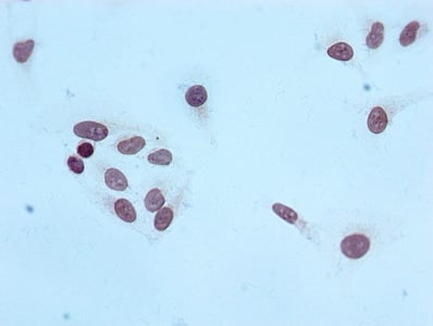 ICC (Immunocytochemistry)
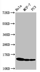
(Immunocytochemistry analysis of AAA235176 diluted at 1:100 and staining in Hela cells performed on a Leica BondTM system. After dewaxing and hydration, antigen retrieval was mediated by high pressure in a citrate buffer (pH 6.0). Section was blocked with 10% normal goat serum 30min at RT. Then primary antibody (1% BSA) was incubated at 4 degree C overnight. The primary is detected by a biotinylated secondary antibody and visualized using an HRP conjugated SP system.)
ICC (Immunocytochemistry)
(Immunocytochemistry analysis of AAA235176 diluted at 1:100 and staining in Hela cells performed on a Leica BondTM system. After dewaxing and hydration, antigen retrieval was mediated by high pressure in a citrate buffer (pH 6.0). Section was blocked with 10% normal goat serum 30min at RT. Then primary antibody (1% BSA) was incubated at 4 degree C overnight. The primary is detected by a biotinylated secondary antibody and visualized using an HRP conjugated SP system.)
Histone H4, Monoclonal Recombinant Antibody (Cat# AAA235176)
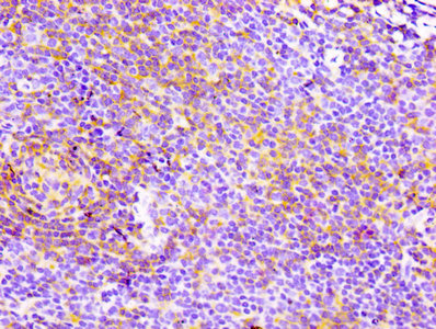
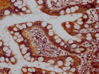
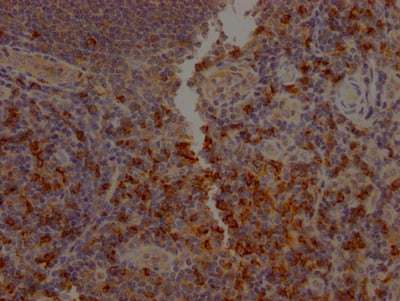
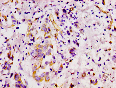
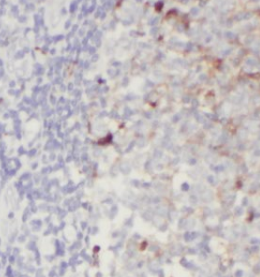
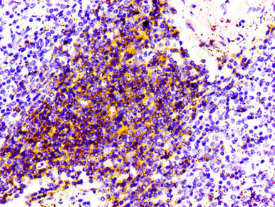 IHC (Immunohistochemisry)
(IHC image of AAA235181 diluted at 1:100 and staining in paraffin-embedded human spleen tissue performed on a Leica BondTM system. After dewaxing and hydration, antigen retrieval was mediated by high pressure in a citrate buffer (pH 6.0). Section was blocked with 10% normal goat serum 30min at RT. Then primary antibody (1% BSA) was incubated at 4 degree C overnight. The primary is detected by a biotinylated secondary antibody and visualized using an HRP conjugated SP system.)
IHC (Immunohistochemisry)
(IHC image of AAA235181 diluted at 1:100 and staining in paraffin-embedded human spleen tissue performed on a Leica BondTM system. After dewaxing and hydration, antigen retrieval was mediated by high pressure in a citrate buffer (pH 6.0). Section was blocked with 10% normal goat serum 30min at RT. Then primary antibody (1% BSA) was incubated at 4 degree C overnight. The primary is detected by a biotinylated secondary antibody and visualized using an HRP conjugated SP system.)
CD4, Monoclonal Recombinant Antibody (Cat# AAA235181)
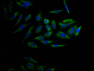
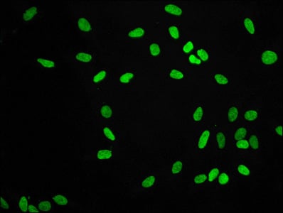 IF (Immunofluorescence)
(Immunofluorescence staining of Hela cells with AAA235186 at 1:56, counter-stained with DAPI. The cells were fixed in 4% formaldehyde, permeabilized using 0.2% Triton X-100 and blocked in 10% normal Goat Serum. The cells were then incubated with the antibody overnight at 4 degree C.The secondary antibody was Alexa Fluor 488-congugated AffiniPure Goat Anti-Rabbit IgG (H+L).)
IF (Immunofluorescence)
(Immunofluorescence staining of Hela cells with AAA235186 at 1:56, counter-stained with DAPI. The cells were fixed in 4% formaldehyde, permeabilized using 0.2% Triton X-100 and blocked in 10% normal Goat Serum. The cells were then incubated with the antibody overnight at 4 degree C.The secondary antibody was Alexa Fluor 488-congugated AffiniPure Goat Anti-Rabbit IgG (H+L).)
Di-methyl-Histone H3.1, Monoclonal Recombinant Antibody (Cat# AAA235186)
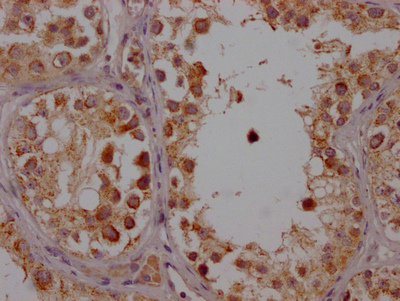
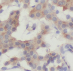
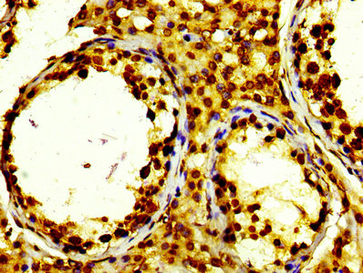 IHC (Immunohiostchemistry)
(IHC image of AAA235188 diluted at 1:100 and staining in paraffin-embedded human testis tissue performed on a Leica BondTM system. After dewaxing and hydration, antigen retrieval was mediated by high pressure in a citrate buffer (pH 6.0). Section was blocked with 10% normal goat serum 30min at RT. Then primary antibody (1% BSA) was incubated at 4 degree C overnight. The primary is detected by a biotinylated secondary antibody and visualized using an HRP conjugated SP system.)
IHC (Immunohiostchemistry)
(IHC image of AAA235188 diluted at 1:100 and staining in paraffin-embedded human testis tissue performed on a Leica BondTM system. After dewaxing and hydration, antigen retrieval was mediated by high pressure in a citrate buffer (pH 6.0). Section was blocked with 10% normal goat serum 30min at RT. Then primary antibody (1% BSA) was incubated at 4 degree C overnight. The primary is detected by a biotinylated secondary antibody and visualized using an HRP conjugated SP system.)
Histone H2B type 1-K, Monoclonal Recombinant Antibody (Cat# AAA235188)
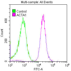 Application Data
(Overlay histogram showing Hela cells stained with AAA235194 (red line) at 1:50. The cells were fixed with 70% Ethylalcohol (18h) and then permeabilized with 0.3% Triton X-100 for 2 min.The cells were then incubated in 1x PBS /10% normal goat serum to block non-specific protein-protein interactions followed by primary antibody for 1 h at 4 degree C.The secondary antibody used was FITC goat anti-rabbit IgG (H+L) at 1/200 dilution for 1 h at 4 degree C. Control antibody (green line) was used under the same conditions. Acquisition of >10, 000 events was performed.)
Application Data
(Overlay histogram showing Hela cells stained with AAA235194 (red line) at 1:50. The cells were fixed with 70% Ethylalcohol (18h) and then permeabilized with 0.3% Triton X-100 for 2 min.The cells were then incubated in 1x PBS /10% normal goat serum to block non-specific protein-protein interactions followed by primary antibody for 1 h at 4 degree C.The secondary antibody used was FITC goat anti-rabbit IgG (H+L) at 1/200 dilution for 1 h at 4 degree C. Control antibody (green line) was used under the same conditions. Acquisition of >10, 000 events was performed.)
ACTA1, Monoclonal Recombinant Antibody (Cat# AAA235194)
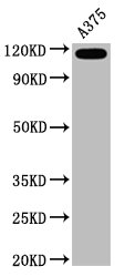
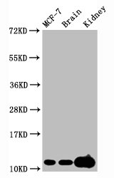
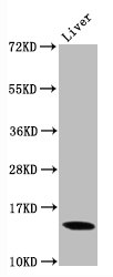
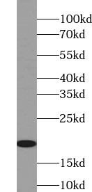
 WB (Western Blot)
(Recombinant protein were subjected to SDS PAGE followed by western blot with AAA247955 (IL1F8 Antibody) at dilution of 1:8000)
WB (Western Blot)
(Recombinant protein were subjected to SDS PAGE followed by western blot with AAA247955 (IL1F8 Antibody) at dilution of 1:8000)
IL1F8, Monoclonal Antibody (Cat# AAA247955)
Protein A+G purification
 WB (Western Blot)
(IL23A transfected HEK293 cells were subjected to SDS-PAGE followed by western blot with AAA247958 (IL23A monoclonal Antibody) at dilution of 1:1000)
WB (Western Blot)
(IL23A transfected HEK293 cells were subjected to SDS-PAGE followed by western blot with AAA247958 (IL23A monoclonal Antibody) at dilution of 1:1000)
IL23A, Monoclonal Antibody (Cat# AAA247958)
Protein A+G purification
 WB (Western Blot)
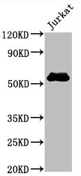
(Jurkat cells were subjected to SDS PAGE followed by western blot with AAA247963 (IL4 antibody) at dilution of 1:1000)
WB (Western Blot)
(Jurkat cells were subjected to SDS PAGE followed by western blot with AAA247963 (IL4 antibody) at dilution of 1:1000)
IL-4, Monoclonal Antibody (Cat# AAA247963)
Protein A+G purification
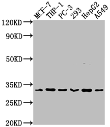
 WB (Western Blot)
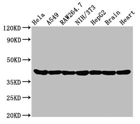
(PC-12, C6, Raw264.7, NIH/3T3 cells were subjected to SDS PAGE followed by western blot with AAA247981 (LPCAT1 Antibody) at dilution of 1:10000)
WB (Western Blot)
(PC-12, C6, Raw264.7, NIH/3T3 cells were subjected to SDS PAGE followed by western blot with AAA247981 (LPCAT1 Antibody) at dilution of 1:10000)
LPCAT1, Monoclonal Antibody (Cat# AAA247981)
Protein A+G purification
 WB (Western Blot)
(Jurkat cells were subjected to SDS PAGE followed by western blot with AAA247982 (LYN antibody) at dilution of 1:1000)
WB (Western Blot)
(Jurkat cells were subjected to SDS PAGE followed by western blot with AAA247982 (LYN antibody) at dilution of 1:1000)
LYN, Monoclonal Antibody (Cat# AAA247982)
Protein A+G purification
 WB (Western Blot)
(HEK-293 cells were subjected to SDS PAGE followed by western blot with AAA247986 (MBIP antibody) at dilution of 1:1000)
WB (Western Blot)
(HEK-293 cells were subjected to SDS PAGE followed by western blot with AAA247986 (MBIP antibody) at dilution of 1:1000)
MBIP, Monoclonal Antibody (Cat# AAA247986)
Protein A+G purification
What are Monoclonal Antibodies?
Monoclonal antibodies are specialized laboratory-produced proteins developed for binding to specific biological antigens or other molecular targets. Since they come from a single cell (or clone), they are especially consistent and accurate in the data they are involved in producing.
This type of antibody material has been shown to be a powerful tool in finding and subsequently destroying harmful cells in an organism, such as those found in cancers or various autoimmune diseases. This makes them excellent aids in medical testing and research, which is why they are so widely used.
AAA Biotech offers a comprehensive range of high-quality monoclonal antibodies that perform effectively in various laboratory tests, including (amongst others) ELISA, western blotting, immunohistochemistry, and flow cytometry. All of the products in our catalog are thoroughly quality tested to make sure that they are reliable and will consistently perform well in your research.
What Are The Uses of Monoclonal Antibodies
Monoclonal antibodies are used in many lab tests, including (amongst others) ELISA, western blotting, immunohistochemistry, and flow cytometry.
ELISA is a test that helps detect a specific substance/analyte in a sample. It uses antibodies (often monoclonal) bound to a solid surface (such as the well of a microplate) to “capture” the substance/analyte in the sample and immobilize it so that the detection antibody component can then bind to it and produce a signal, which can then be measured.
Western blotting identifies specific proteins in a sample. The sample is first separated on a gel, and then antibodies are applied that will typically bind to the target, which will all be localized to a single band in a lane.
Immunohistochemistry helps locate specific proteins in cells or tissue samples using antibodies.
Flow cytometry looks at and sorts cells. It uses antibodies that are conjugated to reporter molecules called “fluorophores”, which, under special lights, emit light themselves, which can then be measured by a detector instrument. For a deeper understanding of these techniques, explore our complete guide to monoclonal antibodies and their benefits.
How Monoclonal Antibodies Are Used as Medicine?
Please note that all of the products listed in AAA Biotech’s also known as AAA Bio or AAABio catalog are strictly for research-use only (RUO).
Monoclonal antibodies can also be used as therapeutic/medical treatments, particularly in the context of cancers. They are designed to find and bind to specific cells or proteins, helping the immune system recognize and attack the cancer. These treatments work in different ways, such as:
- Radioimmunotherapy attaches a small amount of radioactive molecule to the antibody, so it delivers the radiation directly to the cancer cells that the antibody is specifically binding to.
- Antibody-directed enzyme prodrug therapy uses antibodies that are specifically bound to special enzymes. These enzymes activate a harmless drug in the body and turn it into a cancer-killing drug only near the cancer cells—this helps avoid harming healthy cells.
- Immunoliposomes are tiny “bubbles” filled with medicine/drug and coated with antibodies. They carry the drug straight to the cancer cells.
Why Buy Monoclonal Antibodies From Us?
At AAA Biotech, we provide high-performance monoclonal antibodies designed to support a wide range of research needs.
1. Validated for Versatile Applications
The antibodies in our catalog are extensively validated and compatible with multiple techniques, including (but not limited to) ELISA, flow cytometry (FC), immunocytochemistry (ICC), immunofluorescence (IF), immunohistochemistry (IHC), immunoprecipitation (IP), and western blotting (WB).
2. Wide Selection & Specialized Options
We offer antibodies for common and rare species, that are available in various conjugated forms, and also in recombinant formats. Essentially, there is almost anything one might need to meet their experimental model’s requirements.
3. High-Quality Proteins
Our proteins meet high purity standards—90% or more as confirmed by SDS-PAGE. Many are available with tags like His, Flag, GST, or MBP, and we also supply native and biologically active proteins for functional studies.
Frequently Asked Questions
1. Are your monoclonal antibodies validated for specific applications?
Yes, our antibodies are tested and validated for use in methods such as ELISA, western blot, IHC, flow cytometry, and more. Refer to specific product pages or datasheets for individual product information.
2. How do I choose the right monoclonal antibody for my application?
Review the product details directly for application validation, species reactivity, and target information. You may also contact our support team at any time for help.
3. How quickly can I receive my order?
Most orders are processed and shipped within 1–3 business days, depending on product availability and your shipping location.