Filters
▼Clonality
▼Type
▼Reactivity
▼Gene Name
▼Isotype
▼Host
▼Application
▼Clone
▼Monoclonal Antibodies
Get accurate results in your research with our Monoclonal Antibodies, which are specially made to target exactly what you require for your research, and will produce consistent, reliable performance in lab tests.
Viewing 100-150 of 27645 product results
CD34, Monoclonal Antibody (Cat# AAA128360)
CD34, Monoclonal Antibody (Cat# AAA128361)
CD49e, Monoclonal Antibody (Cat# AAA128376)
 WB (Western Blot)
(Western Blot; Sample: Recombinant IL8, Gallus.)
WB (Western Blot)
(Western Blot; Sample: Recombinant IL8, Gallus.)
Interleukin 8 (IL8), Monoclonal Antibody (Cat# AAA134806)
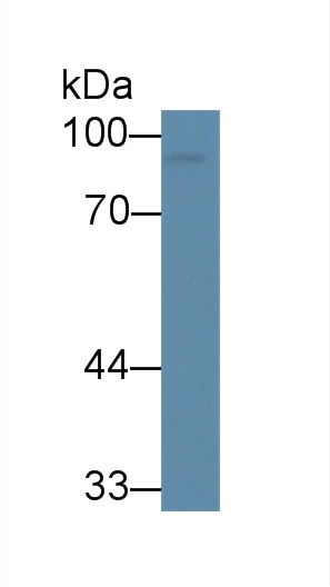
 WB (Western Blot)
(Western Blot: Sample: Recombinant CLU, Human.)
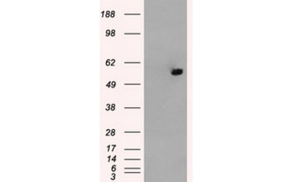
WB (Western Blot)
(Western Blot: Sample: Recombinant CLU, Human.)
Clusterin (CLU), Monoclonal Antibody (Cat# AAA134825)
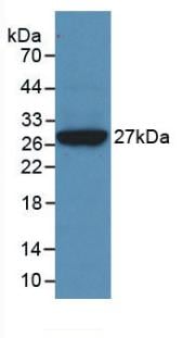
 WB (Western Blot)
(Western Blot: Sample: Recombinant NEU, Human.)
WB (Western Blot)
(Western Blot: Sample: Recombinant NEU, Human.)
Neuraminidase (NEU), Monoclonal Antibody (Cat# AAA134828)


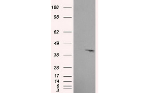
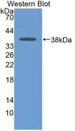
 Knockout Validation
(Knockout Validation: Lane 1: Wild-type HepG2 cell lysate;;Lane 2: ANXA3 knockout HepG2 cell lysate;;Predicted MW: 36kDa ;Observed MW: 36kDa;Primary Ab: 3ug/ml Mouse Anti-Human ANXA3 Antibody;Second Ab: 0.2ug/mL HRP-Linked Caprine Anti-Mouse IgG Polyclonal Antibody;)
Knockout Validation
(Knockout Validation: Lane 1: Wild-type HepG2 cell lysate;;Lane 2: ANXA3 knockout HepG2 cell lysate;;Predicted MW: 36kDa ;Observed MW: 36kDa;Primary Ab: 3ug/ml Mouse Anti-Human ANXA3 Antibody;Second Ab: 0.2ug/mL HRP-Linked Caprine Anti-Mouse IgG Polyclonal Antibody;)
Annexin A3 (ANXA3), Monoclonal Antibody (Cat# AAA134832)
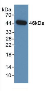
 WB (Western Blot)
(Western Blot: Sample: Recombinant ITIH4, Human)
WB (Western Blot)
(Western Blot: Sample: Recombinant ITIH4, Human)
Inter Alpha-Globulin Inhibitor H4 (ITIH4), Monoclonal Antibody (Cat# AAA134836)
CD106, Monoclonal Antibody (Cat# AAA129069)
CD49e, Monoclonal Antibody (Cat# AAA129070)
 Application Data
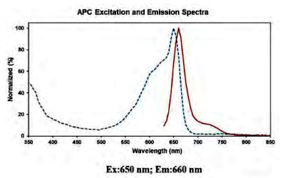
(Conjugation: APC)
Application Data
(Conjugation: APC)
TCRbeta, Monoclonal Antibody (Cat# AAA174749)
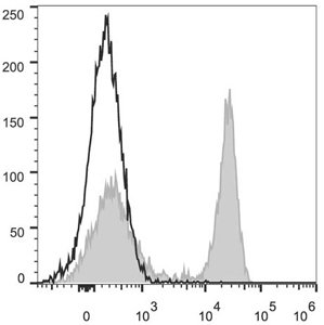 FCM/FACS (Flow Cytometry)
(Each lot of this antibody is quality control tested by flow cytometric analysis. The amount of the reagent is suggested to be used 5 uL of the antibody per test (million cells in 100 uL staining volume or per 100 uL of whole blood). Please check your vial before the experiment. Since applications vary, the appropriate dilutions must be determined for individual use.C57BL/6 murine splenocytes are stained with PerCP/Cyanine5.5 Anti-Mouse CD45R/B220 Antibody (filled gray histogram). Unstained splenocytes (empty black histogram) are used as control.)
FCM/FACS (Flow Cytometry)
(Each lot of this antibody is quality control tested by flow cytometric analysis. The amount of the reagent is suggested to be used 5 uL of the antibody per test (million cells in 100 uL staining volume or per 100 uL of whole blood). Please check your vial before the experiment. Since applications vary, the appropriate dilutions must be determined for individual use.C57BL/6 murine splenocytes are stained with PerCP/Cyanine5.5 Anti-Mouse CD45R/B220 Antibody (filled gray histogram). Unstained splenocytes (empty black histogram) are used as control.)
CD45R/B220, Monoclonal Antibody (Cat# AAA174637)

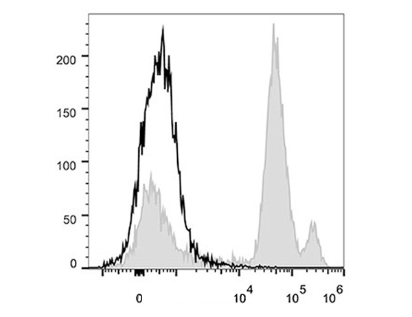 FCM/FACS (Flow Cytometry)
(C57BL/6 murine bone marrow cells are stained with Anti-Mouse Ly6C Monoclonal Antibody(PerCP/Cy5.5 Conjugated)(filled gray histogram). Unstained bone marrow cells (empty black histogram) are used as control.)
FCM/FACS (Flow Cytometry)
(C57BL/6 murine bone marrow cells are stained with Anti-Mouse Ly6C Monoclonal Antibody(PerCP/Cy5.5 Conjugated)(filled gray histogram). Unstained bone marrow cells (empty black histogram) are used as control.)
Ly6C, Monoclonal Antibody (Cat# AAA174647)
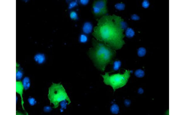
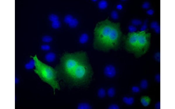
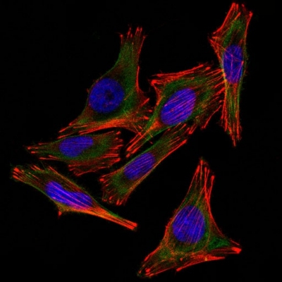
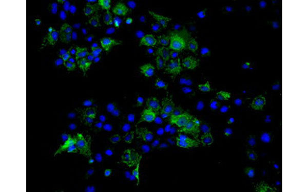 IF (Immunofluorescence)
(Immunofluorescent staining of COS7 cells transiently transfected with recombinant ATP5B protein using ATP5B antibody)
IF (Immunofluorescence)
(Immunofluorescent staining of COS7 cells transiently transfected with recombinant ATP5B protein using ATP5B antibody)
ATP5B, Monoclonal Antibody (Cat# AAA106429)
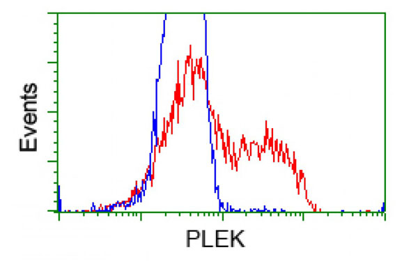
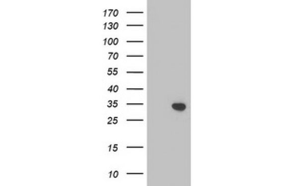
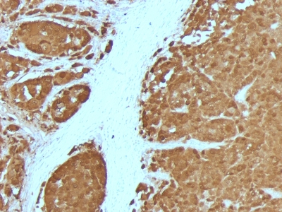
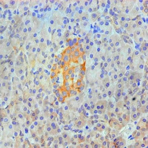
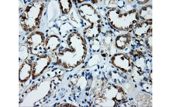 IHC (Immunohistochemisry)
(Immunohistochemical analysis of PLEK protein in paraffin embedded Human kidney tissue using PLEK antibody)
IHC (Immunohistochemisry)
(Immunohistochemical analysis of PLEK protein in paraffin embedded Human kidney tissue using PLEK antibody)
PLEK, Monoclonal Antibody (Cat# AAA108060)
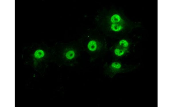
 IF (Immunofluorescence)
(Immunofluorescent staining of COS7 cells transiently transfected with recombinant TOMM34 protein using TOMM34 antibody)
IF (Immunofluorescence)
(Immunofluorescent staining of COS7 cells transiently transfected with recombinant TOMM34 protein using TOMM34 antibody)
TOMM34, Monoclonal Antibody (Cat# AAA108139)
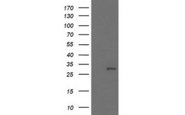
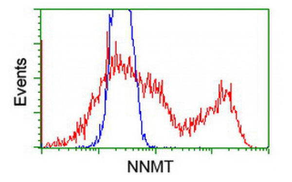
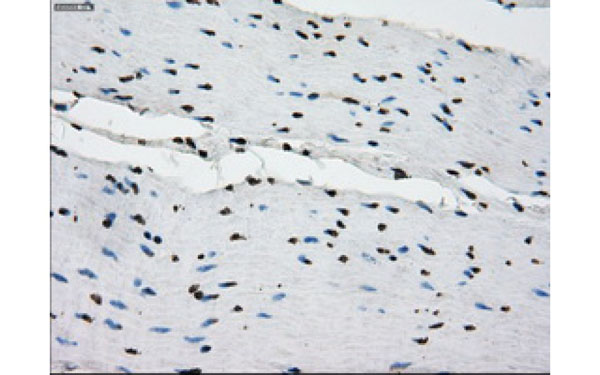
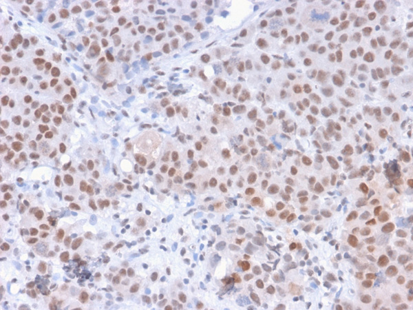
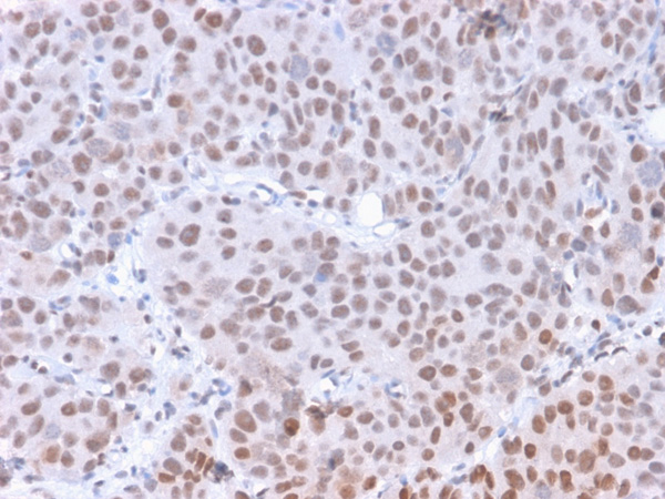
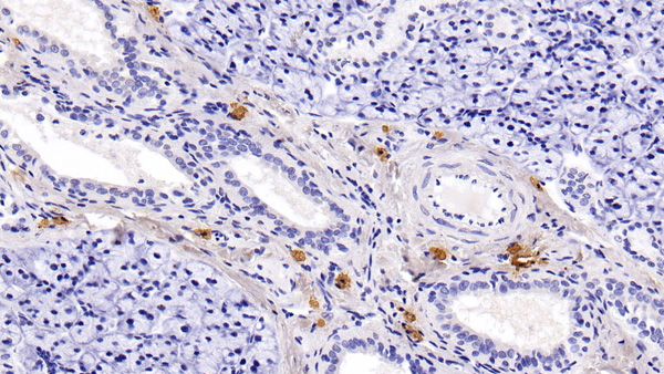
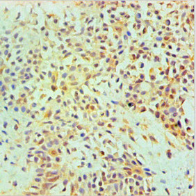
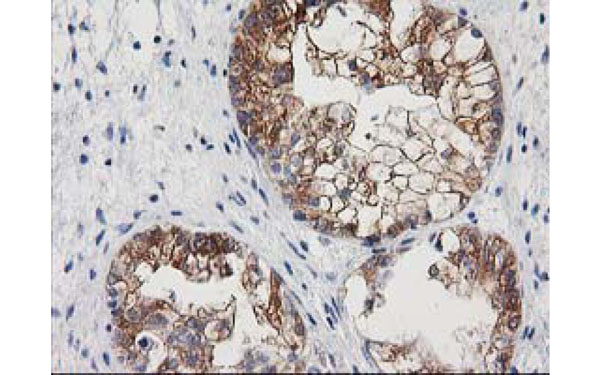 IHC (Immunohistochemisry)
(Immunohistochemical analysis of NNMT protein in paraffin embedded Adenocarcinoma of Human ovary tissue using NNMT antibody)
IHC (Immunohistochemisry)
(Immunohistochemical analysis of NNMT protein in paraffin embedded Adenocarcinoma of Human ovary tissue using NNMT antibody)
NNMT, Monoclonal Antibody (Cat# AAA107173)
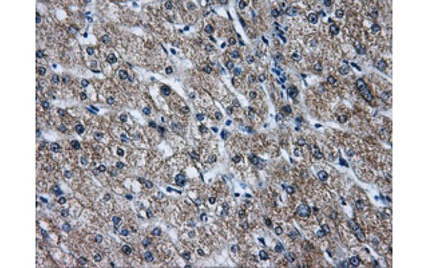
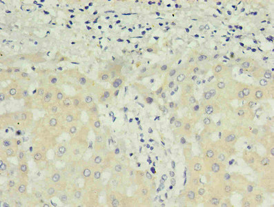
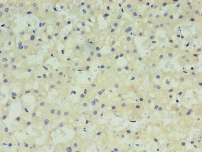
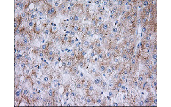 IHC (Immunohistochemisry)
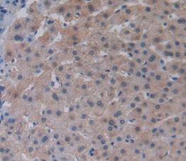
(Immunohistochemical analysis of RAB17 protein in paraffin embedded Human liver tissue using RAB17 antibody)
IHC (Immunohistochemisry)
(Immunohistochemical analysis of RAB17 protein in paraffin embedded Human liver tissue using RAB17 antibody)
RAB17, Monoclonal Antibody (Cat# AAA107259)

 IHC (Immunohistochemisry)
(Immunohistochemical analysis of PRKY protein in paraffin embedded Human colon tissue using PRKY antibody)
IHC (Immunohistochemisry)
(Immunohistochemical analysis of PRKY protein in paraffin embedded Human colon tissue using PRKY antibody)
PRKY, Monoclonal Antibody (Cat# AAA107453)
Mouse anti Human Bence Jones lambda (surface and hidden determinants), Monoclonal Secondary Antibody (Cat# AAA77534)
Streptavidin Streptomysces Avidinii, Monoclonal Antibody (Cat# AAA78064)
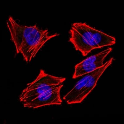 IF (Immunofluorescence)
(Confocal Immunofluorescent analysis of A2058 cells using AF488-labeled Isotype Control Monoclonal Antibody (IgG2a) (Green). F-actin filaments were labeled with DyLight 554 Phalloidin (red). DAPI was used to stain the cell nuclei (blue). (Negative Control))
IF (Immunofluorescence)
(Confocal Immunofluorescent analysis of A2058 cells using AF488-labeled Isotype Control Monoclonal Antibody (IgG2a) (Green). F-actin filaments were labeled with DyLight 554 Phalloidin (red). DAPI was used to stain the cell nuclei (blue). (Negative Control))
S100B, Monoclonal Antibody (Cat# AAA214511)
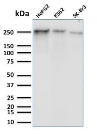 WB (Western Blot)
(Western Blot Analysis of Human HepG2, K562 and SK-Br3 cell lysates using RNA Poll II Mouse Monoclonal Antibody (8A7).)
WB (Western Blot)
(Western Blot Analysis of Human HepG2, K562 and SK-Br3 cell lysates using RNA Poll II Mouse Monoclonal Antibody (8A7).)
RNA Polymerase II CTD Repeat YSPTSPS, Monoclonal Antibody (Cat# AAA215151)

 WB (Western Blot)
(Western Blot: Sample: Recombinant LDHB, Rat.)
WB (Western Blot)
(Western Blot: Sample: Recombinant LDHB, Rat.)
Lactate Dehydrogenase B, Monoclonal Antibody (Cat# AAA141281)
 WB (Western Blot)
(Western Blot: Sample: Recombinant CDK5, Human.)
WB (Western Blot)
(Western Blot: Sample: Recombinant CDK5, Human.)
Cyclin Dependent Kinase 5, Monoclonal Antibody (Cat# AAA141323)
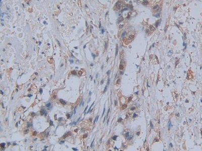
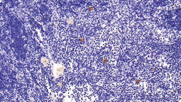
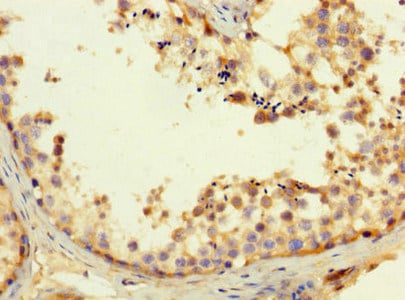
 IHC (Immunohistochemisry)
(DAB staining on IHC-P; Samples: Human Pancreatic cancer Tissue))
IHC (Immunohistochemisry)
(DAB staining on IHC-P; Samples: Human Pancreatic cancer Tissue))
Monocyte Chemotactic Protein 1, Monoclonal Antibody (Cat# AAA141337)
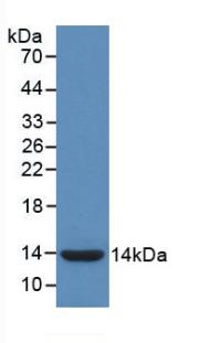
 WB (Western Blot)
(Sample: Recombinant CSTA, Human.)
WB (Western Blot)
(Sample: Recombinant CSTA, Human.)
Cystatin A, Monoclonal Antibody (Cat# AAA141360)

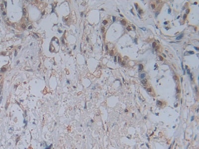
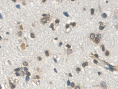
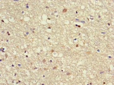
 IHC (Immunohistochemistry)
(DAB staining on IHC-P;Samples: Human Brain Tissue)
IHC (Immunohistochemistry)
(DAB staining on IHC-P;Samples: Human Brain Tissue)
Lysophosphatidylcholine Acyltransferase 3, Monoclonal Antibody (Cat# AAA141361)
Cystatin A (CSTA), Monoclonal Antibody (Cat# AAA149340)
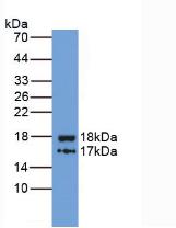
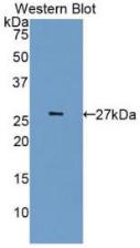
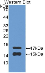
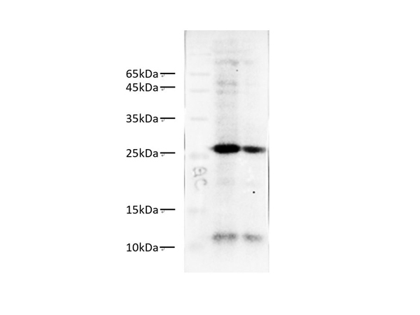
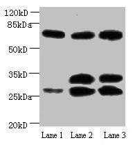
 WB (Western Blot)
(Western Blot; Sample: Lane1: Human Leukocyte lysate; Lane2: Human Lymphocyte lysate; Lane3: Human PBMC lysate Primary Ab: 0.1ug/ml Mouse AntiHuman S100A9 Antibody Second Ab: 0.2ug/mL HRPLinked Caprine AntiMouse IgG Polyclonal Antibody (Catalog: SAA544Mu19))
WB (Western Blot)
(Western Blot; Sample: Lane1: Human Leukocyte lysate; Lane2: Human Lymphocyte lysate; Lane3: Human PBMC lysate Primary Ab: 0.1ug/ml Mouse AntiHuman S100A9 Antibody Second Ab: 0.2ug/mL HRPLinked Caprine AntiMouse IgG Polyclonal Antibody (Catalog: SAA544Mu19))
S100 Calcium Binding Protein A9 (S100A9), Monoclonal Antibody (Cat# AAA151749)
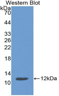
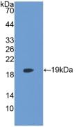
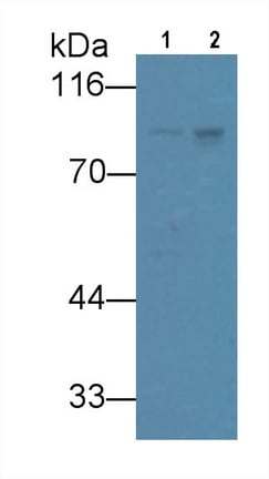 WB (Western Blot)
(Western Blot; Sample: Lane1: Rat Placenta lysate; Lane2: U87MG cell lysate Primary Ab: 3ug/ml Mouse AntiHuman VGF Antibody Second Ab: 0.2ug/mL HRPLinked Caprine AntiMouse IgG Polyclonal Antibody (Catalog: SAA544Mu19))
WB (Western Blot)
(Western Blot; Sample: Lane1: Rat Placenta lysate; Lane2: U87MG cell lysate Primary Ab: 3ug/ml Mouse AntiHuman VGF Antibody Second Ab: 0.2ug/mL HRPLinked Caprine AntiMouse IgG Polyclonal Antibody (Catalog: SAA544Mu19))
VGF Nerve Growth Factor Inducible (VGF), Monoclonal Antibody (Cat# AAA151685)
 IF (Immunofluorescence)
(Immunofluorescent analysis of GFP transfected 293 cells with GFP-Tag monoclonal antibody at dilution of 1:500.)
IF (Immunofluorescence)
(Immunofluorescent analysis of GFP transfected 293 cells with GFP-Tag monoclonal antibody at dilution of 1:500.)
GFP-Tag, Monoclonal Antibody (Cat# AAA178009)
 IHC (Immunohiostchemistry)
(Immunohistochemistry of paraffin-embedded human liver cancer using AAA118979 in 30ug/ml dilute concentrations.)
IHC (Immunohiostchemistry)
(Immunohistochemistry of paraffin-embedded human liver cancer using AAA118979 in 30ug/ml dilute concentrations.)
Alpha-fetoprotein, Monoclonal Antibody (Cat# AAA118979)
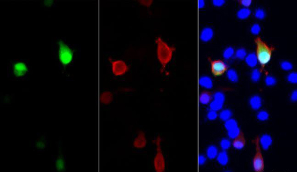
 ICC (Immunocytochemistry)
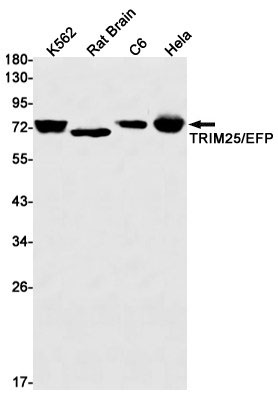
(Immunocytochemistry of TRIM25/EFP (green) in MCF-7 using TRIM25/EFP antibody at dilution 1:20, and DAPI(blue))
ICC (Immunocytochemistry)
(Immunocytochemistry of TRIM25/EFP (green) in MCF-7 using TRIM25/EFP antibody at dilution 1:20, and DAPI(blue))
TRIM25, Monoclonal Antibody (Cat# AAA178811)
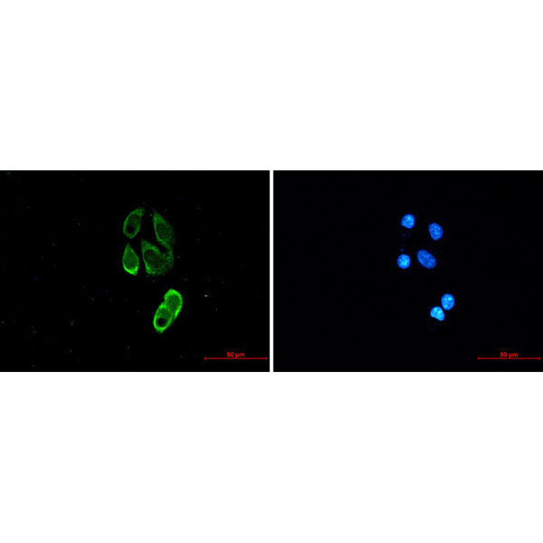
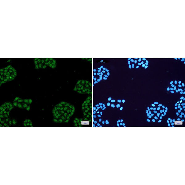 IF (Immunofluorescence)
(Immunofluorescence of NR1D1(green) in Hela cells using NR1D1 Rabbit mAb at dilution 1:50, and DAPI(blue))
IF (Immunofluorescence)
(Immunofluorescence of NR1D1(green) in Hela cells using NR1D1 Rabbit mAb at dilution 1:50, and DAPI(blue))
NR1D1, Monoclonal Antibody (Cat# AAA178868)

 IHC (Immunohistochemisry)
(Immunohistochemistry of NRF1 in paraffin-embedded Human colon cancer tissue using NRF1 Rabbit mAb at dilution 1:50)
IHC (Immunohistochemisry)
(Immunohistochemistry of NRF1 in paraffin-embedded Human colon cancer tissue using NRF1 Rabbit mAb at dilution 1:50)
Nrf1, Monoclonal Antibody (Cat# AAA178869)
 IHC (Immunohiostchemistry)
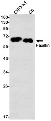
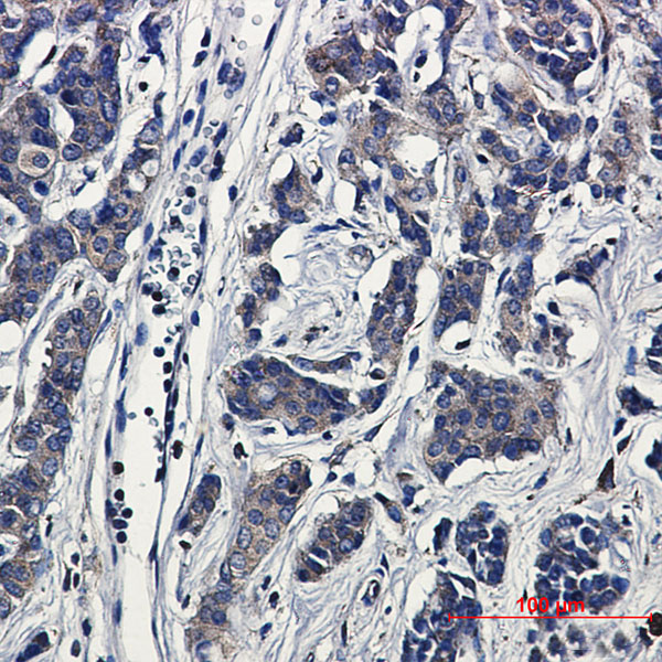
(Immunohistochemical of Paxillin in Human breast cancer tissue using Paxillin antibody at dilution 1:20)
IHC (Immunohiostchemistry)
(Immunohistochemical of Paxillin in Human breast cancer tissue using Paxillin antibody at dilution 1:20)
Paxillin, Monoclonal Antibody (Cat# AAA178872)
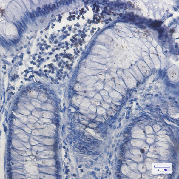
 IHC (Immunohiostchemistry)
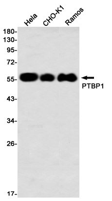
(Immunohistochemistry of PTBP1 in paraffin-embedded Human tonsil using PTBP1 Rabbit mAb at dilution 1:50)
IHC (Immunohiostchemistry)
(Immunohistochemistry of PTBP1 in paraffin-embedded Human tonsil using PTBP1 Rabbit mAb at dilution 1:50)
PTBP1, Monoclonal Antibody (Cat# AAA178876)
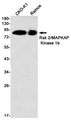
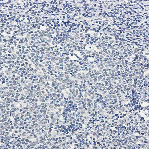
 IHC (Immunohiostchemistry)
(Immunohistochemistry of Rsk 2/MAPKAP Kinase 1b in paraffin-embedded Human lung cancer tissue using Rsk 2/MAPKAP Kinase 1b Rabbit mAb at dilution 1?50)
IHC (Immunohiostchemistry)
(Immunohistochemistry of Rsk 2/MAPKAP Kinase 1b in paraffin-embedded Human lung cancer tissue using Rsk 2/MAPKAP Kinase 1b Rabbit mAb at dilution 1?50)
RSK2, Monoclonal Antibody (Cat# AAA178878)
 IF (Immunofluorescence)
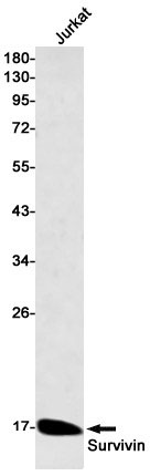
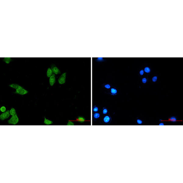
(Immunofluorescence of Survivin (green) in MCF-7 using Survivin antibody at dilution 1:20, and DAPI(blue))
IF (Immunofluorescence)
(Immunofluorescence of Survivin (green) in MCF-7 using Survivin antibody at dilution 1:20, and DAPI(blue))
Survivin, Monoclonal Antibody (Cat# AAA178882)
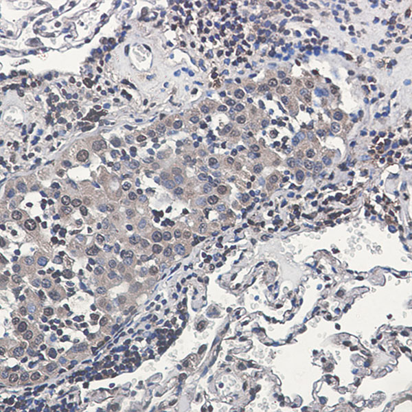
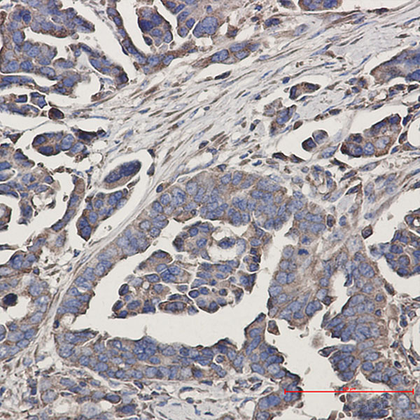
 IHC (Immunohiostchemistry)
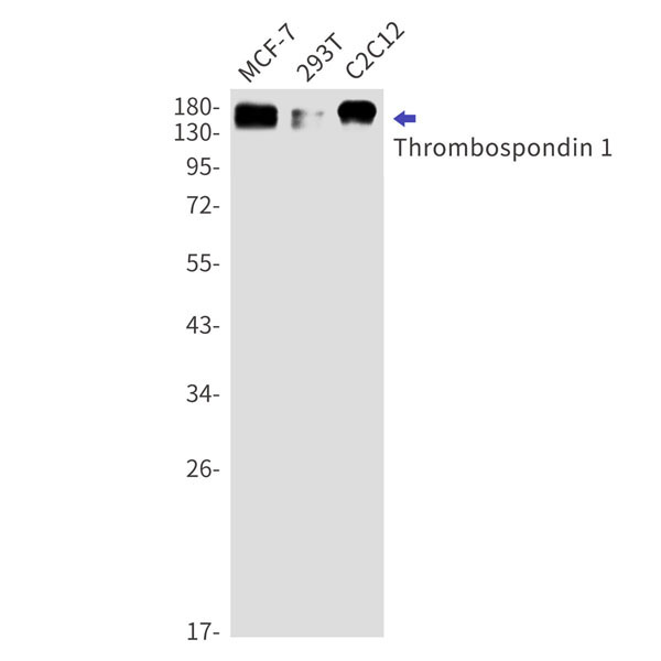
(Immunohistochemistry of Thrombospondin 1 in paraffin-embedded Human Cholangiocarcinoma using Thrombospondin 1 Rabbit mAb at dilution 1:50)
IHC (Immunohiostchemistry)
(Immunohistochemistry of Thrombospondin 1 in paraffin-embedded Human Cholangiocarcinoma using Thrombospondin 1 Rabbit mAb at dilution 1:50)
Thrombospondin 1, Monoclonal Antibody (Cat# AAA178884)
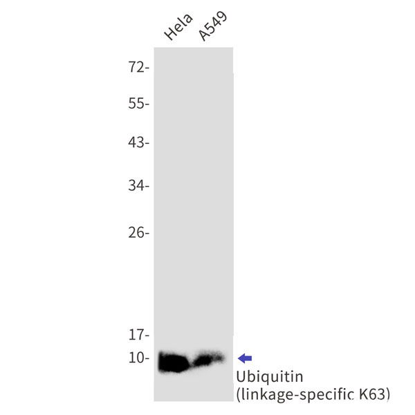
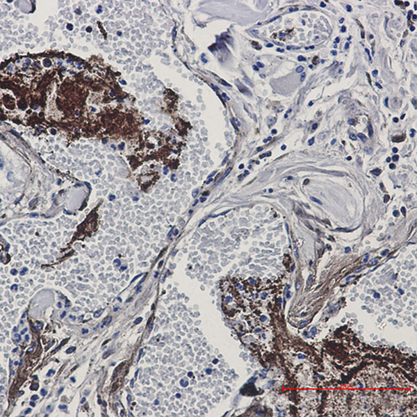
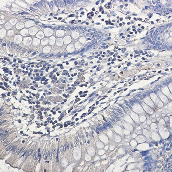 IHC (Immunohistochemisry)
(Immunohistochemistry of Ubiquitin (linkage-specific K63) in paraffin-embedded Human colon cancer tissue using Ubiquitin (linkage-specific K63) Rabbit mAb at dilution 1:50)
IHC (Immunohistochemisry)
(Immunohistochemistry of Ubiquitin (linkage-specific K63) in paraffin-embedded Human colon cancer tissue using Ubiquitin (linkage-specific K63) Rabbit mAb at dilution 1:50)
Ubiquitin K63, Monoclonal Antibody (Cat# AAA178887)
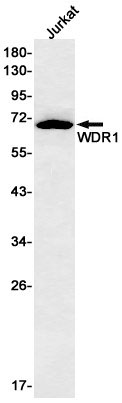
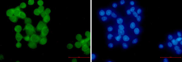
 IF (Immunofluorescence)
(Immunofluorescence of WDR1 (green) in hela using WDR1 Rabbit mAb at dilution 1:50, and DAPI(blue))
IF (Immunofluorescence)
(Immunofluorescence of WDR1 (green) in hela using WDR1 Rabbit mAb at dilution 1:50, and DAPI(blue))
WDR1, Monoclonal Antibody (Cat# AAA178889)
 IF (Immunofluorescence)
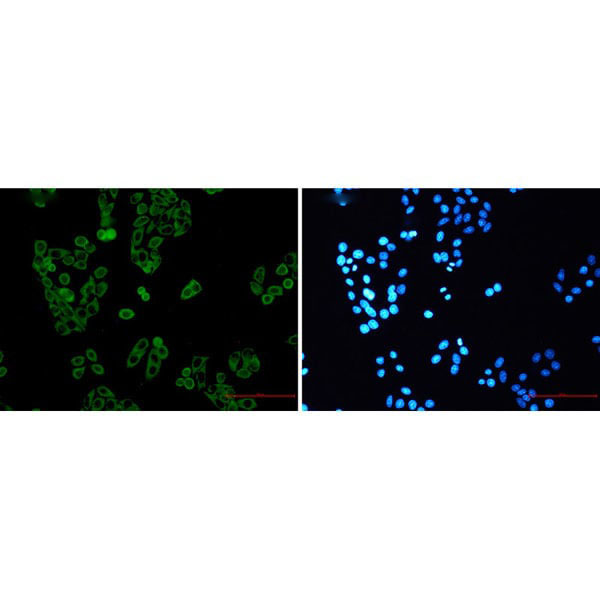
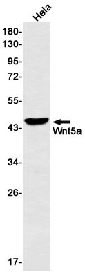
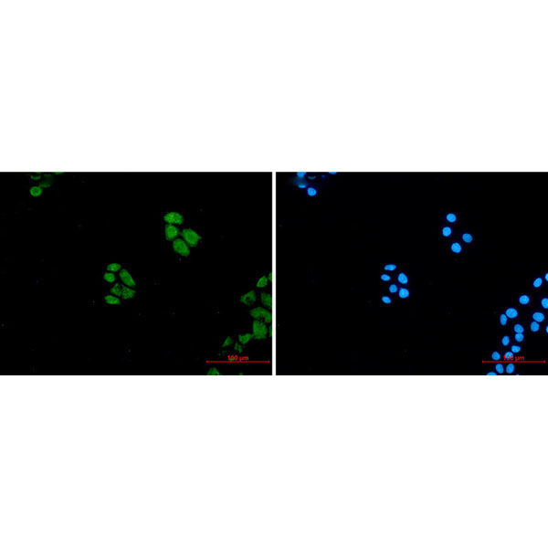
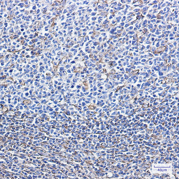
(Immunofluorescence of Wnt5a (green) in hela using Wnt5a Rabbit mAb at dilution 1:50, and DAPI(blue))
IF (Immunofluorescence)
(Immunofluorescence of Wnt5a (green) in hela using Wnt5a Rabbit mAb at dilution 1:50, and DAPI(blue))
Wnt5a, Monoclonal Antibody (Cat# AAA178890)
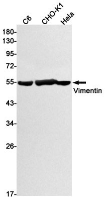
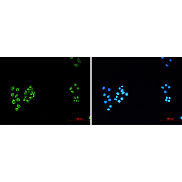 IF (Immunofluorescence)
(Immunofluorescence of Vimentin (green) in Hela using Vimentin antibody at dilution 1:20, and DAPI(blue))
IF (Immunofluorescence)
(Immunofluorescence of Vimentin (green) in Hela using Vimentin antibody at dilution 1:20, and DAPI(blue))
Vimentin, Monoclonal Antibody (Cat# AAA178774)
 IHC (Immunohiostchemistry)
(Immunohistochemistry of paraffin-embedded human breast cancer using AAA118279 in 30ug/ml dilute concentrations.)
IHC (Immunohiostchemistry)
(Immunohistochemistry of paraffin-embedded human breast cancer using AAA118279 in 30ug/ml dilute concentrations.)
RRM1, Monoclonal Antibody (Cat# AAA118279)
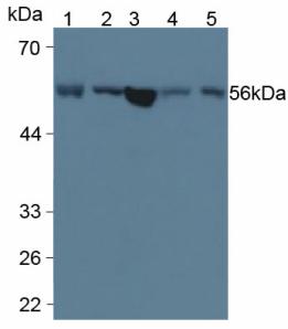
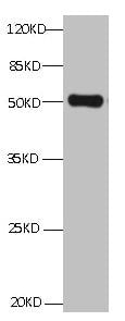 WB (Western Blot)
(All lanes: Mouse anti ANXA2 Monoclonal antibody at 1ug/mllane 1:HepG2 whole cell lysateSecondary Goat polyclonal to Mouse IgG at 1/5000 dilutionPredicted band size:39,41kdObserved band size:50KD)
WB (Western Blot)
(All lanes: Mouse anti ANXA2 Monoclonal antibody at 1ug/mllane 1:HepG2 whole cell lysateSecondary Goat polyclonal to Mouse IgG at 1/5000 dilutionPredicted band size:39,41kdObserved band size:50KD)
ANXA2, Monoclonal Antibody (Cat# AAA118281)
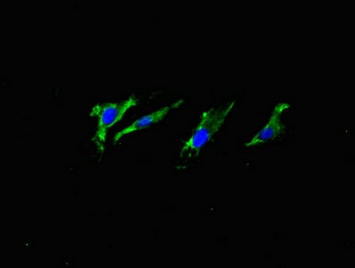
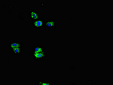 IF (Immunofluorescence)
(Immunofluorescent analysis of HepG2 cells using AAA118932 at a dilution of 1:100 and Alexa Fluor 488-congugated AffiniPure Goat Anti-Rabbit IgG(H+L))
IF (Immunofluorescence)
(Immunofluorescent analysis of HepG2 cells using AAA118932 at a dilution of 1:100 and Alexa Fluor 488-congugated AffiniPure Goat Anti-Rabbit IgG(H+L))
APP, Monoclonal Antibody (Cat# AAA118932)
Ebola virus EBOV Nucleoprotein/NP, Monoclonal Antibody (Cat# AAA176983)
What are Monoclonal Antibodies?
Monoclonal antibodies are specialized laboratory-produced proteins developed for binding to specific biological antigens or other molecular targets. Since they come from a single cell (or clone), they are especially consistent and accurate in the data they are involved in producing.
This type of antibody material has been shown to be a powerful tool in finding and subsequently destroying harmful cells in an organism, such as those found in cancers or various autoimmune diseases. This makes them excellent aids in medical testing and research, which is why they are so widely used.
AAA Biotech offers a comprehensive range of high-quality monoclonal antibodies that perform effectively in various laboratory tests, including (amongst others) ELISA, western blotting, immunohistochemistry, and flow cytometry. All of the products in our catalog are thoroughly quality tested to make sure that they are reliable and will consistently perform well in your research.
What Are The Uses of Monoclonal Antibodies
Monoclonal antibodies are used in many lab tests, including (amongst others) ELISA, western blotting, immunohistochemistry, and flow cytometry.
ELISA is a test that helps detect a specific substance/analyte in a sample. It uses antibodies (often monoclonal) bound to a solid surface (such as the well of a microplate) to “capture” the substance/analyte in the sample and immobilize it so that the detection antibody component can then bind to it and produce a signal, which can then be measured.
Western blotting identifies specific proteins in a sample. The sample is first separated on a gel, and then antibodies are applied that will typically bind to the target, which will all be localized to a single band in a lane.
Immunohistochemistry helps locate specific proteins in cells or tissue samples using antibodies.
Flow cytometry looks at and sorts cells. It uses antibodies that are conjugated to reporter molecules called “fluorophores”, which, under special lights, emit light themselves, which can then be measured by a detector instrument. For a deeper understanding of these techniques, explore our complete guide to monoclonal antibodies and their benefits.
How Monoclonal Antibodies Are Used as Medicine?
Please note that all of the products listed in AAA Biotech’s also known as AAA Bio or AAABio catalog are strictly for research-use only (RUO).
Monoclonal antibodies can also be used as therapeutic/medical treatments, particularly in the context of cancers. They are designed to find and bind to specific cells or proteins, helping the immune system recognize and attack the cancer. These treatments work in different ways, such as:
- Radioimmunotherapy attaches a small amount of radioactive molecule to the antibody, so it delivers the radiation directly to the cancer cells that the antibody is specifically binding to.
- Antibody-directed enzyme prodrug therapy uses antibodies that are specifically bound to special enzymes. These enzymes activate a harmless drug in the body and turn it into a cancer-killing drug only near the cancer cells—this helps avoid harming healthy cells.
- Immunoliposomes are tiny “bubbles” filled with medicine/drug and coated with antibodies. They carry the drug straight to the cancer cells.
Why Buy Monoclonal Antibodies From Us?
At AAA Biotech, we provide high-performance monoclonal antibodies designed to support a wide range of research needs.
1. Validated for Versatile Applications
The antibodies in our catalog are extensively validated and compatible with multiple techniques, including (but not limited to) ELISA, flow cytometry (FC), immunocytochemistry (ICC), immunofluorescence (IF), immunohistochemistry (IHC), immunoprecipitation (IP), and western blotting (WB).
2. Wide Selection & Specialized Options
We offer antibodies for common and rare species, that are available in various conjugated forms, and also in recombinant formats. Essentially, there is almost anything one might need to meet their experimental model’s requirements.
3. High-Quality Proteins
Our proteins meet high purity standards—90% or more as confirmed by SDS-PAGE. Many are available with tags like His, Flag, GST, or MBP, and we also supply native and biologically active proteins for functional studies.
Frequently Asked Questions
1. Are your monoclonal antibodies validated for specific applications?
Yes, our antibodies are tested and validated for use in methods such as ELISA, western blot, IHC, flow cytometry, and more. Refer to specific product pages or datasheets for individual product information.
2. How do I choose the right monoclonal antibody for my application?
Review the product details directly for application validation, species reactivity, and target information. You may also contact our support team at any time for help.
3. How quickly can I receive my order?
Most orders are processed and shipped within 1–3 business days, depending on product availability and your shipping location.