Filters
▼Clonality
▼Type
▼Reactivity
▼Gene Name
▼Isotype
▼Host
▼Application
▼Clone
▼Monoclonal Antibodies
Get accurate results in your research with our Monoclonal Antibodies, which are specially made to target exactly what you require for your research, and will produce consistent, reliable performance in lab tests.
Viewing 150-200 of 27645 product results
 FCM/FACS (Flow Cytometry)
(Overlay histogram showing Hela cells stained with CSB-RA009666A0HU (red line) at 1:50. The cells were fixed with 70% Ethylalcohol (18h) and then permeabilized with 0.3% Triton X-100 for 2 min. The cells were then incubated in 1x PBS /10% normal goat serum to block non-specific protein-protein interactions followed by primary antibody for 1 h at 4 degree C. The secondary antibody used was FITC goat anti-rabbit IgG (H+L) at 1/200 dilution for 1 h at 4 degree C. Control antibody (green line) was used under the same conditions. Acquisition of >10,000 events was performed.)
FCM/FACS (Flow Cytometry)
(Overlay histogram showing Hela cells stained with CSB-RA009666A0HU (red line) at 1:50. The cells were fixed with 70% Ethylalcohol (18h) and then permeabilized with 0.3% Triton X-100 for 2 min. The cells were then incubated in 1x PBS /10% normal goat serum to block non-specific protein-protein interactions followed by primary antibody for 1 h at 4 degree C. The secondary antibody used was FITC goat anti-rabbit IgG (H+L) at 1/200 dilution for 1 h at 4 degree C. Control antibody (green line) was used under the same conditions. Acquisition of >10,000 events was performed.)
GOLM1, Monoclonal Recombinant Antibody (Cat# AAA235548)
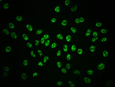
 IF (Immunofluorescence)
(Immunofluorescence staining of Hela cells(treated with 50mM Calyculin A for 30min) with CSB-RA009963A09phHU at 1:100,counter-stained with DAPI. The cells were fixed in 4% formaldehyde, permeabilized using 0.2% Triton X-100 and blocked in 10% normal Goat Serum. The cells were then incubated with the antibody overnight at 4 degree C. The secondary antibody was Alexa Fluor 488-congugated AffiniPure Goat Anti-Rabbit IgG (H+L).)
IF (Immunofluorescence)
(Immunofluorescence staining of Hela cells(treated with 50mM Calyculin A for 30min) with CSB-RA009963A09phHU at 1:100,counter-stained with DAPI. The cells were fixed in 4% formaldehyde, permeabilized using 0.2% Triton X-100 and blocked in 10% normal Goat Serum. The cells were then incubated with the antibody overnight at 4 degree C. The secondary antibody was Alexa Fluor 488-congugated AffiniPure Goat Anti-Rabbit IgG (H+L).)
GSK3B, Monoclonal Recombinant Antibody (Cat# AAA235550)
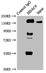 IP (Immunoprecipitation)
(Immunoprecipitating HDAC6 in HepG2 whole cell lysateLane 1: Rabbit control IgG instead of CSB-RA010242A0HU in HepG2 whole cell lysate.For western blotting, a HRP-conjugated Protein G antibody was used as the secondary antibody (1/2000)Lane 2: CSB-RA010242A0HU (3ug) + HepG2 whole cell lysate (500ug)Lane 3: HepG2 whole cell lysate (20ug))
IP (Immunoprecipitation)
(Immunoprecipitating HDAC6 in HepG2 whole cell lysateLane 1: Rabbit control IgG instead of CSB-RA010242A0HU in HepG2 whole cell lysate.For western blotting, a HRP-conjugated Protein G antibody was used as the secondary antibody (1/2000)Lane 2: CSB-RA010242A0HU (3ug) + HepG2 whole cell lysate (500ug)Lane 3: HepG2 whole cell lysate (20ug))
HDAC6, Monoclonal Recombinant Antibody (Cat# AAA235554)
 IF (Immunofluorescence)
(Immunofluorescence staining of Hela cells with CSB-RA010245A0HU at 1:51, counter-stained with DAPI. The cells were fixed in 4% formaldehyde, permeabilized using 0.2% Triton X-100 and blocked in 10% normal Goat Serum. The cells were then incubated with the antibody overnight at 4 degree C. The secondary antibody was Alexa Fluor 488-congugated AffiniPure Goat Anti-Rabbit IgG (H+L).)
IF (Immunofluorescence)
(Immunofluorescence staining of Hela cells with CSB-RA010245A0HU at 1:51, counter-stained with DAPI. The cells were fixed in 4% formaldehyde, permeabilized using 0.2% Triton X-100 and blocked in 10% normal Goat Serum. The cells were then incubated with the antibody overnight at 4 degree C. The secondary antibody was Alexa Fluor 488-congugated AffiniPure Goat Anti-Rabbit IgG (H+L).)
HDAC9, Monoclonal Recombinant Antibody (Cat# AAA235555)
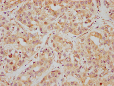
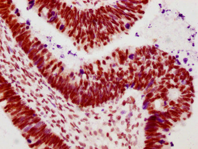
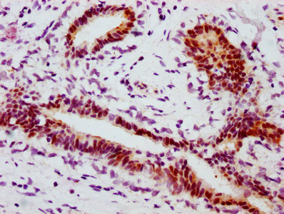
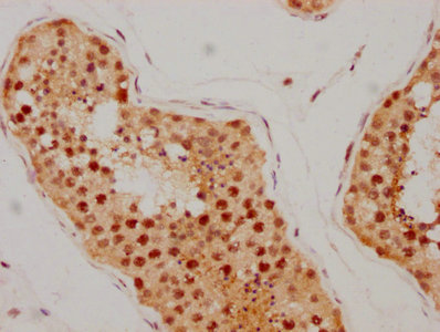
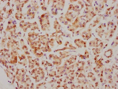
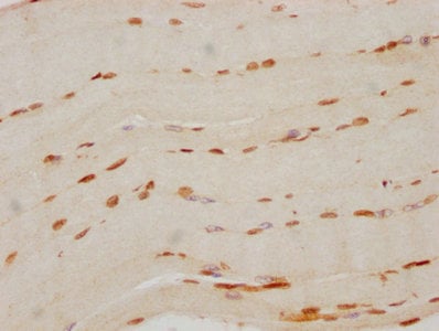
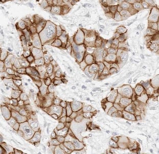
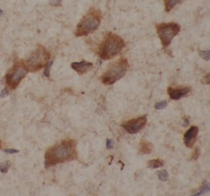
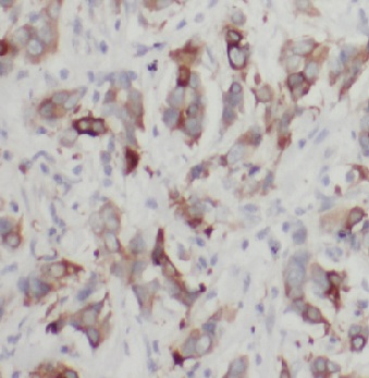
 IHC (Immunohistochemisry)
(IHC image of CSB-RA010833A78phHU diluted at 1:100 and staining in paraffin-embedded human breast cancer performed on a Leica BondTM system. After dewaxing and hydration, antigen retrieval was mediated by high pressure in a citrate buffer (pH 6.0). Section was blocked with 10% normal goat serum 30min at RT. Then primary antibody (1% BSA) was incubated at 4 degree C overnight. The primary is detected by a biotinylated secondary antibody and visualized using an HRP conjugated SP system.)
IHC (Immunohistochemisry)
(IHC image of CSB-RA010833A78phHU diluted at 1:100 and staining in paraffin-embedded human breast cancer performed on a Leica BondTM system. After dewaxing and hydration, antigen retrieval was mediated by high pressure in a citrate buffer (pH 6.0). Section was blocked with 10% normal goat serum 30min at RT. Then primary antibody (1% BSA) was incubated at 4 degree C overnight. The primary is detected by a biotinylated secondary antibody and visualized using an HRP conjugated SP system.)
HSPB1, Monoclonal Recombinant Antibody (Cat# AAA235561)
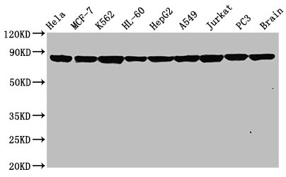
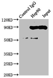 IP (Immunoprecipitation)
(Immunoprecipitating Hsp90 in Hela whole cell lysateLane 1: Rabbit control IgG instead of CSB-RA011087A0HU in Hela whole cell lysate.For western blotting, a HRP-conjugated Protein G antibody was used as the secondary antibody (1/2000)Lane 2: CSB-RA011087A0HU (3ug) + Hela whole cell lysate (500ug)Lane 3: Hela whole cell lysate (20ug))
IP (Immunoprecipitation)
(Immunoprecipitating Hsp90 in Hela whole cell lysateLane 1: Rabbit control IgG instead of CSB-RA011087A0HU in Hela whole cell lysate.For western blotting, a HRP-conjugated Protein G antibody was used as the secondary antibody (1/2000)Lane 2: CSB-RA011087A0HU (3ug) + Hela whole cell lysate (500ug)Lane 3: Hela whole cell lysate (20ug))
HSP90AA1, Monoclonal Recombinant Antibody (Cat# AAA235563)
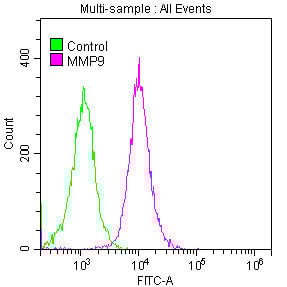 FCM/FACS (Flow Cytometry)
(Overlay histogram showing Jurkat cells stained with CSB-RA014679A0HU (red line) at 1:50. The cells were fixed with 70% Ethylalcohol (18h) and then permeabilized with 0.3% Triton X-100 for 2 min. The cells were then incubated in 1x PBS /10% normal goat serum to block non-specific protein-protein interactions followed by primary antibody for 1 h at 4 degree C. The secondary antibody used was FITC goat anti-rabbit IgG (H+L) at 1/200 dilution for 1 h at 4 degree C. Control antibody (green line) was used under the same conditions. Acquisition of >10,000 events was performed.)
FCM/FACS (Flow Cytometry)
(Overlay histogram showing Jurkat cells stained with CSB-RA014679A0HU (red line) at 1:50. The cells were fixed with 70% Ethylalcohol (18h) and then permeabilized with 0.3% Triton X-100 for 2 min. The cells were then incubated in 1x PBS /10% normal goat serum to block non-specific protein-protein interactions followed by primary antibody for 1 h at 4 degree C. The secondary antibody used was FITC goat anti-rabbit IgG (H+L) at 1/200 dilution for 1 h at 4 degree C. Control antibody (green line) was used under the same conditions. Acquisition of >10,000 events was performed.)
MMP9, Monoclonal Recombinant Antibody (Cat# AAA235573)

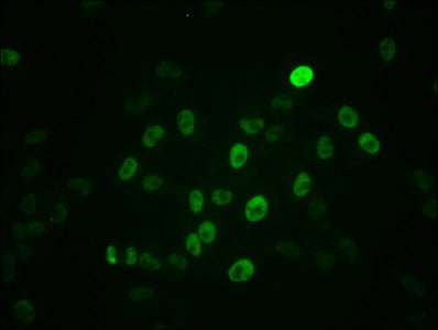 IF (Immunofluorescence)
(Immunofluorescence staining of Hela cells with CSB-RA015270A62phHU at 1:100,counter-stained with DAPI. The cells were fixed in 4% formaldehyde, permeabilized using 0.2% Triton X-100 and blocked in 10% normal Goat Serum. The cells were then incubated with the antibody overnight at 4 degree C. The secondary antibody was Alexa Fluor 488-congugated AffiniPure Goat Anti-Rabbit IgG (H+L).)
IF (Immunofluorescence)
(Immunofluorescence staining of Hela cells with CSB-RA015270A62phHU at 1:100,counter-stained with DAPI. The cells were fixed in 4% formaldehyde, permeabilized using 0.2% Triton X-100 and blocked in 10% normal Goat Serum. The cells were then incubated with the antibody overnight at 4 degree C. The secondary antibody was Alexa Fluor 488-congugated AffiniPure Goat Anti-Rabbit IgG (H+L).)
MYC, Monoclonal Recombinant Antibody (Cat# AAA235574)
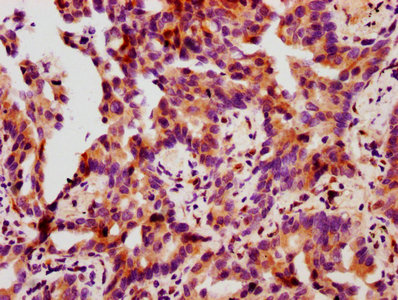
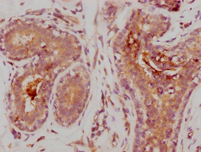
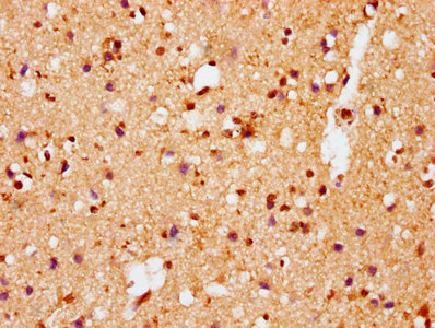
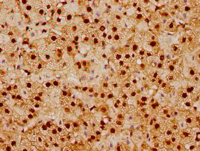
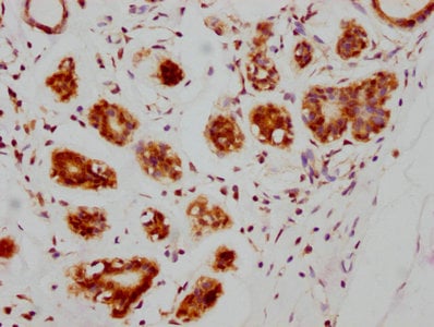
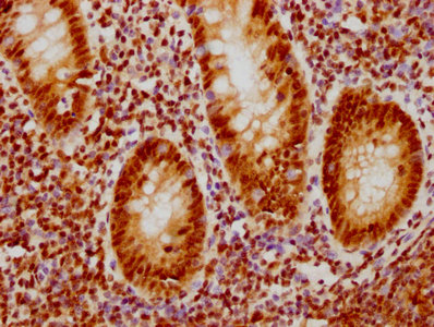
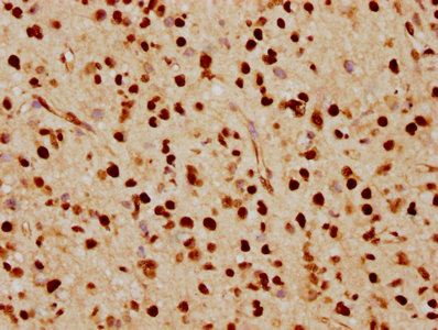
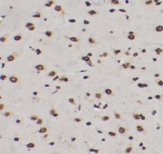
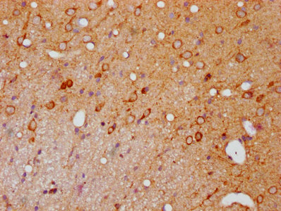 IHC (Immunohiostchemistry)
(IHC image of CSB-RA017407A144phHU diluted at 1:100 and staining in paraffin-embedded rat brain tissue performed on a Leica BondTM system. After dewaxing and hydration, antigen retrieval was mediated by high pressure in a citrate buffer (pH 6.0). Section was blocked with 10% normal goat serum 30min at RT. Then primary antibody (1% BSA) was incubated at 4 degree C overnight. The primary is detected by a biotinylated secondary antibody and visualized using an HRP conjugated SP system.)
IHC (Immunohiostchemistry)
(IHC image of CSB-RA017407A144phHU diluted at 1:100 and staining in paraffin-embedded rat brain tissue performed on a Leica BondTM system. After dewaxing and hydration, antigen retrieval was mediated by high pressure in a citrate buffer (pH 6.0). Section was blocked with 10% normal goat serum 30min at RT. Then primary antibody (1% BSA) was incubated at 4 degree C overnight. The primary is detected by a biotinylated secondary antibody and visualized using an HRP conjugated SP system.)
PAK1/PAK2/PAK3, Monoclonal Recombinant Antibody (Cat# AAA235577)
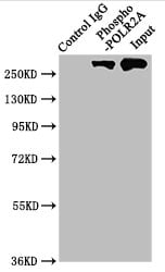 IP (Immunoprecipitation)
(Immunoprecipitating Phospho-POLR2A in Hela whole cell lysateLane 1: Rabbit control IgG(1ug)instead of CSB-RA018327A02phHU in Hela whole cell lysate.For western blotting,a HRP-conjugated Protein G antibody was used as the secondary antibody (1/2000)Lane 2: CSB-RA018327A02phHU(3ug)+ Hela whole cell lysate(1mg)Lane 3: Hela whole cell lysate (20ug))
IP (Immunoprecipitation)
(Immunoprecipitating Phospho-POLR2A in Hela whole cell lysateLane 1: Rabbit control IgG(1ug)instead of CSB-RA018327A02phHU in Hela whole cell lysate.For western blotting,a HRP-conjugated Protein G antibody was used as the secondary antibody (1/2000)Lane 2: CSB-RA018327A02phHU(3ug)+ Hela whole cell lysate(1mg)Lane 3: Hela whole cell lysate (20ug))
POLR2A, Monoclonal Recombinant Antibody (Cat# AAA235579)
 IF (Immunofluorescence)
(Immunofluorescence staining of Hela cells with CSB-RA018694A0HU at 1:36, counter-stained with DAPI. The cells were fixed in 4% formaldehyde, permeabilized using 0.2% Triton X-100 and blocked in 10% normal Goat Serum. The cells were then incubated with the antibody overnight at 4 degree C. The secondary antibody was Alexa Fluor 488-congugated AffiniPure Goat Anti-Rabbit IgG (H+L).)
IF (Immunofluorescence)
(Immunofluorescence staining of Hela cells with CSB-RA018694A0HU at 1:36, counter-stained with DAPI. The cells were fixed in 4% formaldehyde, permeabilized using 0.2% Triton X-100 and blocked in 10% normal Goat Serum. The cells were then incubated with the antibody overnight at 4 degree C. The secondary antibody was Alexa Fluor 488-congugated AffiniPure Goat Anti-Rabbit IgG (H+L).)
PRKAR1A, Monoclonal Recombinant Antibody (Cat# AAA235581)
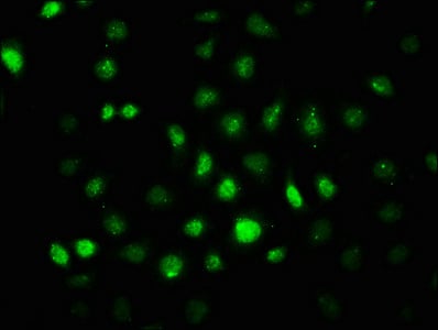
 IF (Immunofluorescence)
(Immunofluorescence staining of HepG2 cells with CSB-RA018699A638phHU at 1:100,counter-stained with DAPI. The cells were fixed in 4% formaldehyde, permeabilized using 0.2% Triton X-100 and blocked in 10% normal Goat Serum. The cells were then incubated with the antibody overnight at 4 degree C. The secondary antibody was Alexa Fluor 488-congugated AffiniPure Goat Anti-Rabbit IgG (H+L).)
IF (Immunofluorescence)
(Immunofluorescence staining of HepG2 cells with CSB-RA018699A638phHU at 1:100,counter-stained with DAPI. The cells were fixed in 4% formaldehyde, permeabilized using 0.2% Triton X-100 and blocked in 10% normal Goat Serum. The cells were then incubated with the antibody overnight at 4 degree C. The secondary antibody was Alexa Fluor 488-congugated AffiniPure Goat Anti-Rabbit IgG (H+L).)
PRKCA, Monoclonal Recombinant Antibody (Cat# AAA235582)
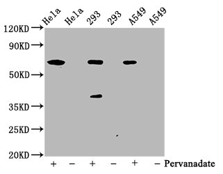
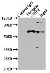 IP (Immunoprecipitation)
(Immunoprecipitating Phospho-PTPN11 in Hela whole cell lysate treated with PervanadateLane 1: Rabbit control IgG(1ug)instead of CSB-RA019025A542phHU in Hela whole cell lysate treated with Pervanadate.For western blotting,a HRP-conjugated Protein G antibody was used as the secondary antibody (1/2000)Lane 2: CSB-RA019025A542phHU(3ug)+ Hela whole cell lysate treated with Pervanadate(1mg)Lane 3: Hela whole cell lysate treated with Pervanadate(20ug))
IP (Immunoprecipitation)
(Immunoprecipitating Phospho-PTPN11 in Hela whole cell lysate treated with PervanadateLane 1: Rabbit control IgG(1ug)instead of CSB-RA019025A542phHU in Hela whole cell lysate treated with Pervanadate.For western blotting,a HRP-conjugated Protein G antibody was used as the secondary antibody (1/2000)Lane 2: CSB-RA019025A542phHU(3ug)+ Hela whole cell lysate treated with Pervanadate(1mg)Lane 3: Hela whole cell lysate treated with Pervanadate(20ug))
PTPN11, Monoclonal Recombinant Antibody (Cat# AAA235583)
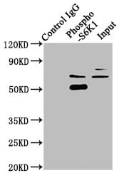 IP (Immunoprecipitation)
(Immunoprecipitating Phospho-RPS6KB1 in Hela whole cell lysateLane 1: Rabbit control IgG(1ug)instead of CSB-RA020470A421phHU in Hela whole cell lysate.For western blotting,a HRP-conjugated Protein G antibody was used as the secondary antibody (1/2000)Lane 2: CSB-RA020470A421phHU(3ug)+ Hela whole cell lysate(1mg)Lane 3: Hela whole cell lysate (20ug))
IP (Immunoprecipitation)
(Immunoprecipitating Phospho-RPS6KB1 in Hela whole cell lysateLane 1: Rabbit control IgG(1ug)instead of CSB-RA020470A421phHU in Hela whole cell lysate.For western blotting,a HRP-conjugated Protein G antibody was used as the secondary antibody (1/2000)Lane 2: CSB-RA020470A421phHU(3ug)+ Hela whole cell lysate(1mg)Lane 3: Hela whole cell lysate (20ug))
RPS6KB1, Monoclonal Recombinant Antibody (Cat# AAA235587)
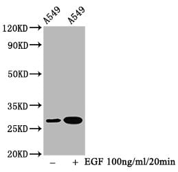
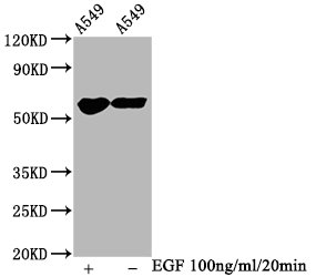
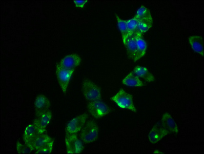
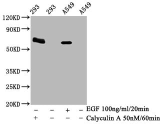
 IF (Immunofluorescence)
(Immunofluorescence staining of Hela cells(treated with 100mM EGF for 20min) with CSB-RA021912A129phHU at 1:206,counter-stained with DAPI. The cells were fixed in 4% formaldehyde, permeabilized using 0.2% Triton X-100 and blocked in 10% normal Goat Serum. The cells were then incubated with the antibody overnight at 4 degree C. The secondary antibody was Alexa Fluor 488-congugated AffiniPure Goat Anti-Rabbit IgG (H+L).)
IF (Immunofluorescence)
(Immunofluorescence staining of Hela cells(treated with 100mM EGF for 20min) with CSB-RA021912A129phHU at 1:206,counter-stained with DAPI. The cells were fixed in 4% formaldehyde, permeabilized using 0.2% Triton X-100 and blocked in 10% normal Goat Serum. The cells were then incubated with the antibody overnight at 4 degree C. The secondary antibody was Alexa Fluor 488-congugated AffiniPure Goat Anti-Rabbit IgG (H+L).)
SNCA, Monoclonal Recombinant Antibody (Cat# AAA235588)
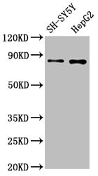
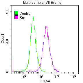 FCM/FACS (Flow Cytometry)
(Overlay histogram showing SH-SY5Y cells stained with CSB-RA022650A0HU (red line) at 1:50. The cells were fixed with 70% Ethylalcohol (18h) and then permeabilized with 0.3% Triton X-100 for 2 min. The cells were then incubated in 1x PBS /10% normal goat serum to block non-specific protein-protein interactions followed by primary antibody for 1 h at 4 degree C. The secondary antibody used was FITC goat anti-rabbit IgG (H+L) at 1/200 dilution for 1 h at 4 degree C. Control antibody (green line) was used under the same conditions. Acquisition of >10,000 events was performed.)
FCM/FACS (Flow Cytometry)
(Overlay histogram showing SH-SY5Y cells stained with CSB-RA022650A0HU (red line) at 1:50. The cells were fixed with 70% Ethylalcohol (18h) and then permeabilized with 0.3% Triton X-100 for 2 min. The cells were then incubated in 1x PBS /10% normal goat serum to block non-specific protein-protein interactions followed by primary antibody for 1 h at 4 degree C. The secondary antibody used was FITC goat anti-rabbit IgG (H+L) at 1/200 dilution for 1 h at 4 degree C. Control antibody (green line) was used under the same conditions. Acquisition of >10,000 events was performed.)
SRC, Monoclonal Recombinant Antibody (Cat# AAA235589)
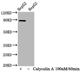
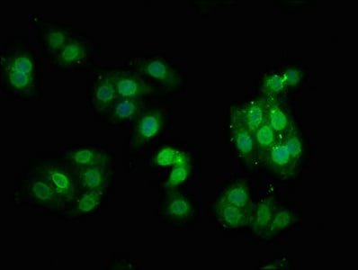
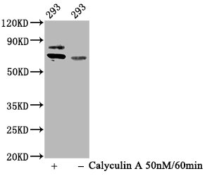
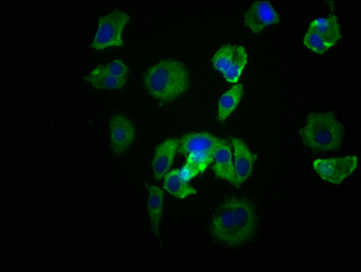
 IF (Immunofluorescence)
(Immunofluorescence staining of HepG2 cells(treated with 100mM Calyculin A for 30min) with CSB-RA022810A727phHU at 1:66,counter-stained with DAPI. The cells were fixed in 4% formaldehyde, permeabilized using 0.2% Triton X-100 and blocked in 10% normal Goat Serum. The cells were then incubated with the antibody overnight at 4 degree C. The secondary antibody was Alexa Fluor 488-congugated AffiniPure Goat Anti-Rabbit IgG (H+L).)
IF (Immunofluorescence)
(Immunofluorescence staining of HepG2 cells(treated with 100mM Calyculin A for 30min) with CSB-RA022810A727phHU at 1:66,counter-stained with DAPI. The cells were fixed in 4% formaldehyde, permeabilized using 0.2% Triton X-100 and blocked in 10% normal Goat Serum. The cells were then incubated with the antibody overnight at 4 degree C. The secondary antibody was Alexa Fluor 488-congugated AffiniPure Goat Anti-Rabbit IgG (H+L).)
STAT1, Monoclonal Recombinant Antibody (Cat# AAA235590)
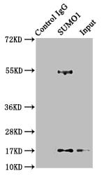
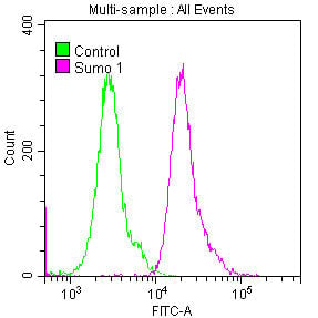 FCM/FACS (Flow Cytometry)
(Flow Cytometry (FC/FACS))
FCM/FACS (Flow Cytometry)
(Flow Cytometry (FC/FACS))
SUMO1, Monoclonal Recombinant Antibody (Cat# AAA235594)
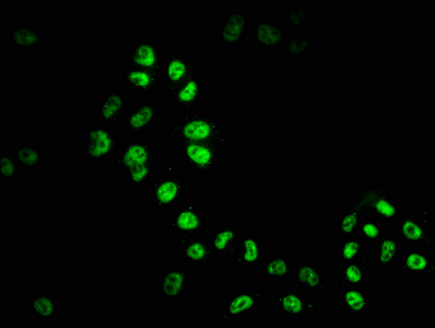
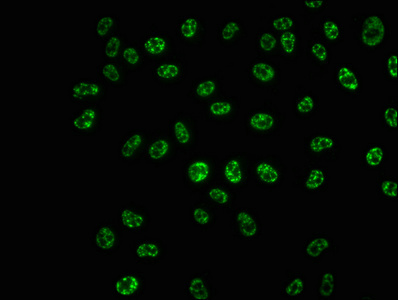
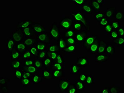
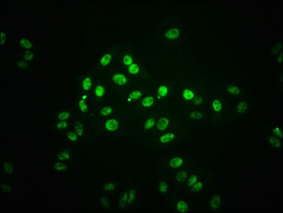
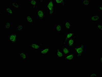
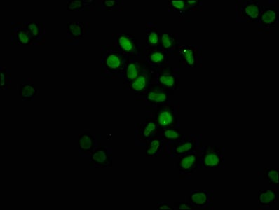
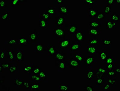 IF (Immunofluorescence)
(Immunofluorescence staining of Hela cells with CSB-RA024077A33phHU at 1:100,counter-stained with DAPI. The cells were fixed in 4% formaldehyde, permeabilized using 0.2% Triton X-100 and blocked in 10% normal Goat Serum. The cells were then incubated with the antibody overnight at 4 degree C. The secondary antibody was Alexa Fluor 488-congugated AffiniPure Goat Anti-Rabbit IgG (H+L).)
IF (Immunofluorescence)
(Immunofluorescence staining of Hela cells with CSB-RA024077A33phHU at 1:100,counter-stained with DAPI. The cells were fixed in 4% formaldehyde, permeabilized using 0.2% Triton X-100 and blocked in 10% normal Goat Serum. The cells were then incubated with the antibody overnight at 4 degree C. The secondary antibody was Alexa Fluor 488-congugated AffiniPure Goat Anti-Rabbit IgG (H+L).)
TP53, Monoclonal Recombinant Antibody (Cat# AAA235595)
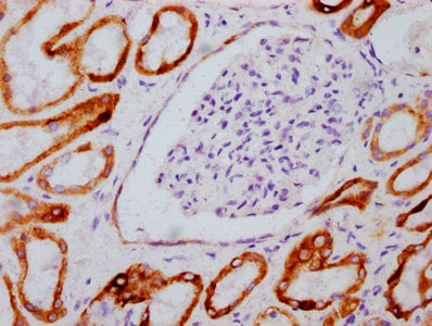
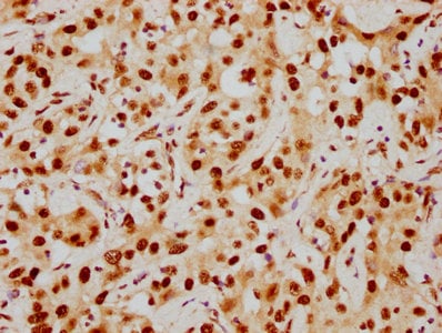
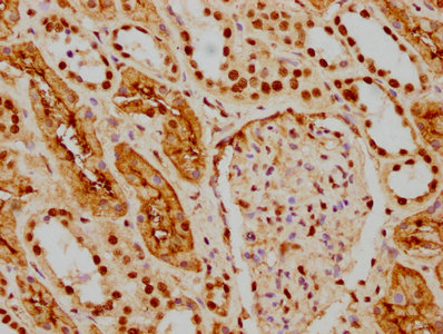
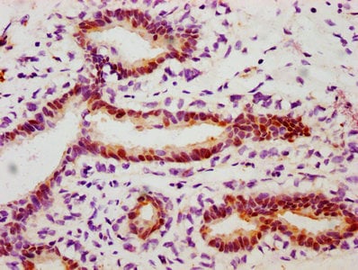
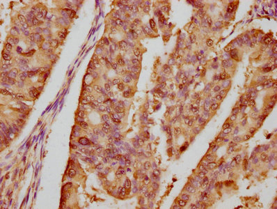
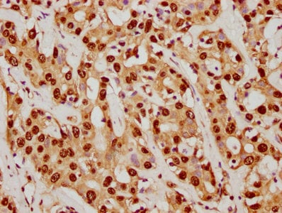
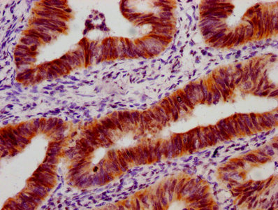
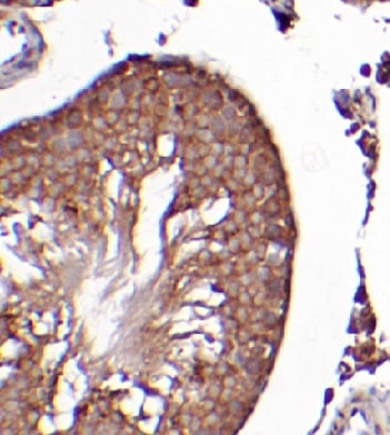
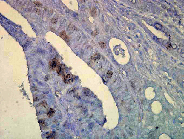
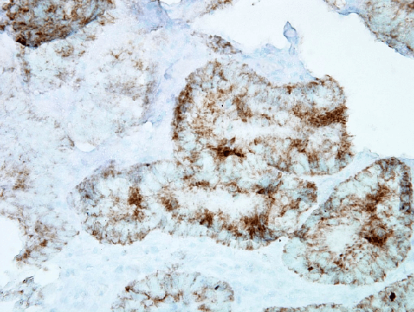
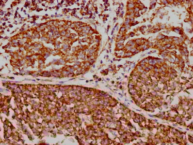 IHC (Immunohistochemisry)
(IHC image of CSB-RA025821A0HU diluted at 1:73.125 and staining in paraffin-embedded human cervical cancer performed on a Leica BondTM system. After dewaxing and hydration, antigen retrieval was mediated by high pressure in a citrate buffer (pH 6.0). Section was blocked with 10% normal goat serum 30min at RT. Then primary antibody (1% BSA) was incubated at 4 degree C overnight. The primary is detected by a biotinylated secondary antibody and visualized using an HRP conjugated SP system.)
IHC (Immunohistochemisry)
(IHC image of CSB-RA025821A0HU diluted at 1:73.125 and staining in paraffin-embedded human cervical cancer performed on a Leica BondTM system. After dewaxing and hydration, antigen retrieval was mediated by high pressure in a citrate buffer (pH 6.0). Section was blocked with 10% normal goat serum 30min at RT. Then primary antibody (1% BSA) was incubated at 4 degree C overnight. The primary is detected by a biotinylated secondary antibody and visualized using an HRP conjugated SP system.)
VDAC1, Monoclonal Recombinant Antibody (Cat# AAA235598)
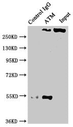 IP (Immunoprecipitation)
(Immunoprecipitating ATM in PC3 whole cell lysateLane 1: Rabbit control IgG instead of CSB-RA618770A0HU in PC3 whole cell lysate.For western blotting, a HRP-conjugated Protein G antibody was used as the secondary antibody (1/2000)Lane 2: CSB-RA618770A0HU (3ug) + PC3 whole cell lysate (500ug)Lane 3: PC3 whole cell lysate (20ug))
IP (Immunoprecipitation)
(Immunoprecipitating ATM in PC3 whole cell lysateLane 1: Rabbit control IgG instead of CSB-RA618770A0HU in PC3 whole cell lysate.For western blotting, a HRP-conjugated Protein G antibody was used as the secondary antibody (1/2000)Lane 2: CSB-RA618770A0HU (3ug) + PC3 whole cell lysate (500ug)Lane 3: PC3 whole cell lysate (20ug))
ATM, Monoclonal Recombinant Antibody (Cat# AAA235602)
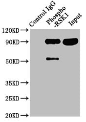 IP (Immunoprecipitation)
(Immunoprecipitating Phospho-RPS6KA1 in Hela whole cell lysateLane 1: Rabbit control IgG(1ug)instead of CSB-RA618984A380phHU in Hela whole cell lysate.For western blotting,a HRP-conjugated Protein G antibody was used as the secondary antibody (1/2000)Lane 2: CSB-RA618984A380phHU(3ug)+ Hela whole cell lysate(1mg)Lane 3: Hela whole cell lysate (20ug))
IP (Immunoprecipitation)
(Immunoprecipitating Phospho-RPS6KA1 in Hela whole cell lysateLane 1: Rabbit control IgG(1ug)instead of CSB-RA618984A380phHU in Hela whole cell lysate.For western blotting,a HRP-conjugated Protein G antibody was used as the secondary antibody (1/2000)Lane 2: CSB-RA618984A380phHU(3ug)+ Hela whole cell lysate(1mg)Lane 3: Hela whole cell lysate (20ug))
RPS6KA1, Monoclonal Recombinant Antibody (Cat# AAA235604)
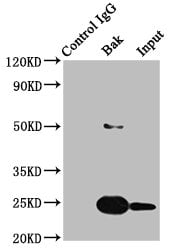
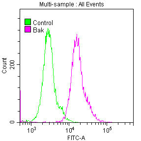 FCM/FACS (Flow Cytometry)
(Overlay histogram showing Hela cells stained with CSB-RA624111A0HU (red line) at 1:50. The cells were fixed with 70% Ethylalcohol (18h) and then permeabilized with 0.3% Triton X-100 for 2 min. The cells were then incubated in 1x PBS /10% normal goat serum to block non-specific protein-protein interactions followed by primary antibody for 1 h at 4 degree C. The secondary antibody used was FITC goat anti-rabbit IgG (H+L) at 1/200 dilution for 1 h at 4 degree C. Control antibody (green line) was used under the same conditions. Acquisition of >10,000 events was performed.)
FCM/FACS (Flow Cytometry)
(Overlay histogram showing Hela cells stained with CSB-RA624111A0HU (red line) at 1:50. The cells were fixed with 70% Ethylalcohol (18h) and then permeabilized with 0.3% Triton X-100 for 2 min. The cells were then incubated in 1x PBS /10% normal goat serum to block non-specific protein-protein interactions followed by primary antibody for 1 h at 4 degree C. The secondary antibody used was FITC goat anti-rabbit IgG (H+L) at 1/200 dilution for 1 h at 4 degree C. Control antibody (green line) was used under the same conditions. Acquisition of >10,000 events was performed.)
BAK1, Monoclonal Recombinant Antibody (Cat# AAA235605)
 IF (Immunofluorescence)
(Immunofluorescence staining of A549 cells with CSB-RA007795A724phHU at 1:100,counter-stained with DAPI. The cells were fixed in 4% formaldehyde, permeabilized using 0.2% Triton X-100 and blocked in 10% normal Goat Serum. The cells were then incubated with the antibody overnight at 4 degree C. The secondary antibody was Alexa Fluor 488-congugated AffiniPure Goat Anti-Rabbit IgG (H+L).)
IF (Immunofluorescence)
(Immunofluorescence staining of A549 cells with CSB-RA007795A724phHU at 1:100,counter-stained with DAPI. The cells were fixed in 4% formaldehyde, permeabilized using 0.2% Triton X-100 and blocked in 10% normal Goat Serum. The cells were then incubated with the antibody overnight at 4 degree C. The secondary antibody was Alexa Fluor 488-congugated AffiniPure Goat Anti-Rabbit IgG (H+L).)
PRKAA2, Monoclonal Recombinant Antibody (Cat# AAA235606)
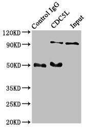 IP (Immunoprecipitation)
(Immunoprecipitating CDC5L in Hela whole cell lysateLane 1: Rabbit control IgG instead of CSB-RA858712A0HU in Hela whole cell lysate.For western blotting, a HRP-conjugated Protein G antibody was used as the secondary antibody (1/2000)Lane 2: CSB-RA858712A0HU (3ug) + Hela whole cell lysate (500ug)Lane 3: Hela whole cell lysate (20ug))
IP (Immunoprecipitation)
(Immunoprecipitating CDC5L in Hela whole cell lysateLane 1: Rabbit control IgG instead of CSB-RA858712A0HU in Hela whole cell lysate.For western blotting, a HRP-conjugated Protein G antibody was used as the secondary antibody (1/2000)Lane 2: CSB-RA858712A0HU (3ug) + Hela whole cell lysate (500ug)Lane 3: Hela whole cell lysate (20ug))
CDC5L, Monoclonal Recombinant Antibody (Cat# AAA235608)
 IF (Immunofluorescence)
(Immunofluorescence staining of Hela cells with CSB-RA880154A0HU at 1:23, counter-stained with DAPI. The cells were fixed in 4% formaldehyde, permeabilized using 0.2% Triton X-100 and blocked in 10% normal Goat Serum. The cells were then incubated with the antibody overnight at 4 degree C. The secondary antibody was Alexa Fluor 488-congugated AffiniPure Goat Anti-Rabbit IgG (H+L).)
IF (Immunofluorescence)
(Immunofluorescence staining of Hela cells with CSB-RA880154A0HU at 1:23, counter-stained with DAPI. The cells were fixed in 4% formaldehyde, permeabilized using 0.2% Triton X-100 and blocked in 10% normal Goat Serum. The cells were then incubated with the antibody overnight at 4 degree C. The secondary antibody was Alexa Fluor 488-congugated AffiniPure Goat Anti-Rabbit IgG (H+L).)
FTO, Monoclonal Recombinant Antibody (Cat# AAA235610)
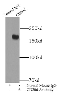
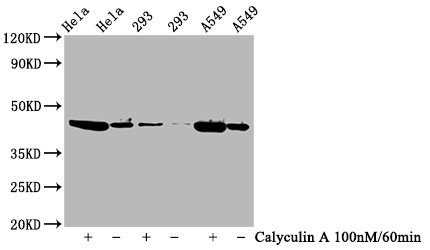
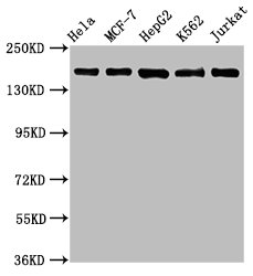
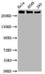
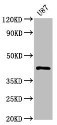
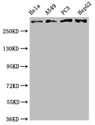
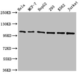
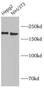
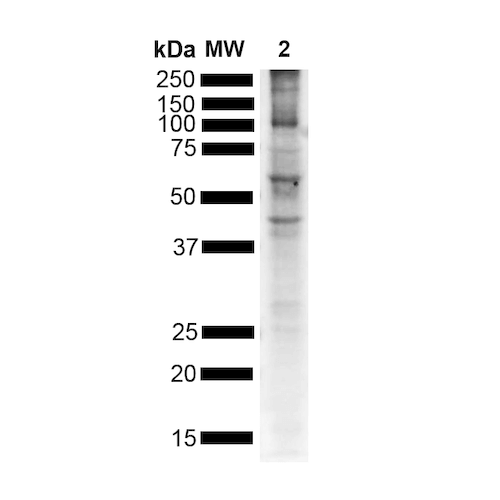
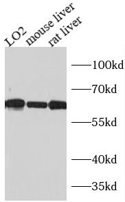
 WB (Western Blot)
(human liver tissue were subjected to SDS PAGE followed by western blot with AAA249032 (CD206 antibody) at dilution of 1:2000)
WB (Western Blot)
(human liver tissue were subjected to SDS PAGE followed by western blot with AAA249032 (CD206 antibody) at dilution of 1:2000)
CD206, Monoclonal Antibody (Cat# AAA249032)
Protein A+G Purification
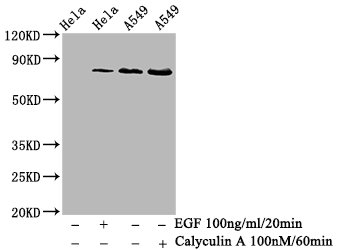
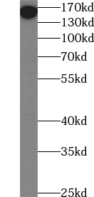
 WB (Western Blot)
(A431 cells were subjected to SDS PAGE followed by western blot (B-catenin antibody) at dilution of 1:5000)
WB (Western Blot)
(A431 cells were subjected to SDS PAGE followed by western blot (B-catenin antibody) at dilution of 1:5000)
beta-Catenin, Monoclonal Antibody (Cat# AAA249042)
Protein A+G Purified
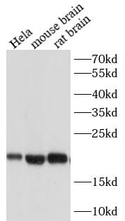
 WB (Western Blot)
(SH-SY5Y cells were subjected to SDS PAGE followed by western blot with AAA249044 (TUBB3 specific antibody) at dilution of 1:30000)
WB (Western Blot)
(SH-SY5Y cells were subjected to SDS PAGE followed by western blot with AAA249044 (TUBB3 specific antibody) at dilution of 1:30000)
TUBB3, Monoclonal Antibody (Cat# AAA249044)
Protein A+G Purified
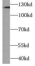 WB (Western Blot)
(Cobalt Chloride treated HeLa cells were subjected to SDS PAGE followed by western blot with AAA249054 (HIF1a Antibody) at dilution of 1:5000)
WB (Western Blot)
(Cobalt Chloride treated HeLa cells were subjected to SDS PAGE followed by western blot with AAA249054 (HIF1a Antibody) at dilution of 1:5000)
HIF1a, Monoclonal Antibody (Cat# AAA249054)
Protein A+G Purified
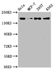
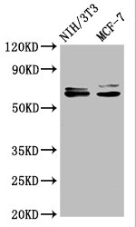
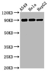
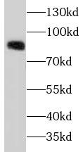
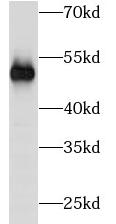
 WB (Western Blot)
(MCF7 cells were subjected to SDS PAGE followed by western blot with AAA249535(FAK Antibody) at dilution of 1:1000.)
WB (Western Blot)
(MCF7 cells were subjected to SDS PAGE followed by western blot with AAA249535(FAK Antibody) at dilution of 1:1000.)
FAK, Monoclonal Antibody (Cat# AAA249535)
 Application Data
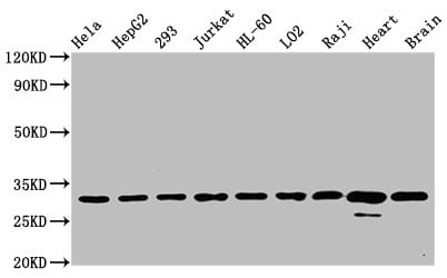
(Various lysates were subjected to SDS PAGE followed by western blot with AAA251230(GPX4 antibody) at dilution of 1:1000)
Application Data
(Various lysates were subjected to SDS PAGE followed by western blot with AAA251230(GPX4 antibody) at dilution of 1:1000)
GPX4, Monoclonal Antibody (Cat# AAA251230)
Protein A+G purification
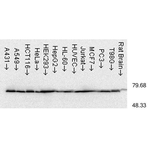
 Application Data
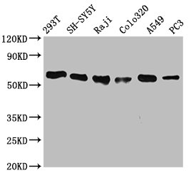
(Various cell lysates were subjected to SDS PAGE followed by western blot with BRD4 antibody at dilution of 1:2000)
Application Data
(Various cell lysates were subjected to SDS PAGE followed by western blot with BRD4 antibody at dilution of 1:2000)
BRD4, Monoclonal Antibody (Cat# AAA251231)
Protein A+G purification
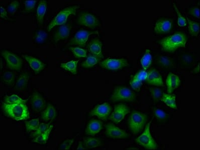
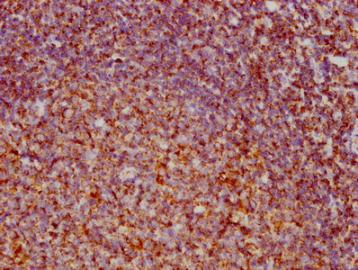
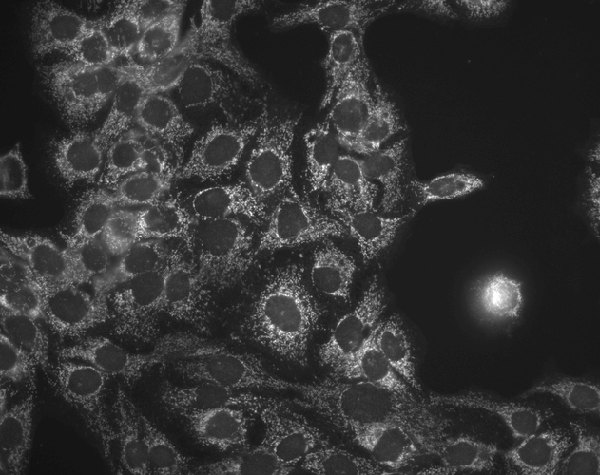
 IF (Immunofluorescence)
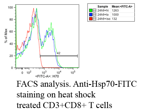
(Fluorescence Activated Cell Sorting analysis using Mouse Anti-Hsp70: FITC Monoclonal Antibody, Clone C92 . Tissue: Heat Shocked CD3+ CD8+ T cells. Species: Mouse. Primary Antibody: Mouse Anti-Hsp70: FITC Monoclonal Antibody at 1:1000. Courtesy of: Cheryl Cameron, Vaccine and Gene Therapy Instit. Florida.)
IF (Immunofluorescence)
(Fluorescence Activated Cell Sorting analysis using Mouse Anti-Hsp70: FITC Monoclonal Antibody, Clone C92 . Tissue: Heat Shocked CD3+ CD8+ T cells. Species: Mouse. Primary Antibody: Mouse Anti-Hsp70: FITC Monoclonal Antibody at 1:1000. Courtesy of: Cheryl Cameron, Vaccine and Gene Therapy Instit. Florida.)
HSP70, Monoclonal Antibody (Cat# AAA253926)
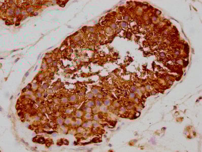
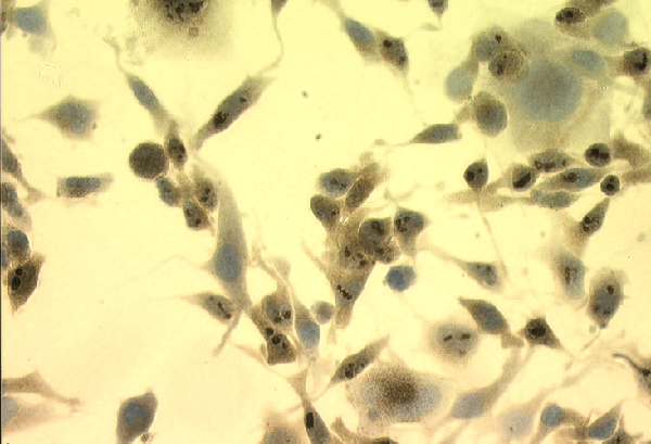
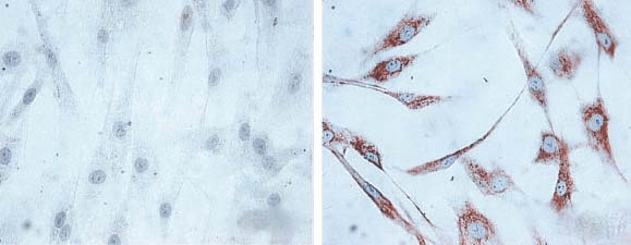 ICC (Immunocytochemistry)
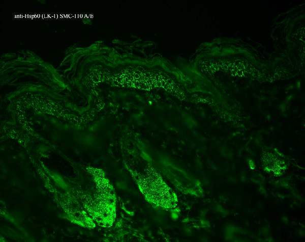
(Immunocytochemistry/Immunofluorescence analysis using Mouse Anti-Hsp60 Monoclonal Antibody, Clone LK1, . Tissue: skin Fibroblasts. Species: Human. Fixation: Cold 100% methanol for 30 minutes at-20 degree C. Primary Antibody: Mouse Anti-Hsp60 Monoclonal Antibody at 1:1000 for 1 hour at RT. Secondary Antibody: DAKO LSAB2 streptavidin-peroxidase system. Counterstain: Mayer Hematoxylin (purple/blue) nuclear stain. Left: control; Right: 24 hours after 7th passage of senescence. Courtesy of: Valentina di Felice, University of Palermo, Italy.)
ICC (Immunocytochemistry)
(Immunocytochemistry/Immunofluorescence analysis using Mouse Anti-Hsp60 Monoclonal Antibody, Clone LK1, . Tissue: skin Fibroblasts. Species: Human. Fixation: Cold 100% methanol for 30 minutes at-20 degree C. Primary Antibody: Mouse Anti-Hsp60 Monoclonal Antibody at 1:1000 for 1 hour at RT. Secondary Antibody: DAKO LSAB2 streptavidin-peroxidase system. Counterstain: Mayer Hematoxylin (purple/blue) nuclear stain. Left: control; Right: 24 hours after 7th passage of senescence. Courtesy of: Valentina di Felice, University of Palermo, Italy.)
HSP60, Monoclonal Antibody (Cat# AAA253929)
 WB (Western Blot)
(Western Blot analysis of Human Heat Shocked cervical cancer cell line (HeLa) lysate showing detection of Hsp60 protein using Mouse Anti-Hsp60 Monoclonal Antibody, Clone LK-2 . Load: 15 ug. Block: 1.5% BSA for 30 minutes at RT. Primary Antibody: Mouse Anti-Hsp60 Monoclonal Antibody at 1:1000 for 2 hours at RT. Secondary Antibody: Sheep Anti-Mouse IgG: HRP for 1 hour at RT.)
WB (Western Blot)
(Western Blot analysis of Human Heat Shocked cervical cancer cell line (HeLa) lysate showing detection of Hsp60 protein using Mouse Anti-Hsp60 Monoclonal Antibody, Clone LK-2 . Load: 15 ug. Block: 1.5% BSA for 30 minutes at RT. Primary Antibody: Mouse Anti-Hsp60 Monoclonal Antibody at 1:1000 for 2 hours at RT. Secondary Antibody: Sheep Anti-Mouse IgG: HRP for 1 hour at RT.)
HSP60, Monoclonal Antibody (Cat# AAA253930)
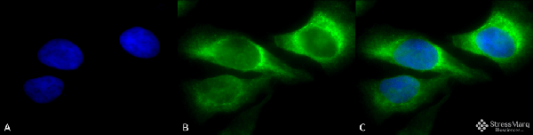
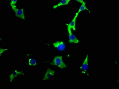
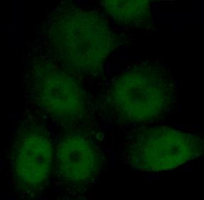
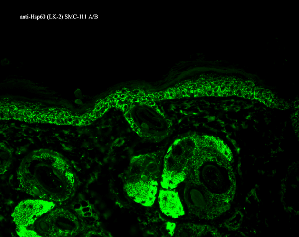
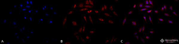 ICC (Immunocytochemistry)
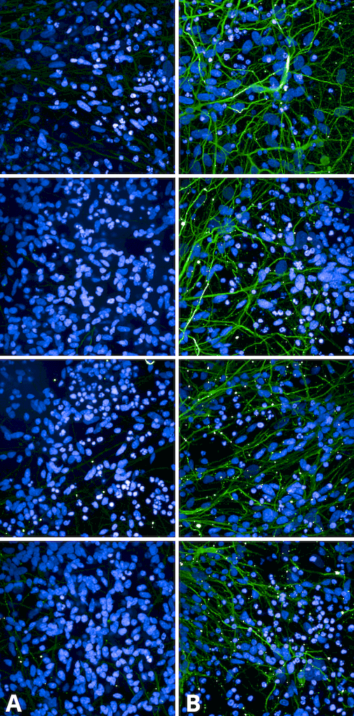
(Immunocytochemistry/Immunofluorescence analysis using Mouse Anti-Hsp47 Monoclonal Antibody, Clone 1C4-1A6 . Tissue: Heat Shocked cervical cancer cells (HeLa). Species: Human. Fixation: 2% Formaldehyde for 20 min at RT. Primary Antibody: Mouse Anti-Hsp47 Monoclonal Antibody at 1:100 for 12 hours at 4 degree C. Secondary Antibody: APC Goat Anti-Mouse (red) at 1:200 for 2 hours at RT. Counterstain: DAPI (blue) nuclear stain at 1:40000 for 2 hours at RT. Localization: Endoplasmic reticulum lumen. Cytoplasm. Magnification: 20x. (A) DAPI (blue) nuclear stain. (B) Anti-Hsp47 Antibody. (C) Composite. Heat Shocked at 42 degree C for 1h.)
ICC (Immunocytochemistry)
(Immunocytochemistry/Immunofluorescence analysis using Mouse Anti-Hsp47 Monoclonal Antibody, Clone 1C4-1A6 . Tissue: Heat Shocked cervical cancer cells (HeLa). Species: Human. Fixation: 2% Formaldehyde for 20 min at RT. Primary Antibody: Mouse Anti-Hsp47 Monoclonal Antibody at 1:100 for 12 hours at 4 degree C. Secondary Antibody: APC Goat Anti-Mouse (red) at 1:200 for 2 hours at RT. Counterstain: DAPI (blue) nuclear stain at 1:40000 for 2 hours at RT. Localization: Endoplasmic reticulum lumen. Cytoplasm. Magnification: 20x. (A) DAPI (blue) nuclear stain. (B) Anti-Hsp47 Antibody. (C) Composite. Heat Shocked at 42 degree C for 1h.)
HSP47, Monoclonal Antibody (Cat# AAA253942)
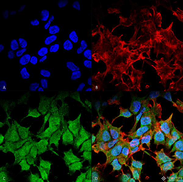
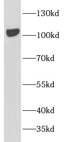
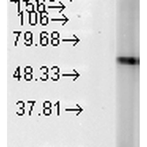
 WB (Western Blot)
(Western Blot analysis of Rat Brain showing detection of ~45 kDa Kir6.1 protein using Mouse Anti-Kir6.1 Monoclonal Antibody, Clone S366-60 at 1:1000 for 16 hours at 4 degree C. Secondary Antibody: Goat Anti-Mouse IgG: HRP at 1:200 for 1 hour at RT. Color Development: ECL solution for 6 min at RT. Predicted/Observed Size: ~45 kDa. Other Band(s): ~100 kDa.)
WB (Western Blot)
(Western Blot analysis of Rat Brain showing detection of ~45 kDa Kir6.1 protein using Mouse Anti-Kir6.1 Monoclonal Antibody, Clone S366-60 at 1:1000 for 16 hours at 4 degree C. Secondary Antibody: Goat Anti-Mouse IgG: HRP at 1:200 for 1 hour at RT. Color Development: ECL solution for 6 min at RT. Predicted/Observed Size: ~45 kDa. Other Band(s): ~100 kDa.)
Kir6.1, Monoclonal Antibody (Cat# AAA253963)
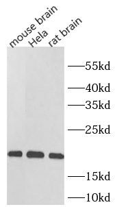
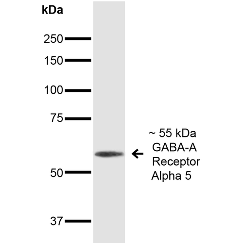 WB (Western Blot)
(Western Blot analysis of Mouse Brain showing detection of ~55 kDa GABA A Receptor Alpha 5 protein using Mouse Anti-GABA A Receptor Alpha 5 Monoclonal Antibody, Clone S415-24 at 1:1000 for 16 hours at 4 degree C. Secondary Antibody: Goat Anti-Mouse IgG: HRP at 1:200 for 1 hour at RT. Predicted/Observed Size: ~55 kDa.)
WB (Western Blot)
(Western Blot analysis of Mouse Brain showing detection of ~55 kDa GABA A Receptor Alpha 5 protein using Mouse Anti-GABA A Receptor Alpha 5 Monoclonal Antibody, Clone S415-24 at 1:1000 for 16 hours at 4 degree C. Secondary Antibody: Goat Anti-Mouse IgG: HRP at 1:200 for 1 hour at RT. Predicted/Observed Size: ~55 kDa.)
GABA A Receptor Alpha 5, Monoclonal Antibody (Cat# AAA253964)
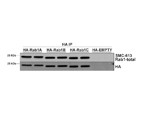 IP (Immunoprecipitation)
(Immunoprecipitation analysis using Mouse Anti-RAB1A Monoclonal Antibody, Clone 4G10 . Tissue: HEK293 cells overexpressing RAB1A, RAB1B, and RAB1C. Species: Human. Primary Antibody: Mouse Anti-RAB1A Monoclonal Antibody .)
IP (Immunoprecipitation)
(Immunoprecipitation analysis using Mouse Anti-RAB1A Monoclonal Antibody, Clone 4G10 . Tissue: HEK293 cells overexpressing RAB1A, RAB1B, and RAB1C. Species: Human. Primary Antibody: Mouse Anti-RAB1A Monoclonal Antibody .)
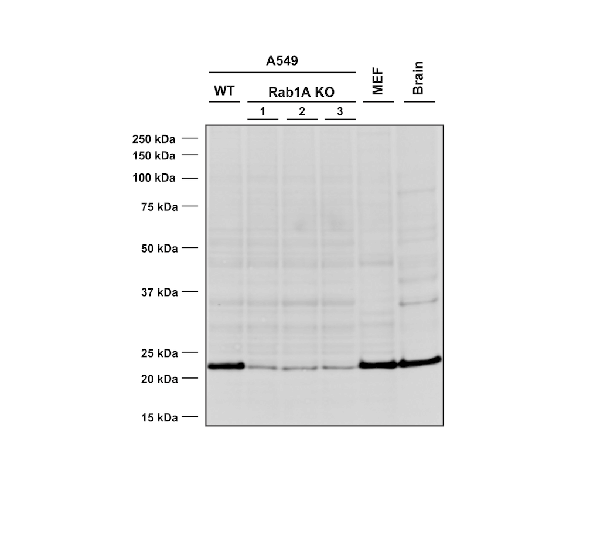
RAB1A, Monoclonal Antibody (Cat# AAA254043)
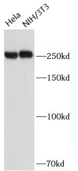
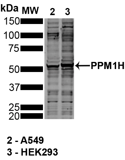 WB (Western Blot)
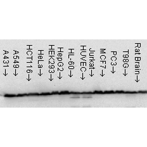
(Western BlotanalysisofHu,Ms,RtKidneyCells(HEK293),AlveolarBasalEpithelialcells(A549)showingdetectionof~55PPM1HproteinusingMouseAnti-PPM1HMonoclonalAntibody,Clone10D2 .Lane1:MWLadder.Lane2:A549.Lane3:HEK293..Load:30ug.Block:5%SkimMilkinTBST,1h,RT.PrimaryAntibody:MouseAnti-PPM1HMonoclonalAntibody at1:1000forOvernight,4C.SecondaryAntibody:Donkeyanti-mouse800(LiCOR)at1:25000for1houratRT.ColorDevelopment:OdysseyCLxWestern Blotimaging.Predicted/ObservedSize:~55.OtherBand(s):~30,~100.Courtesyof:DarioAlessiLab,UniversityofDundee.)
WB (Western Blot)
(Western BlotanalysisofHu,Ms,RtKidneyCells(HEK293),AlveolarBasalEpithelialcells(A549)showingdetectionof~55PPM1HproteinusingMouseAnti-PPM1HMonoclonalAntibody,Clone10D2 .Lane1:MWLadder.Lane2:A549.Lane3:HEK293..Load:30ug.Block:5%SkimMilkinTBST,1h,RT.PrimaryAntibody:MouseAnti-PPM1HMonoclonalAntibody at1:1000forOvernight,4C.SecondaryAntibody:Donkeyanti-mouse800(LiCOR)at1:25000for1houratRT.ColorDevelopment:OdysseyCLxWestern Blotimaging.Predicted/ObservedSize:~55.OtherBand(s):~30,~100.Courtesyof:DarioAlessiLab,UniversityofDundee.)
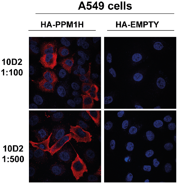
PPM1H IgG1, Monoclonal Antibody (Cat# AAA254073)
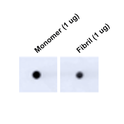
 WB (Western Blot)
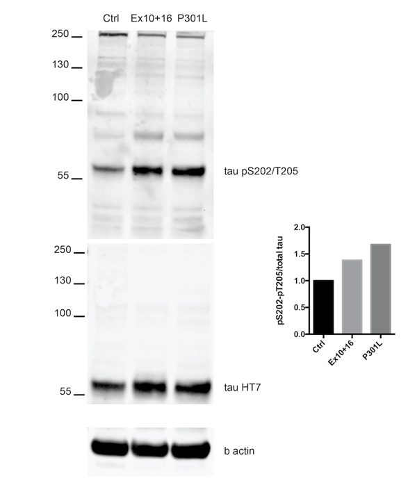
(Western Blot analysis of Human Alpha Synuclein protein using Rabbit Anti-Alpha Synuclein pSer129 Monoclonal Antibody, Clone J18. Lane 1: MW ladder. Lane 2: 0.5 ug human alpha synuclein monomer. Lane 3: 2 ug human alpha synuclein monomer. Lane 4: 0.5 ug human alpha synuclein PFFs. Lane 5: 2 ug human alpha synuclein PFFs. Block: 5% BSA in TBST. Primary Antibody: Rabbit Anti-Alpha Synuclein pSer129 Monoclonal Antibody at 1:500 for 2 hours at RT with shaking. Secondary Antibody: Goat anti-mouse IgG:HRP at 1:4000 for 1 hour at RT with shaking. Color Development: Chemiluminescent for HRP (Moss) for 5 min in RT. It does not detect unphosphorylated alpha synuclein.)
WB (Western Blot)
(Western Blot analysis of Human Alpha Synuclein protein using Rabbit Anti-Alpha Synuclein pSer129 Monoclonal Antibody, Clone J18. Lane 1: MW ladder. Lane 2: 0.5 ug human alpha synuclein monomer. Lane 3: 2 ug human alpha synuclein monomer. Lane 4: 0.5 ug human alpha synuclein PFFs. Lane 5: 2 ug human alpha synuclein PFFs. Block: 5% BSA in TBST. Primary Antibody: Rabbit Anti-Alpha Synuclein pSer129 Monoclonal Antibody at 1:500 for 2 hours at RT with shaking. Secondary Antibody: Goat anti-mouse IgG:HRP at 1:4000 for 1 hour at RT with shaking. Color Development: Chemiluminescent for HRP (Moss) for 5 min in RT. It does not detect unphosphorylated alpha synuclein.)
Alpha Synuclein, Monoclonal Antibody (Cat# AAA253969)

 WB (Western Blot)
(Western Blot analysis of Human Alpha Synuclein protein using Rabbit Anti-Alpha Synuclein pSer129 Monoclonal Antibody, Clone J18. Lane 1: MW ladder. Lane 2: 0.5 ug human alpha synuclein monomer. Lane 3: 2 ug human alpha synuclein monomer. Lane 4: 0.5 ug human alpha synuclein PFFs. Lane 5: 2 ug human alpha synuclein PFFs. Block: 5% BSA in TBST. Primary Antibody: Rabbit Anti-Alpha Synuclein pSer129 Monoclonal Antibody at 1:500 for 2 hours at RT with shaking. Secondary Antibody: Goat anti-mouse IgG:HRP at 1:4000 for 1 hour at RT with shaking. Color Development: Chemiluminescent for HRP (Moss) for 5 min in RT. It does not detect unphosphorylated alpha synuclein.)
WB (Western Blot)
(Western Blot analysis of Human Alpha Synuclein protein using Rabbit Anti-Alpha Synuclein pSer129 Monoclonal Antibody, Clone J18. Lane 1: MW ladder. Lane 2: 0.5 ug human alpha synuclein monomer. Lane 3: 2 ug human alpha synuclein monomer. Lane 4: 0.5 ug human alpha synuclein PFFs. Lane 5: 2 ug human alpha synuclein PFFs. Block: 5% BSA in TBST. Primary Antibody: Rabbit Anti-Alpha Synuclein pSer129 Monoclonal Antibody at 1:500 for 2 hours at RT with shaking. Secondary Antibody: Goat anti-mouse IgG:HRP at 1:4000 for 1 hour at RT with shaking. Color Development: Chemiluminescent for HRP (Moss) for 5 min in RT. It does not detect unphosphorylated alpha synuclein.)
Alpha Synuclein, Monoclonal Antibody (Cat# AAA253972)
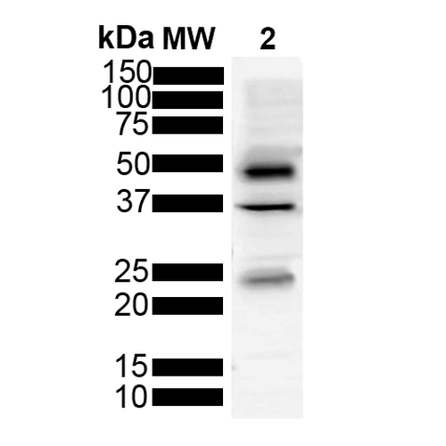
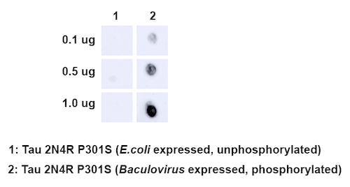 DB (Dot Blot)
(Dot Blot analysis using Rabbit Anti-Tau Monoclonal Antibody, Clone AH36. Species: E. Coli, Baculovirus. Primary Antibody: Rabbit Anti-Tau Monoclonal Antibody at 1:500. Secondary Antibody: Goat anti-rabbit IgG:HRP.)
DB (Dot Blot)
(Dot Blot analysis using Rabbit Anti-Tau Monoclonal Antibody, Clone AH36. Species: E. Coli, Baculovirus. Primary Antibody: Rabbit Anti-Tau Monoclonal Antibody at 1:500. Secondary Antibody: Goat anti-rabbit IgG:HRP.)
Tau, Monoclonal Antibody (Cat# AAA253978)
 DB (Dot Blot)
(Dot Blot analysis using Mouse Anti-Tau Monoclonal Antibody, Clone 3D4 (SMC-608). Tissue: Recombinant Protein. Species: Human. Primary Antibody: Mouse Anti-Tau Monoclonal Antibody (SMC-608) at 1:1000 for 2 hours at RT with shaking. Secondary Antibody: Goat anti-mouse IgG:HRP at 1:5000 for 1 hour at RT with shaking.)
DB (Dot Blot)
(Dot Blot analysis using Mouse Anti-Tau Monoclonal Antibody, Clone 3D4 (SMC-608). Tissue: Recombinant Protein. Species: Human. Primary Antibody: Mouse Anti-Tau Monoclonal Antibody (SMC-608) at 1:1000 for 2 hours at RT with shaking. Secondary Antibody: Goat anti-mouse IgG:HRP at 1:5000 for 1 hour at RT with shaking.)
Tau, Monoclonal Antibody (Cat# AAA253989)

 DB (Dot Blot)
(Dot Blot analysis using Mouse Anti-Tau Monoclonal Antibody, Clone 3D4 (SMC-608). Tissue: Recombinant Protein. Species: Human. Primary Antibody: Mouse Anti-Tau Monoclonal Antibody (SMC-608) at 1:1000 for 2 hours at RT with shaking. Secondary Antibody: Goat anti-mouse IgG:HRP at 1:5000 for 1 hour at RT with shaking.)
DB (Dot Blot)
(Dot Blot analysis using Mouse Anti-Tau Monoclonal Antibody, Clone 3D4 (SMC-608). Tissue: Recombinant Protein. Species: Human. Primary Antibody: Mouse Anti-Tau Monoclonal Antibody (SMC-608) at 1:1000 for 2 hours at RT with shaking. Secondary Antibody: Goat anti-mouse IgG:HRP at 1:5000 for 1 hour at RT with shaking.)
Tau, Monoclonal Antibody (Cat# AAA253993)

 DB (Dot Blot)
(Dot Blot analysis using Mouse Anti-Tau Monoclonal Antibody, Clone 3D4 (SMC-608). Tissue: Recombinant Protein. Species: Human. Primary Antibody: Mouse Anti-Tau Monoclonal Antibody (SMC-608) at 1:1000 for 2 hours at RT with shaking. Secondary Antibody: Goat anti-mouse IgG:HRP at 1:5000 for 1 hour at RT with shaking.)
DB (Dot Blot)
(Dot Blot analysis using Mouse Anti-Tau Monoclonal Antibody, Clone 3D4 (SMC-608). Tissue: Recombinant Protein. Species: Human. Primary Antibody: Mouse Anti-Tau Monoclonal Antibody (SMC-608) at 1:1000 for 2 hours at RT with shaking. Secondary Antibody: Goat anti-mouse IgG:HRP at 1:5000 for 1 hour at RT with shaking.)
Tau, Monoclonal Antibody (Cat# AAA253995)

 WB (Western Blot)
(various lysates were subjected to SDS PAGE followed by western blot with AAA253815(ACC1 antibody) at dilution of 1:2000)
WB (Western Blot)
(various lysates were subjected to SDS PAGE followed by western blot with AAA253815(ACC1 antibody) at dilution of 1:2000)
ACC1, Monoclonal Antibody (Cat# AAA253815)
>=95% as determined by SDS-PAGE

 WB (Western Blot)
(Various lysates were subjected to SDS PAGE followed by western blot with AAA253818(Catalase antibody) at dilution of 1:2000)
WB (Western Blot)
(Various lysates were subjected to SDS PAGE followed by western blot with AAA253818(Catalase antibody) at dilution of 1:2000)
CAT, Monoclonal Antibody (Cat# AAA253818)
>=95% as determined by SDS-PAGE
 WB (Western Blot)
(Various lysates were subjected to SDS PAGE followed by western blot with AAA253819(SOD1 antibody) at dilution of 1:2000)
WB (Western Blot)
(Various lysates were subjected to SDS PAGE followed by western blot with AAA253819(SOD1 antibody) at dilution of 1:2000)
SOD1, Monoclonal Antibody (Cat# AAA253819)
>=95% as determined by SDS-PAGE
What are Monoclonal Antibodies?
Monoclonal antibodies are specialized laboratory-produced proteins developed for binding to specific biological antigens or other molecular targets. Since they come from a single cell (or clone), they are especially consistent and accurate in the data they are involved in producing.
This type of antibody material has been shown to be a powerful tool in finding and subsequently destroying harmful cells in an organism, such as those found in cancers or various autoimmune diseases. This makes them excellent aids in medical testing and research, which is why they are so widely used.
AAA Biotech offers a comprehensive range of high-quality monoclonal antibodies that perform effectively in various laboratory tests, including (amongst others) ELISA, western blotting, immunohistochemistry, and flow cytometry. All of the products in our catalog are thoroughly quality tested to make sure that they are reliable and will consistently perform well in your research.
What Are The Uses of Monoclonal Antibodies
Monoclonal antibodies are used in many lab tests, including (amongst others) ELISA, western blotting, immunohistochemistry, and flow cytometry.
ELISA is a test that helps detect a specific substance/analyte in a sample. It uses antibodies (often monoclonal) bound to a solid surface (such as the well of a microplate) to “capture” the substance/analyte in the sample and immobilize it so that the detection antibody component can then bind to it and produce a signal, which can then be measured.
Western blotting identifies specific proteins in a sample. The sample is first separated on a gel, and then antibodies are applied that will typically bind to the target, which will all be localized to a single band in a lane.
Immunohistochemistry helps locate specific proteins in cells or tissue samples using antibodies.
Flow cytometry looks at and sorts cells. It uses antibodies that are conjugated to reporter molecules called “fluorophores”, which, under special lights, emit light themselves, which can then be measured by a detector instrument. For a deeper understanding of these techniques, explore our complete guide to monoclonal antibodies and their benefits.
How Monoclonal Antibodies Are Used as Medicine?
Please note that all of the products listed in AAA Biotech’s also known as AAA Bio or AAABio catalog are strictly for research-use only (RUO).
Monoclonal antibodies can also be used as therapeutic/medical treatments, particularly in the context of cancers. They are designed to find and bind to specific cells or proteins, helping the immune system recognize and attack the cancer. These treatments work in different ways, such as:
- Radioimmunotherapy attaches a small amount of radioactive molecule to the antibody, so it delivers the radiation directly to the cancer cells that the antibody is specifically binding to.
- Antibody-directed enzyme prodrug therapy uses antibodies that are specifically bound to special enzymes. These enzymes activate a harmless drug in the body and turn it into a cancer-killing drug only near the cancer cells—this helps avoid harming healthy cells.
- Immunoliposomes are tiny “bubbles” filled with medicine/drug and coated with antibodies. They carry the drug straight to the cancer cells.
Why Buy Monoclonal Antibodies From Us?
At AAA Biotech, we provide high-performance monoclonal antibodies designed to support a wide range of research needs.
1. Validated for Versatile Applications
The antibodies in our catalog are extensively validated and compatible with multiple techniques, including (but not limited to) ELISA, flow cytometry (FC), immunocytochemistry (ICC), immunofluorescence (IF), immunohistochemistry (IHC), immunoprecipitation (IP), and western blotting (WB).
2. Wide Selection & Specialized Options
We offer antibodies for common and rare species, that are available in various conjugated forms, and also in recombinant formats. Essentially, there is almost anything one might need to meet their experimental model’s requirements.
3. High-Quality Proteins
Our proteins meet high purity standards—90% or more as confirmed by SDS-PAGE. Many are available with tags like His, Flag, GST, or MBP, and we also supply native and biologically active proteins for functional studies.
Frequently Asked Questions
1. Are your monoclonal antibodies validated for specific applications?
Yes, our antibodies are tested and validated for use in methods such as ELISA, western blot, IHC, flow cytometry, and more. Refer to specific product pages or datasheets for individual product information.
2. How do I choose the right monoclonal antibody for my application?
Review the product details directly for application validation, species reactivity, and target information. You may also contact our support team at any time for help.
3. How quickly can I receive my order?
Most orders are processed and shipped within 1–3 business days, depending on product availability and your shipping location.