Filters
▼Clonality
▼Type
▼Reactivity
▼Gene Name
▼Isotype
▼Host
▼Application
▼Clone
▼Monoclonal Antibodies
Get accurate results in your research with our Monoclonal Antibodies, which are specially made to target exactly what you require for your research, and will produce consistent, reliable performance in lab tests.
Viewing 50-100 of 27645 product results
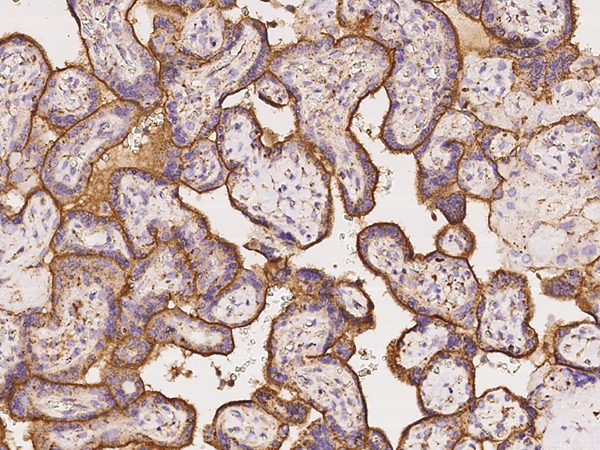
HLA-G1, Monoclonal Recombinant Antibody (Cat# AAA120733)
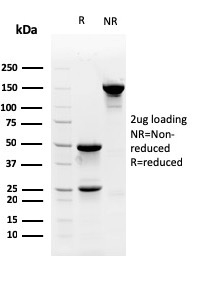
Protein A/G purified from cell culture supernatant.
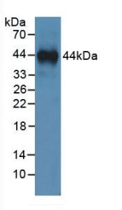
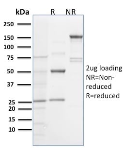 SDS-PAGE
(SDS-PAGE Analysis Purified Spastin Mouse Monoclonal Antibody (Sp 6C6). Confirmation of Purity and Integrity of Antibody.)
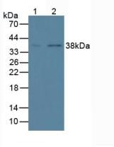
SDS-PAGE
(SDS-PAGE Analysis Purified Spastin Mouse Monoclonal Antibody (Sp 6C6). Confirmation of Purity and Integrity of Antibody.)
Spastin, Monoclonal Antibody (Cat# AAA215205)
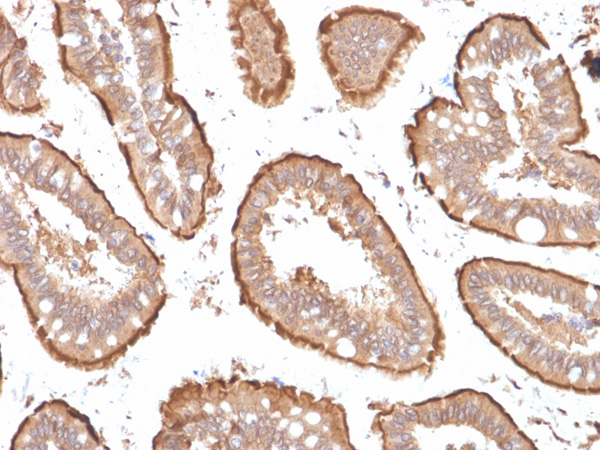
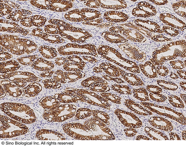
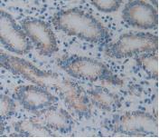
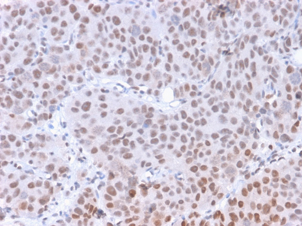
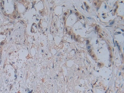
 IHC (Immunohiostchemistry)
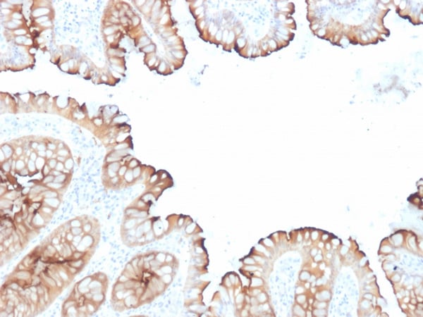
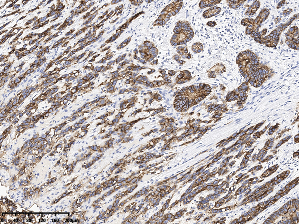
(Formalin-fixed, paraffin-embedded human Colon Carcinoma stained with Villin Mouse Monoclonal Antibody (VIL1/1314 + VIL1/2376).)
IHC (Immunohiostchemistry)
(Formalin-fixed, paraffin-embedded human Colon Carcinoma stained with Villin Mouse Monoclonal Antibody (VIL1/1314 + VIL1/2376).)
Villin, Monoclonal Antibody (Cat# AAA215252)
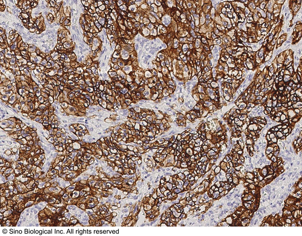
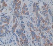
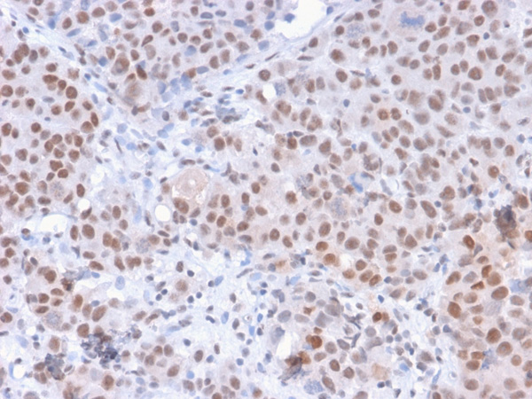
 IHC (Immunohiostchemistry)
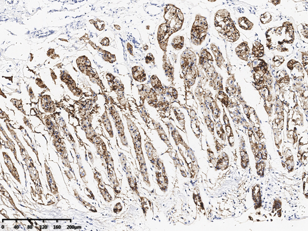
(Formalin-fixed, paraffin-embedded human Renal Cell Carcinoma stained with PAX8 Monoclonal Antibody (PAX8/1491 + PAX8/1492).)
IHC (Immunohiostchemistry)
(Formalin-fixed, paraffin-embedded human Renal Cell Carcinoma stained with PAX8 Monoclonal Antibody (PAX8/1491 + PAX8/1492).)
PAX8, Monoclonal Antibody (Cat# AAA215258)
 IHC (Immunohistochemistry)
(Formalin-fixed, paraffin-embedded human Skin stained with Pan-Cytokeratin Recombinant Rabbit Monoclonal Antibody (KRTH/1576R + KRTL/1577R).)
IHC (Immunohistochemistry)
(Formalin-fixed, paraffin-embedded human Skin stained with Pan-Cytokeratin Recombinant Rabbit Monoclonal Antibody (KRTH/1576R + KRTL/1577R).)
Cytokeratin, Monoclonal Antibody (Cat# AAA214863)
Expected to show broad species reactivity.

 SDS-PAGE
(SDS-PAGE Analysis Purified HPV-18 Mouse Monoclonal Antibody (HPV16 E1/E4). Confirmation of Purity and Integrity of Antibody.)
SDS-PAGE
(SDS-PAGE Analysis Purified HPV-18 Mouse Monoclonal Antibody (HPV16 E1/E4). Confirmation of Purity and Integrity of Antibody.)
HPV16 E1/E4 (Human Papilloma Virus 16), Monoclonal Antibody (Cat# AAA215348)
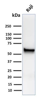
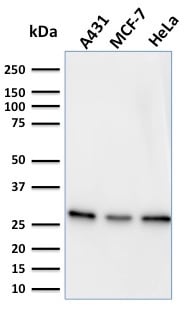 WB (Western Blot)
(Western Blot Analysis of A431, MCF-7 & HeLa cell lysates using Myofibroblast Marker Mouse Monoclonal Antibody (PR 2D3).)
WB (Western Blot)
(Western Blot Analysis of A431, MCF-7 & HeLa cell lysates using Myofibroblast Marker Mouse Monoclonal Antibody (PR 2D3).)
Myofibroblast Marker, Monoclonal Antibody (Cat# AAA215328)
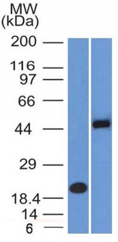
 WB (Western Blot)
(Western blot analysis of SOX1 expression in HeLa cell lysate (AAA124499).Electrophoresis was performed on a 5-20% SDS-PAGE gel at 70V (Stacking gel) / 90V (Resolving gel) for 2-3 hours. The sample well of each lane was loaded with 50ug of sample under reducing conditions.After Electrophoresis, proteins were transferred to a Nitrocellulose membrane at 150mA for 50-90 minutes. Blocked the membrane with 5% Non-fat Milk/ TBS for 1.5 hour at RT. The membrane was incubated with rabbit anti-SOX1 monoclonal antibody overnight at 4 degree C, then washed with TBS-0.1%Tween 3 times with 5 minutes each and probed with a goat anti-rabbit IgG-HRP secondary antibody at a dilution of 1:10000 for 1.5 hour at RT. The signal is developed using an Enhanced Chemiluminescent detection (ECL) kit with Tanon 5200 system. A specific band was detected for SOX1)
WB (Western Blot)
(Western blot analysis of SOX1 expression in HeLa cell lysate (AAA124499).Electrophoresis was performed on a 5-20% SDS-PAGE gel at 70V (Stacking gel) / 90V (Resolving gel) for 2-3 hours. The sample well of each lane was loaded with 50ug of sample under reducing conditions.After Electrophoresis, proteins were transferred to a Nitrocellulose membrane at 150mA for 50-90 minutes. Blocked the membrane with 5% Non-fat Milk/ TBS for 1.5 hour at RT. The membrane was incubated with rabbit anti-SOX1 monoclonal antibody overnight at 4 degree C, then washed with TBS-0.1%Tween 3 times with 5 minutes each and probed with a goat anti-rabbit IgG-HRP secondary antibody at a dilution of 1:10000 for 1.5 hour at RT. The signal is developed using an Enhanced Chemiluminescent detection (ECL) kit with Tanon 5200 system. A specific band was detected for SOX1)
SOX1, Monoclonal Antibody (Cat# AAA124499)
 IHC (Immunohistochemisry)
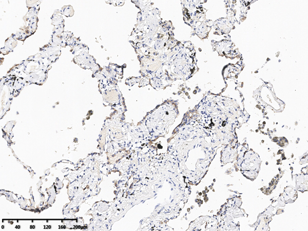
(Immunochemical staining of human Claudin 18 in human lung with mouse monoclonal antibody at 1:2000 dilution, formalin-fixed paraffin embedded sections.)
IHC (Immunohistochemisry)
(Immunochemical staining of human Claudin 18 in human lung with mouse monoclonal antibody at 1:2000 dilution, formalin-fixed paraffin embedded sections.)
Claudin18, Monoclonal Antibody (Cat# AAA258865)
Has cross-reactivity in IHC with Human Claudin 18.1 and Claudin 18.2
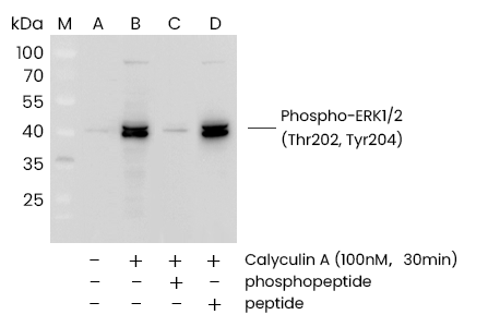
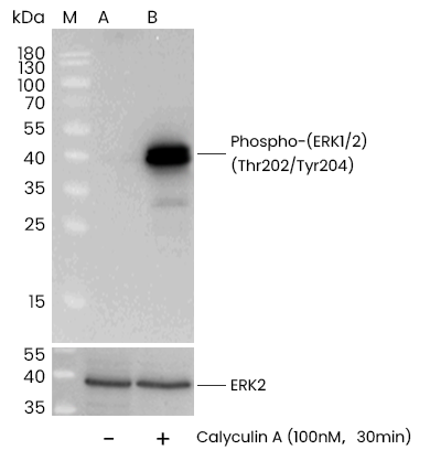
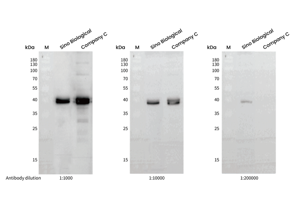 WB (Western Blot)
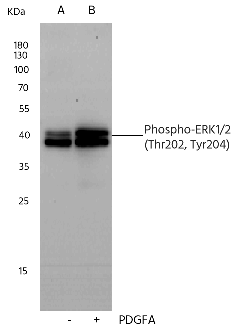
(Western blot analysis of extracts from serum-starved Hela, treated with Calyculin A (100 nM, 30 min), using Phospho-p44/42 MAPK (ERK1/2) (Thr202, Tyr204) rabbit monoclonal Antibody (#AAA258876) and other brands’ antibodies (company C) at dilution of 1:1000, 1:10000 and 1:200000.)
WB (Western Blot)
(Western blot analysis of extracts from serum-starved Hela, treated with Calyculin A (100 nM, 30 min), using Phospho-p44/42 MAPK (ERK1/2) (Thr202, Tyr204) rabbit monoclonal Antibody (#AAA258876) and other brands’ antibodies (company C) at dilution of 1:1000, 1:10000 and 1:200000.)
ERK1/2, Monoclonal Antibody (Cat# AAA258876)
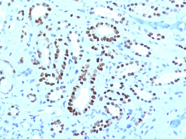
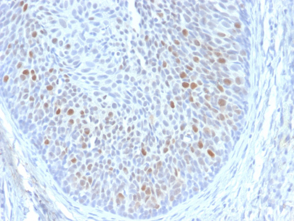
 IHC (Immunohiostchemistry)
(Immunochemical staining of human FOXP3 in human breast carcinoma with mouse monoclonal antibody at 1:200 dilution, formalin-fixed paraffin embedded sections.)
IHC (Immunohiostchemistry)
(Immunochemical staining of human FOXP3 in human breast carcinoma with mouse monoclonal antibody at 1:200 dilution, formalin-fixed paraffin embedded sections.)
FOXP3, Monoclonal Antibody (Cat# AAA258881)
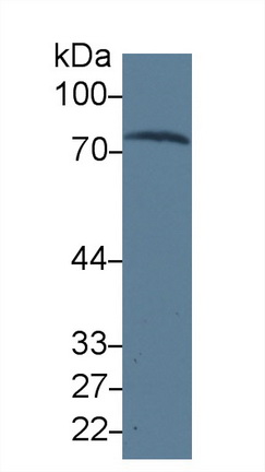
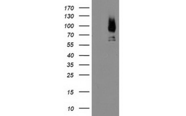
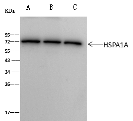 IP (Immunoprecipitation)
(HSPA1A was immunoprecipitated using:Lane A:0.5 mg A431 Whole Cell LysateLane B:0.5 mg A549, Hela Whole Cell Lysate2 uL anti-HSPA1A mouse monoclonal antibody and 60 ug of Immunomagnetic beads Protein A/G.Primary antibody:Anti-HSPA1A mouse monoclonal antibody,at 1:100 dilutionSecondary antibody:Clean-Blot IP Detection Reagent (HRP) at 1:1000dilutionDeveloped using the ECL technique.Performed under reducing conditions.Predicted band size: 70 kDaObserved band size :71 kDa)
IP (Immunoprecipitation)
(HSPA1A was immunoprecipitated using:Lane A:0.5 mg A431 Whole Cell LysateLane B:0.5 mg A549, Hela Whole Cell Lysate2 uL anti-HSPA1A mouse monoclonal antibody and 60 ug of Immunomagnetic beads Protein A/G.Primary antibody:Anti-HSPA1A mouse monoclonal antibody,at 1:100 dilutionSecondary antibody:Clean-Blot IP Detection Reagent (HRP) at 1:1000dilutionDeveloped using the ECL technique.Performed under reducing conditions.Predicted band size: 70 kDaObserved band size :71 kDa)
HSP70, Monoclonal Antibody (Cat# AAA258897)
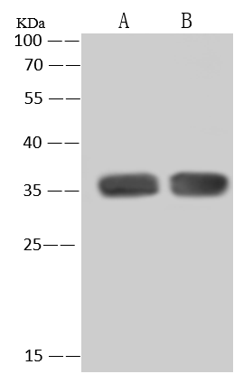
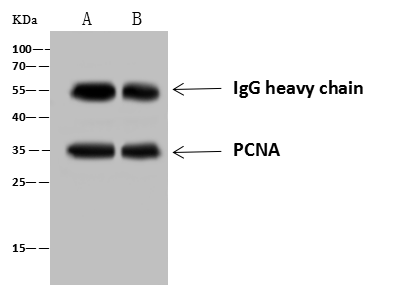 IP (Immunoprecipitation)
(PCNA was immunoprecipitated using:Lane A:0.5 mg HepG2 Whole Cell LysateLane B:0.5 mg MCF7 Whole Cell Lysate4 uL anti-PCNA rabbit monoclonal antibody and 60 ug of Immunomagnetic beads Protein A/G.Primary antibody:Anti-PCNA rabbit monoclonal antibody,at 1:100 dilutionSecondary antibody:Goat Anti-Rabbit IgG (H+L)/HRP at 1/10000 dilutionDeveloped using the ECL technique.Performed under reducing conditions.Predicted band size: 29 kDaObserved band size :35 kDa)
IP (Immunoprecipitation)
(PCNA was immunoprecipitated using:Lane A:0.5 mg HepG2 Whole Cell LysateLane B:0.5 mg MCF7 Whole Cell Lysate4 uL anti-PCNA rabbit monoclonal antibody and 60 ug of Immunomagnetic beads Protein A/G.Primary antibody:Anti-PCNA rabbit monoclonal antibody,at 1:100 dilutionSecondary antibody:Goat Anti-Rabbit IgG (H+L)/HRP at 1/10000 dilutionDeveloped using the ECL technique.Performed under reducing conditions.Predicted band size: 29 kDaObserved band size :35 kDa)
PCNA, Monoclonal Recombinant Antibody (Cat# AAA258901)

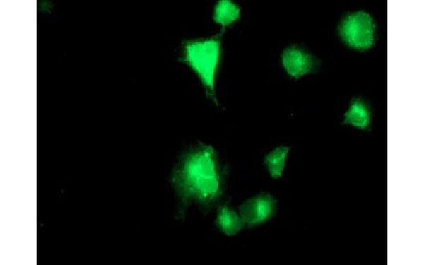
 IF (Immunofluorescence)
(Immunofluorescence staining of SELPLG- in HeLa cells. Cells were fixed with 4% PFA, permeabilzed with 0.1% Triton X-100 in PBS,blocked with 10% serum, and incubated with mouse anti-Human SELPLG- monoclonal antibody (dilution ratio 1:60) at 4? overnight. Then cells were stained with the Alexa Fluor488-conjugated Goat Anti-mouse IgG secondary antibody (green) and counterstained with DAPI (blue).Positive staining was localized to Cytoplasm.)
IF (Immunofluorescence)
(Immunofluorescence staining of SELPLG- in HeLa cells. Cells were fixed with 4% PFA, permeabilzed with 0.1% Triton X-100 in PBS,blocked with 10% serum, and incubated with mouse anti-Human SELPLG- monoclonal antibody (dilution ratio 1:60) at 4? overnight. Then cells were stained with the Alexa Fluor488-conjugated Goat Anti-mouse IgG secondary antibody (green) and counterstained with DAPI (blue).Positive staining was localized to Cytoplasm.)
PSGL-1/CD162, Monoclonal Antibody (Cat# AAA258912)
BAFF/BLyS/TNFSF13B, Monoclonal Antibody (Cat# AAA258720)
 IHC (Immunohiostchemistry)
(Immunochemical staining of human CD274 in human placenta with mouse monoclonal antibody at 1:20000 dilution, formalin-fixed paraffin embedded sections.)
IHC (Immunohiostchemistry)
(Immunochemical staining of human CD274 in human placenta with mouse monoclonal antibody at 1:20000 dilution, formalin-fixed paraffin embedded sections.)
PD-L1/B7-H1, Monoclonal Antibody (Cat# AAA258733)

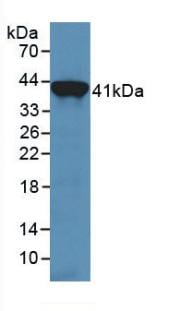
 IP (Immunoprecipitation)
(pCMV3-CD34-TT-WPRE was immunoprecipitated using:Lane A:0.5 mg TF-1 Whole Cell Lysate4 uL anti-pCMV3-CD34-TT-WPRE mouse monoclonal antibody and 60 ug of Immunomagnetic beads Protein A/G.Primary antibody:Anti-pCMV3-CD34-TT-WPRE mouse monoclonal antibody,at 1:100 dilutionSecondary antibody:Rabbit Anti-Mouse IgG F(ab)2/HRP at 1/10000 dilutionDeveloped using the ECL technique.Performed under reducing conditions.Predicted band size: 41 kDaObserved band size :95 kDa)
IP (Immunoprecipitation)
(pCMV3-CD34-TT-WPRE was immunoprecipitated using:Lane A:0.5 mg TF-1 Whole Cell Lysate4 uL anti-pCMV3-CD34-TT-WPRE mouse monoclonal antibody and 60 ug of Immunomagnetic beads Protein A/G.Primary antibody:Anti-pCMV3-CD34-TT-WPRE mouse monoclonal antibody,at 1:100 dilutionSecondary antibody:Rabbit Anti-Mouse IgG F(ab)2/HRP at 1/10000 dilutionDeveloped using the ECL technique.Performed under reducing conditions.Predicted band size: 41 kDaObserved band size :95 kDa)
CD34, Monoclonal Antibody (Cat# AAA258743)
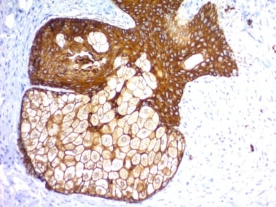
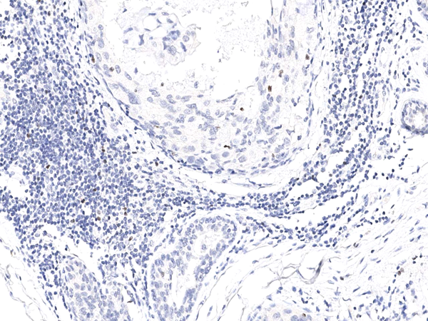
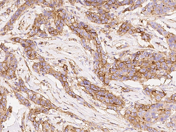
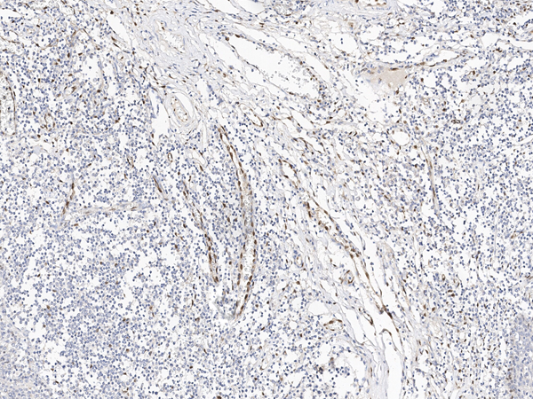
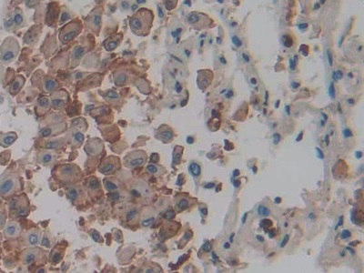
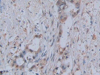
 IHC (Immunohiostchemistry)
(Immunochemical staining of human CAIX in human renal carcinoma with mouse monoclonal antibody (1:200, formalin-fixed paraffin embedded sections).)
IHC (Immunohiostchemistry)
(Immunochemical staining of human CAIX in human renal carcinoma with mouse monoclonal antibody (1:200, formalin-fixed paraffin embedded sections).)
Carbonic Anhydrase IX/CA9, Monoclonal Antibody (Cat# AAA258747)
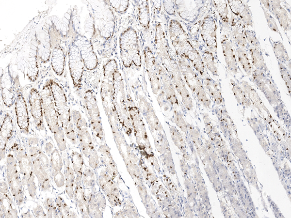
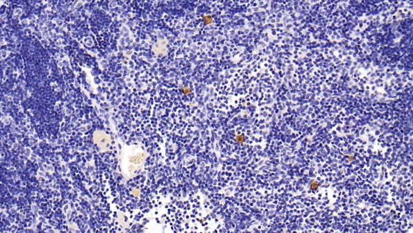
 IHC (Immunohiostchemistry)
(Immunochemical staining of human IL-33 in human tonsil with rabbit monoclonal antibody at 1:1000 dilution, formalin-fixed paraffin embedded sections.)
IHC (Immunohiostchemistry)
(Immunochemical staining of human IL-33 in human tonsil with rabbit monoclonal antibody at 1:1000 dilution, formalin-fixed paraffin embedded sections.)
IL-33, Monoclonal Recombinant Antibody (Cat# AAA258796)
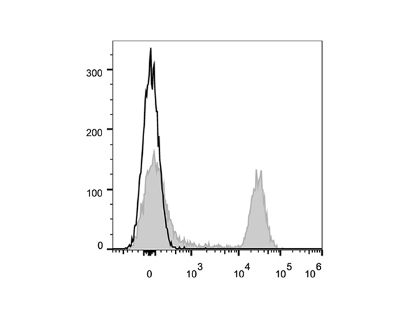
 FCM/FACS (Flow Cytometry)
(C57BL/6 murine splenocytes are stained with Anti-Mouse CD45R/B220 Monoclonal Antibody(Alexa Fluor 647 Conjuaged)(filled gray histogram). Unstained splenocytes (empty black histogram) are used as control.)
FCM/FACS (Flow Cytometry)
(C57BL/6 murine splenocytes are stained with Anti-Mouse CD45R/B220 Monoclonal Antibody(Alexa Fluor 647 Conjuaged)(filled gray histogram). Unstained splenocytes (empty black histogram) are used as control.)
CD45R/B220, Monoclonal Antibody (Cat# AAA174638)
 Knockout Validation
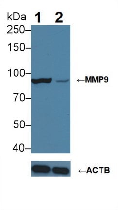
(Knockout Validation: Lane 1: Wild-type HepG2 cell lysate; Lane 2: MMP9 knockout HepG2 cell lysate; Predicted MW: 76kDa Observed MW: 90kDa Primary Ab: 3ug/ml Mouse Anti-Human MMP9 Antibody Second Ab: 0.2ug/mL HRP-Linked Caprine Anti-Mouse IgG Polyclonal Antibody)
Knockout Validation
(Knockout Validation: Lane 1: Wild-type HepG2 cell lysate; Lane 2: MMP9 knockout HepG2 cell lysate; Predicted MW: 76kDa Observed MW: 90kDa Primary Ab: 3ug/ml Mouse Anti-Human MMP9 Antibody Second Ab: 0.2ug/mL HRP-Linked Caprine Anti-Mouse IgG Polyclonal Antibody)
Matrix Metalloproteinase 9, Monoclonal Antibody (Cat# AAA141266)
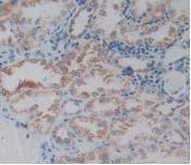
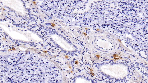
 IHC (Immunohistochemistry)
(DAB staining on IHC-P; Samples: Human Kidney Tissue.)
IHC (Immunohistochemistry)
(DAB staining on IHC-P; Samples: Human Kidney Tissue.)
Epstein Barr Virus Induced Protein 3, Monoclonal Antibody (Cat# AAA141309)
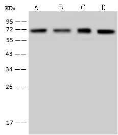
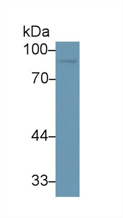
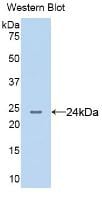
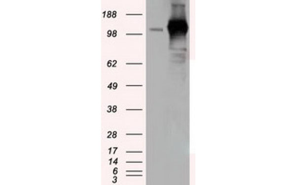
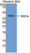
 WB (Western Blot)
(Figure. Western Blot; Sample: Recombinant GLRX3, Human.)
WB (Western Blot)
(Figure. Western Blot; Sample: Recombinant GLRX3, Human.)
Glutaredoxin 3, Monoclonal Antibody (Cat# AAA141322)
Immunoglobulin G1 (IgG1), Monoclonal Antibody (Cat# AAA149780)
Diamine Oxidase (DAO), Monoclonal Antibody (Cat# AAA146124)
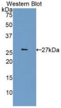
 WB (Western Blot)
(Western Blot: Sample: Recombinant S100A12, Human.)
WB (Western Blot)
(Western Blot: Sample: Recombinant S100A12, Human.)
S100 Calcium Binding Protein A12, Monoclonal Antibody (Cat# AAA144659)
 WB (Western Blot)
(Sample: Human Liver lysate;Primary Ab: 1ug/mL Mouse Anti-Human FABP1 AntibodySecond Ab: 0.2ug/mL HRP-Linked Caprine Anti-Mouse IgG Polyclonal Antibody)
WB (Western Blot)
(Sample: Human Liver lysate;Primary Ab: 1ug/mL Mouse Anti-Human FABP1 AntibodySecond Ab: 0.2ug/mL HRP-Linked Caprine Anti-Mouse IgG Polyclonal Antibody)
Fatty Acid Binding Protein 1 (FABP1), Monoclonal Antibody (Cat# AAA147913)

 IHC (Immunohistochemistry)
(DAB staining on IHC-P; Samples: Human Lung cancer Tissue))
IHC (Immunohistochemistry)
(DAB staining on IHC-P; Samples: Human Lung cancer Tissue))
Interleukin 1 Alpha (IL1a), Monoclonal Antibody (Cat# AAA147927)
Renin (REN), Monoclonal Antibody (Cat# AAA148002)
Neuraminidase (NEU), Monoclonal Antibody (Cat# AAA148005)
CD106, Monoclonal Antibody (Cat# AAA128344)
CD158d, Monoclonal Antibody (Cat# AAA128700)
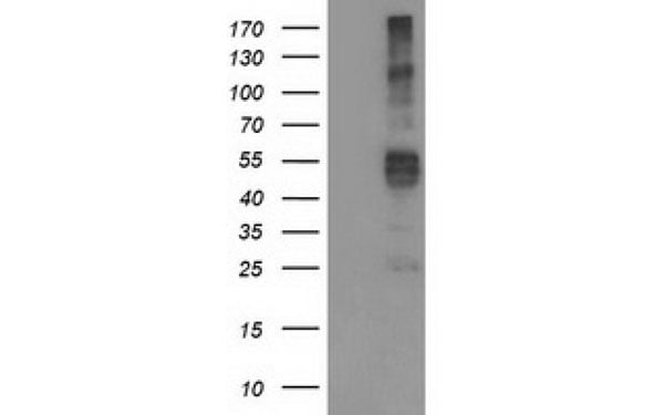
 WB (Western Blot)
(Western Blot: Sample: Recombinant TRF, Human.)
WB (Western Blot)
(Western Blot: Sample: Recombinant TRF, Human.)
Transferrin (TRF), Monoclonal Antibody (Cat# AAA130616)
 Knockout Validation
(Knockout Validation: Lane 1: Wild-type Hela cell lysate;;Lane 2: ANXA1 knockout Hela cell lysate;;Predicted MW: 39kDa ;Observed MW: 39kDa;Primary Ab: 2ug/ml Mouse Anti-Human ANXA1 Antibody;Second Ab: 0.2ug/mL HRP-Linked Caprine Anti-Mouse IgG Polyclonal Antibody;)
Knockout Validation
(Knockout Validation: Lane 1: Wild-type Hela cell lysate;;Lane 2: ANXA1 knockout Hela cell lysate;;Predicted MW: 39kDa ;Observed MW: 39kDa;Primary Ab: 2ug/ml Mouse Anti-Human ANXA1 Antibody;Second Ab: 0.2ug/mL HRP-Linked Caprine Anti-Mouse IgG Polyclonal Antibody;)
Annexin A1 (ANXA1), Monoclonal Antibody (Cat# AAA134805)
Acetylcholinesterase (AchE), Monoclonal Antibody (Cat# AAA134830)
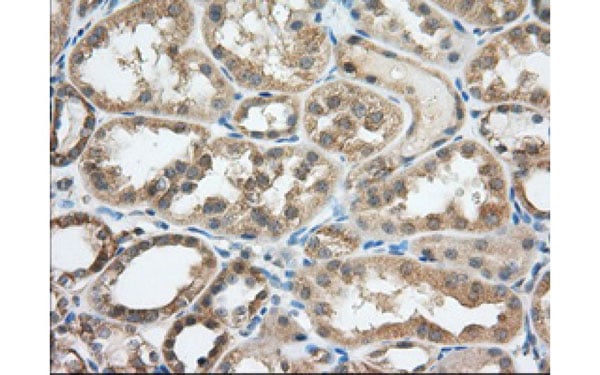
 IHC (Immunohistochemistry)
(DAB staining on IHC-P; Samples: Human Kidney Tissue.)
IHC (Immunohistochemistry)
(DAB staining on IHC-P; Samples: Human Kidney Tissue.)
Anterior Gradient Protein 2 (AGR2), Monoclonal Antibody (Cat# AAA134840)
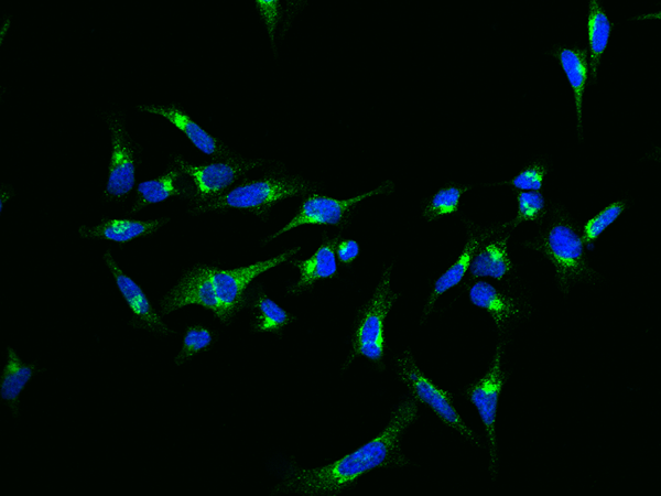
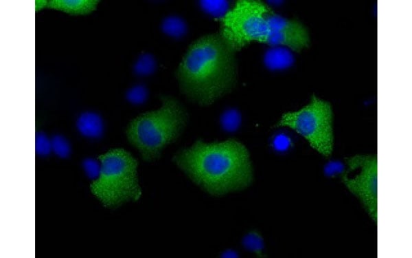 IF (Immunofluorescence)
(Immunofluorescent staining of COS7 cells transiently transfected with recombinant TACC3 protein using TACC3 antibody)
IF (Immunofluorescence)
(Immunofluorescent staining of COS7 cells transiently transfected with recombinant TACC3 protein using TACC3 antibody)
TACC3, Monoclonal Antibody (Cat# AAA107977)
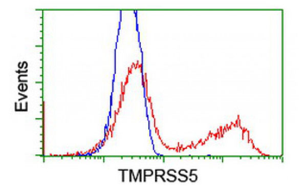
 IF (Immunofluorescence)
(Immunofluorescent staining of COS7 cells transiently transfected with recombinant TMPRSS5 protein using TMPRSS5 antibody)
IF (Immunofluorescence)
(Immunofluorescent staining of COS7 cells transiently transfected with recombinant TMPRSS5 protein using TMPRSS5 antibody)
TMPRSS5, Monoclonal Antibody (Cat# AAA107978)
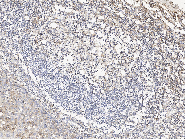
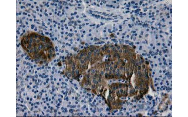 IHC (Immunohiostchemistry)
(Immunohistochemical analysis of LGALS3BP protein in paraffin embedded Human pancreas tissue using LGALS3BP antibody)
IHC (Immunohiostchemistry)
(Immunohistochemical analysis of LGALS3BP protein in paraffin embedded Human pancreas tissue using LGALS3BP antibody)
LGALS3BP, Monoclonal Antibody (Cat# AAA108075)
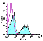
 FCM/FACS (Flow Cytometry)
(Flow Cytometric analysis using TCR beta antibodyStaining of C57BL/6 splenocytes with staining buffer (Autofluorescence) (open histogram) or 0.06 ug of TCR beta antibody (filled histogram). Total viable cells were used for analysis.)
FCM/FACS (Flow Cytometry)
(Flow Cytometric analysis using TCR beta antibodyStaining of C57BL/6 splenocytes with staining buffer (Autofluorescence) (open histogram) or 0.06 ug of TCR beta antibody (filled histogram). Total viable cells were used for analysis.)
Sialosyl-Tn, Monoclonal Antibody (Cat# AAA107363)
Mucin, Monoclonal Antibody (Cat# AAA106277)
Negative to cow.


 Application Data
(Detection limit for recombinant GST tagged TCEA3 is approximately 0.03ng/ml as a capture antibody.)
Application Data
(Detection limit for recombinant GST tagged TCEA3 is approximately 0.03ng/ml as a capture antibody.)
TCEA3, Monoclonal Antibody (Cat# AAA26607)
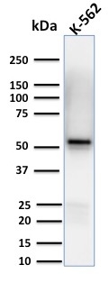
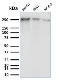 WB (Western Blot)
(Western Blot Analysis of Human HepG2, K562 and SK-Br3 cell lysates using RNA Poll II Mouse Monoclonal Antibody (8A7).)
WB (Western Blot)
(Western Blot Analysis of Human HepG2, K562 and SK-Br3 cell lysates using RNA Poll II Mouse Monoclonal Antibody (8A7).)
RNA Polymerase II CTD Repeat YSPTSPS, Monoclonal Antibody (Cat# AAA215151)
Cystatin A (CSTA), Monoclonal Antibody (Cat# AAA149340)
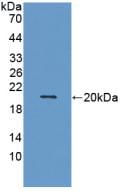
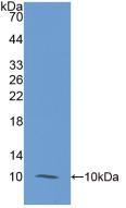
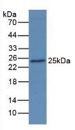
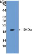
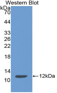 WB (Western Blot)
(Sample: Recombinant CSTA, Human.)
WB (Western Blot)
(Sample: Recombinant CSTA, Human.)
Cystatin A, Monoclonal Antibody (Cat# AAA141360)

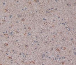 IHC (Immunohistochemistry)
(DAB staining on IHC-P;Samples: Human Brain Tissue)
IHC (Immunohistochemistry)
(DAB staining on IHC-P;Samples: Human Brain Tissue)
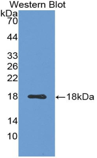
Lysophosphatidylcholine Acyltransferase 3, Monoclonal Antibody (Cat# AAA141361)

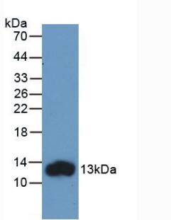
 WB (Western Blot)
(Western Blot: Sample: Recombinant LDHB, Rat.)
WB (Western Blot)
(Western Blot: Sample: Recombinant LDHB, Rat.)
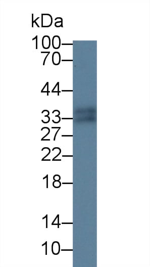
Lactate Dehydrogenase B, Monoclonal Antibody (Cat# AAA141281)
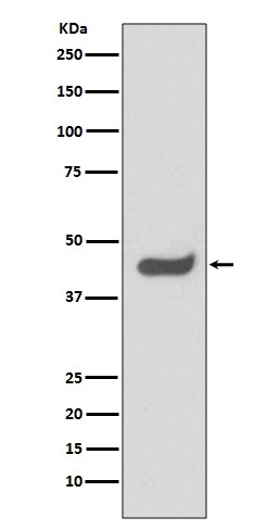
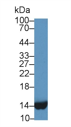
 WB (Western Blot)
(Western Blot: Sample: Recombinant CDK5, Human.)
WB (Western Blot)
(Western Blot: Sample: Recombinant CDK5, Human.)
Cyclin Dependent Kinase 5, Monoclonal Antibody (Cat# AAA141323)
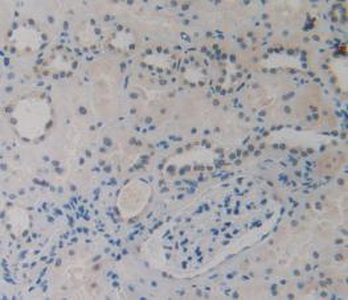
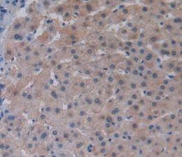
 IHC (Immunohistochemisry)
(DAB staining on IHC-P; Samples: Human Pancreatic cancer Tissue))
IHC (Immunohistochemisry)
(DAB staining on IHC-P; Samples: Human Pancreatic cancer Tissue))
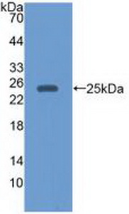
Monocyte Chemotactic Protein 1, Monoclonal Antibody (Cat# AAA141337)
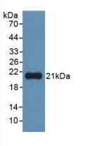
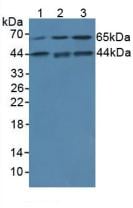
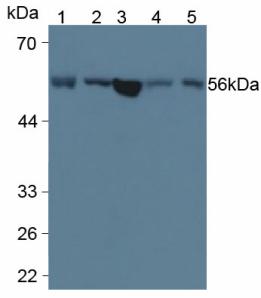
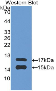
 WB (Western Blot)
(Western Blot; Sample: Lane1: Human Leukocyte lysate; Lane2: Human Lymphocyte lysate; Lane3: Human PBMC lysate Primary Ab: 0.1ug/ml Mouse AntiHuman S100A9 Antibody Second Ab: 0.2ug/mL HRPLinked Caprine AntiMouse IgG Polyclonal Antibody (Catalog: SAA544Mu19))
WB (Western Blot)
(Western Blot; Sample: Lane1: Human Leukocyte lysate; Lane2: Human Lymphocyte lysate; Lane3: Human PBMC lysate Primary Ab: 0.1ug/ml Mouse AntiHuman S100A9 Antibody Second Ab: 0.2ug/mL HRPLinked Caprine AntiMouse IgG Polyclonal Antibody (Catalog: SAA544Mu19))
S100 Calcium Binding Protein A9 (S100A9), Monoclonal Antibody (Cat# AAA151749)
What are Monoclonal Antibodies?
Monoclonal antibodies are specialized laboratory-produced proteins developed for binding to specific biological antigens or other molecular targets. Since they come from a single cell (or clone), they are especially consistent and accurate in the data they are involved in producing.
This type of antibody material has been shown to be a powerful tool in finding and subsequently destroying harmful cells in an organism, such as those found in cancers or various autoimmune diseases. This makes them excellent aids in medical testing and research, which is why they are so widely used.
AAA Biotech offers a comprehensive range of high-quality monoclonal antibodies that perform effectively in various laboratory tests, including (amongst others) ELISA, western blotting, immunohistochemistry, and flow cytometry. All of the products in our catalog are thoroughly quality tested to make sure that they are reliable and will consistently perform well in your research.
What Are The Uses of Monoclonal Antibodies
Monoclonal antibodies are used in many lab tests, including (amongst others) ELISA, western blotting, immunohistochemistry, and flow cytometry.
ELISA is a test that helps detect a specific substance/analyte in a sample. It uses antibodies (often monoclonal) bound to a solid surface (such as the well of a microplate) to “capture” the substance/analyte in the sample and immobilize it so that the detection antibody component can then bind to it and produce a signal, which can then be measured.
Western blotting identifies specific proteins in a sample. The sample is first separated on a gel, and then antibodies are applied that will typically bind to the target, which will all be localized to a single band in a lane.
Immunohistochemistry helps locate specific proteins in cells or tissue samples using antibodies.
Flow cytometry looks at and sorts cells. It uses antibodies that are conjugated to reporter molecules called “fluorophores”, which, under special lights, emit light themselves, which can then be measured by a detector instrument. For a deeper understanding of these techniques, explore our complete guide to monoclonal antibodies and their benefits.
How Monoclonal Antibodies Are Used as Medicine?
Please note that all of the products listed in AAA Biotech’s also known as AAA Bio or AAABio catalog are strictly for research-use only (RUO).
Monoclonal antibodies can also be used as therapeutic/medical treatments, particularly in the context of cancers. They are designed to find and bind to specific cells or proteins, helping the immune system recognize and attack the cancer. These treatments work in different ways, such as:
- Radioimmunotherapy attaches a small amount of radioactive molecule to the antibody, so it delivers the radiation directly to the cancer cells that the antibody is specifically binding to.
- Antibody-directed enzyme prodrug therapy uses antibodies that are specifically bound to special enzymes. These enzymes activate a harmless drug in the body and turn it into a cancer-killing drug only near the cancer cells—this helps avoid harming healthy cells.
- Immunoliposomes are tiny “bubbles” filled with medicine/drug and coated with antibodies. They carry the drug straight to the cancer cells.
Why Buy Monoclonal Antibodies From Us?
At AAA Biotech, we provide high-performance monoclonal antibodies designed to support a wide range of research needs.
1. Validated for Versatile Applications
The antibodies in our catalog are extensively validated and compatible with multiple techniques, including (but not limited to) ELISA, flow cytometry (FC), immunocytochemistry (ICC), immunofluorescence (IF), immunohistochemistry (IHC), immunoprecipitation (IP), and western blotting (WB).
2. Wide Selection & Specialized Options
We offer antibodies for common and rare species, that are available in various conjugated forms, and also in recombinant formats. Essentially, there is almost anything one might need to meet their experimental model’s requirements.
3. High-Quality Proteins
Our proteins meet high purity standards—90% or more as confirmed by SDS-PAGE. Many are available with tags like His, Flag, GST, or MBP, and we also supply native and biologically active proteins for functional studies.
Frequently Asked Questions
1. Are your monoclonal antibodies validated for specific applications?
Yes, our antibodies are tested and validated for use in methods such as ELISA, western blot, IHC, flow cytometry, and more. Refer to specific product pages or datasheets for individual product information.
2. How do I choose the right monoclonal antibody for my application?
Review the product details directly for application validation, species reactivity, and target information. You may also contact our support team at any time for help.
3. How quickly can I receive my order?
Most orders are processed and shipped within 1–3 business days, depending on product availability and your shipping location.