Filters
▼Clonality
▼Type
▼Reactivity
▼Gene Name
▼Isotype
▼Host
▼Application
▼Clone
▼Monoclonal Antibodies
Get accurate results in your research with our Monoclonal Antibodies, which are specially made to target exactly what you require for your research, and will produce consistent, reliable performance in lab tests.
Viewing 3150-3200 of 27645 product results
 IHC (Immunohiostchemistry)
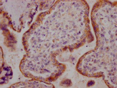
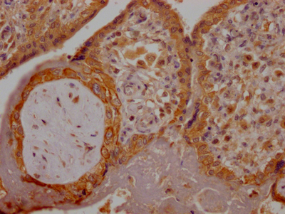
(IHC image diluted at 1:100 and staining in paraffin-embedded human placenta tissue performed on a Leica BondTM system. After dewaxing and hydration, antigen retrieval was mediated by high pressure in a citrate buffer (pH 6.0). Section was blocked with 10% normal goat serum 30min at RT. Then primary antibody (1% BSA) was incubated at 4 degree C overnight. The primary is detected by a Goat anti-rabbit IgG polymer labeled by HRP and visualized using 0.05% DAB.)
IHC (Immunohiostchemistry)
(IHC image diluted at 1:100 and staining in paraffin-embedded human placenta tissue performed on a Leica BondTM system. After dewaxing and hydration, antigen retrieval was mediated by high pressure in a citrate buffer (pH 6.0). Section was blocked with 10% normal goat serum 30min at RT. Then primary antibody (1% BSA) was incubated at 4 degree C overnight. The primary is detected by a Goat anti-rabbit IgG polymer labeled by HRP and visualized using 0.05% DAB.)
ACVR2B, Monoclonal Recombinant Antibody (Cat# AAA243877)
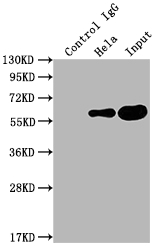
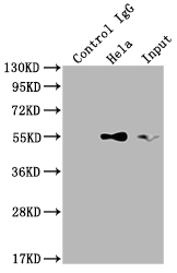
 IP (Immunoprecipitation)
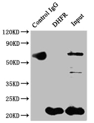
(Immunoprecipitating DHFR in Hela whole cell lysateLane 1: Rabbit control IgG instead of in Hela whole cell lysate. For western blotting,a HRP-conjugated Protein G antibody was used as the secondary antibody (1/2000)Lane 2: Hela whole cell lysate?500ug?Lane 3: Hela whole cell lysate (10ug))
IP (Immunoprecipitation)
(Immunoprecipitating DHFR in Hela whole cell lysateLane 1: Rabbit control IgG instead of in Hela whole cell lysate. For western blotting,a HRP-conjugated Protein G antibody was used as the secondary antibody (1/2000)Lane 2: Hela whole cell lysate?500ug?Lane 3: Hela whole cell lysate (10ug))
DHFR, Monoclonal Recombinant Antibody (Cat# AAA243881)
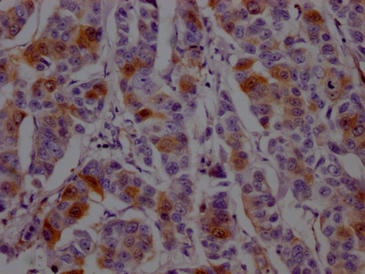
 IHC (Immunohiostchemistry)
(IHC image diluted at 1:100 and staining in paraffin-embedded human placenta tissue performed on a Leica BondTM system. After dewaxing and hydration, antigen retrieval was mediated by high pressure in a citrate buffer (pH 6.0). Section was blocked with 10% normal goat serum 30min at RT. Then primary antibody (1% BSA) was incubated at 4 degree C overnight. The primary is detected by a Goat anti-rabbit IgG polymer labeled by HRP and visualized using 0.05% DAB.)
IHC (Immunohiostchemistry)
(IHC image diluted at 1:100 and staining in paraffin-embedded human placenta tissue performed on a Leica BondTM system. After dewaxing and hydration, antigen retrieval was mediated by high pressure in a citrate buffer (pH 6.0). Section was blocked with 10% normal goat serum 30min at RT. Then primary antibody (1% BSA) was incubated at 4 degree C overnight. The primary is detected by a Goat anti-rabbit IgG polymer labeled by HRP and visualized using 0.05% DAB.)
PGF, Monoclonal Recombinant Antibody (Cat# AAA243893)
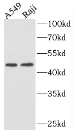
 WB (Western Blot)
(Raji cells were subjected to SDS PAGE followed by western blot with AAA249028 (CD45 Antibody) at dilution of 1:1000)
WB (Western Blot)
(Raji cells were subjected to SDS PAGE followed by western blot with AAA249028 (CD45 Antibody) at dilution of 1:1000)
CD45, Monoclonal Antibody (Cat# AAA249028)
Protein A+G Purification
 WB (Western Blot)
(hela lysates were subjected to SDS PAGE followed by western blot with AAA249031 (Vinculin Antibody) at dilution of 1:5000)
WB (Western Blot)
(hela lysates were subjected to SDS PAGE followed by western blot with AAA249031 (Vinculin Antibody) at dilution of 1:5000)
Vinculin, Monoclonal Antibody (Cat# AAA249031)
Protein A+G Purification
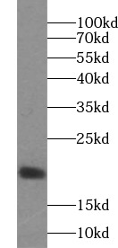
 WB (Western Blot)
(Hela cells were subjected to SDS PAGE followed by western blot with AAA249034 (Cytochrome c antibody) at dilution of 1:20000)
WB (Western Blot)
(Hela cells were subjected to SDS PAGE followed by western blot with AAA249034 (Cytochrome c antibody) at dilution of 1:20000)
Cytochrome C, Monoclonal Antibody (Cat# AAA249034)
Protein A+G Purification
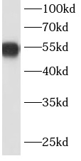
 WB (Western Blot)
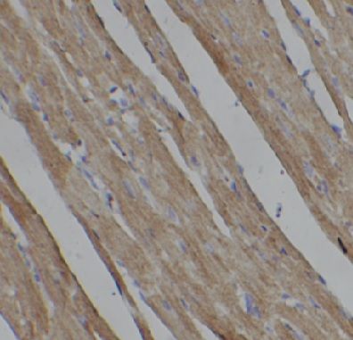
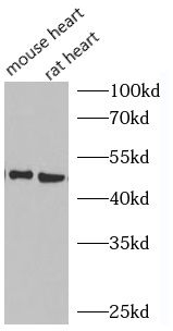
(rat heart tissue were subjected to SDS PAGE followed by western blot with AAA249039 (Visfatin antibody) at dilution of 1:2000)
WB (Western Blot)
(rat heart tissue were subjected to SDS PAGE followed by western blot with AAA249039 (Visfatin antibody) at dilution of 1:2000)
Visfatin, Monoclonal Antibody (Cat# AAA249039)
Protein A+G Purified
 WB (Western Blot)
(Jurkat cells were subjected to SDS PAGE followed by western blot with AAA249050 (CD82 antibody) at dilution of 1:2000)
WB (Western Blot)
(Jurkat cells were subjected to SDS PAGE followed by western blot with AAA249050 (CD82 antibody) at dilution of 1:2000)
CD82, Monoclonal Antibody (Cat# AAA249050)
Protein A+G Purified
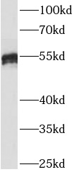
 WB (Western Blot)
(A431 cells were subjected to SDS PAGE followed by western blot with AAA249059 (E-cadherin antibody) at dilution of 1:3000)
WB (Western Blot)
(A431 cells were subjected to SDS PAGE followed by western blot with AAA249059 (E-cadherin antibody) at dilution of 1:3000)
ECAD, Monoclonal Antibody (Cat# AAA249059)
Protein A+G purified
 Application Data
(Hela cells were subjected to SDS PAGE followed by western blot with AAA251232(TOMM20 Antibody) at dilution of 1:5000)
Application Data
(Hela cells were subjected to SDS PAGE followed by western blot with AAA251232(TOMM20 Antibody) at dilution of 1:5000)
TOMM20, Monoclonal Antibody (Cat# AAA251232)
Protein A+G purification
 Application Data
HRPT2, Monoclonal Antibody (Cat# AAA251233)
Protein A+G purification
 Application Data
GSDMD, Monoclonal Antibody (Cat# AAA251234)
Protein A+G purification
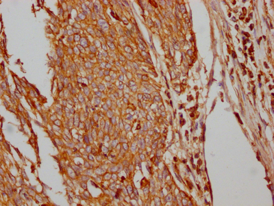
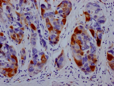
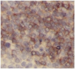
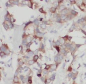
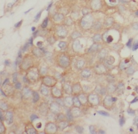
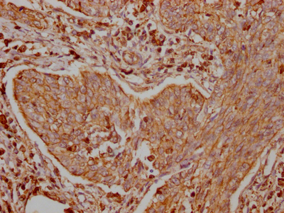
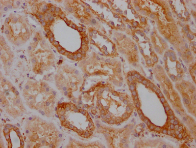
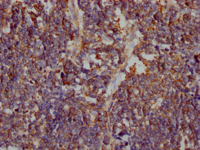 IHC (Immunohiostchemistry)
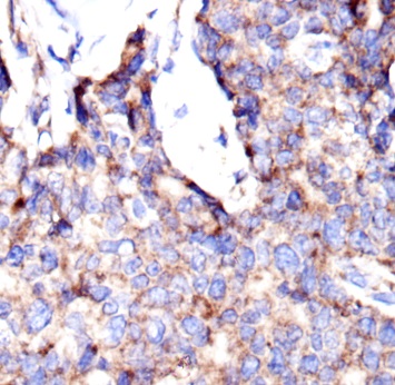
(IHC image diluted at 1:100 and staining in paraffin-embedded human lung cancer performed on a Leica BondTM system. After dewaxing and hydration, antigen retrieval was mediated by high pressure in a citrate buffer (pH 6.0). Section was blocked with 10% normal goat serum 30min at RT. Then primary antibody (1% BSA) was incubated at 4 degree C overnight. The primary is detected by a Goat anti-rabbit IgG polymer labeled by HRP and visualized using 0.05% DAB.)
IHC (Immunohiostchemistry)
(IHC image diluted at 1:100 and staining in paraffin-embedded human lung cancer performed on a Leica BondTM system. After dewaxing and hydration, antigen retrieval was mediated by high pressure in a citrate buffer (pH 6.0). Section was blocked with 10% normal goat serum 30min at RT. Then primary antibody (1% BSA) was incubated at 4 degree C overnight. The primary is detected by a Goat anti-rabbit IgG polymer labeled by HRP and visualized using 0.05% DAB.)
SLC2A1, Monoclonal Recombinant Antibody (Cat# AAA243903)
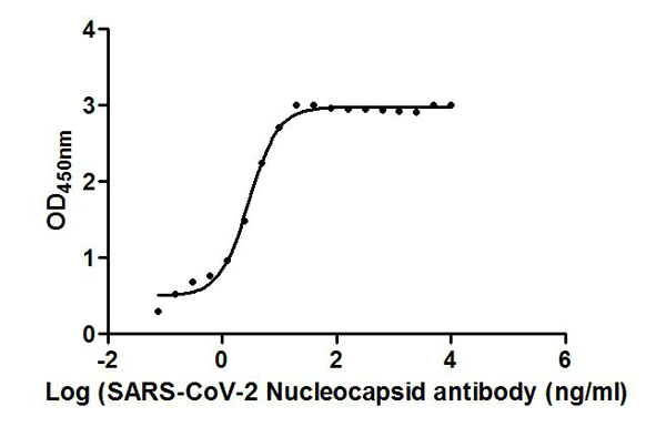
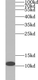
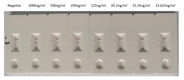 Application Data
(In the Colloidal Gold Immunochromatography Assay detection system, the background of antibody is clean, the detection limit can be as low as 15.625ng/ml (1.09ng/0.07ml), and the sensitivity is very good.)
Application Data
(In the Colloidal Gold Immunochromatography Assay detection system, the background of antibody is clean, the detection limit can be as low as 15.625ng/ml (1.09ng/0.07ml), and the sensitivity is very good.)
COVID 19 Nucleocapsid (NP) Coronavirus, Monoclonal Recombinant Antibody (Cat# AAA243906)
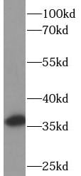
 IP (Immunoprecipitation)
(Immunoprecipitating PKM in Hela whole cell lysateLane 1: Rabbit control IgG instead of in Hela whole cell lysate. For western blotting,a HRP-conjugated Protein G antibody was used as the secondary antibody (1/2000)Lane 2: Hela whole cell lysate?500ug?Lane 3: Hela whole cell lysate (10ug))
IP (Immunoprecipitation)
(Immunoprecipitating PKM in Hela whole cell lysateLane 1: Rabbit control IgG instead of in Hela whole cell lysate. For western blotting,a HRP-conjugated Protein G antibody was used as the secondary antibody (1/2000)Lane 2: Hela whole cell lysate?500ug?Lane 3: Hela whole cell lysate (10ug))
PKM, Monoclonal Recombinant Antibody (Cat# AAA243936)
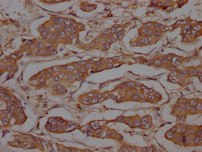
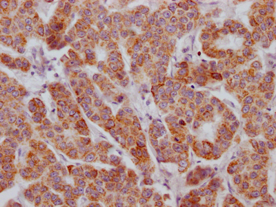
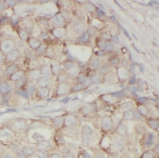
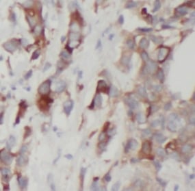
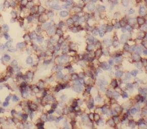
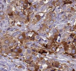
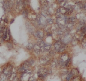
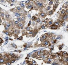
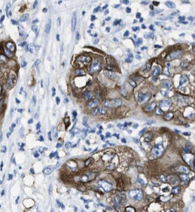
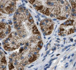
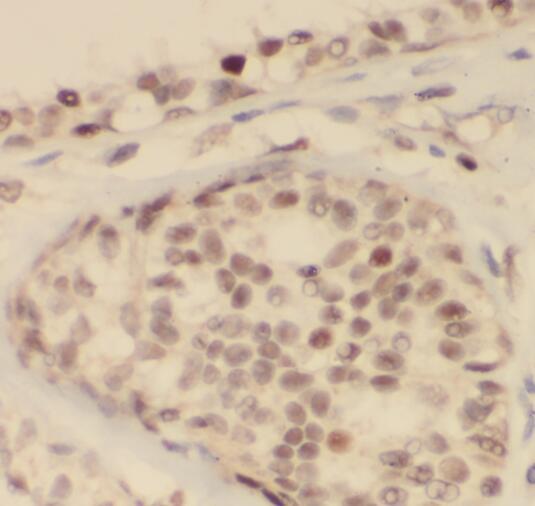
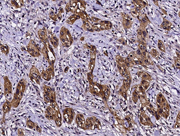
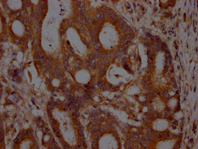 IHC (Immunohiostchemistry)
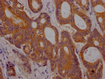
(IHC image diluted at 1:100 and staining in paraffin-embedded human colon cancer performed on a Leica BondTM system. After dewaxing and hydration, antigen retrieval was mediated by high pressure in a citrate buffer (pH 6.0). Section was blocked with 10% normal goat serum 30min at RT. Then primary antibody (1% BSA) was incubated at 4 degree C overnight. The primary is detected by a Goat anti-rabbit IgG polymer labeled by HRP and visualized using 0.05% DAB.)
IHC (Immunohiostchemistry)
(IHC image diluted at 1:100 and staining in paraffin-embedded human colon cancer performed on a Leica BondTM system. After dewaxing and hydration, antigen retrieval was mediated by high pressure in a citrate buffer (pH 6.0). Section was blocked with 10% normal goat serum 30min at RT. Then primary antibody (1% BSA) was incubated at 4 degree C overnight. The primary is detected by a Goat anti-rabbit IgG polymer labeled by HRP and visualized using 0.05% DAB.)
MET, Monoclonal Recombinant Antibody (Cat# AAA243938)
 IP (Immunoprecipitation)
(Immunoprecipitating TRAF2 in Hela whole cell lysateLane 1: Rabbit control IgG instead of in Hela whole cell lysate. For western blotting,a HRP-conjugated Protein G antibody was used as the secondary antibody (1/2000)Lane 2: Hela whole cell lysate?500ug?Lane 3: Hela whole cell lysate (10ug))
IP (Immunoprecipitation)
(Immunoprecipitating TRAF2 in Hela whole cell lysateLane 1: Rabbit control IgG instead of in Hela whole cell lysate. For western blotting,a HRP-conjugated Protein G antibody was used as the secondary antibody (1/2000)Lane 2: Hela whole cell lysate?500ug?Lane 3: Hela whole cell lysate (10ug))
TRAF2, Monoclonal Recombinant Antibody (Cat# AAA243950)
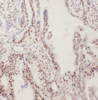
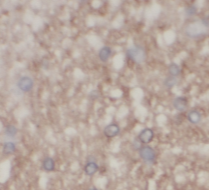
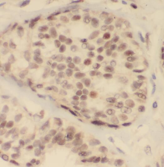
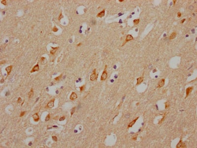 IHC (Immunohiostchemistry)
(IHC image diluted at 1:100 and staining in paraffin-embedded human brain tissue performed on a Leica BondTM system. After dewaxing and hydration, antigen retrieval was mediated by high pressure in a citrate buffer (pH 6.0). Section was blocked with 10% normal goat serum 30min at RT. Then primary antibody (1% BSA) was incubated at 4 degree C overnight. The primary is detected by a Goat anti-rabbit IgG polymer labeled by HRP and visualized using 0.05% DAB.)
IHC (Immunohiostchemistry)
(IHC image diluted at 1:100 and staining in paraffin-embedded human brain tissue performed on a Leica BondTM system. After dewaxing and hydration, antigen retrieval was mediated by high pressure in a citrate buffer (pH 6.0). Section was blocked with 10% normal goat serum 30min at RT. Then primary antibody (1% BSA) was incubated at 4 degree C overnight. The primary is detected by a Goat anti-rabbit IgG polymer labeled by HRP and visualized using 0.05% DAB.)
CHRM3, Monoclonal Recombinant Antibody (Cat# AAA243963)
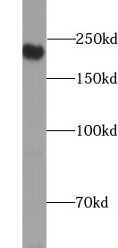
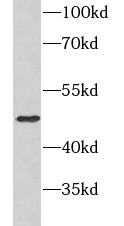
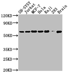
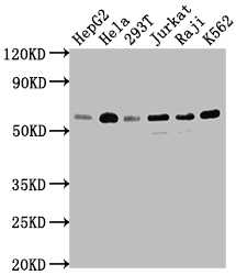
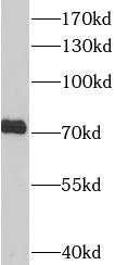
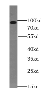
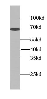
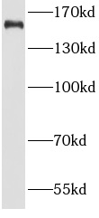
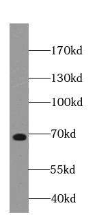
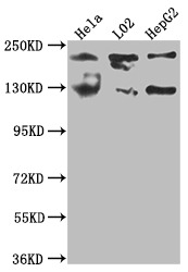 WB (Western Blot)
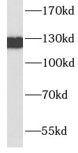
(Western BlotPositive WB detected in: Hela whole cell lysate, L02 whole cell lysate, HepG2 whole cell lysateAll lanes: MGEA5 antibody at 1:1000SecondaryGoat polyclonal to rabbit IgG at 1/50000 dilutionPredicted band size: 103, 96, 77, 97 kDaObserved band size: 130 kDa)
WB (Western Blot)
(Western BlotPositive WB detected in: Hela whole cell lysate, L02 whole cell lysate, HepG2 whole cell lysateAll lanes: MGEA5 antibody at 1:1000SecondaryGoat polyclonal to rabbit IgG at 1/50000 dilutionPredicted band size: 103, 96, 77, 97 kDaObserved band size: 130 kDa)
MGEA5, Monoclonal Recombinant Antibody (Cat# AAA243965)
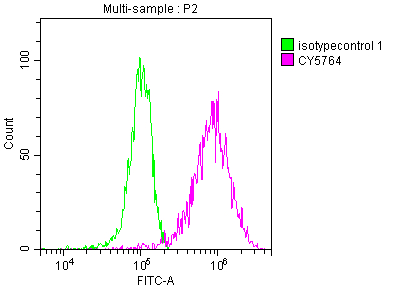
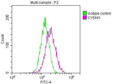
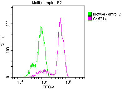 FCM/FACS (Flow Cytometry)
(Overlay histogram showing Hela cells stained with (red line) at 1?50. The cells were fixed with 70% Ethylalcohol (18h) and then incubated in 10% normal goat serum to block non-specific protein-protein interactions followedby the antibody (1ug/1*106cells) for 1 h at 4?.The secondary antibody used was FITC-conjugated goat anti-rabbit IgG (H+L) at 1/200 dilution for 30min at 4?. Control antibody (green line) was Rabbit IgG (1ug/1*106cells) used under the same conditions. Acquisition of >10,000 events was performed.)
FCM/FACS (Flow Cytometry)
(Overlay histogram showing Hela cells stained with (red line) at 1?50. The cells were fixed with 70% Ethylalcohol (18h) and then incubated in 10% normal goat serum to block non-specific protein-protein interactions followedby the antibody (1ug/1*106cells) for 1 h at 4?.The secondary antibody used was FITC-conjugated goat anti-rabbit IgG (H+L) at 1/200 dilution for 30min at 4?. Control antibody (green line) was Rabbit IgG (1ug/1*106cells) used under the same conditions. Acquisition of >10,000 events was performed.)
PTGS1, Monoclonal Recombinant Antibody (Cat# AAA243981)
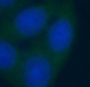
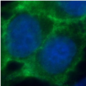
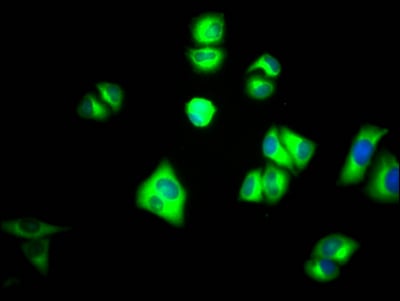
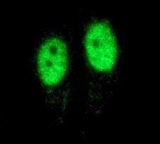
 IF (Immunofluorescence)
(Immunofluorescence staining of HepG2 Cells at 1?50, counter-stained with DAPI. The cells were fixed in 4% formaldehyde, permeated by 0.2% TritonX-100, and blocked in 10% normal Goat Serum. The cells were then incubated with the antibody overnight at 4 degree C. Nuclear DNA was labeled in blue with DAPI. The secondary antibody was FITC-conjugated AffiniPure Goat Anti-Rabbit IgG ?H+L?.)
IF (Immunofluorescence)
(Immunofluorescence staining of HepG2 Cells at 1?50, counter-stained with DAPI. The cells were fixed in 4% formaldehyde, permeated by 0.2% TritonX-100, and blocked in 10% normal Goat Serum. The cells were then incubated with the antibody overnight at 4 degree C. Nuclear DNA was labeled in blue with DAPI. The secondary antibody was FITC-conjugated AffiniPure Goat Anti-Rabbit IgG ?H+L?.)
PTGS2, Monoclonal Recombinant Antibody (Cat# AAA243983)
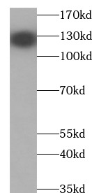
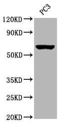
 WB (Western Blot)
(PC-3 cells were subjected to SDS PAGE followed by western blot with AAA248058 (RABEP2 antibody) at dilution of 1:300)
WB (Western Blot)
(PC-3 cells were subjected to SDS PAGE followed by western blot with AAA248058 (RABEP2 antibody) at dilution of 1:300)
RABEP2, Monoclonal Antibody (Cat# AAA248058)
Protein A+G purification
 WB (Western Blot)
(PC-12 cells were subjected to SDS PAGE followed by western blot with AAA248061 (RACGAP1 Antibody) at dilution of 1:600)
WB (Western Blot)
(PC-12 cells were subjected to SDS PAGE followed by western blot with AAA248061 (RACGAP1 Antibody) at dilution of 1:600)
RACGAP1, Monoclonal Antibody (Cat# AAA248061)
Protein A+G purification
 WB (Western Blot)
(Recombinant protein were subjected to SDS PAGE followed by western blot with AAA248075 (S1 tag Antibody) at dilution of 1:10000)
WB (Western Blot)
(Recombinant protein were subjected to SDS PAGE followed by western blot with AAA248075 (S1 tag Antibody) at dilution of 1:10000)
S1 tag, Monoclonal Antibody (Cat# AAA248075)
Purification: Protein A+G purification
 WB (Western Blot)
(mouse brain tissue were subjected to SDS PAGE followed by western blot with AAA248079 (SEC5/EXOC2 antibody) at dilution of 1:5000)
WB (Western Blot)
(mouse brain tissue were subjected to SDS PAGE followed by western blot with AAA248079 (SEC5/EXOC2 antibody) at dilution of 1:5000)
SEC5/EXOC2, Monoclonal Antibody (Cat# AAA248079)
Protein A+G purification
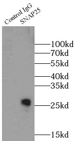
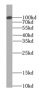
 WB (Western Blot)
(HEK-293 cells were subjected to SDS PAGE followed by western blot with AAA248090 (SNAP25 antibody) at dilution of 1:10000)
WB (Western Blot)
(HEK-293 cells were subjected to SDS PAGE followed by western blot with AAA248090 (SNAP25 antibody) at dilution of 1:10000)
SNAP25, Monoclonal Antibody (Cat# AAA248090)
Protein A+G purification
 WB (Western Blot)
(A431 cells were subjected to SDS PAGE followed by western blot with AAA248104 (TELO2 antibody) at dilution of 1:500)
WB (Western Blot)
(A431 cells were subjected to SDS PAGE followed by western blot with AAA248104 (TELO2 antibody) at dilution of 1:500)
TELO2, Monoclonal Antibody (Cat# AAA248104)
Protein A+G purification
 WB (Western Blot)
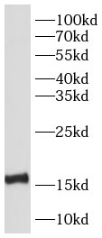
(LPS treated RAW 264.7 cells were subjected to SDS PAGE followed by western blot with AAA248113 (TNF-a Antibody) at dilution of 1:1000)
WB (Western Blot)
(LPS treated RAW 264.7 cells were subjected to SDS PAGE followed by western blot with AAA248113 (TNF-a Antibody) at dilution of 1:1000)
TNF alpha, Monoclonal Antibody (Cat# AAA248113)
Protein A+G purification
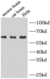
 WB (Western Blot)
(human brain tissue were subjected to SDS PAGE followed by western blot with AAA248120 (TKT antibody) at dilution of 1:1000)
WB (Western Blot)
(human brain tissue were subjected to SDS PAGE followed by western blot with AAA248120 (TKT antibody) at dilution of 1:1000)
Transketolase, Monoclonal Antibody (Cat# AAA248120)
Protein A+G purification
 WB (Western Blot)
(Transfected HEK-293 cells were subjected to SDS PAGE followed by western blot with AAA247962 (IL37 Antibody) at dilution of 1:1000)
WB (Western Blot)
(Transfected HEK-293 cells were subjected to SDS PAGE followed by western blot with AAA247962 (IL37 Antibody) at dilution of 1:1000)
IL37, Monoclonal Antibody (Cat# AAA247962)
 WB (Western Blot)
(HEK-293 cells were subjected to SDS PAGE followed by western blot with AAA247997 (MSH6 Antibody) at dilution of 1:1000)
WB (Western Blot)
(HEK-293 cells were subjected to SDS PAGE followed by western blot with AAA247997 (MSH6 Antibody) at dilution of 1:1000)
MSH6, Monoclonal Antibody (Cat# AAA247997)
Protein A+G purification
 WB (Western Blot)
(fetal human brain tissue were subjected to SDS PAGE followed by western blot with AAA248011 (NESP55,GNAS Antibody) at dilution of 1:2000)
WB (Western Blot)
(fetal human brain tissue were subjected to SDS PAGE followed by western blot with AAA248011 (NESP55,GNAS Antibody) at dilution of 1:2000)
NESP55, GNAS, Monoclonal Antibody (Cat# AAA248011)
Protein A+G purification
 WB (Western Blot)
(L02 cells were subjected to SDS PAGE followed by western blot with AAA248022 (NUMBLIKE Antibody) at dilution of 1:1000)
WB (Western Blot)
(L02 cells were subjected to SDS PAGE followed by western blot with AAA248022 (NUMBLIKE Antibody) at dilution of 1:1000)
NUMBL, Monoclonal Antibody (Cat# AAA248022)
Protein A+G purification
 WB (Western Blot)
(HepG2 cells were subjected to SDS PAGE followed by western blot with AAA248028 (OXA1L antibody) at dilution of 1:1000)
WB (Western Blot)
(HepG2 cells were subjected to SDS PAGE followed by western blot with AAA248028 (OXA1L antibody) at dilution of 1:1000)
OXA1L, Monoclonal Antibody (Cat# AAA248028)
Protein A+G purification
 WB (Western Blot)
(HEK-293 cells were subjected to SDS PAGE followed by western blot with AAA248035 (PCNA Antibody) at dilution of 1:10000)
WB (Western Blot)
(HEK-293 cells were subjected to SDS PAGE followed by western blot with AAA248035 (PCNA Antibody) at dilution of 1:10000)
PCNA, Monoclonal Antibody (Cat# AAA248035)
Protein A+G purification
 WB (Western Blot)
(HeLa cells were subjected to SDS PAGE followed by western blot with AAA248037 (PDCD4 Antibody) at dilution of 1:2000)
WB (Western Blot)
(HeLa cells were subjected to SDS PAGE followed by western blot with AAA248037 (PDCD4 Antibody) at dilution of 1:2000)
PDCD4, Monoclonal Antibody (Cat# AAA248037)
Protein A+G purification
 WB (Western Blot)
(human placenta tissue were subjected to SDS PAGE followed by western blot with AAA247944 (IDS antibody) at dilution of 1:500)
WB (Western Blot)
(human placenta tissue were subjected to SDS PAGE followed by western blot with AAA247944 (IDS antibody) at dilution of 1:500)
Iduronate 2 sulfatase, Monoclonal Antibody (Cat# AAA247944)
Protein A+G purification
 WB (Western Blot)
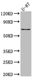
(U-937 cells were subjected to SDS PAGE followed by western blot with AAA247953 (IL-17 antibody) at dilution of 1:500)
WB (Western Blot)
(U-937 cells were subjected to SDS PAGE followed by western blot with AAA247953 (IL-17 antibody) at dilution of 1:500)
IL-17, Monoclonal Antibody (Cat# AAA247953)
 WB (Western Blot)
(HepG2 cells were subjected to SDS PAGE followed by western blot with AAA248132 (USP13 Antibody) at dilution of 1:1000)
WB (Western Blot)
(HepG2 cells were subjected to SDS PAGE followed by western blot with AAA248132 (USP13 Antibody) at dilution of 1:1000)
USP13, Monoclonal Antibody (Cat# AAA248132)
Protein A+G purification
 WB (Western Blot)
(HepG2 cells were subjected to SDS PAGE followed by western blot with AAA248133 (VAPB Antibody) at dilution of 1:1000)
WB (Western Blot)
(HepG2 cells were subjected to SDS PAGE followed by western blot with AAA248133 (VAPB Antibody) at dilution of 1:1000)
VAPB, Monoclonal Antibody (Cat# AAA248133)
Protein A+G purification
 WB (Western Blot)
(HEK-293 cells were subjected to SDS PAGE followed by western blot with AAA248137 (WTAP Antibody) at dilution of 1:2000)
WB (Western Blot)
(HEK-293 cells were subjected to SDS PAGE followed by western blot with AAA248137 (WTAP Antibody) at dilution of 1:2000)
WTAP, Monoclonal Antibody (Cat# AAA248137)
Protein A+G purification
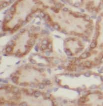
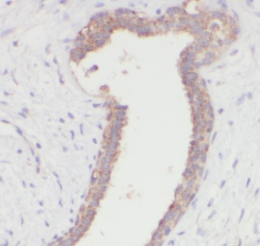
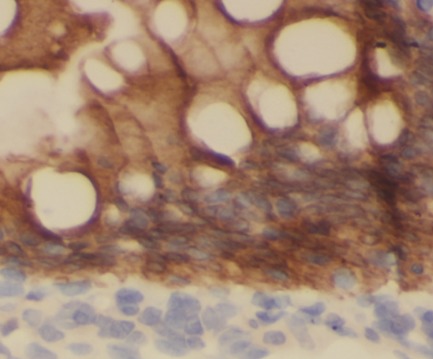
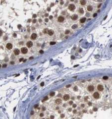
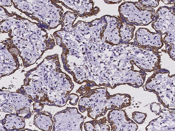
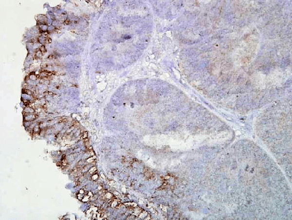
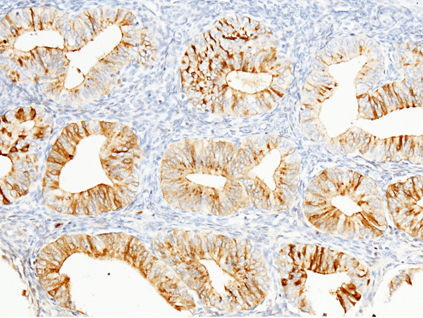 IHC (Immunohiostchemistry)
(Immunochemical staining of human WFDC2 in human endometrium with rabbit monoclonal antibody (1:200, formalin-fixed paraffin embedded sections). Positive staining was localized to the epithelium of uterune gland.)
IHC (Immunohiostchemistry)
(Immunochemical staining of human WFDC2 in human endometrium with rabbit monoclonal antibody (1:200, formalin-fixed paraffin embedded sections). Positive staining was localized to the epithelium of uterune gland.)
HE4, Monoclonal Antibody (Cat# AAA255597)
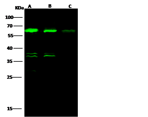
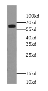
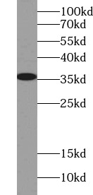
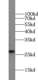
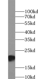
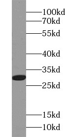
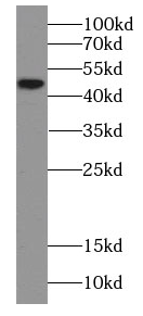
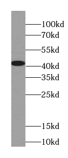
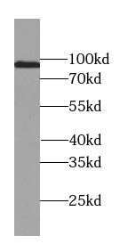
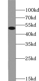
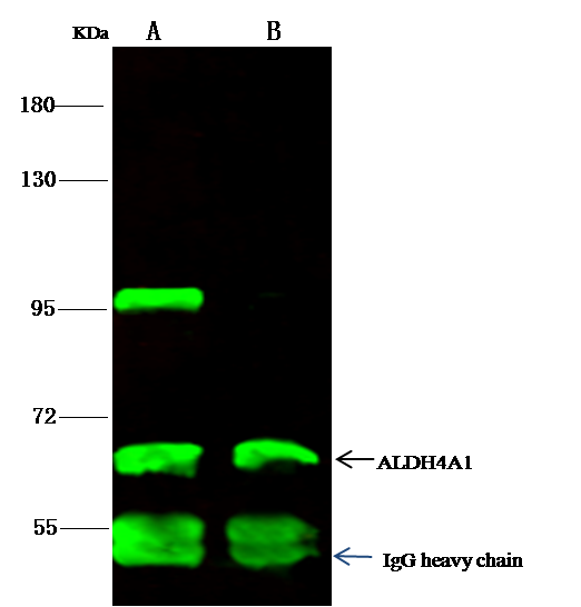 IP (Immunoprecipitation)
(ALDH4A1 was immunoprecipitated using:Lane A:0.5 mg Jurkat Whole Cell LysateLane B:0.5 mg K562 Whole Cell Lysate2 uL anti-ALDH4A1 rabbit monoclonal antibody and 15 ul of 50 % Protein G agarose.Primary antibody:Anti-ALDH4A1 rabbit monoclonal antibody,at 1:200 dilutionSecondary antibody:Dylight 800-labeled antibody to rabbit IgG (H+L), at 1:5000 dilutionDeveloped using the odssey technique.Performed under reducing conditions.Predicted band size: 62 kDaObserved band size: 62 kDa)
IP (Immunoprecipitation)
(ALDH4A1 was immunoprecipitated using:Lane A:0.5 mg Jurkat Whole Cell LysateLane B:0.5 mg K562 Whole Cell Lysate2 uL anti-ALDH4A1 rabbit monoclonal antibody and 15 ul of 50 % Protein G agarose.Primary antibody:Anti-ALDH4A1 rabbit monoclonal antibody,at 1:200 dilutionSecondary antibody:Dylight 800-labeled antibody to rabbit IgG (H+L), at 1:5000 dilutionDeveloped using the odssey technique.Performed under reducing conditions.Predicted band size: 62 kDaObserved band size: 62 kDa)
ALDH4A1, Monoclonal Antibody (Cat# AAA255610)
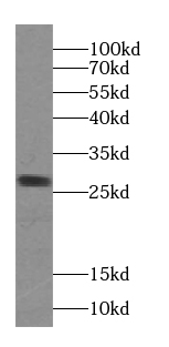
IL18R1, Monoclonal Antibody (Cat# AAA255327)

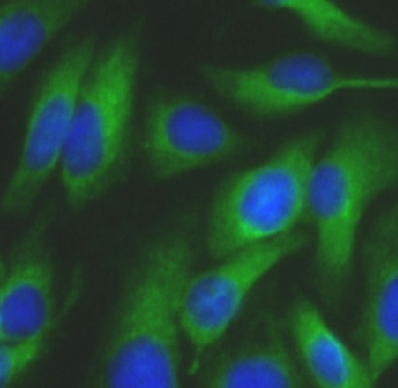
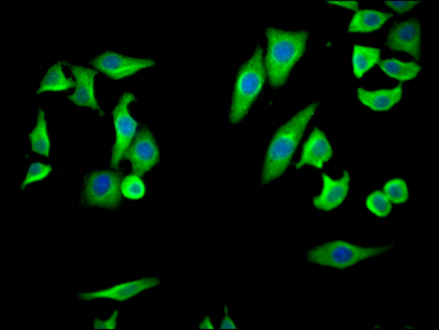
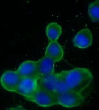
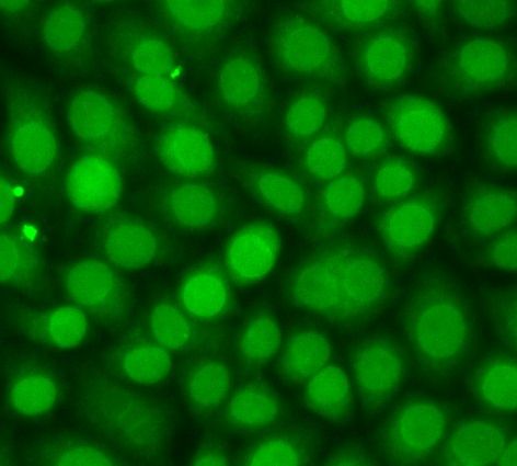 ICC (Immunocytochemistry)
(Immunofluorescent analysis of Hela cells? using AAA253814 (CREB1 antibody) at dilution of 1:200 and Alexa Fluor 488-conjugated Goat Anti-Rabbit IgG(H+L))
ICC (Immunocytochemistry)
(Immunofluorescent analysis of Hela cells? using AAA253814 (CREB1 antibody) at dilution of 1:200 and Alexa Fluor 488-conjugated Goat Anti-Rabbit IgG(H+L))
CREB1, Monoclonal Antibody (Cat# AAA253814)
>=95% as determined by SDS-PAGE
 WB (Western Blot)
(various lysates were subjected to SDS PAGE followed by western blot with AAA253816(GLUT4 antibody) at dilution of 1:1000)
WB (Western Blot)
(various lysates were subjected to SDS PAGE followed by western blot with AAA253816(GLUT4 antibody) at dilution of 1:1000)
GLUT4, Monoclonal Antibody (Cat# AAA253816)
>=95% as determined by SDS-PAGE
 WB (Western Blot)
(various lysates were subjected to SDS PAGE followed by western blot with AAA253817(PPARG antibody) at dilution of 1:1000)
WB (Western Blot)
(various lysates were subjected to SDS PAGE followed by western blot with AAA253817(PPARG antibody) at dilution of 1:1000)
PPARG, Monoclonal Antibody (Cat# AAA253817)
>=95% as determined by SDS-PAGE
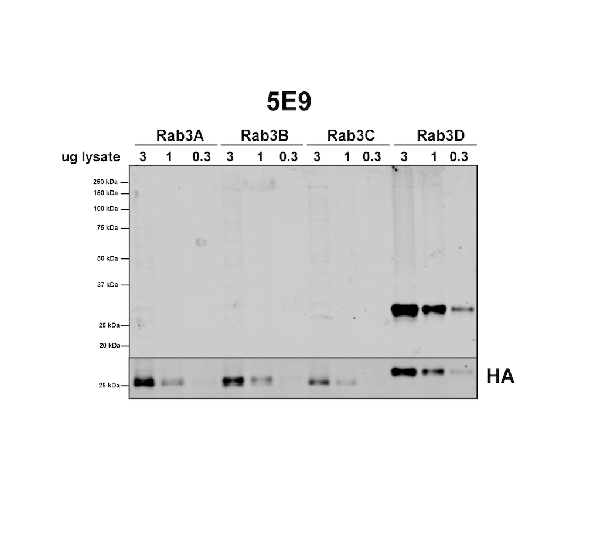
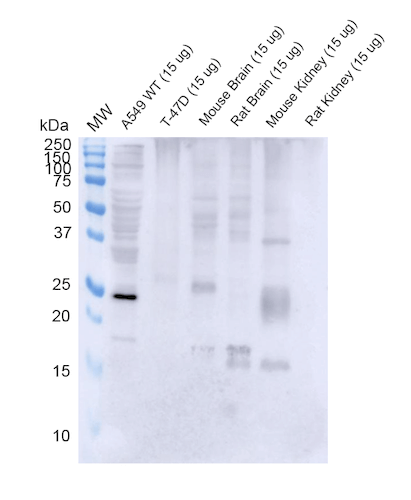 WB (Western Blot)
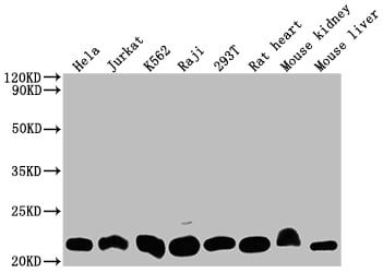
(Western Blot analysis of Human, Mouse, Rat A549, T-47D cells, mouse and rat brain, kidney showing detection of RAB3D protein using Mouse Anti-RAB3D Monoclonal Antibody, Clone 5E9 . Lane 1: MW ladder. Lane 2: WT A549 cells. Lane 3: T-47D cells. Lane 4: mouse brain. Lane 5: rat brain. Lane 6: mouse kidney. Lane 7: rat kidney.. Load: 15 ug. Block: 5% Skim Milk Powder in TBST. Primary Antibody: Mouse Anti-RAB3D Monoclonal Antibody at 1:1000 for 2 hours at RT with shaking. Secondary Antibody: Goat anti-mouse IgG:HRP at 1:4000 for 1 hour at RT with shaking. Color Development: Chemiluminescent for HRP (Moss) for 5 min in RT.)
WB (Western Blot)
(Western Blot analysis of Human, Mouse, Rat A549, T-47D cells, mouse and rat brain, kidney showing detection of RAB3D protein using Mouse Anti-RAB3D Monoclonal Antibody, Clone 5E9 . Lane 1: MW ladder. Lane 2: WT A549 cells. Lane 3: T-47D cells. Lane 4: mouse brain. Lane 5: rat brain. Lane 6: mouse kidney. Lane 7: rat kidney.. Load: 15 ug. Block: 5% Skim Milk Powder in TBST. Primary Antibody: Mouse Anti-RAB3D Monoclonal Antibody at 1:1000 for 2 hours at RT with shaking. Secondary Antibody: Goat anti-mouse IgG:HRP at 1:4000 for 1 hour at RT with shaking. Color Development: Chemiluminescent for HRP (Moss) for 5 min in RT.)
RAB3D, Monoclonal Antibody (Cat# AAA254056)
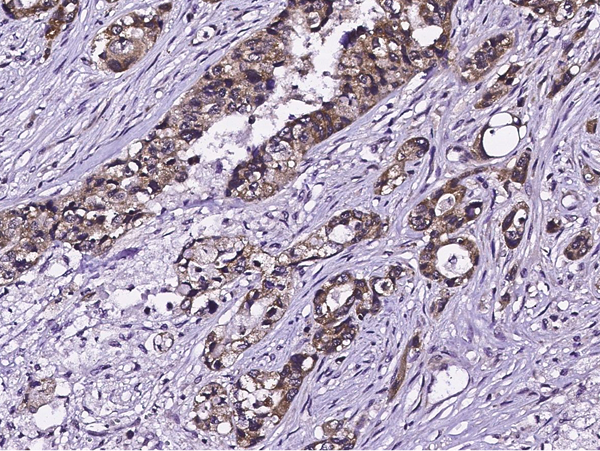
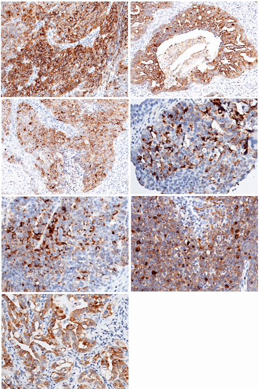
 IHC (Immunohistochemisry)
(Immunochemical staining of human EGFR in human esophageal carcinoma with mouse monoclonal antibody (1:60, formalin-fixed paraffin embedded sections).)
IHC (Immunohistochemisry)
(Immunochemical staining of human EGFR in human esophageal carcinoma with mouse monoclonal antibody (1:60, formalin-fixed paraffin embedded sections).)
EGFR, Monoclonal Antibody (Cat# AAA254114)
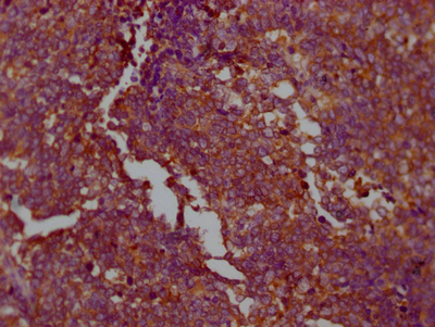
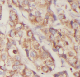
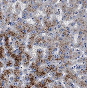
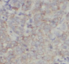
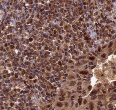
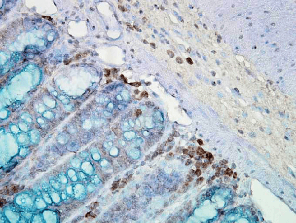 IHC (Immunohiostchemistry)
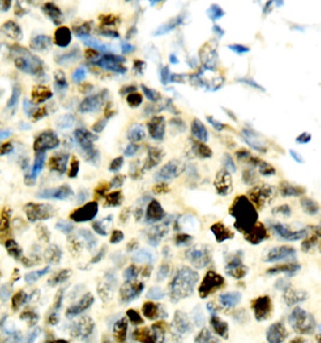
(Immunohistochemistry analysis using Mouse Anti-Hsp90 Monoclonal Antibody, Clone AC-16 . Tissue: inflamed colon. Species: Mouse. Fixation: Formalin. Primary Antibody: Mouse Anti-Hsp90 Monoclonal Antibody at 1:2000 for 12 hours at 4 degree C. Secondary Antibody: Biotin Goat Anti-Mouse at 1:2000 for 1 hour at RT. Counterstain: Mayer Hematoxylin (purple/blue) nuclear stain at 200 ul for 2 minutes at RT. Localization: Inflammatory cells. Magnification: 40x. Mostly inflammatory cells, some mucosa.)
IHC (Immunohiostchemistry)
(Immunohistochemistry analysis using Mouse Anti-Hsp90 Monoclonal Antibody, Clone AC-16 . Tissue: inflamed colon. Species: Mouse. Fixation: Formalin. Primary Antibody: Mouse Anti-Hsp90 Monoclonal Antibody at 1:2000 for 12 hours at 4 degree C. Secondary Antibody: Biotin Goat Anti-Mouse at 1:2000 for 1 hour at RT. Counterstain: Mayer Hematoxylin (purple/blue) nuclear stain at 200 ul for 2 minutes at RT. Localization: Inflammatory cells. Magnification: 40x. Mostly inflammatory cells, some mucosa.)
HSP90, Monoclonal Antibody (Cat# AAA253931)
What are Monoclonal Antibodies?
Monoclonal antibodies are specialized laboratory-produced proteins developed for binding to specific biological antigens or other molecular targets. Since they come from a single cell (or clone), they are especially consistent and accurate in the data they are involved in producing.
This type of antibody material has been shown to be a powerful tool in finding and subsequently destroying harmful cells in an organism, such as those found in cancers or various autoimmune diseases. This makes them excellent aids in medical testing and research, which is why they are so widely used.
AAA Biotech offers a comprehensive range of high-quality monoclonal antibodies that perform effectively in various laboratory tests, including (amongst others) ELISA, western blotting, immunohistochemistry, and flow cytometry. All of the products in our catalog are thoroughly quality tested to make sure that they are reliable and will consistently perform well in your research.
What Are The Uses of Monoclonal Antibodies
Monoclonal antibodies are used in many lab tests, including (amongst others) ELISA, western blotting, immunohistochemistry, and flow cytometry.
ELISA is a test that helps detect a specific substance/analyte in a sample. It uses antibodies (often monoclonal) bound to a solid surface (such as the well of a microplate) to “capture” the substance/analyte in the sample and immobilize it so that the detection antibody component can then bind to it and produce a signal, which can then be measured.
Western blotting identifies specific proteins in a sample. The sample is first separated on a gel, and then antibodies are applied that will typically bind to the target, which will all be localized to a single band in a lane.
Immunohistochemistry helps locate specific proteins in cells or tissue samples using antibodies.
Flow cytometry looks at and sorts cells. It uses antibodies that are conjugated to reporter molecules called “fluorophores”, which, under special lights, emit light themselves, which can then be measured by a detector instrument. For a deeper understanding of these techniques, explore our complete guide to monoclonal antibodies and their benefits.
How Monoclonal Antibodies Are Used as Medicine?
Please note that all of the products listed in AAA Biotech’s also known as AAA Bio or AAABio catalog are strictly for research-use only (RUO).
Monoclonal antibodies can also be used as therapeutic/medical treatments, particularly in the context of cancers. They are designed to find and bind to specific cells or proteins, helping the immune system recognize and attack the cancer. These treatments work in different ways, such as:
- Radioimmunotherapy attaches a small amount of radioactive molecule to the antibody, so it delivers the radiation directly to the cancer cells that the antibody is specifically binding to.
- Antibody-directed enzyme prodrug therapy uses antibodies that are specifically bound to special enzymes. These enzymes activate a harmless drug in the body and turn it into a cancer-killing drug only near the cancer cells—this helps avoid harming healthy cells.
- Immunoliposomes are tiny “bubbles” filled with medicine/drug and coated with antibodies. They carry the drug straight to the cancer cells.
Why Buy Monoclonal Antibodies From Us?
At AAA Biotech, we provide high-performance monoclonal antibodies designed to support a wide range of research needs.
1. Validated for Versatile Applications
The antibodies in our catalog are extensively validated and compatible with multiple techniques, including (but not limited to) ELISA, flow cytometry (FC), immunocytochemistry (ICC), immunofluorescence (IF), immunohistochemistry (IHC), immunoprecipitation (IP), and western blotting (WB).
2. Wide Selection & Specialized Options
We offer antibodies for common and rare species, that are available in various conjugated forms, and also in recombinant formats. Essentially, there is almost anything one might need to meet their experimental model’s requirements.
3. High-Quality Proteins
Our proteins meet high purity standards—90% or more as confirmed by SDS-PAGE. Many are available with tags like His, Flag, GST, or MBP, and we also supply native and biologically active proteins for functional studies.
Frequently Asked Questions
1. Are your monoclonal antibodies validated for specific applications?
Yes, our antibodies are tested and validated for use in methods such as ELISA, western blot, IHC, flow cytometry, and more. Refer to specific product pages or datasheets for individual product information.
2. How do I choose the right monoclonal antibody for my application?
Review the product details directly for application validation, species reactivity, and target information. You may also contact our support team at any time for help.
3. How quickly can I receive my order?
Most orders are processed and shipped within 1–3 business days, depending on product availability and your shipping location.