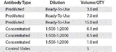
Filters
▼Clonality
▼Type
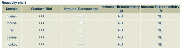
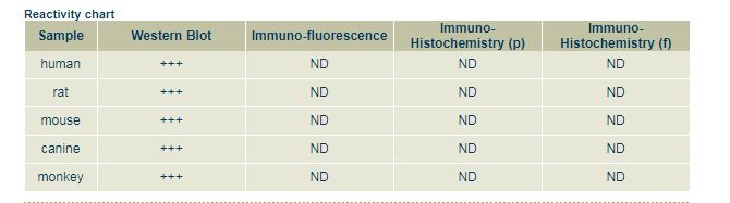
▼Reactivity
▼Gene Name
▼Isotype
▼Host
▼Application
▼Clone
▼Polyclonal Antibodies
At AAA Biotech also known as AAA Bio or AAABio, we provide a broad range of purified polyclonal antibodies (pAbs) that are able to all be browsed online through our website. Due to their high specificity and strong binding affinity, these antibodies are ideal for wide swathes of research and experimental applications.
Our polyclonal antibodies can easily support your work, whether you use them for Western Blotting, Immunocytochemistry (with or without Immunofluorescence used in conjunction), Immunohistochemistry, Immunoprecipitation, and ELISA tests. We highly encourage you to browse our range of pAbs and choose the one that best suits your experimental model.
Viewing 650-700 of 118597 product results
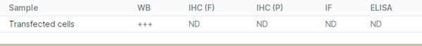
 Application Data
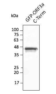
(Anti-ORF3a Ab at 1/2,500 dilution using HEK293 transfected cell lysates at 50 ug per lane; rabbit polyclonal to goat IgG (HRP) at 1/10,000 dilution;)
Application Data
(Anti-ORF3a Ab at 1/2,500 dilution using HEK293 transfected cell lysates at 50 ug per lane; rabbit polyclonal to goat IgG (HRP) at 1/10,000 dilution;)
COVID 19 ORF3a Coronavirus, Polyclonal Antibody (Cat# AAA63175)
 Application Data
(Anti-BCL2 Ab at 1:1,000 dilution; 50 ug of total protein per lane; rabbit polyclonal to goat IgG (HRP) at 1/10,000 dilution;)
Application Data
(Anti-BCL2 Ab at 1:1,000 dilution; 50 ug of total protein per lane; rabbit polyclonal to goat IgG (HRP) at 1/10,000 dilution;)
BCL2, Polyclonal Antibody (Cat# AAA63176)
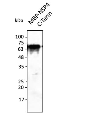 Application Data
(Anti-NSP4 protein Ab at 1/2,500 dilution; lane with 30 ng of recombinant fusion protein (385 aa - stop); rabbit polyclonal to goat IgG (HRP) at 1/10,000 dilution;)
Application Data
(Anti-NSP4 protein Ab at 1/2,500 dilution; lane with 30 ng of recombinant fusion protein (385 aa - stop); rabbit polyclonal to goat IgG (HRP) at 1/10,000 dilution;)
COVID 19 NSP4 Coronavirus, Polyclonal Antibody (Cat# AAA63182)
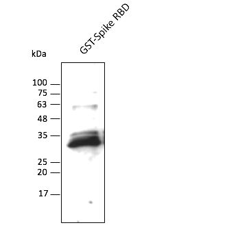 Application Data
(Anti-Spike Ab at 1/2,500 dilution; lane with 30 ng of recombinant fusion protein; chicken polyclonal to goat IgG (HRP) at 1/10,000 dilution;)
Application Data
(Anti-Spike Ab at 1/2,500 dilution; lane with 30 ng of recombinant fusion protein; chicken polyclonal to goat IgG (HRP) at 1/10,000 dilution;)
COVID 19 Spike RBD Domain Coronavirus, Polyclonal Antibody (Cat# AAA63194)

 Application Data
(Anti-Spike Ab at 1/2,500 dilution; lane with 30 ng of recombinant fusion protein; rabbit polyclonal to goat IgG (HRP) at 1/10,000 dilution;)
Application Data
(Anti-Spike Ab at 1/2,500 dilution; lane with 30 ng of recombinant fusion protein; rabbit polyclonal to goat IgG (HRP) at 1/10,000 dilution;)
COVID 19 Spike RBD Domain Coronavirus, Polyclonal Antibody (Cat# AAA63195)
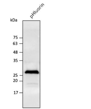
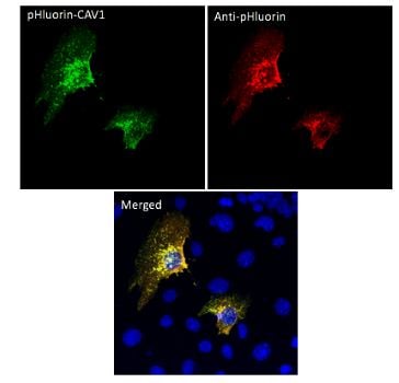
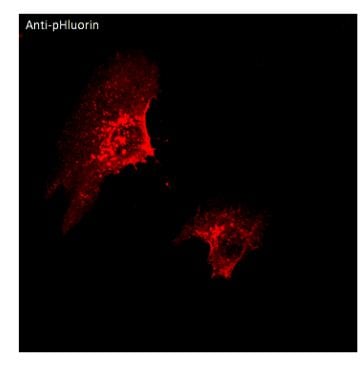 IF (Immunofluorescence)
(Immunofluorescence -anti-pHluorin Ab using hCEC cells transduced with GFP-CAV1; cells were fixed with methanol and anti-pHluorin at 1/250;)
IF (Immunofluorescence)
(Immunofluorescence -anti-pHluorin Ab using hCEC cells transduced with GFP-CAV1; cells were fixed with methanol and anti-pHluorin at 1/250;)
pHluorin, Polyclonal Antibody (Cat# AAA63237)
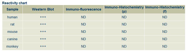
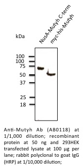 Application Data
MUTYH, Polyclonal Antibody (Cat# AAA63065)

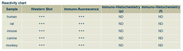
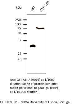 Application Data
GST, Polyclonal Antibody (Cat# AAA63072)
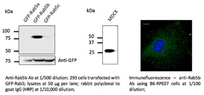 Application Data
Rab5b, Polyclonal Antibody (Cat# AAA63084)
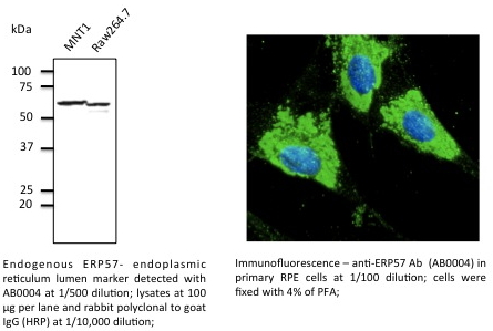 Application Data
ERP57, Polyclonal Antibody (Cat# AAA63085)
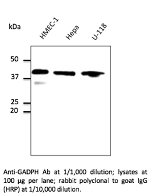
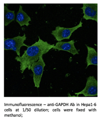 IF (Immunofluorescence)
GAPDH, Polyclonal Antibody (Cat# AAA63089)
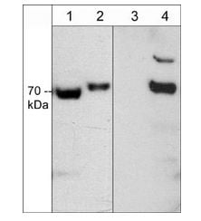
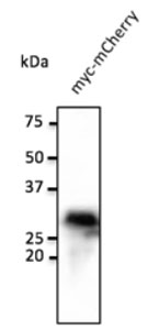 WB (Western Blot)
(Anti-myc tag Ab at 1:1000 dilution; 293 cells tranfected with myc-mCherry (red fluroescent protein); rabbit polyclonal to goat (HRP) at 1:10000 dilution.)
WB (Western Blot)
(Anti-myc tag Ab at 1:1000 dilution; 293 cells tranfected with myc-mCherry (red fluroescent protein); rabbit polyclonal to goat (HRP) at 1:10000 dilution.)
myc, Polyclonal Antibody (Cat# AAA63092)
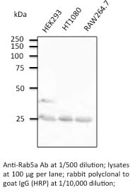
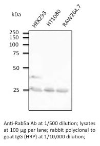 Application Data
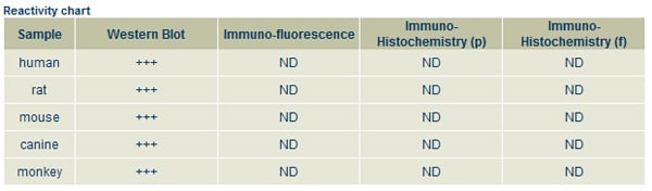
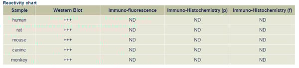
Rab5a, Polyclonal Antibody (Cat# AAA63101)
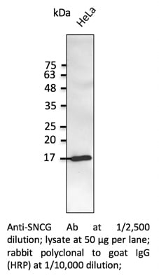 WB (Western Blot)
SNCG, Polyclonal Antibody (Cat# AAA63142)
 WB (Western Blot)
Rab3a, Polyclonal Antibody (Cat# AAA63147)
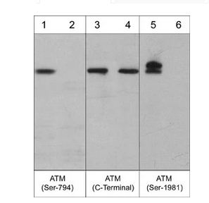
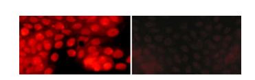 ICC (Immunocytochemistry)
(Immunocytochemical labeling of ATM phosphorylation in calyculin A-treated A431 cells. The cells were labeled with rabbit polyclonal anti-ATM (Ser-794) (AP3631) antibody in the absence (Left) or presence (Right) of blocking peptide (AX3635). The antibody was detected using appropriate secondary antibody conjugated to DyLight 594.)
ICC (Immunocytochemistry)
(Immunocytochemical labeling of ATM phosphorylation in calyculin A-treated A431 cells. The cells were labeled with rabbit polyclonal anti-ATM (Ser-794) (AP3631) antibody in the absence (Left) or presence (Right) of blocking peptide (AX3635). The antibody was detected using appropriate secondary antibody conjugated to DyLight 594.)
ATM, Polyclonal Antibody (Cat# AAA71565)
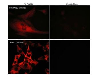 ICC (Immunocytochemistry)
(Immunocytochemical labeling of phosphorylated CRMP2 in mouse C2C12 cells. The cells were probed with CRMP2 (C-terminal region) and CRMP2 (Thr-555) rabbit polyclonal antibodies, then the antibodies were detected using appropriate secondary antibodies conjugated to Cy3. The antibodies were used in the absence (left) or presence (right) of their respective blocking peptide (CX2165 or CX2255).)
ICC (Immunocytochemistry)
(Immunocytochemical labeling of phosphorylated CRMP2 in mouse C2C12 cells. The cells were probed with CRMP2 (C-terminal region) and CRMP2 (Thr-555) rabbit polyclonal antibodies, then the antibodies were detected using appropriate secondary antibodies conjugated to Cy3. The antibodies were used in the absence (left) or presence (right) of their respective blocking peptide (CX2165 or CX2255).)
CRMP2, Polyclonal Antibody (Cat# AAA71605)
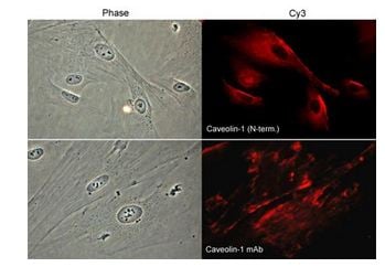 ICC (Immunocytochemistry)
(Immunocytochemical labeling of caveolin-1 in paraformaldehyde-fixed and NP-40-permeabilized rabbit spleen fibroblasts. The cells were labeled with rabbit polyclonal Caveolin-1 (N-terminal region) and mouse monoclonal Caveolin-1 antibodies, and detected using appropriate secondary antibodies conjugated to Cy3. Phase contrast images (left) and immunofluorescent images (right).)
ICC (Immunocytochemistry)
(Immunocytochemical labeling of caveolin-1 in paraformaldehyde-fixed and NP-40-permeabilized rabbit spleen fibroblasts. The cells were labeled with rabbit polyclonal Caveolin-1 (N-terminal region) and mouse monoclonal Caveolin-1 antibodies, and detected using appropriate secondary antibodies conjugated to Cy3. Phase contrast images (left) and immunofluorescent images (right).)
Caveolin-1, Polyclonal Antibody (Cat# AAA71608)
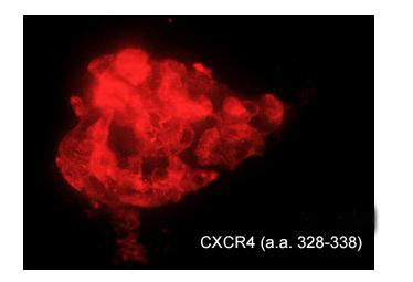 ICC (Immunocytochemistry)
(Immunocytochemical labeling of CXCR4 in chick pluripotent cells. The cells were labeled with rabbit polyclonal CXCR4 (a.a. 328-338) antibody (CP4231), then detected using appropriate secondary antibody (Red). (Image provided by Dr. Yangqing Lu at the Regenerative Bioscience Center, University of Georgia).)
ICC (Immunocytochemistry)
(Immunocytochemical labeling of CXCR4 in chick pluripotent cells. The cells were labeled with rabbit polyclonal CXCR4 (a.a. 328-338) antibody (CP4231), then detected using appropriate secondary antibody (Red). (Image provided by Dr. Yangqing Lu at the Regenerative Bioscience Center, University of Georgia).)
CXCR4, Polyclonal Antibody (Cat# AAA71614)
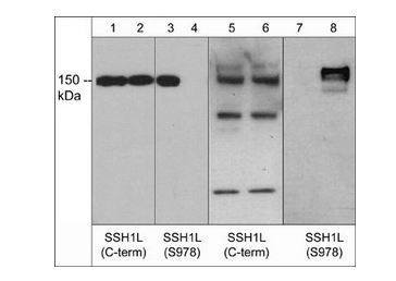
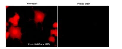 ICC (Immunocytochemistry)
(Immunocytochemical labeling of Slingshot-1L in rat PC12 cells differentiated with NGF. The cells were labeled with rabbit polyclonal anti-Myosin IIA Heavy Chain (a.a. 1936-1950), then detected using appropriate secondary antibody conjugated to Cy3. The antibody was used in the absence (left) or presence (right) of blocking peptide (MX3795).)
ICC (Immunocytochemistry)
(Immunocytochemical labeling of Slingshot-1L in rat PC12 cells differentiated with NGF. The cells were labeled with rabbit polyclonal anti-Myosin IIA Heavy Chain (a.a. 1936-1950), then detected using appropriate secondary antibody conjugated to Cy3. The antibody was used in the absence (left) or presence (right) of blocking peptide (MX3795).)
Myosin IIA Heavy Chain, Polyclonal Antibody (Cat# AAA71661)
 ICC (Immunocytochemistry)
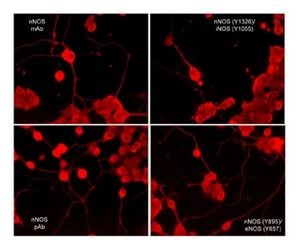
(Immunocytochemical labeling of nNOS phosphorylation in rat PC12 cells differentiated with NGF. The cells were probed with mouse monoclonal (mAb) nNOS (NM4011), and rabbit polyclonal (pAb) nNOS (C-terminal region), nNOS (Tyr-895)/eNOS (Tyr-657), and nNOS (Tyr-1326)/iNOS (Tyr-1055). The antibodies were detected using appropriate secondary antibody conjugated to DyLight 594.)
ICC (Immunocytochemistry)
(Immunocytochemical labeling of nNOS phosphorylation in rat PC12 cells differentiated with NGF. The cells were probed with mouse monoclonal (mAb) nNOS (NM4011), and rabbit polyclonal (pAb) nNOS (C-terminal region), nNOS (Tyr-895)/eNOS (Tyr-657), and nNOS (Tyr-1326)/iNOS (Tyr-1055). The antibodies were detected using appropriate secondary antibody conjugated to DyLight 594.)
eNOS, Polyclonal Antibody (Cat# AAA71675)
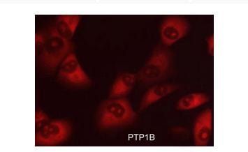 ICC (Immunocytochemistry)
(Immunocytochemical labeling of PTP1B in aldehyde-fixed and NP-40 permeabilized human NCI-H1915 lung carcinoma cells. The cells were labeled with rabbit polyclonal anti-PTP1B (PP2351) antibody. The antibody was detected using appropriate secondary antibody conjugated to DyLight 594.)
ICC (Immunocytochemistry)
(Immunocytochemical labeling of PTP1B in aldehyde-fixed and NP-40 permeabilized human NCI-H1915 lung carcinoma cells. The cells were labeled with rabbit polyclonal anti-PTP1B (PP2351) antibody. The antibody was detected using appropriate secondary antibody conjugated to DyLight 594.)
PTP1B, Polyclonal Antibody (Cat# AAA71692)
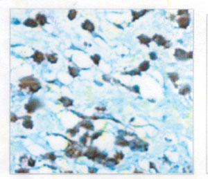
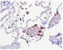
 IHC (Immunohiostchemistry)
(Formalin fixed, citric acid treated parafin sections of mouse cerebral cortex. Sections were probed with anti-Slingshot-1L (SP1711) then anti-Rabbit:HRP before detection using DAB. (Image provided by Carl Hobbs and Dr. Pat Doherty at Wolfson Centre for Age-Related Diseases, King's College London).)
IHC (Immunohiostchemistry)
(Formalin fixed, citric acid treated parafin sections of mouse cerebral cortex. Sections were probed with anti-Slingshot-1L (SP1711) then anti-Rabbit:HRP before detection using DAB. (Image provided by Carl Hobbs and Dr. Pat Doherty at Wolfson Centre for Age-Related Diseases, King's College London).)
Slingshot-1L, Polyclonal Antibody (Cat# AAA71712)
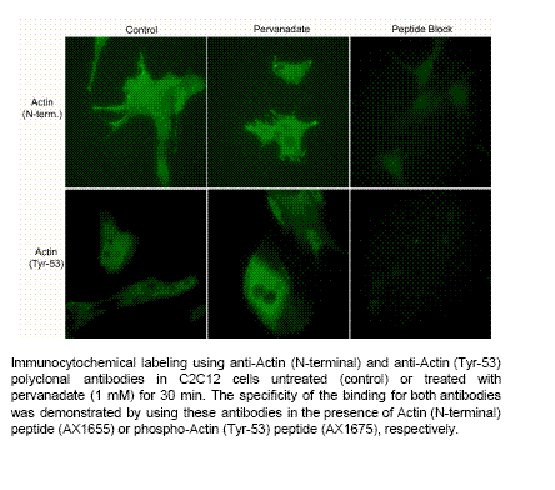
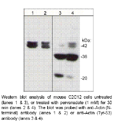 Application Data
Actin (N-terminal Region), Polyclonal Antibody (Cat# AAA71510)
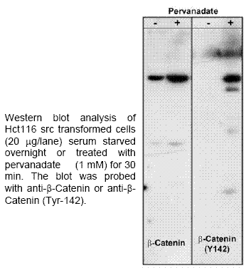
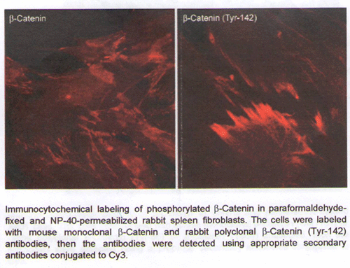 ICC (Immunocytochemistry)
b-Catenin (Tyr-142), Polyclonal Antibody (Cat# AAA71511)
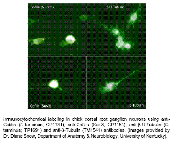
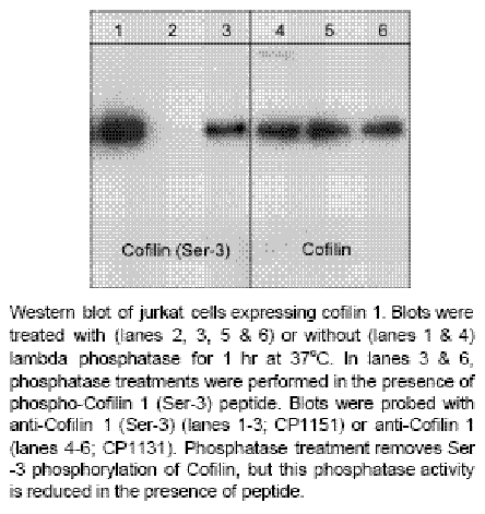 Application Data
Cofilin 1 (N-terminus), Polyclonal Antibody (Cat# AAA71516)
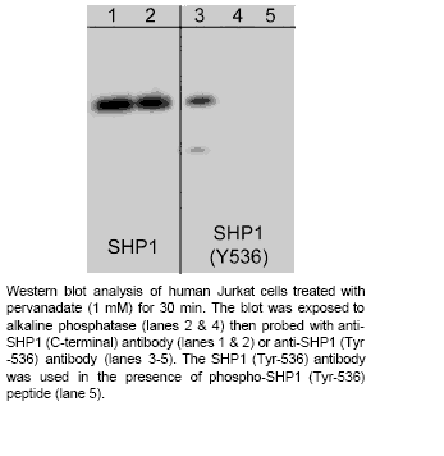 Application Data
SHP1 (Tyr-536), Polyclonal Antibody (Cat# AAA71533)
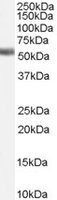
 WB (Western Blot)
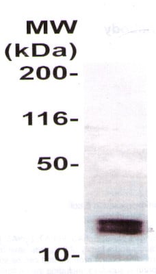
(WB : The full length recombinant protein as immunoblotted by the Rabbit anti-CD Ephrin-A4 (AAA71299) @ 1:500. An immunoreactive band around 22 kDa was observed.)
WB (Western Blot)
(WB : The full length recombinant protein as immunoblotted by the Rabbit anti-CD Ephrin-A4 (AAA71299) @ 1:500. An immunoreactive band around 22 kDa was observed.)
Ephrin A4, Polyclonal Antibody (Cat# AAA71299)
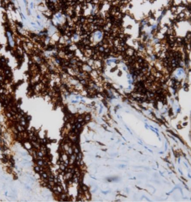
 IHC (Immunohistochemistry)
(Human brain tisuse stained with Anti-GluR 1 antibody AAA71310 at 1:100 for 10 min at room temperature.Staining of formalin-fixed tissue requieres boiling tissue sections in 10 mM Citrate Buffer,pH 6.0 for 10 min followed by cooling at RT for 20 min.)
IHC (Immunohistochemistry)
(Human brain tisuse stained with Anti-GluR 1 antibody AAA71310 at 1:100 for 10 min at room temperature.Staining of formalin-fixed tissue requieres boiling tissue sections in 10 mM Citrate Buffer,pH 6.0 for 10 min followed by cooling at RT for 20 min.)
GluR1, Polyclonal Antibody (Cat# AAA71310)

 IHC (Immunohistochemisry)
(Immunohistochemistry: Human tonsil tissue (FFPE)was immune-stained with Anti-TAFA2 antibody, (Cat#AAA71325) at 1:100 for 10 min @ RT. Staining of formalin-fixed tissue requires boiling tissue sections in 10 mM Citrate Buffer, pH 6.0 for 10 min followed by cooling at RT for 20 min.)
IHC (Immunohistochemisry)
(Immunohistochemistry: Human tonsil tissue (FFPE)was immune-stained with Anti-TAFA2 antibody, (Cat#AAA71325) at 1:100 for 10 min @ RT. Staining of formalin-fixed tissue requires boiling tissue sections in 10 mM Citrate Buffer, pH 6.0 for 10 min followed by cooling at RT for 20 min.)
TAFA2, Polyclonal Antibody (Cat# AAA71325)
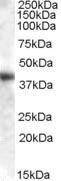
 WB (Western Blot)
(The highly purified Ovomucoid protein from chicken egg white was immunoblotted by Rabbit anti Ovomucoid antibody (Cat#AAA71337) at 1:500. An immunoreactive band is observed at ~28 kDa.)
WB (Western Blot)
(The highly purified Ovomucoid protein from chicken egg white was immunoblotted by Rabbit anti Ovomucoid antibody (Cat#AAA71337) at 1:500. An immunoreactive band is observed at ~28 kDa.)
Ovomucoid, Polyclonal Antibody (Cat# AAA71337)
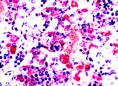
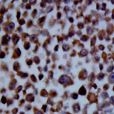 IHC (Immunohistochemistry)
(Immunohistochemistry: The whole cell pellet K562 (FFPE) stained with Rabbit anti-SSEA4 antibody (Cat# 620-500) at 1:200 for 10 min @ RT. Staining of formalin-fixed tissue requires boiling tissue sections in 10 mM Citrate Buffer, pH 6.0 for 10 min followed by cooling at RT for 20 min.)
IHC (Immunohistochemistry)
(Immunohistochemistry: The whole cell pellet K562 (FFPE) stained with Rabbit anti-SSEA4 antibody (Cat# 620-500) at 1:200 for 10 min @ RT. Staining of formalin-fixed tissue requires boiling tissue sections in 10 mM Citrate Buffer, pH 6.0 for 10 min followed by cooling at RT for 20 min.)
SSEA4, Polyclonal Antibody (Cat# AAA71344)
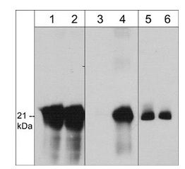
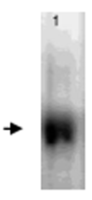
 WB (Western Blot)
(Western Blot: The EGF stimulated HUVEC cell Iysates were resolved onto 10% SDS-PAGE, transferred onto NC membrane, and followed by an immunoblotting with Rabbit anti AXL(Phosphospecific) (Cat# AAA71347) antibody (Lane 1 &2 ) at 1 :500.)
WB (Western Blot)
(Western Blot: The EGF stimulated HUVEC cell Iysates were resolved onto 10% SDS-PAGE, transferred onto NC membrane, and followed by an immunoblotting with Rabbit anti AXL(Phosphospecific) (Cat# AAA71347) antibody (Lane 1 &2 ) at 1 :500.)
AXL/UFO, Polyclonal Antibody (Cat# AAA71347)
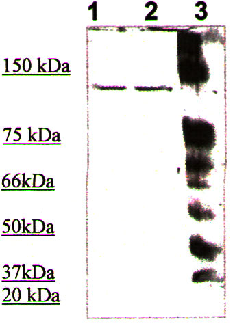
 WB (Western Blot)
(Western Blot: The cell lysate derived from astrocytoma (SW-1783) was resolved onto 12% SDS-PAGE, transferred onto NC membrane, and immunoprobed by Rabbit anti-TIMP-4 (AAA71348) at 1:500. An immunoreactive band is observed around ~25 kDa (1). This band is abolished by pre-incubation with immunizing peptide (2).)
WB (Western Blot)
(Western Blot: The cell lysate derived from astrocytoma (SW-1783) was resolved onto 12% SDS-PAGE, transferred onto NC membrane, and immunoprobed by Rabbit anti-TIMP-4 (AAA71348) at 1:500. An immunoreactive band is observed around ~25 kDa (1). This band is abolished by pre-incubation with immunizing peptide (2).)
TIMP-4, Polyclonal Antibody (Cat# AAA71348)

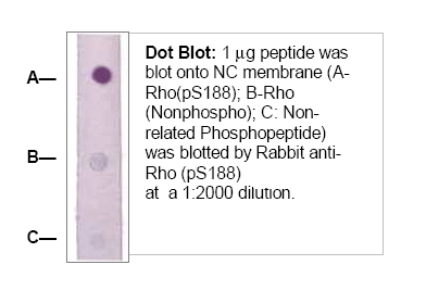 Application Data
Rho (pS188), Polyclonal Antibody (Cat# AAA71406)
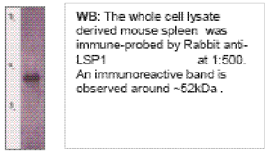 Application Data
LSP, Polyclonal Antibody (Cat# AAA71421)
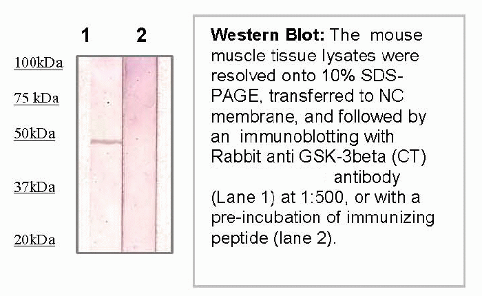 Application Data
GSK-3b, Polyclonal Antibody (Cat# AAA71438)
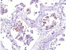
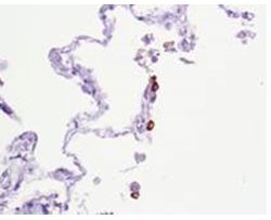
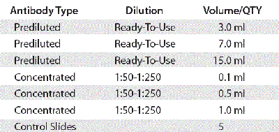
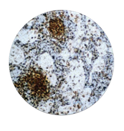
 IHC (Immunohistochemistry)
(Fig. 4 Negative control (lung tissue).)
IHC (Immunohistochemistry)
(Fig. 4 Negative control (lung tissue).)
COVID 19 Spike S Coronavirus, Polyclonal Antibody (Cat# AAA71441)
13(S)-HODE, Polyclonal Antibody (Cat# AAA71782)
 Specificity
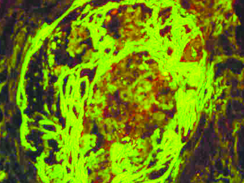
 IF (Immunofluorescence)
(IF of C4c on a FFPE Lupus Positive Tissue)
IF (Immunofluorescence)
(IF of C4c on a FFPE Lupus Positive Tissue)
C4c, Polyclonal Antibody (Cat# AAA59323)
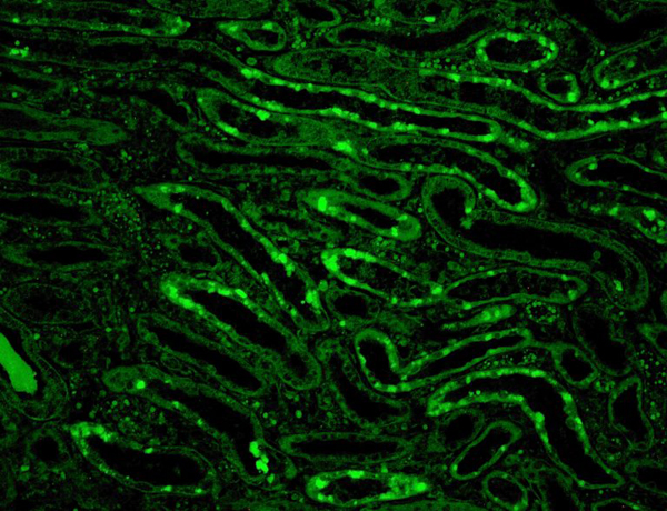
 IF (Immunofluorescence)
(IF of Fibrinogen)
IF (Immunofluorescence)
(IF of Fibrinogen)
Fibrinogen, Polyclonal Antibody (Cat# AAA59324)
 IHC (Immunohiostchemistry)
 IHC (Immunohiostchemistry)
A-1-Antitrypsin, Polyclonal Antibody (Cat# AAA59424)
 IHC (Immunohiostchemistry)
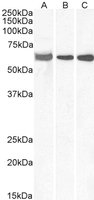
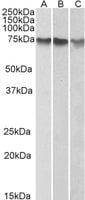 WB (Western Blot)
((1ug/ml) staining of HepG2 (A), K562 (B) and HeLa (C) nuclear lysates (35ug protein in RIPA buffer). Primary incubation was 1 hour. Detected by chemiluminescence.)
WB (Western Blot)
((1ug/ml) staining of HepG2 (A), K562 (B) and HeLa (C) nuclear lysates (35ug protein in RIPA buffer). Primary incubation was 1 hour. Detected by chemiluminescence.)
ATF2, Polyclonal Antibody (Cat# AAA60977)
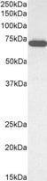
 WB (Western Blot)
((1ug/ml) staining of Human Lung lysate (35ug protein in RIPA buffer). Primary incubation was 1 hour. Detected by chemiluminescence.)
WB (Western Blot)
((1ug/ml) staining of Human Lung lysate (35ug protein in RIPA buffer). Primary incubation was 1 hour. Detected by chemiluminescence.)
Farnesoid X receptor/FXR, Polyclonal Antibody (Cat# AAA60978)
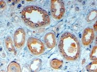 IHC (Immunohistochemistry)
(AAA60982 (4ug/ml) staining of paraffin embedded Human Kidney. Steamed antigen retrieval with citrate buffer pH 6, HRP-staining.)
IHC (Immunohistochemistry)
(AAA60982 (4ug/ml) staining of paraffin embedded Human Kidney. Steamed antigen retrieval with citrate buffer pH 6, HRP-staining.)
LRP6, Polyclonal Antibody (Cat# AAA60982)
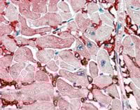 IHC (Immunohiostchemistry)
((2.5ug/ml) staining of paraffin embedded Human Heart. Steamed antigen retrieval with citrate buffer pH 6, AP-staining.)
IHC (Immunohiostchemistry)
((2.5ug/ml) staining of paraffin embedded Human Heart. Steamed antigen retrieval with citrate buffer pH 6, AP-staining.)
GOT1, Polyclonal Antibody (Cat# AAA60996)
 WB (Western Blot)
((0.1ug/ml) staining of Jurkat cell lysate (35ug protein in RIPA buffer). Primary incubation was 1 hour. Detected by chemiluminescence.)
WB (Western Blot)
((0.1ug/ml) staining of Jurkat cell lysate (35ug protein in RIPA buffer). Primary incubation was 1 hour. Detected by chemiluminescence.)
ELF1, Polyclonal Antibody (Cat# AAA60997)
What are Polyclonal Antibodies?
Polyclonal antibodies are antibodies that come from multiple B cell clones of a host animal. The typical hosts used for the majority of polyclonal antibody production are rabbits, goats, sheep, and donkeys. These polyclonal antibodies, once having identified their target, will bind to different epitopes located at different regions or sequences on the same protein/antigen. This ability to bind multiple epitopes is what makes polyclonal antibodies highly sensitive, as explained in our detailed guide on polyclonal antibodies and why they matter.
As a result, they are ideal at locating and binding to the target, even if the target is in very low concentrations (due to many different antibodies being able to bind to the same target molecule, which allows for significant amplification of a downstream signal).
Polyclonal antibodies are typically produced by injecting an antigen into a host animal, which causes the animal’s immune system to attack the foreign antigen by mass generating antibodies against it. After a period of time, serum is collected from the animal and purified using physicochemical fractionation, class-specific affinity purification, and/or antigen-affinity purification.
Key Uses of Polyclonal Antibodies
- Western Blotting: This method is used to find specific proteins in biological samples after separating them by size.
- Immunohistochemistry: IHC helps visualize the location of proteins in tissue sections using various staining techniques.
- ELISA: (Enzyme-Linked Immunosorbent Assay) is typically used to identify specific protein quantities in a sample. ELISAs can be either “Quantitative” or “Qualitative”.
- Flow Cytometry: technique that identifies and measures the specific protein on the surface or inside the cells in a fluid suspension.
- Immunoprecipitation: IP isolates and studies a specific protein from a complex mixture using antibodies.
Why Buy Polyclonal Antibodies from AAA Biotech?
1. Ideal for Various Applications
Our antibodies are generally going to be validated for use in multiple types of assays, including ELISA, Western Blotting, Immunohistochemistry, Immunoprecipitation, amongst others. They are ideal for a wide range of research applications.
2. Rigorous Quality Control
All of the antibodies in our catalog undergo strict quality testing to ensure specificity, sensitivity, and consistent performance. We are confident in the ability of our antibodies to provide you with accurate results.
3. Wide Assortment of Antibodies
Antibodies in our catalog can be found for both common and exotic species, and these antibodies are also available in both conjugated and recombinant forms to suit many diverse experimental needs.
4. Highly Purified
Our antibodies are available in purified forms with over 85% purity, as confirmed by SDS-PAGE. They are also available with tags such as His, Flag, GST, or MBP. We cater to customers worldwide.
FAQ
1. How are polyclonal antibodies produced?
Traditionally, polyclonal antibodies are produced by injecting an antigen into a host animal (such as a rabbit or goat), which then triggers an immune response from the host animal. The animal’s B cells produce antibodies that will recognize different parts of the injected antigen. These antibodies are then collected from the animal’s blood and purified for use.
2. How do polyclonal antibodies differ from monoclonal antibodies?
Polyclonal antibodies are a mix of antibodies that bind to different locations (epitopes) of the same antigen, while monoclonal antibodies are identical and bind to just one specific epitope. This makes polyclonal antibodies more versatile and better at detecting proteins that may be present in low quantities or in altered/modified forms.
3. How should I store polyclonal antibodies?
Polyclonal antibodies should be stored at 4°C for short-term use (up to a few weeks) and at -20°C or -80°C for long-term storage. Avoid repeated freeze-thaw cycles by dividing them into small aliquots. Always check the datasheet for specific storage instructions.