Filters
▼Clonality
▼Type
▼Reactivity
▼Gene Name
▼Isotype
▼Host
▼Application
▼Clone
▼Polyclonal Antibodies
At AAA Biotech also known as AAA Bio or AAABio, we provide a broad range of purified polyclonal antibodies (pAbs) that are able to all be browsed online through our website. Due to their high specificity and strong binding affinity, these antibodies are ideal for wide swathes of research and experimental applications.
Our polyclonal antibodies can easily support your work, whether you use them for Western Blotting, Immunocytochemistry (with or without Immunofluorescence used in conjunction), Immunohistochemistry, Immunoprecipitation, and ELISA tests. We highly encourage you to browse our range of pAbs and choose the one that best suits your experimental model.
Viewing 750-800 of 118597 product results
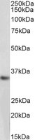
 WB (Western Blot)
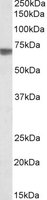
((1ug/ml) staining of Human Cerebellum lysate (35ug protein in RIPA buffer). Primary incubation was 1 hour. Detected by chemiluminescence.)
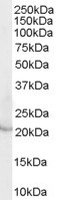
WB (Western Blot)
((1ug/ml) staining of Human Cerebellum lysate (35ug protein in RIPA buffer). Primary incubation was 1 hour. Detected by chemiluminescence.)
CHRNB2, Polyclonal Antibody (Cat# AAA61353)
 IHC (Immunohistochemisry)
((3.8ug/ml) staining of paraffin embedded Human Cerebral Cortex. Steamed antigen retrieval with citrate buffer pH 6, AP-staining.)
IHC (Immunohistochemisry)
((3.8ug/ml) staining of paraffin embedded Human Cerebral Cortex. Steamed antigen retrieval with citrate buffer pH 6, AP-staining.)
STK39/SPAK, Polyclonal Antibody (Cat# AAA61358)
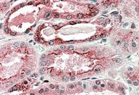
 IHC (Immunohiostchemistry)
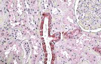
((3.8ug/ml) staining of paraffin embedded Human Kidney. Steamed antigen retrieval with citrate buffer pH 6, AP-staining.)
IHC (Immunohiostchemistry)
((3.8ug/ml) staining of paraffin embedded Human Kidney. Steamed antigen retrieval with citrate buffer pH 6, AP-staining.)
TGFBI, Polyclonal Antibody (Cat# AAA61359)
EBI3, Polyclonal Antibody (Cat# AAA61371)

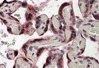
 IHC (Immunohiostchemistry)
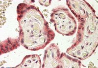
(AAA61374 (3.8ug/ml) staining of paraffin embedded Human Placenta. Steamed antigen retrieval with citrate buffer pH 6, AP-staining.)
IHC (Immunohiostchemistry)
(AAA61374 (3.8ug/ml) staining of paraffin embedded Human Placenta. Steamed antigen retrieval with citrate buffer pH 6, AP-staining.)
TRC8, Polyclonal Antibody (Cat# AAA61374)
 IHC (Immunohiostchemistry)
((3.8ug/ml) staining of paraffin embedded Human Brain Cortex. Steamed antigen retrieval with citrate buffer pH 6, AP-staining.)
IHC (Immunohiostchemistry)
((3.8ug/ml) staining of paraffin embedded Human Brain Cortex. Steamed antigen retrieval with citrate buffer pH 6, AP-staining.)
CAMK2A, Polyclonal Antibody (Cat# AAA61376)
 IF (Immunofluorescence)
((10ug/ml) staining of nuclei HeLa cells (green). Detected by immunofluorescence.)
IF (Immunofluorescence)
((10ug/ml) staining of nuclei HeLa cells (green). Detected by immunofluorescence.)
ODZ3/Teneurin-3, Polyclonal Antibody (Cat# AAA61385)
 IHC (Immunohiostchemistry)
((5ug/ml) staining of paraffin embedded Human Kidney. Steamed antigen retrieval with citrate buffer pH 6, AP-staining.)
IHC (Immunohiostchemistry)
((5ug/ml) staining of paraffin embedded Human Kidney. Steamed antigen retrieval with citrate buffer pH 6, AP-staining.)
GGCX, Polyclonal Antibody (Cat# AAA61389)
 IHC (Immunohiostchemistry)
((3.8ug/ml) staining of paraffin embedded Human Placenta. Steamed antigen retrieval with citrate buffer pH 6, AP-staining.)
IHC (Immunohiostchemistry)
((3.8ug/ml) staining of paraffin embedded Human Placenta. Steamed antigen retrieval with citrate buffer pH 6, AP-staining.)
IDS, Polyclonal Antibody (Cat# AAA61405)
 WB (Western Blot)
((2ug/ml) staining of NIH3T3 lysate (35ug protein in RIPA buffer). Primary incubation was 1 hour. Detected by chemiluminescence.)
WB (Western Blot)
((2ug/ml) staining of NIH3T3 lysate (35ug protein in RIPA buffer). Primary incubation was 1 hour. Detected by chemiluminescence.)
SEC23A, Polyclonal Antibody (Cat# AAA61422)
 WB (Western Blot)
((0.3ug/ml) staining of Rat Brain lysate (35ug protein in RIPA buffer). Primary incubation was 1 hour. Detected by chemiluminescence)
WB (Western Blot)
((0.3ug/ml) staining of Rat Brain lysate (35ug protein in RIPA buffer). Primary incubation was 1 hour. Detected by chemiluminescence)
GPR17, Polyclonal Antibody (Cat# AAA61423)
 WB (Western Blot)
((0.5ug/ml) staining of HeLa lysate (35ug protein in RIPA buffer). Primary incubation was 1 hour. Detected by chemiluminescence.)
WB (Western Blot)
((0.5ug/ml) staining of HeLa lysate (35ug protein in RIPA buffer). Primary incubation was 1 hour. Detected by chemiluminescence.)
F2R/PAR1, Polyclonal Antibody (Cat# AAA61424)
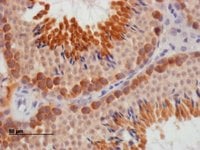 IHC (Immunohiostchemistry)
(Staining of paraffin embedded Mouse Testis.)
IHC (Immunohiostchemistry)
(Staining of paraffin embedded Mouse Testis.)
DAZL, Polyclonal Antibody (Cat# AAA61266)
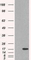 WB (Western Blot)
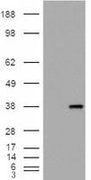
(HEK293 overexpressing SH2D1A (RC204723) and probed (mock transfection in first lane), tested by Origene.)
WB (Western Blot)
(HEK293 overexpressing SH2D1A (RC204723) and probed (mock transfection in first lane), tested by Origene.)
SH2D1A/SLAM associated protein, Polyclonal Antibody (Cat# AAA61280)
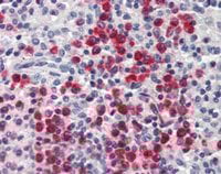
 IHC (Immunohiostchemistry)
((2.5ug/ml) staining of paraffin embedded Human Spleen. Steamed antigen retrieval with citrate buffer pH 6, AP-staining.)
IHC (Immunohiostchemistry)
((2.5ug/ml) staining of paraffin embedded Human Spleen. Steamed antigen retrieval with citrate buffer pH 6, AP-staining.)
ILF2/NF45, Polyclonal Antibody (Cat# AAA61306)
ENOX2/APK1, Polyclonal Antibody (Cat# AAA61310)
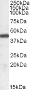
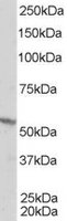
 WB (Western Blot)
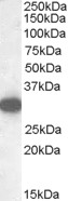
(HEK293 overexpressing ESRRG (RC218233) and probed (mock transfection in first lane), tested by Origene.)
WB (Western Blot)
(HEK293 overexpressing ESRRG (RC218233) and probed (mock transfection in first lane), tested by Origene.)
ESRRG, Polyclonal Antibody (Cat# AAA61326)
 WB (Western Blot)
((0.1ug/ml) staining of NIH3T3 lysate (35ug protein in RIPA buffer). Primary incubation was 1 hour. Detected by chemiluminescence.)
WB (Western Blot)
((0.1ug/ml) staining of NIH3T3 lysate (35ug protein in RIPA buffer). Primary incubation was 1 hour. Detected by chemiluminescence.)
SAR1, Polyclonal Antibody (Cat# AAA61331)
 IHC (Immunohiostchemistry)
((0. 02ug/ml) staining of PFA-perfused cryosection of Mouse Stria terminalis. HRP-staining with Ni-DAB after Biotin-SP-anti-goat amplification. Data obtained by Prof. Erik Hrabovszky, Inst, Exp, Med. , Budapest, Hungary.)
IHC (Immunohiostchemistry)
((0. 02ug/ml) staining of PFA-perfused cryosection of Mouse Stria terminalis. HRP-staining with Ni-DAB after Biotin-SP-anti-goat amplification. Data obtained by Prof. Erik Hrabovszky, Inst, Exp, Med. , Budapest, Hungary.)
Proenkephalin, Polyclonal Antibody (Cat# AAA61340)
Expected from sequence similarity: Mouse, Rat
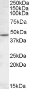
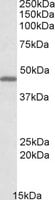
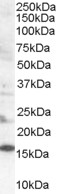
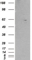
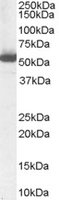
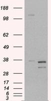
 WB (Western Blot)
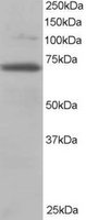
((1ug/ml) staining of Human Testis lysate (35ug protein in RIPA buffer). Primary incubation was 1 hour. Detected by chemiluminescence.)
WB (Western Blot)
((1ug/ml) staining of Human Testis lysate (35ug protein in RIPA buffer). Primary incubation was 1 hour. Detected by chemiluminescence.)
SRD5A2, Polyclonal Antibody (Cat# AAA61346)
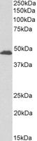
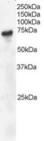
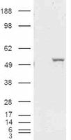
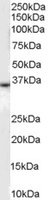
 WB (Western Blot)
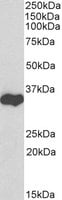
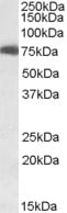
((0.3ug/ml) staining of lysates of cell line HepG2 (35ug protein in RIPA buffer). Primary incubation was 1 hour. Detected by chemiluminescence.)
WB (Western Blot)
((0.3ug/ml) staining of lysates of cell line HepG2 (35ug protein in RIPA buffer). Primary incubation was 1 hour. Detected by chemiluminescence.)
FOXA1/HNF3A, Polyclonal Antibody (Cat# AAA60864)
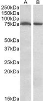
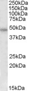
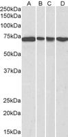
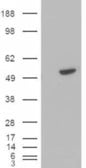
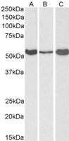
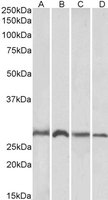 WB (Western Blot)
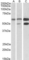
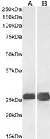
((1ug/ml) staining of Mouse (A, C) and Rat (B, D) Brain (A, B) and Liver (C, D) lysates (35ug protein in RIPA buffer). Primary incubation was 1 hour. Detected by chemiluminescence.)
WB (Western Blot)
((1ug/ml) staining of Mouse (A, C) and Rat (B, D) Brain (A, B) and Liver (C, D) lysates (35ug protein in RIPA buffer). Primary incubation was 1 hour. Detected by chemiluminescence.)
SIAH1, Polyclonal Antibody (Cat# AAA60865)
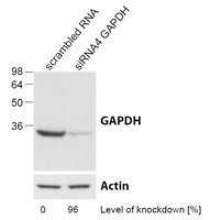
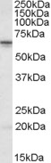
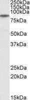
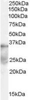
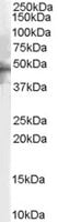
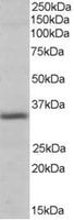
 WB (Western Blot)
((0. 03ug/ml) staining of lysates of cell line HEK293 (35ug protein in RIPA buffer). Primary incubation was 1 hour. Detected by chemiluminescence.)
WB (Western Blot)
((0. 03ug/ml) staining of lysates of cell line HEK293 (35ug protein in RIPA buffer). Primary incubation was 1 hour. Detected by chemiluminescence.)
GAPDH, Polyclonal Antibody (Cat# AAA60874)
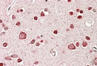
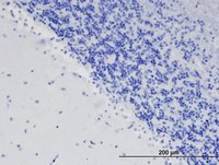
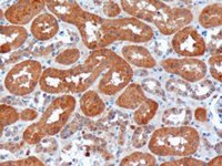
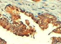
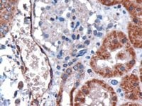
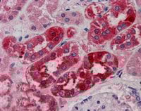 IHC (Immunohiostchemistry)
((5ug/ml) staining of paraffin embedded Human Kidney. Steamed antigen retrieval with citrate buffer pH 6, AP-staining.)
IHC (Immunohiostchemistry)
((5ug/ml) staining of paraffin embedded Human Kidney. Steamed antigen retrieval with citrate buffer pH 6, AP-staining.)
MID2/TRIM1, Polyclonal Antibody (Cat# AAA60883)
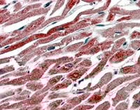
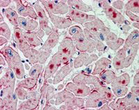 IHC (Immunohiostchemistry)
((2.5ug/ml) staining of paraffin embedded Human Heart. Steamed antigen retrieval with citrate buffer pH 6, AP-staining.)
IHC (Immunohiostchemistry)
((2.5ug/ml) staining of paraffin embedded Human Heart. Steamed antigen retrieval with citrate buffer pH 6, AP-staining.)
ATGL/Desnutrin, Polyclonal Antibody (Cat# AAA60888)

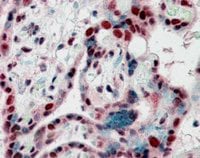 IHC (Immunohiostchemistry)
((2.5ug/ml) staining of paraffin embedded Human Placenta. Steamed antigen retrieval with citrate buffer pH 6, AP-staining.)
IHC (Immunohiostchemistry)
((2.5ug/ml) staining of paraffin embedded Human Placenta. Steamed antigen retrieval with citrate buffer pH 6, AP-staining.)
CBX3/HP1 Gamma, Polyclonal Antibody (Cat# AAA60899)
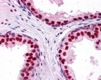 IHC (Immunohiostchemistry)
((2.5ug/ml) staining of paraffin embedded Human Prostate. Steamed antigen retrieval with citrate buffer pH 6, AP-staining. Data obtained using a previous batch.)
IHC (Immunohiostchemistry)
((2.5ug/ml) staining of paraffin embedded Human Prostate. Steamed antigen retrieval with citrate buffer pH 6, AP-staining. Data obtained using a previous batch.)
DDX5/p68 RNA helicase, Polyclonal Antibody (Cat# AAA60903)
 WB (Western Blot)
((0.5ug/ml) staining of Mouse Adipose lysate (35ug protein in RIPA buffer). Primary incubation was 1 hour. Detected by chemiluminescence.)
WB (Western Blot)
((0.5ug/ml) staining of Mouse Adipose lysate (35ug protein in RIPA buffer). Primary incubation was 1 hour. Detected by chemiluminescence.)
Monoglyceride Lipase, Polyclonal Antibody (Cat# AAA60908)
 IHC (Immunohiostchemistry)
((2.5ug/ml) staining of paraffin embedded Human Spleen. Steamed antigen retrieval with citrate buffer pH 6, AP-staining.)
IHC (Immunohiostchemistry)
((2.5ug/ml) staining of paraffin embedded Human Spleen. Steamed antigen retrieval with citrate buffer pH 6, AP-staining.)
Lipocalin 2/NGAL, Polyclonal Antibody (Cat# AAA60915)
 WB (Western Blot)
(HEK293 overexpressing RXRB (RC200516) and probed (mock transfection in first lane), tested by Origene.)
WB (Western Blot)
(HEK293 overexpressing RXRB (RC200516) and probed (mock transfection in first lane), tested by Origene.)
RXR beta, Polyclonal Antibody (Cat# AAA60918)
 WB (Western Blot)
(HEK293 overexpressing BLNK (RC202488) and probed (mock transfection in first lane), tested by Origene.)
WB (Western Blot)
(HEK293 overexpressing BLNK (RC202488) and probed (mock transfection in first lane), tested by Origene.)
BLNK/SLP-65, Polyclonal Antibody (Cat# AAA60921)
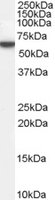
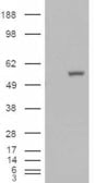
 WB (Western Blot)
((2ug/ml) staining of Human Brain lysate (35ug protein in RIPA buffer). Primary incubation was 1 hour. Detected by chemiluminescence.)
WB (Western Blot)
((2ug/ml) staining of Human Brain lysate (35ug protein in RIPA buffer). Primary incubation was 1 hour. Detected by chemiluminescence.)
Serotonin receptor 1B/HTR1B, Polyclonal Antibody (Cat# AAA60922)
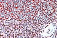
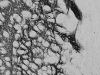
 Application Data
((3.75ug/ml) staining of paraffin embedded Human Colon. Steamed antigen retrieval with citrate bufferpH 6, AP-staining.)
Application Data
((3.75ug/ml) staining of paraffin embedded Human Colon. Steamed antigen retrieval with citrate bufferpH 6, AP-staining.)
PRODH, Polyclonal Antibody (Cat# AAA60931)
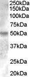
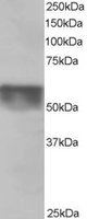
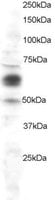
 WB (Western Blot)
((0.1ug/ml) staining of K562 lysate (35ug protein in RIPA buffer). Primary incubation was 1 hour. Detected by chemiluminescence.)
WB (Western Blot)
((0.1ug/ml) staining of K562 lysate (35ug protein in RIPA buffer). Primary incubation was 1 hour. Detected by chemiluminescence.)
CBX8, Polyclonal Antibody (Cat# AAA60947)
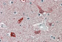
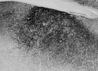
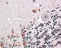
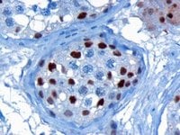
 IHC (Immunohiostchemistry)
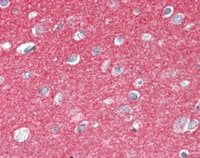
((2.5ug/ml) staining of paraffin embedded Human Cerebral Cortex. Steamed antigen retrieval with citrate buffer pH 6, AP-staining.)
IHC (Immunohiostchemistry)
((2.5ug/ml) staining of paraffin embedded Human Cerebral Cortex. Steamed antigen retrieval with citrate buffer pH 6, AP-staining.)
SNAP25, Polyclonal Antibody (Cat# AAA61037)
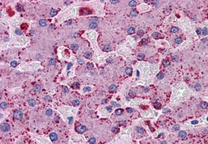
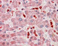 IHC (Immunohistochemisry)
((2.5ug/ml) staining of paraffin embedded Human Liver. Steamed antigen retrieval with citrate buffer pH 6, AP-staining.)
IHC (Immunohistochemisry)
((2.5ug/ml) staining of paraffin embedded Human Liver. Steamed antigen retrieval with citrate buffer pH 6, AP-staining.)
ASC/TMS1, Polyclonal Antibody (Cat# AAA61043)
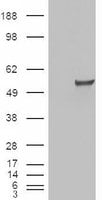
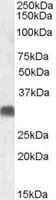
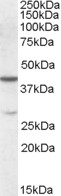
 WB (Western Blot)
((0. 01ug/ml) staining of Human Adipose lysate (35ug protein in RIPA buffer). Primary incubation was 1 hour. Detected by chemiluminescence.)
WB (Western Blot)
((0. 01ug/ml) staining of Human Adipose lysate (35ug protein in RIPA buffer). Primary incubation was 1 hour. Detected by chemiluminescence.)
Perilipin, Polyclonal Antibody (Cat# AAA61046)
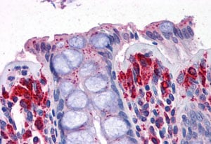
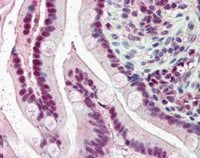 IHC (Immunohistochemisry)
((5ug/ml) staining of paraffin embedded Human Small Intestine. Steamed antigen retrieval with citrate buffer pH 6, AP-staining.)
IHC (Immunohistochemisry)
((5ug/ml) staining of paraffin embedded Human Small Intestine. Steamed antigen retrieval with citrate buffer pH 6, AP-staining.)
KPNA4/IPOA3, Polyclonal Antibody (Cat# AAA61066)
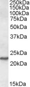
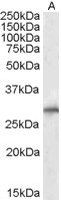
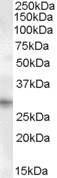 WB (Western Blot)
((1ug/ml) staining of Human Heart lysate (35ug protein in RIPA buffer). Primary incubation was 1 hour. Detected by chemiluminescence.)
WB (Western Blot)
((1ug/ml) staining of Human Heart lysate (35ug protein in RIPA buffer). Primary incubation was 1 hour. Detected by chemiluminescence.)
PRRX1, Polyclonal Antibody (Cat# AAA61080)
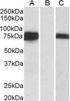 WB (Western Blot)
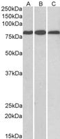
(HEK293 lysate (10ug protein in RIPA buffer) over expressing Human FOXC1 with DYKDDDDK tag probed (0.5ug/ml) in Lane A and probed with anti- DYKDDDDK Tag (1/3000) in lane C. Mock-transfected HEK293 probed (0.5mg/ml) in Lane B. Primary incubations were for 1 hour. Detected by chemiluminescence.)
WB (Western Blot)
(HEK293 lysate (10ug protein in RIPA buffer) over expressing Human FOXC1 with DYKDDDDK tag probed (0.5ug/ml) in Lane A and probed with anti- DYKDDDDK Tag (1/3000) in lane C. Mock-transfected HEK293 probed (0.5mg/ml) in Lane B. Primary incubations were for 1 hour. Detected by chemiluminescence.)
FOXC1, Polyclonal Antibody (Cat# AAA61090)
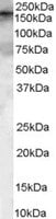 WB (Western Blot)
((0.1ug/ml) staining of HeLa Lysate (35ug protein in RIPA buffer). Primary incubation was 1 hour. Detected by chemiluminescence.)
WB (Western Blot)
((0.1ug/ml) staining of HeLa Lysate (35ug protein in RIPA buffer). Primary incubation was 1 hour. Detected by chemiluminescence.)
GEF5, Polyclonal Antibody (Cat# AAA61101)
 IHC (Immunohiostchemistry)
((2.5ug/ml) staining of paraffin embedded Human Liver. Steamed antigen retrieval with citrate buffer pH 6, AP-staining.)
IHC (Immunohiostchemistry)
((2.5ug/ml) staining of paraffin embedded Human Liver. Steamed antigen retrieval with citrate buffer pH 6, AP-staining.)
RANGAP1, Polyclonal Antibody (Cat# AAA61105)
 IHC (Immunohiostchemistry)
((10ug/ml) staining of paraffin embedded Human Kidney. Microwaved antigen retrieval with citrate buffer pH 6, HRP-staining. (Data have been obtained from a previous batch).)
IHC (Immunohiostchemistry)
((10ug/ml) staining of paraffin embedded Human Kidney. Microwaved antigen retrieval with citrate buffer pH 6, HRP-staining. (Data have been obtained from a previous batch).)
USH1C/Harmonin, Polyclonal Antibody (Cat# AAA61108)
 WB (Western Blot)
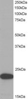
(HEK293 overexpressing ALDH1A1 (RC200723) and probed (mock transfection in first lane), tested by Origene.)
WB (Western Blot)
(HEK293 overexpressing ALDH1A1 (RC200723) and probed (mock transfection in first lane), tested by Origene.)
ALDH1A1, Polyclonal Antibody (Cat# AAA61122)
 IHC (Immunohiostchemistry)
((3.8ug/ml) staining of paraffin embedded Human Heart. Steamed antigen retrieval with citrate buffer pH 6, AP-staining.)
IHC (Immunohiostchemistry)
((3.8ug/ml) staining of paraffin embedded Human Heart. Steamed antigen retrieval with citrate buffer pH 6, AP-staining.)
COX1/PTGS1, Polyclonal Antibody (Cat# AAA61130)

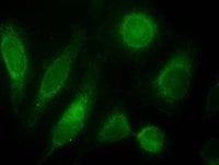
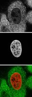 IF (Immunofluorescence)
(Staining (top panel and green) of 4% formaldahyde-fixed HeLa with endogenously expressed HistonH2B-GFP fusion as DNA marker (mid panel and red). Data kindly provided by Francisco Iborra, MRC Mol. Haematol. Unit, Oxford.)
IF (Immunofluorescence)
(Staining (top panel and green) of 4% formaldahyde-fixed HeLa with endogenously expressed HistonH2B-GFP fusion as DNA marker (mid panel and red). Data kindly provided by Francisco Iborra, MRC Mol. Haematol. Unit, Oxford.)
KPNA2/IPOA1, Polyclonal Antibody (Cat# AAA61133)
 WB (Western Blot)
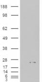
(HEK293 overexpressing ALDH1A1 and probed (mock transfection in first lane).)
WB (Western Blot)
(HEK293 overexpressing ALDH1A1 and probed (mock transfection in first lane).)
ALDH1A1, Polyclonal Antibody (Cat# AAA61136)
 IHC (Immunohistochemistry)
((4ug/ml) staining of paraffin embedded Human Prostate. Steamed antigen retrieval with citrate buffer pH 6, HRP-staining.)
IHC (Immunohistochemistry)
((4ug/ml) staining of paraffin embedded Human Prostate. Steamed antigen retrieval with citrate buffer pH 6, HRP-staining.)
ABCC4/MRP4, Polyclonal Antibody (Cat# AAA61143)
 WB (Western Blot)
(HEK293 overexpressing PIR (RC223111) and probed (mock transfection in first lane), tested by Origene.)
WB (Western Blot)
(HEK293 overexpressing PIR (RC223111) and probed (mock transfection in first lane), tested by Origene.)
Pirin, Polyclonal Antibody (Cat# AAA61152)
 IHC (Immunohiostchemistry)
((3ug/ml) staining of paraffin embedded human kidney. Microwaved antigen retrieval with citrate buffer pH 6, HRP-staining.)
IHC (Immunohiostchemistry)
((3ug/ml) staining of paraffin embedded human kidney. Microwaved antigen retrieval with citrate buffer pH 6, HRP-staining.)
Duffy/FY/DARC, Polyclonal Antibody (Cat# AAA61158)
What are Polyclonal Antibodies?
Polyclonal antibodies are antibodies that come from multiple B cell clones of a host animal. The typical hosts used for the majority of polyclonal antibody production are rabbits, goats, sheep, and donkeys. These polyclonal antibodies, once having identified their target, will bind to different epitopes located at different regions or sequences on the same protein/antigen. This ability to bind multiple epitopes is what makes polyclonal antibodies highly sensitive, as explained in our detailed guide on polyclonal antibodies and why they matter.
As a result, they are ideal at locating and binding to the target, even if the target is in very low concentrations (due to many different antibodies being able to bind to the same target molecule, which allows for significant amplification of a downstream signal).
Polyclonal antibodies are typically produced by injecting an antigen into a host animal, which causes the animal’s immune system to attack the foreign antigen by mass generating antibodies against it. After a period of time, serum is collected from the animal and purified using physicochemical fractionation, class-specific affinity purification, and/or antigen-affinity purification.
Key Uses of Polyclonal Antibodies
- Western Blotting: This method is used to find specific proteins in biological samples after separating them by size.
- Immunohistochemistry: IHC helps visualize the location of proteins in tissue sections using various staining techniques.
- ELISA: (Enzyme-Linked Immunosorbent Assay) is typically used to identify specific protein quantities in a sample. ELISAs can be either “Quantitative” or “Qualitative”.
- Flow Cytometry: technique that identifies and measures the specific protein on the surface or inside the cells in a fluid suspension.
- Immunoprecipitation: IP isolates and studies a specific protein from a complex mixture using antibodies.
Why Buy Polyclonal Antibodies from AAA Biotech?
1. Ideal for Various Applications
Our antibodies are generally going to be validated for use in multiple types of assays, including ELISA, Western Blotting, Immunohistochemistry, Immunoprecipitation, amongst others. They are ideal for a wide range of research applications.
2. Rigorous Quality Control
All of the antibodies in our catalog undergo strict quality testing to ensure specificity, sensitivity, and consistent performance. We are confident in the ability of our antibodies to provide you with accurate results.
3. Wide Assortment of Antibodies
Antibodies in our catalog can be found for both common and exotic species, and these antibodies are also available in both conjugated and recombinant forms to suit many diverse experimental needs.
4. Highly Purified
Our antibodies are available in purified forms with over 85% purity, as confirmed by SDS-PAGE. They are also available with tags such as His, Flag, GST, or MBP. We cater to customers worldwide.
FAQ
1. How are polyclonal antibodies produced?
Traditionally, polyclonal antibodies are produced by injecting an antigen into a host animal (such as a rabbit or goat), which then triggers an immune response from the host animal. The animal’s B cells produce antibodies that will recognize different parts of the injected antigen. These antibodies are then collected from the animal’s blood and purified for use.
2. How do polyclonal antibodies differ from monoclonal antibodies?
Polyclonal antibodies are a mix of antibodies that bind to different locations (epitopes) of the same antigen, while monoclonal antibodies are identical and bind to just one specific epitope. This makes polyclonal antibodies more versatile and better at detecting proteins that may be present in low quantities or in altered/modified forms.
3. How should I store polyclonal antibodies?
Polyclonal antibodies should be stored at 4°C for short-term use (up to a few weeks) and at -20°C or -80°C for long-term storage. Avoid repeated freeze-thaw cycles by dividing them into small aliquots. Always check the datasheet for specific storage instructions.