Filters
▼Clonality
▼Type
▼Reactivity
▼Gene Name
▼Isotype
▼Host
▼Application
▼Clone
▼Polyclonal Antibodies
At AAA Biotech also known as AAA Bio or AAABio, we provide a broad range of purified polyclonal antibodies (pAbs) that are able to all be browsed online through our website. Due to their high specificity and strong binding affinity, these antibodies are ideal for wide swathes of research and experimental applications.
Our polyclonal antibodies can easily support your work, whether you use them for Western Blotting, Immunocytochemistry (with or without Immunofluorescence used in conjunction), Immunohistochemistry, Immunoprecipitation, and ELISA tests. We highly encourage you to browse our range of pAbs and choose the one that best suits your experimental model.
Viewing 450-500 of 118597 product results
 ICC (Immunocytochemistry)
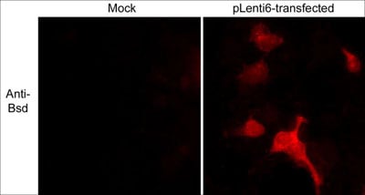
(Immunocytochemical labeling of Blasticidin S Deaminase (Bsd) in 293 cells mock transfected (left) and transiently transfected with pLenti6/TR lentiviral vector (right). The cells were labeled with anti-Bsd (AAA71515) and detected using appropriate secondary antibody conjugated to Texas Red. (Images provided by Charles Mashburn and Dr.George Smith at the University of Kentucky, Spinal Cordand Brain Injury Research Center).)
ICC (Immunocytochemistry)
(Immunocytochemical labeling of Blasticidin S Deaminase (Bsd) in 293 cells mock transfected (left) and transiently transfected with pLenti6/TR lentiviral vector (right). The cells were labeled with anti-Bsd (AAA71515) and detected using appropriate secondary antibody conjugated to Texas Red. (Images provided by Charles Mashburn and Dr.George Smith at the University of Kentucky, Spinal Cordand Brain Injury Research Center).)
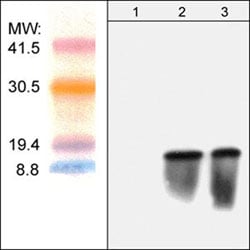
Blasticidin S Deaminase, Polyclonal Antibody (Cat# AAA71515)
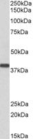
 Application Data
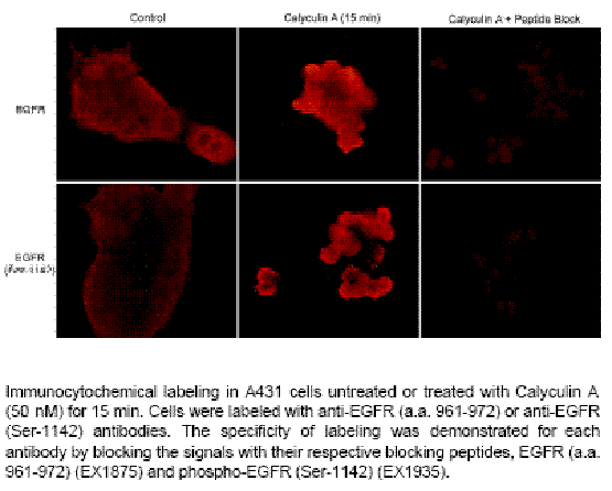
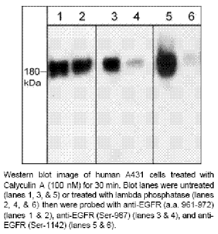
EGFR (Ser-1142), Polyclonal Antibody (Cat# AAA71519)
 Application Data
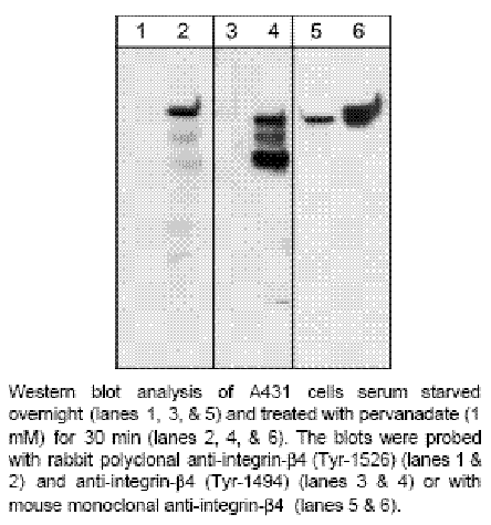
Integrin b4 (Tyr-1494), Polyclonal Antibody (Cat# AAA71523)

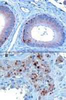
 IHC (Immunohiostchemistry)
(Immunohistochemistry: Human Tonsil tissue (FFPE) stained with Rabbit anti-CCR5 (Cat# AAA71323) at 1:200 for 10 min @ RT. Staining of formalin-fixed tissue requires boiling tissue sections in 10 mM Citrate Buffer, pH 6.0 for 10 min followed by cooling at RT for 20 min.)
IHC (Immunohiostchemistry)
(Immunohistochemistry: Human Tonsil tissue (FFPE) stained with Rabbit anti-CCR5 (Cat# AAA71323) at 1:200 for 10 min @ RT. Staining of formalin-fixed tissue requires boiling tissue sections in 10 mM Citrate Buffer, pH 6.0 for 10 min followed by cooling at RT for 20 min.)
CCR5, Polyclonal Antibody (Cat# AAA71323)
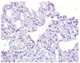
 IHC (Immunohiostchemistry)
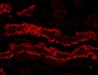
(IHC: Human lung tissue stained with Anti-Tnc antibody, (Cat# AAA71327) at 1:100 for 10 min @ RT. Staining of formalin-fixed tissue requires boiling tissue sections in 10 mM Citrate Buffer, pH 6.0 for 10 min followed by cooling at RT for 20 min.)
IHC (Immunohiostchemistry)
(IHC: Human lung tissue stained with Anti-Tnc antibody, (Cat# AAA71327) at 1:100 for 10 min @ RT. Staining of formalin-fixed tissue requires boiling tissue sections in 10 mM Citrate Buffer, pH 6.0 for 10 min followed by cooling at RT for 20 min.)
Tenascin C, Polyclonal Antibody (Cat# AAA71327)
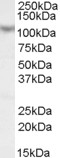
 WB (Western Blot)
(Western Blot: The recombinant protein derived from the full-length (225aa) of H5N1 (5 ug/lane) was resolved onto 10% of SDS-PAGE, transferred onto NC membrane, and immunoblotted by Rabbit anti –H1N1 (CT) antibody at 1:500 . An immunoreactive band around ~30 kDa was observed.)
WB (Western Blot)
(Western Blot: The recombinant protein derived from the full-length (225aa) of H5N1 (5 ug/lane) was resolved onto 10% of SDS-PAGE, transferred onto NC membrane, and immunoblotted by Rabbit anti –H1N1 (CT) antibody at 1:500 . An immunoreactive band around ~30 kDa was observed.)
H1N1, Polyclonal Antibody (Cat# AAA71331)
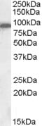
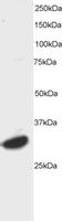
 WB (Western Blot)
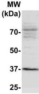
(The whole tissue lysate derived from Mouse embryos was separated in 10% SDS-PAGE, transferred onto NC membrane, and immunoblotted by Rabbit anti –SOX6 (AAA71333) antibody at 1:500. An immunoreactive band around ~94 kDa was observed.)
WB (Western Blot)
(The whole tissue lysate derived from Mouse embryos was separated in 10% SDS-PAGE, transferred onto NC membrane, and immunoblotted by Rabbit anti –SOX6 (AAA71333) antibody at 1:500. An immunoreactive band around ~94 kDa was observed.)
SOX-6, Polyclonal Antibody (Cat# AAA71333)
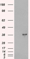
 WB (Western Blot)
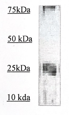
(The cell lysate derived from Jurkat was immunobloted by the Rabbit anti CCR8 (AAA71298) @1:500. An immunoreactive band is observed at ~40 kDa.)
WB (Western Blot)
(The cell lysate derived from Jurkat was immunobloted by the Rabbit anti CCR8 (AAA71298) @1:500. An immunoreactive band is observed at ~40 kDa.)
CCR8, Polyclonal Antibody (Cat# AAA71298)
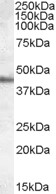
 WB (Western Blot)
(the cell lysate derived from 80% confluence -A431 was immunoprobed by Rabbit anti-Claudin 2 (AAA71313) at 1:500. an immune-reactive band is observed around ~24kDa.)
WB (Western Blot)
(the cell lysate derived from 80% confluence -A431 was immunoprobed by Rabbit anti-Claudin 2 (AAA71313) at 1:500. an immune-reactive band is observed around ~24kDa.)
Claudin 2, Polyclonal Antibody (Cat# AAA71313)
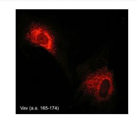 ICC (Immunocytochemistry)
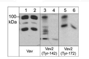
(Immunocytochemical labeling of Vav in paraformaldehyde-fixed and NP-40-permeabilized rabbit spleen fibroblasts. The cells were labeled with rabbit polyclonal Vav (a.a. 165-174), and detected using appropriate secondary antibody conjugated to Cy3.)
ICC (Immunocytochemistry)
(Immunocytochemical labeling of Vav in paraformaldehyde-fixed and NP-40-permeabilized rabbit spleen fibroblasts. The cells were labeled with rabbit polyclonal Vav (a.a. 165-174), and detected using appropriate secondary antibody conjugated to Cy3.)
Vav, Polyclonal Antibody (Cat# AAA71726)
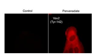 ICC (Immunocytochemistry)
(Immunocytochemical labeling of VAV2 in control and pervanadate-treated human A431 cells. The cells were fixed in paraformaldehyde and permeabilized using NP-40. Then labeled with rabbit polyclonal Vav2 (Tyr-142). The antibody was detected using goat anti-rabbit DyLight 594.)
ICC (Immunocytochemistry)
(Immunocytochemical labeling of VAV2 in control and pervanadate-treated human A431 cells. The cells were fixed in paraformaldehyde and permeabilized using NP-40. Then labeled with rabbit polyclonal Vav2 (Tyr-142). The antibody was detected using goat anti-rabbit DyLight 594.)
Vav2, Polyclonal Antibody (Cat# AAA71727)
 ICC (Immunocytochemistry)
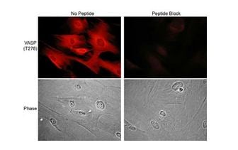
(Immunocytochemical labeling of VASP phosphorylation in rabbit spleen fibroblasts treated with Calyculin A. The cells were labeled with rabbit polyclonal VASP (Thr-278) antibody, then detected using appropriate secondary antibodies conjugated to Cy3. The antibody was used in the absence (top left) or presence (top right) of blocking peptide (VX2785). Corresponding phase images are shown bottom left and right.)
ICC (Immunocytochemistry)
(Immunocytochemical labeling of VASP phosphorylation in rabbit spleen fibroblasts treated with Calyculin A. The cells were labeled with rabbit polyclonal VASP (Thr-278) antibody, then detected using appropriate secondary antibodies conjugated to Cy3. The antibody was used in the absence (top left) or presence (top right) of blocking peptide (VX2785). Corresponding phase images are shown bottom left and right.)
VASP, Polyclonal Antibody (Cat# AAA71729)

 WB (Western Blot)
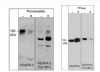
(Left: Western blot image of HUVEC cells untreated (-) or treated with pervanadate (1mM) for 30 min. (+). Right: Western blot image of GST-recombinant VEGFR-2 kinase without (-) or with (+) akaline phosphatase treatment. Both sets of blots were probed with rabbit polyclonal anti-VEGFR-2 (a.a. 1304-1317) or anti-VEGFR-2 (Tyr-801).)
WB (Western Blot)
(Left: Western blot image of HUVEC cells untreated (-) or treated with pervanadate (1mM) for 30 min. (+). Right: Western blot image of GST-recombinant VEGFR-2 kinase without (-) or with (+) akaline phosphatase treatment. Both sets of blots were probed with rabbit polyclonal anti-VEGFR-2 (a.a. 1304-1317) or anti-VEGFR-2 (Tyr-801).)
VEGFR-2, Polyclonal Antibody (Cat# AAA71730)
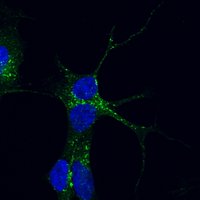
 ICC (Immunocytochemistry)
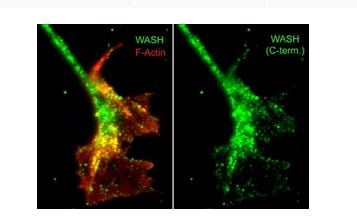
(Immunocytochemical labeling of WASH relative to F-actin in chick DRG neurons. The cells were labeled with rabbit polyclonal WASH (C-terminal region) antibody (AAA71732), then the antibody was detected using appropriate secondary antibody (Green). On the left, this WASH labeling is compared to F-actin staining (Red). (Image provided by Dr. Gianluca Gallo at Drexel University).)
ICC (Immunocytochemistry)
(Immunocytochemical labeling of WASH relative to F-actin in chick DRG neurons. The cells were labeled with rabbit polyclonal WASH (C-terminal region) antibody (AAA71732), then the antibody was detected using appropriate secondary antibody (Green). On the left, this WASH labeling is compared to F-actin staining (Red). (Image provided by Dr. Gianluca Gallo at Drexel University).)
WASH, Polyclonal Antibody (Cat# AAA71732)
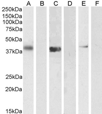
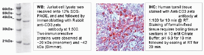 Application Data
CD3 zeta, Polyclonal Antibody (Cat# AAA71436)
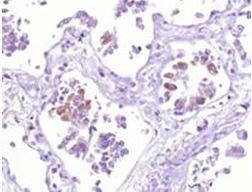
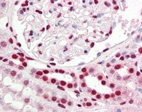
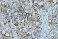
 IHC (Immunohistochemistry)
(Fig. 4 Negative control (lung tissue).)
IHC (Immunohistochemistry)
(Fig. 4 Negative control (lung tissue).)
COVID 19 Spike S Coronavirus, Polyclonal Antibody (Cat# AAA71442)
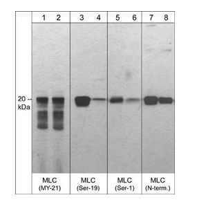
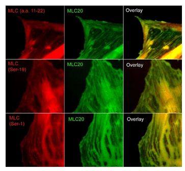 ICC (Immunocytochemistry)
(Immunocytochemical labeling of phosphorylated MLC in paraformaldehyde fixed A7r5 cells. The cells were dual-labeled with anti-MLC (MM3441; middle) and anti-MLC (MP4201; top left), anti-MLC (Ser-19) (MP4221; middle left) and anti-MLC (Ser-1) (MP3461; bottom left). Goat anti-Mouse DyLight 488 and Goat anti-Rabbit DyLight 594 were used for detection of primary antibodies. The overlay of staining patterns are shown to the right.)
ICC (Immunocytochemistry)
(Immunocytochemical labeling of phosphorylated MLC in paraformaldehyde fixed A7r5 cells. The cells were dual-labeled with anti-MLC (MM3441; middle) and anti-MLC (MP4201; top left), anti-MLC (Ser-19) (MP4221; middle left) and anti-MLC (Ser-1) (MP3461; bottom left). Goat anti-Mouse DyLight 488 and Goat anti-Rabbit DyLight 594 were used for detection of primary antibodies. The overlay of staining patterns are shown to the right.)
Myosin, Polyclonal Antibody (Cat# AAA71664)
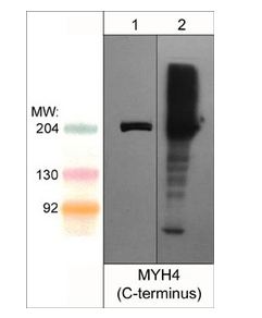 WB (Western Blot)
(Western blot analysis MYH4 in mouse C2C12 (lane 1) and mouse extraocular muscle (lane 2). Both lanes of the blot were probed with rabbit polyclonal anti-MYH4/MyHC-IIB (C-terminus) at 1:1000.)
WB (Western Blot)
(Western blot analysis MYH4 in mouse C2C12 (lane 1) and mouse extraocular muscle (lane 2). Both lanes of the blot were probed with rabbit polyclonal anti-MYH4/MyHC-IIB (C-terminus) at 1:1000.)
Myosin 4/MyHC-IIB, Polyclonal Antibody (Cat# AAA71665)
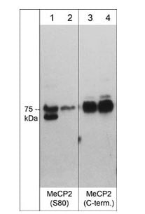
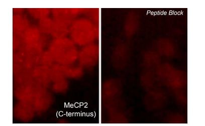 ICC (Immunocytochemistry)
(Immunocytochemical labeling of MeCP2 in rat PC12 cells differentiated with NGF. The cells were probed with MeCP2 (C-terminus) rabbit polyclonal antibody (MP4591) in the absence (left) or presence (right) of blocking peptide (MX4595). The antibody was detected using appropriate secondary antibody conjugated to DyLight 594.)
ICC (Immunocytochemistry)
(Immunocytochemical labeling of MeCP2 in rat PC12 cells differentiated with NGF. The cells were probed with MeCP2 (C-terminus) rabbit polyclonal antibody (MP4591) in the absence (left) or presence (right) of blocking peptide (MX4595). The antibody was detected using appropriate secondary antibody conjugated to DyLight 594.)
MeCP2, Polyclonal Antibody (Cat# AAA71668)
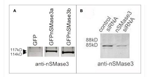
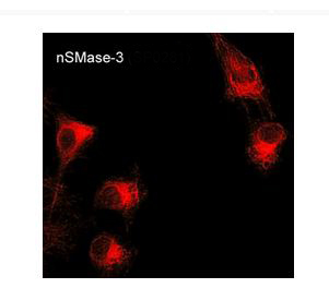 ICC (Immunocytochemistry)
(Immunocytochemical labeling of nSMase3 in aldehyde-fixed and NP-40-permeabilized mouse C2C12 cells. The cells were labeled with rabbit polyclonal anti-nSMase3 (SP0281) antibody. The antibody was detected using appropriate secondary antibody conjugated to DyLight 594.)
ICC (Immunocytochemistry)
(Immunocytochemical labeling of nSMase3 in aldehyde-fixed and NP-40-permeabilized mouse C2C12 cells. The cells were labeled with rabbit polyclonal anti-nSMase3 (SP0281) antibody. The antibody was detected using appropriate secondary antibody conjugated to DyLight 594.)
nSMase3, Polyclonal Antibody (Cat# AAA71708)
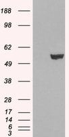
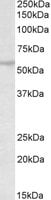
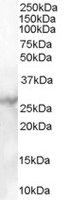
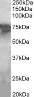
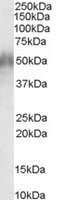
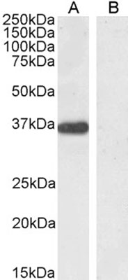 WB (Western Blot)
(AAA60889 (0.5ug/ml) staining of Human Pancreas lysate (35ug protein in RIPA buffer). Detected by chemiluminescence.)
WB (Western Blot)
(AAA60889 (0.5ug/ml) staining of Human Pancreas lysate (35ug protein in RIPA buffer). Detected by chemiluminescence.)
PTF1A/PFT1-P48, Polyclonal Antibody (Cat# AAA60889)
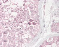
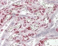
 IHC (Immunohiostchemistry)
((5ug/ml) staining of paraffin embedded Human Pancreas. Steamed antigen retrieval with citrate buffer pH 6, AP-staining.)
IHC (Immunohiostchemistry)
((5ug/ml) staining of paraffin embedded Human Pancreas. Steamed antigen retrieval with citrate buffer pH 6, AP-staining.)
RBP1, Polyclonal Antibody (Cat# AAA60890)
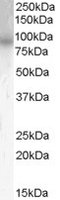
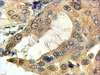
 IHC (Immunohiostchemistry)
((2ug/ml) staining of paraffin embedded Human Breast. Steamed antigen retrieval with citrate buffer pH 9, HRP-staining.)
IHC (Immunohiostchemistry)
((2ug/ml) staining of paraffin embedded Human Breast. Steamed antigen retrieval with citrate buffer pH 9, HRP-staining.)
KAP1/TRIM28, Polyclonal Antibody (Cat# AAA60896)
 IHC (Immunohistochemisry)
((2.5ug/ml) staining of paraffin embedded Human Kidney. Steamed antigen retrieval with citrate buffer pH 6, AP-staining.)
IHC (Immunohistochemisry)
((2.5ug/ml) staining of paraffin embedded Human Kidney. Steamed antigen retrieval with citrate buffer pH 6, AP-staining.)
IRF6, Polyclonal Antibody (Cat# AAA60897)
OCT6/POU3F1, Polyclonal Antibody (Cat# AAA60900)
 IHC (Immunohiostchemistry)
((3.8ug/ml) staining of paraffin embedded Human Testis. Steamed antigen retrieval with citrate buffer pH 6, AP-staining.)
IHC (Immunohiostchemistry)
((3.8ug/ml) staining of paraffin embedded Human Testis. Steamed antigen retrieval with citrate buffer pH 6, AP-staining.)
RNF36/TRIM69, Polyclonal Antibody (Cat# AAA60901)
 WB (Western Blot)
(HEK293 overexpressing Human PDE4B (RC211956) and probed (mock transfection in first lane), tested by Origene.)
WB (Western Blot)
(HEK293 overexpressing Human PDE4B (RC211956) and probed (mock transfection in first lane), tested by Origene.)
Phosphodiesterase 4B, Polyclonal Antibody (Cat# AAA60912)
Timp2L, Polyclonal Antibody (Cat# AAA60914)
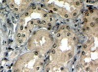 IHC (Immunohiostchemistry)
((4ug/ml) staining of paraffin embedded Human Kidney. Steamed antigen retrieval with citrate buffer pH 6, HRP-staining. Similar results with antigen retrieval at pH9.)
IHC (Immunohiostchemistry)
((4ug/ml) staining of paraffin embedded Human Kidney. Steamed antigen retrieval with citrate buffer pH 6, HRP-staining. Similar results with antigen retrieval at pH9.)
MRP5, Polyclonal Antibody (Cat# AAA60926)
CHD7, Polyclonal Antibody (Cat# AAA60929)
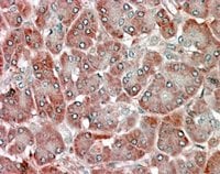
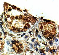
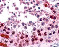
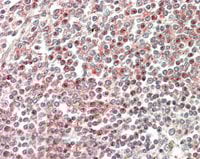
 IHC (Immunohiostchemistry)
((0.5ug/ml) staining of paraffin embedded Human hepatocelluar carcinoma. Data obtained from C. Furer, Visceral Surgery, University of Bern, Switzerland.)
IHC (Immunohiostchemistry)
((0.5ug/ml) staining of paraffin embedded Human hepatocelluar carcinoma. Data obtained from C. Furer, Visceral Surgery, University of Bern, Switzerland.)
NDRG1, Polyclonal Antibody (Cat# AAA60933)
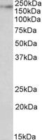
 WB (Western Blot)
((1ug/ml) staining of Human Heart lysate (35ug protein in RIPA buffer). Primary incubation was 1 hour. Detected by chemiluminescence.)
WB (Western Blot)
((1ug/ml) staining of Human Heart lysate (35ug protein in RIPA buffer). Primary incubation was 1 hour. Detected by chemiluminescence.)
MBL2/Mannan-Binding Lectin, Polyclonal Antibody (Cat# AAA60942)
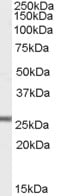
 WB (Western Blot)
((0.1ug/ml) staining of Rat Brain lysate (35ug protein in RIPA buffer). Primary incubation was 1 hour. Detected by chemiluminescence.)
WB (Western Blot)
((0.1ug/ml) staining of Rat Brain lysate (35ug protein in RIPA buffer). Primary incubation was 1 hour. Detected by chemiluminescence.)
Kalirin, Polyclonal Antibody (Cat# AAA60950)
 IF (Immunofluorescence)
((20ug/ml) staining of PFA-perfused cryosection of Porcin Kidney. Microwave antigen retrieval with citrate buffer pH 3, CY3-staining. Data obtained from Dr. Hrvoje Brzica, University of Zagreb, Croatia)
IF (Immunofluorescence)
((20ug/ml) staining of PFA-perfused cryosection of Porcin Kidney. Microwave antigen retrieval with citrate buffer pH 3, CY3-staining. Data obtained from Dr. Hrvoje Brzica, University of Zagreb, Croatia)
GPX3, Polyclonal Antibody (Cat# AAA60952)
 IHC (Immunohiostchemistry)
((2ug/ml) staining of paraffin embedded Human Tonsil. Steamed antigen retrieval with citrate buffer pH 6, HRP-staining.)
IHC (Immunohiostchemistry)
((2ug/ml) staining of paraffin embedded Human Tonsil. Steamed antigen retrieval with citrate buffer pH 6, HRP-staining.)
Swiprosin 1/EFHD2, Polyclonal Antibody (Cat# AAA60953)
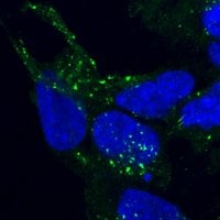
 IF (Immunofluorescence)
(staining of PFA-fixed and saponin permeabilized SHSY5Y and detected with FITC in confocal microscopy. Data obtained from Dr. M. Schallburg Nielsen, Aarhus University Denmark.)
IF (Immunofluorescence)
(staining of PFA-fixed and saponin permeabilized SHSY5Y and detected with FITC in confocal microscopy. Data obtained from Dr. M. Schallburg Nielsen, Aarhus University Denmark.)
VPS35/MEM3, Polyclonal Antibody (Cat# AAA60959)
ITPR3, Polyclonal Antibody (Cat# AAA60961)
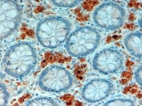 IHC (Immunohiostchemistry)
((2ug/ml) staining of paraffin embedded Human Intestine. Steamed antigen retrieval with citrate buffer pH 6, HRP-staining.)
IHC (Immunohiostchemistry)
((2ug/ml) staining of paraffin embedded Human Intestine. Steamed antigen retrieval with citrate buffer pH 6, HRP-staining.)
APOA4, Polyclonal Antibody (Cat# AAA60965)
KIF5A, Polyclonal Antibody (Cat# AAA60981)
 IHC (Immunohiostchemistry)
((3.8ug/ml) staining of paraffin embedded Human Testis. Steamed antigen retrieval with citrate buffer pH 6, AP-staining.)
IHC (Immunohiostchemistry)
((3.8ug/ml) staining of paraffin embedded Human Testis. Steamed antigen retrieval with citrate buffer pH 6, AP-staining.)
ATF7, Polyclonal Antibody (Cat# AAA60998)
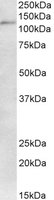
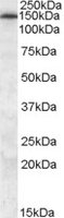
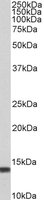
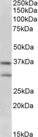 WB (Western Blot)
((0.3ug/ml) staining of Human Brain ((Hippocampus) lysate (35ug protein in RIPA buffer). Primary incubation was 1 hour. Detected by chemiluminescence.)
WB (Western Blot)
((0.3ug/ml) staining of Human Brain ((Hippocampus) lysate (35ug protein in RIPA buffer). Primary incubation was 1 hour. Detected by chemiluminescence.)
FGF23, Polyclonal Antibody (Cat# AAA61001)
 IHC (Immunohiostchemistry)
((3.8ug/ml) staining of paraffin embedded Human Placenta. Steamed antigen retrieval with citrate buffer pH 6, AP-staining.)
IHC (Immunohiostchemistry)
((3.8ug/ml) staining of paraffin embedded Human Placenta. Steamed antigen retrieval with citrate buffer pH 6, AP-staining.)
MAML1/Mastermind, Polyclonal Antibody (Cat# AAA61004)
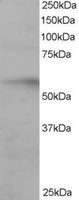
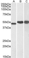
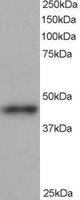
 WB (Western Blot)
((2ug/ml) staining of Human Liver lysate (35ug protein in RIPA buffer). Primary incubation was 1 hour. Detected by chemiluminescence.)
WB (Western Blot)
((2ug/ml) staining of Human Liver lysate (35ug protein in RIPA buffer). Primary incubation was 1 hour. Detected by chemiluminescence.)
PCK2/PEPCK-M, Polyclonal Antibody (Cat# AAA61018)
 IHC (Immunohistochemistry)
((4ug/ml) staining of paraffin embedded Human Prostate. Steamed antigen retrieval with Tris/EDTA buffer pH 9, HRP-staining.)
IHC (Immunohistochemistry)
((4ug/ml) staining of paraffin embedded Human Prostate. Steamed antigen retrieval with Tris/EDTA buffer pH 9, HRP-staining.)
SRD5A1/5-alpha reductase 1, Polyclonal Antibody (Cat# AAA61027)
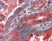 IHC (Immunohiostchemistry)
((3.8ug/ml) staining of paraffin embedded Human Uterus. Steamed antigen retrieval with citrate buffer pH 6, AP-staining.)
IHC (Immunohiostchemistry)
((3.8ug/ml) staining of paraffin embedded Human Uterus. Steamed antigen retrieval with citrate buffer pH 6, AP-staining.)
ARP1 homolog B, Polyclonal Antibody (Cat# AAA61034)
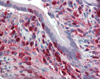 IHC (Immunohiostchemistry)
((2ug/ml) staining of paraffin embedded Human Small Intestine. Steamed antigen retrieval with citrate buffer pH 6, HRP-staining.)
IHC (Immunohiostchemistry)
((2ug/ml) staining of paraffin embedded Human Small Intestine. Steamed antigen retrieval with citrate buffer pH 6, HRP-staining.)
S100A4/CAPL, Polyclonal Antibody (Cat# AAA61038)
 IHC (Immunohiostchemistry)
((5ug/ml) staining of paraffin embedded Human Spleen. Steamed antigen retrieval with citrate buffer pH 6, AP-staining.)
IHC (Immunohiostchemistry)
((5ug/ml) staining of paraffin embedded Human Spleen. Steamed antigen retrieval with citrate buffer pH 6, AP-staining.)
CD2BP2, Polyclonal Antibody (Cat# AAA61040)
SNM1/DCLRE1A, Polyclonal Antibody (Cat# AAA61042)
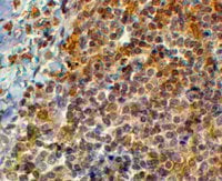
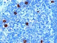 IHC (Immunohiostchemistry)
((1ug/ml) staining of paraffin embedded Human Tonsil. Microwaved antigen retrieval with citrate buffer pH6, HRP-staining.)
IHC (Immunohiostchemistry)
((1ug/ml) staining of paraffin embedded Human Tonsil. Microwaved antigen retrieval with citrate buffer pH6, HRP-staining.)
DOK5, Polyclonal Antibody (Cat# AAA61044)
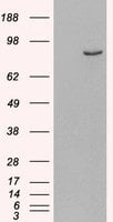
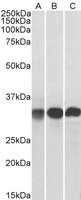
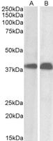 WB (Western Blot)
((0. 01ug/ml) staining of Daudi (A) and Molt4 (B) cell lysate (35ug protein in RIPA buffer). Primary incubation was 1 hour. Detected by chemiluminescence.)
WB (Western Blot)
((0. 01ug/ml) staining of Daudi (A) and Molt4 (B) cell lysate (35ug protein in RIPA buffer). Primary incubation was 1 hour. Detected by chemiluminescence.)
SET/I2 alpha PP2A, Polyclonal Antibody (Cat# AAA61056)
What are Polyclonal Antibodies?
Polyclonal antibodies are antibodies that come from multiple B cell clones of a host animal. The typical hosts used for the majority of polyclonal antibody production are rabbits, goats, sheep, and donkeys. These polyclonal antibodies, once having identified their target, will bind to different epitopes located at different regions or sequences on the same protein/antigen. This ability to bind multiple epitopes is what makes polyclonal antibodies highly sensitive, as explained in our detailed guide on polyclonal antibodies and why they matter.
As a result, they are ideal at locating and binding to the target, even if the target is in very low concentrations (due to many different antibodies being able to bind to the same target molecule, which allows for significant amplification of a downstream signal).
Polyclonal antibodies are typically produced by injecting an antigen into a host animal, which causes the animal’s immune system to attack the foreign antigen by mass generating antibodies against it. After a period of time, serum is collected from the animal and purified using physicochemical fractionation, class-specific affinity purification, and/or antigen-affinity purification.
Key Uses of Polyclonal Antibodies
- Western Blotting: This method is used to find specific proteins in biological samples after separating them by size.
- Immunohistochemistry: IHC helps visualize the location of proteins in tissue sections using various staining techniques.
- ELISA: (Enzyme-Linked Immunosorbent Assay) is typically used to identify specific protein quantities in a sample. ELISAs can be either “Quantitative” or “Qualitative”.
- Flow Cytometry: technique that identifies and measures the specific protein on the surface or inside the cells in a fluid suspension.
- Immunoprecipitation: IP isolates and studies a specific protein from a complex mixture using antibodies.
Why Buy Polyclonal Antibodies from AAA Biotech?
1. Ideal for Various Applications
Our antibodies are generally going to be validated for use in multiple types of assays, including ELISA, Western Blotting, Immunohistochemistry, Immunoprecipitation, amongst others. They are ideal for a wide range of research applications.
2. Rigorous Quality Control
All of the antibodies in our catalog undergo strict quality testing to ensure specificity, sensitivity, and consistent performance. We are confident in the ability of our antibodies to provide you with accurate results.
3. Wide Assortment of Antibodies
Antibodies in our catalog can be found for both common and exotic species, and these antibodies are also available in both conjugated and recombinant forms to suit many diverse experimental needs.
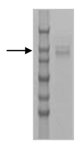
4. Highly Purified
Our antibodies are available in purified forms with over 85% purity, as confirmed by SDS-PAGE. They are also available with tags such as His, Flag, GST, or MBP. We cater to customers worldwide.
FAQ
1. How are polyclonal antibodies produced?
Traditionally, polyclonal antibodies are produced by injecting an antigen into a host animal (such as a rabbit or goat), which then triggers an immune response from the host animal. The animal’s B cells produce antibodies that will recognize different parts of the injected antigen. These antibodies are then collected from the animal’s blood and purified for use.
2. How do polyclonal antibodies differ from monoclonal antibodies?
Polyclonal antibodies are a mix of antibodies that bind to different locations (epitopes) of the same antigen, while monoclonal antibodies are identical and bind to just one specific epitope. This makes polyclonal antibodies more versatile and better at detecting proteins that may be present in low quantities or in altered/modified forms.
3. How should I store polyclonal antibodies?
Polyclonal antibodies should be stored at 4°C for short-term use (up to a few weeks) and at -20°C or -80°C for long-term storage. Avoid repeated freeze-thaw cycles by dividing them into small aliquots. Always check the datasheet for specific storage instructions.