Filters
▼Clonality
▼Type
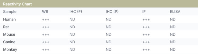
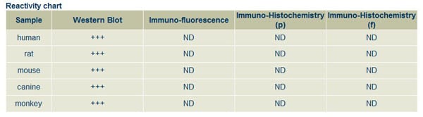
▼Reactivity
▼Gene Name
▼Isotype
▼Host
▼Application
▼Clone
▼Polyclonal Antibodies
At AAA Biotech also known as AAA Bio or AAABio, we provide a broad range of purified polyclonal antibodies (pAbs) that are able to all be browsed online through our website. Due to their high specificity and strong binding affinity, these antibodies are ideal for wide swathes of research and experimental applications.
Our polyclonal antibodies can easily support your work, whether you use them for Western Blotting, Immunocytochemistry (with or without Immunofluorescence used in conjunction), Immunohistochemistry, Immunoprecipitation, and ELISA tests. We highly encourage you to browse our range of pAbs and choose the one that best suits your experimental model.
Viewing 600-650 of 118597 product results
 WB (Western Blot)
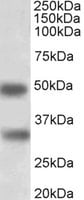
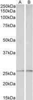
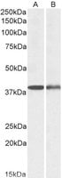
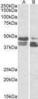
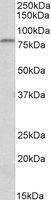
(KLF5 antibody (pAb) tested by Western Blot. Nuclear extract of K562 cells (20 ug) probed with the KLF5 antibody (pAb) at a dilution of 1:2,000.)
WB (Western Blot)
(KLF5 antibody (pAb) tested by Western Blot. Nuclear extract of K562 cells (20 ug) probed with the KLF5 antibody (pAb) at a dilution of 1:2,000.)
KLF5, Polyclonal Antibody (Cat# AAA59976)

 WB (Western Blot)
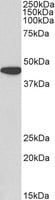
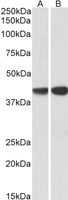
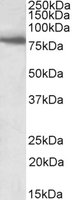
(LXR-a antibody (pAb) tested by Western blot. Whole cell extract of Hep G2 cells (30 ug) probed with LXR-a antibody (pAb) at a dilution of 1:500.)
WB (Western Blot)
(LXR-a antibody (pAb) tested by Western blot. Whole cell extract of Hep G2 cells (30 ug) probed with LXR-a antibody (pAb) at a dilution of 1:500.)
LXR-alpha, Polyclonal Antibody (Cat# AAA59987)

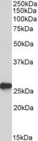
 WB (Western Blot)
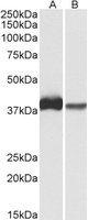
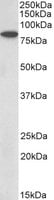
(LXR-b pAb tested by Western blot. Detection of LXR-b by Western blot analysis. HeLa whole-cell extract (20 ug) was probed with LXR-b pAb at a 1:1000 dilution.)
WB (Western Blot)
(LXR-b pAb tested by Western blot. Detection of LXR-b by Western blot analysis. HeLa whole-cell extract (20 ug) was probed with LXR-b pAb at a 1:1000 dilution.)
LXR-beta, Polyclonal Antibody (Cat# AAA59988)
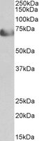
 WB (Western Blot)
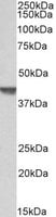
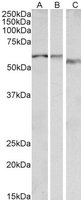
(MOF / MYST1 antibody (pAb) tested by Western blot. Whole-cell extract of HeLa cells (30 ug) probed with MOF / MYST1 pAb at a dilution of 1:500.)
WB (Western Blot)
(MOF / MYST1 antibody (pAb) tested by Western blot. Whole-cell extract of HeLa cells (30 ug) probed with MOF / MYST1 pAb at a dilution of 1:500.)
MOF/MYST1, Polyclonal Antibody (Cat# AAA59998)

 WB (Western Blot)
(Elk-1 antibody (pAb) tested by Western blot. Whole cell extract of HeLa cells (40 ug) probed with Elk-1 antibody (1:500).)
WB (Western Blot)
(Elk-1 antibody (pAb) tested by Western blot. Whole cell extract of HeLa cells (40 ug) probed with Elk-1 antibody (1:500).)
Elk-1, Polyclonal Antibody (Cat# AAA60007)
[Met5]-Enkephalin, Polyclonal Antibody (Cat# AAA58823)
Calcitonin (Salmon), Polyclonal Antibody (Cat# AAA58826)
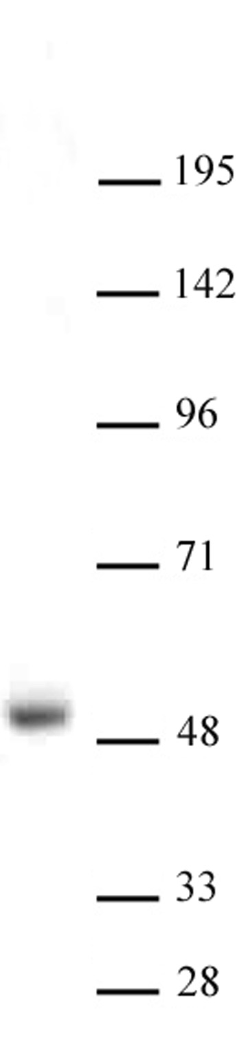
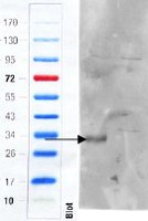
 WB (Western Blot)
((1ug/ml) staining of U937 lysate (35ug protein in RIPA buffer). Primary incubation was 1 hour. Detected by chemiluminescence.)
WB (Western Blot)
((1ug/ml) staining of U937 lysate (35ug protein in RIPA buffer). Primary incubation was 1 hour. Detected by chemiluminescence.)
CD68, Polyclonal Antibody (Cat# AAA61782)
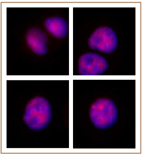
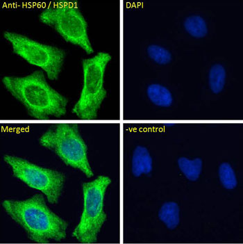 IF (Immunofluorescence)
(AAA61783 Immunofluorescence analysis of paraformaldehyde fixed HeLa cells, permeabilized with 0.15% Triton. Primary incubation 1hr (10ug/ml) followed by Alexa Fluor 488 secondary antibody (4ug/ml), showing Mitochondrial staining. The nuclear stain is DAPI (blue). Negative control: Unimmunized goat IgG (10ug/ml) followed by Alexa Fluor 488 secondary antibody (4ug/ml).)
IF (Immunofluorescence)
(AAA61783 Immunofluorescence analysis of paraformaldehyde fixed HeLa cells, permeabilized with 0.15% Triton. Primary incubation 1hr (10ug/ml) followed by Alexa Fluor 488 secondary antibody (4ug/ml), showing Mitochondrial staining. The nuclear stain is DAPI (blue). Negative control: Unimmunized goat IgG (10ug/ml) followed by Alexa Fluor 488 secondary antibody (4ug/ml).)
HSP60/HSPD1, Polyclonal Antibody (Cat# AAA61783)
Expected from sequence similarity: Human, Dog, Pig, Cow
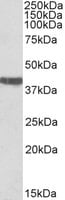
 WB (Western Blot)
((0.1ug/ml) staining of HeLa lysate (35ug protein in RIPA buffer). Primary incubation was 1 hour. Detected by chemiluminescence.)
WB (Western Blot)
((0.1ug/ml) staining of HeLa lysate (35ug protein in RIPA buffer). Primary incubation was 1 hour. Detected by chemiluminescence.)
EGFR, Polyclonal Antibody (Cat# AAA61788)
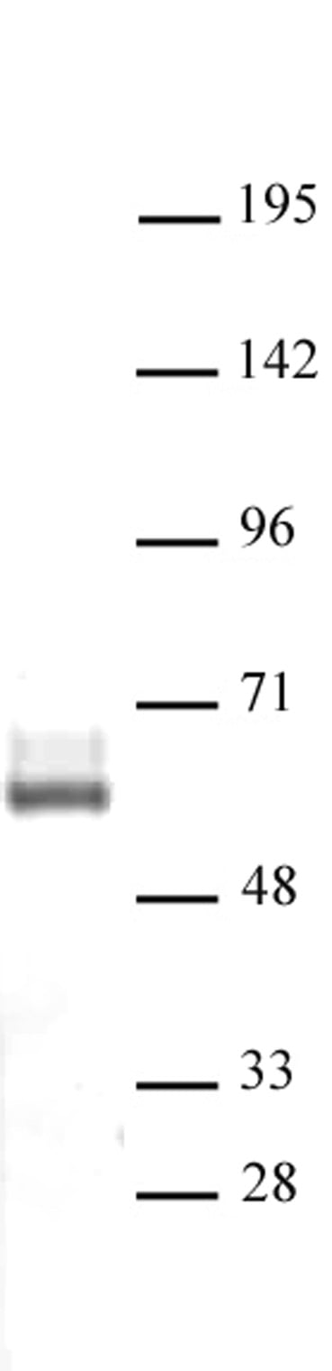
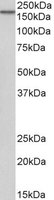
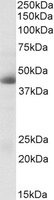
 WB (Western Blot)
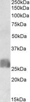
((0.1ug/ml) staining of Rat Testis lysate (35ug protein in RIPA buffer). Primary incubation was 1 hour. Detected by chemiluminescence.)
WB (Western Blot)
((0.1ug/ml) staining of Rat Testis lysate (35ug protein in RIPA buffer). Primary incubation was 1 hour. Detected by chemiluminescence.)
SRRT/ARS2, Polyclonal Antibody (Cat# AAA61790)
HSULF-1/sulfatase 1, Polyclonal Antibody (Cat# AAA61791)
 IHC (Immunohistochemistry)
(Biotinylated (0.03ug/ml) staining of Human Kidney lysate (35ug protein in RIPA buffer), exactly mirroring its parental non-biotinylated product. Primary incubation was 1 hour. Detected by chemiluminescence, using streptavidin-HRP and using NAP blocker as a substitute for skimmed milk.)
IHC (Immunohistochemistry)
(Biotinylated (0.03ug/ml) staining of Human Kidney lysate (35ug protein in RIPA buffer), exactly mirroring its parental non-biotinylated product. Primary incubation was 1 hour. Detected by chemiluminescence, using streptavidin-HRP and using NAP blocker as a substitute for skimmed milk.)
Argininosuccinate synthetase 1, Polyclonal Antibody (Cat# AAA61804)
Expected from sequence similarity: Human, Mouse, Rat, Dog
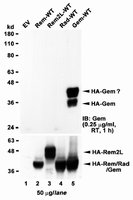
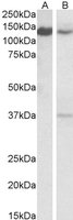
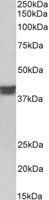
 WB (Western Blot)
((1ug/ml) staining of Human Spleen lysate (35ug protein in RIPA buffer). Primary incubation was 1 hour. Detected by chemiluminescence.)
WB (Western Blot)
((1ug/ml) staining of Human Spleen lysate (35ug protein in RIPA buffer). Primary incubation was 1 hour. Detected by chemiluminescence.)
GEM, Polyclonal Antibody (Cat# AAA61694)
 IHC (Immunohiostchemistry)
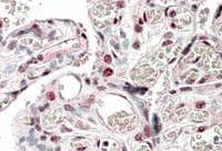
((5ug/ml) staining of paraffin embedded Human Placenta. Steamed antigen retrieval with citrate buffer pH 6, AP-staining.)
IHC (Immunohiostchemistry)
((5ug/ml) staining of paraffin embedded Human Placenta. Steamed antigen retrieval with citrate buffer pH 6, AP-staining.)
CREB3L2, Polyclonal Antibody (Cat# AAA61696)
 WB (Western Blot)
((0.5ug/ml) staining of HEK293 lysate (35ug protein in RIPA buffer). Primary incubation was 1 hour. Detected by chemiluminescence.)
WB (Western Blot)
((0.5ug/ml) staining of HEK293 lysate (35ug protein in RIPA buffer). Primary incubation was 1 hour. Detected by chemiluminescence.)
NPC1, Polyclonal Antibody (Cat# AAA61698)
 WB (Western Blot)
((1ug/ml) staining of NIH3T3 lysate (35ug protein in RIPA buffer). Primary incubation was 1 hour. Detected by chemiluminescence.)
WB (Western Blot)
((1ug/ml) staining of NIH3T3 lysate (35ug protein in RIPA buffer). Primary incubation was 1 hour. Detected by chemiluminescence.)
MYH9, Polyclonal Antibody (Cat# AAA61701)
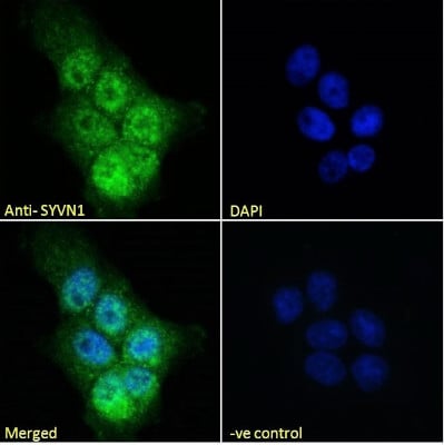
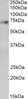
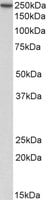
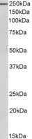
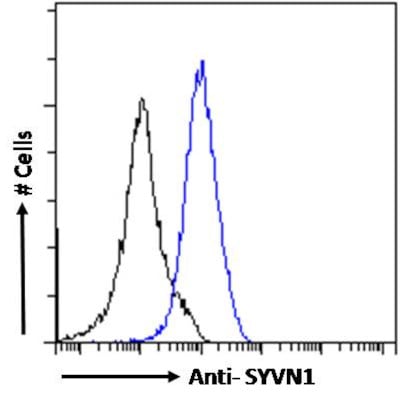 FCM/FACS (Flow Cytometry)
(AAA61704 Flow cytometric analysis of paraformaldehyde fixed A431 cells (blue line), permeabilized with 0.5% Triton. Primary incubation 1hr (10ug/ml) followed by Alexa Fluor 488 secondary antibody (1ug/ml). IgG control: Unimmunized goat IgG (black line) followed by Alexa Fluor 488 secondary antibody.)
FCM/FACS (Flow Cytometry)
(AAA61704 Flow cytometric analysis of paraformaldehyde fixed A431 cells (blue line), permeabilized with 0.5% Triton. Primary incubation 1hr (10ug/ml) followed by Alexa Fluor 488 secondary antibody (1ug/ml). IgG control: Unimmunized goat IgG (black line) followed by Alexa Fluor 488 secondary antibody.)
SYVN1, Polyclonal Antibody (Cat# AAA61704)
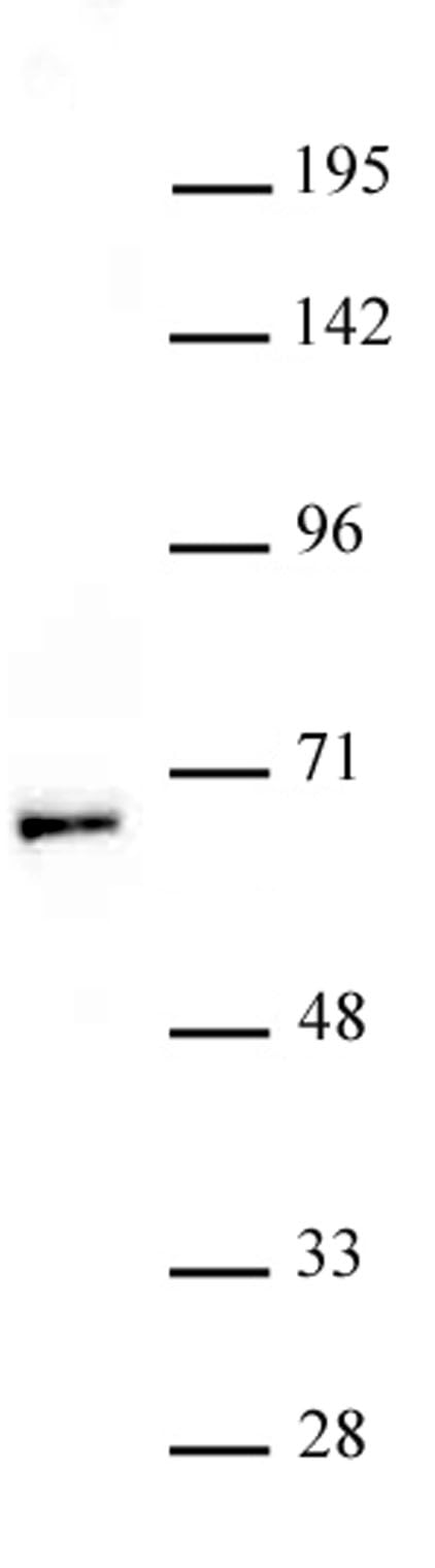
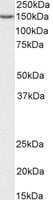
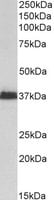
 WB (Western Blot)
((1ug/ml) staining of Pig Kidney lysate (35ug protein in RIPA buffer). Primary incubation was 1 hour. Detected by chemiluminescence.)
WB (Western Blot)
((1ug/ml) staining of Pig Kidney lysate (35ug protein in RIPA buffer). Primary incubation was 1 hour. Detected by chemiluminescence.)
HOXA5, Polyclonal Antibody (Cat# AAA61715)
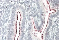 IHC (Immunohiostchemistry)
((5ug/ml) staining of paraffin embedded Human Uterus. Steamed antigen retrieval with citrate buffer pH 6, AP-staining.)
IHC (Immunohiostchemistry)
((5ug/ml) staining of paraffin embedded Human Uterus. Steamed antigen retrieval with citrate buffer pH 6, AP-staining.)
BCAR1, Polyclonal Antibody (Cat# AAA61719)
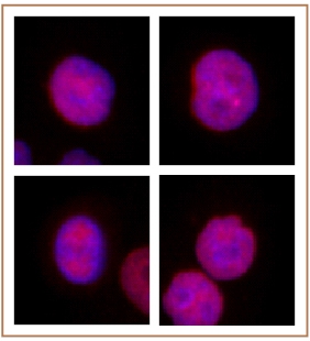
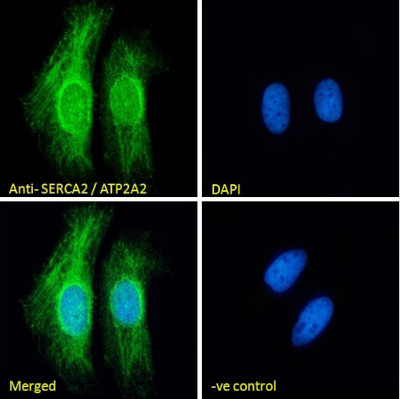 IF (Immunofluorescence)
(Immunofluorescence analysis of paraformaldehyde fixed HeLa cells, permeabilized with 0.15% Triton. Primary incubation 1hr (10ug/ml) followed by Alexa Fluor 488 secondary antibody (2ug/ml), showing endoplasmic reticulum staining. The nuclear stain is DAPI (blue). Negative control: Unimmunized goat IgG (10ug/ml) followed by Alexa Fluor 488 secondary antibody (2ug/ml).)
IF (Immunofluorescence)
(Immunofluorescence analysis of paraformaldehyde fixed HeLa cells, permeabilized with 0.15% Triton. Primary incubation 1hr (10ug/ml) followed by Alexa Fluor 488 secondary antibody (2ug/ml), showing endoplasmic reticulum staining. The nuclear stain is DAPI (blue). Negative control: Unimmunized goat IgG (10ug/ml) followed by Alexa Fluor 488 secondary antibody (2ug/ml).)
SERCA2/ATP2A2, Polyclonal Antibody (Cat# AAA61737)
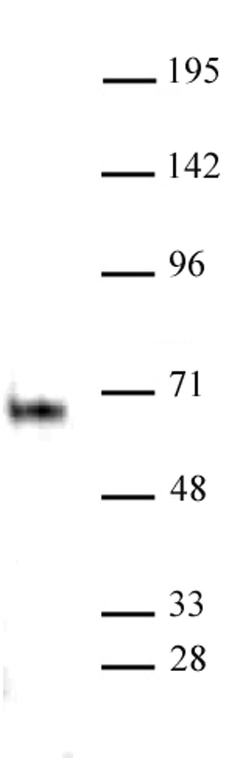
 WB (Western Blot)
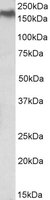
((0.1ug/ml) staining of Mouse Liver lysate (35ug protein in RIPA buffer). Primary incubation was 1 hour. Detected by chemiluminescence.)
WB (Western Blot)
((0.1ug/ml) staining of Mouse Liver lysate (35ug protein in RIPA buffer). Primary incubation was 1 hour. Detected by chemiluminescence.)
peroxiredoxin 6, Polyclonal Antibody (Cat# AAA61745)
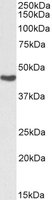
 WB (Western Blot)
((0.1ug/ml) staining of Human Kidney lysate (35ug protein in RIPA buffer). Primary incubation was 1 hour. Detected by chemiluminescence.)
WB (Western Blot)
((0.1ug/ml) staining of Human Kidney lysate (35ug protein in RIPA buffer). Primary incubation was 1 hour. Detected by chemiluminescence.)
AMACR, Polyclonal Antibody (Cat# AAA61751)
 WB (Western Blot)
((0.5ug/ml) staining of Human Heart lysate (35ug protein in RIPA buffer). Primary incubation was 1 hour. Detected by chemiluminescence.)
WB (Western Blot)
((0.5ug/ml) staining of Human Heart lysate (35ug protein in RIPA buffer). Primary incubation was 1 hour. Detected by chemiluminescence.)
TCF4, Polyclonal Antibody (Cat# AAA61752)
 WB (Western Blot)
((0.3ug/ml) staining of Pig Spleen lysate (35ug protein in RIPA buffer). Primary incubation was 1 hour. Detected by chemiluminescence.)
WB (Western Blot)
((0.3ug/ml) staining of Pig Spleen lysate (35ug protein in RIPA buffer). Primary incubation was 1 hour. Detected by chemiluminescence.)
CAPG, Polyclonal Antibody (Cat# AAA61758)
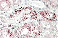 IHC (Immunohiostchemistry)
((5ug/ml) staining of paraffin embedded Human Kidney. Steamed antigen retrieval with citrate buffer pH 6, AP-staining.)
IHC (Immunohiostchemistry)
((5ug/ml) staining of paraffin embedded Human Kidney. Steamed antigen retrieval with citrate buffer pH 6, AP-staining.)
carbonic anhydrase XII, Polyclonal Antibody (Cat# AAA61761)
 WB (Western Blot)
((1ug/ml) staining of fetal Mouse Brain lysate (35ug protein in RIPA buffer). Primary incubation was 1 hour. Detected by chemiluminescence.)
WB (Western Blot)
((1ug/ml) staining of fetal Mouse Brain lysate (35ug protein in RIPA buffer). Primary incubation was 1 hour. Detected by chemiluminescence.)
MSI2/musashi-2, Polyclonal Antibody (Cat# AAA61763)
LRP1, Polyclonal Antibody (Cat# AAA61768)
 WB (Western Blot)
((2ug/ml) staining of Human Adipose lysate (35ug protein in RIPA buffer). Primary incubation was 1 hour. Detected by chemiluminescence.)
WB (Western Blot)
((2ug/ml) staining of Human Adipose lysate (35ug protein in RIPA buffer). Primary incubation was 1 hour. Detected by chemiluminescence.)
CFD/adipsin, Polyclonal Antibody (Cat# AAA61774)
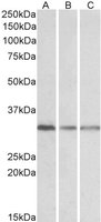
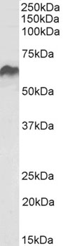
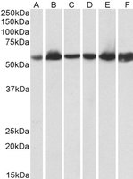
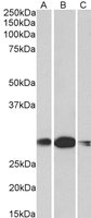
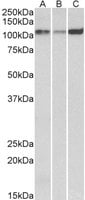
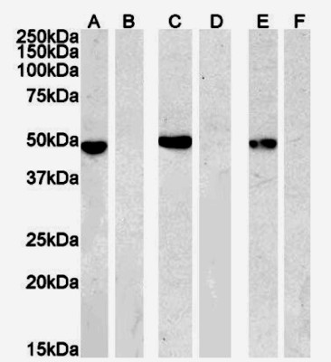
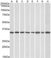 WB (Western Blot)
((0.1ug/ml) staining of NIH3T3 (A), HeK293 (B), HeLa (C, A431(D), A549 (E), MCF7 (F), Jurkat (G), K562 (H) lysates (35ug protein in RIPA buffer). Primary incubation was 1 hour. Detected by chemiluminescence.)
WB (Western Blot)
((0.1ug/ml) staining of NIH3T3 (A), HeK293 (B), HeLa (C, A431(D), A549 (E), MCF7 (F), Jurkat (G), K562 (H) lysates (35ug protein in RIPA buffer). Primary incubation was 1 hour. Detected by chemiluminescence.)
GNB2L1, Polyclonal Antibody (Cat# AAA61631)
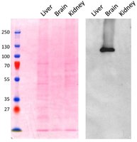 Application Data
((0.1ug/ml) staining of Mouse Brain lysate (~5ug protein in SDSPAGE buffer). The left panel shows the same blot stained with Ponceau red for total protein stain as the loading control before labelling. Primary incubation was 1 hour. Detected by chemiluminescence. Data obtained from Prof. M Robinson, CIMR, Cambridge, UK)
Application Data
((0.1ug/ml) staining of Mouse Brain lysate (~5ug protein in SDSPAGE buffer). The left panel shows the same blot stained with Ponceau red for total protein stain as the loading control before labelling. Primary incubation was 1 hour. Detected by chemiluminescence. Data obtained from Prof. M Robinson, CIMR, Cambridge, UK)
AP2A1, Polyclonal Antibody (Cat# AAA61644)
 WB (Western Blot)
((0. 03ug/ml) staining of HeLa (lane 1) and Human Colon cancer (lane two) lysate (35ug protein in RIPA buffer). Primary incubation was 1 hour. Detected by chemiluminescence.)
WB (Western Blot)
((0. 03ug/ml) staining of HeLa (lane 1) and Human Colon cancer (lane two) lysate (35ug protein in RIPA buffer). Primary incubation was 1 hour. Detected by chemiluminescence.)
perilipin 3/TIP47, Polyclonal Antibody (Cat# AAA61647)
Sox9b, Polyclonal Antibody (Cat# AAA61664)
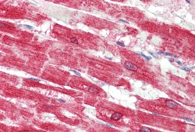
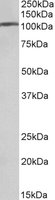
 WB (Western Blot)
((0.5ug/ml) staining of Rat Skeletal Muscle lysate (35ug protein in RIPA buffer). Primary incubation was 1 hour. Detected by chemiluminescence.)
WB (Western Blot)
((0.5ug/ml) staining of Rat Skeletal Muscle lysate (35ug protein in RIPA buffer). Primary incubation was 1 hour. Detected by chemiluminescence.)
BBS7, Polyclonal Antibody (Cat# AAA61666)
 WB (Western Blot)
((0.1ug/ml) staining of Pig Heart lysate (35ug protein in RIPA buffer). Primary incubation was 1 hour. Detected by chemiluminescence.)
WB (Western Blot)
((0.1ug/ml) staining of Pig Heart lysate (35ug protein in RIPA buffer). Primary incubation was 1 hour. Detected by chemiluminescence.)
ADRA1B, Polyclonal Antibody (Cat# AAA61669)
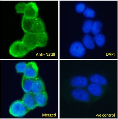
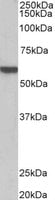
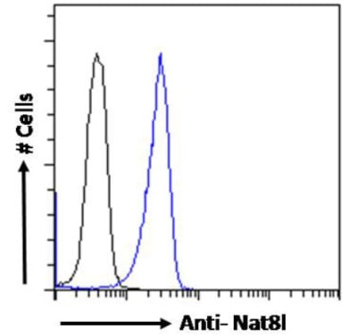 FCM/FACS (Flow Cytometry)
(Flow cytometric analysis of paraformaldehyde fixed Kelly cells (blue line), permeabilized with 0.5% Triton. Primary incubation 1hr (10ug/ml) followed by Alexa Fluor 488 secondary antibody (1ug/ml). IgG control: Unimmunized goat IgG (black line) followed by Alexa Fluor 488 secondary antibody.)
FCM/FACS (Flow Cytometry)
(Flow cytometric analysis of paraformaldehyde fixed Kelly cells (blue line), permeabilized with 0.5% Triton. Primary incubation 1hr (10ug/ml) followed by Alexa Fluor 488 secondary antibody (1ug/ml). IgG control: Unimmunized goat IgG (black line) followed by Alexa Fluor 488 secondary antibody.)
Nat8l, Polyclonal Antibody (Cat# AAA61680)
Expected from sequence similarity: Human, Mouse, Rat, Dog, Cow
 WB (Western Blot)
((1ug/ml) staining of Mouse Colon (wt, left lane, knock-out right lane) lysate (35ug protein in RIPA buffer). Primary incubation was 1 hour. Detected by infrared fluorescence (Odyssey). Data obtained from anonymous customer)
WB (Western Blot)
((1ug/ml) staining of Mouse Colon (wt, left lane, knock-out right lane) lysate (35ug protein in RIPA buffer). Primary incubation was 1 hour. Detected by infrared fluorescence (Odyssey). Data obtained from anonymous customer)
Cd97, Polyclonal Antibody (Cat# AAA61683)
 WB (Western Blot)
((0. 01ug/ml) staining of Pig Heart lysate (35ug protein in RIPA buffer). Primary incubation was 1 hour. Detected by chemiluminescence.)
WB (Western Blot)
((0. 01ug/ml) staining of Pig Heart lysate (35ug protein in RIPA buffer). Primary incubation was 1 hour. Detected by chemiluminescence.)
cardiac troponin T, Polyclonal Antibody (Cat# AAA61688)
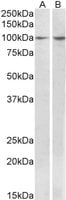
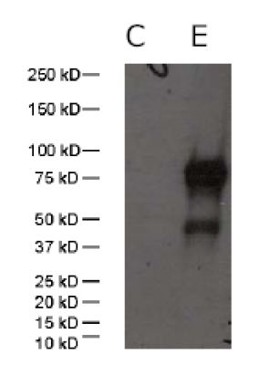
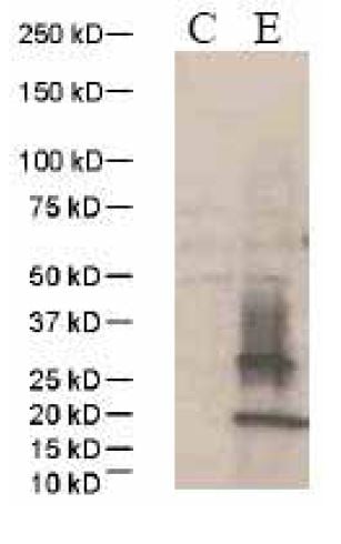 Application Data
(C: 293 cell extract control; E: 293 cell expressing human PD-1 (a.a. 1-167))
Application Data
(C: 293 cell extract control; E: 293 cell expressing human PD-1 (a.a. 1-167))
PD-1, Polyclonal Antibody (Cat# AAA62111)
 Application Data
NA (A/Chicken/HongKong/NT366/03)(H9N2), Polyclonal Antibody (Cat# AAA62114)
 Application Data
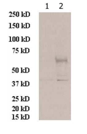
(1: 293 cell extract control; 2: 293 cell expressing p24 (Clade A))
Application Data
(1: 293 cell extract control; 2: 293 cell expressing p24 (Clade A))
p24 (HIV-1/Clade A), Polyclonal Antibody (Cat# AAA62116)
 Application Data
NS1 (A/California/06/2009)(H1N1), Polyclonal Antibody (Cat# AAA62121)
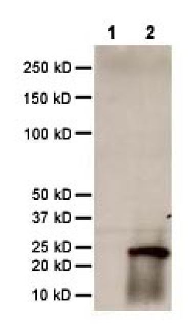
 Application Data
(WB:C: 293 cell extract controlE: 293 cell expressingNote: The upper band is HA, while the lower band is HA1)
Application Data
(WB:C: 293 cell extract controlE: 293 cell expressingNote: The upper band is HA, while the lower band is HA1)
HA1 (A/Chicken/NY/29878/91)(H2N2), Polyclonal Antibody (Cat# AAA62123)
H3(H3N2)(A/Wisconsin/67/X-161/2005), Polyclonal Antibody (Cat# AAA62126)
M2 (A/chicken/Korea/GH2/2007(H9N2), Polyclonal Antibody (Cat# AAA62128)
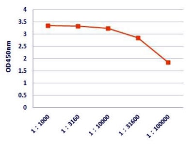 ELISA
(Figure 1. Titration curves of anti-spike rabbit polyclonal antibody to HCoV-HKU1 spike protein. 96-well corning EILSA plate was coated with HKU1 spike protein at a concentration of 2 ug/ml.)
ELISA
(Figure 1. Titration curves of anti-spike rabbit polyclonal antibody to HCoV-HKU1 spike protein. 96-well corning EILSA plate was coated with HKU1 spike protein at a concentration of 2 ug/ml.)
Spike (HCoV-HKU1), Polyclonal Antibody (Cat# AAA62219)
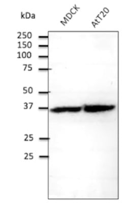
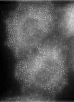
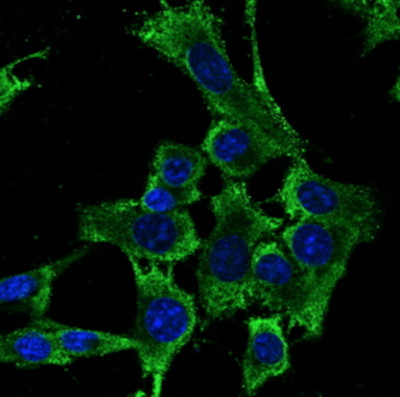 IF (Immunofluorescence)
(Confocal immunofluorescence of Hepa1-6 cells with RPS6 Ab at 1/100 dilution; methanol fixation)
IF (Immunofluorescence)
(Confocal immunofluorescence of Hepa1-6 cells with RPS6 Ab at 1/100 dilution; methanol fixation)
RPS6, Polyclonal Antibody (Cat# AAA63052)
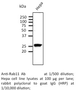 Application Data
Rab11, Polyclonal Antibody (Cat# AAA63057)
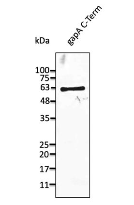 Application Data
(Anti-gapA Ab at 1/1,000 dilution; lane with 30 ng of recombinant fusion protein (176 aa - stop) from Microbacterium sp; rabbit polyclonal to goat IgG (HRP) at 1/10,000 dilution;)
Application Data
(Anti-gapA Ab at 1/1,000 dilution; lane with 30 ng of recombinant fusion protein (176 aa - stop) from Microbacterium sp; rabbit polyclonal to goat IgG (HRP) at 1/10,000 dilution;)
gapA, Polyclonal Antibody (Cat# AAA63169)
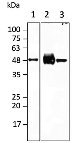 Application Data
(Anti-TAU Ab at 1/2,000 dilution; lanes with 40 ug of total lysates; 1 - SH-SY5Y, 2 - brain, 3 - mouse new born head; chicken polyclonal to goat IgG conjugated to HRP (AB1125) at 1/10,000 dilution;)
Application Data
(Anti-TAU Ab at 1/2,000 dilution; lanes with 40 ug of total lysates; 1 - SH-SY5Y, 2 - brain, 3 - mouse new born head; chicken polyclonal to goat IgG conjugated to HRP (AB1125) at 1/10,000 dilution;)
TAU, Polyclonal Antibody (Cat# AAA63172)
What are Polyclonal Antibodies?
Polyclonal antibodies are antibodies that come from multiple B cell clones of a host animal. The typical hosts used for the majority of polyclonal antibody production are rabbits, goats, sheep, and donkeys. These polyclonal antibodies, once having identified their target, will bind to different epitopes located at different regions or sequences on the same protein/antigen. This ability to bind multiple epitopes is what makes polyclonal antibodies highly sensitive, as explained in our detailed guide on polyclonal antibodies and why they matter.
As a result, they are ideal at locating and binding to the target, even if the target is in very low concentrations (due to many different antibodies being able to bind to the same target molecule, which allows for significant amplification of a downstream signal).
Polyclonal antibodies are typically produced by injecting an antigen into a host animal, which causes the animal’s immune system to attack the foreign antigen by mass generating antibodies against it. After a period of time, serum is collected from the animal and purified using physicochemical fractionation, class-specific affinity purification, and/or antigen-affinity purification.
Key Uses of Polyclonal Antibodies
- Western Blotting: This method is used to find specific proteins in biological samples after separating them by size.
- Immunohistochemistry: IHC helps visualize the location of proteins in tissue sections using various staining techniques.
- ELISA: (Enzyme-Linked Immunosorbent Assay) is typically used to identify specific protein quantities in a sample. ELISAs can be either “Quantitative” or “Qualitative”.
- Flow Cytometry: technique that identifies and measures the specific protein on the surface or inside the cells in a fluid suspension.
- Immunoprecipitation: IP isolates and studies a specific protein from a complex mixture using antibodies.
Why Buy Polyclonal Antibodies from AAA Biotech?
1. Ideal for Various Applications
Our antibodies are generally going to be validated for use in multiple types of assays, including ELISA, Western Blotting, Immunohistochemistry, Immunoprecipitation, amongst others. They are ideal for a wide range of research applications.
2. Rigorous Quality Control
All of the antibodies in our catalog undergo strict quality testing to ensure specificity, sensitivity, and consistent performance. We are confident in the ability of our antibodies to provide you with accurate results.
3. Wide Assortment of Antibodies
Antibodies in our catalog can be found for both common and exotic species, and these antibodies are also available in both conjugated and recombinant forms to suit many diverse experimental needs.
4. Highly Purified
Our antibodies are available in purified forms with over 85% purity, as confirmed by SDS-PAGE. They are also available with tags such as His, Flag, GST, or MBP. We cater to customers worldwide.
FAQ
1. How are polyclonal antibodies produced?
Traditionally, polyclonal antibodies are produced by injecting an antigen into a host animal (such as a rabbit or goat), which then triggers an immune response from the host animal. The animal’s B cells produce antibodies that will recognize different parts of the injected antigen. These antibodies are then collected from the animal’s blood and purified for use.
2. How do polyclonal antibodies differ from monoclonal antibodies?
Polyclonal antibodies are a mix of antibodies that bind to different locations (epitopes) of the same antigen, while monoclonal antibodies are identical and bind to just one specific epitope. This makes polyclonal antibodies more versatile and better at detecting proteins that may be present in low quantities or in altered/modified forms.
3. How should I store polyclonal antibodies?
Polyclonal antibodies should be stored at 4°C for short-term use (up to a few weeks) and at -20°C or -80°C for long-term storage. Avoid repeated freeze-thaw cycles by dividing them into small aliquots. Always check the datasheet for specific storage instructions.