Filters
▼Clonality
▼Type
▼Reactivity
▼Gene Name
▼Isotype
▼Host
▼Application
▼Clone
▼Polyclonal Antibodies
At AAA Biotech also known as AAA Bio or AAABio, we provide a broad range of purified polyclonal antibodies (pAbs) that are able to all be browsed online through our website. Due to their high specificity and strong binding affinity, these antibodies are ideal for wide swathes of research and experimental applications.
Our polyclonal antibodies can easily support your work, whether you use them for Western Blotting, Immunocytochemistry (with or without Immunofluorescence used in conjunction), Immunohistochemistry, Immunoprecipitation, and ELISA tests. We highly encourage you to browse our range of pAbs and choose the one that best suits your experimental model.
Viewing 1500-1550 of 118597 product results
 FCM/FACS (Flow Cytometry)
(Flow cytometric analysis of paraformaldehyde fixed HepG2 cells (blue line), permeabilized with 0.5% Triton.Primary incubation 1hr (10ug/ml) followed by Alexa Fluor 488 secondary antibody (1ug/ml).IgG control: Unimmunized goat IgG (black line) followed by Alexa Fluor 488 secondary antibody.)
FCM/FACS (Flow Cytometry)
(Flow cytometric analysis of paraformaldehyde fixed HepG2 cells (blue line), permeabilized with 0.5% Triton.Primary incubation 1hr (10ug/ml) followed by Alexa Fluor 488 secondary antibody (1ug/ml).IgG control: Unimmunized goat IgG (black line) followed by Alexa Fluor 488 secondary antibody.)
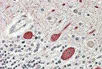
ABCB5, Polyclonal Antibody (Cat# AAA61316)
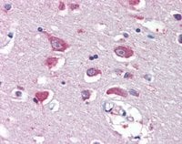 IHC (Immunohiostchemistry)
((5ug/ml) staining of paraffin embedded Human Cortex. Steamed antigen retrieval with citrate buffer pH 6, AP-staining.)
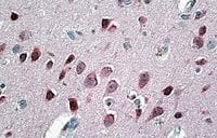
IHC (Immunohiostchemistry)
((5ug/ml) staining of paraffin embedded Human Cortex. Steamed antigen retrieval with citrate buffer pH 6, AP-staining.)
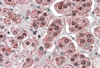
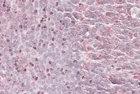
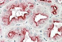
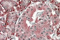
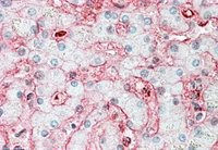
GRIA4, Polyclonal Antibody (Cat# AAA61330)
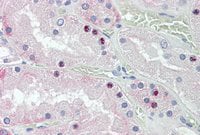 IHC (Immunohiostchemistry)
((3.8ug/ml) staining of paraffin embedded Human Kidney. Steamed antigen retrieval with citrate buffer pH 6, AP-staining.)
IHC (Immunohiostchemistry)
((3.8ug/ml) staining of paraffin embedded Human Kidney. Steamed antigen retrieval with citrate buffer pH 6, AP-staining.)
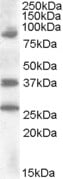
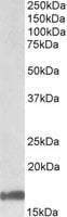
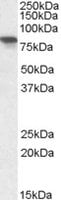
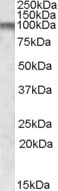
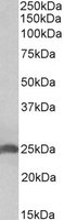
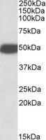
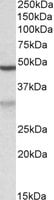
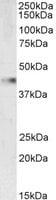
HOXD10, Polyclonal Antibody (Cat# AAA61335)
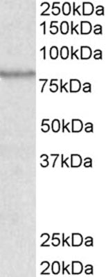
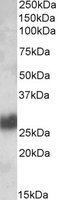
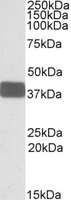
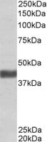
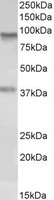
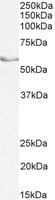
 WB (Western Blot)
((0.1ug/ml) staining of Human Liver lysate (35ug protein in RIPA buffer). Primary incubation was 1 hour. Detected by chemiluminescence.)
WB (Western Blot)
((0.1ug/ml) staining of Human Liver lysate (35ug protein in RIPA buffer). Primary incubation was 1 hour. Detected by chemiluminescence.)
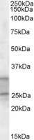
ACO1/Aconitase 1, Polyclonal Antibody (Cat# AAA61336)
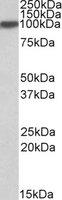
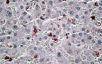
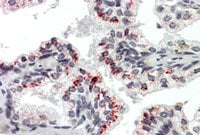
HIVEP2/MIBP1, Polyclonal Antibody (Cat# AAA61342)
 IHC (Immunohiostchemistry)
((3.8ug/ml) staining of paraffin embedded Human Adrenal Gland. Steamed antigen retrieval with citrate buffer pH 6, AP-staining.)
IHC (Immunohiostchemistry)
((3.8ug/ml) staining of paraffin embedded Human Adrenal Gland. Steamed antigen retrieval with citrate buffer pH 6, AP-staining.)
KPNA6, Polyclonal Antibody (Cat# AAA61347)
 IHC (Immunohistochemistry)
((6ug/ml) staining of paraffin embedded Human Liver. Steamed antigen retrieval with citrate buffer pH 6, AP-staining.)
IHC (Immunohistochemistry)
((6ug/ml) staining of paraffin embedded Human Liver. Steamed antigen retrieval with citrate buffer pH 6, AP-staining.)
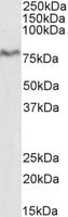
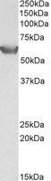
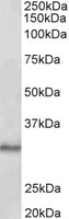
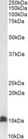
CCL20, Polyclonal Antibody (Cat# AAA61348)
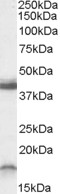
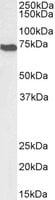
 WB (Western Blot)
((0.1ug/ml) staining of Human Tonsil lysate (35ug protein in RIPA buffer). Primary incubation was 1 hour. Detected by chemiluminescence.)
WB (Western Blot)
((0.1ug/ml) staining of Human Tonsil lysate (35ug protein in RIPA buffer). Primary incubation was 1 hour. Detected by chemiluminescence.)
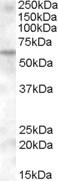
CD90/Thy1, Polyclonal Antibody (Cat# AAA61360)
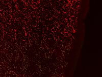 IF (Immunofluorescence)
((0.3ug/ml) staining of PFA-perfused cryosection of Human Hypothalamus. Antigen retrieval with citrate buffer pH 6 at 80C for 30min, Cy3-staining. Data obtained by Prof. Erik Hrabovszky, Inst, Exp, Med. , Budapest, Hungary.)
IF (Immunofluorescence)
((0.3ug/ml) staining of PFA-perfused cryosection of Human Hypothalamus. Antigen retrieval with citrate buffer pH 6 at 80C for 30min, Cy3-staining. Data obtained by Prof. Erik Hrabovszky, Inst, Exp, Med. , Budapest, Hungary.)
GAL, Polyclonal Antibody (Cat# AAA61364)
 IHC (Immunohistochemisry)
((3.8ug/ml) staining of paraffin embedded Human Cortex. Steamed antigen retrieval with citrate buffer pH 6, AP-staining.)
IHC (Immunohistochemisry)
((3.8ug/ml) staining of paraffin embedded Human Cortex. Steamed antigen retrieval with citrate buffer pH 6, AP-staining.)
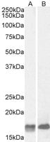
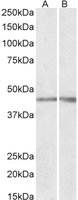
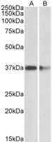
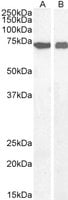
Neurogranin precursor, Polyclonal Antibody (Cat# AAA61369)
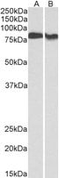
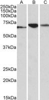
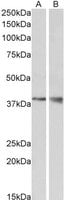 WB (Western Blot)
((1ug/ml) staining of Mouse Kidney (A) and Brain (B) lysate (35ug protein in RIPA buffer). Primary incubation was 1 hour. Detected by chemiluminescence.)
WB (Western Blot)
((1ug/ml) staining of Mouse Kidney (A) and Brain (B) lysate (35ug protein in RIPA buffer). Primary incubation was 1 hour. Detected by chemiluminescence.)
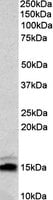
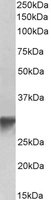
WNT15/WNT9B, Polyclonal Antibody (Cat# AAA61375)
 IHC (Immunohiostchemistry)
((3.8ug/ml) staining of paraffin embedded Human Tonsil. Steamed antigen retrieval with citrate buffer pH 6, AP-staining.)
IHC (Immunohiostchemistry)
((3.8ug/ml) staining of paraffin embedded Human Tonsil. Steamed antigen retrieval with citrate buffer pH 6, AP-staining.)
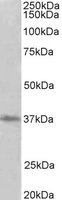
SEPT6, Polyclonal Antibody (Cat# AAA61384)
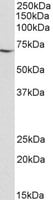
 WB (Western Blot)
((1ug/ml) staining of Human Skeletal Muscle lysate (35ug protein in RIPA buffer). Primary incubation was 1 hour. Detected by chemiluminescence.)
WB (Western Blot)
((1ug/ml) staining of Human Skeletal Muscle lysate (35ug protein in RIPA buffer). Primary incubation was 1 hour. Detected by chemiluminescence.)
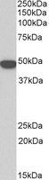
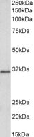
FBXO32, Polyclonal Antibody (Cat# AAA61391)
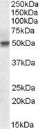
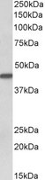
 WB (Western Blot)
((0.3ug/ml) staining of Rat Brain lysate (35ug protein in RIPA buffer). Primary incubation was 1 hour. Detected by chemiluminescence.)
WB (Western Blot)
((0.3ug/ml) staining of Rat Brain lysate (35ug protein in RIPA buffer). Primary incubation was 1 hour. Detected by chemiluminescence.)
Ramp1, Polyclonal Antibody (Cat# AAA61396)
 WB (Western Blot)
((0.1ug/ml) staining of Pig Pancreas lysate (35ug protein in RIPA buffer). Primary incubation was 1 hour. Detected by chemiluminescence.)
WB (Western Blot)
((0.1ug/ml) staining of Pig Pancreas lysate (35ug protein in RIPA buffer). Primary incubation was 1 hour. Detected by chemiluminescence.)
XPNPEP1, Polyclonal Antibody (Cat# AAA61401)
 IHC (Immunohiostchemistry)
((3.8ug/ml) staining of paraffin embedded Human Tonsil. Steamed antigen retrieval with citrate buffer pH 6, AP-staining.)
IHC (Immunohiostchemistry)
((3.8ug/ml) staining of paraffin embedded Human Tonsil. Steamed antigen retrieval with citrate buffer pH 6, AP-staining.)
PRDM1/MEL1, Polyclonal Antibody (Cat# AAA61407)
 WB (Western Blot)
((1ug/ml) staining of Human Frontal Cortex lysate (35ug protein in RIPA buffer). Primary incubation was 1 hour. Detected by chemiluminescence.)
WB (Western Blot)
((1ug/ml) staining of Human Frontal Cortex lysate (35ug protein in RIPA buffer). Primary incubation was 1 hour. Detected by chemiluminescence.)
CPT1C, Polyclonal Antibody (Cat# AAA61409)
 IHC (Immunohistochemistry)
((3.8ug/ml) staining of paraffin embedded Human Cerebellum. Steamed antigen retrieval with citrate buffer pH 6, AP-staining.)
IHC (Immunohistochemistry)
((3.8ug/ml) staining of paraffin embedded Human Cerebellum. Steamed antigen retrieval with citrate buffer pH 6, AP-staining.)
MTNR1B, Polyclonal Antibody (Cat# AAA61418)
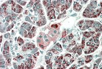 IHC (Immunohiostchemistry)
((5ug/ml) staining of paraffin embedded Human Pancreas. Microwaved antigen retrieval with Tris/EDTA buffer pH9, HRP-staining.)
IHC (Immunohiostchemistry)
((5ug/ml) staining of paraffin embedded Human Pancreas. Microwaved antigen retrieval with Tris/EDTA buffer pH9, HRP-staining.)
PEBP4, Polyclonal Antibody (Cat# AAA61430)
 WB (Western Blot)
((2ug/ml) staining of Peripheral Blood Lymphocytes lysate (35ug protein in RIPA buffer). Primary incubation was 1 hour. Detected by chemiluminescence.)
WB (Western Blot)
((2ug/ml) staining of Peripheral Blood Lymphocytes lysate (35ug protein in RIPA buffer). Primary incubation was 1 hour. Detected by chemiluminescence.)
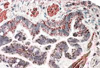
CD34-T, Polyclonal Antibody (Cat# AAA61667)
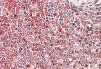 IHC (Immunohiostchemistry)
((5ug/ml) staining of paraffin embedded Human Spleen. Steamed antigen retrieval with citrate buffer pH 6, AP-staining.)
IHC (Immunohiostchemistry)
((5ug/ml) staining of paraffin embedded Human Spleen. Steamed antigen retrieval with citrate buffer pH 6, AP-staining.)
PRKCB, Polyclonal Antibody (Cat# AAA61670)
 WB (Western Blot)
((0. 01ug/ml) staining of Pig Heart lysate (35ug protein in RIPA buffer). Primary incubation was 1 hour. Detected by chemiluminescence.)
WB (Western Blot)
((0. 01ug/ml) staining of Pig Heart lysate (35ug protein in RIPA buffer). Primary incubation was 1 hour. Detected by chemiluminescence.)
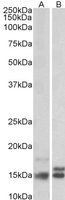
MDH1/MOR2, Polyclonal Antibody (Cat# AAA61673)
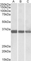
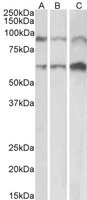
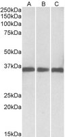
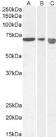
 WB (Western Blot)
((0.1ug/ml) staining of adult Mouse (A), fetal Mouse (B), adult Rat (C) and adult Pig (D) Heart lysates (35ug protein in RIPA buffer). Primary incubation was 1 hour. Detected by chemiluminescence.)
WB (Western Blot)
((0.1ug/ml) staining of adult Mouse (A), fetal Mouse (B), adult Rat (C) and adult Pig (D) Heart lysates (35ug protein in RIPA buffer). Primary incubation was 1 hour. Detected by chemiluminescence.)
TNNI3, Polyclonal Antibody (Cat# AAA61674)
 WB (Western Blot)
((0.1ug/ml) staining of A549 lysate (35ug protein in RIPA buffer). Primary incubation was 1 hour. Detected by chemiluminescence.)
WB (Western Blot)
((0.1ug/ml) staining of A549 lysate (35ug protein in RIPA buffer). Primary incubation was 1 hour. Detected by chemiluminescence.)
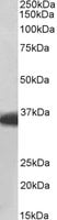
AOC3/VAP1, Polyclonal Antibody (Cat# AAA61692)

 WB (Western Blot)
((0.1ug/ml) staining of Mouse (A), Rat (B) and Pig (C) Heart lysate (35ug protein in RIPA buffer). Primary incubation was 1 hour. Detected by chemiluminescence.)
WB (Western Blot)
((0.1ug/ml) staining of Mouse (A), Rat (B) and Pig (C) Heart lysate (35ug protein in RIPA buffer). Primary incubation was 1 hour. Detected by chemiluminescence.)
ADAM12, Polyclonal Antibody (Cat# AAA61699)
 IHC (Immunohiostchemistry)
(AAA61706 (5ug/ml) staining of paraffin embedded Human Liver. Steamed antigen retrieval with citrate buffer pH 6, AP-staining.)
IHC (Immunohiostchemistry)
(AAA61706 (5ug/ml) staining of paraffin embedded Human Liver. Steamed antigen retrieval with citrate buffer pH 6, AP-staining.)
cytokeratin 19, Polyclonal Antibody (Cat# AAA61706)
 IHC (Immunohiostchemistry)
((5ug/ml) staining of paraffin embedded Human Kidney. Steamed antigen retrieval with citrate buffer pH 6, AP-staining.)
IHC (Immunohiostchemistry)
((5ug/ml) staining of paraffin embedded Human Kidney. Steamed antigen retrieval with citrate buffer pH 6, AP-staining.)
Apolipoprotein D, Polyclonal Antibody (Cat# AAA61711)
 WB (Western Blot)
((0.1ug/ml) staining of Mouse brain lysate (35ug protein in RIPA buffer). Primary incubation was 1 hour. Detected by chemiluminescence.)
WB (Western Blot)
((0.1ug/ml) staining of Mouse brain lysate (35ug protein in RIPA buffer). Primary incubation was 1 hour. Detected by chemiluminescence.)
GOLPH3, Polyclonal Antibody (Cat# AAA61716)
 WB (Western Blot)
((0.1ug/ml) staining of Pig Liver lysate (35ug protein in RIPA buffer). Primary incubation was 1 hour. Detected by chemiluminescence)
WB (Western Blot)
((0.1ug/ml) staining of Pig Liver lysate (35ug protein in RIPA buffer). Primary incubation was 1 hour. Detected by chemiluminescence)
keratin 18, Polyclonal Antibody (Cat# AAA61723)
 WB (Western Blot)
((1ug/ml) staining of Pig Kidney lysates (35ug protein in RIPA buffer). Primary incubation was 1 hour. Detected by chemiluminescence.)
WB (Western Blot)
((1ug/ml) staining of Pig Kidney lysates (35ug protein in RIPA buffer). Primary incubation was 1 hour. Detected by chemiluminescence.)
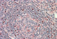
NPHS2/SRN1, Polyclonal Antibody (Cat# AAA61728)
 IHC (Immunohiostchemistry)
((5ug/ml) staining of paraffin embedded Human Spleen. Steamed antigen retrieval with citrate buffer pH 6, AP-staining.)
IHC (Immunohiostchemistry)
((5ug/ml) staining of paraffin embedded Human Spleen. Steamed antigen retrieval with citrate buffer pH 6, AP-staining.)
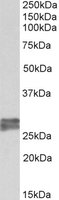
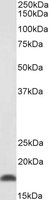
EDF1, Polyclonal Antibody (Cat# AAA61740)
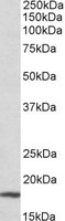 WB (Western Blot)
((0.3ug/ml) staining of Mouse Liver lysate (35ug protein in RIPA buffer). Primary incubation was 1 hour. Detected by chemiluminescence.)
WB (Western Blot)
((0.3ug/ml) staining of Mouse Liver lysate (35ug protein in RIPA buffer). Primary incubation was 1 hour. Detected by chemiluminescence.)
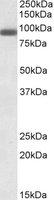
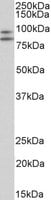
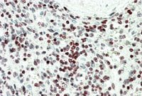
P19ARF, Polyclonal Antibody (Cat# AAA61588)
 Application Data
((0.25 ug/ml) staining of homogenized Mouse salivary gland tissue. (20 ?g protein per lane). Detected by chemiluminescence.)
Application Data
((0.25 ug/ml) staining of homogenized Mouse salivary gland tissue. (20 ?g protein per lane). Detected by chemiluminescence.)
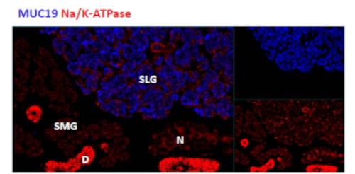
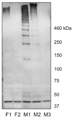
Mucin 19/Apomucin, Polyclonal Antibody (Cat# AAA61611)
 IHC (Immunohiostchemistry)
((3.8ug/ml) staining of paraffin embedded Human Pancreas. Steamed antigen retrieval with citrate buffer pH 6, AP-staining.)
IHC (Immunohiostchemistry)
((3.8ug/ml) staining of paraffin embedded Human Pancreas. Steamed antigen retrieval with citrate buffer pH 6, AP-staining.)
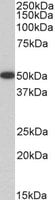
PDIA2, Polyclonal Antibody (Cat# AAA61618)
 WB (Western Blot)
((0.1ug/ml) staining of Rat Brain lysate (35ug protein in RIPA buffer). Primary incubation was 1 hour. Detected by chemiluminescence.)
WB (Western Blot)
((0.1ug/ml) staining of Rat Brain lysate (35ug protein in RIPA buffer). Primary incubation was 1 hour. Detected by chemiluminescence.)
Htr3b, Polyclonal Antibody (Cat# AAA61621)
 WB (Western Blot)
(AAA61623(0.5ug/ml) staining of HEK293 lysate (35ug protein in RIPA buffer). Primary incubation was 1 hour. Detected by chemiluminescence.)
WB (Western Blot)
(AAA61623(0.5ug/ml) staining of HEK293 lysate (35ug protein in RIPA buffer). Primary incubation was 1 hour. Detected by chemiluminescence.)
SLC22A3, Polyclonal Antibody (Cat# AAA61623)
 IHC (Immunohiostchemistry)
((5ug/ml) staining of paraffin embedded Human Lung. Steamed antigen retrieval with citrate buffer pH 6, AP-staining.)
IHC (Immunohiostchemistry)
((5ug/ml) staining of paraffin embedded Human Lung. Steamed antigen retrieval with citrate buffer pH 6, AP-staining.)
MOB2, Polyclonal Antibody (Cat# AAA61639)
 IHC (Immunohiostchemistry)
((5ug/ml) staining of paraffin embedded Human Tonsil. Steamed antigen retrieval with citrate buffer pH 6, AP-staining.)
IHC (Immunohiostchemistry)
((5ug/ml) staining of paraffin embedded Human Tonsil. Steamed antigen retrieval with citrate buffer pH 6, AP-staining.)
CTDSP1, Polyclonal Antibody (Cat# AAA61642)

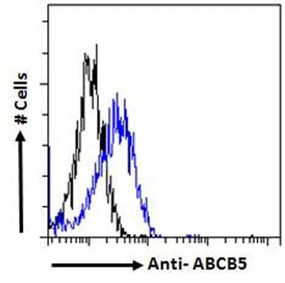
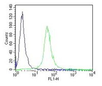
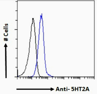 FCM/FACS (Flow Cytometry)
(Flow cytometric analysis of paraformaldehyde fixed A549 cells (blue line), permeabilized with 0.5% Triton. Primary incubation 1hr (10ug/ml) followed by Alexa Fluor 488 secondary antibody (1ug/ml). IgG control: Unimmunized goat IgG (black line) followed by Alexa Fluor 488 secondary antibody.)
FCM/FACS (Flow Cytometry)
(Flow cytometric analysis of paraformaldehyde fixed A549 cells (blue line), permeabilized with 0.5% Triton. Primary incubation 1hr (10ug/ml) followed by Alexa Fluor 488 secondary antibody (1ug/ml). IgG control: Unimmunized goat IgG (black line) followed by Alexa Fluor 488 secondary antibody.)
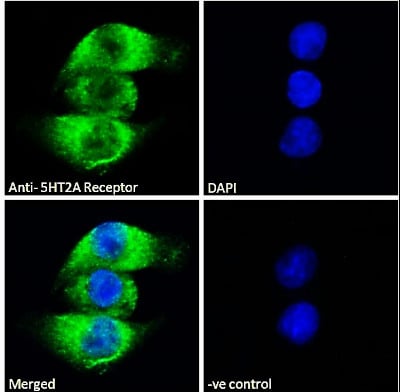
5HT2A Receptor, Polyclonal Antibody (Cat# AAA61649)
Expected from sequence similarity: Human
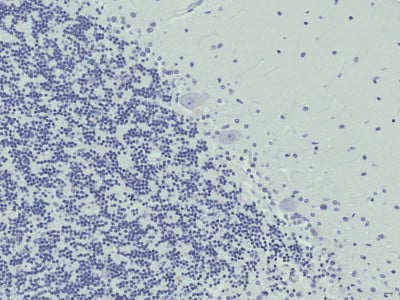
 IHC (Immunohiostchemistry)
((5ug/ml) staining of paraffin embedded Human Liver. Steamed antigen retrieval with citrate buffer pH 6, AP-staining.)
IHC (Immunohiostchemistry)
((5ug/ml) staining of paraffin embedded Human Liver. Steamed antigen retrieval with citrate buffer pH 6, AP-staining.)
Stomatin, Polyclonal Antibody (Cat# AAA61651)
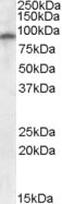
 WB (Western Blot)
((2ug/ml) staining of Pig Brain lysate (35ug protein in RIPA buffer). Primary incubation was 1 hour. Detected by chemiluminescence.)
WB (Western Blot)
((2ug/ml) staining of Pig Brain lysate (35ug protein in RIPA buffer). Primary incubation was 1 hour. Detected by chemiluminescence.)
PRMT2, Polyclonal Antibody (Cat# AAA61653)
 WB (Western Blot)
((0.1ug/ml) staining of Pig Heart lysates (35ug protein in RIPA buffer). Primary incubation was 1 hour. Detected by chemiluminescence.)
WB (Western Blot)
((0.1ug/ml) staining of Pig Heart lysates (35ug protein in RIPA buffer). Primary incubation was 1 hour. Detected by chemiluminescence.)
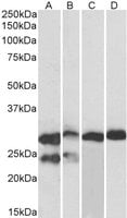
MDH1/MOR2, Polyclonal Antibody (Cat# AAA61658)
 WB (Western Blot)
((0.3ug/ml) staining of Mouse (A) and Rat (B) Spleen lysates (35ug protein in RIPA buffer). Primary incubation was 1 hour. Detected by chemiluminescence.)
WB (Western Blot)
((0.3ug/ml) staining of Mouse (A) and Rat (B) Spleen lysates (35ug protein in RIPA buffer). Primary incubation was 1 hour. Detected by chemiluminescence.)
ACTN1, Polyclonal Antibody (Cat# AAA61748)

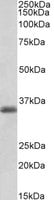
 WB (Western Blot)
((0.3ug/ml) staining of Pig Ovary lysate (35ug protein in RIPA buffer). Primary incubation was 1 hour. Detected by chemiluminescence.)
WB (Western Blot)
((0.3ug/ml) staining of Pig Ovary lysate (35ug protein in RIPA buffer). Primary incubation was 1 hour. Detected by chemiluminescence.)
NR5A2/LRH1, Polyclonal Antibody (Cat# AAA61756)
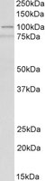
dysferlin, Polyclonal Antibody (Cat# AAA61769)
 WB (Western Blot)
((0.03ug/ml) staining of Mouse (A) and Rat (B) Kidney lysate (35ug protein in RIPA buffer). Primary incubation was 1 hour. Detected by chemiluminescence)
WB (Western Blot)
((0.03ug/ml) staining of Mouse (A) and Rat (B) Kidney lysate (35ug protein in RIPA buffer). Primary incubation was 1 hour. Detected by chemiluminescence)
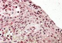
AIFM1, Polyclonal Antibody (Cat# AAA61770)
 IHC (Immunohiostchemistry)
((3.8ug/ml) staining of paraffin embedded Human Prostate. Steamed antigen retrieval with citrate buffer pH 6, AP-staining.)
IHC (Immunohiostchemistry)
((3.8ug/ml) staining of paraffin embedded Human Prostate. Steamed antigen retrieval with citrate buffer pH 6, AP-staining.)
Pleiotrophin, Polyclonal Antibody (Cat# AAA61772)
Expected from sequence similarity: Human, Mouse, Rat
 WB (Western Blot)
((1ug/ml) staining of Mouse Thymus lysate (35ug protein in RIPA buffer). Primary incubation was 1 hour. Detected by chemiluminescence.)
WB (Western Blot)
((1ug/ml) staining of Mouse Thymus lysate (35ug protein in RIPA buffer). Primary incubation was 1 hour. Detected by chemiluminescence.)
VASP, Polyclonal Antibody (Cat# AAA61778)
 WB (Western Blot)
(Biotinylated (0.1ug/ml) staining of Mouse Duodenum lysate (35ug protein in RIPA buffer), exactly mirroring its parental non-biotinylated product. Primary incubation was 1 hour. Detected by chemiluminescence, using streptavidin-HRP and using NAP blocker as a substitute for skimmed milk.)
WB (Western Blot)
(Biotinylated (0.1ug/ml) staining of Mouse Duodenum lysate (35ug protein in RIPA buffer), exactly mirroring its parental non-biotinylated product. Primary incubation was 1 hour. Detected by chemiluminescence, using streptavidin-HRP and using NAP blocker as a substitute for skimmed milk.)
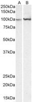
FABP2, Polyclonal Antibody (Cat# AAA61785)
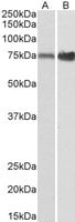
 WB (Western Blot)
(HEK293 lysate (10ug protein in RIPA buffer) overexpressing Human CSF1R with C-terminal MYC tag probed with (1ug/ml) in Lane A and probed with anti-MYC Tag (1/1000) in lane C. Mock-transfected HEK293 probed with (1mg/ml) in Lane B. Primary incubations were for 1 hour. Detected by chemiluminescence.)
WB (Western Blot)
(HEK293 lysate (10ug protein in RIPA buffer) overexpressing Human CSF1R with C-terminal MYC tag probed with (1ug/ml) in Lane A and probed with anti-MYC Tag (1/1000) in lane C. Mock-transfected HEK293 probed with (1mg/ml) in Lane B. Primary incubations were for 1 hour. Detected by chemiluminescence.)
CSF1R, Polyclonal Antibody (Cat# AAA61539)
What are Polyclonal Antibodies?
Polyclonal antibodies are antibodies that come from multiple B cell clones of a host animal. The typical hosts used for the majority of polyclonal antibody production are rabbits, goats, sheep, and donkeys. These polyclonal antibodies, once having identified their target, will bind to different epitopes located at different regions or sequences on the same protein/antigen. This ability to bind multiple epitopes is what makes polyclonal antibodies highly sensitive, as explained in our detailed guide on polyclonal antibodies and why they matter.
As a result, they are ideal at locating and binding to the target, even if the target is in very low concentrations (due to many different antibodies being able to bind to the same target molecule, which allows for significant amplification of a downstream signal).
Polyclonal antibodies are typically produced by injecting an antigen into a host animal, which causes the animal’s immune system to attack the foreign antigen by mass generating antibodies against it. After a period of time, serum is collected from the animal and purified using physicochemical fractionation, class-specific affinity purification, and/or antigen-affinity purification.
Key Uses of Polyclonal Antibodies
- Western Blotting: This method is used to find specific proteins in biological samples after separating them by size.
- Immunohistochemistry: IHC helps visualize the location of proteins in tissue sections using various staining techniques.
- ELISA: (Enzyme-Linked Immunosorbent Assay) is typically used to identify specific protein quantities in a sample. ELISAs can be either “Quantitative” or “Qualitative”.
- Flow Cytometry: technique that identifies and measures the specific protein on the surface or inside the cells in a fluid suspension.
- Immunoprecipitation: IP isolates and studies a specific protein from a complex mixture using antibodies.
Why Buy Polyclonal Antibodies from AAA Biotech?
1. Ideal for Various Applications
Our antibodies are generally going to be validated for use in multiple types of assays, including ELISA, Western Blotting, Immunohistochemistry, Immunoprecipitation, amongst others. They are ideal for a wide range of research applications.
2. Rigorous Quality Control
All of the antibodies in our catalog undergo strict quality testing to ensure specificity, sensitivity, and consistent performance. We are confident in the ability of our antibodies to provide you with accurate results.
3. Wide Assortment of Antibodies
Antibodies in our catalog can be found for both common and exotic species, and these antibodies are also available in both conjugated and recombinant forms to suit many diverse experimental needs.
4. Highly Purified
Our antibodies are available in purified forms with over 85% purity, as confirmed by SDS-PAGE. They are also available with tags such as His, Flag, GST, or MBP. We cater to customers worldwide.
FAQ
1. How are polyclonal antibodies produced?
Traditionally, polyclonal antibodies are produced by injecting an antigen into a host animal (such as a rabbit or goat), which then triggers an immune response from the host animal. The animal’s B cells produce antibodies that will recognize different parts of the injected antigen. These antibodies are then collected from the animal’s blood and purified for use.
2. How do polyclonal antibodies differ from monoclonal antibodies?
Polyclonal antibodies are a mix of antibodies that bind to different locations (epitopes) of the same antigen, while monoclonal antibodies are identical and bind to just one specific epitope. This makes polyclonal antibodies more versatile and better at detecting proteins that may be present in low quantities or in altered/modified forms.
3. How should I store polyclonal antibodies?
Polyclonal antibodies should be stored at 4°C for short-term use (up to a few weeks) and at -20°C or -80°C for long-term storage. Avoid repeated freeze-thaw cycles by dividing them into small aliquots. Always check the datasheet for specific storage instructions.