Filters
▼Clonality
▼Type
▼Reactivity
▼Gene Name
▼Isotype
▼Host
▼Application
▼Clone
▼Polyclonal Antibodies
At AAA Biotech also known as AAA Bio or AAABio, we provide a broad range of purified polyclonal antibodies (pAbs) that are able to all be browsed online through our website. Due to their high specificity and strong binding affinity, these antibodies are ideal for wide swathes of research and experimental applications.
Our polyclonal antibodies can easily support your work, whether you use them for Western Blotting, Immunocytochemistry (with or without Immunofluorescence used in conjunction), Immunohistochemistry, Immunoprecipitation, and ELISA tests. We highly encourage you to browse our range of pAbs and choose the one that best suits your experimental model.
Viewing 1650-1700 of 118597 product results
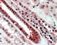
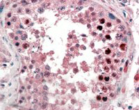
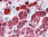
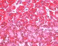 IHC (Immunohiostchemistry)
((5ug/ml) staining of paraffin embedded Human Pancreas. Steamed antigen retrieval with citrate buffer pH 6, AP-staining.)
IHC (Immunohiostchemistry)
((5ug/ml) staining of paraffin embedded Human Pancreas. Steamed antigen retrieval with citrate buffer pH 6, AP-staining.)
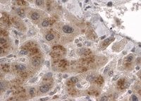
4E-T/EIF4ENIF1, Polyclonal Antibody (Cat# AAA60811)
 IHC (Immunohistochemisry)
((2.5ug/ml) staining of paraffin embedded Human Small Intestine. Steamed antigen retrieval with citrate buffer pH 6, AP-staining.)
IHC (Immunohistochemisry)
((2.5ug/ml) staining of paraffin embedded Human Small Intestine. Steamed antigen retrieval with citrate buffer pH 6, AP-staining.)
FABP2, Polyclonal Antibody (Cat# AAA60829)
 Application Data
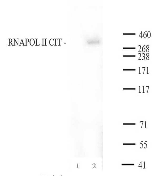
RNA pol II CTD Cit1810, Polyclonal Antibody (Cat# AAA60352)
 Application Data
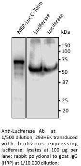
Luciferase, Polyclonal Antibody (Cat# AAA63093)
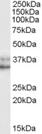
 WB (Western Blot)
(AAA61675 (1ug/ml) staining of Mouse Liver lysate (35ug protein in RIPA buffer). Primary incubation was 1 hour. Detected by chemiluminescence.)
WB (Western Blot)
(AAA61675 (1ug/ml) staining of Mouse Liver lysate (35ug protein in RIPA buffer). Primary incubation was 1 hour. Detected by chemiluminescence.)
Lamin B1, Polyclonal Antibody (Cat# AAA61675)
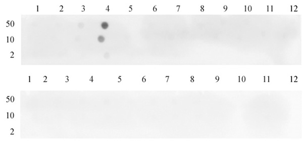
 DB (Dot Blot)
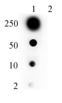
(Histone H3 dimethyl Lys36 antibody (pAb) tested by dot blot analysis. Dot blot analysis was used to confirm the specificity of Histone H3 dimethyl Lys36 antibody for dimethyl Lys36 histone H3. Methylated peptides corresponding to the immunogen and related sequences derived from histone H3 were spotted onto PVDF and probed with Histone H3 dimethyl Lys36 antibody at 1 ug/ml. The amount of peptide (picomoles) spotted is indicated next to each row. Top panel: Lane 1: unmodified Lys4. Lane 2: monomethyl Lys4. Lane 3: dimethyl Lys4. Lane 4: trimethyl Lys4. Lane 5: unmodified Lys9, 14, 18. Lane 6: monomethyl Lys9. Lane 7: dimethyl Lys9. Lane 8: trimethyl Lys9. Lane 9: dimethyl Lys14. Lane 10: monomethyl Lys18. Lane 11: dimethyl Lys18. Lane 12: trimethyl Lys18. Bottom panel: Lane 1: Unmodified Lys23. Lane 2: Monomethyl Lys23. Lane 3: Dimethyl Lys23. Lane 4: Trimethyl Lys23. Lane 5: unmodified Lys27. Lane 6: monomethyl Lys27. Lane 7: dimethyl Lys27. Lane 8: trimethyl Lys27. Lane 9: unmodified Lys36. Lane 10: monomethyl Lys36. Lane 11: dimethyl Lys36. Lane 12: trimethyl Lys36.)
DB (Dot Blot)
(Histone H3 dimethyl Lys36 antibody (pAb) tested by dot blot analysis. Dot blot analysis was used to confirm the specificity of Histone H3 dimethyl Lys36 antibody for dimethyl Lys36 histone H3. Methylated peptides corresponding to the immunogen and related sequences derived from histone H3 were spotted onto PVDF and probed with Histone H3 dimethyl Lys36 antibody at 1 ug/ml. The amount of peptide (picomoles) spotted is indicated next to each row. Top panel: Lane 1: unmodified Lys4. Lane 2: monomethyl Lys4. Lane 3: dimethyl Lys4. Lane 4: trimethyl Lys4. Lane 5: unmodified Lys9, 14, 18. Lane 6: monomethyl Lys9. Lane 7: dimethyl Lys9. Lane 8: trimethyl Lys9. Lane 9: dimethyl Lys14. Lane 10: monomethyl Lys18. Lane 11: dimethyl Lys18. Lane 12: trimethyl Lys18. Bottom panel: Lane 1: Unmodified Lys23. Lane 2: Monomethyl Lys23. Lane 3: Dimethyl Lys23. Lane 4: Trimethyl Lys23. Lane 5: unmodified Lys27. Lane 6: monomethyl Lys27. Lane 7: dimethyl Lys27. Lane 8: trimethyl Lys27. Lane 9: unmodified Lys36. Lane 10: monomethyl Lys36. Lane 11: dimethyl Lys36. Lane 12: trimethyl Lys36.)
Histone H3K36me2, Polyclonal Antibody (Cat# AAA59943)
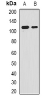
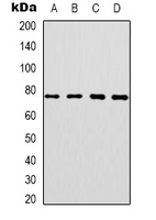
 WB (Western Blot)
(ETO / RUNX1T1 antibody (pAb) tested by Western Blot: K-562 nuclear extract (20 ug per lane) probed with the ETO / RUNX1T1 antibody (pAb) at a dilution of 1:500.)
WB (Western Blot)
(ETO / RUNX1T1 antibody (pAb) tested by Western Blot: K-562 nuclear extract (20 ug per lane) probed with the ETO / RUNX1T1 antibody (pAb) at a dilution of 1:500.)
ETO/RUNX1T1, Polyclonal Antibody (Cat# AAA59980)

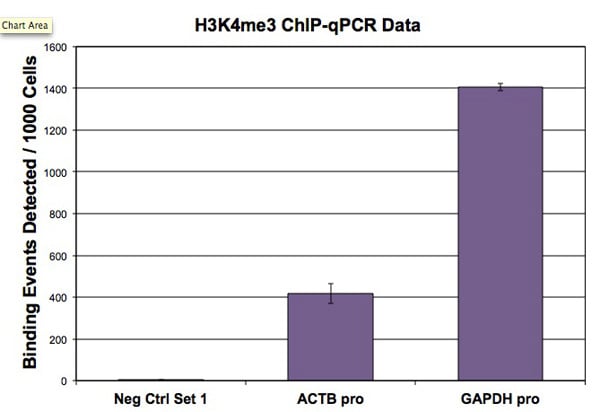

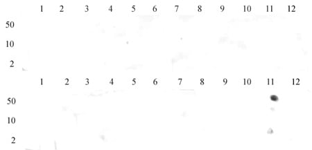
 DB (Dot Blot)
(Histone H3K4me3 antibody (pAb) tested by dot blot analysis. Dot blot was used to confirm specificity of Histone H3K4me3 antibody. Peptides corresponding to regions around major sites of histone H3 methylation (lysine 4, lysine 9, lysine 27) were spotted onto PVDF and probed with antibody at a dilution of 1:500. The amount of peptide (in picomoles) spotted is indicated next to each row. Top panel - Lane 1: unmodified H3K4. Lane 2: H3K4me1. Lane 3: H3K4me2. Lane 4: H3K4me3. Lane 5: unmodified H3K9. Lane 6: H3K9me1. Lane 7: H3K9me2. Lane 8: H3K9me3. Lane 9: unmodified H3K79. Lane 10: H3K79me1. Lane 11: H3K79me2. Lane 12: H3K79me3. Bottom panel - Lane 1: unmodified H3K23. Lane 2: H3K23me1. Lane 3: H3K23me3. Lane 4: H3K23me3. Lane 5: unmodified H3K27. Lane 6: H3K27me1. Lane 7: H3K27me2. Lane 8: H3K27me3. Lane 9: unmodified H3K36. Lane 10: H3K36me1. Lane 11: H3K36me2. Lane 12: H3K36me3.)
DB (Dot Blot)
(Histone H3K4me3 antibody (pAb) tested by dot blot analysis. Dot blot was used to confirm specificity of Histone H3K4me3 antibody. Peptides corresponding to regions around major sites of histone H3 methylation (lysine 4, lysine 9, lysine 27) were spotted onto PVDF and probed with antibody at a dilution of 1:500. The amount of peptide (in picomoles) spotted is indicated next to each row. Top panel - Lane 1: unmodified H3K4. Lane 2: H3K4me1. Lane 3: H3K4me2. Lane 4: H3K4me3. Lane 5: unmodified H3K9. Lane 6: H3K9me1. Lane 7: H3K9me2. Lane 8: H3K9me3. Lane 9: unmodified H3K79. Lane 10: H3K79me1. Lane 11: H3K79me2. Lane 12: H3K79me3. Bottom panel - Lane 1: unmodified H3K23. Lane 2: H3K23me1. Lane 3: H3K23me3. Lane 4: H3K23me3. Lane 5: unmodified H3K27. Lane 6: H3K27me1. Lane 7: H3K27me2. Lane 8: H3K27me3. Lane 9: unmodified H3K36. Lane 10: H3K36me1. Lane 11: H3K36me2. Lane 12: H3K36me3.)
Histone H3K4me3, Polyclonal Antibody (Cat# AAA59808)
 IHC (Immunohiostchemistry)
((3.8ug/ml) staining of paraffin embedded Human Kidney. Steamed antigen retrieval with citrate buffer pH 6, AP-staining.)
IHC (Immunohiostchemistry)
((3.8ug/ml) staining of paraffin embedded Human Kidney. Steamed antigen retrieval with citrate buffer pH 6, AP-staining.)
TCF3/ITF1, Polyclonal Antibody (Cat# AAA61087)
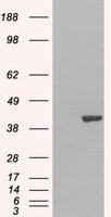
 WB (Western Blot)
(Staining (0.5ug/ml) of Jurkat lysate (RIPA buffer, 30ug total protein per lane). Primary incubated for 1 hour. Detected by western blot using chemiluminescence.)
WB (Western Blot)
(Staining (0.5ug/ml) of Jurkat lysate (RIPA buffer, 30ug total protein per lane). Primary incubated for 1 hour. Detected by western blot using chemiluminescence.)
GRAP2/GRID/Grf40, Polyclonal Antibody (Cat# AAA61110)
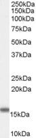
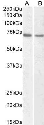
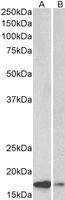
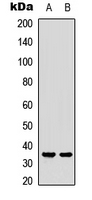
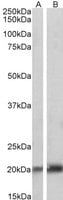
 WB (Western Blot)
((1ug/ml) staining of Mouse Skeletal Muscle lysate (35ug protein in RIPA buffer). Primary incubation was 1 hour. Detected by chemiluminescence.)
WB (Western Blot)
((1ug/ml) staining of Mouse Skeletal Muscle lysate (35ug protein in RIPA buffer). Primary incubation was 1 hour. Detected by chemiluminescence.)
Integrin alpha 11, Polyclonal Antibody (Cat# AAA61564)
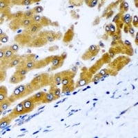
 IHC (Immunohiostchemistry)
((2ug/ml) staining of paraffin embedded Human Liver. Steamed antigen retrieval with citrate buffer pH 6, HRP-staining.)
IHC (Immunohiostchemistry)
((2ug/ml) staining of paraffin embedded Human Liver. Steamed antigen retrieval with citrate buffer pH 6, HRP-staining.)
CLPP, Polyclonal Antibody (Cat# AAA60885)
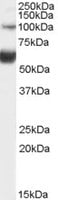
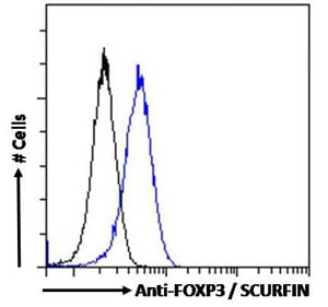
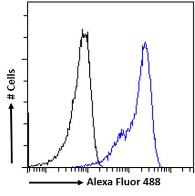 FCM/FACS (Flow Cytometry)
(AAA60905 Flow cytometric analysis of paraformaldehyde fixed U937 cells (blue line), permeabilized with 0.5%Triton.Primary incubation 1hr (10ug/ml) followed by Alexa Fluor 488 secondary antibody (1ug/ml).IgG control: Unimmunized goat IgG (black line) followed by Alexa Fluor 488 secondary antibody.)
FCM/FACS (Flow Cytometry)
(AAA60905 Flow cytometric analysis of paraformaldehyde fixed U937 cells (blue line), permeabilized with 0.5%Triton.Primary incubation 1hr (10ug/ml) followed by Alexa Fluor 488 secondary antibody (1ug/ml).IgG control: Unimmunized goat IgG (black line) followed by Alexa Fluor 488 secondary antibody.)
VDR, Polyclonal Antibody (Cat# AAA60905)
Expected from sequence similarity: Human, Mouse, Rat, Dog
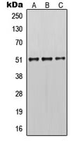
 WB (Western Blot)
(Staining (0.5ug/ml) of A431 lysate (RIPA buffer, 35ug total protein per lane). Primary incubated for 1 hour. Detected by western blot using chemiluminescence.)
WB (Western Blot)
(Staining (0.5ug/ml) of A431 lysate (RIPA buffer, 35ug total protein per lane). Primary incubated for 1 hour. Detected by western blot using chemiluminescence.)
Annexin A2, Polyclonal Antibody (Cat# AAA60930)
 WB (Western Blot)
((1ug/ml) staining of Human Brain (Cerebellum) lysate (35ug protein in RIPA buffer). Primary incubation was 1 hour. Detected by chemiluminescence.)
WB (Western Blot)
((1ug/ml) staining of Human Brain (Cerebellum) lysate (35ug protein in RIPA buffer). Primary incubation was 1 hour. Detected by chemiluminescence.)
GLuR5/GRIK1, Polyclonal Antibody (Cat# AAA60891)
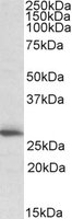
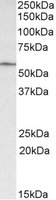
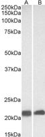
 WB (Western Blot)
((0.1ug/ml) staining of Human Liver lysate (35ug protein in RIPA buffer). Primary incubation was 1 hour. Detected by chemiluminescence.)
WB (Western Blot)
((0.1ug/ml) staining of Human Liver lysate (35ug protein in RIPA buffer). Primary incubation was 1 hour. Detected by chemiluminescence.)
TCF2/VHNF1, Polyclonal Antibody (Cat# AAA61008)
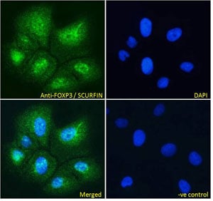
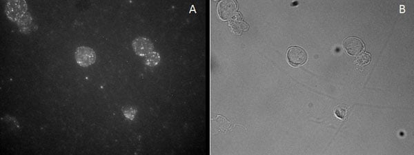 IF (Immunofluorescence)
(Staining of CD25-sorted (Treg) Human blood cells gathered by cytospin and detected by FITC (A) and in phase contrast (B).)
IF (Immunofluorescence)
(Staining of CD25-sorted (Treg) Human blood cells gathered by cytospin and detected by FITC (A) and in phase contrast (B).)
FOXP3/SCURFIN, Polyclonal Antibody (Cat# AAA61014)
 IHC (Immunohiostchemistry)
(AAA61652 (5ug/ml) staining of paraffin embedded Human Cerebellum. Steamed antigen retrieval with citrate buffer pH 6, AP-staining.)
IHC (Immunohiostchemistry)
(AAA61652 (5ug/ml) staining of paraffin embedded Human Cerebellum. Steamed antigen retrieval with citrate buffer pH 6, AP-staining.)
Alpha-synuclein, Polyclonal Antibody (Cat# AAA61652)
Expected from sequence similarity: Human, Mouse, Rat, Dog, Pig
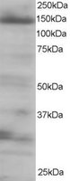
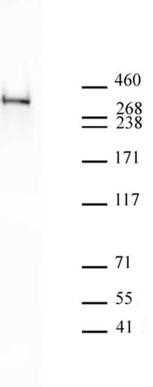
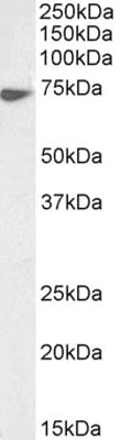
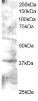
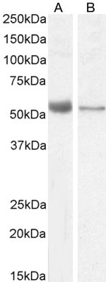
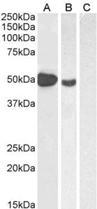
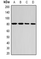
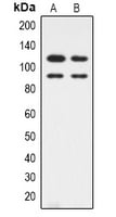
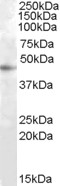
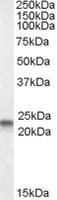
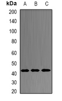
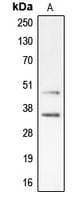
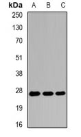
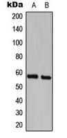
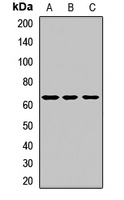
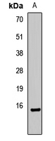
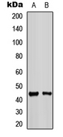
 WB (Western Blot)
((1ug/ml) staining of Mouse Heart lysate (35ug protein in RIPA buffer). Primary incubation was 1 hour. Detected by chemiluminescence.)
WB (Western Blot)
((1ug/ml) staining of Mouse Heart lysate (35ug protein in RIPA buffer). Primary incubation was 1 hour. Detected by chemiluminescence.)
GLP-1-R, Polyclonal Antibody (Cat# AAA61687)
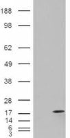
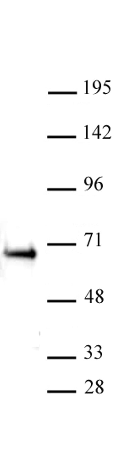
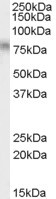
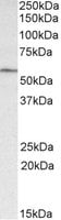
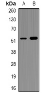
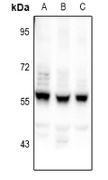
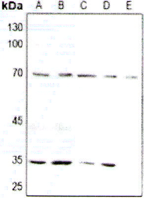
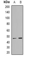
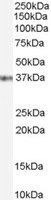
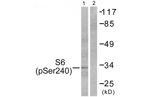 WB (Western Blot)
(Western blot analysis of extracts from HeLa cells, treated with TNF-a (20ng/ml, 2mins), using S6 Ribosomal Protein (Phospho-Ser240) antibody.)
WB (Western Blot)
(Western blot analysis of extracts from HeLa cells, treated with TNF-a (20ng/ml, 2mins), using S6 Ribosomal Protein (Phospho-Ser240) antibody.)
S6, Polyclonal Antibody (Cat# AAA224459)
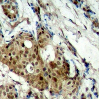
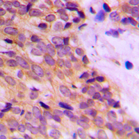
 IHC (Immunohiostchemistry)
(Immunohistochemical analysis of EGFR staining in human breast cancer formalin fixed paraffin embedded tissue section. The section was pre-treated using heat mediated antigen retrieval with sodium citrate buffer (pH 6.0). The section was then incubated with the antibody at room temperature and detected using an HRP conjugated compact polymer system. DAB was used as the chromogen. The section was then counterstained with haematoxylin and mounted with DPX.)
IHC (Immunohiostchemistry)
(Immunohistochemical analysis of EGFR staining in human breast cancer formalin fixed paraffin embedded tissue section. The section was pre-treated using heat mediated antigen retrieval with sodium citrate buffer (pH 6.0). The section was then incubated with the antibody at room temperature and detected using an HRP conjugated compact polymer system. DAB was used as the chromogen. The section was then counterstained with haematoxylin and mounted with DPX.)
EGFR, Polyclonal Antibody (Cat# AAA220963)
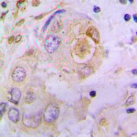
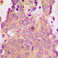
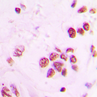
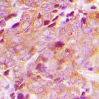
 IHC (Immunohiostchemistry)
(Immunohistochemical analysis of GPD2 staining in human liver cancer formalin fixed paraffin embedded tissue section. The section was pre-treated using heat mediated antigen retrieval with sodium citrate buffer (pH 6.0). The section was then incubated with the antibody at room temperature and detected using an HRP conjugated compact polymer system. DAB was used as the chromogen. The section was then counterstained with haematoxylin and mounted with DPX.)
IHC (Immunohiostchemistry)
(Immunohistochemical analysis of GPD2 staining in human liver cancer formalin fixed paraffin embedded tissue section. The section was pre-treated using heat mediated antigen retrieval with sodium citrate buffer (pH 6.0). The section was then incubated with the antibody at room temperature and detected using an HRP conjugated compact polymer system. DAB was used as the chromogen. The section was then counterstained with haematoxylin and mounted with DPX.)
GPD2, Polyclonal Antibody (Cat# AAA222124)
 IF (Immunofluorescence)
(Immunofluorescent analysis of NEK2 staining in U2OS cells. Formalin-fixed cells were permeabilized with 0.1% Triton X-100 in TBS for 5-10 minutes and blocked with 3% BSA-PBS for 30 minutes at room temperature. Cells were probed with the primary antibody in 3% BSA-PBS and incubated overnight at 4 °C in a humidified chamber. Cells were washed with PBST and incubated with a DyLight 594-conjugated secondary antibody (red) in PBS at room temperature in the dark.)
IF (Immunofluorescence)
(Immunofluorescent analysis of NEK2 staining in U2OS cells. Formalin-fixed cells were permeabilized with 0.1% Triton X-100 in TBS for 5-10 minutes and blocked with 3% BSA-PBS for 30 minutes at room temperature. Cells were probed with the primary antibody in 3% BSA-PBS and incubated overnight at 4 °C in a humidified chamber. Cells were washed with PBST and incubated with a DyLight 594-conjugated secondary antibody (red) in PBS at room temperature in the dark.)
NEK2, Polyclonal Antibody (Cat# AAA222311)
 IHC (Immunohiostchemistry)
(Immunohistochemical analysis of CEP55 staining in human breast cancer formalin fixed paraffin embedded tissue section. The section was pre-treated using heat mediated antigen retrieval with sodium citrate buffer (pH 6.0). The section was then incubated with the antibody at room temperature and detected using an HRP conjugated compact polymer system. DAB was used as the chromogen. The section was then counterstained with haematoxylin and mounted with DPX.)
IHC (Immunohiostchemistry)
(Immunohistochemical analysis of CEP55 staining in human breast cancer formalin fixed paraffin embedded tissue section. The section was pre-treated using heat mediated antigen retrieval with sodium citrate buffer (pH 6.0). The section was then incubated with the antibody at room temperature and detected using an HRP conjugated compact polymer system. DAB was used as the chromogen. The section was then counterstained with haematoxylin and mounted with DPX.)
CEP55, Polyclonal Antibody (Cat# AAA220030)
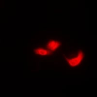
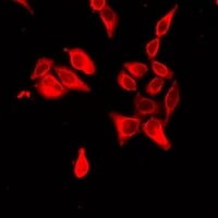
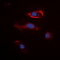 IF (Immunofluorescence)
(Immunofluorescent analysis of FGF22 staining in K562 cells. Formalin-fixed cells were permeabilized with 0.1% Triton X-100 in TBS for 5-10 minutes and blocked with 3% BSA-PBS for 30 minutes at room temperature. Cells were probed with the primary antibody in 3% BSA-PBS and incubated overnight at 4 °C in a humidified chamber. Cells were washed with PBST and incubated with a DyLight 594-conjugated secondary antibody (red) in PBS at room temperature in the dark. DAPI was used to stain the cell nuclei (blue).)
IF (Immunofluorescence)
(Immunofluorescent analysis of FGF22 staining in K562 cells. Formalin-fixed cells were permeabilized with 0.1% Triton X-100 in TBS for 5-10 minutes and blocked with 3% BSA-PBS for 30 minutes at room temperature. Cells were probed with the primary antibody in 3% BSA-PBS and incubated overnight at 4 °C in a humidified chamber. Cells were washed with PBST and incubated with a DyLight 594-conjugated secondary antibody (red) in PBS at room temperature in the dark. DAPI was used to stain the cell nuclei (blue).)
FGF22, Polyclonal Antibody (Cat# AAA220528)
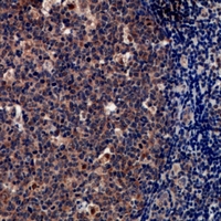
 IHC (Immunohiostchemistry)
(Immunohistochemical analysis of Caspase 3 (pS150) staining in human tonsil formalin fixed paraffin embedded tissue section. The section was pre-treated using heat mediated antigen retrieval with sodium citrate buffer (pH 6.0). The section was then incubated with the antibody at room temperature and detected using an HRP conjugated compact polymer system. DAB was used as the chromogen. The section was then counterstained with haematoxylin and mounted with DPX.)
IHC (Immunohiostchemistry)
(Immunohistochemical analysis of Caspase 3 (pS150) staining in human tonsil formalin fixed paraffin embedded tissue section. The section was pre-treated using heat mediated antigen retrieval with sodium citrate buffer (pH 6.0). The section was then incubated with the antibody at room temperature and detected using an HRP conjugated compact polymer system. DAB was used as the chromogen. The section was then counterstained with haematoxylin and mounted with DPX.)
Caspase 3, Polyclonal Antibody (Cat# AAA223505)
 IHC (Immunohiostchemistry)
(Immunohistochemical analysis of CDK11B staining in human breast cancer formalin fixed paraffin embedded tissue section. The section was pre-treated using heat mediated antigen retrieval with sodium citrate buffer (pH 6.0). The section was then incubated with the antibody at room temperature and detected using an HRP conjugated compact polymer system. DAB was used as the chromogen. The section was then counterstained with haematoxylin and mounted with DPX.)
IHC (Immunohiostchemistry)
(Immunohistochemical analysis of CDK11B staining in human breast cancer formalin fixed paraffin embedded tissue section. The section was pre-treated using heat mediated antigen retrieval with sodium citrate buffer (pH 6.0). The section was then incubated with the antibody at room temperature and detected using an HRP conjugated compact polymer system. DAB was used as the chromogen. The section was then counterstained with haematoxylin and mounted with DPX.)
CDK11B, Polyclonal Antibody (Cat# AAA223553)
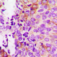
 IHC (Immunohiostchemistry)
(Immunohistochemical analysis of Osteopontin staining in human lung cancer formalin fixed paraffin embedded tissue section. The section was pre-treated using heat mediated antigen retrieval with sodium citrate buffer (pH 6.0). The section was then incubated with the antibody at room temperature and detected using an HRP conjugated compact polymer system. DAB was used as the chromogen. The section was then counterstained with haematoxylin and mounted with DPX.)
IHC (Immunohiostchemistry)
(Immunohistochemical analysis of Osteopontin staining in human lung cancer formalin fixed paraffin embedded tissue section. The section was pre-treated using heat mediated antigen retrieval with sodium citrate buffer (pH 6.0). The section was then incubated with the antibody at room temperature and detected using an HRP conjugated compact polymer system. DAB was used as the chromogen. The section was then counterstained with haematoxylin and mounted with DPX.)
Osteopontin, Polyclonal Antibody (Cat# AAA220347)
 IHC (Immunohiostchemistry)
(Immunohistochemical analysis of Tryptase delta staining in human tonsil formalin fixed paraffin embedded tissue section. The section was pre-treated using heat mediated antigen retrieval with sodium citrate buffer (pH 6.0). The section was then incubated with the antibody at room temperature and detected using an HRP conjugated compact polymer system. DAB was used as the chromogen. The section was then counterstained with haematoxylin and mounted with DPX.)
IHC (Immunohiostchemistry)
(Immunohistochemical analysis of Tryptase delta staining in human tonsil formalin fixed paraffin embedded tissue section. The section was pre-treated using heat mediated antigen retrieval with sodium citrate buffer (pH 6.0). The section was then incubated with the antibody at room temperature and detected using an HRP conjugated compact polymer system. DAB was used as the chromogen. The section was then counterstained with haematoxylin and mounted with DPX.)
Tryptase delta, Polyclonal Antibody (Cat# AAA220074)
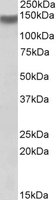
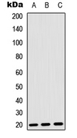
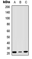
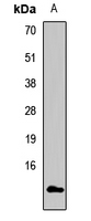
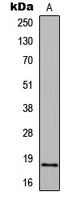
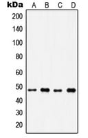
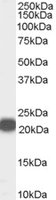
 WB (Western Blot)
((1ug/ml) staining of HepG2 cell lysate (35ug protein in RIPA buffer). Primary incubation was 1 hour. Detected by chemiluminescence.)
WB (Western Blot)
((1ug/ml) staining of HepG2 cell lysate (35ug protein in RIPA buffer). Primary incubation was 1 hour. Detected by chemiluminescence.)
RASSF6, Polyclonal Antibody (Cat# AAA61328)
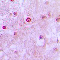
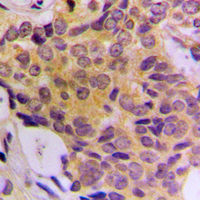
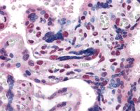 IHC (Immunohiostchemistry)
((3.8ug/ml) staining of paraffin embedded Human Placenta. Steamed antigen retrieval with citrate buffer pH 6, AP-staining.)
IHC (Immunohiostchemistry)
((3.8ug/ml) staining of paraffin embedded Human Placenta. Steamed antigen retrieval with citrate buffer pH 6, AP-staining.)
GBX2, Polyclonal Antibody (Cat# AAA61117)
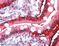
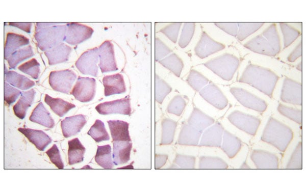
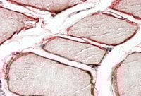 IHC (Immunohistochemistry)
((3.8ug/ml) staining of paraffin embedded Human Skeletal Muscle. Steamed antigen retrieval with citrate buffer pH 6, AP-staining.)
IHC (Immunohistochemistry)
((3.8ug/ml) staining of paraffin embedded Human Skeletal Muscle. Steamed antigen retrieval with citrate buffer pH 6, AP-staining.)
CAV3, Polyclonal Antibody (Cat# AAA61412)
 IF (Immunofluorescence)
(Immunofluorescent analysis of PRDM7 staining in A549 cells. Formalin-fixed cells were permeabilized with 0.1% Triton X-100 in TBS for 5-10 minutes and blocked with 3% BSA-PBS for 30 minutes at room temperature. Cells were probed with the primary antibody in 3% BSA-PBS and incubated overnight at 4 °C in a humidified chamber. Cells were washed with PBST and incubated with a DyLight 594-conjugated secondary antibody (red) in PBS at room temperature in the dark.)
IF (Immunofluorescence)
(Immunofluorescent analysis of PRDM7 staining in A549 cells. Formalin-fixed cells were permeabilized with 0.1% Triton X-100 in TBS for 5-10 minutes and blocked with 3% BSA-PBS for 30 minutes at room temperature. Cells were probed with the primary antibody in 3% BSA-PBS and incubated overnight at 4 °C in a humidified chamber. Cells were washed with PBST and incubated with a DyLight 594-conjugated secondary antibody (red) in PBS at room temperature in the dark.)
PRDM7, Polyclonal Antibody (Cat# AAA220190)
 IHC (Immunohiostchemistry)
(Immunohistochemical analysis of NSG1 staining in human breast cancer formalin fixed paraffin embedded tissue section. The section was pre-treated using heat mediated antigen retrieval with sodium citrate buffer (pH 6.0). The section was then incubated with the antibody at room temperature and detected using an HRP conjugated compact polymer system. DAB was used as the chromogen. The section was then counterstained with haematoxylin and mounted with DPX.)
IHC (Immunohiostchemistry)
(Immunohistochemical analysis of NSG1 staining in human breast cancer formalin fixed paraffin embedded tissue section. The section was pre-treated using heat mediated antigen retrieval with sodium citrate buffer (pH 6.0). The section was then incubated with the antibody at room temperature and detected using an HRP conjugated compact polymer system. DAB was used as the chromogen. The section was then counterstained with haematoxylin and mounted with DPX.)
NSG1, Polyclonal Antibody (Cat# AAA221536)
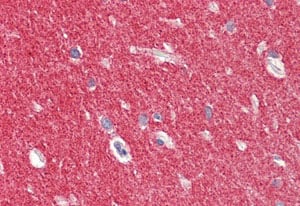
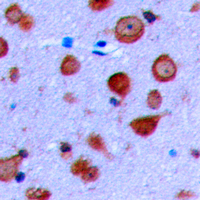
 IHC (Immunohiostchemistry)
(Immunohistochemical analysis of CDK10 staining in human brain formalin fixed paraffin embedded tissue section. The section was pre-treated using heat mediated antigen retrieval with sodium citrate buffer (pH 6.0). The section was then incubated with the antibody at room temperature and detected using an HRP conjugated compact polymer system. DAB was used as the chromogen. The section was then counterstained with haematoxylin and mounted with DPX.)
IHC (Immunohiostchemistry)
(Immunohistochemical analysis of CDK10 staining in human brain formalin fixed paraffin embedded tissue section. The section was pre-treated using heat mediated antigen retrieval with sodium citrate buffer (pH 6.0). The section was then incubated with the antibody at room temperature and detected using an HRP conjugated compact polymer system. DAB was used as the chromogen. The section was then counterstained with haematoxylin and mounted with DPX.)
CDK10, Polyclonal Antibody (Cat# AAA223334)
 IF (Immunofluorescence)
(Immunofluorescent analysis of POLR2F staining in HeLa cells. Formalin-fixed cells were permeabilized with 0.1% Triton X-100 in TBS for 5-10 minutes and blocked with 3% BSA-PBS for 30 minutes at room temperature. Cells were probed with the primary antibody in 3% BSA-PBS and incubated overnight at 4 °C in a humidified chamber. Cells were washed with PBST and incubated with a DyLight 594-conjugated secondary antibody (red) in PBS at room temperature in the dark.)
IF (Immunofluorescence)
(Immunofluorescent analysis of POLR2F staining in HeLa cells. Formalin-fixed cells were permeabilized with 0.1% Triton X-100 in TBS for 5-10 minutes and blocked with 3% BSA-PBS for 30 minutes at room temperature. Cells were probed with the primary antibody in 3% BSA-PBS and incubated overnight at 4 °C in a humidified chamber. Cells were washed with PBST and incubated with a DyLight 594-conjugated secondary antibody (red) in PBS at room temperature in the dark.)
POLR2F, Polyclonal Antibody (Cat# AAA222376)
 IF (Immunofluorescence)
(Immunofluorescent analysis of EIF6 staining in Hela cells. Formalin-fixed cells were permeabilized with 0.1% Triton X-100 in TBS for 5-10 minutes and blocked with 3% BSA-PBS for 30 minutes at room temperature. Cells were probed with the primary antibody in 3% BSA-PBS and incubated overnight at 4 °C in a humidified chamber. Cells were washed with PBST and incubated with a DyLight 594-conjugated secondary antibody (red) in PBS at room temperature in the dark.)
IF (Immunofluorescence)
(Immunofluorescent analysis of EIF6 staining in Hela cells. Formalin-fixed cells were permeabilized with 0.1% Triton X-100 in TBS for 5-10 minutes and blocked with 3% BSA-PBS for 30 minutes at room temperature. Cells were probed with the primary antibody in 3% BSA-PBS and incubated overnight at 4 °C in a humidified chamber. Cells were washed with PBST and incubated with a DyLight 594-conjugated secondary antibody (red) in PBS at room temperature in the dark.)
EIF6, Polyclonal Antibody (Cat# AAA222264)
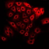
 IF (Immunofluorescence)
(Immunofluorescent analysis of Cytokeratin 14 staining in MCF7 cells. Formalin-fixed cells were permeabilized with 0.1% Triton X-100 in TBS for 5-10 minutes and blocked with 3% BSA-PBS for 30 minutes at room temperature. Cells were probed with the primary antibody in 3% BSA-PBS and incubated overnight at 4 °C in a humidified chamber. Cells were washed with PBST and incubated with a DyLight 594-conjugated secondary antibody (red) in PBS at room temperature in the dark.)
IF (Immunofluorescence)
(Immunofluorescent analysis of Cytokeratin 14 staining in MCF7 cells. Formalin-fixed cells were permeabilized with 0.1% Triton X-100 in TBS for 5-10 minutes and blocked with 3% BSA-PBS for 30 minutes at room temperature. Cells were probed with the primary antibody in 3% BSA-PBS and incubated overnight at 4 °C in a humidified chamber. Cells were washed with PBST and incubated with a DyLight 594-conjugated secondary antibody (red) in PBS at room temperature in the dark.)
Cytokeratin 14/16, Polyclonal Antibody (Cat# AAA222495)
 IHC (Immunohiostchemistry)
(Immunohistochemical analysis of CDK5RAP3 staining in human breast cancer formalin fixed paraffin embedded tissue section. The section was pre-treated using heat mediated antigen retrieval with sodium citrate buffer (pH 6.0). The section was then incubated with the antibody at room temperature and detected using an HRP conjugated compact polymer system. DAB was used as the chromogen. The section was then counterstained with haematoxylin and mounted with DPX.)
IHC (Immunohiostchemistry)
(Immunohistochemical analysis of CDK5RAP3 staining in human breast cancer formalin fixed paraffin embedded tissue section. The section was pre-treated using heat mediated antigen retrieval with sodium citrate buffer (pH 6.0). The section was then incubated with the antibody at room temperature and detected using an HRP conjugated compact polymer system. DAB was used as the chromogen. The section was then counterstained with haematoxylin and mounted with DPX.)
CDK5RAP3, Polyclonal Antibody (Cat# AAA220439)
 IF (Immunofluorescence)
(Immunofluorescent analysis of RNF40 staining in MCF7 cells. Formalin-fixed cells were permeabilized with 0.1% Triton X-100 in TBS for 5-10 minutes and blocked with 3% BSA-PBS for 30 minutes at room temperature. Cells were probed with the primary antibody in 3% BSA-PBS and incubated overnight at 4 °C in a humidified chamber. Cells were washed with PBST and incubated with a DyLight 594-conjugated secondary antibody (red) in PBS at room temperature in the dark.)
IF (Immunofluorescence)
(Immunofluorescent analysis of RNF40 staining in MCF7 cells. Formalin-fixed cells were permeabilized with 0.1% Triton X-100 in TBS for 5-10 minutes and blocked with 3% BSA-PBS for 30 minutes at room temperature. Cells were probed with the primary antibody in 3% BSA-PBS and incubated overnight at 4 °C in a humidified chamber. Cells were washed with PBST and incubated with a DyLight 594-conjugated secondary antibody (red) in PBS at room temperature in the dark.)
RNF40, Polyclonal Antibody (Cat# AAA222111)
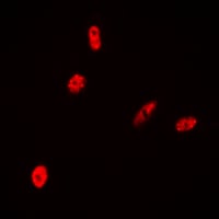 IF (Immunofluorescence)
(Immunofluorescent analysis of Histone H2B staining in HeLa cells. Formalin-fixed cells were permeabilized with 0.1% Triton X-100 in TBS for 5-10 minutes and blocked with 3% BSA-PBS for 30 minutes at room temperature. Cells were probed with the primary antibody in 3% BSA-PBS and incubated overnight at 4 °C in a humidified chamber. Cells were washed with PBST and incubated with a DyLight 594-conjugated secondary antibody (red) in PBS at room temperature in the dark.)
IF (Immunofluorescence)
(Immunofluorescent analysis of Histone H2B staining in HeLa cells. Formalin-fixed cells were permeabilized with 0.1% Triton X-100 in TBS for 5-10 minutes and blocked with 3% BSA-PBS for 30 minutes at room temperature. Cells were probed with the primary antibody in 3% BSA-PBS and incubated overnight at 4 °C in a humidified chamber. Cells were washed with PBST and incubated with a DyLight 594-conjugated secondary antibody (red) in PBS at room temperature in the dark.)
Histone H2B, Polyclonal Antibody (Cat# AAA222646)
 IF (Immunofluorescence)
(Immunofluorescent analysis of NPT2 staining in HeLa cells. Formalin-fixed cells were permeabilized with 0.1% Triton X-100 in TBS for 5-10 minutes and blocked with 3% BSA-PBS for 30 minutes at room temperature. Cells were probed with the primary antibody in 3% BSA-PBS and incubated overnight at 4 °C in a humidified chamber. Cells were washed with PBST and incubated with a DyLight 594-conjugated secondary antibody (red) in PBS at room temperature in the dark.)
IF (Immunofluorescence)
(Immunofluorescent analysis of NPT2 staining in HeLa cells. Formalin-fixed cells were permeabilized with 0.1% Triton X-100 in TBS for 5-10 minutes and blocked with 3% BSA-PBS for 30 minutes at room temperature. Cells were probed with the primary antibody in 3% BSA-PBS and incubated overnight at 4 °C in a humidified chamber. Cells were washed with PBST and incubated with a DyLight 594-conjugated secondary antibody (red) in PBS at room temperature in the dark.)
NPT2, Polyclonal Antibody (Cat# AAA222398)
 IF (Immunofluorescence)
(Immunofluorescent analysis of IP6K2 staining in A549 cells. Formalin-fixed cells were permeabilized with 0.1% Triton X-100 in TBS for 5-10 minutes and blocked with 3% BSA-PBS for 30 minutes at room temperature. Cells were probed with the primary antibody in 3% BSA-PBS and incubated overnight at 4 °C in a humidified chamber. Cells were washed with PBST and incubated with a DyLight 594-conjugated secondary antibody (red) in PBS at room temperature in the dark.)
IF (Immunofluorescence)
(Immunofluorescent analysis of IP6K2 staining in A549 cells. Formalin-fixed cells were permeabilized with 0.1% Triton X-100 in TBS for 5-10 minutes and blocked with 3% BSA-PBS for 30 minutes at room temperature. Cells were probed with the primary antibody in 3% BSA-PBS and incubated overnight at 4 °C in a humidified chamber. Cells were washed with PBST and incubated with a DyLight 594-conjugated secondary antibody (red) in PBS at room temperature in the dark.)
IP6K2, Polyclonal Antibody (Cat# AAA223200)
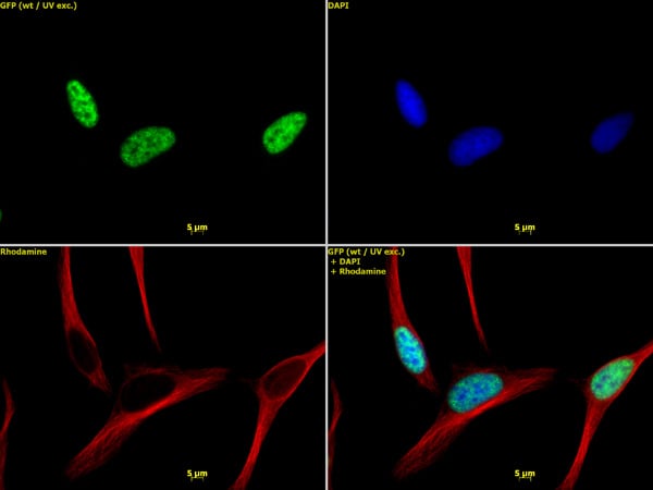
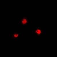
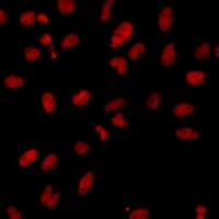
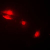
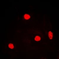
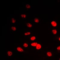
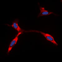
 IF (Immunofluorescence)
(Immunofluorescent analysis of Histone H3 (MonoMethyl R26) staining in HEK293T cells. Formalin-fixed cells were permeabilized with 0.1% Triton X-100 in TBS for 5-10 minutes and blocked with 3% BSA-PBS for 30 minutes at room temperature. Cells were probed with the primary antibody in 3% BSA-PBS and incubated overnight at 4 °C in a humidified chamber. Cells were washed with PBST and incubated with a DyLight 594-conjugated secondary antibody (red) in PBS at room temperature in the dark.)
IF (Immunofluorescence)
(Immunofluorescent analysis of Histone H3 (MonoMethyl R26) staining in HEK293T cells. Formalin-fixed cells were permeabilized with 0.1% Triton X-100 in TBS for 5-10 minutes and blocked with 3% BSA-PBS for 30 minutes at room temperature. Cells were probed with the primary antibody in 3% BSA-PBS and incubated overnight at 4 °C in a humidified chamber. Cells were washed with PBST and incubated with a DyLight 594-conjugated secondary antibody (red) in PBS at room temperature in the dark.)
Histone H3, Polyclonal Antibody (Cat# AAA223122)

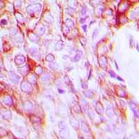
 IHC (Immunohiostchemistry)
(Immunohistochemical analysis of Focal Adhesion Kinase (pY397) staining in human breast cancer formalin fixed paraffin embedded tissue section. The section was pre-treated using heat mediated antigen retrieval with sodium citrate buffer (pH 6.0). The section was then incubated with the antibody at room temperature and detected using an HRP conjugated compact polymer system. DAB was used as the chromogen. The section was then counterstained with haematoxylin and mounted with DPX.)
IHC (Immunohiostchemistry)
(Immunohistochemical analysis of Focal Adhesion Kinase (pY397) staining in human breast cancer formalin fixed paraffin embedded tissue section. The section was pre-treated using heat mediated antigen retrieval with sodium citrate buffer (pH 6.0). The section was then incubated with the antibody at room temperature and detected using an HRP conjugated compact polymer system. DAB was used as the chromogen. The section was then counterstained with haematoxylin and mounted with DPX.)
Focal Adhesion Kinase, Polyclonal Antibody (Cat# AAA223456)
 IF (Immunofluorescence)
(Immunofluorescent analysis of Kir6.2 staining in HeLa cells. Formalin-fixed cells were permeabilized with 0.1% Triton X-100 in TBS for 5-10 minutes and blocked with 3% BSA-PBS for 30 minutes at room temperature. Cells were probed with the primary antibody in 3% BSA-PBS and incubated overnight at 4 °C in a humidified chamber. Cells were washed with PBST and incubated with a DyLight 594-conjugated secondary antibody (red) in PBS at room temperature in the dark. DAPI was used to stain the cell nuclei (blue).)
IF (Immunofluorescence)
(Immunofluorescent analysis of Kir6.2 staining in HeLa cells. Formalin-fixed cells were permeabilized with 0.1% Triton X-100 in TBS for 5-10 minutes and blocked with 3% BSA-PBS for 30 minutes at room temperature. Cells were probed with the primary antibody in 3% BSA-PBS and incubated overnight at 4 °C in a humidified chamber. Cells were washed with PBST and incubated with a DyLight 594-conjugated secondary antibody (red) in PBS at room temperature in the dark. DAPI was used to stain the cell nuclei (blue).)
Kir6.2, Polyclonal Antibody (Cat# AAA223891)
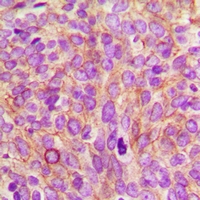
 IHC (Immunohiostchemistry)
(Immunohistochemical analysis of CEP76 staining in human breast cancer formalin fixed paraffin embedded tissue section. The section was pre-treated using heat mediated antigen retrieval with sodium citrate buffer (pH 6.0). The section was then incubated with the antibody at room temperature and detected using an HRP conjugated compact polymer system. DAB was used as the chromogen. The section was then counterstained with haematoxylin and mounted with DPX.)
IHC (Immunohiostchemistry)
(Immunohistochemical analysis of CEP76 staining in human breast cancer formalin fixed paraffin embedded tissue section. The section was pre-treated using heat mediated antigen retrieval with sodium citrate buffer (pH 6.0). The section was then incubated with the antibody at room temperature and detected using an HRP conjugated compact polymer system. DAB was used as the chromogen. The section was then counterstained with haematoxylin and mounted with DPX.)
CEP76, Polyclonal Antibody (Cat# AAA223573)
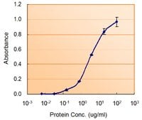 Application Data
((5ug/ml) as the reporter with as the capture rabbit antibody (5ug/ml).)
Application Data
((5ug/ml) as the reporter with as the capture rabbit antibody (5ug/ml).)
NANOG, Polyclonal Antibody (Cat# AAA60948)
 IHC (Immunohiostchemistry)
((2.5ug/ml) staining of paraffin embedded Human Adrenal Gland. Steamed antigen retrieval with citrate buffer pH 6, AP-staining.)
IHC (Immunohiostchemistry)
((2.5ug/ml) staining of paraffin embedded Human Adrenal Gland. Steamed antigen retrieval with citrate buffer pH 6, AP-staining.)
PEBP1/RKIP, Polyclonal Antibody (Cat# AAA61077)
What are Polyclonal Antibodies?
Polyclonal antibodies are antibodies that come from multiple B cell clones of a host animal. The typical hosts used for the majority of polyclonal antibody production are rabbits, goats, sheep, and donkeys. These polyclonal antibodies, once having identified their target, will bind to different epitopes located at different regions or sequences on the same protein/antigen. This ability to bind multiple epitopes is what makes polyclonal antibodies highly sensitive, as explained in our detailed guide on polyclonal antibodies and why they matter.
As a result, they are ideal at locating and binding to the target, even if the target is in very low concentrations (due to many different antibodies being able to bind to the same target molecule, which allows for significant amplification of a downstream signal).
Polyclonal antibodies are typically produced by injecting an antigen into a host animal, which causes the animal’s immune system to attack the foreign antigen by mass generating antibodies against it. After a period of time, serum is collected from the animal and purified using physicochemical fractionation, class-specific affinity purification, and/or antigen-affinity purification.
Key Uses of Polyclonal Antibodies
- Western Blotting: This method is used to find specific proteins in biological samples after separating them by size.
- Immunohistochemistry: IHC helps visualize the location of proteins in tissue sections using various staining techniques.
- ELISA: (Enzyme-Linked Immunosorbent Assay) is typically used to identify specific protein quantities in a sample. ELISAs can be either “Quantitative” or “Qualitative”.
- Flow Cytometry: technique that identifies and measures the specific protein on the surface or inside the cells in a fluid suspension.
- Immunoprecipitation: IP isolates and studies a specific protein from a complex mixture using antibodies.
Why Buy Polyclonal Antibodies from AAA Biotech?
1. Ideal for Various Applications
Our antibodies are generally going to be validated for use in multiple types of assays, including ELISA, Western Blotting, Immunohistochemistry, Immunoprecipitation, amongst others. They are ideal for a wide range of research applications.
2. Rigorous Quality Control
All of the antibodies in our catalog undergo strict quality testing to ensure specificity, sensitivity, and consistent performance. We are confident in the ability of our antibodies to provide you with accurate results.
3. Wide Assortment of Antibodies
Antibodies in our catalog can be found for both common and exotic species, and these antibodies are also available in both conjugated and recombinant forms to suit many diverse experimental needs.
4. Highly Purified
Our antibodies are available in purified forms with over 85% purity, as confirmed by SDS-PAGE. They are also available with tags such as His, Flag, GST, or MBP. We cater to customers worldwide.
FAQ
1. How are polyclonal antibodies produced?
Traditionally, polyclonal antibodies are produced by injecting an antigen into a host animal (such as a rabbit or goat), which then triggers an immune response from the host animal. The animal’s B cells produce antibodies that will recognize different parts of the injected antigen. These antibodies are then collected from the animal’s blood and purified for use.
2. How do polyclonal antibodies differ from monoclonal antibodies?
Polyclonal antibodies are a mix of antibodies that bind to different locations (epitopes) of the same antigen, while monoclonal antibodies are identical and bind to just one specific epitope. This makes polyclonal antibodies more versatile and better at detecting proteins that may be present in low quantities or in altered/modified forms.
3. How should I store polyclonal antibodies?
Polyclonal antibodies should be stored at 4°C for short-term use (up to a few weeks) and at -20°C or -80°C for long-term storage. Avoid repeated freeze-thaw cycles by dividing them into small aliquots. Always check the datasheet for specific storage instructions.