Filters
▼Clonality
▼Type
▼Reactivity
▼Gene Name
▼Isotype
▼Host
▼Application
▼Clone
▼Polyclonal Antibodies
At AAA Biotech also known as AAA Bio or AAABio, we provide a broad range of purified polyclonal antibodies (pAbs) that are able to all be browsed online through our website. Due to their high specificity and strong binding affinity, these antibodies are ideal for wide swathes of research and experimental applications.
Our polyclonal antibodies can easily support your work, whether you use them for Western Blotting, Immunocytochemistry (with or without Immunofluorescence used in conjunction), Immunohistochemistry, Immunoprecipitation, and ELISA tests. We highly encourage you to browse our range of pAbs and choose the one that best suits your experimental model.
Viewing 1550-1600 of 118597 product results
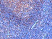
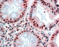
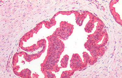
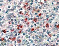 IHC (Immunohiostchemistry)
(Staining of paraffin embedded Human Spleen. Steamed antigen retrieval with citrate buffer pH 6, AP-staining.)
IHC (Immunohiostchemistry)
(Staining of paraffin embedded Human Spleen. Steamed antigen retrieval with citrate buffer pH 6, AP-staining.)
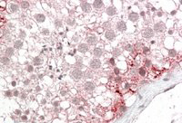
CD14, Polyclonal Antibody (Cat# AAA61193)
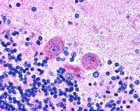 IHC (Immunohistochemistry)
((2.5ug/ml) staining of paraffin embedded Human Brain. Steamed antigen retrieval with citrate buffer pH 6, AP-staining.)
IHC (Immunohistochemistry)
((2.5ug/ml) staining of paraffin embedded Human Brain. Steamed antigen retrieval with citrate buffer pH 6, AP-staining.)
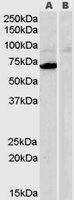
SNX26/TCGAP, Polyclonal Antibody (Cat# AAA61195)
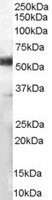
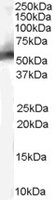
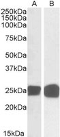
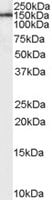
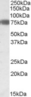
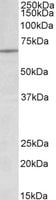
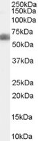
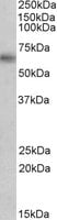
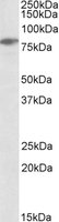 WB (Western Blot)
(AAA61214 (0.3ug/ml) staining of Jurkat lysate (35ug protein in RIPA buffer). Primary incubation was 1 hour. Detected by chemiluminescence.)
WB (Western Blot)
(AAA61214 (0.3ug/ml) staining of Jurkat lysate (35ug protein in RIPA buffer). Primary incubation was 1 hour. Detected by chemiluminescence.)
SLP76/LCP2, Polyclonal Antibody (Cat# AAA61214)
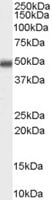
 WB (Western Blot)
(HEK293 overexpressing MAOA (RC207276) and probed (mock transfection in first lane), tested by Origene.)
WB (Western Blot)
(HEK293 overexpressing MAOA (RC207276) and probed (mock transfection in first lane), tested by Origene.)
Monoamine Oxidase A, Polyclonal Antibody (Cat# AAA61222)
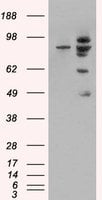
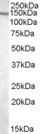
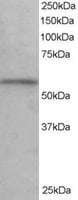
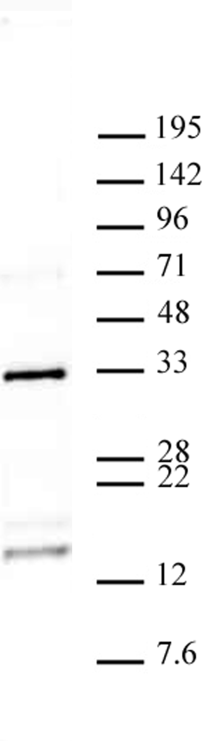
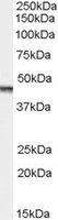
 WB (Western Blot)
((0. 05ug/ml) staining of Human Cerebellum (A), Human Frontal Cortex (B), Human Hippocampus (C) Mouse Fetal Brain (D), Mouse Brain (E) and Rat Brain (F) lysates (35ug protein in RIPA buffer). Primary incubation was 1 hour. Detected by chemiluminescence.)
WB (Western Blot)
((0. 05ug/ml) staining of Human Cerebellum (A), Human Frontal Cortex (B), Human Hippocampus (C) Mouse Fetal Brain (D), Mouse Brain (E) and Rat Brain (F) lysates (35ug protein in RIPA buffer). Primary incubation was 1 hour. Detected by chemiluminescence.)
PARK7/DJ-1, Polyclonal Antibody (Cat# AAA60984)
FENS1/WDFY1, Polyclonal Antibody (Cat# AAA60985)
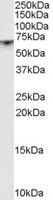
 WB (Western Blot)
(Staining (0.1ug/ml) of Jurkat lysate (RIPA buffer, 35ug total protein per lane). Primary incubated for 1 hour. Detected by western blot using chemiluminescence.)
WB (Western Blot)
(Staining (0.1ug/ml) of Jurkat lysate (RIPA buffer, 35ug total protein per lane). Primary incubated for 1 hour. Detected by western blot using chemiluminescence.)
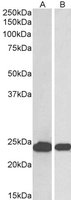
Nucleophosmin/NPM1, Polyclonal Antibody (Cat# AAA60987)
 IHC (Immunohistochemistry)
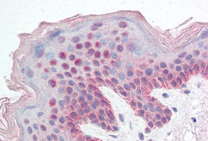
(AAA60991 (5ug/ml) staining of paraffin embedded Human Skin. Steamed antigen retrieval with citrate buffer pH 6, AP-staining.)
IHC (Immunohistochemistry)
(AAA60991 (5ug/ml) staining of paraffin embedded Human Skin. Steamed antigen retrieval with citrate buffer pH 6, AP-staining.)
FBXL10/PCCX2, Polyclonal Antibody (Cat# AAA60991)
 Application Data
((0.5ug/ml) as the reporter with as the capture rabbit antibody (2.5ug/ml).)
Application Data
((0.5ug/ml) as the reporter with as the capture rabbit antibody (2.5ug/ml).)
FOXO4/MLLT7, Polyclonal Antibody (Cat# AAA60995)
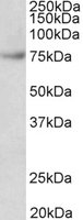
 WB (Western Blot)
((0.1ug/ml) staining of Rat Testis lysate (35ug protein in RIPA buffer). Primary incubation was 1 hour. Detected by chemiluminescence.)
WB (Western Blot)
((0.1ug/ml) staining of Rat Testis lysate (35ug protein in RIPA buffer). Primary incubation was 1 hour. Detected by chemiluminescence.)
Sall4, Polyclonal Antibody (Cat# AAA61009)
 WB (Western Blot)
(HEK293 overexpressing MDM2 (RC219518) and probed (mock transfection in first lane), tested by Origene.)
WB (Western Blot)
(HEK293 overexpressing MDM2 (RC219518) and probed (mock transfection in first lane), tested by Origene.)
MDM2, Polyclonal Antibody (Cat# AAA61023)
 WB (Western Blot)
((0.5ug/ml) staining of Rat Brain lysate (35ug protein in RIPA buffer). Primary incubation was 1 hour. Detected by chemiluminescence.)
WB (Western Blot)
((0.5ug/ml) staining of Rat Brain lysate (35ug protein in RIPA buffer). Primary incubation was 1 hour. Detected by chemiluminescence.)
PAK1, Polyclonal Antibody (Cat# AAA61032)
 Application Data
(1: 293 cell extract control; 2: 293 cell expressing gp41 (HXBc2))
Application Data
(1: 293 cell extract control; 2: 293 cell expressing gp41 (HXBc2))
gp41 (Clade B), Polyclonal Antibody (Cat# AAA62104)
HBsAg (Recombivax HB), Polyclonal Antibody (Cat# AAA62131)
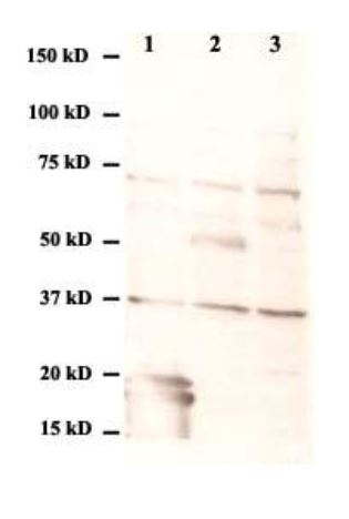 Application Data
(Western Blot:1. 293 cell expressing E (DIII) protein of West Nile virus2. 293 cell expressing E protein of West Nile virus3. 293 cell extract control)
Application Data
(Western Blot:1. 293 cell expressing E (DIII) protein of West Nile virus2. 293 cell expressing E protein of West Nile virus3. 293 cell extract control)
E (West Nile Virus), Polyclonal Antibody (Cat# AAA62132)
M2 (A/northern shoverl/Mississippi/11OS145/2011)(H7N9), Polyclonal Antibody (Cat# AAA62133)
 WB (Western Blot)
((0.3ug/ml) staining of Mouse Liver lysate (35ug protein in RIPA buffer). Primary incubation was 1 hour. Detected by chemiluminescence.)
WB (Western Blot)
((0.3ug/ml) staining of Mouse Liver lysate (35ug protein in RIPA buffer). Primary incubation was 1 hour. Detected by chemiluminescence.)
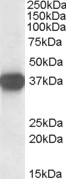
AGTR1/AT1, Polyclonal Antibody (Cat# AAA61020)
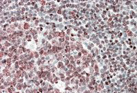
 IHC (Immunohistochemistry)
((0.3ug/ml) staining of paraffin embedded Human Tonsil. Microwaved antigen retrieval with citrate buffer pH6, HRP-staining.)
IHC (Immunohistochemistry)
((0.3ug/ml) staining of paraffin embedded Human Tonsil. Microwaved antigen retrieval with citrate buffer pH6, HRP-staining.)
GRB2, Polyclonal Antibody (Cat# AAA61154)
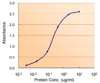
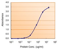
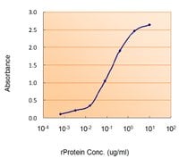 Application Data
((1.5ug/ml) as the reporter with as the capture rabbit antibody (2.5ug/ml).)
Application Data
((1.5ug/ml) as the reporter with as the capture rabbit antibody (2.5ug/ml).)
VHL, Polyclonal Antibody (Cat# AAA61220)
 WB (Western Blot)
((0.5ug/ml) staining of Human Breast Cancer lysate (35ug protein in RIPA buffer). Primary incubation was 1 hour. Detected by chemiluminescence.)
WB (Western Blot)
((0.5ug/ml) staining of Human Breast Cancer lysate (35ug protein in RIPA buffer). Primary incubation was 1 hour. Detected by chemiluminescence.)
MMP12, Polyclonal Antibody (Cat# AAA61478)
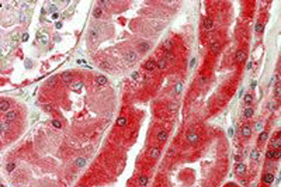
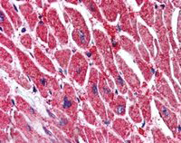
 IHC (Immunohistochemistry)
((3.8ug/ml) staining of paraffin embedded Human Heart. Steamed antigen retrieval with citrate buffer pH 6, AP-staining.)
IHC (Immunohistochemistry)
((3.8ug/ml) staining of paraffin embedded Human Heart. Steamed antigen retrieval with citrate buffer pH 6, AP-staining.)
Desmoplakin, Polyclonal Antibody (Cat# AAA61506)
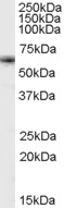
 WB (Western Blot)
((1ug/ml) staining of Human Brain lysate (35ug protein in RIPA buffer). Primary incubation was 1 hour. Detected by chemiluminescence.)
WB (Western Blot)
((1ug/ml) staining of Human Brain lysate (35ug protein in RIPA buffer). Primary incubation was 1 hour. Detected by chemiluminescence.)
Leptin Receptor, Polyclonal Antibody (Cat# AAA61224)
 IHC (Immunohiostchemistry)
((2.5ug/ml) staining of paraffin embedded Human Colon. Steamed antigen retrieval with citrate buffer pH 6, AP-staining.)
IHC (Immunohiostchemistry)
((2.5ug/ml) staining of paraffin embedded Human Colon. Steamed antigen retrieval with citrate buffer pH 6, AP-staining.)
MBD2, Polyclonal Antibody (Cat# AAA61255)
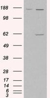
 DB (Dot Blot)
(Histone H3 monomethyl Lys23 pAb tested by dot blot analysis. Dot blot analysis was used to confirm the specificity of Histone H3 monomethyl Lys23 pAb for monomethyl Lys23 histone H3. Methylated peptides corresponding to the immunogen and related sequences derived from histone H3 were spotted onto PVDF and probed with the antibody at 1:5,000. The amount of peptide (picomoles) spotted is indicated next to each row. Top row: Lane 1: Unmod H3 aa 1-10 peptide. Lane 2: Monomethyl lysine 4. Lane 3: Dimethyl lysine 4. Lane 4: Trimethyl lysine 4. Lane 5: Unmodified H3 aa 6-22 peptide. Lane 6: Monomethyl lysine 9. Lane 7: Dimethyl lysine 9. Lane 8: Trimethyl lysine 9. Bottom row: Lane 1: Dimethyl lysine 14. Lane 2: Monomethyl lysine 18. Lane 3: Dimethyl lysine 18. Lane 4: Trimethyl lysine 18. Lane 5: Unmod H3 aa 18-27 peptide. Lane 6: Monomethyl lysine 23. Lane 7: Dimethyl lysine 23. Lane 8: Trimethyl lysine 23. Lane 9: Unmod H3 aa 22-32 peptide. Lane 10: Monomethyl lysine 27. Lane 11: Dimethyl lysine 27. Lane 12: Trimethyl lysine 27.)
DB (Dot Blot)
(Histone H3 monomethyl Lys23 pAb tested by dot blot analysis. Dot blot analysis was used to confirm the specificity of Histone H3 monomethyl Lys23 pAb for monomethyl Lys23 histone H3. Methylated peptides corresponding to the immunogen and related sequences derived from histone H3 were spotted onto PVDF and probed with the antibody at 1:5,000. The amount of peptide (picomoles) spotted is indicated next to each row. Top row: Lane 1: Unmod H3 aa 1-10 peptide. Lane 2: Monomethyl lysine 4. Lane 3: Dimethyl lysine 4. Lane 4: Trimethyl lysine 4. Lane 5: Unmodified H3 aa 6-22 peptide. Lane 6: Monomethyl lysine 9. Lane 7: Dimethyl lysine 9. Lane 8: Trimethyl lysine 9. Bottom row: Lane 1: Dimethyl lysine 14. Lane 2: Monomethyl lysine 18. Lane 3: Dimethyl lysine 18. Lane 4: Trimethyl lysine 18. Lane 5: Unmod H3 aa 18-27 peptide. Lane 6: Monomethyl lysine 23. Lane 7: Dimethyl lysine 23. Lane 8: Trimethyl lysine 23. Lane 9: Unmod H3 aa 22-32 peptide. Lane 10: Monomethyl lysine 27. Lane 11: Dimethyl lysine 27. Lane 12: Trimethyl lysine 27.)
Histone H3K23me1, Polyclonal Antibody (Cat# AAA59870)
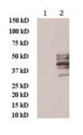
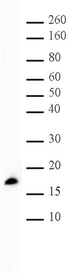
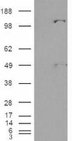
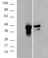
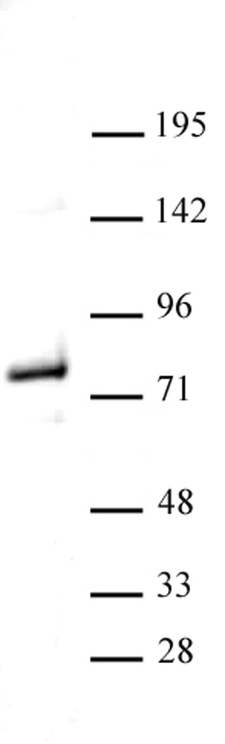 WB (Western Blot)
(Western blot of Menin antibody. Nuclear extract of HEK293 cells (20 ?g) probed with Menin antibody (1:1,000).)
WB (Western Blot)
(Western blot of Menin antibody. Nuclear extract of HEK293 cells (20 ?g) probed with Menin antibody (1:1,000).)
Menin, Polyclonal Antibody (Cat# AAA59962)
GP (Zarie Ebolavirus/Mayinga 1976), Polyclonal Antibody (Cat# AAA62130)
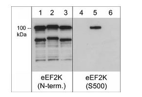
 WB (Western Blot)
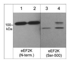
(Western blot image of C2C12 untreated (lanes 1 & 3) or treated with calyculin A (100nM) for 30 min. (lanes 2 & 4). The blot was probed with anti-eEF2K (N-terminus) (lanes 1 & 2) and anti-eEF2K (Ser-500) (lanes 3 & 4).)
WB (Western Blot)
(Western blot image of C2C12 untreated (lanes 1 & 3) or treated with calyculin A (100nM) for 30 min. (lanes 2 & 4). The blot was probed with anti-eEF2K (N-terminus) (lanes 1 & 2) and anti-eEF2K (Ser-500) (lanes 3 & 4).)
eEF2K, Polyclonal Antibody (Cat# AAA71634)
 WB (Western Blot)
((1ug/ml) staining of Human Bone Marrow lysate (35ug protein in RIPA buffer). Primary incubation was 1 hour. Detected by chemiluminescence.)
WB (Western Blot)
((1ug/ml) staining of Human Bone Marrow lysate (35ug protein in RIPA buffer). Primary incubation was 1 hour. Detected by chemiluminescence.)
DLX5, Polyclonal Antibody (Cat# AAA61453)

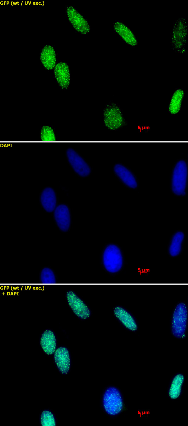
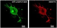 IF (Immunofluorescence)
(HEK293 overexpressing Human srGAP2 and probed at 2.5ug/ml in the right panel. Data kindly provided by Ms. Ya-Jing Mi and Dr. Wei-Lin Jin, Institute of Neurosciences, Shanghai Jiao Tong University.)
IF (Immunofluorescence)
(HEK293 overexpressing Human srGAP2 and probed at 2.5ug/ml in the right panel. Data kindly provided by Ms. Ya-Jing Mi and Dr. Wei-Lin Jin, Institute of Neurosciences, Shanghai Jiao Tong University.)
SRGAP2, Polyclonal Antibody (Cat# AAA61469)
 IHC (Immunohiostchemistry)
((3.8ug/ml) staining of paraffin embedded Human Testis. Steamed antigen retrieval with citrate buffer pH 6, AP-staining.)
IHC (Immunohiostchemistry)
((3.8ug/ml) staining of paraffin embedded Human Testis. Steamed antigen retrieval with citrate buffer pH 6, AP-staining.)
ABCD3, Polyclonal Antibody (Cat# AAA61395)
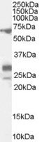
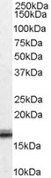
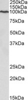
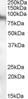
 WB (Western Blot)
((0.3ug/ml) staining of Human Adipose lysate (35ug protein in RIPA buffer). Primary incubation was 1 hour. Detected by chemiluminescence.)
WB (Western Blot)
((0.3ug/ml) staining of Human Adipose lysate (35ug protein in RIPA buffer). Primary incubation was 1 hour. Detected by chemiluminescence.)
EBF1/COE1, Polyclonal Antibody (Cat# AAA61403)
 IHC (Immunohiostchemistry)
((5ug/ml) staining of paraffin embedded Human Human Tonsil. Steamed antigen retrieval with citrate buffer pH 6, AP-staining.)
IHC (Immunohiostchemistry)
((5ug/ml) staining of paraffin embedded Human Human Tonsil. Steamed antigen retrieval with citrate buffer pH 6, AP-staining.)
Fubp1, Polyclonal Antibody (Cat# AAA61654)
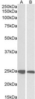
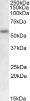
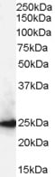
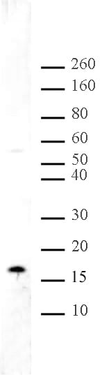
 WB (Western Blot)
((0.2ug/ml) staining of Jurkat lysate (35ug protein in RIPA buffer). Primary incubation was 1 hour. Detected by chemiluminescence.)
WB (Western Blot)
((0.2ug/ml) staining of Jurkat lysate (35ug protein in RIPA buffer). Primary incubation was 1 hour. Detected by chemiluminescence.)
Lamin B1, Polyclonal Antibody (Cat# AAA61657)
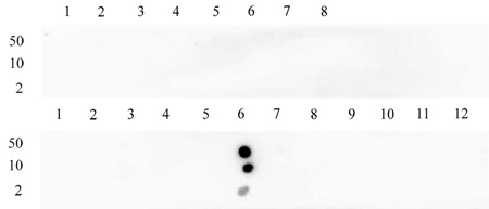
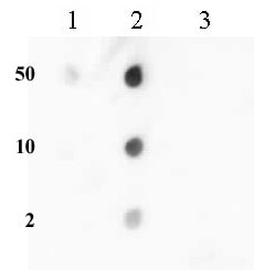
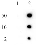
 DB (Dot Blot)
(Histone H3K56me1 antibody (pAb) tested by dot blot analysis. Dot blot analysis was used to confirm the specificity of Histone H3 monomethyl Lys56 pAb for monomethyl Lys56 histone H3. Methylated peptides corresponding to the immunogen and related peptides were spotted onto PVDF and probed with at 1:10,000. The amount of peptide (picomoles) spotted is indicated next to each row. Lane 1: Unmodified lysine 56 peptide. Lane 2: Monomethyl lysine 56 peptide. Lane 3: Dimethyl lysine 56 peptide. Lane 4: Trimethyl lysine 56 peptide. No detection of peptides (unmodified, mono-, di-, or trimethylated) corresponding to lysine 4, lysine 9 or lysine 27 was observed with this antibody.)
DB (Dot Blot)
(Histone H3K56me1 antibody (pAb) tested by dot blot analysis. Dot blot analysis was used to confirm the specificity of Histone H3 monomethyl Lys56 pAb for monomethyl Lys56 histone H3. Methylated peptides corresponding to the immunogen and related peptides were spotted onto PVDF and probed with at 1:10,000. The amount of peptide (picomoles) spotted is indicated next to each row. Lane 1: Unmodified lysine 56 peptide. Lane 2: Monomethyl lysine 56 peptide. Lane 3: Dimethyl lysine 56 peptide. Lane 4: Trimethyl lysine 56 peptide. No detection of peptides (unmodified, mono-, di-, or trimethylated) corresponding to lysine 4, lysine 9 or lysine 27 was observed with this antibody.)
Histone H3K56me1, Polyclonal Antibody (Cat# AAA59855)
 IHC (Immunohiostchemistry)
(AAA61713 (5ug/ml) staining of paraffin embedded Human Prostate. Steamed antigen retrieval with citrate buffer Ph 6, AP-staining)
IHC (Immunohiostchemistry)
(AAA61713 (5ug/ml) staining of paraffin embedded Human Prostate. Steamed antigen retrieval with citrate buffer Ph 6, AP-staining)
CDC48/ VCP/YDL126C, Polyclonal Antibody (Cat# AAA61713)
Expected from sequence similarity: Saccharomyces cerevisiae S288c, Human, Mouse, Rat, Dog, Cow


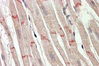
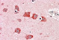
 IHC (Immunohistochemisry)
((5ug/ml) staining of paraffin embedded Mouse Cerebral Cortex. Steamed antigen retrieval with citrate buffer pH 6, DAB-staining.)
IHC (Immunohistochemisry)
((5ug/ml) staining of paraffin embedded Mouse Cerebral Cortex. Steamed antigen retrieval with citrate buffer pH 6, DAB-staining.)
contactin 1, Polyclonal Antibody (Cat# AAA61735)
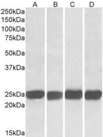
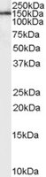
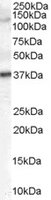
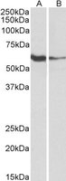 WB (Western Blot)
((1ug/ml) staining of Human Tonsil (A) and Daudi (B) lysates (35ug protein in RIPA buffer). Primary incubation was 1 hour. Detected by chemiluminescence.)
WB (Western Blot)
((1ug/ml) staining of Human Tonsil (A) and Daudi (B) lysates (35ug protein in RIPA buffer). Primary incubation was 1 hour. Detected by chemiluminescence.)
FCRL2, Polyclonal Antibody (Cat# AAA61765)
 WB (Western Blot)
(HEK293 overexpressing Neuregulin3 (RC207955) and probed (mock transfection in first lane), tested by Origene.)
WB (Western Blot)
(HEK293 overexpressing Neuregulin3 (RC207955) and probed (mock transfection in first lane), tested by Origene.)
Neuroligin 3, Polyclonal Antibody (Cat# AAA60968)
 WB (Western Blot)
(HEK293 overexpressing Human Pyruvate Carboxylase (RC221895) and probed (mock transfection in first lane), tested by Origene.)
WB (Western Blot)
(HEK293 overexpressing Human Pyruvate Carboxylase (RC221895) and probed (mock transfection in first lane), tested by Origene.)
Pyruvate Carboxylase, Polyclonal Antibody (Cat# AAA60973)
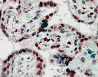 IHC (Immunohiostchemistry)
((2.5ug/ml) staining of paraffin embedded Human Placenta. Steamed antigen retrieval with citrate buffer pH 6, AP-staining.)
IHC (Immunohiostchemistry)
((2.5ug/ml) staining of paraffin embedded Human Placenta. Steamed antigen retrieval with citrate buffer pH 6, AP-staining.)
CBX1/HP1-Beta, Polyclonal Antibody (Cat# AAA60843)
 IHC (Immunohiostchemistry)
((5ug/ml) staining of paraffin embedded Human Heart. Steamed antigen retrieval with citrate buffer pH 6, AP-staining.)
IHC (Immunohiostchemistry)
((5ug/ml) staining of paraffin embedded Human Heart. Steamed antigen retrieval with citrate buffer pH 6, AP-staining.)
KPNA3/IPOA4, Polyclonal Antibody (Cat# AAA60860)
 IHC (Immunohiostchemistry)
((3.8ug/ml) staining of paraffin embedded Human Testis. Steamed antigen retrieval with citrate buffer pH 6, AP-staining.)
IHC (Immunohiostchemistry)
((3.8ug/ml) staining of paraffin embedded Human Testis. Steamed antigen retrieval with citrate buffer pH 6, AP-staining.)
Galanin Receptor 3, Polyclonal Antibody (Cat# AAA60882)
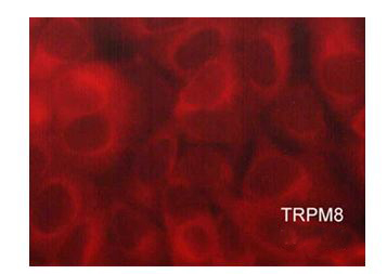 ICC (Immunocytochemistry)
(Immunocytochemical labeling of TRPM8 in paraformaldehyde fixed and NP-40 permeabilized MCF-7 cells. The cells were labeled with rabbit polyclonal anti-TRPM8 (TP5701). The antibody was detected using goat anti-rabbit DyLight 594.)
ICC (Immunocytochemistry)
(Immunocytochemical labeling of TRPM8 in paraformaldehyde fixed and NP-40 permeabilized MCF-7 cells. The cells were labeled with rabbit polyclonal anti-TRPM8 (TP5701). The antibody was detected using goat anti-rabbit DyLight 594.)
TRPM8, Polyclonal Antibody (Cat# AAA71723)
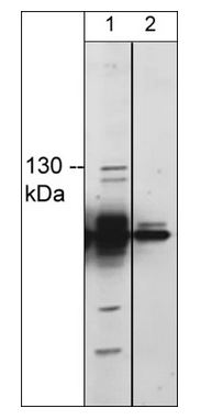
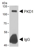 WB (Western Blot)
(HEK293 lysate overexpressing Human DYKDDDDK-tagged PKD1 was used to immunoprecipitate PKD1 with 2ug The precipitate was subsequently probed in Western blot using at 1ug/ml. The secondary anti-goat picks up the heavy chain of used for the immunoprecipitation (annotated as IgG). Data kindly obtained from Dr Peter Storz, Mayo Clinic, USA)
WB (Western Blot)
(HEK293 lysate overexpressing Human DYKDDDDK-tagged PKD1 was used to immunoprecipitate PKD1 with 2ug The precipitate was subsequently probed in Western blot using at 1ug/ml. The secondary anti-goat picks up the heavy chain of used for the immunoprecipitation (annotated as IgG). Data kindly obtained from Dr Peter Storz, Mayo Clinic, USA)
PRKD1, Polyclonal Antibody (Cat# AAA61794)
Tested: Human
Expected from sequence similarity: Human, Mouse, Rat, Pig
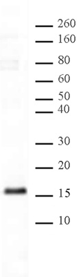
 DB (Dot Blot)
(Histone H3K36me2 antibody tested by dot blot analysis. Dot blot analysis was used to confirm the specificity of Histone H3 dimethyl Lys36 antibody for histone H3 dimethyl Lys36. Methylated peptides corresponding to the immunogen were spotted onto PVDF and probed with the antibody at 1:5,000. The amount of peptide (picomoles) spotted is indicated next to each row. Lane 1: unmodified Lys36 peptide. Lane 2: monomethyl-Lys36 peptide. Lane 3: dimethyl-Lys36 peptide. Lane 3: trimethyl- Lys36 peptide.)
DB (Dot Blot)
(Histone H3K36me2 antibody tested by dot blot analysis. Dot blot analysis was used to confirm the specificity of Histone H3 dimethyl Lys36 antibody for histone H3 dimethyl Lys36. Methylated peptides corresponding to the immunogen were spotted onto PVDF and probed with the antibody at 1:5,000. The amount of peptide (picomoles) spotted is indicated next to each row. Lane 1: unmodified Lys36 peptide. Lane 2: monomethyl-Lys36 peptide. Lane 3: dimethyl-Lys36 peptide. Lane 3: trimethyl- Lys36 peptide.)
Histone H3K36me2, Polyclonal Antibody (Cat# AAA59806)
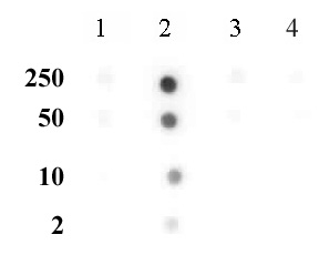
 DB (Dot Blot)
(Histone H2AQ104me1 (pAb) tested by dot blot analysis. Dot blot analysis was used to confirm the specificity of Histone H2AQ104me1 pAb for monomethyl-glu104 of histone H2A. Decreasing amounts of modified and unmodified peptides were spotted onto PVDF and probed with the antibody at a dilution of 1:5,000. Lane 1: Unmodified glutamine 104 of H2A peptide. Lane 2: Monomethylated glutamine 104 of H2A peptide.)
DB (Dot Blot)
(Histone H2AQ104me1 (pAb) tested by dot blot analysis. Dot blot analysis was used to confirm the specificity of Histone H2AQ104me1 pAb for monomethyl-glu104 of histone H2A. Decreasing amounts of modified and unmodified peptides were spotted onto PVDF and probed with the antibody at a dilution of 1:5,000. Lane 1: Unmodified glutamine 104 of H2A peptide. Lane 2: Monomethylated glutamine 104 of H2A peptide.)
Histone H2AQ104me1, Polyclonal Antibody (Cat# AAA60054)
 IHC (Immunohiostchemistry)
((5ug/ml) staining of paraffin embedded Human Cortex. Steamed antigen retrieval with citrate buffer pH 6, AP-staining.)
IHC (Immunohiostchemistry)
((5ug/ml) staining of paraffin embedded Human Cortex. Steamed antigen retrieval with citrate buffer pH 6, AP-staining.)
DKK3/REIC, Polyclonal Antibody (Cat# AAA61287)
 Application Data
((0.5ug/ml) as the reporter with as the capture rabbit antibody (2.5ug/ml).)
Application Data
((0.5ug/ml) as the reporter with as the capture rabbit antibody (2.5ug/ml).)
MAOB, Polyclonal Antibody (Cat# AAA61069)
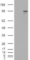
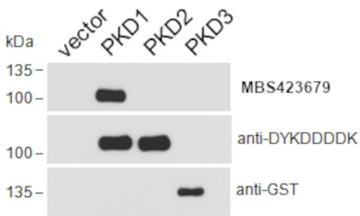
 WB (Western Blot)
(HEK293 overexpressing Aurora kinase A (RC212018) with C-terminal tag (DYKDDDDK) and probed with anti-DYKDDDDK in the left panel and in the right panel (vector-only transfection in first and fourth lanes).)
WB (Western Blot)
(HEK293 overexpressing Aurora kinase A (RC212018) with C-terminal tag (DYKDDDDK) and probed with anti-DYKDDDDK in the left panel and in the right panel (vector-only transfection in first and fourth lanes).)
Aurora kinase A/AURKA, Polyclonal Antibody (Cat# AAA61082)
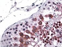
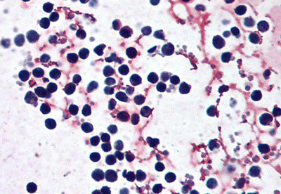 IHC (Immunohistochemistry)
(AAA61488 (5ug/ml) staining of paraffin embedded Human peripheral blood leukocytes. Steamed antigen retrieval with citrate buffer pH 6, AP-staining.)
IHC (Immunohistochemistry)
(AAA61488 (5ug/ml) staining of paraffin embedded Human peripheral blood leukocytes. Steamed antigen retrieval with citrate buffer pH 6, AP-staining.)
Cryopirin/NALP3/NLRP3, Polyclonal Antibody (Cat# AAA61488)
Expected from sequence similarity: Human
What are Polyclonal Antibodies?
Polyclonal antibodies are antibodies that come from multiple B cell clones of a host animal. The typical hosts used for the majority of polyclonal antibody production are rabbits, goats, sheep, and donkeys. These polyclonal antibodies, once having identified their target, will bind to different epitopes located at different regions or sequences on the same protein/antigen. This ability to bind multiple epitopes is what makes polyclonal antibodies highly sensitive, as explained in our detailed guide on polyclonal antibodies and why they matter.
As a result, they are ideal at locating and binding to the target, even if the target is in very low concentrations (due to many different antibodies being able to bind to the same target molecule, which allows for significant amplification of a downstream signal).
Polyclonal antibodies are typically produced by injecting an antigen into a host animal, which causes the animal’s immune system to attack the foreign antigen by mass generating antibodies against it. After a period of time, serum is collected from the animal and purified using physicochemical fractionation, class-specific affinity purification, and/or antigen-affinity purification.
Key Uses of Polyclonal Antibodies
- Western Blotting: This method is used to find specific proteins in biological samples after separating them by size.
- Immunohistochemistry: IHC helps visualize the location of proteins in tissue sections using various staining techniques.
- ELISA: (Enzyme-Linked Immunosorbent Assay) is typically used to identify specific protein quantities in a sample. ELISAs can be either “Quantitative” or “Qualitative”.
- Flow Cytometry: technique that identifies and measures the specific protein on the surface or inside the cells in a fluid suspension.
- Immunoprecipitation: IP isolates and studies a specific protein from a complex mixture using antibodies.
Why Buy Polyclonal Antibodies from AAA Biotech?
1. Ideal for Various Applications
Our antibodies are generally going to be validated for use in multiple types of assays, including ELISA, Western Blotting, Immunohistochemistry, Immunoprecipitation, amongst others. They are ideal for a wide range of research applications.
2. Rigorous Quality Control
All of the antibodies in our catalog undergo strict quality testing to ensure specificity, sensitivity, and consistent performance. We are confident in the ability of our antibodies to provide you with accurate results.
3. Wide Assortment of Antibodies
Antibodies in our catalog can be found for both common and exotic species, and these antibodies are also available in both conjugated and recombinant forms to suit many diverse experimental needs.
4. Highly Purified
Our antibodies are available in purified forms with over 85% purity, as confirmed by SDS-PAGE. They are also available with tags such as His, Flag, GST, or MBP. We cater to customers worldwide.
FAQ
1. How are polyclonal antibodies produced?
Traditionally, polyclonal antibodies are produced by injecting an antigen into a host animal (such as a rabbit or goat), which then triggers an immune response from the host animal. The animal’s B cells produce antibodies that will recognize different parts of the injected antigen. These antibodies are then collected from the animal’s blood and purified for use.
2. How do polyclonal antibodies differ from monoclonal antibodies?
Polyclonal antibodies are a mix of antibodies that bind to different locations (epitopes) of the same antigen, while monoclonal antibodies are identical and bind to just one specific epitope. This makes polyclonal antibodies more versatile and better at detecting proteins that may be present in low quantities or in altered/modified forms.
3. How should I store polyclonal antibodies?
Polyclonal antibodies should be stored at 4°C for short-term use (up to a few weeks) and at -20°C or -80°C for long-term storage. Avoid repeated freeze-thaw cycles by dividing them into small aliquots. Always check the datasheet for specific storage instructions.