Loading...

Skip to main content

Call us on + 1 (800) 604-9114 for more information about our products

Have a question or looking for a specific datasheet Manual/COA/MSDS?
Submit a Question

Interested to get a quote about our products?
Request a Quote

Polyclonal Antibodies

Viewing 2100-2150 of 3252 product results


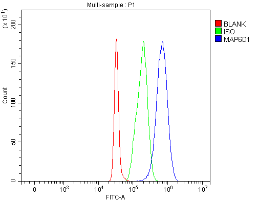
FCM (Flow Cytometry) (Figure 8. Flow Cytometry analysis of HeLa cells using anti-MRPS22 antibody (AAA19618).Overlay histogram showing HeLa cells stained with AAA19618 (Blue line). The cells were blocked with 10% normal goat serum. And then incubated with rabbit anti-MRPS22 Antibody (AAA19618, 1 ug/1x10^6 cells) for 30 min at 20 degree C. DyLight488 conjugated goat anti-rabbit IgG was used as secondary antibody for 30 minutes at 20 degree C. Isotype control antibody (Green line) was rabbit IgG (1 ug/1x10^6) used under the same conditions. Unlabelled sample (Red line) was also used as a control.)

MRPS22, Polyclonal Antibody (Cat# AAA19618)

Full Name
Anti-MRPS22 Antibody Picoband
Gene Names
MRPS22; GIBT; GK002; C3orf5; COXPD5; RPMS22; MRP-S22
Reactivity
Human
Applications
Western Blot (WB), Immunohistochemistry (IHC), Immunocytochemistry (ICC), Immunofluorescence (IF), Flow Cytometry (FC/FACS), ELISA (EIA)
Purity
Immunogen affinity purified.
Pricing
FCM (Flow Cytometry) (Figure 9. Flow Cytometry analysis of HepG2 cells using anti-DUSP6 antibody (AAA19449).Overlay histogram showing HepG2 cells stained with AAA19449 (Blue line). The cells were blocked with 10% normal goat serum. And then incubated with rabbit anti-DUSP6 Antibody (AAA19449, 1 ug/1x10^6 cells) for 30 min at 20 degree C. DyLight488 conjugated goat anti-rabbit IgG was used as secondary antibody for 30 minutes at 20 degree C. Isotype control antibody (Green line) was rabbit IgG (1 ug/1x10^6) used under the same conditions. Unlabelled sample (Red line) was also used as a control.)

DUSP6, Polyclonal Antibody (Cat# AAA19449)

Full Name
Anti-DUSP6 Antibody Picoband
Gene Names
DUSP6; MKP3; PYST1
Reactivity
Human, Mouse, Rat
Applications
ELISA (EIA), Flow Cytometry (FC/FACS), Immunofluorescence (IF), Immunohistochemistry (IHC), Immunocytochemistry (ICC), Western Blot (WB)
Purity
Immunogen affinity purified.
Pricing
FCM (Flow Cytometry) (Figure 11. Flow Cytometry analysis of Jurkat cells using anti-G3BP/G3BP1 antibody (AAA19450).Overlay histogram showing Jurkat cells stained with AAA19450 (Blue line). The cells were blocked with 10% normal goat serum. And then incubated with rabbit anti-G3BP/G3BP1 Antibody (AAA19450, 1 ug/1x10^6 cells) for 30 min at 20 degree C. DyLight488 conjugated goat anti-rabbit IgG was used as secondary antibody for 30 minutes at 20 degree C. Isotype control antibody (Green line) was rabbit IgG (1 ug/1x10^6) used under the same conditions. Unlabelled sample (Red line) was also used as a control.)

G3BP/G3BP1, Polyclonal Antibody (Cat# AAA19450)

Full Name
Anti-G3BP/G3BP1 Antibody Picoband
Gene Names
G3BP1; G3BP; HDH-VIII
Reactivity
Human
Applications
Western Blot (WB), Immunohistochemistry (IHC), Immunocytochemistry (ICC), Immunofluorescence (IF), Flow Cytometry (FC/FACS)
Purity
Immunogen affinity purified.
Pricing
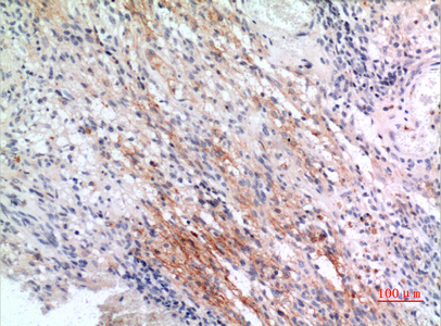
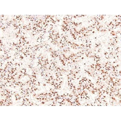
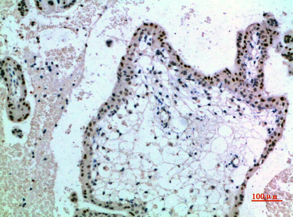
IHC (Immunohistochemistry) (Figure 7. IHC analysis of COL6A1 using anti-COL6A1 antibody (AAA19451).COL6A1 was detected in a paraffin-embedded section of human placenta tissue. Heat mediated antigen retrieval was performed in EDTA buffer (pH 8.0, epitope retrieval solution). The tissue section was blocked with 10% goat serum. The tissue section was then incubated with 2 ug/ml rabbit anti-COL6A1 Antibody (AAA19451) overnight at 4 degree C. Biotinylated goat anti-rabbit IgG was used as secondary antibody and incubated for 30 minutes at 37 degree C. The tissue section was developed using Strepavidin-Biotin-Complex (SABC) with DAB as the chromogen.)

COL6A1, Polyclonal Antibody (Cat# AAA19451)

Full Name
Anti-COL6A1 Antibody Picoband
Gene Names
COL6A1; OPLL
Reactivity
Human
Applications
Western Blot (WB), Immunohistochemistry (IHC), ELISA (EIA)
Purity
Immunogen affinity purified.
Pricing
FCM (Flow Cytometry) (Figure 10. Flow Cytometry analysis of SiHa cells using anti-SFPQ antibody (AAA19452).Overlay histogram showing SiHa cells stained with AAA19452 (Blue line). The cells were blocked with 10% normal goat serum. And then incubated with rabbit anti-SFPQ Antibody (AAA19452, 1 ug/1x10^6 cells) for 30 min at 20 degree C. DyLight488 conjugated goat anti-rabbit IgG was used as secondary antibody for 30 minutes at 20 degree C. Isotype control antibody (Green line) was rabbit IgG (1 ug/1x10^6) used under the same conditions. Unlabelled sample (Red line) was also used as a control.)

SFPQ, Polyclonal Antibody (Cat# AAA19452)

Full Name
Anti-SFPQ Antibody Picoband
Gene Names
SFPQ; PSF; POMP100; PPP1R140
Reactivity
Human, Mouse, Rat
Applications
Western Blot (WB), Immunohistochemistry (IHC), Immunocytochemistry (ICC), Immunofluorescence (IF), Flow Cytometry (FC/FACS)
Purity
Immunogen affinity purified.
Pricing

Chlamydia trachomatis, Polyclonal Antibody (Cat# AAA14333)

Full Name
Chlamydia trachomatis antibody
Reactivity
To be determined by end-user.
Applications
ELISA (EIA), Immunofluorescence (IF), Immunohistochemistry (IHC), Western Blot (WB).
Purity
> 95% pure
Pricing
FCM (Flow Cytometry) (Figure 7. Flow Cytometry analysis of HeLa cells using anti-delta 1 Catenin/CAS/CTNND1 antibody (AAA19453).Overlay histogram showing HeLa cells stained with AAA19453 (Blue line). The cells were blocked with 10% normal goat serum. And then incubated with rabbit anti-delta 1 Catenin/CAS/CTNND1 Antibody (AAA19453, 1 ug/1x10^6 cells) for 30 min at 20 degree C. DyLight488 conjugated goat anti-rabbit IgG was used as secondary antibody for 30 minutes at 20 degree C. Isotype control antibody (Green line) was rabbit IgG (1 ug/1x10^6) used under the same conditions. Unlabelled sample (Red line) was also used as a control.)

delta 1 Catenin/CAS/CTNND1, Polyclonal Antibody (Cat# AAA19453)

Full Name
Anti-delta 1 Catenin/CAS/CTNND1 Antibody Picoband
Gene Names
CTNND1; CAS; p120; CTNND; P120CAS; P120CTN; p120(CAS); p120(CTN)
Reactivity
Human, Mouse, Rat
Applications
Western Blot (WB), Immunohistochemistry (IHC), Immunocytochemistry (ICC), Immunofluorescence (IF), Flow Cytometry (FC/FACS), ELISA (EIA)
Purity
Immunogen affinity purified.
Pricing
FCM (Flow Cytometry) (Figure 9. Flow Cytometry analysis of HeLa cells using anti-ACADM/MCAD antibody (AAA19455).Overlay histogram showing HeLa cells stained with AAA19455 (Blue line). The cells were blocked with 10% normal goat serum. And then incubated with rabbit anti-ACADM/MCAD Antibody (AAA19455, 1 ug/1x10^6 cells) for 30 min at 20 degree C. DyLight488 conjugated goat anti-rabbit IgG was used as secondary antibody for 30 minutes at 20 degree C. Isotype control antibody (Green line) was rabbit IgG (1 ug/1x10^6) used under the same conditions. Unlabelled sample (Red line) was also used as a control.)

ACADM/MCAD, Polyclonal Antibody (Cat# AAA19455)

Full Name
Anti-ACADM/MCAD Antibody Picoband
Gene Names
ACADM; MCAD; ACAD1; MCADH
Reactivity
Human, Mouse, Rat
Applications
Western Blot (WB), Immunohistochemistry (IHC), Immunocytochemistry (ICC), Immunofluorescence (IF), Flow Cytometry (FC/FACS)
Purity
Immunogen affinity purified.
Pricing
FCM (Flow Cytometry) (Figure 8. Flow Cytometry analysis of THP-1 cells using anti-NOX2/gp91phox/CYBB antibody (AAA19218).Overlay histogram showing THP-1 cells stained with AAA19218 (Blue line). The cells were blocked with 10% normal goat serum. And then incubated with rabbit anti-NOX2/gp91phox/CYBB Antibody (AAA19218, 1μg/1x106 cells) for 30 min at 20 degree C. DyLight®488 conjugated goat anti-rabbit IgG (5-10μg/1x106 cells) was used as secondary antibody for 30 minutes at 20 degree C. Isotype control antibody (Green line) was rabbit IgG (1μg/1x106) used under the same conditions. Unlabelled sample (Red line) was also used as a control.)

NOX2/gp91phox/CYBB, Polyclonal Antibody (Cat# AAA19218)

Full Name
Anti-NOX2/gp91phox/CYBB Antibody
Gene Names
CYBB; CGD; NOX2; AMCBX2; GP91-1; GP91PHOX; p91-PHOX; GP91-PHOX
Reactivity
Human
Applications
Western Blot (WB), Immunohistochemistry-Paraffin (IHC-P), Immunocytochemistry (ICC), Immunofluorescence (IF), Flow Cytometry (FC/FACS/FCM), Direct ELISA (EIA)
Purity
Immunogen affinity purified.
Pricing
FCM (Flow Cytometry) (Figure 11. Flow Cytometry analysis of U937 cells using anti-NSF antibody (AAA19222).Overlay histogram showing U937 cells stained with AAA19222 (Blue line). The cells were blocked with 10% normal goat serum. And then incubated with rabbit anti-NSF Antibody (AAA19222, 1μg/1x106 cells) for 30 min at 20 degree C. DyLight®488 conjugated goat anti-rabbit IgG (5-10μg/1x106 cells) was used as secondary antibody for 30 minutes at 20 degree C. Isotype control antibody (Green line) was rabbit IgG (1μg/1x106) used under the same conditions. Unlabelled sample (Red line) was also used as a control.)

NSF, Polyclonal Antibody (Cat# AAA19222)

Full Name
Anti-NSF Antibody
Gene Names
NSF; SKD2
Reactivity
Human, Mouse, Rat
Applications
Western Blot (WB), Immunohistochemistry-Paraffin (IHC-P), Immunocytochemistry (ICC), Immunofluorescence (IF), Flow Cytometry (FC/FACS/FCM), Direct ELISA (EIA)
Purity
Immunogen affinity purified.
Pricing
IF (Immunofluorescence) (Figure 6. IF analysis of SGK1 using anti- SGK1 antibody (AAA19225).SGK1 was detected in immunocytochemical section of A549 cells. Enzyme antigen retrieval was performed using IHC enzyme antigen retrieval reagent for 15 mins. The cells were blocked with 10% goat serum. And then incubated with 4μg/mL rabbit anti- SGK1 Antibody (AAA19225) overnight at 4 degree C. DyLight®488 Conjugated Goat Anti-Rabbit IgG was used as secondary antibody at 1:100 dilution and incubated for 30 minutes at 37 degree C. The section was counterstained with DAPI. Visualize using a fluorescence microscope and filter sets appropriate for the label used.)

SGK1, Polyclonal Antibody (Cat# AAA19225)

Full Name
Anti-SGK1 Antibody
Gene Names
SGK1; SGK
Reactivity
Human
Applications
Western Blot (WB), Immunohistochemistry-Paraffin (IHC-P), Immunocytochemistry (ICC), Immunofluorescence (IF), Flow Cytometry (FC/FACS/FCM)
Purity
Immunogen affinity purified.
Pricing
FCM (Flow Cytometry) (Figure 7. Flow Cytometry analysis of HELA cells using anti-MCU antibody (AAA19226).Overlay histogram showing HELA cells stained with AAA19226 (Blue line). The cells were blocked with 10% normal goat serum. And then incubated with rabbit anti-MCU Antibody (AAA19226, 1μg/1x106 cells) for 30 min at 20 degree C. DyLight®488 conjugated goat anti-rabbit IgG (5-10μg/1x106 cells) was used as secondary antibody for 30 minutes at 20 degree C. Isotype control antibody (Green line) was rabbit IgG (1μg/1x106) used under the same conditions. Unlabelled sample (Red line) was also used as a control.)

MCU, Polyclonal Antibody (Cat# AAA19226)

Full Name
Anti-MCU Antibody
Gene Names
MCU; C10orf42; CCDC109A
Reactivity
Human, Mouse, Rat, Monkey
Applications
Western Blot (WB), Immunohistochemistry-Paraffin (IHC-P), Flow Cytometry (FC/FACS/FCM), Direct ELISA (EIA)
Purity
Immunogen affinity purified.
Pricing
FCM (Flow Cytometry) (Figure 6. Flow Cytometry analysis of CACO-2 cells using anti-PLTP antibody (AAA19255).Overlay histogram showing CACO-2 cells stained with AAA19255 (Blue line). The cells were blocked with 10% normal goat serum. And then incubated with rabbit anti-PLTP Antibody (AAA19255, 1μg/1x106 cells) for 30 min at 20 degree C. DyLight®488 conjugated goat anti-rabbit IgG (5-10μg/1x106 cells) was used as secondary antibody for 30 minutes at 20 degree C. Isotype control antibody (Green line) was rabbit IgG (1μg/1x106) used under the same conditions. Unlabelled sample (Red line) was also used as a control.)

PLTP, Polyclonal Antibody (Cat# AAA19255)

Full Name
Anti-PLTP Antibody
Gene Names
PLTP; BPIFE; HDLCQ9
Reactivity
Human
Applications
Western Blot (WB), Immunohistochemistry-Paraffin (IHC-P), Immunocytochemistry (ICC), Immunofluorescence (IF), Flow Cytometry (FC/FACS/FCM), Direct ELISA (EIA)
Purity
Immunogen affinity purified.
Pricing
FCM (Flow Cytometry) (Figure 7. Flow Cytometry analysis of THP-1 cells using anti-PCBP2/hnRNP E2 antibody (AAA19259).Overlay histogram showing THP-1 cells stained with AAA19259 (Blue line). The cells were blocked with 10% normal goat serum. And then incubated with rabbit anti-PCBP2/hnRNP E2 Antibody (AAA19259, 1μg/1x106 cells) for 30 min at 20 degree C. DyLight®488 conjugated goat anti-rabbit IgG (5-10μg/1x106 cells) was used as secondary antibody for 30 minutes at 20 degree C. Isotype control antibody (Green line) was rabbit IgG (1μg/1x106) used under the same conditions. Unlabelled sample (Red line) was also used as a control.)

PCBP2/hnRNP E2, Polyclonal Antibody (Cat# AAA19259)

Full Name
Anti-PCBP2/hnRNP E2 Antibody
Gene Names
PCBP2; HNRPE2; HNRNPE2; hnRNP-E2
Reactivity
Human, Mouse, Rat
Applications
Western Blot (WB), Immunohistochemistry-Paraffin (IHC-P), Flow Cytometry (FC/FACS/FCM), Direct ELISA (EIA)
Purity
Immunogen affinity purified.
Pricing
FCM (Flow Cytometry) (Figure 7. Flow Cytometry analysis of U87 cells using anti-PDE6 beta/PDE6B antibody (AAA19261).Overlay histogram showing U87 cells stained with AAA19261 (Blue line). The cells were blocked with 10% normal goat serum. And then incubated with rabbit anti-PDE6 beta/PDE6B Antibody (AAA19261,1μg/1x106 cells) for 30 min at 20 degree C. DyLight®488 conjugated goat anti-rabbit IgG (5-10μg/1x106 cells) was used as secondary antibody for 30 minutes at 20 degree C. Isotype control antibody (Green line) was rabbit IgG (1μg/1x106) used under the same conditions. Unlabelled sample (Red line) was also used as a control.)

PDE6 beta/PDE6B, Polyclonal Antibody (Cat# AAA19261)

Full Name
Anti-PDE6 beta/PDE6B Antibody
Reactivity
Human, Mouse, Rat
Applications
Western Blot (WB), Immunohistochemistry-Paraffin (IHC-P), Immunocytochemistry (ICC), Immunofluorescence (IF), Flow Cytometry (FC/FACS/FCM), Direct ELISA (EIA)
Purity
Immunogen affinity purified.
Pricing
FCM (Flow Cytometry) (Figure 8. Flow Cytometry analysis of C6 cells using anti-non-muscle Myosin IIB/MYH10 antibody (AAA19262).Overlay histogram showing C6 cells stained with AAA19262 (Blue line). The cells were blocked with 10% normal goat serum. And then incubated with rabbit anti-non-muscle Myosin IIB/MYH10 Antibody (AAA19262,1μg/1x106 cells) for 30 min at 20 degree C. DyLight®488 conjugated goat anti-rabbit IgG (5-10μg/1x106 cells) was used as secondary antibody for 30 minutes at 20 degree C. Isotype control antibody (Green line) was rabbit IgG (1μg/1x106) used under the same conditions. Unlabelled sample (Red line) was also used as a control.)

non-muscle Myosin IIB/MYH10, Polyclonal Antibody (Cat# AAA19262)

Full Name
Anti-non-muscle Myosin IIB/MYH10 Antibody
Gene Names
MYH10; NMMHCB; NMMHC-IIB
Reactivity
Human, Mouse, Rat
Applications
Western Blot (WB), Immunohistochemistry-Paraffin (IHC-P), Immunocytochemistry (ICC), Immunofluorescence (IF), Flow Cytometry (FC/FACS/FCM)
Purity
Immunogen affinity purified.
Pricing
FCM (Flow Cytometry) (Figure 7. Flow Cytometry analysis of SiHa cells using anti-Aconitase 1/ACO1 antibody (AAA19267).Overlay histogram showing SiHa cells stained with AAA19267 (Blue line). The cells were blocked with 10% normal goat serum. And then incubated with rabbit anti-Aconitase 1/ACO1 Antibody (AAA19267,1μg/1x106 cells) for 30 min at 20 degree C. DyLight®488 conjugated goat anti-rabbit IgG (5-10μg/1x106 cells) was used as secondary antibody for 30 minutes at 20 degree C. Isotype control antibody (Green line) was rabbit IgG (1μg/1x106) used under the same conditions. Unlabelled sample (Red line) was also used as a control.)

Aconitase 1/ACO1, Polyclonal Antibody (Cat# AAA19267)

Full Name
Anti-Aconitase 1/ACO1 Antibody
Gene Names
ACO1; IRP1; ACONS; IREB1; IREBP; IREBP1
Reactivity
Human, Mouse, Monkey, Rat
Applications
Western Blot (WB), Immunohistochemistry-Paraffin (IHC-P), Immunocytochemistry (ICC), Immunofluorescence (IF), Flow Cytometry (FC/FACS/FCM), Direct ELISA (EIA)
Purity
Immunogen affinity purified.
Pricing
FCM (Flow Cytometry) (Figure 11. Flow Cytometry analysis of THP-1 cells using anti-EPRS1/PARS antibody (AAA19268).Overlay histogram showing THP-1 cells stained with AAA19268 (Blue line). The cells were blocked with 10% normal goat serum. And then incubated with rabbit anti-EPRS1/PARS Antibody (AAA19268, 1μg/1x106 cells) for 30 min at 20 degree C. DyLight®488 conjugated goat anti-rabbit IgG (5-10μg/1x106 cells) was used as secondary antibody for 30 minutes at 20 degree C. Isotype control antibody (Green line) was rabbit IgG (1μg/1x106) used under the same conditions. Unlabelled sample (Red line) was also used as a control.)

EPRS1/PARS, Polyclonal Antibody (Cat# AAA19268)

Full Name
Anti-EPRS1/PARS Antibody
Gene Names
EPRS; EARS; PARS; QARS; QPRS; PIG32; GLUPRORS
Reactivity
Human, Mouse, Rat
Applications
Western Blot (WB), Immunohistochemistry-Paraffin (IHC-P), Immunocytochemistry (ICC), Immunofluorescence (IF), Flow Cytometry (FC/FACS/FCM), Direct ELISA (EIA)
Purity
Immunogen affinity purified.
Pricing
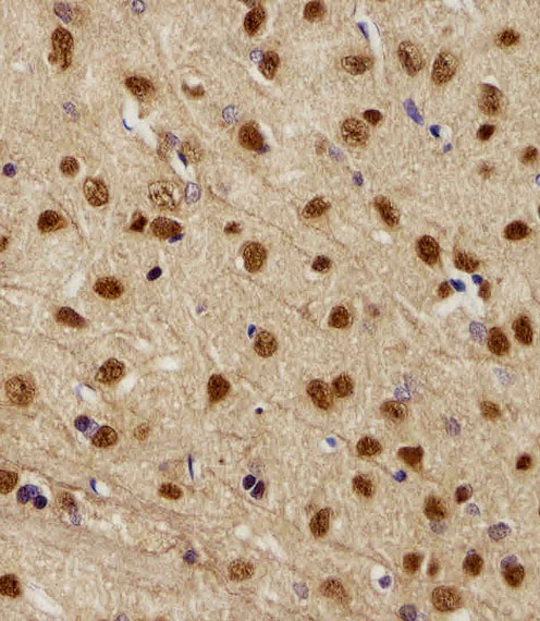
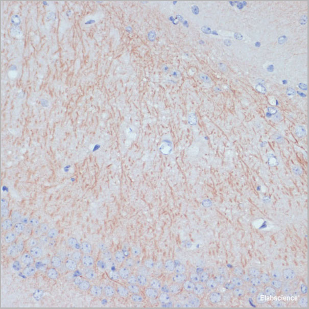
IHC (Immunohistchemistry) (Figure 6. IHC analysis of Synaptopodin/SYNPO using anti-Synaptopodin/SYNPO antibody (AAA19271).Synaptopodin/SYNPO was detected in paraffin-embedded section of mouse brain tissue. Heat mediated antigen retrieval was performed in EDTA buffer (pH8. 0, epitope retrieval solution). The tissue section was blocked with 10% goat serum. The tissue section was then incubated with 2μg/ml rabbit anti-Synaptopodin/SYNPO Antibody (AAA19271) overnight at 4 degree C. Biotinylated goat anti-rabbit IgG was used as secondary antibody and incubated for 30 minutes at 37 degree C. The tissue section was developed using Strepavidin-Biotin-Complex (SABC) (Catalog # with DAB as the chromogen.)

Synaptopodin/SYNPO, Polyclonal Antibody (Cat# AAA19271)

Full Name
Anti-Synaptopodin/SYNPO Antibody
Reactivity
Human, Mouse, Rat
Applications
Western Blot (WB), Immunohistochemistry-Paraffin (IHC-P), Immunocytochemistry (ICC), Immunofluorescence (IF), Flow Cytometry (FC/FACS/FCM)
Purity
Immunogen affinity purified.
Pricing
FCM (Flow Cytometry) (Figure 8. Flow Cytometry analysis of U20S cells using anti-SV2A antibody (AAA19279).Overlay histogram showing U20S cells stained with AAA19279 (Blue line). The cells were blocked with 10% normal goat serum. And then incubated with rabbit anti-SV2A Antibody (AAA19279, 1μg/1x106 cells) for 30 min at 20 degree C. DyLight®488 conjugated goat anti-rabbit IgG (5-10μg/1x106 cells) was used as secondary antibody for 30 minutes at 20 degree C. Isotype control antibody (Green line) was rabbit IgG (1μg/1x106) used under the same conditions. Unlabelled sample (Red line) was also used as a control.)

SV2A, Polyclonal Antibody (Cat# AAA19279)

Full Name
Anti-SV2A Antibody
Gene Names
SV2A; SV2
Reactivity
Human, Mouse, Rat
Applications
Western Blot (WB), Immunohistochemistry-Paraffin (IHC-P), Flow Cytometry (FC/FACS/FCM)
Purity
Immunogen affinity purified.
Pricing
FCM (Flow Cytometry) (Figure 7. Flow Cytometry analysis of Hela cells using anti-ATG9A antibody (AAA19280).Overlay histogram showing Hela cells stained with AAA19280 (Blue line). The cells were blocked with 10% normal goat serum. And then incubated with rabbit anti-ATG9A Antibody (AAA19280, 1μg/1x106 cells) for 30 min at 20 degree C. DyLight®488 conjugated goat anti-rabbit IgG (5-10μg/1x106 cells) was used as secondary antibody for 30 minutes at 20 degree C. Isotype control antibody (Green line) was rabbit IgG (1μg/1x106) used under the same conditions. Unlabelled sample (Red line) was also used as a control.)

ATG9A, Polyclonal Antibody (Cat# AAA19280)

Full Name
Anti-ATG9A Antibody
Gene Names
ATG9A; mATG9; APG9L1; MGD3208
Reactivity
Human, Mouse, Rat
Applications
Western Blot (WB), Immunohistochemistry-Paraffin (IHC-P), Flow Cytometry (FC/FACS/FCM), Direct ELISA (EIA)
Purity
Immunogen affinity purified.
Pricing
IF (Immunofluorescence) (Immunofluorescence of CCL2 in human spleen tissue with CCL2 antibody at 20 μg/ml.)

CCL2, Polyclonal Antibody (Cat# AAA10976)

Full Name
CCL2 Antibody
Gene Names
CCL2; HC11; MCAF; MCP1; MCP-1; SCYA2; GDCF-2; SMC-CF; HSMCR30
Reactivity
Human, Mouse, Rat
Applications
ELISA (EIA), Western Blot (WB), Immunohistochemistry-paraffin (IHC-P), Immunofluorescence (IF)
Purity
CCL2 Antibody is affinity chromatography purified via peptide column.
Pricing
FCM (Flow Cytometry) (Figure 6. Flow Cytometry analysis of U87 cells using anti-SPG3A/ATL1 antibody (AAA19441).Overlay histogram showing U87 cells stained with AAA19441 (Blue line). The cells were blocked with 10% normal goat serum. And then incubated with rabbit anti-SPG3A/ATL1 Antibody (AAA19441, 1 ug/1x10^6 cells) for 30 min at 20 degree C. DyLight488 conjugated goat anti-rabbit IgG was used as secondary antibody for 30 minutes at 20 degree C. Isotype control antibody (Green line) was rabbit IgG (1 ug/1x10^6) used under the same conditions. Unlabelled sample (Red line) was also used as a control.)

SPG3A/ATL1, Polyclonal Antibody (Cat# AAA19441)

Full Name
Anti-SPG3A/ATL1 Antibody Picoband
Gene Names
ATL1; FSP1; GBP3; SPG3; HSN1D; SPG3A; AD-FSP; atlastin1
Reactivity
Human
Applications
Flow Cytometry (FC/FACS), Immunohistochemistry (IHC), Western Blot (WB)
Purity
Immunogen affinity purified.
Pricing
FCM (Flow Cytometry) (Figure 10. Flow Cytometry analysis of JK cells using anti-MTHFD1 antibody (AAA19442).Overlay histogram showing JK cells stained with AAA19442 (Blue line). The cells were blocked with 10% normal goat serum. And then incubated with rabbit anti-MTHFD1 Antibody (AAA19442, 1 ug/1x10^6 cells) for 30 min at 20 degree C. DyLight488 conjugated goat anti-rabbit IgG was used as secondary antibody for 30 minutes at 20 degree C. Isotype control antibody (Green line) was rabbit IgG (1 ug/1x10^6) used under the same conditions. Unlabelled sample (Red line) was also used as a control.)

MTHFD1, Polyclonal Antibody (Cat# AAA19442)

Full Name
Anti-MTHFD1 Antibody Picoband
Gene Names
MTHFD1; MTHFC; MTHFD
Reactivity
Human, Mouse, Rat
Applications
Western Blot (WB), Immunohistochemistry (IHC), Immunocytochemistry (ICC), Immunofluorescence (IF), Flow Cytometry (FC/FACS), ELISA (EIA)
Purity
Immunogen affinity purified.
Pricing
IF (Immunofluorescence) (Figure 8. IF analysis of c-Jun/JUN using anti-c-Jun/JUN antibody (AAA19443).c-Jun/JUN was detected in an immunocytochemical section of A431 cells. Enzyme antigen retrieval was performed using IHC enzyme antigen retrieval reagent (AR0022) for 15 mins. The cells were blocked with 10% goat serum. And then incubated with 5 ug/mL rabbit anti-c-Jun/JUN Antibody (AAA19443) overnight at 4 degree C. DyLight488 Conjugated Goat Anti-Rabbit IgG was used as secondary antibody at 1:100 dilution and incubated for 30 minutes at 37 degree C. The section was counterstained with DAPI. Visualize using a fluorescence microscope and filter sets appropriate for the label used.)

c-Jun/JUN, Polyclonal Antibody (Cat# AAA19443)

Full Name
Anti-c-Jun/JUN Antibody Picoband
Gene Names
JUN; AP1; AP-1; c-Jun
Reactivity
Human
Applications
Western Blot (WB), Immunohistochemistry (IHC), Immunocytochemistry (ICC), Immunofluorescence (IF)
Purity
Immunogen affinity purified.
Pricing
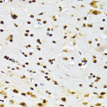
IHC (Immunohistochemistry) (Figure 8. IHC analysis of FOXP1 using anti-FOXP1 antibody (AAA19227).FOXP1 was detected in paraffin-embedded section of rat brain tissue. Heat mediated antigen retrieval was performed in EDTA buffer (pH8. 0, epitope retrieval solution). The tissue section was blocked with 10% goat serum. The tissue section was then incubated with 2μg/ml rabbit anti-FOXP1 Antibody (AAA19227) overnight at 4 degree C. Biotinylated goat anti-rabbit IgG was used as secondary antibody and incubated for 30 minutes at 37 degree C. The tissue section was developed using Strepavidin-Biotin-Complex (SABC) (Catalog # with DAB as the chromogen.)

FOXP1, Polyclonal Antibody (Cat# AAA19227)

Full Name
Anti-FOXP1 Antibody
Gene Names
FOXP1; QRF1; 12CC4; hFKH1B; HSPC215
Reactivity
Human, Mouse, Rat, Monkey
Applications
Western Blot (WB), Immunohistochemistry-Paraffin (IHC-P)
Purity
Immunogen affinity purified.
Pricing
IF (Immunofluorescence) (Figure 6. IF analysis of EEF2 using anti- EEF2 antibody (AAA19228).EEF2 was detected in immunocytochemical section of MCF-7 cells. Enzyme antigen retrieval was performed using IHC enzyme antigen retrieval reagent for 15 mins. The cells were blocked with 10% goat serum. And then incubated with 5μg/mL rabbit anti-EEF2 Antibody (AAA19228) overnight at 4 degree C. DyLight®488 Conjugated Goat Anti-Rabbit IgG was used as secondary antibody at 1:100 dilution and incubated for 30 minutes at 37 degree C. The section was counterstained with DAPI. Visualize using a fluorescence microscope and filter sets appropriate for the label used.)

EEF2, Polyclonal Antibody (Cat# AAA19228)

Full Name
Anti-EEF2 Antibody
Gene Names
EEF2; EF2; EF-2; EEF-2
Reactivity
Human
Applications
Western Blot (WB), Immunohistochemistry-Paraffin (IHC-P), Immunocytochemistry (ICC), Immunofluorescence (IF), Direct ELISA (EIA)
Purity
Immunogen affinity purified.
Pricing
FCM (Flow Cytometry) (Figure 6. Flow Cytometry analysis of NRK cells using anti-SHANK3 antibody (AAA19237).Overlay histogram showing NRK cells stained with AAA19237 (Blue line). The cells were blocked with 10% normal goat serum. And then incubated with rabbit anti-SHANK3 Antibody (AAA19237,1μg/1x106 cells) for 30 min at 20 degree C. DyLight®488 conjugated goat anti-rabbit IgG (5-10μg/1x106 cells) was used as secondary antibody for 30 minutes at 20 degree C. Isotype control antibody (Green line) was rabbit IgG (1μg/1x106) used under the same conditions. Unlabelled sample (Red line) was also used as a control.)

SHANK3, Polyclonal Antibody (Cat# AAA19237)

Full Name
Anti-SHANK3 Antibody
Gene Names
SHANK3; PSAP2; SCZD15; PROSAP2; SPANK-2; DEL22q13.3
Reactivity
Human, Mouse, Rat
Applications
Western Blot (WB), Immunohistochemistry-Paraffin (IHC-P), Flow Cytometry (FC/FACS/FCM), Direct ELISA (EIA)
Purity
Immunogen affinity purified.
Pricing
FCM (Flow Cytometry) (Figure 6. Flow Cytometry analysis of MCF-7 cells using anti-ALK-1/ACVRL1 antibody (AAA19242).Overlay histogram showing MCF-7 cells stained with AAA19242 (Blue line). The cells were blocked with 10% normal goat serum. And then incubated with rabbit anti-ALK-1/ACVRL1 Antibody (AAA19242, 1μg/1x106 cells) for 30 min at 20 degree C. DyLight®488 conjugated goat anti-rabbit IgG (5-10μg/1x106 cells) was used as secondary antibody for 30 minutes at 20 degree C. Isotype control antibody (Green line) was rabbit IgG (1μg/1x106) used under the same conditions. Unlabelled sample (Red line) was also used as a control.)

ALK-1/ACVRL1, Polyclonal Antibody (Cat# AAA19242)

Full Name
Anti-ALK-1/ACVRL1 Antibody
Gene Names
ACVRL1; HHT; ALK1; HHT2; ORW2; SKR3; ALK-1; TSR-I; ACVRLK1
Reactivity
Human, Mouse, Rat
Applications
Western Blot (WB), Immunohistochemistry-Paraffin (IHC-P), Flow Cytometry (FC/FACS/FCM), Direct ELISA (EIA)
Purity
Immunogen affinity purified.
Pricing
IF (Immunofluorescence) (Figure 6. IF analysis of UNG using anti-UNG antibody (AAA19245).UNG was detected in immunocytochemical section of MCF-7 cells. Enzyme antigen retrieval was performed using IHC enzyme antigen retrieval reagent for 15 mins. The cells were blocked with 10% goat serum. And then incubated with 5μg/mL rabbit anti- UNG Antibody (AAA19245) overnight at 4 degree C. DyLight®594 Conjugated Goat Anti-Rabbit IgG (BA1142) was used as secondary antibody at 1:100 dilution and incubated for 30 minutes at 37 degree C. The section was counterstained with DAPI. Visualize using a fluorescence microscope and filter sets appropriate for the label used.)

UNG, Polyclonal Antibody (Cat# AAA19245)

Full Name
Anti-UNG Antibody
Gene Names
UNG; DGU; UDG; UNG1; UNG2; HIGM4; HIGM5; UNG15
Reactivity
Human, Mouse, Rat, Monkey
Applications
Western Blot (WB), Immunohistochemistry-Paraffin (IHC-P), Immunocytochemistry (ICC), Immunofluorescence (IF), Flow Cytometry (FC/FACS/FCM), Direct ELISA (EIA)
Purity
Immunogen affinity purified.
Pricing
FCM (Flow Cytometry) (Figure 7. Flow Cytometry analysis of U251 cells using anti-CGKI/PRKG1 antibody (AAA19246).Overlay histogram showing U251 cells stained with AAA19246 (Blue line). The cells were blocked with 10% normal goat serum. And then incubated with rabbit anti-CGKI/PRKG1 Antibody (AAA19246, 1μg/1x106 cells) for 30 min at 20 degree C. DyLight®488 conjugated goat anti-rabbit IgG (5-10μg/1x106 cells) was used as secondary antibody for 30 minutes at 20 degree C. Isotype control antibody (Green line) was rabbit IgG (1μg/1x106) used under the same conditions. Unlabelled sample (Red line) was also used as a control.)

cGKI/PRKG1, Polyclonal Antibody (Cat# AAA19246)

Full Name
Anti-cGKI/PRKG1 Antibody
Gene Names
PRKG1; 1; PKG; cGK; AAT8; cGK1; cGKI; cGK 1; PRKG1B; PRKGR1B; cGKI-BETA; cGKI-alpha
Reactivity
Human, Mouse, Rat
Applications
Western Blot (WB), Immunohistochemistry-Paraffin (IHC-P), Immunocytochemistry (ICC), Immunofluorescence (IF), Flow Cytometry (FC/FACS/FCM), Direct ELISA (EIA)
Purity
Immunogen affinity purified.
Pricing
FCM (Flow Cytometry) (Figure 8. Flow Cytometry analysis of RH35 cells using anti-TORC1/CRTC1 antibody (AAA19251).Overlay histogram showing RH35 cells stained with AAA19251 (Blue line). The cells were blocked with 10% normal goat serum. And then incubated with rabbit anti-TORC1/CRTC1 Antibody (AAA19251, 1μg/1x106 cells) for 30 min at 20 degree C. DyLight®488 conjugated goat anti-rabbit IgG (5-10μg/1x106 cells) was used as secondary antibody for 30 minutes at 20 degree C. Isotype control antibody (Green line) was rabbit IgG (1μg/1x106) used under the same conditions. Unlabelled sample (Red line) was also used as a control.)

TORC1/CRTC1, Polyclonal Antibody (Cat# AAA19251)

Full Name
Anti-TORC1/CRTC1 Antibody
Gene Names
CRTC1; MECT1; TORC1; TORC-1; WAMTP1
Reactivity
Human, Mouse, Rat
Applications
Western Blot (WB), Immunohistochemistry-Paraffin (IHC-P), Immunocytochemistry (ICC), Immunofluorescence (IF), Flow Cytometry (FC/FACS/FCM)
Purity
Immunogen affinity purified.
Pricing
IF (Immunofluorescence) (Figure 11. IF analysis of H Cadherin/CDH13 using anti- H Cadherin/CDH13 antibody (AAA19252).H Cadherin/CDH13 was detected in immunocytochemical section of SIHA cells. Enzyme antigen retrieval was performed using IHC enzyme antigen retrieval reagent for 15 mins. The cells were blocked with 10% goat serum. And then incubated with 5μg/mL rabbit anti- H Cadherin/CDH13 Antibody (AAA19252) overnight at 4 degree C. DyLight®488 Conjugated Goat Anti-Rabbit IgG was used as secondary antibody at 1:100 dilution and incubated for 30 minutes at 37 degree C. The section was counterstained with DAPI. Visualize using a fluorescence microscope and filter sets appropriate for the label used.)

H Cadherin/CDH13, Polyclonal Antibody (Cat# AAA19252)

Full Name
Anti-H Cadherin/CDH13 Antibody
Gene Names
CDH13; CDHH; P105
Reactivity
Human, Rat
Applications
Western Blot (WB), Immunohistochemistry-Paraffin (IHC-P), Immunocytochemistry (ICC), Immunofluorescence (IF), Flow Cytometry (FC/FACS/FCM), Direct ELISA (EIA)
Purity
Immunogen affinity purified.
Pricing
FCM (Flow Cytometry) (Figure 6. Flow Cytometry analysis of A431 cells using anti-TMPRSS3 antibody (AAA19289).Overlay histogram showing A431 cells stained with AAA19289 (Blue line). The cells were blocked with 10% normal goat serum. And then incubated with rabbit anti-TMPRSS3 Antibody (AAA19289, 1μg/1x106 cells) for 30 min at 20 degree C. DyLight®488 conjugated goat anti-rabbit IgG (5-10μg/1x106 cells) was used as secondary antibody for 30 minutes at 20 degree C. Isotype control antibody (Green line) was rabbit IgG (1μg/1x106) used under the same conditions. Unlabelled sample (Red line) was also used as a control.)

TMPRSS3, Polyclonal Antibody (Cat# AAA19289)

Full Name
Anti-TMPRSS3 Antibody
Gene Names
TMPRSS3; DFNB8; DFNB10; ECHOS1; TADG12
Reactivity
Human, Mouse, Rat
Applications
Western Blot (WB), Immunohistochemistry-Paraffin (IHC-P), Flow Cytometry (FC/FACS/FCM), Direct ELISA (EIA)
Purity
Immunogen affinity purified.
Pricing
FCM (Flow Cytometry) (Figure 6. Flow Cytometry analysis of PC-3 cells using anti-MitoNEET/CISD1 antibody (AAA19293).Overlay histogram showing PC-3 cells stained with AAA19293 (Blue line). The cells were blocked with 10% normal goat serum. And then incubated with rabbit anti-MitoNEET/CISD1 Antibody (AAA19293,1μg/1x106 cells) for 30 min at 20 degree C. DyLight®488 conjugated goat anti-rabbit IgG (5-10μg/1x106 cells) was used as secondary antibody for 30 minutes at 20 degree C. Isotype control antibody (Green line) was rabbit IgG (1μg/1x106) used under the same conditions. Unlabelled sample (Red line) was also used as a control.)

MitoNEET/CISD1, Polyclonal Antibody (Cat# AAA19293)

Full Name
Anti-MitoNEET/CISD1 Antibody
Gene Names
CISD1; ZCD1; MDS029; C10orf70; mitoNEET
Reactivity
Human, Mouse, Rat, Monkey
Applications
Western Blot (WB), Immunohistochemistry-Paraffin (IHC-P), Flow Cytometry (FC/FACS/FCM), Direct ELISA (EIA)
Purity
Immunogen affinity purified.
Pricing
FCM (Flow Cytometry) (Figure 7. Flow Cytometry analysis of HEPG2 cells using anti-ALDH1L1 antibody (AAA19297).Overlay histogram showing HEPG2 cells stained with AAA19297 (Blue line). The cells were blocked with 10% normal goat serum. And then incubated with rabbit anti-ALDH1L1 Antibody (AAA19297 1μg/1x106 cells) for 30 min at 20 degree C. DyLight®488 conjugated goat anti-rabbit IgG (5-10μg/1x106 cells) was used as secondary antibody for 30 minutes at 20 degree C. Isotype control antibody (Green line) was rabbit IgG (1μg/1x106) used under the same conditions. Unlabelled sample (Red line) was also used as a control.)

ALDH1L1, Polyclonal Antibody (Cat# AAA19297)

Full Name
Anti-ALDH1L1 Antibody
Gene Names
ALDH1L1; FDH; FTHFD; 10-fTHF; 10-FTHFDH
Reactivity
Human, Mouse, Rat, Monkey
Applications
Western Blot (WB), Immunohistochemistry-Paraffin (IHC-P), Immunocytochemistry (ICC), Immunofluorescence (IF), Flow Cytometry (FC/FACS/FCM), Direct ELISA (EIA)
Purity
Immunogen affinity purified.
Pricing
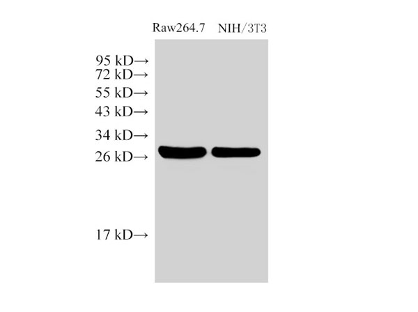
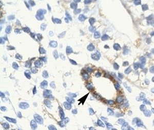
WB (Western Blot) (25 ug of the indicated Human whole cell extracts was loaded onto a 12% SDS-PAGE gel. 1 ug/mL of the antibody was used in this experiment. Canonical 37 kDa isoform is identified. Doublet may reflect phosphorylation.)

RAX, Polyclonal Antibody (Cat# AAA23397)

Full Name
RAX antibody - N-terminal region
Gene Names
RAX; RX; MCOP3
Reactivity
Tested Species Reactivity: Human
Predicted Species Reactivity: Human, Mouse, Rat, Cow, Dog, Guinea Pig, Horse, Rabbit
Applications
Immunohistochemistry (IHC), Western Blot (WB)
Purity
Affinity Purified
Pricing
FCM (Flow Cytometry) (Figure 10. Flow Cytometry analysis of 293T cells using anti-XRCC4 antibody (AAA19407).Overlay histogram showing 293T cells stained with AAA19407 (Blue line). The cells were blocked with 10% normal goat serum. And then incubated with rabbit anti-XRCC4 Antibody (AAA19407, 1 ug/1x10^6 cells) for 30 min at 20 degree C. DyLight488 conjugated goat anti-rabbit IgG was used as secondary antibody for 30 minutes at 20 degree C. Isotype control antibody (Green line) was rabbit IgG (1 ug/1x10^6) used under the same conditions. Unlabelled sample (Red line) was also used as a control.)

XRCC4, Polyclonal Antibody (Cat# AAA19407)

Full Name
Anti-XRCC4 Antibody Picoband
Reactivity
Human
Applications
Western Blot (WB), Immunohistochemistry (IHC), Immunocytochemistry (ICC), Immunofluorescence (IF), Flow Cytometry (FC/FACS), ELISA (EIA)
Purity
Immunogen affinity purified.
Pricing
FCM (Flow Cytometry) (Figure 8. Flow Cytometry analysis of U87 cells using anti-IFI16 antibody (AAA19409).Overlay histogram showing U87 cells stained with AAA19409 (Blue line). The cells were blocked with 10% normal goat serum. And then incubated with rabbit anti-IFI16 Antibody (AAA19409, 1 ug/1x10^6 cells) for 30 min at 20 degree C. DyLight488 conjugated goat anti-rabbit IgG was used as secondary antibody for 30 minutes at 20 degree C. Isotype control antibody (Green line) was rabbit IgG (1 ug/1x10^6) used under the same conditions. Unlabelled sample (Red line) was also used as a control.)

IFI16, Polyclonal Antibody (Cat# AAA19409)

Full Name
Anti-IFI16 Antibody Picoband
Gene Names
IFI16; PYHIN2; IFNGIP1
Reactivity
Human, Mouse, Rat
Applications
Western Blot (WB), Immunohistochemistry (IHC), Flow Cytometry (FC/FACS), ELISA (EIA)
Purity
Immunogen affinity purified.
Pricing
FCM (Flow Cytometry) (Figure 6. Flow Cytometry analysis of U937 cells using anti-BDH1 antibody (AAA19328).Overlay histogram showing U937 cells stained with AAA19328 (Blue line). The cells were blocked with 10% normal goat serum. And then incubated with rabbit anti-BDH1 Antibody (AAA19328,1μg/1x106 cells) for 30 min at 20 degree C. DyLight®488 conjugated goat anti-rabbit IgG (5-10μg/1x106 cells) was used as secondary antibody for 30 minutes at 20 degree C. Isotype control antibody (Green line) was rabbit IgG (1μg/1x106) used under the same conditions. Unlabelled sample (Red line) was also used as a control.)

BDH1, Polyclonal Antibody (Cat# AAA19328)

Full Name
Anti-BDH1 Antibody
Gene Names
BDH1; BDH; SDR9C1
Reactivity
Human, Rat
Applications
Western Blot (WB), Immunohistochemistry-Paraffin (IHC-P), Immunocytochemistry (ICC), Immunofluorescence (IF), Flow Cytometry (FC/FACS/FCM), Direct ELISA (EIA)
Purity
Immunogen affinity purified.
Pricing

Interleukin 1 Beta, Polyclonal Antibody (Cat# AAA20864)

Full Name
Polyclonal Antibody to Interleukin 1 Beta (IL1b)
Reactivity
Bos taurus; Bovine (Cattle)
Applications
Western Blot (WB); Immunohistochemistry (IHC); Immunocytochemistry (ICC); Immunoprecipitation (IP).
Purity
Antigen-specific affinity chromatography followed by Protein A affinity chromatography
Pricing

Phospholipase A2 Receptor 1, Polyclonal Antibody (Cat# AAA20865)

Full Name
Polyclonal Antibody to Phospholipase A2 Receptor 1 (PLA2R1)
Gene Names
Pla2r1; Pla2r; Pla2-r; Pla2g1br
Reactivity
Mus musculus (Mouse)
Applications
Western Blot (WB); Immunohistochemistry (IHC); Immunocytochemistry (ICC); Immunoprecipitation (IP).
Purity
Purification: Antigen-specific affinity chromatography followed by Protein A affinity chromatography
Pricing
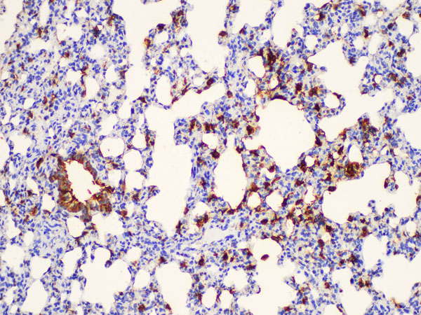
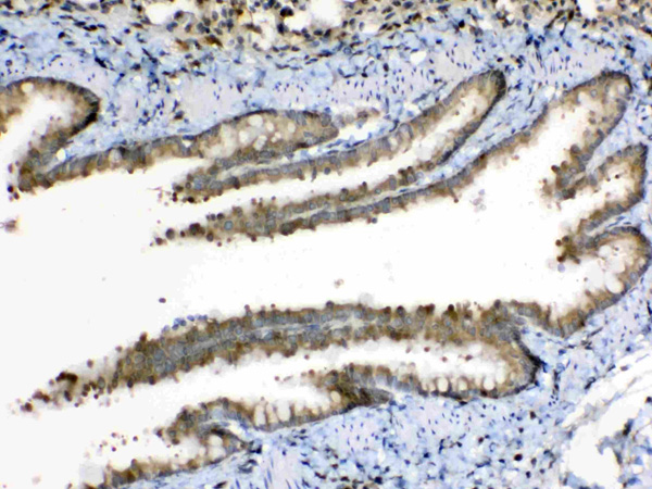
IHC (Immunohistochemistry) (DAB staining on IHC-P; Samples: Human Kidney Tissue.)

Extracellular Matrix Metalloproteinase Inducer, Polyclonal Antibody (Cat# AAA20867)

Full Name
Polyclonal Antibody to Extracellular Matrix Metalloproteinase Inducer (EMMPRIN)
Gene Names
BSG; OK; 5F7; TCSF; CD147; EMMPRIN
Reactivity
Human
Applications
Western Blot (WB), Immunocytochemistry (ICC), Immunohistochemistry (IHC) Formalin/Paraffin, ELISA (EIA)
Purity
Affinity Chromatography
Pricing
FCM (Flow Cytometry) (Figure 6. Flow Cytometry analysis of A431 cells using anti-NDUFB5 antibody (AAA19333).Overlay histogram showing A431 cells stained with AAA19333 (Blue line). The cells were blocked with 10% normal goat serum. And then incubated with rabbit anti-NDUFB5 Antibody (AAA19333, 1μg/1x106 cells) for 30 min at 20 degree C. DyLight®488 conjugated goat anti-rabbit IgG (5-10μg/1x106 cells) was used as secondary antibody for 30 minutes at 20 degree C. Isotype control antibody (Green line) was rabbit IgG (1μg/1x106) used under the same conditions. Unlabelled sample (Red line) was also used as a control.)

NDUFB5, Polyclonal Antibody (Cat# AAA19333)

Full Name
Anti-NDUFB5 Antibody
Gene Names
NDUFB5; SGDH; CISGDH
Reactivity
Human, Mouse, Rat
Applications
Western Blot (WB), Immunohistochemistry-Paraffin (IHC-P), Immunocytochemistry (ICC), Immunofluorescence (IF), Flow Cytometry (FC/FACS/FCM), Direct ELISA (EIA)
Purity
Immunogen affinity purified.
Pricing

Prostaglandin D2 Synthase, Polyclonal Antibody (Cat# AAA20870)

Full Name
Polyclonal Antibody to Prostaglandin D2 Synthase, Brain (PGD2S)
Gene Names
PTGDS; PDS; PGD2; PGDS; LPGDS; PGDS2; L-PGDS
Reactivity
Homo sapiens (Human)
Applications
Western Blot (WB), Immunocytochemistry (ICC), Immunohistochemistry (IHC) Formalin/Paraffin, Immunoprecipitation (IP)
Purity
Antigen-specific affinity chromatography followed by Protein A affinity chromatography
Pricing
FCM (Flow Cytometry) (Figure 13. Flow Cytometry analysis of A431 cells using anti-LSM8 antibody (AAA19337).Overlay histogram showing A431 cells stained with AAA19337 (Blue line). The cells were blocked with 10% normal goat serum. And then incubated with rabbit anti-LSM8 Antibody (AAA19337, 1μg/1x106 cells) for 30 min at 20 degree C. DyLight®488 conjugated goat anti-rabbit IgG (5-10μg/1x106 cells) was used as secondary antibody for 30 minutes at 20 degree C. Isotype control antibody (Green line) was rabbit IgG (1μg/1x106) used under the same conditions. Unlabelled sample (Red line) was also used as a control.)

LSM8, Polyclonal Antibody (Cat# AAA19337)

Full Name
Anti-LSM8 Antibody
Reactivity
Human, Mouse, Rat
Applications
Western Blot (WB), Immunohistochemistry-Paraffin (IHC-P), Immunocytochemistry (ICC), Immunofluorescence (IF), Flow Cytometry (FC/FACS/FCM), Direct ELISA (EIA)
Purity
Immunogen affinity purified.
Pricing
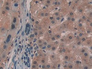
IHC (Immunohistchemistry) (DAB staining on IHC-P;Samples: Human Liver Tissue;Primary Ab: 30ug/ml Rabbit Anti-Human IL35 AntibodySecond Ab: 2ug/mL HRP-Linked Caprine Anti-Rabbit IgG Polyclonal Antibody (Catalog:))

Interleukin 35 (IL35), Polyclonal Antibody (Cat# AAA20105)

Full Name
Polyclonal Antibody to Interleukin 35 (IL35)
Reactivity
Homo sapiens (Human)
Applications
Western Blot (WB); Immunohistochemistry (IHC); Immunocytochemistry (ICC); Immunoprecipitation (IP).
Purity
Antigen-specific affinity chromatography followed by Protein A affinity chromatography
Pricing
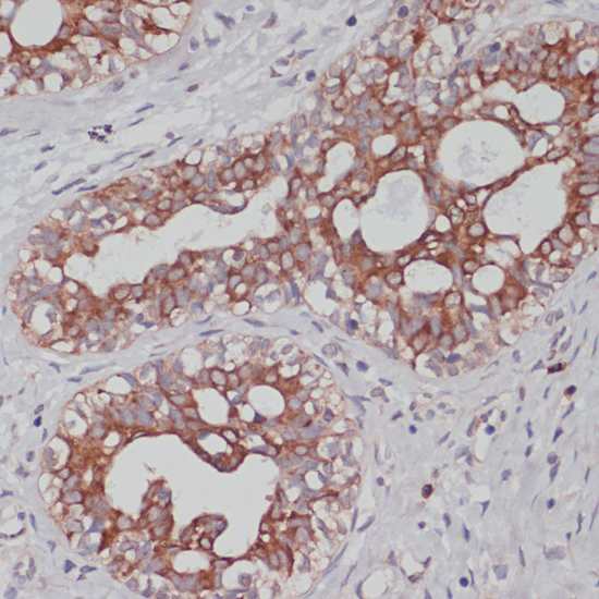
IHC (Immunohistochemistry) (DAB staining on IHC-P;Samples: Human Renal cancer Tissue;Primary Ab: 20ug/ml Rabbit Anti-Human GaA AntibodySecond Ab: 2ug/mL HRP-Linked Caprine Anti-Rabbit IgG Polyclonal Antibody)

Glucosidase Alpha, Acid (GaA), Polyclonal Antibody (Cat# AAA20106)

Full Name
Polyclonal Antibody to Glucosidase Alpha, Acid (GaA)
Gene Names
GAA; LYAG
Reactivity
Homo sapiens (Human)
Applications
Western Blot (WB); Immunohistochemistry (IHC); Immunocytochemistry (ICC); Immunoprecipitation (IP).
Purity
Antigen-specific affinity chromatography followed by Protein A affinity chromatography
Pricing
FCM (Flow Cytometry) (Figure 9. Flow Cytometry analysis of HL-60 cells using anti-NDUFB10 antibody (AAA19339).Overlay histogram showing HL-60 cells stained with AAA19339 (Blue line). The cells were blocked with 10% normal goat serum. And then incubated with rabbit anti-NDUFB10 Antibody (AAA19339, 1μg/1x106 cells) for 30 min at 20 degree C. DyLight®488 conjugated goat anti-rabbit IgG (5-10μg/1x106 cells) was used as secondary antibody for 30 minutes at 20 degree C. Isotype control antibody (Green line) was rabbit IgG (1μg/1x106) used under the same conditions. Unlabelled sample (Red line) was also used as a control.)

NDUFB10, Polyclonal Antibody (Cat# AAA19339)

Full Name
Anti-NDUFB10 Antibody
Gene Names
NDUFB10; PDSW
Reactivity
Human, Mouse, Rat
Applications
Western Blot (WB), Immunohistochemistry-Paraffin (IHC-P), Immunocytochemistry (ICC), Immunofluorescence (IF), Flow Cytometry (FC/FACS/FCM), Direct ELISA (EIA)
Purity
Immunogen affinity purified.
Pricing
IF (Immunofluorescence) (mmunofluorescence of Grik1 in mouse brain tissue with Grik1 Antibody at 20 ?g/mL. Green: Grik1 antibody (AAA10916) Red: Phylloidin stainingBlue: DAPI staining)

Grik1, Polyclonal Antibody (Cat# AAA10916)

Full Name
Grik1 Antibody
Gene Names
GRIK1; EAA3; EEA3; GLR5; GLUR5; GluK1
Reactivity
Human, Mouse
Applications
ELISA (EIA), Western Blot (WB), Immunohistochemistry (IHC-p), Immunofluorescence (IF)
Purity
Grik1 Antibody is affinity chromatography purified via peptide column.
Pricing

Submit a Question

Please complete the form below and a representative will contact you as soon as possible.

Request more Information

Please complete the form below and a representative will contact you as soon as possible.

Request a Manual

Please complete the form below and a representative will contact you as soon as possible.

Request a Quote

Please complete the form below and a representative will contact you as soon as possible.