
Filters
Clonality
Type
Reactivity
Gene Name
Isotype
Host
Application
Clone
10000 results for "Recombinant Proteins" - showing 4950-5000
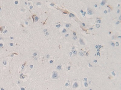
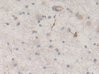 IHC (Immunohiostchemistry)
(DAB staining on IHCP;Sample: Human Glioma Tissue; Primary Ab: 30ug/ml Mouse AntiHuman NES AntibodySecond Ab: 2ug/mL HRPLinked Caprine AntiMouse IgG Polyclonal Antibody(Catalog: SAA544Mu19))
IHC (Immunohiostchemistry)
(DAB staining on IHCP;Sample: Human Glioma Tissue; Primary Ab: 30ug/ml Mouse AntiHuman NES AntibodySecond Ab: 2ug/mL HRPLinked Caprine AntiMouse IgG Polyclonal Antibody(Catalog: SAA544Mu19))
Nestin (NES), Monoclonal Antibody (Cat# AAA151548)
Full Name
Monoclonal Antibody to Nestin (NES)
Gene Names
NES; Nbla00170
Reactivity
Human
Applications
Immunoprecipitation, Immunocytochemistry, Immunohistochemistry, Western Blot
Purity
Protein A + Protein G affinity chromatography
Pricing
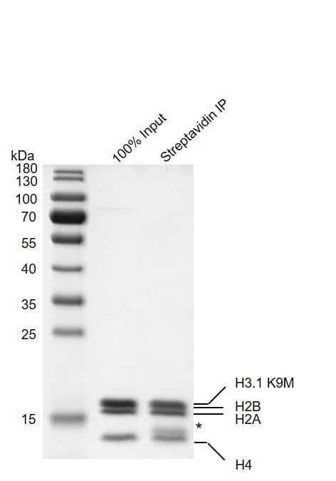
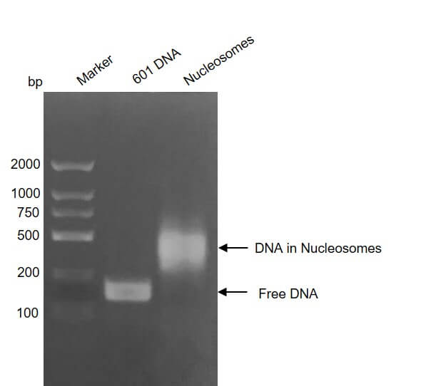
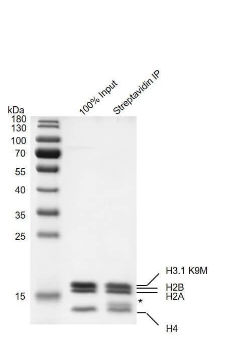 SDS-PAGE
(Streptavidin pull down assay for Recombinant Mononucleosomes H3.1 (K9M) - biotin 24 ug biotinylated mononucleosomes were incubated with 10 ul streptavidin beads for 1 hr at 4 degree C. Streptavidin beads were washed 3 times with 1 ml binding buffer. Then the beads were added to 60 ul 2×SDS loading buffer and boiled for 10 min at 95 degree C. 2.4 ul samples were loaded and run on a 12.5% SDS-PAGE gel and stained with Coomassie Blue. * indicates streptavidin. The SDS-PAGE gel result shows that >90% of biotinylated mononucleosomes were pulled down by streptavidin beads.)
SDS-PAGE
(Streptavidin pull down assay for Recombinant Mononucleosomes H3.1 (K9M) - biotin 24 ug biotinylated mononucleosomes were incubated with 10 ul streptavidin beads for 1 hr at 4 degree C. Streptavidin beads were washed 3 times with 1 ml binding buffer. Then the beads were added to 60 ul 2×SDS loading buffer and boiled for 10 min at 95 degree C. 2.4 ul samples were loaded and run on a 12.5% SDS-PAGE gel and stained with Coomassie Blue. * indicates streptavidin. The SDS-PAGE gel result shows that >90% of biotinylated mononucleosomes were pulled down by streptavidin beads.)
Mononucleosomes H3.1, Recombinant Protein (Cat# AAA60192)
Full Name
Recombinant Mononucleosomes H3.1 (K9M)-biotin
Pricing
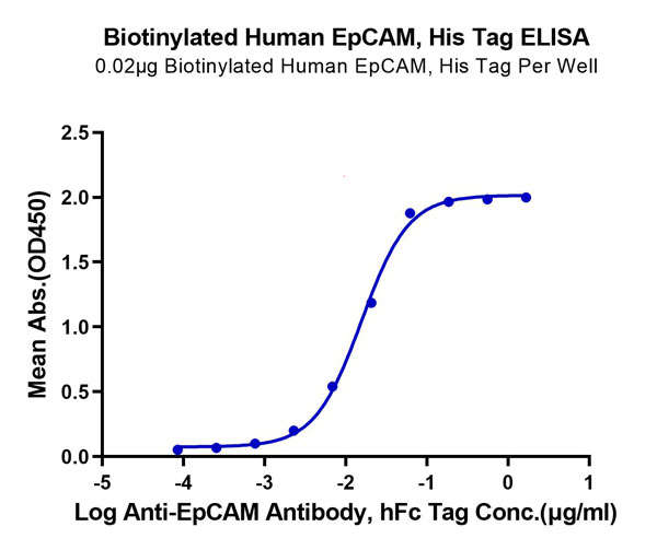 ELISA
(Biotinylated Human EpCAM/TROP1 Protein ELISA DataImmobilized Biotinylated Human EpCAM, His Tag at 0.2?g/ml (100ul/Well) on the plate. Dose response curve for Anti-EpCAM Antibody, hFc Tag with the EC50 of 15.6ng/ml determined by ELISA.)
ELISA
(Biotinylated Human EpCAM/TROP1 Protein ELISA DataImmobilized Biotinylated Human EpCAM, His Tag at 0.2?g/ml (100ul/Well) on the plate. Dose response curve for Anti-EpCAM Antibody, hFc Tag with the EC50 of 15.6ng/ml determined by ELISA.)
EpCAM/TROP1, Recombinant Protein (Cat# AAA76226)
Full Name
Biotinylated Human EpCAM/TROP1 Protein Recombinant
Gene Names
EPCAM; ESA; KSA; M4S1; MK-1; DIAR5; EGP-2; EGP40; KS1/4; MIC18; TROP1; EGP314; HNPCC8; TACSTD1
Purity
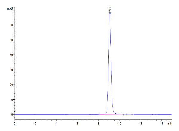
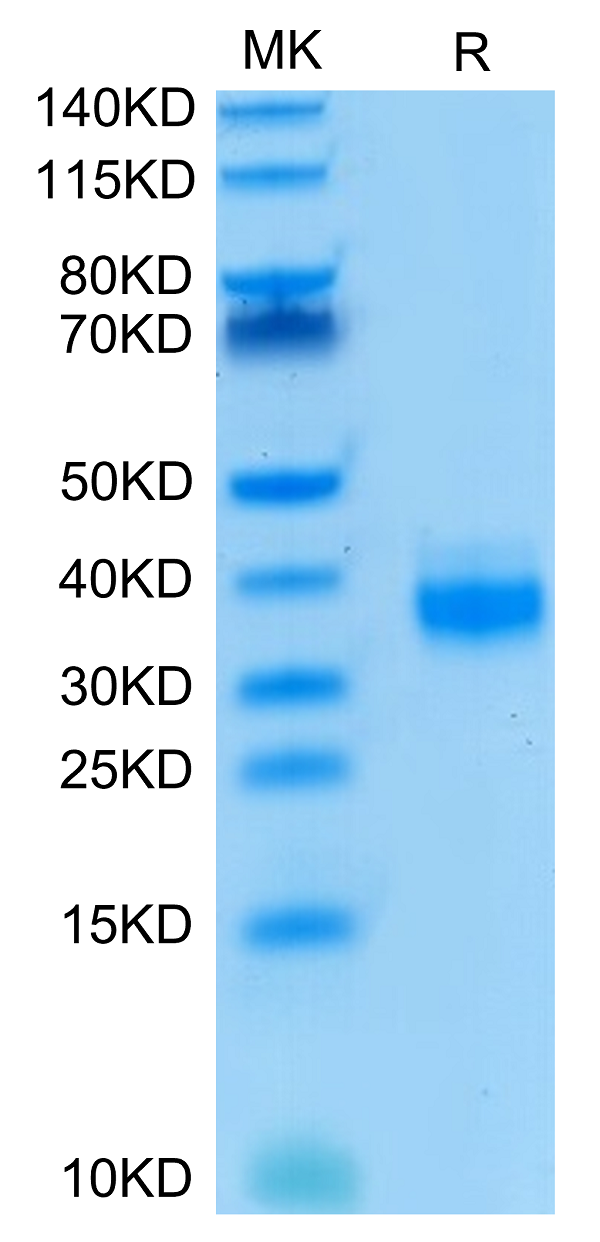
>95%, as determined by SDS-PAGE and HPLC
Pricing
 Bioactivity
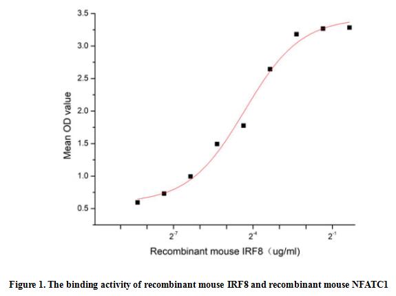
(Interferon regulatory factor 8 (IRF8), also known as interferon consensus sequence-binding protein, is a transcription factor that plays central roles in the regulation of myeloid cell fate. In both mice and humans, IRF8 is required for the differentiation of most monocyte and dendritic cell subsets, but suppresses neutrophil production. IRF8 can both act as a transcriptional activator or repressor, and it acts as a transcriptional repressor of osteoclast differentiation factors such as NFATC1 and EEIG1. Thus a functional binding ELISA assay was conducted to detect the interaction of recombinant mouse IRF8 and recombinant mouse NFATC1. Briefly, IRF8 was diluted serially in PBS with 0.01% BSA (pH 7.4). Duplicate samples of 100 ul were then transferred to NFATC1-coated microtiter wells and incubated for 1h at 37 degree C. Wells were washed with PBST and incubated for 1h with anti-IRF8 pAb, then aspirated and washed 3 times. After incubation with HRP labelled secondary antibody for 1h at 37 degree C, wells were aspirated and washed 5 times. With the addition of substrate solution, wells were incubated 15-25 minutes at 37 degree C. Finally, add 50 uL stop solution to the wells and read at 450/630 nm immediately. The binding activity of recombinant mouse IRF8 and recombinant mouse NFATC1 was shown in Figure 1, the EC50 for this effect is 0.05 ug/mL.)
Bioactivity
(Interferon regulatory factor 8 (IRF8), also known as interferon consensus sequence-binding protein, is a transcription factor that plays central roles in the regulation of myeloid cell fate. In both mice and humans, IRF8 is required for the differentiation of most monocyte and dendritic cell subsets, but suppresses neutrophil production. IRF8 can both act as a transcriptional activator or repressor, and it acts as a transcriptional repressor of osteoclast differentiation factors such as NFATC1 and EEIG1. Thus a functional binding ELISA assay was conducted to detect the interaction of recombinant mouse IRF8 and recombinant mouse NFATC1. Briefly, IRF8 was diluted serially in PBS with 0.01% BSA (pH 7.4). Duplicate samples of 100 ul were then transferred to NFATC1-coated microtiter wells and incubated for 1h at 37 degree C. Wells were washed with PBST and incubated for 1h with anti-IRF8 pAb, then aspirated and washed 3 times. After incubation with HRP labelled secondary antibody for 1h at 37 degree C, wells were aspirated and washed 5 times. With the addition of substrate solution, wells were incubated 15-25 minutes at 37 degree C. Finally, add 50 uL stop solution to the wells and read at 450/630 nm immediately. The binding activity of recombinant mouse IRF8 and recombinant mouse NFATC1 was shown in Figure 1, the EC50 for this effect is 0.05 ug/mL.)
Interferon Regulatory Factor 8 (IRF8), Active Protein (Cat# AAA161858)
Full Name
Active Interferon Regulatory Factor 8 (IRF8)
Gene Names
Irf8; Myls; ICSBP; IRF-8; Icsbp1; AI893568
Applications
Activity Assay, Cell Culture
Purity
Greater than 95% by SDS-PAGE
Pricing

 Application Data
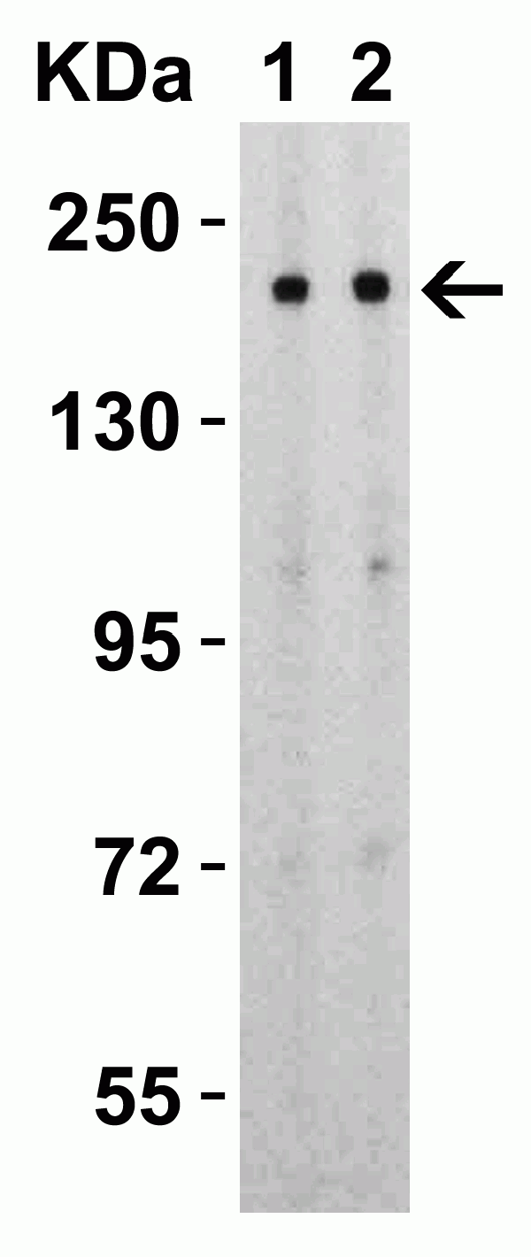
(Figure 4 Overexpression Validation in Spike Transfected 293 CellsLoading: 10 μg per lane of Spike transfected 293 cell lysate. Antibodies: SARS-CoV-2 (COVID-19) Spike, 9123, 1h incubation at RT in 5% NFDM/TBST. Secondary: Goat anti-rabbit IgG HRP conjugate at 1:10000 dilution. Lane 1: 1 μg/mL and Lane 2: 2 μg/mL)
Application Data
(Figure 4 Overexpression Validation in Spike Transfected 293 CellsLoading: 10 μg per lane of Spike transfected 293 cell lysate. Antibodies: SARS-CoV-2 (COVID-19) Spike, 9123, 1h incubation at RT in 5% NFDM/TBST. Secondary: Goat anti-rabbit IgG HRP conjugate at 1:10000 dilution. Lane 1: 1 μg/mL and Lane 2: 2 μg/mL)
COVID 19 Spike S2 Coronavirus, Polyclonal Antibody (Cat# AAA40907)
Full Name
SARS-CoV-2 (COVID-19) Spike S2 Antibody
Reactivity
Virus
Applications
Immunohistochemistry, Immunofluorescence, Western Blot
Purity
SARS-CoV-2 (COVID-19) Spike Antibody is affinity chromatography purified via peptide column
Pricing
 WB (Western Blot)
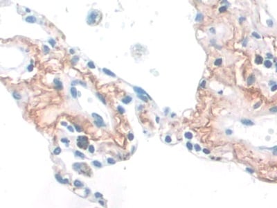
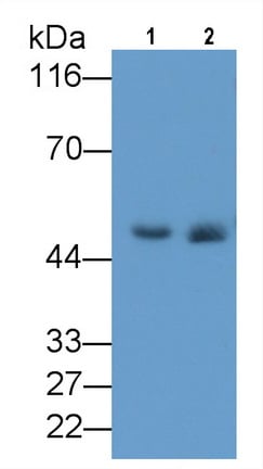
(Western Blot; Sample: Lane1: Rat Lung lysate; Lane2: Rat Skin lysate Primary Ab: 2ug/ml Mouse AntiHuman ELN Antibody Second Ab: 0.2ug/mL HRPLinked Rabbit AntiMouse IgG Polyclonal Antibody (Catalog: SAA544Mu19))
WB (Western Blot)
(Western Blot; Sample: Lane1: Rat Lung lysate; Lane2: Rat Skin lysate Primary Ab: 2ug/ml Mouse AntiHuman ELN Antibody Second Ab: 0.2ug/mL HRPLinked Rabbit AntiMouse IgG Polyclonal Antibody (Catalog: SAA544Mu19))
Elastin (ELN), Monoclonal Antibody (Cat# AAA151700)
Full Name
Monoclonal Antibody to Elastin (ELN)
Gene Names
ELN; WS; WBS; SVAS
Reactivity
Human, Rat
Applications
Immunoprecipitation, Immunocytochemistry, Immunohistochemistry, Western Blot
Purity
Protein A + Protein G affinity chromatography
Pricing
 Bioactivity
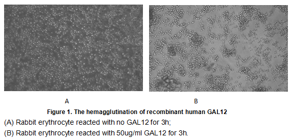
(Galectin-12 is a member of a family of mammalian lectins known as galectins. The galectins constitute a large family of carbohydrate-binding proteins that function in many systems both intracellularly and following secretion. Galectins contain either one or two carbohydrate recognition domains (CRR) which mediate recognition of N-acetyl-lactosamine-containing glycoproteins. Individual galectins differ in their tissue distribution and in their carbohydrate-binding specificities. Galectin-12 is predominantly expressed in adipose tissue and detected also in macrophages and other leukocytes. It plays an important role in cell-cell adhesion, cell-matrix interactions, macrophage activation, angiogenesis, metastasis, apoptosis. In this case, we chose rabbit erythrocyte (RaE) to assay its ability of agglutination. A general procedure for hemagglutination assay (or haemagglutination assay; HA) is as follows, two-fold dilute the recombinant Hu GAL12 with 0.9% sodium chloride injection, add 50uL a serial dilution of GAL12 to each well of a U or V-bottom shaped 96-well microtiter plate. The final well serves as a negative control with no GAL12, replace with 50uL 0.9% sodium chloride injection. Then add 50uL 1% rabbit erythrocyte to each well and mixed gently. The plate is incubated for 3 hours at room temperature. The results are shown in Figure 1. It was obvious that the minimal effective concentration of GAL12 is 0.781 ug/mL.)
Bioactivity
(Galectin-12 is a member of a family of mammalian lectins known as galectins. The galectins constitute a large family of carbohydrate-binding proteins that function in many systems both intracellularly and following secretion. Galectins contain either one or two carbohydrate recognition domains (CRR) which mediate recognition of N-acetyl-lactosamine-containing glycoproteins. Individual galectins differ in their tissue distribution and in their carbohydrate-binding specificities. Galectin-12 is predominantly expressed in adipose tissue and detected also in macrophages and other leukocytes. It plays an important role in cell-cell adhesion, cell-matrix interactions, macrophage activation, angiogenesis, metastasis, apoptosis. In this case, we chose rabbit erythrocyte (RaE) to assay its ability of agglutination. A general procedure for hemagglutination assay (or haemagglutination assay; HA) is as follows, two-fold dilute the recombinant Hu GAL12 with 0.9% sodium chloride injection, add 50uL a serial dilution of GAL12 to each well of a U or V-bottom shaped 96-well microtiter plate. The final well serves as a negative control with no GAL12, replace with 50uL 0.9% sodium chloride injection. Then add 50uL 1% rabbit erythrocyte to each well and mixed gently. The plate is incubated for 3 hours at room temperature. The results are shown in Figure 1. It was obvious that the minimal effective concentration of GAL12 is 0.781 ug/mL.)
Galectin 12 (GAL12), Active Protein (Cat# AAA152174)
Full Name
Active Galectin 12 (GAL12)
Gene Names
LGALS12; GAL12; GRIP1
Applications
Activity Assay, Cell Culture
Purity
Greater than 97% by SDS-PAGE
Pricing
 ELISA
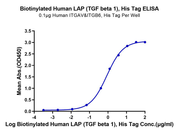
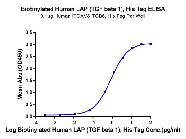
(Biotinylated Human LAP (TGF beta 1) Protein ELISA DataImmobilized Human ITGAV&ITGB6, His Tag at 1?g/ml (100?l/well) on the plate. Dose response curve for Biotinylated Human LAP (TGF beta 1) , His Tag with the EC50 of 0.86?g/ml determined by ELISA.)
ELISA
(Biotinylated Human LAP (TGF beta 1) Protein ELISA DataImmobilized Human ITGAV&ITGB6, His Tag at 1?g/ml (100?l/well) on the plate. Dose response curve for Biotinylated Human LAP (TGF beta 1) , His Tag with the EC50 of 0.86?g/ml determined by ELISA.)
LAP (TGF beta 1), Recombinant Protein (Cat# AAA76336)
Full Name
Biotinylated Human LAP (TGF beta 1) Protein Recombinant
Gene Names
TGFB1; CED; LAP; DPD1; TGFB; TGFbeta
Purity
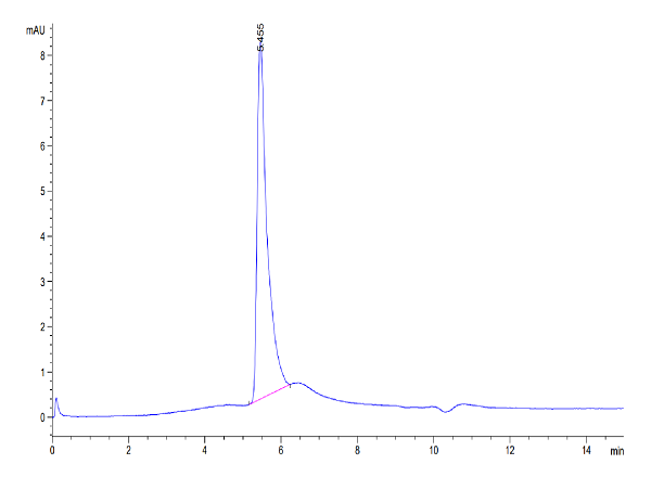
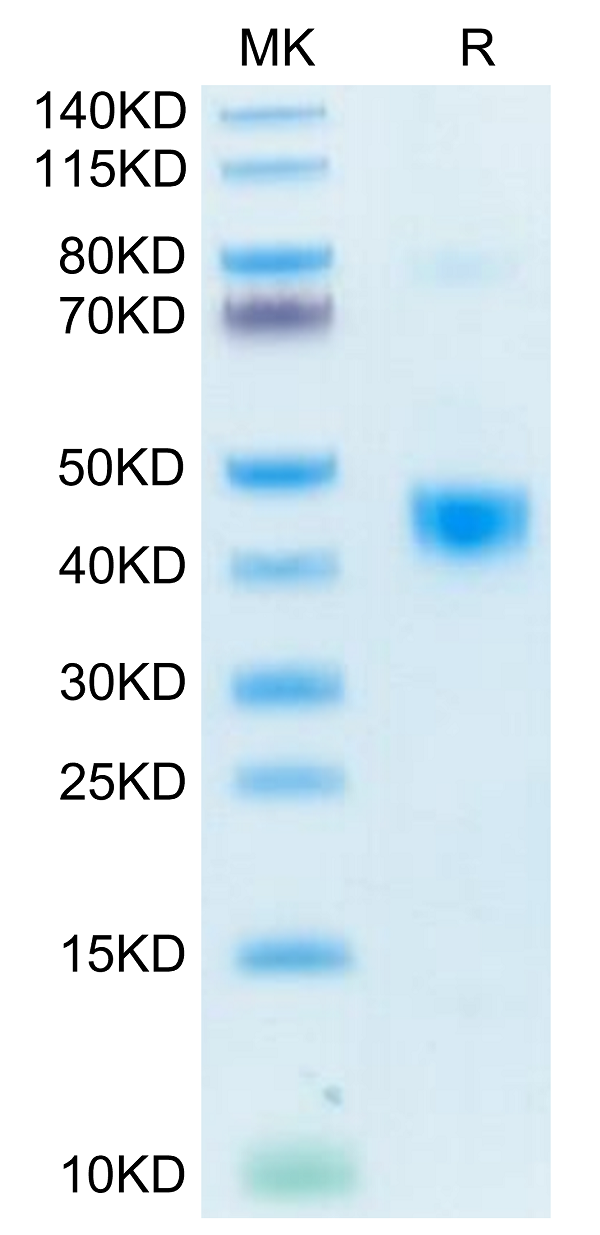
>95%, as determined by SDS-PAGE and HPLC
Pricing
 IHC (Immunohiostchemistry)
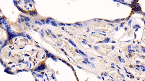
(DAB staining on IHCP; Samples: Human Placenta Tissue; Primary Ab: 20ug/ml Mouse AntiHuman CYPB Antibody Second Ab: 2ug/mL HRPLinked Caprine AntiMouse IgG Polyclonal Antibody (Catalog: SAA544Mu19))
IHC (Immunohiostchemistry)
(DAB staining on IHCP; Samples: Human Placenta Tissue; Primary Ab: 20ug/ml Mouse AntiHuman CYPB Antibody Second Ab: 2ug/mL HRPLinked Caprine AntiMouse IgG Polyclonal Antibody (Catalog: SAA544Mu19))
Cyclophilin B (CYPB), Monoclonal Antibody (Cat# AAA151509)
Full Name
Monoclonal Antibody to Cyclophilin B (CYPB)
Gene Names
PPIB; OI9; CYPB; SCYLP; CYP-S1
Reactivity
Human, Mouse, Rat, Pig, Bovine
Applications
Immunoprecipitation, Immunocytochemistry, Immunohistochemistry, Western Blot
Purity
Protein A + Protein G affinity chromatography
Pricing
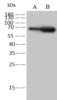 WB (Western Blot)
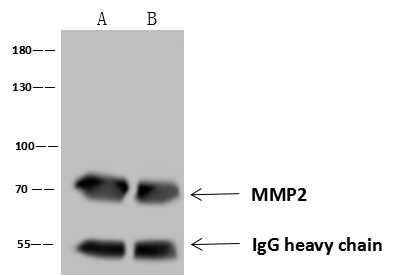
(Anti-MMP2 rabbit polyclonal antibody at 1:500 dilutionLane A: A375 Whole Cell LysateLane B: HepG2 Whole Cell LysateLysates/proteins at 30 ug per lane.SecondaryGoat Anti-Rabbit IgG (H+L)/HRP at 1/10000 dilution.Developed using the ECL technique.Performed under reducing conditions.Predicted band size:73 kDaObserved band size:73 kDa)
WB (Western Blot)
(Anti-MMP2 rabbit polyclonal antibody at 1:500 dilutionLane A: A375 Whole Cell LysateLane B: HepG2 Whole Cell LysateLysates/proteins at 30 ug per lane.SecondaryGoat Anti-Rabbit IgG (H+L)/HRP at 1/10000 dilution.Developed using the ECL technique.Performed under reducing conditions.Predicted band size:73 kDaObserved band size:73 kDa)
MMP-2, Polyclonal Antibody (Cat# AAA258731)
Full Name
Anti-MMP-2 Antibody, Rabbit Polyclonal
Reactivity
Human
Applications
Immunoprecipitation, Western Blot
Purity
Protein A & Antigen Affinity
Pricing
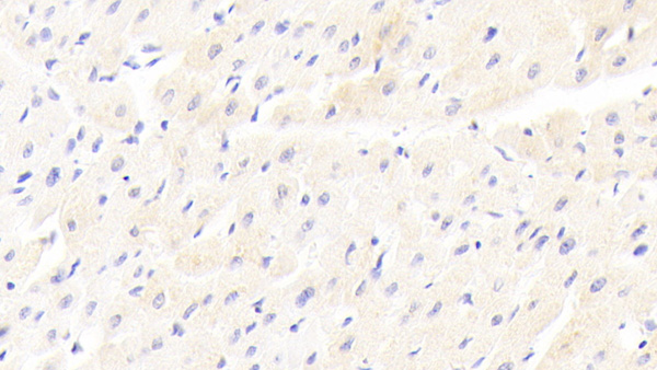
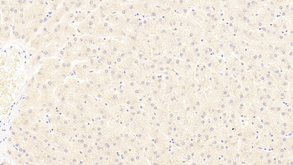 IHC (Immunohiostchemistry)
(DAB staining on IHCP;Sample: Human Liver Tissue; Primary Ab: 20ug/ml Mouse AntiHuman LRP6 AntibodySecond Ab: 2ug/mL HRPLinked Caprine AntiMouse IgG Polyclonal Antibody(Catalog: SAA544Mu19))
IHC (Immunohiostchemistry)
(DAB staining on IHCP;Sample: Human Liver Tissue; Primary Ab: 20ug/ml Mouse AntiHuman LRP6 AntibodySecond Ab: 2ug/mL HRPLinked Caprine AntiMouse IgG Polyclonal Antibody(Catalog: SAA544Mu19))
Low Density Lipoprotein Receptor Related Protein 6 (LRP6), Monoclonal Antibody (Cat# AAA151831)
Full Name
Monoclonal Antibody to Low Density Lipoprotein Receptor Related Protein 6 (LRP6)
Gene Names
LRP6; ADCAD2
Reactivity
Human, Pig
Applications
Immunoprecipitation, Immunocytochemistry, Immunohistochemistry, Western Blot
Purity
Protein A + Protein G affinity chromatography
Pricing
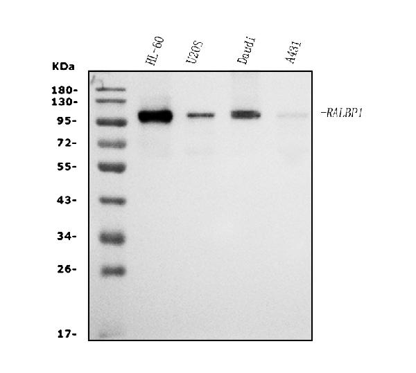
 FCM/FACS (Flow Cytometry)
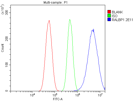
(Figure 2. Flow Cytometry analysis of U87 cells using anti-RALBP1 antibody (AAA125916).Overlay histogram showing U87 cells stained with AAA125916 (Blue line). The cells were blocked with 10% normal goat serum. And then incubated with mouse anti-RALBP1 Antibody (AAA125916, 1μg/1x106 cells) for 30 min at 20 degree C. DyLight®488 conjugated goat anti-mouse IgG (BA1126, 5-10μg/1x106 cells) was used as secondary antibody for 30 minutes at 20 degree C. Isotype control antibody (Green line) was mouse IgG (1μg/1x106) used under the same conditions. Unlabelled sample (Red line) was also used as a control.)
FCM/FACS (Flow Cytometry)
(Figure 2. Flow Cytometry analysis of U87 cells using anti-RALBP1 antibody (AAA125916).Overlay histogram showing U87 cells stained with AAA125916 (Blue line). The cells were blocked with 10% normal goat serum. And then incubated with mouse anti-RALBP1 Antibody (AAA125916, 1μg/1x106 cells) for 30 min at 20 degree C. DyLight®488 conjugated goat anti-mouse IgG (BA1126, 5-10μg/1x106 cells) was used as secondary antibody for 30 minutes at 20 degree C. Isotype control antibody (Green line) was mouse IgG (1μg/1x106) used under the same conditions. Unlabelled sample (Red line) was also used as a control.)
RALBP1, Monoclonal Antibody (Cat# AAA125916)
Full Name
Anti-RALBP1 Antibody (monoclonal, 2E11)
Gene Names
RALBP1; RIP1; RLIP1; RLIP76
Reactivity
Human
Applications
Flow Cytometry, Western Blot
Purity
Immunogen affinity purified.
Pricing
 Application Data
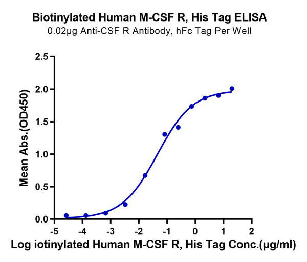
(Biotinylated Human M-CSF R/CSF1R/CD115 Protein SPR DataBiotinylated Human M-CSF R, His-Avi Tag immobilized on CM5 Chip can bind Human M-CSF, No Tag with an affinity constant of 0.81 ?M as determined in SPR assay (Biacore T200).)
Application Data
(Biotinylated Human M-CSF R/CSF1R/CD115 Protein SPR DataBiotinylated Human M-CSF R, His-Avi Tag immobilized on CM5 Chip can bind Human M-CSF, No Tag with an affinity constant of 0.81 ?M as determined in SPR assay (Biacore T200).)
M-CSF R/CSF1R/CD115, Recombinant Protein (Cat# AAA76234)
Full Name
Biotinylated Human M-CSF R/CSF1R/CD115 Protein Recombinant
Gene Names
CSF1R; FMS; CSFR; FIM2; HDLS; C-FMS; CD115; CSF-1R; M-CSF-R
Purity
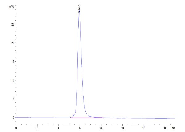
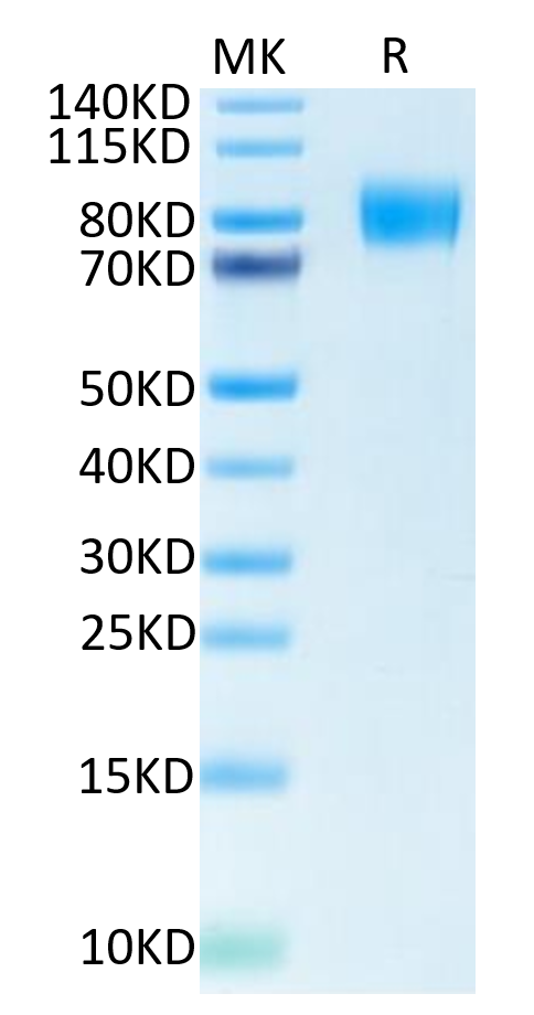
>95%, as determined by SDS-PAGE and HPLC
Pricing
 FCM/FACS (Flow Cytometry)
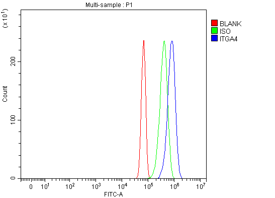
(Figure 2. Flow Cytometry analysis of NIH/3T3 cells using anti-Integrin Alpha 4/Itga4 antibody (AAA126990).Overlay histogram showing NIH/3T3 cells stained with AAA126990 (Blue line). The cells were fixed with 4% paraformaldehyde and blocked with 10% normal goat serum. And then incubated with rabbit anti-Integrin Alpha 4/Itga4 Antibody (AAA126990, 1ug/1x106 cells) for 30 min at 20 degree C. DyLight488 conjugated goat anti-rabbit IgG was used as secondary antibody for 30 minutes at 20 degree C. Isotype control antibody (Green line) was rabbit IgG (1ug/1x106) used under the same conditions. Unlabelled sample (Red line) was also used as a control.)
FCM/FACS (Flow Cytometry)
(Figure 2. Flow Cytometry analysis of NIH/3T3 cells using anti-Integrin Alpha 4/Itga4 antibody (AAA126990).Overlay histogram showing NIH/3T3 cells stained with AAA126990 (Blue line). The cells were fixed with 4% paraformaldehyde and blocked with 10% normal goat serum. And then incubated with rabbit anti-Integrin Alpha 4/Itga4 Antibody (AAA126990, 1ug/1x106 cells) for 30 min at 20 degree C. DyLight488 conjugated goat anti-rabbit IgG was used as secondary antibody for 30 minutes at 20 degree C. Isotype control antibody (Green line) was rabbit IgG (1ug/1x106) used under the same conditions. Unlabelled sample (Red line) was also used as a control.)
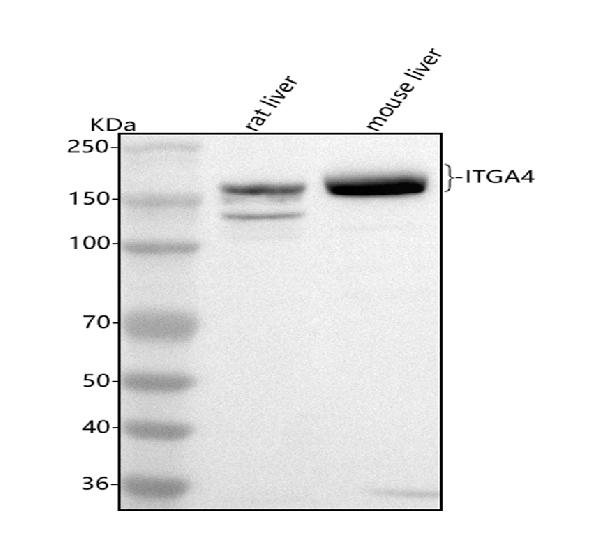
Integrin Alpha 4/Itga4, Polyclonal Antibody (Cat# AAA126990)
Full Name
Anti-Integrin Alpha 4/Itga4 Antibody Picoband
Gene Names
Itga4; CD49D; Itga4B
Reactivity
Mouse, Rat
Applications
Flow Cytometry, Western Blot
Purity
Immunogen affinity purified.
Pricing
 FCM/FACS (Flow Cytometry)
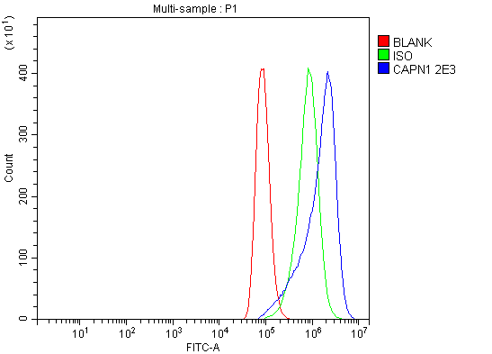
(Figure 2. Flow Cytometry analysis of A549 cells using anti-Calpain 1 antibody (AAA125920).Overlay histogram showing A549 cells stained with AAA125920 (Blue line). The cells were blocked with 10% normal goat serum. And then incubated with mouse anti-Calpain 1 Antibody (AAA125920, 1μg/1x106 cells) for 30 min at 20 degree C. DyLight®488 conjugated goat anti-mouse IgG (BA1126, 5-10μg/1x106 cells) was used as secondary antibody for 30 minutes at 20 degree C. Isotype control antibody (Green line) was mouse IgG (1μg/1x106) used under the same conditions. Unlabelled sample (Red line) was also used as a control.)
FCM/FACS (Flow Cytometry)
(Figure 2. Flow Cytometry analysis of A549 cells using anti-Calpain 1 antibody (AAA125920).Overlay histogram showing A549 cells stained with AAA125920 (Blue line). The cells were blocked with 10% normal goat serum. And then incubated with mouse anti-Calpain 1 Antibody (AAA125920, 1μg/1x106 cells) for 30 min at 20 degree C. DyLight®488 conjugated goat anti-mouse IgG (BA1126, 5-10μg/1x106 cells) was used as secondary antibody for 30 minutes at 20 degree C. Isotype control antibody (Green line) was mouse IgG (1μg/1x106) used under the same conditions. Unlabelled sample (Red line) was also used as a control.)
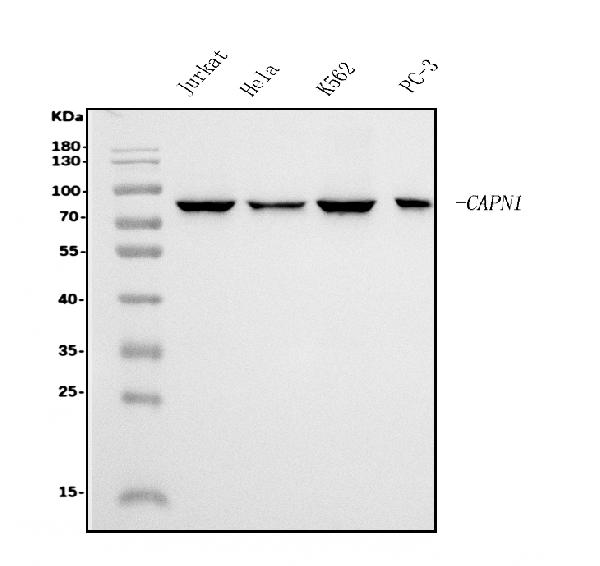
Calpain 1, Monoclonal Antibody (Cat# AAA125920)
Full Name
Anti-Calpain 1 Antibody (monoclonal, 2E3)
Gene Names
CAPN1; CANP; muCL; CANP1; CANPL1; muCANP
Reactivity
Human
Applications
Flow Cytometry, Western Blot
Purity
Immunogen affinity purified.
Pricing
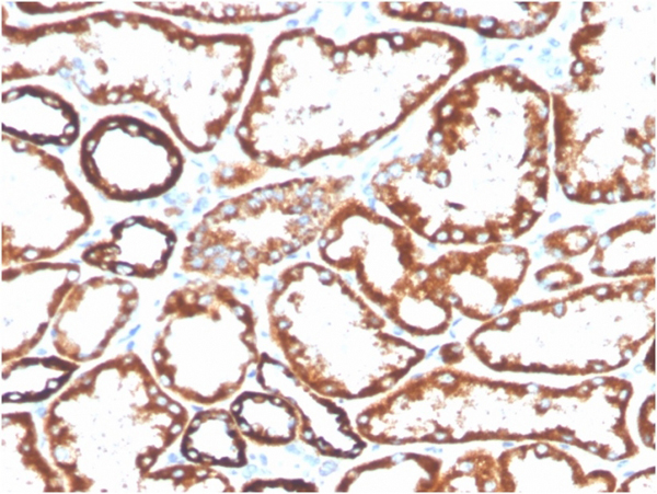
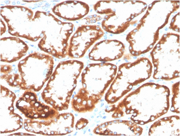
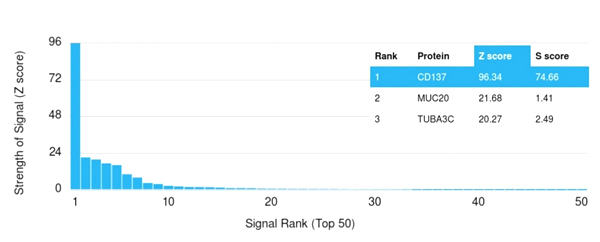 Application Data
(Analysis of Protein Array containing more than 19,000 full-length human proteins using CD137-Monospecific Mouse Monoclonal Antibody (4-1BB/3201). Z- and S- Score: The Z-score represents the strength of a signal that a monoclonal antibody (MAb) (in combination with a fluorescently-tagged anti-IgG secondary antibody) produces when binding to a particular protein on the HuProtTM array. Z-scores are described in units of standard deviations (SD's) above the mean value of all signals generated on that array. If targets on HuProtTM are arranged in descending order of the Z-score, the S-score is the difference (also in units of SD's) between the Z-score. S-score therefore represents the relative target specificity of a MAb to its intended target. A MAb is considered to specific to its intended target, if the MAb has an S-score of at least 2.5. For example, if a MAb binds to protein X with a Z-score of 43 and to protein Y with a Z-score of 14, then the S-score for the binding of that MAb to protein X is equal to 29.)
Application Data
(Analysis of Protein Array containing more than 19,000 full-length human proteins using CD137-Monospecific Mouse Monoclonal Antibody (4-1BB/3201). Z- and S- Score: The Z-score represents the strength of a signal that a monoclonal antibody (MAb) (in combination with a fluorescently-tagged anti-IgG secondary antibody) produces when binding to a particular protein on the HuProtTM array. Z-scores are described in units of standard deviations (SD's) above the mean value of all signals generated on that array. If targets on HuProtTM are arranged in descending order of the Z-score, the S-score is the difference (also in units of SD's) between the Z-score. S-score therefore represents the relative target specificity of a MAb to its intended target. A MAb is considered to specific to its intended target, if the MAb has an S-score of at least 2.5. For example, if a MAb binds to protein X with a Z-score of 43 and to protein Y with a Z-score of 14, then the S-score for the binding of that MAb to protein X is equal to 29.)
CD137/4-1BB/TNFRSF9, Monoclonal Antibody (Cat# AAA215484)
Full Name
CD137/4-1BB/TNFRSF9
Gene Names
TNFRSF9; ILA; 4-1BB; CD137; CDw137
Reactivity
Human
Applications
Immunohistochemistry, Immunofluorescence, Flow Cytometry
Purity
Purified Ab conjugated to Biotin
Pricing

 Application Data
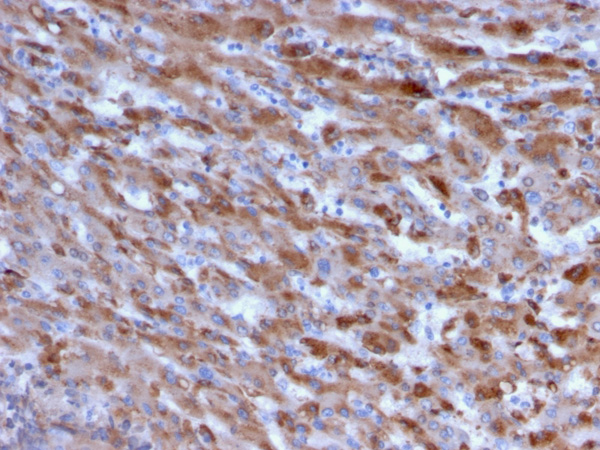
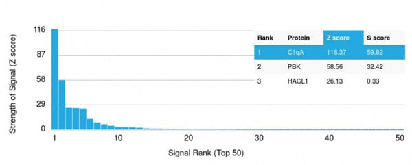
(Analysis of Protein Array containing more than 19,000 full-length human proteins using C1QA Mouse Monoclonal Antibody (C1QA/2955).Z- and S- Score: The Z-score represents the strength of a signal that a monoclonal antibody (MAb) (in combination with a fluorescently-tagged anti-IgG secondary antibody) produces when binding to a particular protein on the HuProtTM array. Z-scores are described in units of standard deviations (SD's) above the mean value of all signals generated on that array. If targets on HuProtTM are arranged in descending order of the Z-score, the S-score is the difference (also in units of SD's) between the Z-score. S-score therefore represents the relative target specificity of a MAb to its intended target. A MAb is considered to specific to its intended target, if the MAb has an S-score of at least 2.5. For example, if a MAb binds to protein X with a Z-score of 43 and to protein Y with a Z-score of 14, then the S-score for the binding of that MAb to protein X is equal to 29.)
Application Data
(Analysis of Protein Array containing more than 19,000 full-length human proteins using C1QA Mouse Monoclonal Antibody (C1QA/2955).Z- and S- Score: The Z-score represents the strength of a signal that a monoclonal antibody (MAb) (in combination with a fluorescently-tagged anti-IgG secondary antibody) produces when binding to a particular protein on the HuProtTM array. Z-scores are described in units of standard deviations (SD's) above the mean value of all signals generated on that array. If targets on HuProtTM are arranged in descending order of the Z-score, the S-score is the difference (also in units of SD's) between the Z-score. S-score therefore represents the relative target specificity of a MAb to its intended target. A MAb is considered to specific to its intended target, if the MAb has an S-score of at least 2.5. For example, if a MAb binds to protein X with a Z-score of 43 and to protein Y with a Z-score of 14, then the S-score for the binding of that MAb to protein X is equal to 29.)
C1QA/Complement C1q A-Chain, Monoclonal Antibody (Cat# AAA215223)
Full Name
C1QA/Complement C1q A-Chain
Reactivity
Human
Applications
Immunohistochemistry
Purity
Purified Ab with BSA and Azide at 200ug/ml OR Purified Ab WITHOUT BSA and Azide at 1.0mg/ml
Pricing
 Application Data
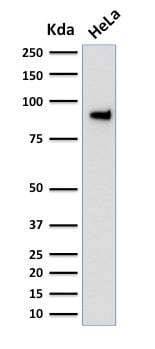
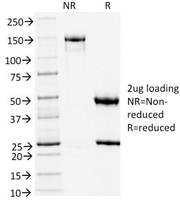
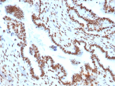
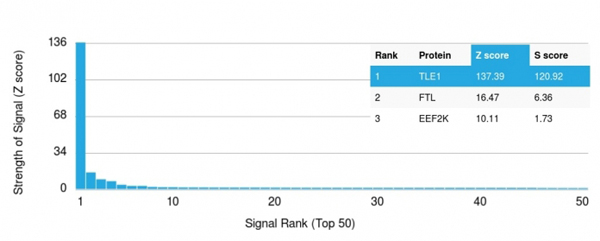
(Analysis of Protein Array containing more than 19, 000 full-length human proteins using TLE1 Mouse Monoclonal Antibody (TLE1/2051). Z- and S- Score: The Z-score represents the strength of a signal that a monoclonal antibody (MAb) (in combination with a fluorescently-tagged anti-IgG secondary antibody) produces when binding to a particular protein on the HuProtTM array. Z-scores are described in units of standard deviations (SD's) above the mean value of all signals generated on that array. If targets on HuProtTM are arranged in descending order of the Z-score, the S-score is the difference (also in units of SD's) between the Z-score. S-score therefore represents the relative target specificity of a MAb to its intended target. A MAb is considered to specific to its intended target, if the MAb has an S-score of at least 2.5. For example, if a MAb binds to protein X with a Z-score of 43 and to protein Y with a Z-score of 14, then the S-score for the binding of that MAb to protein X is equal to 29.)
Application Data
(Analysis of Protein Array containing more than 19, 000 full-length human proteins using TLE1 Mouse Monoclonal Antibody (TLE1/2051). Z- and S- Score: The Z-score represents the strength of a signal that a monoclonal antibody (MAb) (in combination with a fluorescently-tagged anti-IgG secondary antibody) produces when binding to a particular protein on the HuProtTM array. Z-scores are described in units of standard deviations (SD's) above the mean value of all signals generated on that array. If targets on HuProtTM are arranged in descending order of the Z-score, the S-score is the difference (also in units of SD's) between the Z-score. S-score therefore represents the relative target specificity of a MAb to its intended target. A MAb is considered to specific to its intended target, if the MAb has an S-score of at least 2.5. For example, if a MAb binds to protein X with a Z-score of 43 and to protein Y with a Z-score of 14, then the S-score for the binding of that MAb to protein X is equal to 29.)
TLE1, Monoclonal Antibody (Cat# AAA214699)
Full Name
TLE1 (Synovial Sarcoma Marker)
Gene Names
TLE1; ESG; ESG1; GRG1
Reactivity
Human. Others not known.
Applications
Immunohistochemistry, Western Blot
Pricing
 IHC (Immunohiostchemistry)
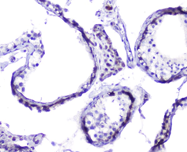
(Figure 3. IHC analysis of Hsp105 using anti-Hsp105 antibody.Hsp105 was detected in paraffin-embedded section of human intestinal cancer tissues. Heat mediated antigen retrieval was performed in citrate buffer (pH6, epitope retrieval solution) for 20 mins. The tissue section was blocked with 10% goat serum. The tissue section was then incubated with 2ug/ml rabbit anti-Hsp105 antibody.Hsp105 was detected in paraffin-embedded section of human intestinal cancer tissues. Heat mediated antigen retrieval was performed in citrate buffer (pH6, epitope retrieval solution) for 20 mins. The tissue section was blocked with 10% goat serum. The tissue section was then incubated with 2ug/ml rabbit anti-Hsp105 antibody.Hsp105 was detected in paraffin-embedded section of human intestinal cancer tissues. Heat mediated antigen retrieval was performed in citrate buffer (pH6, epitope retrieval solution) for 20 mins. The tissue section was blocked with 10% goat serum. The tissue section was then incubated with 2ug/ml rabbit anti-Hsp105 antibody.Hsp105 was detected in paraffin-embedded section of human testis tissues. Heat mediated antigen retrieval was performed in citrate buffer (pH6, epitope retrieval solution) for 20 mins. The tissue section was blocked with 10% goat serum. The tissue section was then incubated with 2ug/ml rabbit anti-Hsp105 Antibody overnight at 4 degree C. Biotinylated goat anti-rabbit IgG was used as secondary antibody and incubated for 30 minutes at 37 degree C. The tissue section was developed using Strepavidin-Biotin-Complex (SABC) with DAB as the chromogen.)
IHC (Immunohiostchemistry)
(Figure 3. IHC analysis of Hsp105 using anti-Hsp105 antibody.Hsp105 was detected in paraffin-embedded section of human intestinal cancer tissues. Heat mediated antigen retrieval was performed in citrate buffer (pH6, epitope retrieval solution) for 20 mins. The tissue section was blocked with 10% goat serum. The tissue section was then incubated with 2ug/ml rabbit anti-Hsp105 antibody.Hsp105 was detected in paraffin-embedded section of human intestinal cancer tissues. Heat mediated antigen retrieval was performed in citrate buffer (pH6, epitope retrieval solution) for 20 mins. The tissue section was blocked with 10% goat serum. The tissue section was then incubated with 2ug/ml rabbit anti-Hsp105 antibody.Hsp105 was detected in paraffin-embedded section of human intestinal cancer tissues. Heat mediated antigen retrieval was performed in citrate buffer (pH6, epitope retrieval solution) for 20 mins. The tissue section was blocked with 10% goat serum. The tissue section was then incubated with 2ug/ml rabbit anti-Hsp105 antibody.Hsp105 was detected in paraffin-embedded section of human testis tissues. Heat mediated antigen retrieval was performed in citrate buffer (pH6, epitope retrieval solution) for 20 mins. The tissue section was blocked with 10% goat serum. The tissue section was then incubated with 2ug/ml rabbit anti-Hsp105 Antibody overnight at 4 degree C. Biotinylated goat anti-rabbit IgG was used as secondary antibody and incubated for 30 minutes at 37 degree C. The tissue section was developed using Strepavidin-Biotin-Complex (SABC) with DAB as the chromogen.)
Hsp105/HSPH1, Polyclonal Antibody (Cat# AAA124956)
Full Name
Anti-Hsp105/HSPH1 Antibody
Gene Names
HSPH1; HSP105; HSP105A; HSP105B; NY-CO-25
Reactivity
Human, Mouse, Rat
Applications
Direct ELISA, Immunohistochemistry, Western Blot
Purity
Immunogen Affinity Purified
Pricing
 Bioactivity
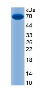
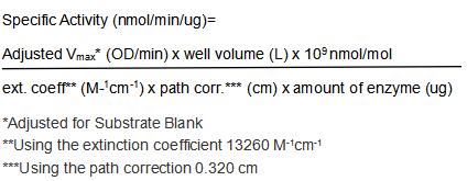
(The classical role of ACHE is to terminate cholinergic neurotransmission by hydrolysis of acetylcholine (ACH). ACHE is thought to be involved in the pathology of Alzheimer's disease (AD) by accelerating the assembly of A beta peptides into fibrillar species through forming complexes with A beta via the peripheral anionic site on ACHE. ACHE inhibitors have been used to delay symptoms of AD patients by virtue of their ability to enhance ACH availability, as well as reduce amyloidogenesis and subsequent neurotoxicity. Its involvement in the cholinergic anti-inflammatory pathway connects ACHE with a possible marker of low-grade systemic inflammation in obesity, hypertension, coronary heart disease, and AD. The activity of recombinant mouse ACHE was measured by its ability to cleave Acetylthiocholine in the assay buffer 0.1 M sodium phosphate, 0.05% (w/v) Brij-35, pH 7.5. 50 uL of various concentrations of rmACHE (diluted by Assay Buffer) was added into the 96-well clear plate and the reaction was started by the addition of 50 ul substrate mixture of 200 uM acetylthiocholine and 100 uM DTNB. The final well serves as a negative control with no rmACHE, replaced with 50ul assay buffer and 50 ul substrate mixture. Read plate in kinetic mode for 5 minutes at an absorbance of 405 nm. The specific activity of recombinant mouse ACHE is >2200 nmol/min/ug.)
Bioactivity
(The classical role of ACHE is to terminate cholinergic neurotransmission by hydrolysis of acetylcholine (ACH). ACHE is thought to be involved in the pathology of Alzheimer's disease (AD) by accelerating the assembly of A beta peptides into fibrillar species through forming complexes with A beta via the peripheral anionic site on ACHE. ACHE inhibitors have been used to delay symptoms of AD patients by virtue of their ability to enhance ACH availability, as well as reduce amyloidogenesis and subsequent neurotoxicity. Its involvement in the cholinergic anti-inflammatory pathway connects ACHE with a possible marker of low-grade systemic inflammation in obesity, hypertension, coronary heart disease, and AD. The activity of recombinant mouse ACHE was measured by its ability to cleave Acetylthiocholine in the assay buffer 0.1 M sodium phosphate, 0.05% (w/v) Brij-35, pH 7.5. 50 uL of various concentrations of rmACHE (diluted by Assay Buffer) was added into the 96-well clear plate and the reaction was started by the addition of 50 ul substrate mixture of 200 uM acetylthiocholine and 100 uM DTNB. The final well serves as a negative control with no rmACHE, replaced with 50ul assay buffer and 50 ul substrate mixture. Read plate in kinetic mode for 5 minutes at an absorbance of 405 nm. The specific activity of recombinant mouse ACHE is >2200 nmol/min/ug.)
Acetylcholinesterase (ACHE), Active Protein (Cat# AAA161851)
Full Name
Active Acetylcholinesterase (ACHE)
Applications
Activity Assay, Cell Culture
Purity
Greater than 90% by SDS-PAGE
Pricing
 Application Data
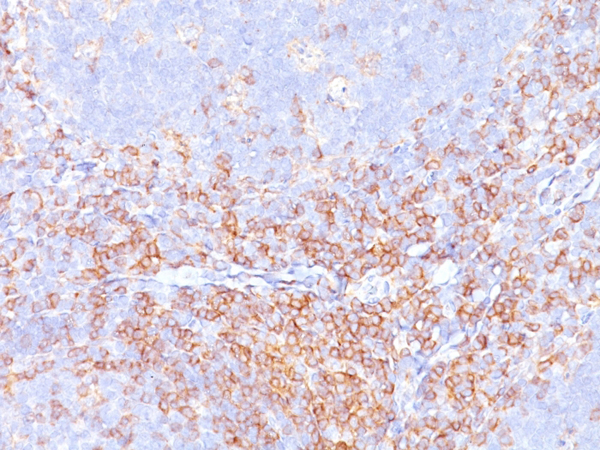
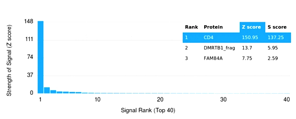
(Analysis of Protein Array containing more than 19,000 full-length human proteins using CD4 Recombinant Monospecific Rabbit Monoclonal Antibody (C4/3619R) Z- and S- Score: The Z-score represents the strength of a signal that a monoclonal antibody (MAb) (in combination with a fluorescently-tagged anti-IgG secondary antibody) produces when binding to a particular protein on the HuProtTM array. Z-scores are described in units of standard deviations (SD's) above the mean value of all signals generated on that array. If targets on HuProtTM are arranged in descending order of the Z-score, the S-score is the difference (also in units of SD's) between the Z-score. S-score therefore represents the relative target specificity of a MAb to its intended target. A MAb is considered to specific to its intended target, if the MAb has an S-score of at least 2.5. For example, if a MAb binds to protein X with a Z-score of 43 and to protein Y with a Z-score of 14, then the S-score for the binding of that MAb to protein X is equal to 29.)
Application Data
(Analysis of Protein Array containing more than 19,000 full-length human proteins using CD4 Recombinant Monospecific Rabbit Monoclonal Antibody (C4/3619R) Z- and S- Score: The Z-score represents the strength of a signal that a monoclonal antibody (MAb) (in combination with a fluorescently-tagged anti-IgG secondary antibody) produces when binding to a particular protein on the HuProtTM array. Z-scores are described in units of standard deviations (SD's) above the mean value of all signals generated on that array. If targets on HuProtTM are arranged in descending order of the Z-score, the S-score is the difference (also in units of SD's) between the Z-score. S-score therefore represents the relative target specificity of a MAb to its intended target. A MAb is considered to specific to its intended target, if the MAb has an S-score of at least 2.5. For example, if a MAb binds to protein X with a Z-score of 43 and to protein Y with a Z-score of 14, then the S-score for the binding of that MAb to protein X is equal to 29.)
CD4, Monoclonal Antibody (Cat# AAA215432)
Full Name
CD4 (T-Helper/Inducer Cell Marker)
Gene Names
CD4; CD4mut
Reactivity
Human
Applications
Immunohistochemistry
Purity
Purified Ab with BSA and Azide at 200ug/ml or Purified Ab with BSA and Azide at 200ug/ml or Purified Ab WITHOUT BSA and Azide at 1.0mg/ml
Pricing
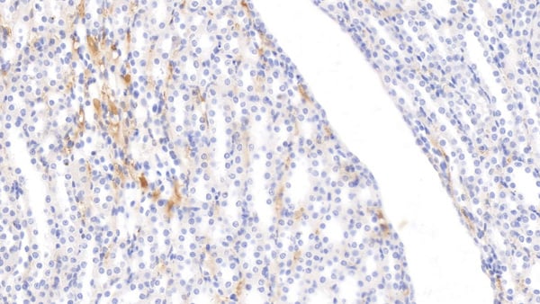
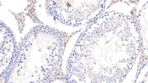
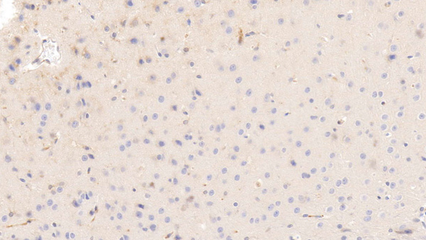
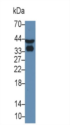 WB (Western Blot)
(Western Blot; Sample: Rat Serum Primary Ab: 3ug/ml Mouse AntiRat APOE Antibody Second Ab: 0.2ug/mL HRPLinked Caprine AntiMouse IgG Polyclonal Antibody (Catalog: SAA544Mu19))
WB (Western Blot)
(Western Blot; Sample: Rat Serum Primary Ab: 3ug/ml Mouse AntiRat APOE Antibody Second Ab: 0.2ug/mL HRPLinked Caprine AntiMouse IgG Polyclonal Antibody (Catalog: SAA544Mu19))
Apolipoprotein E (APOE), Monoclonal Antibody (Cat# AAA151609)
Full Name
Monoclonal Antibody to Apolipoprotein E (APOE)
Gene Names
Apoe; APOEA
Reactivity
Rat
Applications
Immunoprecipitation, Immunocytochemistry, Immunohistochemistry, Western Blot
Purity
Protein A + Protein G affinity chromatography
Pricing

 WB (Western Blot)
(Western Blot; Sample: Human SalivaPrimary Ab: 1ug/ml Mouse AntiHuman PADI4 AntibodySecond Ab: 0.2ug/mL HRPLinked Caprine AntiMouse IgG Polyclonal Antibody(Catalog: SAA544Mu19))
WB (Western Blot)
(Western Blot; Sample: Human SalivaPrimary Ab: 1ug/ml Mouse AntiHuman PADI4 AntibodySecond Ab: 0.2ug/mL HRPLinked Caprine AntiMouse IgG Polyclonal Antibody(Catalog: SAA544Mu19))
Peptidyl Arginine Deiminase Type IV (PADI4), Monoclonal Antibody (Cat# AAA151871)
Full Name
Monoclonal Antibody to Peptidyl Arginine Deiminase Type IV (PADI4)
Gene Names
PADI4; PAD; PAD4; PDI4; PDI5; PADI5
Reactivity
Human
Applications
Immunoprecipitation, Immunocytochemistry, Immunohistochemistry, Western Blot
Purity
Protein A + Protein G affinity chromatography
Pricing
 Application Data
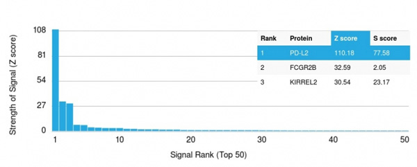
(Analysis of Protein Array containing more than 19,000 full-length human proteins using PD-L2 Mouse Monoclonal Antibody (PDL2/2676) Z- and S- Score: The Z-score represents the strength of a signal that a monoclonal antibody (MAb) (in combination with a fluorescently-tagged anti-IgG secondary antibody) produces when binding to a particular protein on the HuProtTM array. Z-scores are described in units of standard deviations (SD’s) above the mean value of all signals generated on that array. If targets on HuProtTM are arranged in descending order of the Z-score, the S-score is the difference (also in units of SD’s) between the Z-score. S-score therefore represents the relative target specificity of a MAb to its intended target. A MAb is considered to specific to its intended target, if the MAb has an S-score of at least 2.5. For example, if a MAb binds to protein X with a Z-score of 43 and to protein Y with a Z-score of 14, then the S-score for the binding of that MAb to protein X is equal to 29.)
Application Data
(Analysis of Protein Array containing more than 19,000 full-length human proteins using PD-L2 Mouse Monoclonal Antibody (PDL2/2676) Z- and S- Score: The Z-score represents the strength of a signal that a monoclonal antibody (MAb) (in combination with a fluorescently-tagged anti-IgG secondary antibody) produces when binding to a particular protein on the HuProtTM array. Z-scores are described in units of standard deviations (SD’s) above the mean value of all signals generated on that array. If targets on HuProtTM are arranged in descending order of the Z-score, the S-score is the difference (also in units of SD’s) between the Z-score. S-score therefore represents the relative target specificity of a MAb to its intended target. A MAb is considered to specific to its intended target, if the MAb has an S-score of at least 2.5. For example, if a MAb binds to protein X with a Z-score of 43 and to protein Y with a Z-score of 14, then the S-score for the binding of that MAb to protein X is equal to 29.)
PD-L2/PDCD1LG2/CD273, Monoclonal Antibody (Cat# AAA215267)
Full Name
PD-L2/PDCD1LG2/CD273
Reactivity
Human
Applications
Western Blot, Immunofluorescence, Flow Cytometry
Purity
Purified Ab with BSA and Azide at 200ug/ml OR Purified Ab WITHOUT BSA and Azide at 1.0mg/ml
Pricing
 Application Data
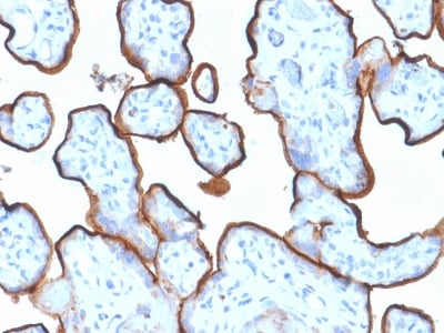
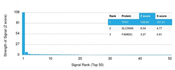
(Analysis of Protein Array containing >19, 000 full-length human proteins using CD71 Mouse Monoclonal Antibody (TFRC/1839) Z- and S- Score: The Z-score represents the strength of a signal that a monoclonal antibody (MAb) (in combination with a fluorescently-tagged anti-IgG secondary antibody) produces when binding to a particular protein on the HuProtTM array. Z-scores are described in units of standard deviations (SD's) above the mean value of all signals generated on that array. If targets on HuProtTM are arranged in descending order of the Z-score, the S-score is the difference (also in units of SD's) between the Z-score. S-score therefore represents the relative target specificity of a MAb to its intended target. A MAb is considered to specific to its intended target, if the MAb has an S-score of at least 2.5. For example, if a MAb binds to protein X with a Z-score of 43 and to protein Y with a Z-score of 14, then the S-score for the binding of that MAb to protein X is equal to 29.)
Application Data
(Analysis of Protein Array containing >19, 000 full-length human proteins using CD71 Mouse Monoclonal Antibody (TFRC/1839) Z- and S- Score: The Z-score represents the strength of a signal that a monoclonal antibody (MAb) (in combination with a fluorescently-tagged anti-IgG secondary antibody) produces when binding to a particular protein on the HuProtTM array. Z-scores are described in units of standard deviations (SD's) above the mean value of all signals generated on that array. If targets on HuProtTM are arranged in descending order of the Z-score, the S-score is the difference (also in units of SD's) between the Z-score. S-score therefore represents the relative target specificity of a MAb to its intended target. A MAb is considered to specific to its intended target, if the MAb has an S-score of at least 2.5. For example, if a MAb binds to protein X with a Z-score of 43 and to protein Y with a Z-score of 14, then the S-score for the binding of that MAb to protein X is equal to 29.)
CD71/Transferrin Receptor (TFRC), Monoclonal Antibody (Cat# AAA214697)
Full Name
CD71/Transferrin Receptor (TFRC) (Extracellular Domain)
Gene Names
TFRC; T9; TR; TFR; p90; CD71; TFR1; TRFR; IMD46
Reactivity
Human. Others not known.
Applications
Immunohistochemistry, Western Blot
Pricing
 FCM/FACS (Flow Cytometry)
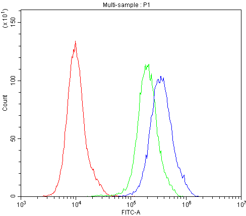
(Figure 5. Flow Cytometry analysis of THP-1 cells using anti-CD163 antibody (AAA124594).Overlay histogram showing THP-1 cells stained with AAA124594 (Blue line).The cells were blocked with 10% normal goat serum. And then incubated with rabbit anti-CD163 Antibody (AAA124594,1ug/1x10^6 cells) for 30 min at 20 degree C. DyLight®488 conjugated goat anti-rabbit IgG (5-10ug/1x10^6 cells) was used as secondary antibody for 30 minutes at 20 degree C. Isotype control antibody (Green line) was rabbit IgG (1ug/1x106) used under the same conditions. Unlabelled sample (Red line) was also used as a control.)
FCM/FACS (Flow Cytometry)
(Figure 5. Flow Cytometry analysis of THP-1 cells using anti-CD163 antibody (AAA124594).Overlay histogram showing THP-1 cells stained with AAA124594 (Blue line).The cells were blocked with 10% normal goat serum. And then incubated with rabbit anti-CD163 Antibody (AAA124594,1ug/1x10^6 cells) for 30 min at 20 degree C. DyLight®488 conjugated goat anti-rabbit IgG (5-10ug/1x10^6 cells) was used as secondary antibody for 30 minutes at 20 degree C. Isotype control antibody (Green line) was rabbit IgG (1ug/1x106) used under the same conditions. Unlabelled sample (Red line) was also used as a control.)
CD163, Antibody (Cat# AAA124594)
Full Name
Anti-CD163 Picoband antibody
Gene Names
CD163; M130; MM130; SCARI1
Reactivity
Human, Mouse, Rat
Applications
Flow Cytometry, Immunocytochemistry, Immunohistochemistry, Immunohistochemistry, Western Blot
Purity
Immunogen affinity purified
Pricing
 Application Data
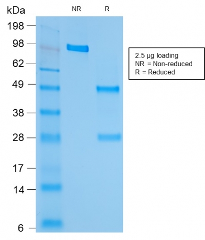
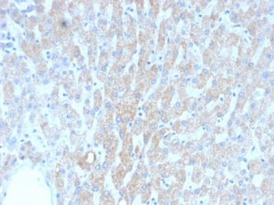
(Analysis of Protein Array containing more than 19, 000 full-length human proteins using HSP60 Mouse Monoclonal Antibody (rGROEL/780).Z- and S- Score: The Z-score represents the strength of a signal that a monoclonal antibody (MAb) (in combination with a fluorescently-tagged anti-IgG secondary antibody) produces when binding to a particular protein on the HuProtTM array. Z-scores are described in units of standard deviations (SD's) above the mean value of all signals generated on that array. If targets on HuProtTM are arranged in descending order of the Z-score, the S-score is the difference (also in units of SD's) between the Z-score. S-score therefore represents the relative target specificity of a MAb to its intended target. A MAb is considered to specific to its intended target, if the MAb has an S-score of at least 2.5. For example, if a MAb binds to protein X with a Z-score of 43 and to protein Y with a Z-score of 14, then the S-score for the binding of that MAb to protein X is equal to 29.)
Application Data
(Analysis of Protein Array containing more than 19, 000 full-length human proteins using HSP60 Mouse Monoclonal Antibody (rGROEL/780).Z- and S- Score: The Z-score represents the strength of a signal that a monoclonal antibody (MAb) (in combination with a fluorescently-tagged anti-IgG secondary antibody) produces when binding to a particular protein on the HuProtTM array. Z-scores are described in units of standard deviations (SD's) above the mean value of all signals generated on that array. If targets on HuProtTM are arranged in descending order of the Z-score, the S-score is the difference (also in units of SD's) between the Z-score. S-score therefore represents the relative target specificity of a MAb to its intended target. A MAb is considered to specific to its intended target, if the MAb has an S-score of at least 2.5. For example, if a MAb binds to protein X with a Z-score of 43 and to protein Y with a Z-score of 14, then the S-score for the binding of that MAb to protein X is equal to 29.)
HSP60 (Heat Shock Protein 60), Monoclonal Antibody (Cat# AAA214389)
Full Name
HSP60 (Heat Shock Protein 60) (Mitochondrial Marker)
Gene Names
HSPD1; HLD4; CPN60; GROEL; HSP60; HSP65; SPG13; HSP-60; HuCHA60
Reactivity
Human, Mouse, Rat, Hamster, Sheep, Rabbit, Cow, Dog, Pig, Monkey, Chicken, Xenopus laevis, Drosophila.
Does not react with Bacteria, Helminths, Spinach. Others not known.
Does not react with Bacteria, Helminths, Spinach. Others not known.
Applications
Immunohistochemistry, Western Blot, Immunofluorescence, Flow Cytometry
Pricing
 Application Data
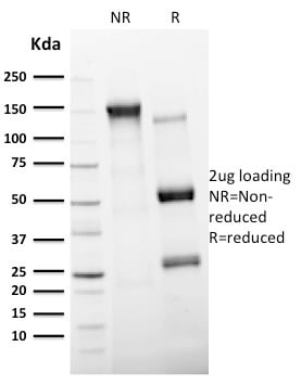
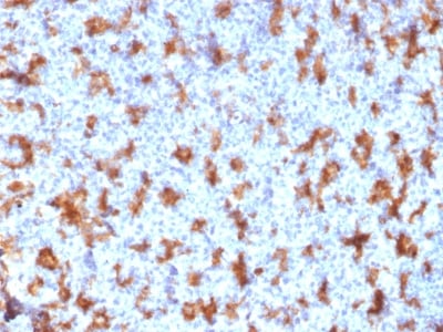
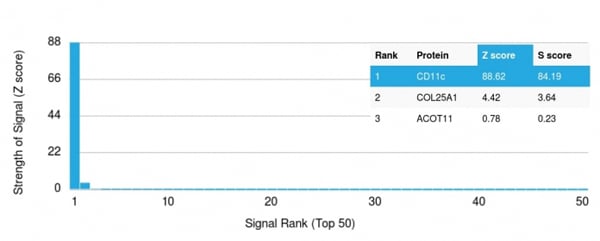
(Analysis of Protein Array containing >19, 000 full-length human proteins using CD11c Mouse Monoclonal Antibody (ITGAX/2507) Z- and S- Score: The Z-score represents the strength of a signal that a monoclonal antibody (MAb) (in combination with a fluorescently-tagged anti-IgG secondary antibody) produces when binding to a particular protein on the HuProtTM array. Z-scores are described in units of standard deviations (SD's) above the mean value of all signals generated on that array. If targets on HuProtTM are arranged in descending order of the Z-score, the S-score is the difference (also in units of SD's) between the Z-score. S-score therefore represents the relative target specificity of a MAb to its intended target. A MAb is considered to specific to its intended target, if the MAb has an S-score of at least 2.5. For example, if a MAb binds to protein X with a Z-score of 43 and to protein Y with a Z-score of 14, then the S-score for the binding of that MAb to protein X is equal to 29.)
Application Data
(Analysis of Protein Array containing >19, 000 full-length human proteins using CD11c Mouse Monoclonal Antibody (ITGAX/2507) Z- and S- Score: The Z-score represents the strength of a signal that a monoclonal antibody (MAb) (in combination with a fluorescently-tagged anti-IgG secondary antibody) produces when binding to a particular protein on the HuProtTM array. Z-scores are described in units of standard deviations (SD's) above the mean value of all signals generated on that array. If targets on HuProtTM are arranged in descending order of the Z-score, the S-score is the difference (also in units of SD's) between the Z-score. S-score therefore represents the relative target specificity of a MAb to its intended target. A MAb is considered to specific to its intended target, if the MAb has an S-score of at least 2.5. For example, if a MAb binds to protein X with a Z-score of 43 and to protein Y with a Z-score of 14, then the S-score for the binding of that MAb to protein X is equal to 29.)
CD11c, Monoclonal Antibody (Cat# AAA214660)
Full Name
CD11c (Dendritic Cell Marker)
Gene Names
ITGAX; CD11C; SLEB6
Reactivity
Human
Applications
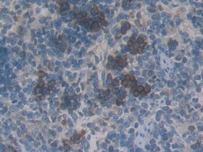
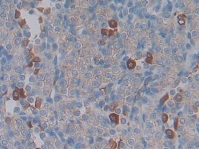
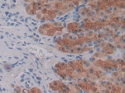
Immunohistochemistry
Pricing
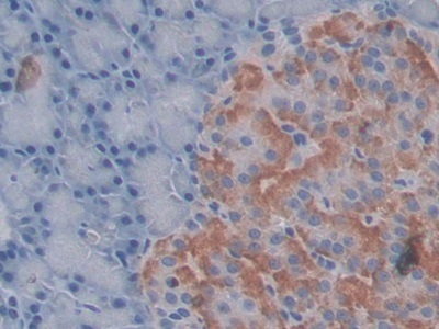 IHC (Immunohistochemistry)
(DAB staining on IHCP;Sample: Rat Pancreas Tissue; Primary Ab: 30ug/ml Mouse AntiRat CTGF AntibodySecond Ab: 2ug/mL HRPLinked Caprine AntiMouse IgG Polyclonal Antibody(Catalog: SAA544Mu19))
IHC (Immunohistochemistry)
(DAB staining on IHCP;Sample: Rat Pancreas Tissue; Primary Ab: 30ug/ml Mouse AntiRat CTGF AntibodySecond Ab: 2ug/mL HRPLinked Caprine AntiMouse IgG Polyclonal Antibody(Catalog: SAA544Mu19))
Connective Tissue Growth Factor (CTGF), Monoclonal Antibody (Cat# AAA151432)
Full Name
Monoclonal Antibody to Connective Tissue Growth Factor (CTGF)
Gene Names
Ctgf; CTGRP
Reactivity
Rat
Applications
Immunoprecipitation, Immunocytochemistry, Immunohistochemistry, Western Blot
Purity
Protein A + Protein G affinity chromatography
Pricing
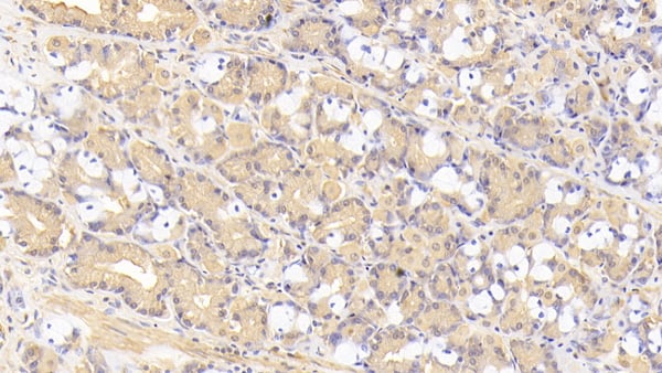
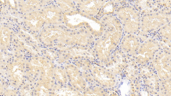
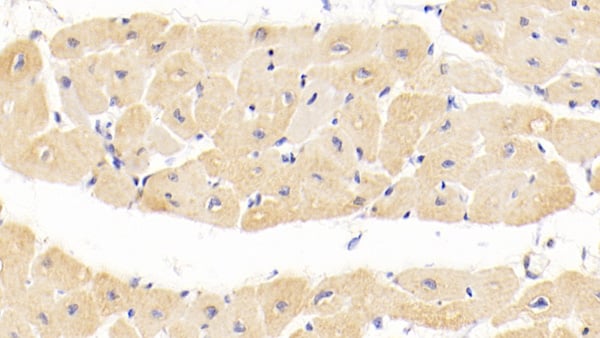 IHC (Immunohistochemisry)
(DAB staining on IHCP;Sample: Human Cardiac Muscle Tissue; Primary Ab: 20ug/ml Mouse AntiHuman TNC AntibodySecond Ab: 2ug/mL HRPLinked Caprine AntiMouse IgG Polyclonal Antibody(Catalog: SAA544Mu19))
IHC (Immunohistochemisry)
(DAB staining on IHCP;Sample: Human Cardiac Muscle Tissue; Primary Ab: 20ug/ml Mouse AntiHuman TNC AntibodySecond Ab: 2ug/mL HRPLinked Caprine AntiMouse IgG Polyclonal Antibody(Catalog: SAA544Mu19))
Tenascin C (TNC), Monoclonal Antibody (Cat# AAA151774)
Full Name
Monoclonal Antibody to Tenascin C (TNC)
Gene Names
TNC; GP; JI; TN; HXB; GMEM; TN-C; 150-225
Reactivity
Human, Pig
Applications
Immunoprecipitation, Immunocytochemistry, Immunohistochemistry, Western Blot
Purity
Protein A + Protein G affinity chromatography
Pricing
 IHC (Immunohistochemistry)
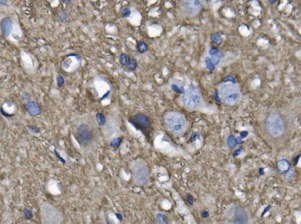
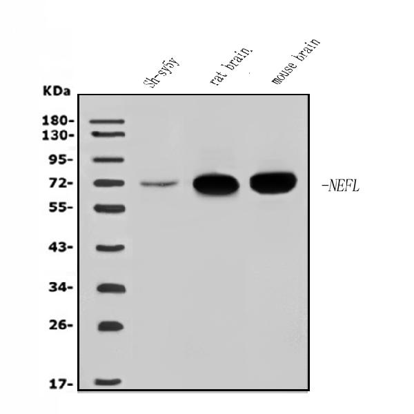
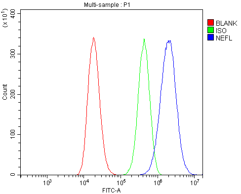
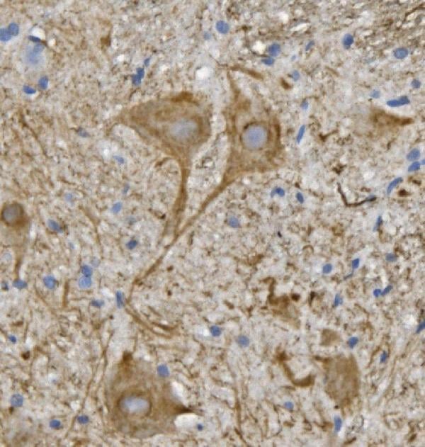
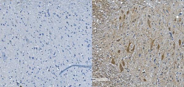
(Figure 5. IHC analysis of NEFL/NF-L using anti-NEFL/NF-L antibody (AAA125655).NEFL/NF-L was detected in paraffin-embedded section of mouse spinal cord tissue. Heat mediated antigen retrieval was performed in EDTA buffer (pH8. 0, epitope retrieval solution). The tissue section was blocked with 10% goat serum. The tissue section was then incubated with 2μg/ml rabbit anti-NEFL/NF-L Antibody (AAA125655) overnight at 4 degree C. Biotinylated goat anti-rabbit IgG was used as secondary antibody and incubated for 30 minutes at 37 degree C. The tissue section was developed using Strepavidin-Biotin-Complex (SABC) with DAB as the chromogen.)
IHC (Immunohistochemistry)
(Figure 5. IHC analysis of NEFL/NF-L using anti-NEFL/NF-L antibody (AAA125655).NEFL/NF-L was detected in paraffin-embedded section of mouse spinal cord tissue. Heat mediated antigen retrieval was performed in EDTA buffer (pH8. 0, epitope retrieval solution). The tissue section was blocked with 10% goat serum. The tissue section was then incubated with 2μg/ml rabbit anti-NEFL/NF-L Antibody (AAA125655) overnight at 4 degree C. Biotinylated goat anti-rabbit IgG was used as secondary antibody and incubated for 30 minutes at 37 degree C. The tissue section was developed using Strepavidin-Biotin-Complex (SABC) with DAB as the chromogen.)
NEFL/NF-L, Polyclonal Antibody (Cat# AAA125655)
Full Name
Anti-NEFL/NF-L Antibody
Gene Names
NEFL; NFL; NF-L; NF68; CMT1F; CMT2E; PPP1R110
Reactivity
Human, Mouse, Rat
Applications
Direct ELISA, Flow Cytometry, Immunohistochemistry, Western Blot
Purity
Immunogen affinity purified.
Pricing
 WB (Western Blot)
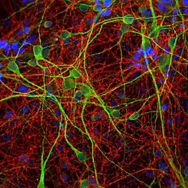
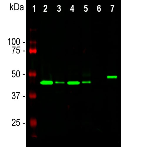
(Western blot analysis of different tissue and cell lysates using mouse mAb to GAP43, MO22170, dilution 1:5,000, in green: [1] protein standard (red), [2] rat brain, [3] rat spinal cord, [4] mouse brain, [5] mouse spinal cord, [6] C6 cells, [7] SH-SY5Y cells. The single band at the 43kDa mark corresponds to the GAP43 protein.)
WB (Western Blot)
(Western blot analysis of different tissue and cell lysates using mouse mAb to GAP43, MO22170, dilution 1:5,000, in green: [1] protein standard (red), [2] rat brain, [3] rat spinal cord, [4] mouse brain, [5] mouse spinal cord, [6] C6 cells, [7] SH-SY5Y cells. The single band at the 43kDa mark corresponds to the GAP43 protein.)
GAP43, Monoclonal Antibody (Cat# AAA76610)
Full Name
GAP43
Gene Names
GAP43; B-50; PP46
Reactivity
Human, Mouse, Rat
Applications
Immunofluorescence, Immunohistochemistry, Western Blot, Immunocytochemistry
Purity
Affinity Purified
Pricing
 IF (Immunofluorescence)
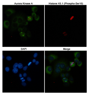
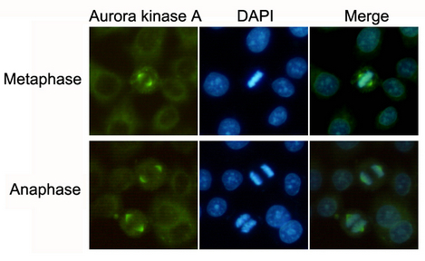
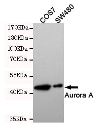
(Immunofluorescent analysis of Hela cells labeled with Aurora Kinase A(200525.dilution 1:50) mouse mAb (green) and Histone H3.1 (Phospho-Ser10)(310045.dilution 1:100) Rabbit pAb (red). DAPI was used to stain nucleus(blue).)
IF (Immunofluorescence)
(Immunofluorescent analysis of Hela cells labeled with Aurora Kinase A(200525.dilution 1:50) mouse mAb (green) and Histone H3.1 (Phospho-Ser10)(310045.dilution 1:100) Rabbit pAb (red). DAPI was used to stain nucleus(blue).)
Aurora Kinase A, Monoclonal Antibody (Cat# AAA320331)
Full Name
Aurora Kinase A Mouse mAb
Gene Names
AURKA; AIK; ARK1; AURA; BTAK; STK6; STK7; STK15; AURORA2; PPP1R47
Reactivity
Human, Monkey
Applications
Immunocytochemistry, Immunofluorescence, Western Blot
Purity
The antibody was affinity-purified from mouse ascites by affinity-chromatography using epitope-specific immunogen.
Pricing
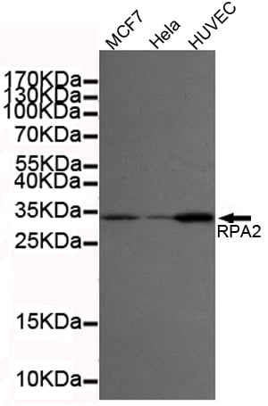 ICC (Immunocytochemistry)
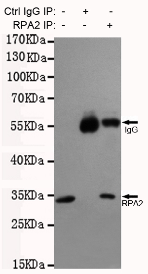
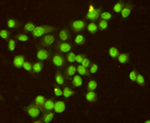
(Immunocytochemistry staining of HeLa cells fixed with -20? Methanol and using anti-RPA32/RPA2 antibody (dilution 1:200).)
ICC (Immunocytochemistry)
(Immunocytochemistry staining of HeLa cells fixed with -20? Methanol and using anti-RPA32/RPA2 antibody (dilution 1:200).)
RPA32/RPA2, Monoclonal Antibody (Cat# AAA320375)
Full Name
RPA32/RPA2 Mouse mAb
Gene Names
RPA2; REPA2; RPA32
Reactivity
Human
Applications
Immunocytochemistry, Immunoprecipitation, Western Blot
Purity
The antibody was affinity-purified from mouse ascites by affinity-chromatography using epitope-specific immunogen.
Pricing
 FCM/FACS (Flow Cytometry)
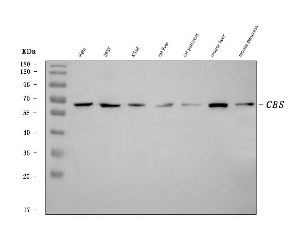
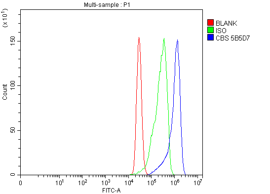
(Figure 2. Flow Cytometry analysis of MCF-7 cells using anti-CBS antibody (AAA126860).Overlay histogram showing MCF-7 cells stained with AAA126860 (Blue line). The cells were blocked with 10% normal goat serum. And then incubated with mouse anti-CBS Antibody (AAA126860, 1 ug/1x10^6 cells) for 30 min at 20 degree C. DyLight488 conjugated goat anti-mouse IgG was used as secondary antibody for 30 minutes at 20 degree C. Isotype control antibody (Green line) was mouse IgG (1 ug/1x10^6) used under the same conditions. Unlabelled sample (Red line) was also used as a control.)
FCM/FACS (Flow Cytometry)
(Figure 2. Flow Cytometry analysis of MCF-7 cells using anti-CBS antibody (AAA126860).Overlay histogram showing MCF-7 cells stained with AAA126860 (Blue line). The cells were blocked with 10% normal goat serum. And then incubated with mouse anti-CBS Antibody (AAA126860, 1 ug/1x10^6 cells) for 30 min at 20 degree C. DyLight488 conjugated goat anti-mouse IgG was used as secondary antibody for 30 minutes at 20 degree C. Isotype control antibody (Green line) was mouse IgG (1 ug/1x10^6) used under the same conditions. Unlabelled sample (Red line) was also used as a control.)
CBS, Monoclonal Antibody (Cat# AAA126860)
Full Name
Anti-CBS Antibody Picoband (monoclonal, 5B5D7)
Gene Names
CBS; HIP4
Reactivity
Human, Mouse, Rat
Applications
Flow Cytometry, Western Blot
Purity
Immunogen affinity purified.
Pricing
 FCM/FACS (Flow Cytometry)
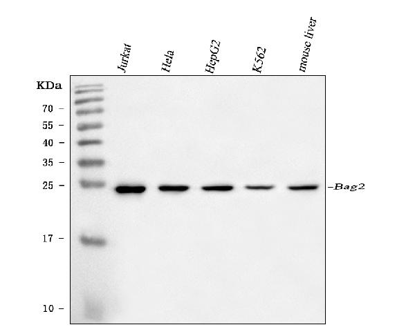
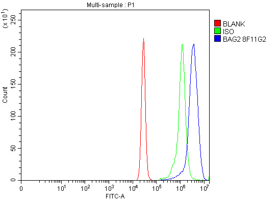
(Figure 2. Flow Cytometry analysis of K562 cells using anti-BAG2 antibody (AAA126942).Overlay histogram showing K562 cells stained with AAA126942 (Blue line). The cells were blocked with 10% normal goat serum. And then incubated with mouse anti-BAG2 Antibody (AAA126942, 1 ug/1x10^6 cells) for 30 min at 20 degree C. DyLight488 conjugated goat anti-mouse IgG was used as secondary antibody for 30 minutes at 20 degree C. Isotype control antibody (Green line) was mouse IgG (1 ug/1x10^6) used under the same conditions. Unlabelled sample (Red line) was also used as a control.)
FCM/FACS (Flow Cytometry)
(Figure 2. Flow Cytometry analysis of K562 cells using anti-BAG2 antibody (AAA126942).Overlay histogram showing K562 cells stained with AAA126942 (Blue line). The cells were blocked with 10% normal goat serum. And then incubated with mouse anti-BAG2 Antibody (AAA126942, 1 ug/1x10^6 cells) for 30 min at 20 degree C. DyLight488 conjugated goat anti-mouse IgG was used as secondary antibody for 30 minutes at 20 degree C. Isotype control antibody (Green line) was mouse IgG (1 ug/1x10^6) used under the same conditions. Unlabelled sample (Red line) was also used as a control.)
BAG2, Monoclonal Antibody (Cat# AAA126942)
Full Name
Anti-BAG2 Antibody Picoband (monoclonal, 8F11G2)
Gene Names
BAG2; BAG-2; dJ417I1.2
Reactivity
Human, Mouse
Applications
Flow Cytometry, Western Blot
Purity
Immunogen affinity purified.
Pricing
 FCM/FACS (Flow Cytometry)
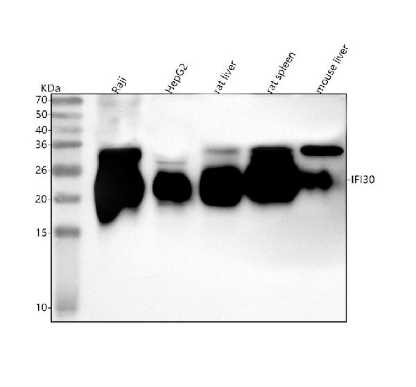
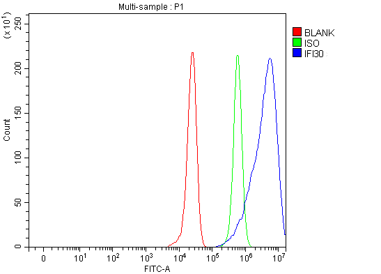
(Figure 2. Flow Cytometry analysis of HepG2 cells using anti-IFI30 antibody (AAA127324).Overlay histogram showing HepG2 cells stained with AAA127324 (Blue line). The cells were fixed with 4% paraformaldehyde and blocked with 10% normal goat serum. And then incubated with rabbit anti-IFI30 Antibody (AAA127324, 1ug/1x106 cells) for 30 min at 20 degree C. DyLight488 conjugated goat anti-rabbit IgG was used as secondary antibody for 30 minutes at 20 degree C. Isotype control antibody (Green line) was rabbit IgG (1ug/1x106) used under the same conditions. Unlabelled sample (Red line) was also used as a control.)
FCM/FACS (Flow Cytometry)
(Figure 2. Flow Cytometry analysis of HepG2 cells using anti-IFI30 antibody (AAA127324).Overlay histogram showing HepG2 cells stained with AAA127324 (Blue line). The cells were fixed with 4% paraformaldehyde and blocked with 10% normal goat serum. And then incubated with rabbit anti-IFI30 Antibody (AAA127324, 1ug/1x106 cells) for 30 min at 20 degree C. DyLight488 conjugated goat anti-rabbit IgG was used as secondary antibody for 30 minutes at 20 degree C. Isotype control antibody (Green line) was rabbit IgG (1ug/1x106) used under the same conditions. Unlabelled sample (Red line) was also used as a control.)
IFI30, Polyclonal Antibody (Cat# AAA127324)
Full Name
Anti-IFI30 Antibody Picoband
Gene Names
IFI30; GILT; IP30; IP-30; IFI-30
Reactivity
Human, Mouse, Rat
Applications
Flow Cytometry, Western Blot
Purity
Immunogen affinity purified.
Pricing
 IF (Immunofluorescence)
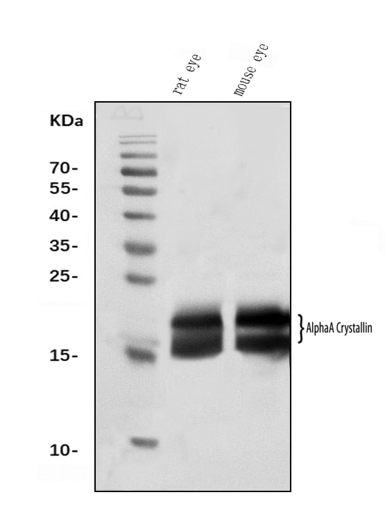
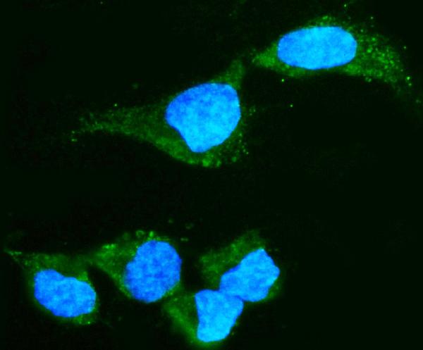
(Figure 2. IF analysis of Alpha A Crystallin using anti-Alpha A Crystallin antibody (AAA126900).Alpha A Crystallin was detected in an immunocytochemical section of HepG2 cells. Enzyme antigen retrieval was performed using IHC enzyme antigen retrieval reagent (AR0022) for 15 mins. The cells were blocked with 10% goat serum. And then incubated with 5 ug/mL mouse anti-Alpha A Crystallin Antibody (AAA126900) overnight at 4 degree C. DyLight488 Conjugated Goat Anti-Mouse IgG was used as secondary antibody at 1:100 dilution and incubated for 30 minutes at 37 degree C. The section was counterstained with DAPI. Visualize using a fluorescence microscope and filter sets appropriate for the label used.)
IF (Immunofluorescence)
(Figure 2. IF analysis of Alpha A Crystallin using anti-Alpha A Crystallin antibody (AAA126900).Alpha A Crystallin was detected in an immunocytochemical section of HepG2 cells. Enzyme antigen retrieval was performed using IHC enzyme antigen retrieval reagent (AR0022) for 15 mins. The cells were blocked with 10% goat serum. And then incubated with 5 ug/mL mouse anti-Alpha A Crystallin Antibody (AAA126900) overnight at 4 degree C. DyLight488 Conjugated Goat Anti-Mouse IgG was used as secondary antibody at 1:100 dilution and incubated for 30 minutes at 37 degree C. The section was counterstained with DAPI. Visualize using a fluorescence microscope and filter sets appropriate for the label used.)
Alpha A Crystallin, Monoclonal Antibody (Cat# AAA126900)
Full Name
Anti-Alpha A Crystallin Antibody Picoband (monoclonal, 10B9)
Gene Names
CRYAA; CRYA1; HSPB4; CTRCT9
Reactivity
Human, Mouse, Rat
Applications
Immunofluorescence, Immunocytochemistry, Western Blot
Purity
Immunogen affinity purified.
Pricing
 FCM/FACS (Flow Cytometry)
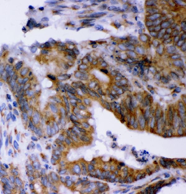
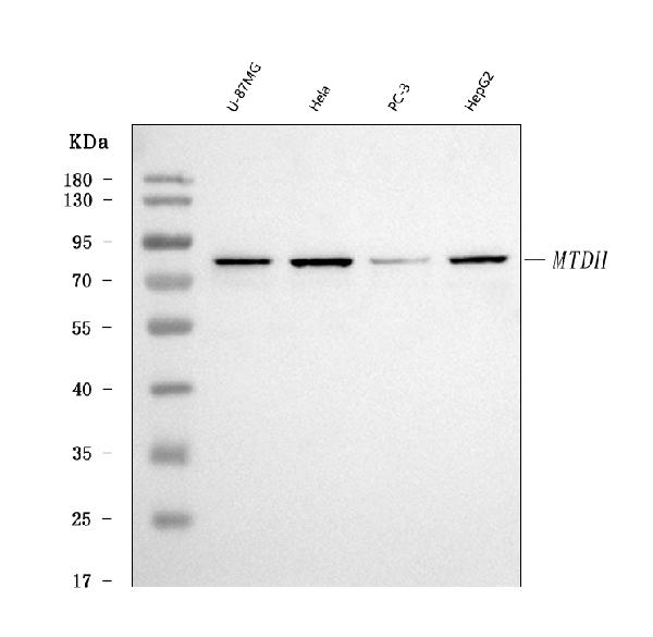
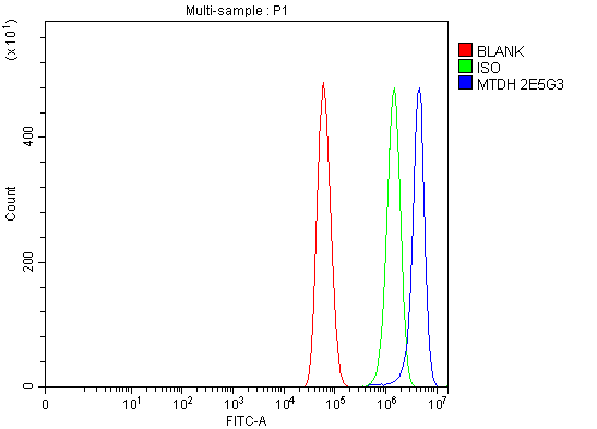
(Figure 3. Flow Cytometry analysis of U87 cells using anti-LYRIC antibody (AAA126933).Overlay histogram showing U87 cells stained with AAA126933 (Blue line). The cells were blocked with 10% normal goat serum. And then incubated with mouse anti-LYRIC Antibody (AAA126933, 1 ug/1x10^6 cells) for 30 min at 20 degree C. DyLight488 conjugated goat anti-mouse IgG was used as secondary antibody for 30 minutes at 20 degree C. Isotype control antibody (Green line) was mouse IgG (1 ug/1x10^6) used under the same conditions. Unlabelled sample (Red line) was also used as a control.)
FCM/FACS (Flow Cytometry)
(Figure 3. Flow Cytometry analysis of U87 cells using anti-LYRIC antibody (AAA126933).Overlay histogram showing U87 cells stained with AAA126933 (Blue line). The cells were blocked with 10% normal goat serum. And then incubated with mouse anti-LYRIC Antibody (AAA126933, 1 ug/1x10^6 cells) for 30 min at 20 degree C. DyLight488 conjugated goat anti-mouse IgG was used as secondary antibody for 30 minutes at 20 degree C. Isotype control antibody (Green line) was mouse IgG (1 ug/1x10^6) used under the same conditions. Unlabelled sample (Red line) was also used as a control.)
LYRIC, Monoclonal Antibody (Cat# AAA126933)
Full Name
Anti-LYRIC Antibody Picoband (monoclonal, 2E5G3)
Gene Names
MTDH; 3D3; AEG1; AEG-1; LYRIC; LYRIC/3D3
Reactivity
Human
Applications
Flow Cytometry, Immunohistochemistry, Western Blot
Purity
Immunogen affinity purified.
Pricing
 FCM/FACS (Flow Cytometry)
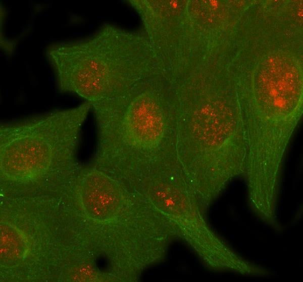
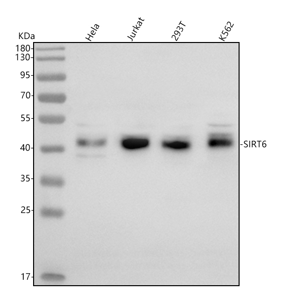
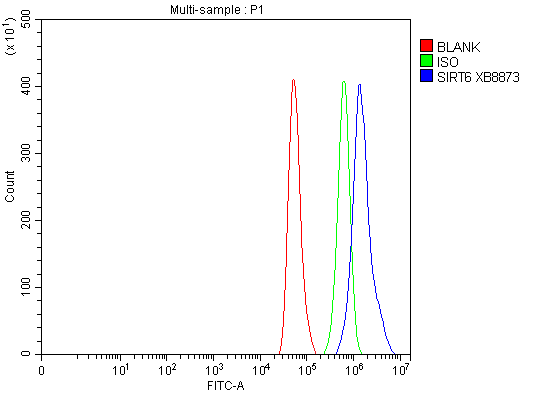
(Figure 3. Flow Cytometry analysis of Hela cells using anti-SIRT6 antibody (AAA127000).Overlay histogram showing Hela cells stained with AAA127000 (Blue line). To facilitate intracellular staining, cells were fixed with 4% paraformaldehyde and permeabilized with permeabilization buffer. The cells were blocked with 10% normal goat serum. And then incubated with rabbit anti-SIRT6 Antibody (AAA127000, 1ug/1x106 cells) for 30 min at 20 degree C. DyLight488 conjugated goat anti-rabbit IgG was used as secondary antibody for 30 minutes at 20 degree C. Isotype control antibody (Green line) was rabbit IgG (1ug/1x106) used under the same conditions. Unlabelled sample (Red line) was also used as a control.)
FCM/FACS (Flow Cytometry)
(Figure 3. Flow Cytometry analysis of Hela cells using anti-SIRT6 antibody (AAA127000).Overlay histogram showing Hela cells stained with AAA127000 (Blue line). To facilitate intracellular staining, cells were fixed with 4% paraformaldehyde and permeabilized with permeabilization buffer. The cells were blocked with 10% normal goat serum. And then incubated with rabbit anti-SIRT6 Antibody (AAA127000, 1ug/1x106 cells) for 30 min at 20 degree C. DyLight488 conjugated goat anti-rabbit IgG was used as secondary antibody for 30 minutes at 20 degree C. Isotype control antibody (Green line) was rabbit IgG (1ug/1x106) used under the same conditions. Unlabelled sample (Red line) was also used as a control.)
SIRT6, Polyclonal Antibody (Cat# AAA127000)
Full Name
Anti-SIRT6 Antibody Picoband
Gene Names
SIRT6; SIR2L6
Reactivity
Human
Applications
Western Blot, Immunofluorescence, Immunocytochemistry, Flow Cytometry
Purity
Immunogen affinity purified.
Pricing
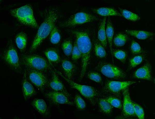
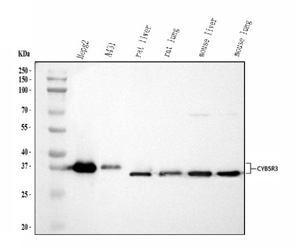
 FCM/FACS (Flow Cytometry)
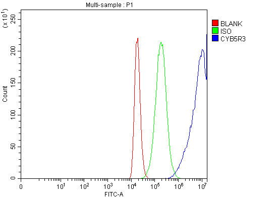
(Figure 4. Flow Cytometry analysis of 293T cells using anti-CYB5R3 antibody (AAA126312).Overlay histogram showing 293T cells stained with AAA126312 (Blue line). The cells were blocked with 10% normal goat serum. And then incubated with rabbit anti-CYB5R3 Antibody (AAA126312, 1 ug/1x10^6 cells) for 30 min at 20 degree C. DyLight488 conjugated goat anti-rabbit IgG was used as secondary antibody for 30 minutes at 20 degree C. Isotype control antibody (Green line) was rabbit IgG (1 ug/1x10^6) used under the same conditions. Unlabelled sample (Red line) was also used as a control.)
FCM/FACS (Flow Cytometry)
(Figure 4. Flow Cytometry analysis of 293T cells using anti-CYB5R3 antibody (AAA126312).Overlay histogram showing 293T cells stained with AAA126312 (Blue line). The cells were blocked with 10% normal goat serum. And then incubated with rabbit anti-CYB5R3 Antibody (AAA126312, 1 ug/1x10^6 cells) for 30 min at 20 degree C. DyLight488 conjugated goat anti-rabbit IgG was used as secondary antibody for 30 minutes at 20 degree C. Isotype control antibody (Green line) was rabbit IgG (1 ug/1x10^6) used under the same conditions. Unlabelled sample (Red line) was also used as a control.)
CYB5R3, Polyclonal Antibody (Cat# AAA126312)
Full Name
Anti-CYB5R3 Antibody Picoband
Gene Names
CYB5R3; B5R; DIA1
Reactivity
Human, Mouse, Rat
Applications
Flow Cytometry, Immunofluorescence, Immunocytochemistry, Western Blot
Purity
Immunogen affinity purified.
Pricing
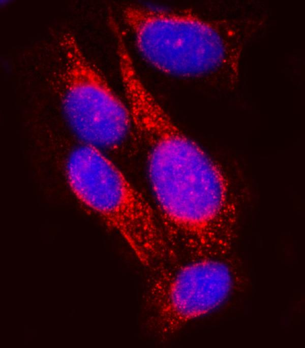
 FCM/FACS (Flow Cytometry)
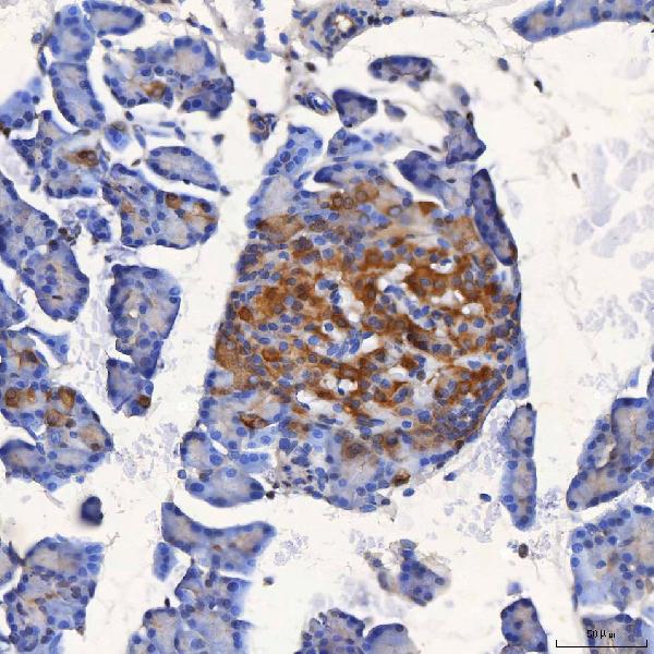
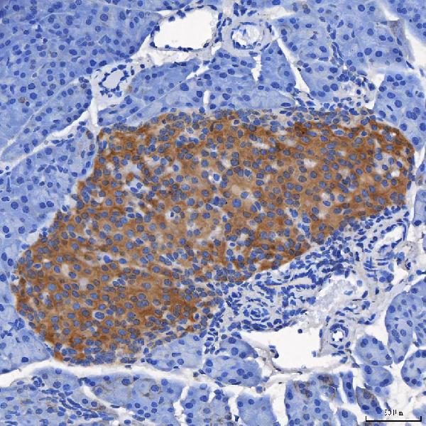
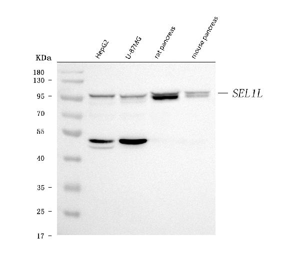
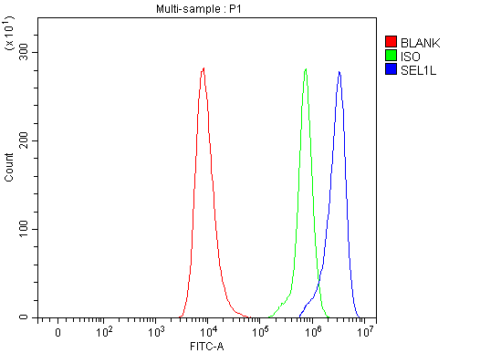
(Figure 5. Flow Cytometry analysis of HEL cells using anti-SEL1L antibody (AAA126301).Overlay histogram showing HEL cells stained with AAA126301 (Blue line). The cells were blocked with 10% normal goat serum. And then incubated with rabbit anti-SEL1L Antibody (AAA126301, 1 ug/1x10^6 cells) for 30 min at 20 degree C. DyLight488 conjugated goat anti-rabbit IgG was used as secondary antibody for 30 minutes at 20 degree C. Isotype control antibody (Green line) was rabbit IgG (1 ug/1x10^6) used under the same conditions. Unlabelled sample (Red line) was also used as a control.)
FCM/FACS (Flow Cytometry)
(Figure 5. Flow Cytometry analysis of HEL cells using anti-SEL1L antibody (AAA126301).Overlay histogram showing HEL cells stained with AAA126301 (Blue line). The cells were blocked with 10% normal goat serum. And then incubated with rabbit anti-SEL1L Antibody (AAA126301, 1 ug/1x10^6 cells) for 30 min at 20 degree C. DyLight488 conjugated goat anti-rabbit IgG was used as secondary antibody for 30 minutes at 20 degree C. Isotype control antibody (Green line) was rabbit IgG (1 ug/1x10^6) used under the same conditions. Unlabelled sample (Red line) was also used as a control.)
SEL1L, Polyclonal Antibody (Cat# AAA126301)
Full Name
Anti-SEL1L Antibody Picoband
Gene Names
SEL1L; SEL1L1; PRO1063; SEL1-LIKE
Reactivity
Human, Mouse, Rat
Applications
Flow Cytometry, Immunofluorescence, Immunocytochemistry, Immunohistochemistry, Western Blot
Purity
Immunogen affinity purified.
Pricing
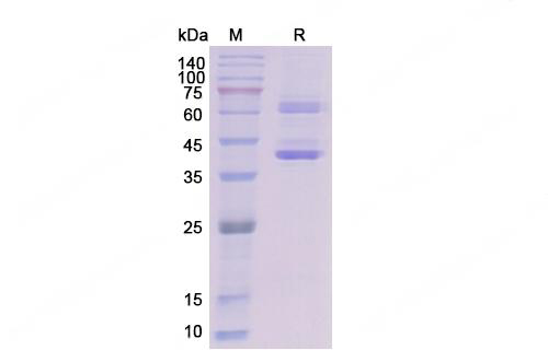 SDS-PAGE
(SDS PAGE for recombinant Dog NGF / Beta-NGF)
SDS-PAGE
(SDS PAGE for recombinant Dog NGF / Beta-NGF)
NGF/Beta-NGF, Recombinant Protein (Cat# AAA120622)
Full Name
Recombinant Dog NGF/Beta-NGF Protein, C-Fc
Applications
Western Blot
Purity
>80% as determined by SDS-PAGE
Pricing
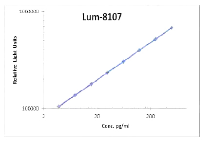 Standard Curve (Sample)
BMP-2, CLIA Kit (Cat# AAA318523)
Full Name
BMP-2 (Human) LumiAb ELISA Kit
Gene Names
BMP2; BDA2; BMP2A; SSFSC
Reactivity
Human
Pricing
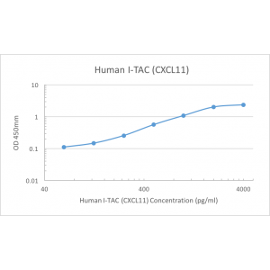 Standard Curve (Sample)
I-TAC (CXCL11), ELISA Kit (Cat# AAA79386)
Full Name
Human I-TAC (CXCL11) Pre-Coated ELISA Kit
Reactivity
Human
Pricing
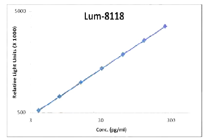 Standard Curve (Sample)
GM-CSF, CLIA Kit (Cat# AAA318530)
Full Name
GM-CSF (Human) LumiAb ELISA Kit
Gene Names
CSF2; CSF; GMCSF
Reactivity
Human
Pricing
 Standard Curve (Sample)
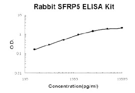
SFRP5, ELISA Kit (Cat# AAA124850)
Full Name
Rabbit SFRP5 PicoKine ELISA Kit
Gene Names
SFRP5; SARP3
Reactivity
Rabbit
There is no detectable cross-reactivity with other relevant proteins.
There is no detectable cross-reactivity with other relevant proteins.
Applications
ELISA
Pricing
 Standard Curve (Sample)
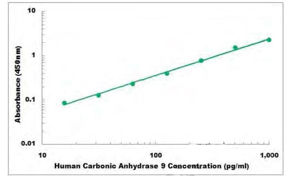
Carbonic Anhydrase 9, ELISA Kit (Cat# AAA105999)
Full Name
Human Carbonic Anhydrase 9 ELISA Kit
Gene Names
CA9; MN; CAIX
Reactivity
Human
Applications
Sandwich ELISA
Pricing
 SDS-PAGE
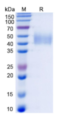
(SDS PAGE for Recombinant Human MICA*004:01 Protein)
SDS-PAGE
(SDS PAGE for Recombinant Human MICA*004:01 Protein)
MICA*004:01, Recombinant Protein (Cat# AAA120653)
Full Name
Recombinant Human MICA*004:01 Protein, C-His
Applications
Western Blot
Purity
>90% as determined by SDS-PAGE
Pricing
 Bioactivity
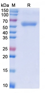
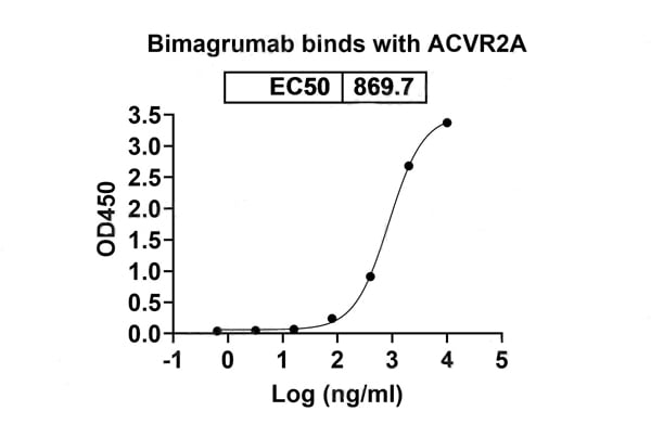
(Detects Bimagrumab in indirect ELISAs.)
Bioactivity
(Detects Bimagrumab in indirect ELISAs.)
ACVR2A, Recombinant Protein (Cat# AAA120571)
Full Name
Recombinant Human ACVR2A Protein, C-Fc
Gene Names
ACVR2A; ACVR2; ACTRII
Applications
Western Blot
Purity
>90% as determined by SDS-PAGE
Pricing