Filters
Clonality
Type
Reactivity
Gene Name
Isotype
Host
Application
Clone
6648 results for "Human Protein" - showing 6400-6450


 IHC (Immunohistchemistry)
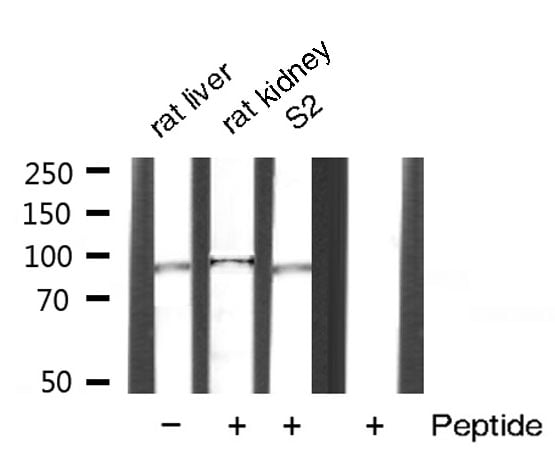
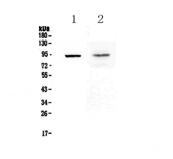
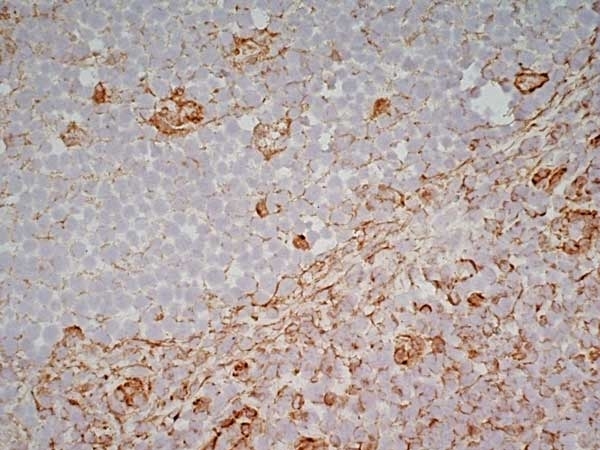
(Figure 6. IHC analysis of Cytoglobin using anti- Cytoglobin antibody (AAA11681).Cytoglobin was detected in paraffin-embedded section of human lung cancer tissues. Heat mediated antigen retrieval was performed in citrate buffer (pH6, epitope retrieval solution) for 20 mins. The tissue section was blocked with 10% goat serum. The tissue section was then incubated with 1ug/ml rabbit anti- Cytoglobin Antibody (AAA11681) overnight at 4 degree C. Biotinylated goat anti-rabbit IgG was used as secondary antibody and incubated for 30 minutes at 37 degree C. The tissue section was developed using Strepavidin-Biotin-Complex (SABC) with DAB as the chromogen.)
IHC (Immunohistchemistry)
(Figure 6. IHC analysis of Cytoglobin using anti- Cytoglobin antibody (AAA11681).Cytoglobin was detected in paraffin-embedded section of human lung cancer tissues. Heat mediated antigen retrieval was performed in citrate buffer (pH6, epitope retrieval solution) for 20 mins. The tissue section was blocked with 10% goat serum. The tissue section was then incubated with 1ug/ml rabbit anti- Cytoglobin Antibody (AAA11681) overnight at 4 degree C. Biotinylated goat anti-rabbit IgG was used as secondary antibody and incubated for 30 minutes at 37 degree C. The tissue section was developed using Strepavidin-Biotin-Complex (SABC) with DAB as the chromogen.)
Cytoglobin, Polyclonal Antibody (Cat# AAA11681)
Full Name
Anti-Cytoglobin Antibody
Gene Names
CYGB; HGB; STAP
Reactivity
Human, Mouse, Rat
Applications
WB, IHC
Purity
Immunogen affinity purified.
Pricing


 Application Data
(Formalin fixed, paraffin embedded human breast cancer biopsy stained with Mouse anti Human estrogen receptor beta5 antibody followed by HRP polymer detection and DAB substrate development following heat mediated antigen retrieval using citrate buffer at pH6.2 (low power))
Application Data
(Formalin fixed, paraffin embedded human breast cancer biopsy stained with Mouse anti Human estrogen receptor beta5 antibody followed by HRP polymer detection and DAB substrate development following heat mediated antigen retrieval using citrate buffer at pH6.2 (low power))
ESTROGEN RECEPTOR BETA 5, Monoclonal Antibody (Cat# AAA12216)
Full Name
MOUSE ANTI HUMAN ESTROGEN RECEPTOR BETA 5
Gene Names
ESR2; Erb; ESRB; ESTRB; NR3A2; ER-BETA; ESR-BETA
Applications
WB
Pricing


 Application Data
(Analysis of Protein Array containing more than 19, 000 full-length human proteins using Mouse Emerin Monoclonal Antibody (EMD/2168) Z- and S- Score: The Z-score represents the strength of a signal that a monoclonal antibody (MAb) (in combination with a fluorescently-tagged anti-IgG secondary antibody) produces when binding to a particular protein on the HuProtTM array. Z-scores are described in units of standard deviations (SD's) above the mean value of all signals generated on that array. If targets on HuProtTM are arranged in descending order of the Z-score, the S-score is the difference (also in units of SD's) between the Z-score. S-score therefore represents the relative target specificity of a MAb to its intended target. A MAb is considered to specific to its intended target, if the MAb has an S-score of at least 2.5. For example, if a MAb binds to protein X with a Z-score of 43 and to protein Y with a Z-score of 14, then the S-score for the binding of that MAb to protein X is equal to 29.)
Application Data
(Analysis of Protein Array containing more than 19, 000 full-length human proteins using Mouse Emerin Monoclonal Antibody (EMD/2168) Z- and S- Score: The Z-score represents the strength of a signal that a monoclonal antibody (MAb) (in combination with a fluorescently-tagged anti-IgG secondary antibody) produces when binding to a particular protein on the HuProtTM array. Z-scores are described in units of standard deviations (SD's) above the mean value of all signals generated on that array. If targets on HuProtTM are arranged in descending order of the Z-score, the S-score is the difference (also in units of SD's) between the Z-score. S-score therefore represents the relative target specificity of a MAb to its intended target. A MAb is considered to specific to its intended target, if the MAb has an S-score of at least 2.5. For example, if a MAb binds to protein X with a Z-score of 43 and to protein Y with a Z-score of 14, then the S-score for the binding of that MAb to protein X is equal to 29.)
Emerin, Monoclonal Antibody (Cat# AAA23899)
Full Name
Emerin (Papillary Thyroid Carcinoma and EDMD Marker)
Gene Names
EMD; STA; EDMD; LEMD5
Reactivity
Human. Others not known.
Applications
IHC
Pricing


 Application Data
(At 25 degree C. The primary antibody was diluted at 1/200 and incubated with the sample for 1 hour at 37 degree C. An Alexa Fluor 594 conjugated goat anti-rabbit IgG (H+L) Ab, diluted at 1/600, was used as the secondary antibody.)
Application Data
(At 25 degree C. The primary antibody was diluted at 1/200 and incubated with the sample for 1 hour at 37 degree C. An Alexa Fluor 594 conjugated goat anti-rabbit IgG (H+L) Ab, diluted at 1/600, was used as the secondary antibody.)
E2F-2, Polyclonal Antibody (Cat# AAA31340)
Full Name
E2F-2 Antibody
Gene Names
E2F2; E2F-2
Reactivity
Human, Mouse
Predicted Reactivity: Pig (100%), Bovine (100%), Horse (100%), Sheep (100%), Rabbit (90%), Dog (100%)
Predicted Reactivity: Pig (100%), Bovine (100%), Horse (100%), Sheep (100%), Rabbit (90%), Dog (100%)
Applications
WB, IHC, IF, ICC, EIA
Purity
The antiserum was purified by peptide affinity chromatography using SulfoLink Coupling Resin
Pricing


 IHC (Immunohistchemistry)
(At 1/100 staining Mouse testis tissue by IHC-P. The sample was formaldehyde fixed and a heat mediated antigen retrieval step in citrate buffer was performed. The sample was then blocked and incubated with the primary antibody at 4 degree C overnight. An HRP conjugated anti-Rabbit antibody was used as the secondary antibody.)
IHC (Immunohistchemistry)
(At 1/100 staining Mouse testis tissue by IHC-P. The sample was formaldehyde fixed and a heat mediated antigen retrieval step in citrate buffer was performed. The sample was then blocked and incubated with the primary antibody at 4 degree C overnight. An HRP conjugated anti-Rabbit antibody was used as the secondary antibody.)
PKC theta, Polyclonal Antibody (Cat# AAA31290)
Full Name
Phospho-PKC theta (Ser695) Antibody
Gene Names
PRKCQ; PRKCT; nPKC-theta
Reactivity
Human, Mouse, Rat
Applications
IHC, EIA
Purity
The antibody is from purified rabbit serum by affinity purification via sequential chromatography on phospho-peptide and non-phospho-peptide affinity columns.
Pricing


 IF (Immunofluorescence)
(Fluorescent confocal image of Hela cell stained with XRCC6 Antibody (C-term). Hela cells were fixed with 4% PFA (20 min), permeabilized with Triton X-100 (0.1%, 10 min), then incubated with XRCC6 primary antibody (1:25, 1 h at 37 degree). For secondary antibody, Alexa Fluor 488 conjugated donkey anti-rabbit antibody (green) was used (1:400, 50 min at 37 degree).Cytoplasmic actin was counterstained with Alexa Fluor 555 (red) conjugated Phalloidin (7units/ml, 1 h at 37 degree). Nuclei were counterstained with DAPI (blue) (10 ug/ml, 10 min). XRCC6 immunoreactivity is localized to nucleus significantly and Cytoplasm weakly.)
IF (Immunofluorescence)
(Fluorescent confocal image of Hela cell stained with XRCC6 Antibody (C-term). Hela cells were fixed with 4% PFA (20 min), permeabilized with Triton X-100 (0.1%, 10 min), then incubated with XRCC6 primary antibody (1:25, 1 h at 37 degree). For secondary antibody, Alexa Fluor 488 conjugated donkey anti-rabbit antibody (green) was used (1:400, 50 min at 37 degree).Cytoplasmic actin was counterstained with Alexa Fluor 555 (red) conjugated Phalloidin (7units/ml, 1 h at 37 degree). Nuclei were counterstained with DAPI (blue) (10 ug/ml, 10 min). XRCC6 immunoreactivity is localized to nucleus significantly and Cytoplasm weakly.)
XRCC6, Polyclonal Antibody (Cat# AAA28768)
Full Name
XRCC6 Antibody (C-term)
Gene Names
XRCC6; ML8; KU70; TLAA; CTC75; CTCBF; G22P1
Reactivity
Human
Applications
WB, EIA, IHC, FC/FACS, IF
Purity
Peptide Affinity Purified Rabbit Polyclonal Antibody (Pab)
Pricing


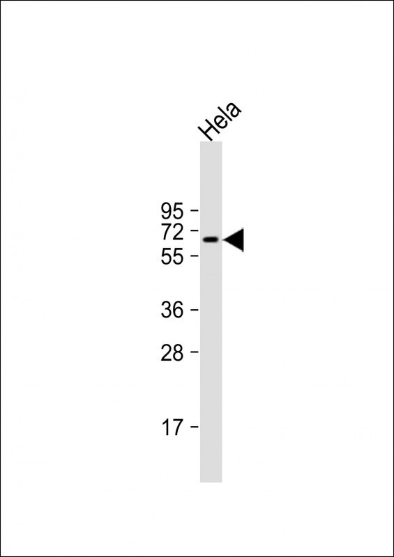
 WB (Western Blot)
(Detection of NFS1 by Western blot.Samples: Whole cell lysate from human HEK293 (H, 25 ug), mouse NIH3T3 (M, 25 ug) and rat F2408 (R, 25 ug) cells. [Lot No. 2635E1a-1]Predicted molecular weight: 50 kDa)
WB (Western Blot)
(Detection of NFS1 by Western blot.Samples: Whole cell lysate from human HEK293 (H, 25 ug), mouse NIH3T3 (M, 25 ug) and rat F2408 (R, 25 ug) cells. [Lot No. 2635E1a-1]Predicted molecular weight: 50 kDa)
NFS1, Monoclonal Antibody (Cat# AAA10614)
Full Name
Mouse monoclonal antibody Anti-Human NFS1
Gene Names
NFS1; IscS; NIFS; HUSSY-08
Reactivity
Human
Applications
DB, WB
Pricing


 WB (Western Blot)
(Western blot analysis of extracts of various sample, using Catenin-beta antibody.)
WB (Western Blot)
(Western blot analysis of extracts of various sample, using Catenin-beta antibody.)
Catenin-beta, Polyclonal Antibody (Cat# AAA31090)
Full Name
Catenin-beta Antibody
Gene Names
CTNNB1; EVR7; CTNNB; MRD19; armadillo
Reactivity
Human, Mouse, Rat
Applications
WB, IHC, IF, ICC, EIA
Purity
The antiserum was purified by peptide affinity chromatography using SulfoLink Coupling Resin.
Pricing


 IF (Immunofluorescence)
(Immunofluorescent analysis of 4% paraformaldehyde-fixed, 0.1% Triton X-100 permeabilized MCF-7 (human breast cancer cell line) cells labeling Pdx1 with at 1:25 dilution, followed by DyLight 488-conjugated IgG goat anti-rabbit secondary antibody at 1:200 dilution (green). Immunofluorescence image showing cytoplasm staining on MCF-7 cell line. Cytoplasmic actin is detected with DyLight 554 Phalloidin (PD18466410) at 1:100 dilution (red). The nuclear counter stain is DAPI (blue).)
IF (Immunofluorescence)
(Immunofluorescent analysis of 4% paraformaldehyde-fixed, 0.1% Triton X-100 permeabilized MCF-7 (human breast cancer cell line) cells labeling Pdx1 with at 1:25 dilution, followed by DyLight 488-conjugated IgG goat anti-rabbit secondary antibody at 1:200 dilution (green). Immunofluorescence image showing cytoplasm staining on MCF-7 cell line. Cytoplasmic actin is detected with DyLight 554 Phalloidin (PD18466410) at 1:100 dilution (red). The nuclear counter stain is DAPI (blue).)
OPN-a/b, Polyclonal Antibody (Cat# AAA26857)
Full Name
OPN-a/b, NT (SPP1, BNSP, OPN, Osteopontin, Bone sialoprotein 1, Nephropontin, Secreted phosphoprotein 1, Urinary stone protein, Uropontin) (APC)
Gene Names
SPP1; OPN; BNSP; BSPI; ETA-1
Reactivity
Human
Applications
WB, IHC, IF
Purity
Purified by Protein A and Peptide Affinity Chromatography.
Pricing


 IF (Immunofluorescence)
(Immunofluorescent analysis of 4% paraformaldehyde-fixed, 0.1% Triton X-100 permeabilized MCF-7 (human breast cancer cell line) cells labeling Pdx1 with at 1:25 dilution, followed by DyLight 488-conjugated IgG goat anti-rabbit secondary antibody at 1:200 dilution (green). Immunofluorescence image showing cytoplasm staining on MCF-7 cell line. Cytoplasmic actin is detected with DyLight 554 Phalloidin (PD18466410) at 1:100 dilution (red). The nuclear counter stain is DAPI (blue).)
IF (Immunofluorescence)
(Immunofluorescent analysis of 4% paraformaldehyde-fixed, 0.1% Triton X-100 permeabilized MCF-7 (human breast cancer cell line) cells labeling Pdx1 with at 1:25 dilution, followed by DyLight 488-conjugated IgG goat anti-rabbit secondary antibody at 1:200 dilution (green). Immunofluorescence image showing cytoplasm staining on MCF-7 cell line. Cytoplasmic actin is detected with DyLight 554 Phalloidin (PD18466410) at 1:100 dilution (red). The nuclear counter stain is DAPI (blue).)
OPN-a/b, Polyclonal Antibody (Cat# AAA26860)
Full Name
OPN-a/b, NT (SPP1, BNSP, OPN, Osteopontin, Bone sialoprotein 1, Nephropontin, Secreted phosphoprotein 1, Urinary stone protein, Uropontin) (MaxLight 405)
Gene Names
SPP1; OPN; BNSP; BSPI; ETA-1
Reactivity
Human
Applications
WB, IHC, IF
Purity
Purified by Protein A and Peptide Affinity Chromatography.
Pricing


 IHC (Immunohistchemistry)
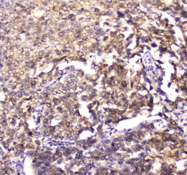
(Figure 6. IHC analysis of CPI17 alpha using anti- CPI17 alpha antibody (AAA19176).CPI17 alpha was detected in paraffin-embedded section of human placenta tissues. Heat mediated antigen retrieval was performed in citrate buffer (pH6, epitope retrieval solution) for 20 mins. The tissue section was blocked with 10% goat serum. The tissue section was then incubated with 1ug/ml rabbit anti- CPI17 alpha Antibody (AAA19176) overnight at 4 degree C. Biotinylated goat anti-rabbit IgG was used as secondary antibody and incubated for 30 minutes at 37 degree C. The tissue section was developed using Strepavidin-Biotin-Complex (SABC) with DAB as the chromogen.)
IHC (Immunohistchemistry)
(Figure 6. IHC analysis of CPI17 alpha using anti- CPI17 alpha antibody (AAA19176).CPI17 alpha was detected in paraffin-embedded section of human placenta tissues. Heat mediated antigen retrieval was performed in citrate buffer (pH6, epitope retrieval solution) for 20 mins. The tissue section was blocked with 10% goat serum. The tissue section was then incubated with 1ug/ml rabbit anti- CPI17 alpha Antibody (AAA19176) overnight at 4 degree C. Biotinylated goat anti-rabbit IgG was used as secondary antibody and incubated for 30 minutes at 37 degree C. The tissue section was developed using Strepavidin-Biotin-Complex (SABC) with DAB as the chromogen.)
CPI17 alpha, Polyclonal Antibody (Cat# AAA19176)
Full Name
Anti-CPI17 alpha Picoband Antibody
Gene Names
PPP1R14A; CPI17; CPI-17; PPP1INL
Reactivity
Human, Mouse, Rat
No cross reactivity with other proteins
No cross reactivity with other proteins
Applications
IHC, WB
Purity
Immunogen affinity purified
Pricing


 IF (Immunofluorescence)
(Immunofluorescent analysis of 4% paraformaldehyde-fixed, 0.1% Triton X-100 permeabilized MCF-7 (human breast cancer cell line) cells labeling Pdx1 with at 1:25 dilution, followed by DyLight 488-conjugated IgG goat anti-rabbit secondary antibody at 1:200 dilution (green). Immunofluorescence image showing cytoplasm staining on MCF-7 cell line. Cytoplasmic actin is detected with DyLight 554 Phalloidin (PD18466410) at 1:100 dilution (red). The nuclear counter stain is DAPI (blue).)
IF (Immunofluorescence)
(Immunofluorescent analysis of 4% paraformaldehyde-fixed, 0.1% Triton X-100 permeabilized MCF-7 (human breast cancer cell line) cells labeling Pdx1 with at 1:25 dilution, followed by DyLight 488-conjugated IgG goat anti-rabbit secondary antibody at 1:200 dilution (green). Immunofluorescence image showing cytoplasm staining on MCF-7 cell line. Cytoplasmic actin is detected with DyLight 554 Phalloidin (PD18466410) at 1:100 dilution (red). The nuclear counter stain is DAPI (blue).)
OPN-a/b, Polyclonal Antibody (Cat# AAA26859)
Full Name
OPN-a/b, NT (SPP1, BNSP, OPN, Osteopontin, Bone sialoprotein 1, Nephropontin, Secreted phosphoprotein 1, Urinary stone protein, Uropontin) (FITC)
Gene Names
SPP1; OPN; BNSP; BSPI; ETA-1
Reactivity
Human
Applications
WB, IHC, IF
Purity
Purified by Protein A and Peptide Affinity Chromatography.
Pricing


 IF (Immunofluorescence)
(Immunofluorescent analysis of 4% paraformaldehyde-fixed, 0.1% Triton X-100 permeabilized MCF-7 (human breast cancer cell line) cells labeling Pdx1 with at 1:25 dilution, followed by DyLight 488-conjugated IgG goat anti-rabbit secondary antibody at 1:200 dilution (green). Immunofluorescence image showing cytoplasm staining on MCF-7 cell line. Cytoplasmic actin is detected with DyLight 554 Phalloidin (PD18466410) at 1:100 dilution (red). The nuclear counter stain is DAPI (blue).)
IF (Immunofluorescence)
(Immunofluorescent analysis of 4% paraformaldehyde-fixed, 0.1% Triton X-100 permeabilized MCF-7 (human breast cancer cell line) cells labeling Pdx1 with at 1:25 dilution, followed by DyLight 488-conjugated IgG goat anti-rabbit secondary antibody at 1:200 dilution (green). Immunofluorescence image showing cytoplasm staining on MCF-7 cell line. Cytoplasmic actin is detected with DyLight 554 Phalloidin (PD18466410) at 1:100 dilution (red). The nuclear counter stain is DAPI (blue).)
OPN-a/b, Polyclonal Antibody (Cat# AAA26864)
Full Name
OPN-a/b, NT (SPP1, BNSP, OPN, Osteopontin, Bone sialoprotein 1, Nephropontin, Secreted phosphoprotein 1, Urinary stone protein, Uropontin) (MaxLight 750)
Gene Names
SPP1; OPN; BNSP; BSPI; ETA-1
Reactivity
Human
Applications
WB, IHC, IF
Purity
Purified by Protein A and Peptide Affinity Chromatography.
Pricing


 IF (Immunofluorescence)
(Immunofluorescent analysis of 4% paraformaldehyde-fixed, 0.1% Triton X-100 permeabilized MCF-7 (human breast cancer cell line) cells labeling Pdx1 with at 1:25 dilution, followed by DyLight 488-conjugated IgG goat anti-rabbit secondary antibody at 1:200 dilution (green). Immunofluorescence image showing cytoplasm staining on MCF-7 cell line. Cytoplasmic actin is detected with DyLight 554 Phalloidin (PD18466410) at 1:100 dilution (red). The nuclear counter stain is DAPI (blue).)
IF (Immunofluorescence)
(Immunofluorescent analysis of 4% paraformaldehyde-fixed, 0.1% Triton X-100 permeabilized MCF-7 (human breast cancer cell line) cells labeling Pdx1 with at 1:25 dilution, followed by DyLight 488-conjugated IgG goat anti-rabbit secondary antibody at 1:200 dilution (green). Immunofluorescence image showing cytoplasm staining on MCF-7 cell line. Cytoplasmic actin is detected with DyLight 554 Phalloidin (PD18466410) at 1:100 dilution (red). The nuclear counter stain is DAPI (blue).)
OPN-a/b, Polyclonal Antibody (Cat# AAA26863)
Full Name
OPN-a/b, NT (SPP1, BNSP, OPN, Osteopontin, Bone sialoprotein 1, Nephropontin, Secreted phosphoprotein 1, Urinary stone protein, Uropontin) (MaxLight 650)
Gene Names
SPP1; OPN; BNSP; BSPI; ETA-1
Reactivity
Human
Applications
WB, IHC, IF
Purity
Purified by Protein A and Peptide Affinity Chromatography.
Pricing


 IF (Immunofluorescence)
(Immunofluorescent analysis of 4% paraformaldehyde-fixed, 0.1% Triton X-100 permeabilized MCF-7 (human breast cancer cell line) cells labeling Pdx1 with at 1:25 dilution, followed by DyLight 488-conjugated IgG goat anti-rabbit secondary antibody at 1:200 dilution (green). Immunofluorescence image showing cytoplasm staining on MCF-7 cell line. Cytoplasmic actin is detected with DyLight 554 Phalloidin (PD18466410) at 1:100 dilution (red). The nuclear counter stain is DAPI (blue).)
IF (Immunofluorescence)
(Immunofluorescent analysis of 4% paraformaldehyde-fixed, 0.1% Triton X-100 permeabilized MCF-7 (human breast cancer cell line) cells labeling Pdx1 with at 1:25 dilution, followed by DyLight 488-conjugated IgG goat anti-rabbit secondary antibody at 1:200 dilution (green). Immunofluorescence image showing cytoplasm staining on MCF-7 cell line. Cytoplasmic actin is detected with DyLight 554 Phalloidin (PD18466410) at 1:100 dilution (red). The nuclear counter stain is DAPI (blue).)
OPN-a/b, Polyclonal Antibody (Cat# AAA26862)
Full Name
OPN-a/b, NT (SPP1, BNSP, OPN, Osteopontin, Bone sialoprotein 1, Nephropontin, Secreted phosphoprotein 1, Urinary stone protein, Uropontin) (MaxLight 550)
Gene Names
SPP1; OPN; BNSP; BSPI; ETA-1
Reactivity
Human
Applications
WB, IHC, IF
Purity
Purified by Protein A and Peptide Affinity Chromatography.
Pricing


 Application Data
(Published customer image:Phycoerythrin conjugated Mouse anti Human CD83 antibody, clone HB15e used for the evaluation of CD83 expression on monocyte derived dendritic cells by flow cytometry.Phenotypic characterization of immunogenic and tolerogenic moDC populations by flow cytometry. Monocytes were negatively selected from PBMC using magnetic beads. Immature moDC were generated with IL-4 and GM-CSF for 6 days. 15d-PGJ2 (PGJ2 DC) and dexamethasone plus 1alpha,25-dihydroxyvitamin were added to generate tolerogenic moDC, respectively (PGJ2 DC and Dex/VD3 DC). To generate immunogenic moDC, immature moDC were stimulated for 24 h with LPS, polyI:C and a cytokine cocktail containing TNF-alpha, IL-1beta, IL-6 and PGE2, respectively. The phenotypes of the cells were analyzed by flow cytometry. Live cells were gated according to FSC/SSC. One representative experiment out of three is shown.From: Sprater F, Hovden A-O, Appel S (2012)Expression of ESE-3 Isoforms in Immunogenic and Tolerogenic Human Monocyte-Derived Dendritic Cells.PLoS ONE 7(11): e49577.)
Application Data
(Published customer image:Phycoerythrin conjugated Mouse anti Human CD83 antibody, clone HB15e used for the evaluation of CD83 expression on monocyte derived dendritic cells by flow cytometry.Phenotypic characterization of immunogenic and tolerogenic moDC populations by flow cytometry. Monocytes were negatively selected from PBMC using magnetic beads. Immature moDC were generated with IL-4 and GM-CSF for 6 days. 15d-PGJ2 (PGJ2 DC) and dexamethasone plus 1alpha,25-dihydroxyvitamin were added to generate tolerogenic moDC, respectively (PGJ2 DC and Dex/VD3 DC). To generate immunogenic moDC, immature moDC were stimulated for 24 h with LPS, polyI:C and a cytokine cocktail containing TNF-alpha, IL-1beta, IL-6 and PGE2, respectively. The phenotypes of the cells were analyzed by flow cytometry. Live cells were gated according to FSC/SSC. One representative experiment out of three is shown.From: Sprater F, Hovden A-O, Appel S (2012)Expression of ESE-3 Isoforms in Immunogenic and Tolerogenic Human Monocyte-Derived Dendritic Cells.PLoS ONE 7(11): e49577.)
CD83, Monoclonal Antibody (Cat# AAA12281)
Full Name
MOUSE ANTI HUMAN CD83: FITC
Reactivity
Human
Applications
FC/FACS
Pricing


 WB (Western Blot)
(PLD6 Antibody (Center) (Cat# AAA28684) western blot analysis in mouse testis tissue lysates (35ug/lane).This demonstrates the PLD6 antibody detected the PLD6 protein (arrow).)
WB (Western Blot)
(PLD6 Antibody (Center) (Cat# AAA28684) western blot analysis in mouse testis tissue lysates (35ug/lane).This demonstrates the PLD6 antibody detected the PLD6 protein (arrow).)
PLD6, Polyclonal Antibody (Cat# AAA28684)
Full Name
PLD6 Antibody (Center)
Gene Names
PLD6; ZUC
Reactivity
Human, Mouse
Applications
WB, EIA
Purity
This antibody is purified through a protein A column, followed by peptide affinity purification.
Pricing


 IHC (Immunohistochemistry)
(At 1/100 staining Human mammary cancer and adjacent normal tissues by IHC-P. The sample was formaldehyde fixed and a heat mediated antigen retrieval step in citrate buffer was performed. The sample was then blocked and incubated with the primary antibody at 4 degree C overnight. An HRP conjugated anti-Rabbit antibody was used as the secondary antibody.)
IHC (Immunohistochemistry)
(At 1/100 staining Human mammary cancer and adjacent normal tissues by IHC-P. The sample was formaldehyde fixed and a heat mediated antigen retrieval step in citrate buffer was performed. The sample was then blocked and incubated with the primary antibody at 4 degree C overnight. An HRP conjugated anti-Rabbit antibody was used as the secondary antibody.)
Jak2, Polyclonal Antibody (Cat# AAA31311)
Full Name
Phospho-Jak2 (Tyr1008) Antibody
Gene Names
JAK2; JTK10; THCYT3
Reactivity
Human, Mouse, Rat
Applications
WB, IHC, EIA
Purity
The antibody is from purified rabbit serum by affinity purification via sequential chromatography on phospho-peptide and non-phospho-peptide affinity columns.
Pricing


 Application Data
(Analysis of Protein Array containing more than 19,000 full-length human proteins using Cytokeratin 15 Mouse Monoclonal Antibody (KRT15/2959). Z- and S- Score: The Z-score represents the strength of a signal that a monoclonal antibody (MAb) (in combination with a fluorescently-tagged anti-IgG secondary antibody) produces when binding to a particular protein on the HuProtTM array. Z-scores are described in units of standard deviations (SD's) above the mean value of all signals generated on that array. If targets on HuProtTM are arranged in descending order of the Z-score, the S-score is the difference (also in units of SD's) between the Z-score. S-score therefore represents the relative target specificity of a MAb to its intended target. A MAb is considered to specific to its intended target, if the MAb has an S-score of at least 2.5. For example, if a MAb binds to protein X with a Z-score of 43 and to protein Y with a Z-score of 14, then the S-score for the binding of that MAb to protein X is equal to 29.)
Application Data
(Analysis of Protein Array containing more than 19,000 full-length human proteins using Cytokeratin 15 Mouse Monoclonal Antibody (KRT15/2959). Z- and S- Score: The Z-score represents the strength of a signal that a monoclonal antibody (MAb) (in combination with a fluorescently-tagged anti-IgG secondary antibody) produces when binding to a particular protein on the HuProtTM array. Z-scores are described in units of standard deviations (SD's) above the mean value of all signals generated on that array. If targets on HuProtTM are arranged in descending order of the Z-score, the S-score is the difference (also in units of SD's) between the Z-score. S-score therefore represents the relative target specificity of a MAb to its intended target. A MAb is considered to specific to its intended target, if the MAb has an S-score of at least 2.5. For example, if a MAb binds to protein X with a Z-score of 43 and to protein Y with a Z-score of 14, then the S-score for the binding of that MAb to protein X is equal to 29.)
Cytokeratin 15, Monoclonal Antibody (Cat# AAA23924)
Full Name
Cytokeratin 15 (Esophageal Squamous Cell Carcinoma Marker)
Gene Names
KRT15; K15; CK15; K1CO
Reactivity
Human
Applications
IF, WB, IHC
Purity
Purified Ab with BSA and Azide at 200ug/ml OR Purified Ab WITHOUT BSA and Azide at 1.0mg/ml
Pricing


 WB (Western Blot)
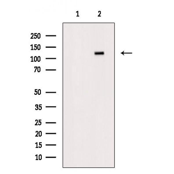
(Western blot analysis of HDAC5 expression in mouse brain tissue lysates, The lane on the right is treated with the antigen-specific peptide.)
WB (Western Blot)
(Western blot analysis of HDAC5 expression in mouse brain tissue lysates, The lane on the right is treated with the antigen-specific peptide.)
HDAC5, Polyclonal Antibody (Cat# AAA31097)
Full Name
HDAC5 Antibody
Gene Names
HDAC5; HD5; NY-CO-9
Reactivity
Human, Mouse
Applications
WB, IHC, IF, ICC, EIA
Purity
The antiserum was purified by peptide affinity chromatography using SulfoLink Coupling Resin.
Pricing


 Application Data
(At 25 degree C. Samples were then incubated with primary Ab(At 37 degree C. An AlexaFluor594 conjugated goat anti-rabbit IgG(H+L) Ab(Red) and an AlexaFluor488 conjugated goat anti-mouse IgG(H+L) Ab(Green) were used as the secondary antibody.The nuclear counter stain is DAPI (blue).)
Application Data
(At 25 degree C. Samples were then incubated with primary Ab(At 37 degree C. An AlexaFluor594 conjugated goat anti-rabbit IgG(H+L) Ab(Red) and an AlexaFluor488 conjugated goat anti-mouse IgG(H+L) Ab(Green) were used as the secondary antibody.The nuclear counter stain is DAPI (blue).)
MCM2, Polyclonal Antibody (Cat# AAA31328)
Full Name
Phospho-MCM2 (Ser108) Antibody
Gene Names
MCM2; BM28; CCNL1; CDCL1; cdc19; D3S3194; MITOTIN
Reactivity
Human, Mouse, Rat
Applications
WB, IHC, IF, ICC, EIA
Purity
The antibody is from purified rabbit serum by affinity purification via sequential chromatography on phospho-peptide and non-phospho-peptide affinity columns.
Pricing


 Application Data
(Staining of J774 cells with Rat anti Mouse F4/80 antigen Biotin)
Application Data
(Staining of J774 cells with Rat anti Mouse F4/80 antigen Biotin)
F4/80, Monoclonal Antibody (Cat# AAA12161)
Full Name
RAT ANTI MOUSE F4/80:APC
Gene Names
Emr1; Ly71; F4/80; Gpf480; TM7LN3; DD7A5-7; EGF-TM7
Applications
FC/FACS
Pricing


 WB (Western Blot)
(Western Blot Analysis of human liver tissue lysate using Prohibitin Mouse Monoclonal Antibody (PHB/3225).)
WB (Western Blot)
(Western Blot Analysis of human liver tissue lysate using Prohibitin Mouse Monoclonal Antibody (PHB/3225).)
Prohibitin, Monoclonal Antibody (Cat# AAA23933)
Full Name
Prohibitin (Mitochondrial Marker)
Gene Names
PHB; PHB1; HEL-215; HEL-S-54e
Reactivity
Human
Applications
WB, IF, IHC
Purity
Purified Ab with BSA and Azide at 200ug/ml OR Purified Ab WITHOUT BSA and Azide at 1.0mg/ml
Pricing


 Application Data
(Staining of KG1 lymphocytes with Mouse anti Human CD59:FITC)
Application Data
(Staining of KG1 lymphocytes with Mouse anti Human CD59:FITC)
CD59, Monoclonal Antibody (Cat# AAA11916)
Full Name
MOUSE ANTI HUMAN CD59
Gene Names
CD59; 1F5; EJ16; EJ30; EL32; G344; MIN1; MIN2; MIN3; MIRL; HRF20; MACIF; MEM43; MIC11; MSK21; 16.3A5; HRF-20; MAC-IP; p18-20
Applications
EIA, EM, FC/FACS, IF, IP, WB
Pricing


 Application Data
(Staining of J774 cells with Rat anti Mouse F4/80 antigen Biotin)
Application Data
(Staining of J774 cells with Rat anti Mouse F4/80 antigen Biotin)
F4/80, Monoclonal Antibody (Cat# AAA12172)
Full Name
RAT ANTI MOUSE F4/80:RPE
Gene Names
Emr1; Ly71; F4/80; Gpf480; TM7LN3; DD7A5-7; EGF-TM7
Applications
FC/FACS, IF
Pricing


 Application Data
(Staining of KG1 lymphocytes with Mouse anti Human CD59:FITC)
Application Data
(Staining of KG1 lymphocytes with Mouse anti Human CD59:FITC)
CD59, Monoclonal Antibody (Cat# AAA11917)
Full Name
MOUSE ANTI HUMAN CD59
Gene Names
CD59; 1F5; EJ16; EJ30; EL32; G344; MIN1; MIN2; MIN3; MIRL; HRF20; MACIF; MEM43; MIC11; MSK21; 16.3A5; HRF-20; MAC-IP; p18-20
Applications
EIA, EM, FC/FACS, IF, IP, WB
Pricing


 Application Data
(Staining of J774 cells with Rat anti Mouse F4/80 antigen Biotin)
Application Data
(Staining of J774 cells with Rat anti Mouse F4/80 antigen Biotin)
F4/80, Monoclonal Antibody (Cat# AAA12163)
Full Name
RAT ANTI MOUSE F4/80:Biotin
Gene Names
Emr1; Ly71; F4/80; Gpf480; TM7LN3; DD7A5-7; EGF-TM7
Applications
FC/FACS
Pricing


 IHC (Immunohistochemistry)
(Figure 8. IHC analysis of beta Catenin using anti-beta Catenin antibody (AAA19129).beta Catenin was detected in paraffin-embedded section of rat small intestine tissue. Heat mediated antigen retrieval was performed in citrate buffer (pH6, epitope retrieval solution) for 20 mins. The tissue section was blocked with 10% goat serum. The tissue section was then incubated with 1ug/ml rabbit anti-beta Catenin Antibody (AAA19129) overnight at 4 degree C. Biotinylated goat anti-rabbit IgG was used as secondary antibody and incubated for 30 minutes at 37 degree C. The tissue section was developed using Strepavidin-Biotin-Complex (SABC) with DAB as the chromogen.)
IHC (Immunohistochemistry)
(Figure 8. IHC analysis of beta Catenin using anti-beta Catenin antibody (AAA19129).beta Catenin was detected in paraffin-embedded section of rat small intestine tissue. Heat mediated antigen retrieval was performed in citrate buffer (pH6, epitope retrieval solution) for 20 mins. The tissue section was blocked with 10% goat serum. The tissue section was then incubated with 1ug/ml rabbit anti-beta Catenin Antibody (AAA19129) overnight at 4 degree C. Biotinylated goat anti-rabbit IgG was used as secondary antibody and incubated for 30 minutes at 37 degree C. The tissue section was developed using Strepavidin-Biotin-Complex (SABC) with DAB as the chromogen.)
beta Catenin, Polyclonal Antibody (Cat# AAA19129)
Full Name
Anti-beta Catenin Picoband Antibody
Gene Names
CTNNB1; EVR7; CTNNB; MRD19; armadillo
Reactivity
Human, Mouse, Rat
No cross reactivity with other proteins.
No cross reactivity with other proteins.
Applications
EIA, IHC, WB
Purity
Immunogen affinity purified
Pricing


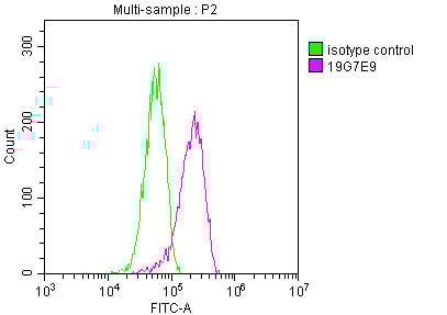 FCM (Flow Cytometry)
(Overlay histogram showing Hela cells stained with (red line) at 1:100. The cells were fixed in 4% formaldehyde and permeated by 0.2% TritonX-100. Then 10% normal goat serum was Incubated to block non-specific protein-protein interactions followed by the antibody (1ug/1*106cells) for 1 h at 4 degree C. The secondary antibody used was FITC-conjugated Goat Anti-Mouse IgG(H+L) at 1/100 dilution for 30min at 4 degree C. Isotype control antibody (green line) was mouse IgG2b (1ug/1*106cells) used under the same conditions. Acquisition of >10,000 events was performed.)
FCM (Flow Cytometry)
(Overlay histogram showing Hela cells stained with (red line) at 1:100. The cells were fixed in 4% formaldehyde and permeated by 0.2% TritonX-100. Then 10% normal goat serum was Incubated to block non-specific protein-protein interactions followed by the antibody (1ug/1*106cells) for 1 h at 4 degree C. The secondary antibody used was FITC-conjugated Goat Anti-Mouse IgG(H+L) at 1/100 dilution for 30min at 4 degree C. Isotype control antibody (green line) was mouse IgG2b (1ug/1*106cells) used under the same conditions. Acquisition of >10,000 events was performed.)
YWHAZ, Monoclonal Antibody (Cat# AAA27050)
Full Name
YWHAZ Monoclonal Antibody
Gene Names
YWHAZ; YWHAD; KCIP-1; 14-3-3-zeta
Reactivity
Human, Mouse, Rat
Applications
EIA, WB, IHC, IF, FC/FACS
Purity
>95%, Protein A purified
Pricing


 FCM (Flow Cytometry)
(Figure 6. Flow Cytometry analysis of THP-1 cells using anti-TLR1 antibody (AAA19134).Overlay histogram showing THP-1 cells stained with AAA19134 (Blue line).The cells were blocked with 10% normal goat serum. And then incubated with rabbit anti-TLR1 Antibody (AAA19134,1ug/1x10^6 cells) for 30 min at 20 degree C. DyLight®488 conjugated goat anti-rabbit IgG (5-10ug/1x10^6 cells) was used as secondary antibody for 30 minutes at 20 degree C. Isotype control antibody (Green line) was rabbit IgG (1ug/1x106) used under the same conditions. Unlabelled sample (Red line) was also used as a control.)
FCM (Flow Cytometry)
(Figure 6. Flow Cytometry analysis of THP-1 cells using anti-TLR1 antibody (AAA19134).Overlay histogram showing THP-1 cells stained with AAA19134 (Blue line).The cells were blocked with 10% normal goat serum. And then incubated with rabbit anti-TLR1 Antibody (AAA19134,1ug/1x10^6 cells) for 30 min at 20 degree C. DyLight®488 conjugated goat anti-rabbit IgG (5-10ug/1x10^6 cells) was used as secondary antibody for 30 minutes at 20 degree C. Isotype control antibody (Green line) was rabbit IgG (1ug/1x106) used under the same conditions. Unlabelled sample (Red line) was also used as a control.)
TLR1, Polyclonal Antibody (Cat# AAA19134)
Full Name
Anti-TLR1 Picoband antibody
Gene Names
TLR1; TIL; CD281; rsc786; TIL. LPRS5
Reactivity
Human, Mouse, Rat
No cross reactivity with other proteins.
No cross reactivity with other proteins.
Applications
EIA, FC/FACS, IHC, ICC, WB
Pricing


 IHC (Immunohistchemistry)
(Figure 6. IHC analysis of HP1 gamma using anti-HP1 gamma antibody (AAA19145).HP1 gamma was detected in paraffin-embedded section of rat small intestine tissue. Heat mediated antigen retrieval was performed in citrate buffer (pH6, epitope retrieval solution) for 20 mins. The tissue section was blocked with 10% goat serum. The tissue section was then incubated with 1ug/ml rabbit anti-HP1 gamma Antibody (AAA19145) overnight at 4 degree C. Biotinylated goat anti-rabbit IgG was used as secondary antibody and incubated for 30 minutes at 37 degree C. The tissue section was developed using Strepavidin-Biotin-Complex (SABC) with DAB as the chromogen.)
IHC (Immunohistchemistry)
(Figure 6. IHC analysis of HP1 gamma using anti-HP1 gamma antibody (AAA19145).HP1 gamma was detected in paraffin-embedded section of rat small intestine tissue. Heat mediated antigen retrieval was performed in citrate buffer (pH6, epitope retrieval solution) for 20 mins. The tissue section was blocked with 10% goat serum. The tissue section was then incubated with 1ug/ml rabbit anti-HP1 gamma Antibody (AAA19145) overnight at 4 degree C. Biotinylated goat anti-rabbit IgG was used as secondary antibody and incubated for 30 minutes at 37 degree C. The tissue section was developed using Strepavidin-Biotin-Complex (SABC) with DAB as the chromogen.)
HP1 gamma, Polyclonal Antibody (Cat# AAA19145)
Full Name
Anti-HP1 gamma Picoband antibody
Gene Names
CBX3; HECH; HP1-GAMMA; HP1Hs-gamma
Reactivity
Human, Mouse, Rat
No cross reactivity with other proteins.
No cross reactivity with other proteins.
Applications
EIA, IHC, WB
Pricing


 IHC (Immunohistchemistry)
(Figure 6. IHC analysis of Cyclophilin B using anti-Cyclophilin B antibody (AAA19167).Cyclophilin B was detected in paraffin-embedded section of rat thyroid gland tissue. Heat mediated antigen retrieval was performed in citrate buffer (pH6, epitope retrieval solution) for 20 mins. The tissue section was blocked with 10% goat serum. The tissue section was then incubated with 1ug/ml rabbit anti-Cyclophilin B Antibody (AAA19167) overnight at 4 degree C. Biotinylated goat anti-rabbit IgG was used as secondary antibody and incubated for 30 minutes at 37 degree C. The tissue section was developed using Strepavidin-Biotin-Complex (SABC) with DAB as the chromogen.)
IHC (Immunohistchemistry)
(Figure 6. IHC analysis of Cyclophilin B using anti-Cyclophilin B antibody (AAA19167).Cyclophilin B was detected in paraffin-embedded section of rat thyroid gland tissue. Heat mediated antigen retrieval was performed in citrate buffer (pH6, epitope retrieval solution) for 20 mins. The tissue section was blocked with 10% goat serum. The tissue section was then incubated with 1ug/ml rabbit anti-Cyclophilin B Antibody (AAA19167) overnight at 4 degree C. Biotinylated goat anti-rabbit IgG was used as secondary antibody and incubated for 30 minutes at 37 degree C. The tissue section was developed using Strepavidin-Biotin-Complex (SABC) with DAB as the chromogen.)
Cyclophilin B, Polyclonal Antibody (Cat# AAA19167)
Full Name
Anti-Cyclophilin B Picoband Antibody
Gene Names
PPIB; OI9; CYPB; SCYLP; CYP-S1; HEL-S-39
Reactivity
Human, Mouse, Rat
No cross reactivity with other proteins.
No cross reactivity with other proteins.
Applications
EIA, IHC, WB
Purity
Immunogen affinity purified
Pricing


 IHC (Immunohistchemistry)
(At 1/100 staining Mouse liver tissue by IHC-P. The sample was formaldehyde fixed and a heat mediated antigen retrieval step in citrate buffer was performed. The sample was then blocked and incubated with the primary antibody at 4 degree C overnight. An HRP conjugated anti-Rabbit antibody was used as the secondary antibody.)
IHC (Immunohistchemistry)
(At 1/100 staining Mouse liver tissue by IHC-P. The sample was formaldehyde fixed and a heat mediated antigen retrieval step in citrate buffer was performed. The sample was then blocked and incubated with the primary antibody at 4 degree C overnight. An HRP conjugated anti-Rabbit antibody was used as the secondary antibody.)
NDEL1, Polyclonal Antibody (Cat# AAA31462)
Full Name
Phospho-NDEL1 (Thr219) Antibody
Gene Names
NDEL1; EOPA; NDE2; NUDEL; MITAP1; NDE1L1
Reactivity
Human, Mouse, Rat
Predicted Reactivity: Pig (100%), Bovine (100%), Horse (100%), Sheep (100%), Rabbit (100%), Dog (100%), Chicken (100%), Xenopus (100%)
Predicted Reactivity: Pig (100%), Bovine (100%), Horse (100%), Sheep (100%), Rabbit (100%), Dog (100%), Chicken (100%), Xenopus (100%)
Applications
WB, IHC, EIA
Purity
The antibody is from purified rabbit serum by affinity purification via sequential chromatography on phospho-peptide and non-phospho-peptide affinity columns.
Pricing


 Application Data
(Published customer image:Phycoerythrin conjugated Mouse anti Human CD83 antibody, clone HB15e used for the evaluation of CD83 expression on monocyte derived dendritic cells by flow cytometry.Phenotypic characterization of immunogenic and tolerogenic moDC populations by flow cytometry. Monocytes were negatively selected from PBMC using magnetic beads. Immature moDC were generated with IL-4 and GM-CSF for 6 days. 15d-PGJ2 (PGJ2 DC) and dexamethasone plus 1alpha,25-dihydroxyvitamin were added to generate tolerogenic moDC, respectively (PGJ2 DC and Dex/VD3 DC). To generate immunogenic moDC, immature moDC were stimulated for 24 h with LPS, polyI:C and a cytokine cocktail containing TNF-alpha, IL-1beta, IL-6 and PGE2, respectively. The phenotypes of the cells were analyzed by flow cytometry. Live cells were gated according to FSC/SSC. One representative experiment out of three is shown.From: Sprater F, Hovden A-O, Appel S (2012)Expression of ESE-3 Isoforms in Immunogenic and Tolerogenic Human Monocyte-Derived Dendritic Cells.PLoS ONE 7(11): e49577.)
Application Data
(Published customer image:Phycoerythrin conjugated Mouse anti Human CD83 antibody, clone HB15e used for the evaluation of CD83 expression on monocyte derived dendritic cells by flow cytometry.Phenotypic characterization of immunogenic and tolerogenic moDC populations by flow cytometry. Monocytes were negatively selected from PBMC using magnetic beads. Immature moDC were generated with IL-4 and GM-CSF for 6 days. 15d-PGJ2 (PGJ2 DC) and dexamethasone plus 1alpha,25-dihydroxyvitamin were added to generate tolerogenic moDC, respectively (PGJ2 DC and Dex/VD3 DC). To generate immunogenic moDC, immature moDC were stimulated for 24 h with LPS, polyI:C and a cytokine cocktail containing TNF-alpha, IL-1beta, IL-6 and PGE2, respectively. The phenotypes of the cells were analyzed by flow cytometry. Live cells were gated according to FSC/SSC. One representative experiment out of three is shown.From: Sprater F, Hovden A-O, Appel S (2012)Expression of ESE-3 Isoforms in Immunogenic and Tolerogenic Human Monocyte-Derived Dendritic Cells.PLoS ONE 7(11): e49577.)
CD83, Monoclonal Antibody (Cat# AAA12280)
Full Name
MOUSE ANTI HUMAN CD83
Reactivity
Human
Applications
FC/FACS, IHC, IHC, IP
Pricing


 Application Data
(Published customer image:Phycoerythrin conjugated Mouse anti Human CD83 antibody, clone HB15e used for the evaluation of CD83 expression on monocyte derived dendritic cells by flow cytometry.Phenotypic characterization of immunogenic and tolerogenic moDC populations by flow cytometry. Monocytes were negatively selected from PBMC using magnetic beads. Immature moDC were generated with IL-4 and GM-CSF for 6 days. 15d-PGJ2 (PGJ2 DC) and dexamethasone plus 1alpha,25-dihydroxyvitamin were added to generate tolerogenic moDC, respectively (PGJ2 DC and Dex/VD3 DC). To generate immunogenic moDC, immature moDC were stimulated for 24 h with LPS, polyI:C and a cytokine cocktail containing TNF-alpha, IL-1beta, IL-6 and PGE2, respectively. The phenotypes of the cells were analyzed by flow cytometry. Live cells were gated according to FSC/SSC. One representative experiment out of three is shown.From: Sprater F, Hovden A-O, Appel S (2012)Expression of ESE-3 Isoforms in Immunogenic and Tolerogenic Human Monocyte-Derived Dendritic Cells.PLoS ONE 7(11): e49577.)
Application Data
(Published customer image:Phycoerythrin conjugated Mouse anti Human CD83 antibody, clone HB15e used for the evaluation of CD83 expression on monocyte derived dendritic cells by flow cytometry.Phenotypic characterization of immunogenic and tolerogenic moDC populations by flow cytometry. Monocytes were negatively selected from PBMC using magnetic beads. Immature moDC were generated with IL-4 and GM-CSF for 6 days. 15d-PGJ2 (PGJ2 DC) and dexamethasone plus 1alpha,25-dihydroxyvitamin were added to generate tolerogenic moDC, respectively (PGJ2 DC and Dex/VD3 DC). To generate immunogenic moDC, immature moDC were stimulated for 24 h with LPS, polyI:C and a cytokine cocktail containing TNF-alpha, IL-1beta, IL-6 and PGE2, respectively. The phenotypes of the cells were analyzed by flow cytometry. Live cells were gated according to FSC/SSC. One representative experiment out of three is shown.From: Sprater F, Hovden A-O, Appel S (2012)Expression of ESE-3 Isoforms in Immunogenic and Tolerogenic Human Monocyte-Derived Dendritic Cells.PLoS ONE 7(11): e49577.)
CD83, Monoclonal Antibody (Cat# AAA12283)
Full Name
MOUSE ANTI HUMAN CD83
Reactivity
Human
Applications
FC/FACS, IHC, IHC
Pricing


 FCM (Flow Cytometry)
(Flow cytometric analysis of Hela cells with Hsp70 antibody at 1/50 dilution (blue) compared with an unlabelled control (cells without incubation with primary antibody; red). Alexa Fluor 488-conjugated goat anti rabbit IgG was used as the secondary antibody)
FCM (Flow Cytometry)
(Flow cytometric analysis of Hela cells with Hsp70 antibody at 1/50 dilution (blue) compared with an unlabelled control (cells without incubation with primary antibody; red). Alexa Fluor 488-conjugated goat anti rabbit IgG was used as the secondary antibody)
Hsp70, Monoclonal Antibody (Cat# AAA29972)
Full Name
Hsp70 Antibody
Gene Names
HSPA1B; HSP72; HSPA1; HSX70; HSP70-1; HSP70-2; HSP70.1; HSP70.2; HSP70-1B
Reactivity
Human, Mouse, Rat
Applications
WB, ICC, IF, IHC, FC/FACS
Purity
ProA affinity purified
Pricing


 IHC (Immunohistchemistry)
(Immunohistochemical analysis of paraffin-embedded bladder cancer tissues using UBE2I mouse mAb with DAB staining.)
IHC (Immunohistchemistry)
(Immunohistochemical analysis of paraffin-embedded bladder cancer tissues using UBE2I mouse mAb with DAB staining.)
Ubc9, Monoclonal Antibody (Cat# AAA14109)
Full Name
Anti-Ubc9 Mouse mAb
Gene Names
UBE2I; P18; UBC9; C358B7.1
Reactivity
Human, Mouse, Rat
Applications
WB
Pricing


 IHC (Immunohistochemistry)
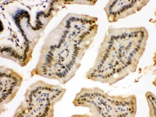
(At 1/100 staining Human prostate cancer by IHC-P. The sample was formaldehyde fixed and a heat mediated antigen retrieval step in citrate buffer was performed. The sample was then blocked and incubated with the primary antibody at 4 degree C overnight. An HRP conjugated anti-Rabbit antibody was used as the secondary antibody.)
IHC (Immunohistochemistry)
(At 1/100 staining Human prostate cancer by IHC-P. The sample was formaldehyde fixed and a heat mediated antigen retrieval step in citrate buffer was performed. The sample was then blocked and incubated with the primary antibody at 4 degree C overnight. An HRP conjugated anti-Rabbit antibody was used as the secondary antibody.)
hFANCI, Polyclonal Antibody (Cat# AAA31282)
Full Name
Phospho-hFANCI (Ser565) Antibody
Gene Names
FANCI; KIAA1794
Reactivity
Human, Mouse, Rat
Applications
IHC, EIA
Purity
The antibody is from purified rabbit serum by affinity purification via sequential chromatography on phospho-peptide and non-phospho-peptide affinity columns.
Pricing


 IP (Immunoprecipitation)
(Immunoprecipitating ENO1 in HepG2 whole cell lysate Lane 1: Mouse control IgG (1ug) instead of in HepG2 whole cell lysate. For western blotting, a HRP-conjugated Protein G antibody was used as the secondary antibody (1/5000) Lane 2: (1ul) + HepG2 whole cell lysate (500ug) Lane 3: HepG2 whole cell lysate (10ug))
IP (Immunoprecipitation)
(Immunoprecipitating ENO1 in HepG2 whole cell lysate Lane 1: Mouse control IgG (1ug) instead of in HepG2 whole cell lysate. For western blotting, a HRP-conjugated Protein G antibody was used as the secondary antibody (1/5000) Lane 2: (1ul) + HepG2 whole cell lysate (500ug) Lane 3: HepG2 whole cell lysate (10ug))
ENO1, Monoclonal Antibody (Cat# AAA27044)
Full Name
ENO1 Monoclonal Antibody
Gene Names
ENO1; NNE; PPH; MPB1; ENO1L1
Reactivity
Human, Mouse, Rat, Rabbit
Applications
EIA, WB, IF, FC/FACS, IP
Purity
>95%, Protein G purified
Pricing


 IP (Immunoprecipitation)
(Immunoprecipitating PD-L2 in Hela whole cell lysate Lane 1: Mouse control IgG instead of in Hela whole cell lysate. Lane 2: (2ul) + Hela whole cell lysate (500ug) Lane 3: Hela whole cell lysate (20ug) For western blotting, the blot was detected at 1:2000, and a HRP-conjugated Protein G antibody was used as the secondary antibody at 1:2000 )
IP (Immunoprecipitation)
(Immunoprecipitating PD-L2 in Hela whole cell lysate Lane 1: Mouse control IgG instead of in Hela whole cell lysate. Lane 2: (2ul) + Hela whole cell lysate (500ug) Lane 3: Hela whole cell lysate (20ug) For western blotting, the blot was detected at 1:2000, and a HRP-conjugated Protein G antibody was used as the secondary antibody at 1:2000 )
PD-L2, Monoclonal Antibody (Cat# AAA27047)
Full Name
PD-L2 Monoclonal Antibody
Gene Names
PDCD1LG2; B7DC; Btdc; PDL2; CD273; PD-L2; PDCD1L2; bA574F11.2
Reactivity
Human
Applications
EIA, WB, IHC, IF, FC/FACS, IP
Purity
>95%, Protein A purified
Pricing


 IHC (Immunohistochemistry)
(At 1/100 staining Human gastric cancer and adjacent normal tissues by IHC-P. The sample was formaldehyde fixed and a heat mediated antigen retrieval step in citrate buffer was performed. The sample was then blocked and incubated with the primary antibody at 4 degree C overnight. An HRP conjugated anti-Rabbit antibody was used as the secondary antibody.)
IHC (Immunohistochemistry)
(At 1/100 staining Human gastric cancer and adjacent normal tissues by IHC-P. The sample was formaldehyde fixed and a heat mediated antigen retrieval step in citrate buffer was performed. The sample was then blocked and incubated with the primary antibody at 4 degree C overnight. An HRP conjugated anti-Rabbit antibody was used as the secondary antibody.)
CAMKK1/2, Polyclonal Antibody (Cat# AAA31402)
Full Name
Phospho-CAMKK1/2 (Ser458/Ser495) Antibody
Gene Names
CAMKK1; CAMKKA
Reactivity
Human, Mouse, Rat
Predicted Reactivity: Pig (100%), Bovine (100%), Sheep (100%), Rabbit (100%), Dog (100%), Chicken (100%), Xenopus (100%)
Predicted Reactivity: Pig (100%), Bovine (100%), Sheep (100%), Rabbit (100%), Dog (100%), Chicken (100%), Xenopus (100%)
Applications
WB, IHC, EIA
Purity
The antibody is from purified rabbit serum by affinity purification via sequential chromatography on phospho-peptide and non-phospho-peptide affinity columns.
Pricing


 IHC (Immunohistochemistry)
(Figure 8. IHC analysis of PRDM1/Blimp1 using anti-PRDM1/Blimp1 antibody (AAA19133).PRDM1/Blimp1 was detected in paraffin-embedded section of rat small intestine tissue. Heat mediated antigen retrieval was performed in citrate buffer (pH6, epitope retrieval solution) for 20 mins. The tissue section was blocked with 10% goat serum. The tissue section was then incubated with 1ug/ml rabbit anti-PRDM1/Blimp1 Antibody (AAA19133) overnight at 4 degree C. Biotinylated goat anti-rabbit IgG was used as secondary antibody and incubated for 30 minutes at 37 degree C. The tissue section was developed using Strepavidin-Biotin-Complex (SABC) with DAB as the chromogen.)
IHC (Immunohistochemistry)
(Figure 8. IHC analysis of PRDM1/Blimp1 using anti-PRDM1/Blimp1 antibody (AAA19133).PRDM1/Blimp1 was detected in paraffin-embedded section of rat small intestine tissue. Heat mediated antigen retrieval was performed in citrate buffer (pH6, epitope retrieval solution) for 20 mins. The tissue section was blocked with 10% goat serum. The tissue section was then incubated with 1ug/ml rabbit anti-PRDM1/Blimp1 Antibody (AAA19133) overnight at 4 degree C. Biotinylated goat anti-rabbit IgG was used as secondary antibody and incubated for 30 minutes at 37 degree C. The tissue section was developed using Strepavidin-Biotin-Complex (SABC) with DAB as the chromogen.)
PRDM1/Blimp1, Polyclonal Antibody (Cat# AAA19133)
Full Name
Anti-PRDM1/Blimp1 Picoband antibody
Gene Names
PRDM1; BLIMP1; PRDI-BF1
Reactivity
Human, Mouse, Rat
No cross reactivity with other proteins.
No cross reactivity with other proteins.
Applications
EIA, IHC, WB
Pricing


 IHC (Immunohistochemistry)
(At 1/200 staining Human kidney tissue sections by IHC-P. The tissue was formaldehyde fixed and a heat mediated antigen retrieval step in citrate buffer was performed. The tissue was then blocked and incubated with the antibody for 1.5 hours at 22 degree C. An HRP conjugated goat anti-rabbit antibody was used as the secondary antibody.)
IHC (Immunohistochemistry)
(At 1/200 staining Human kidney tissue sections by IHC-P. The tissue was formaldehyde fixed and a heat mediated antigen retrieval step in citrate buffer was performed. The tissue was then blocked and incubated with the antibody for 1.5 hours at 22 degree C. An HRP conjugated goat anti-rabbit antibody was used as the secondary antibody.)
CtBP1/2, Polyclonal Antibody (Cat# AAA31423)
Full Name
Phospho-CtBP1/2 (Ser158/Ser164) Antibody
Gene Names
CTBP1; BARS
Reactivity
Human, Mouse, Rat
Predicted Reactivity: Pig (100%), Zebrafish (82%), Bovine (100%), Horse (100%), Dog (100%), Chicken (100%), Xenopus (100%)
Predicted Reactivity: Pig (100%), Zebrafish (82%), Bovine (100%), Horse (100%), Dog (100%), Chicken (100%), Xenopus (100%)
Applications
WB, IHC, EIA
Purity
The antibody is from purified rabbit serum by affinity purification via sequential chromatography on phospho-peptide and non-phospho-peptide affinity columns.
Pricing


 IHC (Immunohistchemistry)
(Figure 6. IHC analysis of NSF using anti-NSF antibody (AAA19136).NSF was detected in paraffin-embedded section of human mammary cancer tissue. Heat mediated antigen retrieval was performed in citrate buffer (pH6, epitope retrieval solution) for 20 mins. The tissue section was blocked with 10% goat serum. The tissue section was then incubated with 1ug/ml rabbit anti-NSF Antibody (AAA19136) overnight at 4 degree C. Biotinylated goat anti-rabbit IgG was used as secondary antibody and incubated for 30 minutes at 37 degree C. The tissue section was developed using Strepavidin-Biotin-Complex (SABC) with DAB as the chromogen.)
IHC (Immunohistchemistry)
(Figure 6. IHC analysis of NSF using anti-NSF antibody (AAA19136).NSF was detected in paraffin-embedded section of human mammary cancer tissue. Heat mediated antigen retrieval was performed in citrate buffer (pH6, epitope retrieval solution) for 20 mins. The tissue section was blocked with 10% goat serum. The tissue section was then incubated with 1ug/ml rabbit anti-NSF Antibody (AAA19136) overnight at 4 degree C. Biotinylated goat anti-rabbit IgG was used as secondary antibody and incubated for 30 minutes at 37 degree C. The tissue section was developed using Strepavidin-Biotin-Complex (SABC) with DAB as the chromogen.)
NSF, Polyclonal Antibody (Cat# AAA19136)
Full Name
Anti-NSF Picoband antibody
Gene Names
NSF; SKD2; SEC18
Reactivity
Human, Mouse, Rat
No cross reactivity with other proteins.
No cross reactivity with other proteins.
Applications
EIA, IHC, WB
Pricing


 WB (Western Blot)
(Figure 9 KD Validation of PUMA (Han et al., 2010)Immunoblot analyses of Tet-induced p53 cells treated with NOXA, Puma, Bim or non-targeting siRNAs that were utilized in this experiment. PUMA protein levels were markedly reduced in PUMA KD cells detected by anti-PUMA antibodies (3041).)
WB (Western Blot)
(Figure 9 KD Validation of PUMA (Han et al., 2010)Immunoblot analyses of Tet-induced p53 cells treated with NOXA, Puma, Bim or non-targeting siRNAs that were utilized in this experiment. PUMA protein levels were markedly reduced in PUMA KD cells detected by anti-PUMA antibodies (3041).)
PUMA, Polyclonal Antibody (Cat# AAA10943)
Full Name
PUMA Antibody
Gene Names
BBC3; JFY1; PUMA; JFY-1
Reactivity
Human, Mouse
Applications
EIA, WB, ICC, IF
Purity
PUMA Antibody is affinity chromatography purified via peptide column.
Pricing


 IHC (Immunohistchemistry)
(Figure 6. IHC analysis of 14-3-3 zeta/delta using anti-14-3-3 zeta/delta antibody (AAA19144).14-3-3 zeta/delta was detected in paraffin-embedded section of rat kidney tissue. Heat mediated antigen retrieval was performed in citrate buffer (pH6, epitope retrieval solution) for 20 mins. The tissue section was blocked with 10% goat serum. The tissue section was then incubated with 1ug/ml rabbit anti-14-3-3 zeta/delta Antibody (AAA19144) overnight at 4 degree C. Biotinylated goat anti-rabbit IgG was used as secondary antibody and incubated for 30 minutes at 37 degree C. The tissue section was developed using Strepavidin-Biotin-Complex (SABC) with DAB as the chromogen.)
IHC (Immunohistchemistry)
(Figure 6. IHC analysis of 14-3-3 zeta/delta using anti-14-3-3 zeta/delta antibody (AAA19144).14-3-3 zeta/delta was detected in paraffin-embedded section of rat kidney tissue. Heat mediated antigen retrieval was performed in citrate buffer (pH6, epitope retrieval solution) for 20 mins. The tissue section was blocked with 10% goat serum. The tissue section was then incubated with 1ug/ml rabbit anti-14-3-3 zeta/delta Antibody (AAA19144) overnight at 4 degree C. Biotinylated goat anti-rabbit IgG was used as secondary antibody and incubated for 30 minutes at 37 degree C. The tissue section was developed using Strepavidin-Biotin-Complex (SABC) with DAB as the chromogen.)
14-3-3 zeta/delta, Polyclonal Antibody (Cat# AAA19144)
Full Name
Anti-14-3-3 zeta/delta Picoband antibody
Gene Names
YWHAZ; HEL4; YWHAD; KCIP-1; HEL-S-3; HEL-S-93; 14-3-3-zeta
Reactivity
Human, Mouse, Rat
No cross reactivity with other proteins.
No cross reactivity with other proteins.
Applications
IHC, WB
Pricing


 IHC (Immunohistochemistry)
(Figure 7. IHC analysis of splicing factor 1 using anti-splicing factor 1 antibody (AAA19141).splicing factor 1 was detected in paraffin-embedded section of rat small intestine tissue. Heat mediated antigen retrieval was performed in citrate buffer (pH6, epitope retrieval solution) for 20 mins. The tissue section was blocked with 10% goat serum. The tissue section was then incubated with 1ug/ml rabbit anti-splicing factor 1 Antibody (AAA19141) overnight at 4 degree C. Biotinylated goat anti-rabbit IgG was used as secondary antibody and incubated for 30 minutes at 37 degree C. The tissue section was developed using Strepavidin-Biotin-Complex (SABC) with DAB as the chromogen.)
IHC (Immunohistochemistry)
(Figure 7. IHC analysis of splicing factor 1 using anti-splicing factor 1 antibody (AAA19141).splicing factor 1 was detected in paraffin-embedded section of rat small intestine tissue. Heat mediated antigen retrieval was performed in citrate buffer (pH6, epitope retrieval solution) for 20 mins. The tissue section was blocked with 10% goat serum. The tissue section was then incubated with 1ug/ml rabbit anti-splicing factor 1 Antibody (AAA19141) overnight at 4 degree C. Biotinylated goat anti-rabbit IgG was used as secondary antibody and incubated for 30 minutes at 37 degree C. The tissue section was developed using Strepavidin-Biotin-Complex (SABC) with DAB as the chromogen.)
splicing factor 1, Polyclonal Antibody (Cat# AAA19141)
Full Name
Anti-splicing factor 1 Picoband antibody
Gene Names
SF1; BBP; MBBP; ZFM1; ZNF162; D11S636; ZCCHC25
Reactivity
Human, Mouse, Rat
No cross reactivity with other proteins.
No cross reactivity with other proteins.
Applications
EIA, IHC, WB
Pricing


 IHC (Immunohistchemistry)
(Figure 6. IHC analysis of MAP1LC3A using anti-MAP1LC3A antibody (AAA19151).MAP1LC3A was detected in paraffin-embedded section of rat brain tissue. Heat mediated antigen retrieval was performed in citrate buffer (pH6, epitope retrieval solution) for 20 mins. The tissue section was blocked with 10% goat serum. The tissue section was then incubated with 1ug/ml rabbit anti-MAP1LC3A Antibody (AAA19151) overnight at 4 degree C. Biotinylated goat anti-rabbit IgG was used as secondary antibody and incubated for 30 minutes at 37 degree C. The tissue section was developed using Strepavidin-Biotin-Complex (SABC) with DAB as the chromogen.)
IHC (Immunohistchemistry)
(Figure 6. IHC analysis of MAP1LC3A using anti-MAP1LC3A antibody (AAA19151).MAP1LC3A was detected in paraffin-embedded section of rat brain tissue. Heat mediated antigen retrieval was performed in citrate buffer (pH6, epitope retrieval solution) for 20 mins. The tissue section was blocked with 10% goat serum. The tissue section was then incubated with 1ug/ml rabbit anti-MAP1LC3A Antibody (AAA19151) overnight at 4 degree C. Biotinylated goat anti-rabbit IgG was used as secondary antibody and incubated for 30 minutes at 37 degree C. The tissue section was developed using Strepavidin-Biotin-Complex (SABC) with DAB as the chromogen.)
MAP1LC3A, Polyclonal Antibody (Cat# AAA19151)
Full Name
Anti-MAP1LC3A Picoband antibody
Gene Names
MAP1LC3A; LC3; LC3A; ATG8E; MAP1ALC3; MAP1BLC3
Reactivity
Human, Mouse, Rat
No cross reactivity with other proteins.
No cross reactivity with other proteins.
Applications
EIA, IHC, WB
Pricing


 ICC (Immunocytochemistry)
(Figure 6. IHC analysis of LYRIC using anti-LYRIC antibody (AAA11650).LYRIC was detected in immunocytochemical section of SMMC-7721 cell. Heat mediated antigen retrieval was performed in citrate buffer (pH6, epitope retrieval solution) for 20 mins. The tissue section was blocked with 10% goat serum. The tissue section was then incubated with 1ug/ml rabbit anti-LYRIC Antibody (AAA11650) overnight at 4 degree C. Biotinylated goat anti-rabbit IgG was used as secondary antibody and incubated for 30 minutes at 37 degree C. The tissue section was developed using Strepavidin-Biotin-Complex (SABC) with DAB as the chromogen.)
ICC (Immunocytochemistry)
(Figure 6. IHC analysis of LYRIC using anti-LYRIC antibody (AAA11650).LYRIC was detected in immunocytochemical section of SMMC-7721 cell. Heat mediated antigen retrieval was performed in citrate buffer (pH6, epitope retrieval solution) for 20 mins. The tissue section was blocked with 10% goat serum. The tissue section was then incubated with 1ug/ml rabbit anti-LYRIC Antibody (AAA11650) overnight at 4 degree C. Biotinylated goat anti-rabbit IgG was used as secondary antibody and incubated for 30 minutes at 37 degree C. The tissue section was developed using Strepavidin-Biotin-Complex (SABC) with DAB as the chromogen.)
LYRIC, Polyclonal Antibody (Cat# AAA11650)
Full Name
Anti-LYRIC Antibody
Gene Names
MTDH; 3D3; AEG1; AEG-1; LYRIC; LYRIC/3D3
Reactivity
Human, Mouse, Rat
Applications
WB, IHC
Purity
Immunogen Affinity Purified
Pricing


 ICC (Immunocytochemistry)
(Figure 6. IHC analysis of EIF6 using anti-EIF6 antibody (AAA11655).EIF6 was detected in immunocytochemical section of SW480 cell. Heat mediated antigen retrieval was performed in citrate buffer (pH6, epitope retrieval solution) for 20 mins. The tissue section was blocked with 10% goat serum. The tissue section was then incubated with 1ug/ml rabbit anti-EIF6 Antibody (AAA11655) overnight at 4 degree C. Biotinylated goat anti-rabbit IgG was used as secondary antibody and incubated for 30 minutes at 37 degree C. The tissue section was developed using Strepavidin-Biotin-Complex (SABC) with DAB as the chromogen.)
ICC (Immunocytochemistry)
(Figure 6. IHC analysis of EIF6 using anti-EIF6 antibody (AAA11655).EIF6 was detected in immunocytochemical section of SW480 cell. Heat mediated antigen retrieval was performed in citrate buffer (pH6, epitope retrieval solution) for 20 mins. The tissue section was blocked with 10% goat serum. The tissue section was then incubated with 1ug/ml rabbit anti-EIF6 Antibody (AAA11655) overnight at 4 degree C. Biotinylated goat anti-rabbit IgG was used as secondary antibody and incubated for 30 minutes at 37 degree C. The tissue section was developed using Strepavidin-Biotin-Complex (SABC) with DAB as the chromogen.)
EIF6, Polyclonal Antibody (Cat# AAA11655)
Full Name
Anti-EIF6 Antibody
Gene Names
EIF6; CAB; EIF3A; eIF-6; p27BBP; ITGB4BP; b(2)gcn; p27(BBP)
Reactivity
Human, Mouse, Rat
Applications
WB, IHC
Purity
Immunogen Affinity Purified
Pricing